Embed Size (px)
Citation preview
Continue
NCCN Clinical Practice Guidelines in Oncology™
ThyroidCarcinoma
V.1.2008
www.nccn.org
Version 1.2008, 09/03/08 © 2008 National Comprehensive Cancer Network, Inc. All rights reserved. These guidelines and this illustration may not be reproduced in any form without the express written permission of NCCN.
Guidelines IndexThyroid Carcinoma TOC
Staging, Discussion, ReferencesPractice Guidelinesin Oncology – v.1.2008NCCN
®
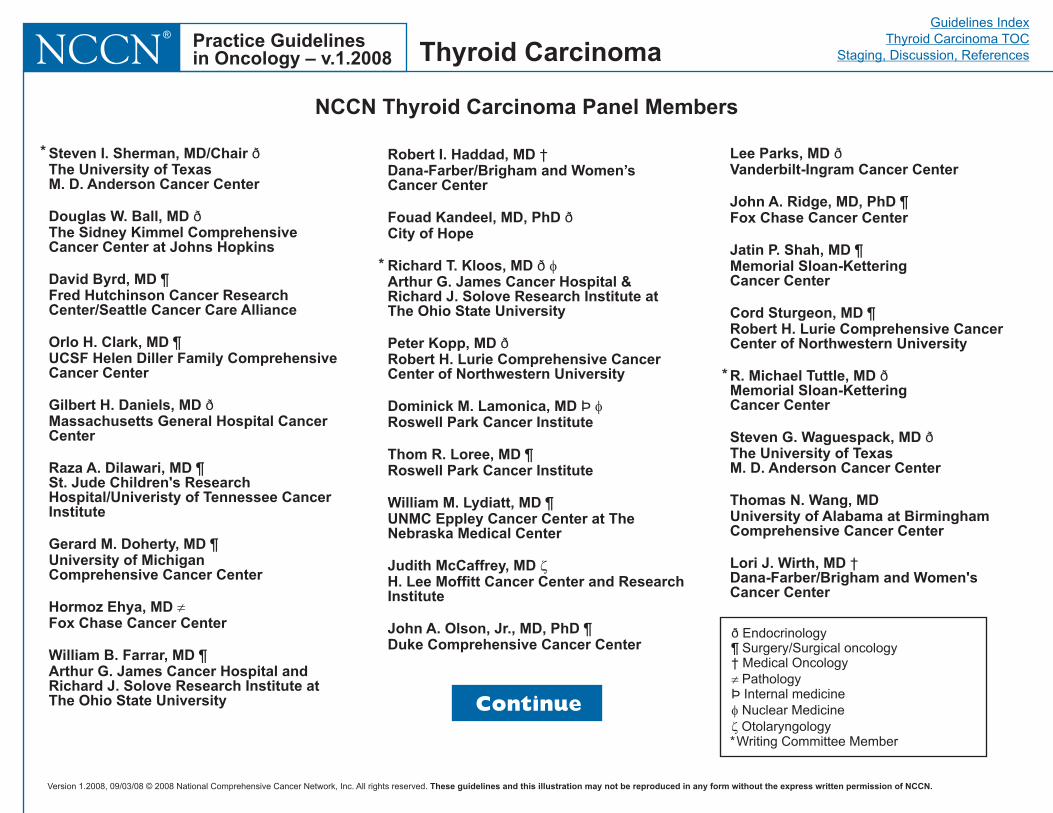
NCCN Thyroid Carcinoma Panel Members
Steven I. Sherman, MD/ChairThe University of TexasM. D. Anderson Cancer Center
Douglas W. Ball, MDThe Sidney Kimmel ComprehensiveCancer Center at Johns Hopkins
David Byrd, MDFred Hutchinson Cancer ResearchCenter/Seattle Cancer Care Alliance
Orlo H. Clark, MDUCSF Helen Diller Family ComprehensiveCancer Center
Gilbert H. Daniels, MDMassachusetts General Hospital CancerCenter
Raza A. Dilawari, MDSt. Jude Children's ResearchHospital/Univeristy of Tennessee CancerInstitute
Gerard M. Doherty, MDUniversity of MichiganComprehensive Cancer Center
Hormoz Ehya, MDFox Chase Cancer Center
William B. Farrar, MDArthur G. James Cancer Hospital andRichard J. Solove Research Institute atThe Ohio State University
ð
ð
¶
¶
ð
¶
¶
¶
�
Robert I. Haddad, MDDana-Farber/Brigham and Women’sCancer Center
Fouad Kandeel, MD, PhDCity of Hope
Richard T. Kloos, MDArthur G. James Cancer Hospital &Richard J. Solove Research Institute atThe Ohio State University
Peter Kopp, MDRobert H. Lurie Comprehensive CancerCenter of Northwestern University
Dominick M. Lamonica, MDRoswell Park Cancer Institute
Thom R. Loree, MDRoswell Park Cancer Institute
William M. Lydiatt, MDUNMC Eppley Cancer Center at TheNebraska Medical Center
Judith McCaffrey, MDH. Lee Moffitt Cancer Center and ResearchInstitute
John A. Olson, Jr., MD, PhDDuke Comprehensive Cancer Center
†
ð
ð
ð
Þ
¶
¶
¶
�
�
�
Lee Parks, MDVanderbilt-Ingram Cancer Center
John A. Ridge, MD, PhDFox Chase Cancer Center
Jatin P. Shah, MDMemorial Sloan-KetteringCancer Center
Cord Sturgeon, MDRobert H. Lurie Comprehensive CancerCenter of Northwestern University
R. Michael Tuttle, MDMemorial Sloan-KetteringCancer Center
Steven G. Waguespack, MDThe University of TexasM. D. Anderson Cancer Center
Thomas N. Wang, MDUniversity of Alabama at BirminghamComprehensive Cancer Center
Lori J. Wirth, MD
ð
¶
¶
¶
ð
ð
†Dana-Farber/Brigham and Women'sCancer Center
*
ð Endocrinology¶ Surgery/Surgical oncology
PathologyÞ Internal medicine
Nuclear MedicineOtolaryngology
*Writing Committee Member
† Medical Oncology�
�
�
Continue
Thyroid Carcinoma
*
*
Version 1.2008, 09/03/08 © 2008 National Comprehensive Cancer Network, Inc. All rights reserved. These guidelines and this illustration may not be reproduced in any form without the express written permission of NCCN.
Guidelines IndexThyroid Carcinoma TOC
Staging, Discussion, ReferencesPractice Guidelinesin Oncology – v.1.2008NCCN
®
Table of Contents
Papillary Carcinoma
Medullary Carcinoma
NCCN Thyroid Carcinoma Panel Members
Summary of Guidelines Updates
Nodule Evaluation (THYR-1)
Follicular Carcinoma (FOLL-1)
Hürthle Cell Carcinoma ( -1)
Diagnosed by FNA (MEDU-1)
Germline mutation of RET proto-oncogene (MEDU-2)
Anaplastic Carcinoma (ANAP-1)
Guidelines Index
Print the Thyroid Carcinoma Guideline
�
�
�
�
FNA positive (PAP-1)
Incidental finding postlobectomy (PAP-2)
HÜRT
These guidelines are a statement of evidence and consensus of the authors regarding their views of currently accepted approaches to treatment.Any clinician seeking to apply or consult these guidelines is expected to use independent medical judgment in the context of individual clinicalcircumstances to determine any patient's care or treatment. The National Comprehensive Cancer Network makes no representations nor warrantiesof any kind whatsoever regarding their content, use, or application and disclaims any responsibility for their application or use in any way. Theseguidelines are copyrighted by National Comprehensive Cancer Network. All rights reserved. These guidelines and the illustrations herein may notbe reproduced in any form without the express written permission of NCCN. ©2008.
For help using thesedocuments, please click here
Staging
Discussion
References
Clinical Trials:
Categories of Evidence andConsensus:NCCN
All recommendationsare Category 2A unless otherwisespecified.
See
Thebelieves that the best managementfor any cancer patient is in a clinicaltrial. Participation in clinical trials isespecially encouraged.
NCCN
To find clinical trials online at NCCNmember institutions, click here:nccn.org/clinical_trials/physician.html
NCCN Categories of Evidenceand Consensus
Thyroid Carcinoma
Version 1.2008, 09/03/08 © 2008 National Comprehensive Cancer Network, Inc. All rights reserved. These guidelines and this illustration may not be reproduced in any form without the express written permission of NCCN.
Guidelines IndexThyroid Carcinoma TOC
Staging, Discussion, ReferencesPractice Guidelinesin Oncology – v.1.2008NCCN
®
Summary of the Guidelines updates
UPDATES
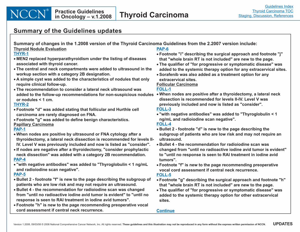
Summary of changes in the 1.2008 version of the Thyroid Carcinoma Guidelines from the 2.2007 version include:
Thyroid Carcinoma
Thyroid Nodule Evaluation
Papillary Carcinoma
Follicular Carcinoma
�
�
�
THYR-1
THYR-2
PAP-1
PAP-4
PAP-5
PAP-6
FOLL-1
FOLL-3
FOLL-4
FOLL-5
�
�
�
�
�
�
�
�
�
�
�
�
�
�
�
MEN2 replaced hyperparathyroidism under the listing of diseasesassociated with thyroid cancer.The central and neck compartments were added to ultrasound in theworkup section with a category 2B designation.A simple cyst was added to the characteristics of nodules that onlyrequire clinical follow-up.The recommendation to consider a lateral neck ultrasound wasadded to the follow-up recommendations for non-suspicious nodulesor nodules < 1 cm.
Footnote "d" was added stating that follicular and Hurthle cellcarcinoma are rarely diagnosed on FNA.Footnote "g" was added to define benign characteristics.
When nodes are positive by ultrasound or FNA cytology after athyroidectomy, a lateral neck dissection is recommended for levels II-IV. Level V was previously included and now is listed as "consider".If nodes are negative after a thyroidectomy, "consider prophylacticneck dissection" was added with a category 2B recommendation.
"with negative antibodies" was added to "Thyroglobulin < 1 ng/mLand radioiodine scan negative".
Bullet 2 - footnote "f" is new to the page describing the subgroup ofpatients who are low risk and may not require an ultrasound.Bullet 4 - the recommendation for radioiodine scan was changedfrom "until no radioactive iodine avid tumor is evident" to "until noresponse is seen to RAI treatment in iodine avid tumors".
Footnote "i" describing the surgical approach and footnote "j"that "whole brain RT is not included" are new to the page.The qualifier of "for progressive or symptomatic disease" wasadded to the systemic therapy option for any extracervical sites.
�
�
�
�
Footnote "h" is new to the page recommending preoperative vocalcord assessment if central neck recurrence.
Sorafenib was also added as a treatment option for anyextracervical sites.
When nodes are positive after a thyroidectomy, a lateral neckdissection is recommended for levels II-IV. Level V waspreviously included and now is listed as "consider".
"with negative antibodies" was added to "Thyroglobulin < 1ng/mL and radioiodine scan negative".
Bullet 2 - footnote "d" is new to the page describing thesubgroup of patients who are low risk and may not require anultrasound.Bullet 4 - the recommendation for radioiodine scan waschanged from "until no radioactive iodine avid tumor is evident"to "until no response is seen to RAI treatment in iodine avidtumors".Footnote "f" is new to the page recommending preoperativevocal cord assessment if central neck recurrence.
Footnote "g" describing the surgical approach and footnote "h"that "whole brain RT is not included" are new to the page.The qualifier of "for progressive or symptomatic disease" wasadded to the systemic therapy option for other extracervicalsites.
Continue
Version 1.2008, 09/03/08 © 2008 National Comprehensive Cancer Network, Inc. All rights reserved. These guidelines and this illustration may not be reproduced in any form without the express written permission of NCCN.
Guidelines IndexThyroid Carcinoma TOC
Staging, Discussion, ReferencesPractice Guidelinesin Oncology – v.1.2008NCCN
®
Summary of the Guidelines updates
UPDATES
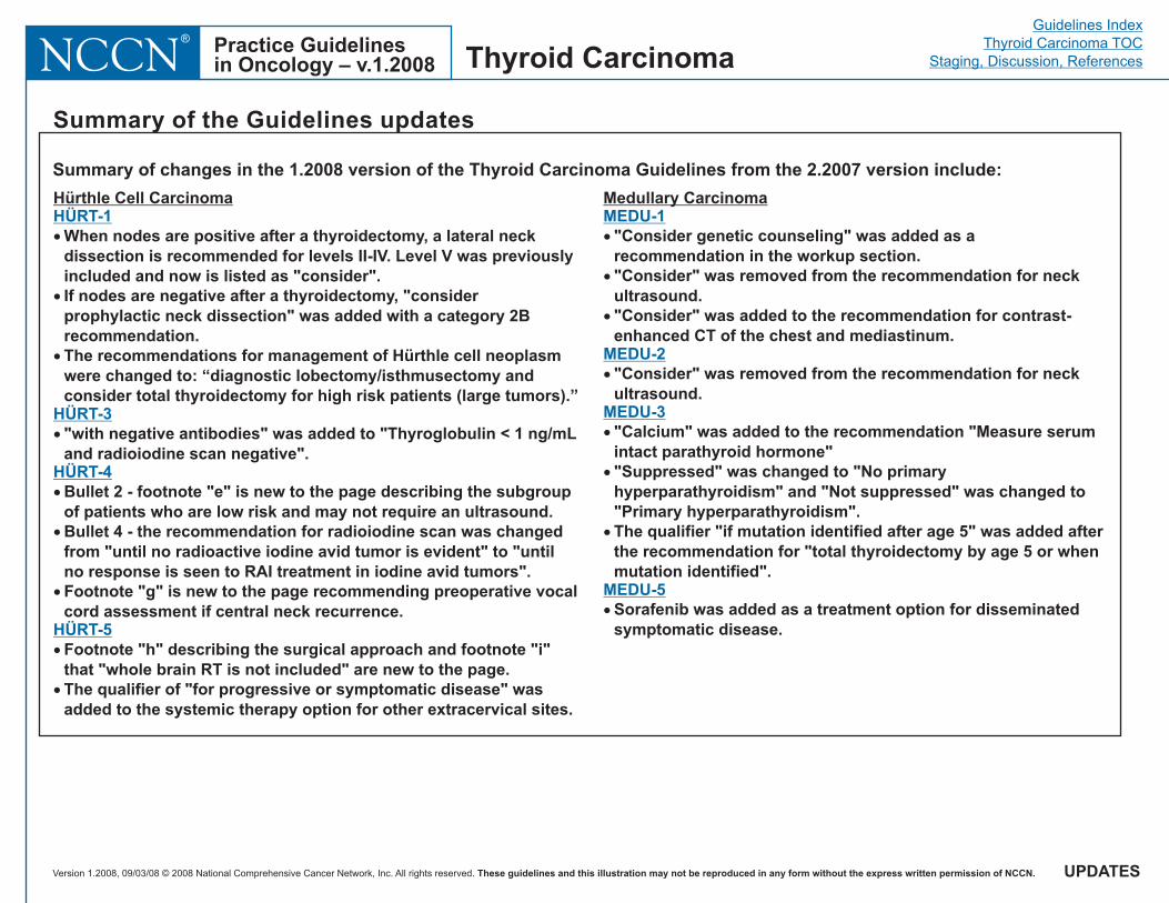
Summary of changes in the 1.2008 version of the Thyroid Carcinoma Guidelines from the 2.2007 version include:
Thyroid Carcinoma
Hürthle Cell Carcinoma Medullary CarcinomaHÜRT-1
HÜRT-3
HÜRT-4
HÜRT-5
MEDU-1
MEDU-2
MEDU-3
MEDU-5
�
�
�
�
�
�
�
�
�
�
�
�
�
�
�
When nodes are positive after a thyroidectomy, a lateral neckdissection is recommended for levels II-IV. Level V was previouslyincluded and now is listed as "consider".If nodes are negative after a thyroidectomy, "considerprophylactic neck dissection" was added with a category 2Brecommendation.The recommendations for management of Hürthle cell neoplasmwere changed to: “diagnostic lobectomy/isthmusectomy andconsider total thyroidectomy for high risk patients (large tumors).”
"with negative antibodies" was added to "Thyroglobulin < 1 ng/mLand radioiodine scan negative".
Footnote "h" describing the surgical approach and footnote "i"that "whole brain RT is not included" are new to the page.The qualifier of "for progressive or symptomatic disease" wasadded to the systemic therapy option for other extracervical sites.
Bullet 2 - footnote "e" is new to the page describing the subgroupof patients who are low risk and may not require an ultrasound.Bullet 4 - the recommendation for radioiodine scan was changedfrom "until no radioactive iodine avid tumor is evident" to "untilno response is seen to RAI treatment in iodine avid tumors".Footnote "g" is new to the page recommending preoperative vocalcord assessment if central neck recurrence.
"Consider genetic counseling" was added as arecommendation in the workup section."Consider" was removed from the recommendation for neckultrasound."Consider" was added to the recommendation for contrast-enhanced CT of the chest and mediastinum.
"Consider" was removed from the recommendation for neckultrasound.
"Calcium" was added to the recommendation "Measure serumintact parathyroid hormone""Suppressed" was changed to "No primaryhyperparathyroidism" and "Not suppressed" was changed to"Primary hyperparathyroidism".The qualifier "if mutation identified after age 5" was added afterthe recommendation for "total thyroidectomy by age 5 or whenmutation identified".
Sorafenib was added as a treatment option for disseminatedsymptomatic disease.
�
�
Version 1.2008, 09/03/08 © 2008 National Comprehensive Cancer Network, Inc. All rights reserved. These guidelines and this illustration may not be reproduced in any form without the express written permission of NCCN.
Guidelines IndexThyroid Carcinoma TOC
Staging, Discussion, ReferencesPractice Guidelinesin Oncology – v.1.2008NCCN
®
Note: All recommendations are category 2A unless otherwise indicated.Clinical Trials: NCCN believes that the best management of any cancer patient is in a clinical trial. Participation in clinical trials is especially encouraged.
Thyroid Carcinoma
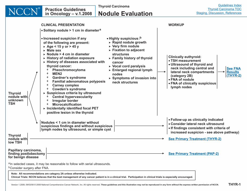
Nodule Evaluation
WORKUPCLINICAL PRESENTATION
Clinically euthyroid:��
��
TSH measurementUltrasound of thyroid andneck including central andlateral neck compartments(category 2B)FNA of noduleFNA of clinically suspiciouslymph nodes
�
�
�
Follow-up as clinically indicatedConsider lateral neck ultrasoundIf findings consistent with criteria ofincreased suspicion - see above pathway)
Thyroidnodule withunknownTSH
Nodules < 1 cm in diameter withoutsuspicious findings and without suspiciouslymph nodes by ultrasound, or simple cyst
�
�
Solitary nodule > 1 cm in diameter
Increased suspicion if anyof the following are present:
Age < 15 y or > 45 yMale sexNodule > 4 cm in diameterHistory of radiation exposureHistory of diseases associated withthyroid cancer:
Gardner’s syndromeFamilial adenomatous polyposisCarney complexCowden’s syndrome
Suspicious criteria by ultrasoundCentral hypervascularity
MicrocalcificationIncidentally identified focal PETpositive lesion in the thyroid
a
�
�
�
�
�
�
�
�
�
�
�
�
�
�
�
�
PheochromocytomaMEN2
Irregular border
� Highly suspicious:Rapid nodule growth
b
�
�
�
�
�
�
�
Very firm noduleFixation to adjacentstructuresFamily history of thyroidcancerVocal cord paralysisEnlarged regional lymphnodesSymptoms of invasion intoneck structures
See FNAResults(THYR-2)
a
bIn selected cases, it may be reasonable to follow with serial ultrasounds.Consider surgery after FNA.
Papillary carcinoma,finding postlobectomyfor benign disease
See Primary Treatment (PAP-2)
THYR-1
Thyroidnodule withlow TSH
See Primary Treatment (THYR-2)
Version 1.2008, 09/03/08 © 2008 National Comprehensive Cancer Network, Inc. All rights reserved. These guidelines and this illustration may not be reproduced in any form without the express written permission of NCCN.
Guidelines IndexThyroid Carcinoma TOC
Staging, Discussion, ReferencesPractice Guidelinesin Oncology – v.1.2008NCCN
®
Note: All recommendations are category 2A unless otherwise indicated.Clinical Trials: NCCN believes that the best management of any cancer patient is in a clinical trial. Participation in clinical trials is especially encouraged.
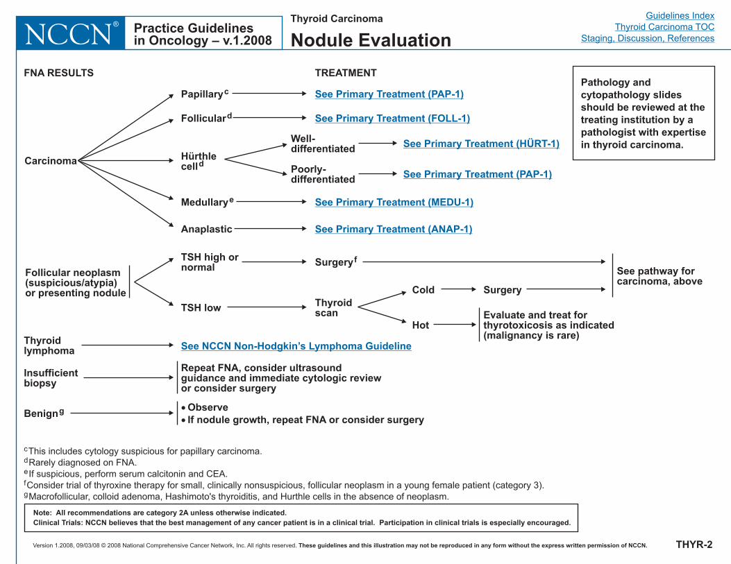
c
d
e
f
g
This includes cytology suspicious for papillary carcinoma.Rarely diagnosed on FNA.If suspicious, perform serum calcitonin and CEA.Consider trial of thyroxine therapy for small, clinically nonsuspicious, follicular neoplasm in a young female patient (category 3).Macrofollicular, colloid adenoma, Hashimoto's thyroiditis, and Hurthle cells in the absence of neoplasm.
FNA RESULTS TREATMENT
Carcinoma
See pathway forcarcinoma, above
Follicular neoplasm(suspicious/atypia)or presenting nodule
Benigng
Insufficientbiopsy
Surgeryf
�
�
ObserveIf nodule growth, repeat FNA or consider surgery
Repeat FNA, consider ultrasoundguidance and immediate cytologic reviewor consider surgery
Papillaryc
Hürthlecelld
See Primary Treatment (PAP-1)
See Primary Treatment (FOLL-1)
See Primary Treatment (HÜRT-1)
See Primary Treatment (MEDU-1)
See Primary Treatment (ANAP-1)Anaplastic
Hot
Cold SurgeryThyroidscan
Folliculard
Medullarye
TSH high ornormal
TSH lowEvaluate and treat forthyrotoxicosis as indicated(malignancy is rare)Thyroid
lymphoma See NCCN Non-Hodgkin’s Lymphoma Guideline
THYR-2
Pathology andcytopathology slidesshould be reviewed at thetreating institution by apathologist with expertisein thyroid carcinoma.
Thyroid Carcinoma
Nodule Evaluation
See Primary Treatment (PAP-1)
Well-differentiated
Poorly-differentiated
Version 1.2008, 09/03/08 © 2008 National Comprehensive Cancer Network, Inc. All rights reserved. These guidelines and this illustration may not be reproduced in any form without the express written permission of NCCN.
Guidelines IndexThyroid Carcinoma TOC
Staging, Discussion, ReferencesPractice Guidelinesin Oncology – v.1.2008NCCN
®
Note: All recommendations are category 2A unless otherwise indicated.Clinical Trials: NCCN believes that the best management of any cancer patient is in a clinical trial. Participation in clinical trials is especially encouraged.
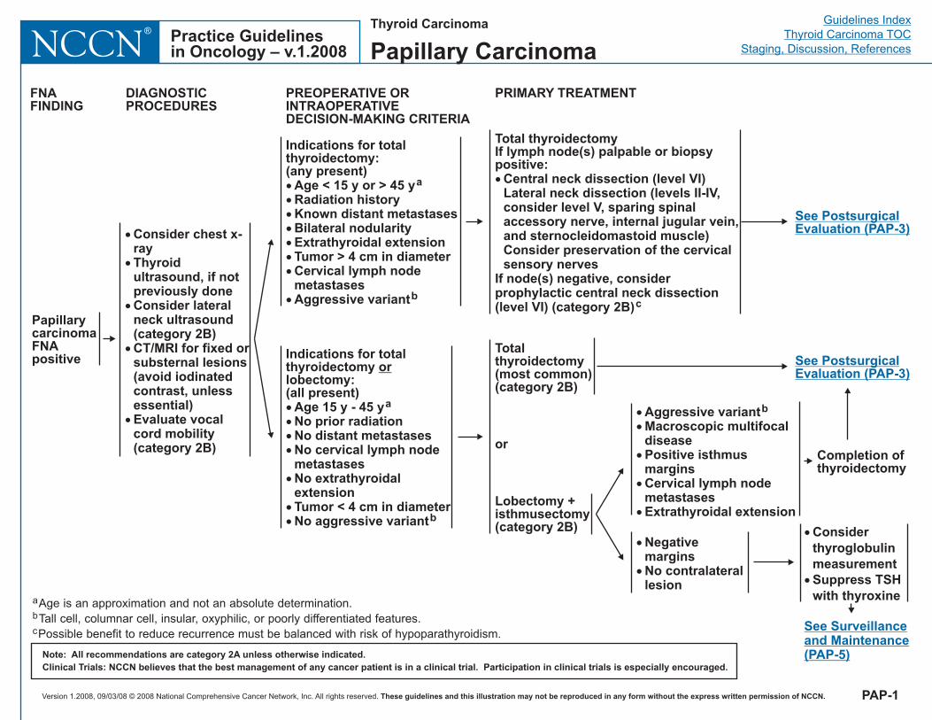
Thyroid Carcinoma
Papillary Carcinoma
PapillarycarcinomaFNApositive
�
�
�
�
�
Consider chest x-rayThyroidultrasound, if notpreviously doneConsider lateralneck ultrasound(category 2B)CT/MRI for fixed orsubsternal lesions(avoid iodinatedcontrast, unlessessential)Evaluate vocalcord mobility(category 2B)
Indications for totalthyroidectomy:(any present)
Ag
�������
�
Age < 15 y or > 45 yRadiation historyKnown distant metastasesBilateral nodularityExtrathyroidal extensionTumor > 4 cm in diameterCervical lymph nodemetastases
gressive variant
a
b
Indications for totalthyroidectomylobectomy:(all present)
or
����
�
��
Age 15 y - 45 yNo prior radiationNo distant metastasesNo cervical lymph nodemetastasesNo extrathyroidalextensionTumor < 4 cm in diameterNo aggressive variant
a
b
PREOPERATIVE ORINTRAOPERATIVEDECISION-MAKING CRITERIA
DIAGNOSTICPROCEDURES
FNAFINDING
Total thyroidectomyIf lymph node(s) palpable or biopsypositive:� Central neck dissection (level VI)
Lateral neck dissection (levels II-IV,consider level V, sparing spinalaccessory nerve, internal jugular vein,and sternocleidomastoid muscle)Consider preservation of the cervicalsensory nerves
If node(s) negative, considerprophylactic central neck dissection(level VI) (category 2B)c
Totalthyroidectomy(most common)(category 2B)
or
Lobectomy +isthmusectomy(category 2B)
PRIMARY TREATMENT
��
�
�
�
Aggressive variantMacroscopic multifocaldiseasePositive isthmusmarginsCervical lymph nodemetastasesExtrathyroidal extension
b
�
�
NegativemarginsNo contralaterallesion
See PostsurgicalEvaluation (PAP-3)
See PostsurgicalEvaluation (PAP-3)
a
b
c
Age is an approximation and not an absolute determination.Tall cell, columnar cell, insular, oxyphilic, or poorly differentiated features.Possible benefit to reduce recurrence must be balanced with risk of hypoparathyroidism.
Completion ofthyroidectomy
�
�
ConsiderthyroglobulinmeasurementSuppress TSHwith thyroxine
See Surveillanceand Maintenance(PAP-5)
PAP-1
Version 1.2008, 09/03/08 © 2008 National Comprehensive Cancer Network, Inc. All rights reserved. These guidelines and this illustration may not be reproduced in any form without the express written permission of NCCN.
Guidelines IndexThyroid Carcinoma TOC
Staging, Discussion, ReferencesPractice Guidelinesin Oncology – v.1.2008NCCN
®
Note: All recommendations are category 2A unless otherwise indicated.Clinical Trials: NCCN believes that the best management of any cancer patient is in a clinical trial. Participation in clinical trials is especially encouraged.
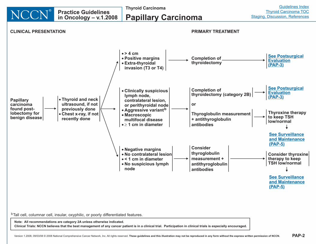
Completion ofthyroidectomy
�
��
�
Clinically suspiciouslymph node,contralateral lesion,or pAggressive variantMacroscopicmultifocal disease
1 cm in diameter
erithyroidal nodeb
�
����
Negative marginsNo contralateral lesion< 1 cm in diameterNo suspicious lymphnode
PRIMARY TREATMENT
���
> 4 cmPositive marginsExtra-thyroidalinvasion (T3 or T4)
Papillarycarcinomafound post-lobectomy forbenign disease
�
�
Thyroid and neckultrasound, if notpreviously doneChest x-ray, if notrecently done
CLINICAL PRESENTATION
See PostsurgicalEvaluation(PAP-3)
PAP-2
bTall cell, columnar cell, insular, oxyphilic, or poorly differentiated features.
Thyroid Carcinoma
Papillary Carcinoma
Considerthyroglobulinmeasurement +antithyroglobulinantibodies
Completion ofthyroidectomy (category 2B)
or
Thyroglobulin measurement+ antithyroglobulinantibodies
Consider thyroxinetherapy to keepTSH low/normal
See Surveillanceand Maintenance(PAP-5)
See PostsurgicalEvaluation(PAP-3)
Thyroxine therapyto keep TSHlow/normal
See Surveillanceand Maintenance(PAP-5)
Version 1.2008, 09/03/08 © 2008 National Comprehensive Cancer Network, Inc. All rights reserved. These guidelines and this illustration may not be reproduced in any form without the express written permission of NCCN.
Guidelines IndexThyroid Carcinoma TOC
Staging, Discussion, ReferencesPractice Guidelinesin Oncology – v.1.2008NCCN
®
Note: All recommendations are category 2A unless otherwise indicated.Clinical Trials: NCCN believes that the best management of any cancer patient is in a clinical trial. Participation in clinical trials is especially encouraged.
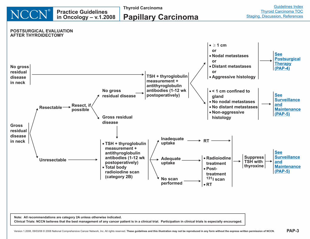
TSH + thyroglobulinmeasurement +antithyroglobulinantibodies (1-12 wkpostoperatively)
POSTSURGICAL EVALUATIONAFTER THYROIDECTOMY
No grossresidualdiseasein neck
Grossresidualdiseasein neck
SuppressTSH withthyroxine
SeeSurveillanceandMaintenance(PAP-5)
Unresectable
Resectable Resect, ifpossible
No grossresidual disease
Gross residualdisease
Inadequateuptake
No scanperformed
RT
�
�
�
RadioiodinetreatmentPost-treatment
I scanRT
131
SeePostsurgicalTherapy(PAP-4)
�
�
TSH + thyroglobulinmeasurement +antithyroglobulinantibodies (1-12 wkpostoperatively)Total bodyradioiodine scan(category 2B)
PAP-3
Adequateuptake
Thyroid Carcinoma
Papillary Carcinoma
�
�
�
�
< 1 cm confined toglandNo nodal metastasesNo distant metastasesNon-aggressivehistology
SeeSurveillanceandMaintenance(PAP-5)
�
�
�
�
1 cmorNodal metastasesorDistant metastasesorAggressive histology
�
Version 1.2008, 09/03/08 © 2008 National Comprehensive Cancer Network, Inc. All rights reserved. These guidelines and this illustration may not be reproduced in any form without the express written permission of NCCN.
Guidelines IndexThyroid Carcinoma TOC
Staging, Discussion, ReferencesPractice Guidelinesin Oncology – v.1.2008NCCN
®
Note: All recommendations are category 2A unless otherwise indicated.Clinical Trials: NCCN believes that the best management of any cancer patient is in a clinical trial. Participation in clinical trials is especially encouraged.
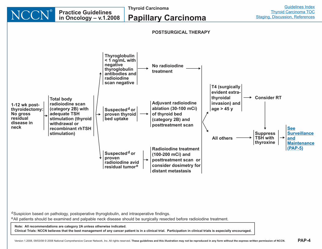
Adjuvant radioiodineablation (30-100 mCi)of thyroid bed(category 2B) andposttreatment scan
RadioiodinemCi
posttreatment scan orconsider dosimetry fordistant metastasis
treatment(100-200 ) and
SuppressTSH withthyroxine
POSTSURGICAL THERAPY
T4 (surgicallyevident extra-thyroidalinvasion) andage > 45 y
All others
Consider RT
SeeSurveillanceandMaintenance(PAP-5)
1-12 wk post-thyroidectomy:No grossresidualdisease inneck
Suspected orprovenradioiodine avidresidual tumor
d
e
Suspected orproven thyroidbed uptake
d
Thyroglobulin< 1 ng/mL withnegativethyroglobulinantibodies andradioiodinescan negative
No radioiodinetreatment
d
eSuspicion based on pathology, postoperative thyroglobulin, and intraoperative findings.All patients should be examined and palpable neck disease should be surgically resected before radioiodine treatment.
PAP-4
Thyroid Carcinoma
Papillary Carcinoma
Total bodyradioiodine scan(category 2B) withadequate TSHstimulation (thyroidwithdrawal orrecombinant rhTSHstimulation)
Version 1.2008, 09/03/08 © 2008 National Comprehensive Cancer Network, Inc. All rights reserved. These guidelines and this illustration may not be reproduced in any form without the express written permission of NCCN.
Guidelines IndexThyroid Carcinoma TOC
Staging, Discussion, ReferencesPractice Guidelinesin Oncology – v.1.2008NCCN
®
Note: All recommendations are category 2A unless otherwise indicated.Clinical Trials: NCCN believes that the best management of any cancer patient is in a clinical trial. Participation in clinical trials is especially encouraged.
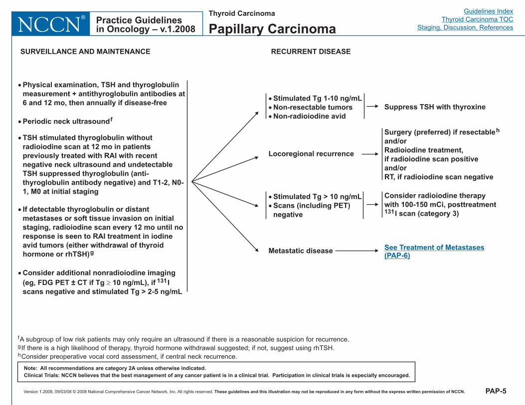
�
�
�
�
�
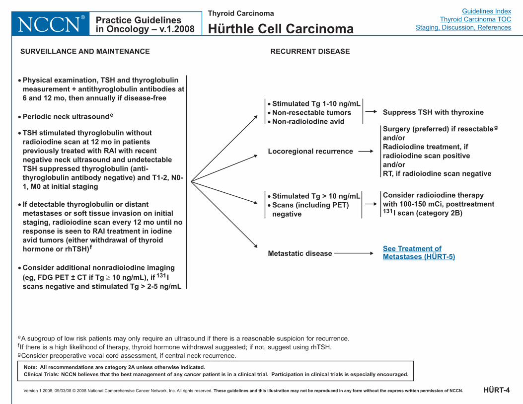
Physical examination, TSH and thyroglobulinmeasurement + antithyroglobulin antibodies at6 and 12 mo, then annually if disease-free
Consider additional nonradioiodine imaging(eg, FDG PET ± CT if Tg 10 ng/mL), if Iscans negative and stimulated Tg > 2-5 ng/mL
Periodic neck ultrasound
TSH stimulated thyroglobulin withoutradioiodine scan at 12 mo in patientspreviously treated with RAI with recentnegative neck ultrasound and undetectableTSH suppressed thyroglobulin (anti-thyroglobulin antibody negative) and T1-2, N0-1, M0 at initial staging
If detectable thyroglobulin or distantmetastases or soft tissue invasion on initialstaging, radioiodine scan every 12 mo until noresponse is seen to RAI treatment in iodineavid tumors (either withdrawal of thyroidhormone or rhTSH)
f
g
131�
SURVEILLANCE AND MAINTENANCE
Locoregional recurrence
Metastatic disease
�
�
Tg > 10 ng mLScans (including PET)negative
Stimulated / Consider therapywith 100-150 mCi, posttreatment
I scan (category 3)
radioiodine
131
Surgery (preferred) if resectableand/orRadioiodine treatment,if radioiodine scan positiveand/orRT, if radioiodine scan negative
h
See Treatment of Metastases(PAP-6)
RECURRENT DISEASE
f
g
h
A subgroup of low risk patients may only require an ultrasound if there is a reasonable suspicion for recurrence.If there is a high likelihood of therapy, thyroid hormone withdrawal suggested; if not, suggest using rhTSH.Consider preoperative vocal cord assessment, if central neck recurrence.
PAP-5
Thyroid Carcinoma
Papillary Carcinoma
�
�
�
Stimulated Tg 1-10 ng/mLNon-resectable tumorsNon-radioiodine avid
Suppress TSH with thyroxine
Version 1.2008, 09/03/08 © 2008 National Comprehensive Cancer Network, Inc. All rights reserved. These guidelines and this illustration may not be reproduced in any form without the express written permission of NCCN.
Guidelines IndexThyroid Carcinoma TOC
Staging, Discussion, ReferencesPractice Guidelinesin Oncology – v.1.2008NCCN
®
Note: All recommendations are category 2A unless otherwise indicated.Clinical Trials: NCCN believes that the best management of any cancer patient is in a clinical trial. Participation in clinical trials is especially encouraged.
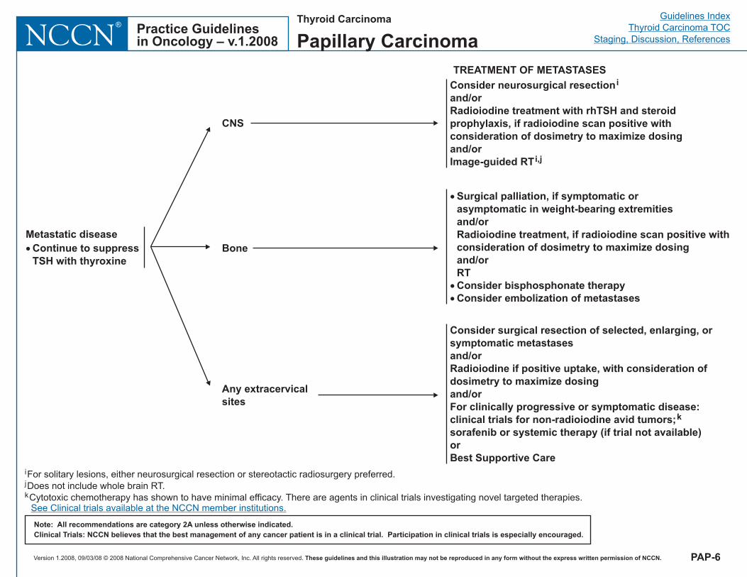
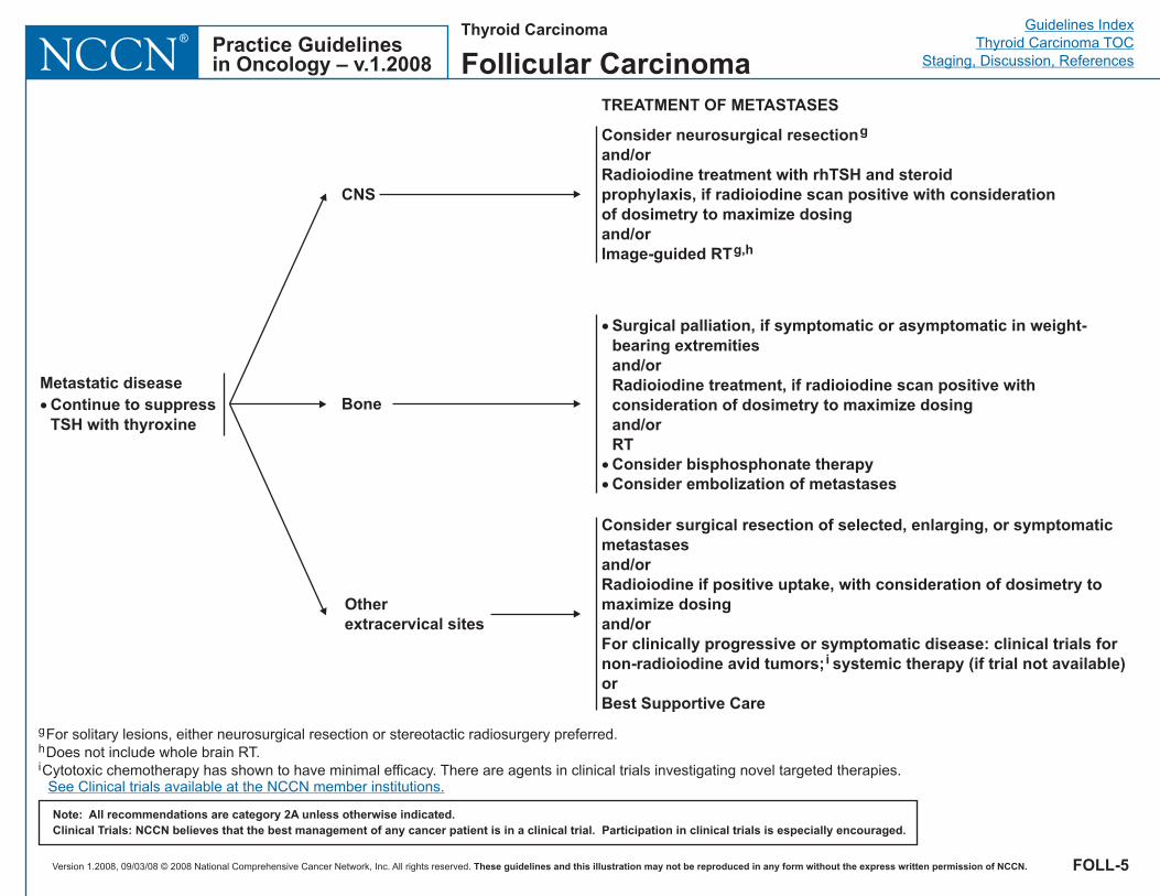
TREATMENT OF METASTASES
CNS
Consider neurosurgical resection
Radioiodine treatment with rhTSH and steroidprophylaxis,
Image-guided RT
i
and/or
if radioiodine scan positive withconsideration of dosimetry to maximize dosingand/or
i,j
Bone
�
�
�
Surgical palliation, if symptomatic orasymptomatic in weight-bearing extremities
Radioiodine treatment, if radioiodine scan positiveand/or
and/orRTConsider bisphosphonate therapyConsider embolization of metastases
withconsideration of dosimetry to maximize dosing
Any extracervicalsites
Consider surgical resection of selected, enlarging, orsymptomatic metastasesand/orRadioiodine if positive uptake, with consideration ofdosimetry to maximize dosingand/orFor clinically progressive or symptomatic disease:clinical trials for non-radioiodine avid tumors;sorafenib or systemic therapy (if trial not available)orBest Supportive Care
k
Metastatic disease� Continue to suppress
TSH with thyroxine
PAP-6
Thyroid Carcinoma
Papillary Carcinoma
i
jFor solitary lesions, either neurosurgical resection or stereotactic radiosurgery preferred.Does not include whole brain RT.
kCytotoxic chemotherapy has shown to have minimal efficacy. There are agents in clinical trials investigating novel targeted therapies.See Clinical trials available at the NCCN member institutions.
Version 1.2008, 09/03/08 © 2008 National Comprehensive Cancer Network, Inc. All rights reserved. These guidelines and this illustration may not be reproduced in any form without the express written permission of NCCN.
Guidelines IndexThyroid Carcinoma TOC
Staging, Discussion, ReferencesPractice Guidelinesin Oncology – v.1.2008NCCN
®Thyroid Carcinoma
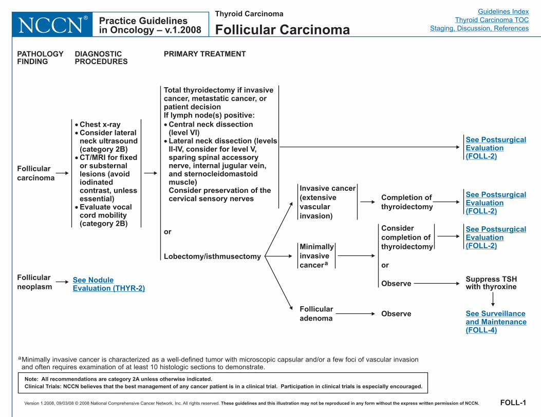
Follicular Carcinoma
Note: All recommendations are category 2A unless otherwise indicated.Clinical Trials: NCCN believes that the best management of any cancer patient is in a clinical trial. Participation in clinical trials is especially encouraged.
Follicularcarcinoma
Total thyroidectomy if invasivecancer, metastatic cancer, orpatient decisionIf lymph node(s) positive:
Central neck dissection(level VI)Lateral neck dissection (levelsII-IV, consider for level V,sparing spinal accessorynerve, internal jugular vein,and sternocleidomastoidmuscle)Consider preservation of thecervical sensory nerves
�
�
or
Lobectomy/isthmusectomy
PATHOLOGYFINDING
DIAGNOSTICPROCEDURES
PRIMARY TREATMENT
��
�
�
Chest x-rayConsider lateralneck ultrasound(category 2B)CT/MRI for fixedor substernallesions (avoidiodinatedcontrast, unlessessential)Evaluate vocalcord mobility(category 2B)
See PostsurgicalEvaluation(FOLL-2)
Minimallyinvasivecancera
Follicularadenoma
Invasive cancer(extensivevascularinvasion)
Considercompletion ofthyroidectomy
or
Observe
Observe
Completion ofthyroidectomy
aMinimally invasive cancer is characterized as a well-defined tumor with microscopic capsular and/or a few foci of vascular invasionand often requires examination of at least 10 histologic sections to demonstrate.
See PostsurgicalEvaluation(FOLL-2)
See PostsurgicalEvaluation(FOLL-2)
FOLL-1
Follicularneoplasm
See NoduleEvaluation (THYR-2)
Suppress TSHwith thyroxine
See Surveillanceand Maintenance(FOLL-4)
Version 1.2008, 09/03/08 © 2008 National Comprehensive Cancer Network, Inc. All rights reserved. These guidelines and this illustration may not be reproduced in any form without the express written permission of NCCN.
Guidelines IndexThyroid Carcinoma TOC
Staging, Discussion, ReferencesPractice Guidelinesin Oncology – v.1.2008NCCN
®
Note: All recommendations are category 2A unless otherwise indicated.Clinical Trials: NCCN believes that the best management of any cancer patient is in a clinical trial. Participation in clinical trials is especially encouraged.
POSTSURGICAL EVALUATIONAFTER THYROIDECTOMY
No grossresidualdiseasein neck
Grossresidualdiseasein neck
SuppressTSH withthyroxine
SeeSurveillanceandMaintenance(FOLL-4)
Unresectable
Resectable Resect, ifpossible
No grossresidual disease
Gross residualdisease
RT�
�
TSH + thyroglobulinmeasurement +antithyroglobulinantibodies (1-12 wkpostoperatively)Total bodyradioiodine scan(category 2B)
FOLL-2
No scanperformed
�
�
�
RadioiodinetreatmentPost-treatment
I scanRT
131
Thyroid Carcinoma
Follicular Carcinoma
Inadequateuptake
Adequateuptake
TSH + thyroglobulinmeasurement +antithyroglobulinantibodies (1-12 wkpostoperatively)
SeePostsurgicalTherapy(FOLL-3)
�
�
�
�
< 1 cm confined toglandNo nodal metastasesNo distant metastasesNon-aggressivehistology
SeeSurveillanceandMaintenance(FOLL-4)
�
�
�
�
1 cmorNodal metastasesorDistant metastasesorAggressive histology
�
Version 1.2008, 09/03/08 © 2008 National Comprehensive Cancer Network, Inc. All rights reserved. These guidelines and this illustration may not be reproduced in any form without the express written permission of NCCN.
Guidelines IndexThyroid Carcinoma TOC
Staging, Discussion, ReferencesPractice Guidelinesin Oncology – v.1.2008NCCN
®
Note: All recommendations are category 2A unless otherwise indicated.Clinical Trials: NCCN believes that the best management of any cancer patient is in a clinical trial. Participation in clinical trials is especially encouraged.
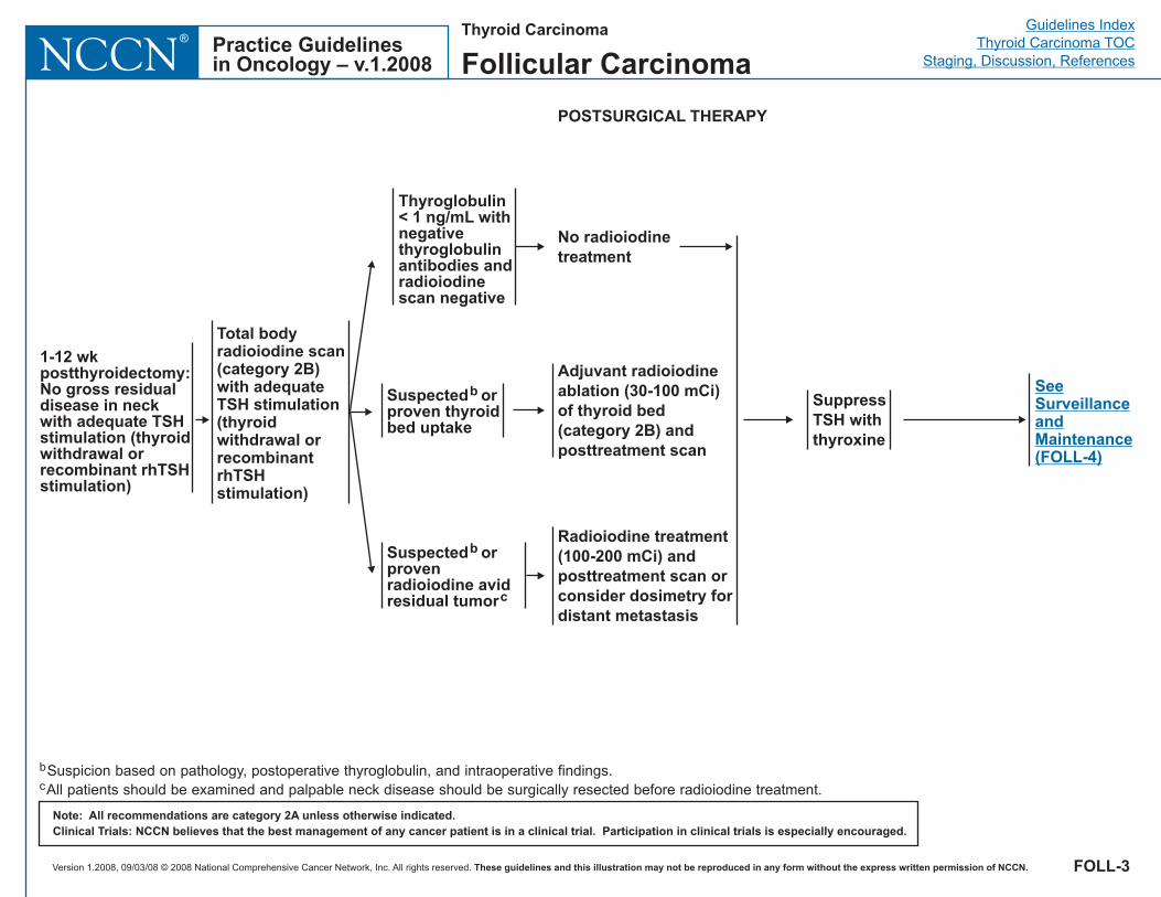
SuppressTSH withthyroxine
SeeSurveillanceandMaintenance(FOLL-4)
FOLL-3
Thyroid Carcinoma
Follicular Carcinoma
Adjuvant radioiodineablation (30-100 mCi)of thyroid bed(category 2B) andposttreatment scan
RadioiodinemCi
posttreatment scan orconsider dosimetry fordistant metastasis
treatment(100-200 ) and
POSTSURGICAL THERAPY
1-12 wkpostthyroidectomy:No gross residualdisease in neckwith adequate TSHstimulation (thyroidwithdrawal orrecombinant rhTSHstimulation)
Suspected orprovenradioiodine avidresidual tumor
b
c
Suspected orproven thyroidbed uptake
b
No radioiodinetreatment
Total bodyradioiodine scan(category 2B)with adequateTSH stimulation(thyroidwithdrawal orrecombinantrhTSHstimulation)
b
cSuspicion based on pathology, postoperative thyroglobulin, and intraoperative findings.All patients should be examined and palpable neck disease should be surgically resected before radioiodine treatment.
Thyroglobulin< 1 ng/mL withnegativethyroglobulinantibodies andradioiodinescan negative
Version 1.2008, 09/03/08 © 2008 National Comprehensive Cancer Network, Inc. All rights reserved. These guidelines and this illustration may not be reproduced in any form without the express written permission of NCCN.
Guidelines IndexThyroid Carcinoma TOC
Staging, Discussion, ReferencesPractice Guidelinesin Oncology – v.1.2008NCCN
®
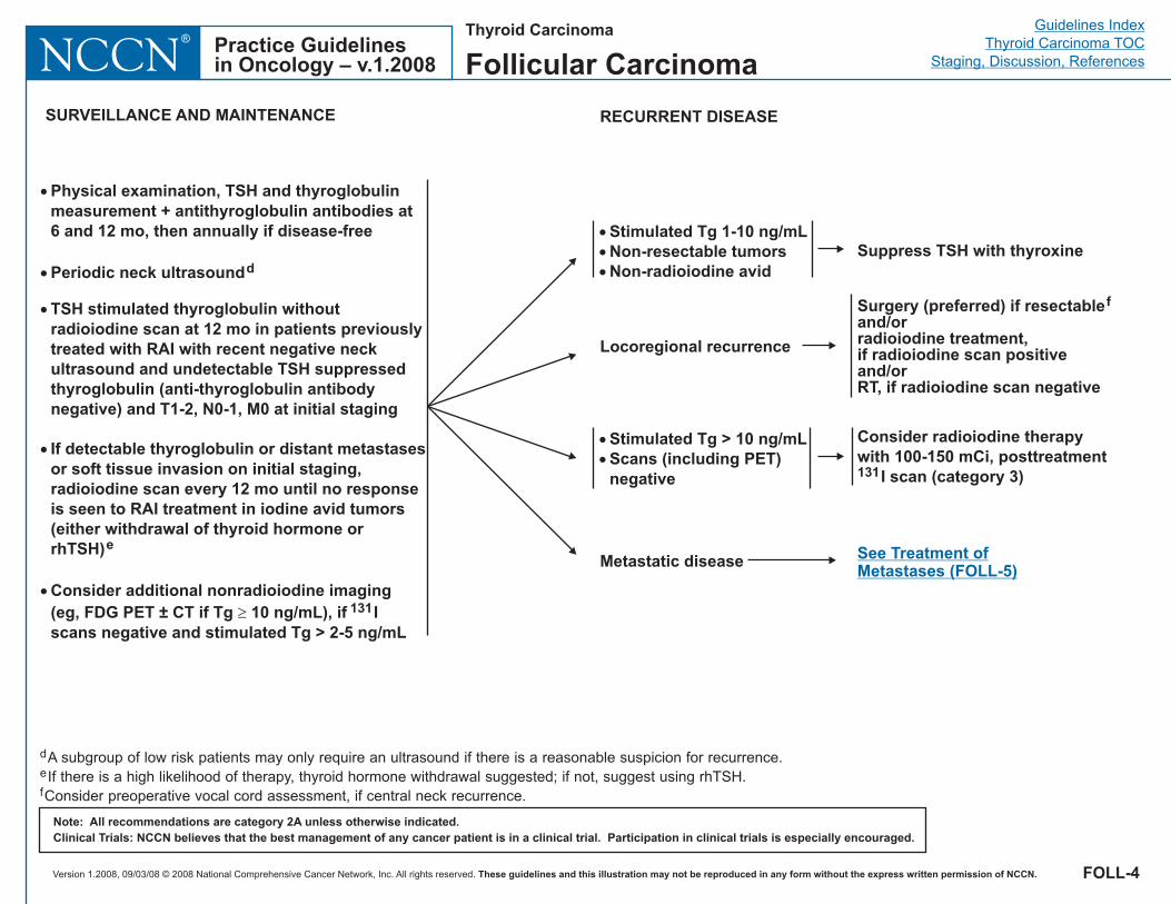
SURVEILLANCE AND MAINTENANCE
Locoregional recurrence
Metastatic disease
Surgery (preferred) if resectableand/orradioiodine treatment,if radioiodine scan positiveand/or
f
RT, if radioiodine scan negative
See Treatment ofMetastases (FOLL-5)
RECURRENT DISEASE
Note: All recommendations are category 2A unless otherwise indicated.Clinical Trials: NCCN believes that the best management of any cancer patient is in a clinical trial. Participation in clinical trials is especially encouraged.
d
e
f
A subgroup of low risk patients may only require an ultrasound if there is a reasonable suspicion for recurrence.If there is a high likelihood of therapy, thyroid hormone withdrawal suggested; if not, suggest using rhTSH.Consider preoperative vocal cord assessment, if central neck recurrence.
FOLL-4
Thyroid Carcinoma
Follicular Carcinoma
�
�
�
Stimulated Tg 1-10 ng/mLNon-resectable tumorsNon-radioiodine avid
Suppress TSH with thyroxine
Consider therapywith 100-150 mCi, posttreatment
I scan (category 3)
radioiodine
131
�
�
�
�
�
Physical examination, TSH and thyroglobulinmeasurement + antithyroglobulin antibodies at6 and 12 mo, then annually if disease-free
Periodic neck ultrasound
Consider additional nonradioiodine imaging(eg, FDG PET ± CT if Tg 10 ng/mL), if Iscans negative and stimulated Tg > 2-5 ng/mL
d
TSH stimulated thyroglobulin withoutradioiodine scan at 12 mo in patients previouslytreated with RAI with recent negative neckultrasound and undetectable TSH suppressedthyroglobulin (anti-thyroglobulin antibodynegative) and T1-2, N0-1, M0 at initial staging
If detectable thyroglobulin or distant metastasesor soft tissue invasion on initial staging,radioiodine scan every 12 mo until no responseis seen to RAI treatment in iodine avid tumors(either withdrawal of thyroid hormone orrhTSH)e
131�
�
�
Tg > 10 ng mLScans (including PET)negative
Stimulated /
Version 1.2008, 09/03/08 © 2008 National Comprehensive Cancer Network, Inc. All rights reserved. These guidelines and this illustration may not be reproduced in any form without the express written permission of NCCN.
Guidelines IndexThyroid Carcinoma TOC
Staging, Discussion, ReferencesPractice Guidelinesin Oncology – v.1.2008NCCN
®
TREATMENT OF METASTASES
CNS
Consider neurosurgical resection
Radioiodine treatment with rhTSH and steroidprophylaxis, if radioiodine scan positive with considerationof dosimetry to maximize dosing
Image-guided RT
g
g,h
and/or
and/or
Bone
�
�
�
Surgical palliation, if symptomatic or asymptomatic in weight-bearing extremities
Radioiodine treatment, if radioiodine scan positiveand/or
and/orRTConsider bisphosphonate therapyConsider embolization of metastases
withconsideration of dosimetry to maximize dosing
Note: All recommendations are category 2A unless otherwise indicated.Clinical Trials: NCCN believes that the best management of any cancer patient is in a clinical trial. Participation in clinical trials is especially encouraged.
Otherextracervical sites
Consider surgical resection of selected, enlarging, or symptomaticmetastasesand/orRadioiodine if positive uptake, with consideration of dosimetry tomaximize dosingand/orFor clinically progressive or symptomatic disease: clinical trials fornon-radioiodine avid tumors; systemic therapy (if trial not available)orBest Supportive Care
i
FOLL-5
Thyroid Carcinoma
Follicular Carcinoma
g
hFor solitary lesions, either neurosurgical resection or stereotactic radiosurgery preferred.Does not include whole brain RT.
iCytotoxic chemotherapy has shown to have minimal efficacy. There are agents in clinical trials investigating novel targeted therapies.See Clinical trials available at the NCCN member institutions.
Metastatic disease� Continue to suppress
TSH with thyroxine
Version 1.2008, 09/03/08 © 2008 National Comprehensive Cancer Network, Inc. All rights reserved. These guidelines and this illustration may not be reproduced in any form without the express written permission of NCCN.
Guidelines IndexThyroid Carcinoma TOC
Staging, Discussion, ReferencesPractice Guidelinesin Oncology – v.1.2008NCCN
®Thyroid Carcinoma
Hürthle Cell Carcinoma
Note: All recommendations are category 2A unless otherwise indicated.Clinical Trials: NCCN believes that the best management of any cancer patient is in a clinical trial. Participation in clinical trials is especially encouraged.
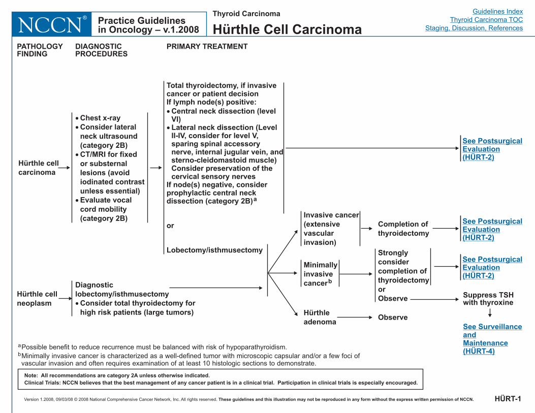
Hürthle cellcarcinoma
Total thyroidectomy, if invasivecancer or patient decisionIf lymph node(s) positive:
Central neck dissection (levelVI)
Consider preservation of thecervical sensory nerves
If node(s) negative, considerprophylactic central neckdissection (category 2B)
�
� Lateral neck dissection (LevelII-IV, consider for level V,sparing spinal accessorynerve, internal jugular vein, andsterno-cleidomastoid muscle)
or
Lobectomy/isthmusectomy
a
PATHOLOGYFINDING
DIAGNOSTICPROCEDURES
PRIMARY TREATMENT
�
�
�
�
Chest x-ray
voidiodinated contrastunless essential)Evaluate vocalcord mobility(category 2B)
Consider lateralneck ultrasound(category 2B)CT/MRI for fixedor substernallesions (a
Minimallyinvasivecancerb
Hürthleadenoma
Invasive cancer(extensivevascularinvasion)
Stronglyconsidercompletion ofthyroidectomyorObserve
Observe
Completion ofthyroidectomy
Suppress TSHwith thyroxine
See SurveillanceandMaintenance(H RT-4)Ü
See PostsurgicalEvaluation(H RT-2)Ü
See PostsurgicalEvaluation(H RT-2)Ü
See PostsurgicalEvaluation(H RT-2)Ü
a
bPossible benefit to reduce recurrence must be balanced with risk of hypoparathyroidism.Minimally invasive cancer is characterized as a well-defined tumor with microscopic capsular and/or a few foci ofvascular invasion and often requires examination of at least 10 histologic sections to demonstrate.
H RT-1Ü
H rthle cellneoplasm
üDiagnosticlobectomy/isthmusectomy
Consider total thyroidectomy forhigh risk patients (large tumors)
�
Version 1.2008, 09/03/08 © 2008 National Comprehensive Cancer Network, Inc. All rights reserved. These guidelines and this illustration may not be reproduced in any form without the express written permission of NCCN.
Guidelines IndexThyroid Carcinoma TOC
Staging, Discussion, ReferencesPractice Guidelinesin Oncology – v.1.2008NCCN
®
Note: All recommendations are category 2A unless otherwise indicated.Clinical Trials: NCCN believes that the best management of any cancer patient is in a clinical trial. Participation in clinical trials is especially encouraged.
POSTSURGICAL EVALUATIONAFTER THYROIDECTOMY
Grossresidualdiseasein neck
SuppressTSH withthyroxine
SeeSurveillanceandMaintenance(H RT-4)Ü
Unresectable
Resectable Resect, ifpossible
No grossresidual disease
Gross residualdisease
�
�
TSH + thyroglobulinmeasurement +antithyroglobulinantibodies (1-12 wkpostoperatively)Total bodyradioiodine scan(category 2B)
H RT-2Ü
RT
No scanperformed
�
�
�
RadioiodinetreatmentPost-treatment
I scanRT
131
No grossresidualdiseasein neck
Thyroid Carcinoma
Hürthle Cell Carcinoma
Inadequateuptake
Adequateuptake
TSH + thyroglobulinmeasurement +antithyroglobulinantibodies (1-12 wkpostoperatively)
SeePostsurgicalTherapy(H RT-3)Ü
�
�
�
�
< 1 cm confined toglandNo nodal metastasesNo distant metastasesNon-aggressivehistology
�
�
�
�
1 cmorNodal metastasesorDistant metastasesorAggressive histology
�
SeeSurveillanceandMaintenance(H RT-4)Ü
Version 1.2008, 09/03/08 © 2008 National Comprehensive Cancer Network, Inc. All rights reserved. These guidelines and this illustration may not be reproduced in any form without the express written permission of NCCN.
Guidelines IndexThyroid Carcinoma TOC
Staging, Discussion, ReferencesPractice Guidelinesin Oncology – v.1.2008NCCN
®
Note: All recommendations are category 2A unless otherwise indicated.Clinical Trials: NCCN believes that the best management of any cancer patient is in a clinical trial. Participation in clinical trials is especially encouraged.
SeeSurveillanceandMaintenance(H RT-4)Ü
SuppressTSH withthyroxine
T4 (surgicallyevident extra-thyroidalinvasion) andage > 45 y
All others
Consider RT
H RT-3Ü
Thyroid Carcinoma
Hürthle Cell Carcinoma
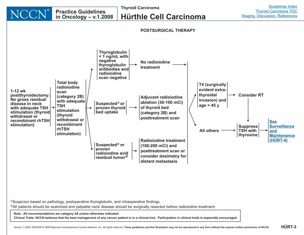
Adjuvant radioiodineablation (30-100 mCi)of thyroid bed(category 2B) andposttreatment scan
RadioiodinemCi
posttreatment scan orconsider dosimetry fordistant metastasis
treatment(100-200 ) and
POSTSURGICAL THERAPY
1-12 wkpostthyroidectomy:No gross residualdisease in neckwith adequate TSHstimulation (thyroidwithdrawal orrecombinant rhTSHstimulation)
Suspected orprovenradioiodine avidresidual tumor
c
d
Suspected orproven thyroidbed uptake
c
No radioiodinetreatment
Total bodyradioiodinescan(category 2B)with adequateTSHstimulation(thyroidwithdrawal orrecombinantrhTSHstimulation)
c
dSuspicion based on pathology, postoperative thyroglobulin, and intraoperative findings.All patients should be examined and palpable neck disease should be surgically resected before radioiodine treatment.
Thyroglobulin< 1 ng/mL withnegativethyroglobulinantibodies andradioiodinescan negative
Version 1.2008, 09/03/08 © 2008 National Comprehensive Cancer Network, Inc. All rights reserved. These guidelines and this illustration may not be reproduced in any form without the express written permission of NCCN.
Guidelines IndexThyroid Carcinoma TOC
Staging, Discussion, ReferencesPractice Guidelinesin Oncology – v.1.2008NCCN
®
SURVEILLANCE AND MAINTENANCE
Locoregional recurrence
Metastatic disease
Surgery (preferred) if resectableand/orRadioiodine treatment, ifradioiodine scan positiveand/orRT, if radioiodine scan negative
g
See Treatment ofMetastases (H RT-5)Ü
RECURRENT DISEASE
Note: All recommendations are category 2A unless otherwise indicated.Clinical Trials: NCCN believes that the best management of any cancer patient is in a clinical trial. Participation in clinical trials is especially encouraged.
e
f
g
A subgroup of low risk patients may only require an ultrasound if there is a reasonable suspicion for recurrence.If there is a high likelihood of therapy, thyroid hormone withdrawal suggested; if not, suggest using rhTSH.Consider preoperative vocal cord assessment, if central neck recurrence.
H RT-4Ü
Thyroid Carcinoma
Hürthle Cell Carcinoma
�
�
�
�
�
Physical examination, TSH and thyroglobulinmeasurement + antithyroglobulin antibodies at6 and 12 mo, then annually if disease-free
Consider additional nonradioiodine imaging(eg, FDG PET ± CT if Tg 10 ng/mL), if Iscans negative and stimulated Tg > 2-5 ng/mL
Periodic neck ultrasound
TSH stimulated thyroglobulin withoutradioiodine scan at 12 mo in patientspreviously treated with RAI with recentnegative neck ultrasound and undetectableTSH suppressed thyroglobulin (anti-thyroglobulin antibody negative) and T1-2, N0-1, M0 at initial staging
If detectable thyroglobulin or distantmetastases or soft tissue invasion on initialstaging, radioiodine scan every 12 mo until noresponse is seen to RAI treatment in iodineavid tumors (either withdrawal of thyroidhormone or rhTSH)
e
f
131�
�
�
�
Stimulated Tg 1-10 ng/mLNon-resectable tumorsNon-radioiodine avid
Suppress TSH with thyroxine
Consider therapywith 100-150 mCi, posttreatment
I scan (category 2B)
radioiodine
131
�
�
Tg > 10 ng mLScans (including PET)negative
Stimulated /
Version 1.2008, 09/03/08 © 2008 National Comprehensive Cancer Network, Inc. All rights reserved. These guidelines and this illustration may not be reproduced in any form without the express written permission of NCCN.
Guidelines IndexThyroid Carcinoma TOC
Staging, Discussion, ReferencesPractice Guidelinesin Oncology – v.1.2008NCCN
®
Note: All recommendations are category 2A unless otherwise indicated.Clinical Trials: NCCN believes that the best management of any cancer patient is in a clinical trial. Participation in clinical trials is especially encouraged.
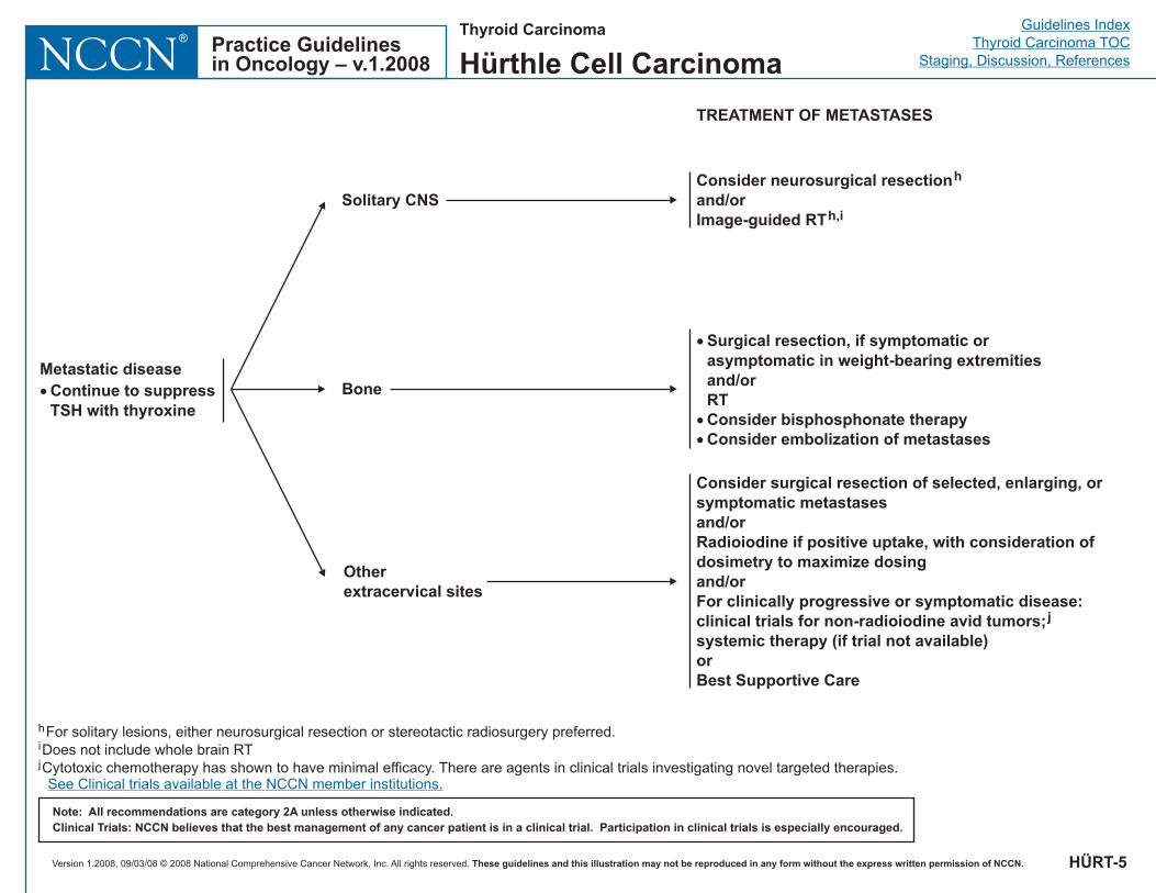
TREATMENT OF METASTASES
Solitary CNSConsider neurosurgical resectionh
and/orImage-guided RTh,i
Bone
�
�
�
Surgical resection, if symptomatic orasymptomatic in weight-bearing extremitiesand/orRTConsider bisphosphonate therapyConsider embolization of metastases
Otherextracervical sites
Consider surgical resection of selected, enlarging, orsymptomatic metastasesand/orRadioiodine if positive uptake, with consideration ofdosimetry to maximize dosingand/or
orBest Supportive Care
For clinically progressive or symptomatic disease:clinical trials for non-radioiodine avid tumors;systemic therapy (if trial not available)
j
H RT-5Ü
Thyroid Carcinoma
Hürthle Cell Carcinoma
Metastatic disease� Continue to suppress
TSH with thyroxine
h
i
j
For solitary lesions, either neurosurgical resection or stereotactic radiosurgery preferred.Does not include whole brain RTCytotoxic chemotherapy has shown to have minimal efficacy. There are agents in clinical trials investigating novel targeted therapies.See Clinical trials available at the NCCN member institutions.
Version 1.2008, 09/03/08 © 2008 National Comprehensive Cancer Network, Inc. All rights reserved. These guidelines and this illustration may not be reproduced in any form without the express written permission of NCCN.
Guidelines IndexThyroid Carcinoma TOC
Staging, Discussion, ReferencesPractice Guidelinesin Oncology – v.1.2008NCCN
®Thyroid Carcinoma
Medullary Carcinoma
Note: All recommendations are category 2A unless otherwise indicated.Clinical Trials: NCCN believes that the best management of any cancer patient is in a clinical trial. Participation in clinical trials is especially encouraged.
MEDU-1
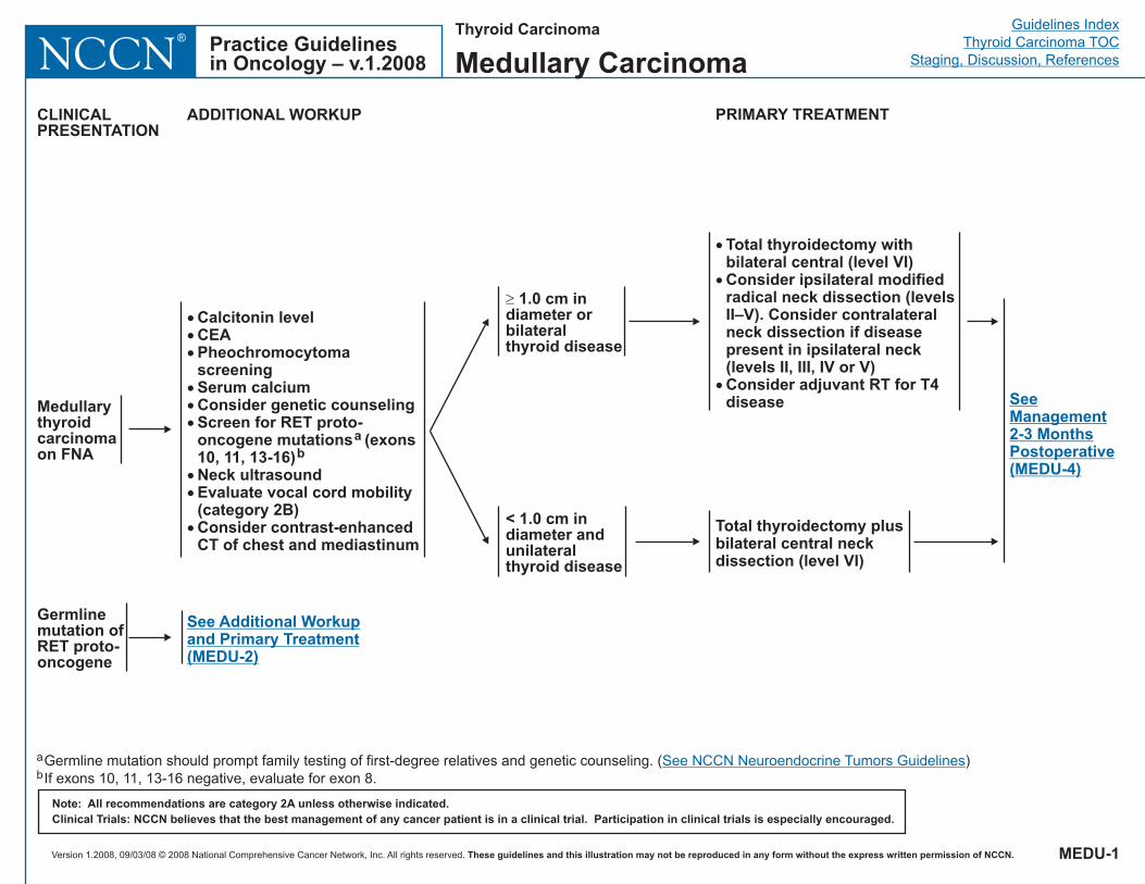
Medullarythyroidcarcinomaon FNA
���
���
��
�
Calcitonin levelCEAPheochromocytomascreeningSerum calciumConsider genetic counselingScreen for RET proto-oncogene mutations (exons10, 11, 13-16)Neck ultrasoundEvaluate vocal cord mobility(category 2B)Consider contrast-enhancedCT of chest and mediastinum
ab
� 1.0 cm indiameter orbilateralthyroid disease
< 1.0 cm indiameter andunilateralthyroid disease
�
�
�
Total thyroidectomy withbilateral central (level VI)Consider ipsilateral modifiedradical neck dissection (levelsII–V). Consider contralateralneck dissection if diseasepresent in ipsilateral neck(levels II, III, IV or V)Consider adjuvant RT for T4disease
Total thyroidectomy plusbilateral central neckdissection (level VI)
CLINICALPRESENTATION
ADDITIONAL WORKUP PRIMARY TREATMENT
SeeManagement2-3 MonthsPostoperative(MEDU-4)
See Additional Workupand Primary Treatment(MEDU-2)
Germlinemutation ofRET proto-oncogene
a
bGermline mutation should prompt family testing of first-degree relatives and genetic counseling. ( )If exons 10, 11, 13-16 negative, evaluate for exon 8.
See NCCN Neuroendocrine Tumors Guidelines
Version 1.2008, 09/03/08 © 2008 National Comprehensive Cancer Network, Inc. All rights reserved. These guidelines and this illustration may not be reproduced in any form without the express written permission of NCCN.
Guidelines IndexThyroid Carcinoma TOC
Staging, Discussion, ReferencesPractice Guidelinesin Oncology – v.1.2008NCCN
®
Note: All recommendations are category 2A unless otherwise indicated.Clinical Trials: NCCN believes that the best management of any cancer patient is in a clinical trial. Participation in clinical trials is especially encouraged.
CLINICALPRESENTATION
ADDITIONAL WORKUP PRIMARY TREATMENT
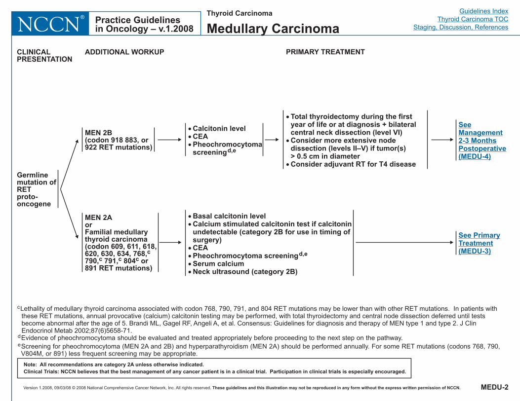
Germlinemutation ofRETproto-oncogene
MEN 2B(codon 918 883, or922 RET mutations)
MEN 2AorFamilial medullarythyroid carcinoma(codon 609, 611, 618,620, 630, 634, 768,790, 791, 804 or891 RET mutations)
cc c c
���
Calcitonin levelCEAPheochromocytomascreeningd,e
��
����
Basal calcitonin levelCalcium stimulated calcitonin test if calcitoninundetectable (category 2B for use in timing ofsurgery)CEAPheochromocytoma screeningSerum calciumNeck ultrasound (category 2B)
d,e
c
d
e
Lethality of medullary thyroid carcinoma associated with codon 768, 790, 791, and 804 RET mutations may be lower than with other RET mutations. In patients withthese RET mutations, annual provocative (calcium) calcitonin testing may be performed, with total thyroidectomy and central node dissection deferred until testsbecome abnormal after the age of 5. Brandi ML, Gagel RF, Angeli A, et al. Consensus: Guidelines for diagnosis and therapy of MEN type 1 and type 2. J ClinEndocrinol Metab 2002;87(6)5658-71.Evidence of pheochromocytoma should be evaluated and treated appropriately before proceeding to the next step on the pathway.Screening for pheochromocytoma (MEN 2A and 2B) and hyperparathyroidism (MEN 2A) should be performed annually. For some RET mutations (codons 768, 790,V804M, or 891) less frequent screening may be appropriate.
�
�
�
Total thyroidectomy during the firstyear of life or at diagnosis + bilateralcentral neck dissection (level VI)Consider more extensive nodedissection (levels II–V) if tumor(s)> 0.5 cm in diameterConsider adjuvant RT for T4 disease
See PrimaryTreatment(MEDU-3)
SeeManagement2-3 MonthsPostoperative(MEDU-4)
MEDU-2
Thyroid Carcinoma
Medullary Carcinoma
Version 1.2008, 09/03/08 © 2008 National Comprehensive Cancer Network, Inc. All rights reserved. These guidelines and this illustration may not be reproduced in any form without the express written permission of NCCN.
Guidelines IndexThyroid Carcinoma TOC
Staging, Discussion, ReferencesPractice Guidelinesin Oncology – v.1.2008NCCN
®
Note: All recommendations are category 2A unless otherwise indicated.Clinical Trials: NCCN believes that the best management of any cancer patient is in a clinical trial. Participation in clinical trials is especially encouraged.
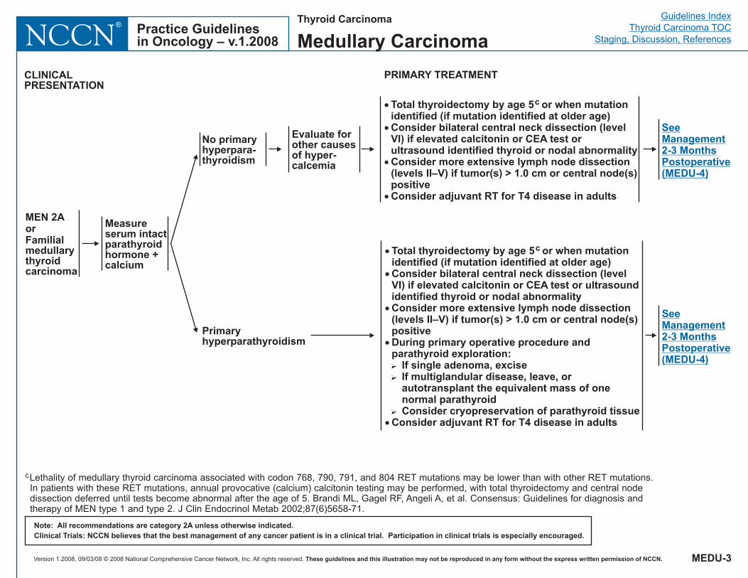
Measureserum intactparathyroidhormone +calcium
Primaryhyperparathyroidism
No primaryhyperpara-thyroidism
Evaluate forother causesof hyper-calcemia
�
�
�
�
Total thyroidectomy by age 5 or when mutationidentified (if mutation identified at older age)Consider bilateral central neck dissection (levelVI) if elevated calcitonin or CEA test orultrasound identified thyroid or nodal abnormalityConsider more extensive lymph node dissection(levels II–V) if tumor(s) > 1.0 cm or central node(s)positiveConsider adjuvant RT for T4 disease in adults
c
�
�
�
�
�
Total thyroidectomy by age 5 or when mutationidentified (if mutation identified at older age)Consider bilateral central neck dissection (levelVI) if elevated calcitonin or CEA test or ultrasoundidentified thyroid or nodal abnormalityConsider more extensive lymph node dissection(levels II–V) if tumor(s) > 1.0 cm or central node(s)positiveDuring primary operative procedure andparathyroid exploration:
If single adenoma, exciseIf multiglandular disease, leave, orautotransplant the equivalent mass of onenormal parathyroidConsider cryopreservation of parathyroid tissue
Consider adjuvant RT for T4 disease in adults
c
�
�
�
MEN 2AorFamilialmedullarythyroidcarcinoma
PRIMARY TREATMENT
SeeManagement2-3 MonthsPostoperative(MEDU-4)
CLINICALPRESENTATION
SeeManagement2-3 MonthsPostoperative(MEDU-4)
MEDU-3
cLethality of medullary thyroid carcinoma associated with codon 768, 790, 791, and 804 RET mutations may be lower than with other RET mutations.In patients with these RET mutations, annual provocative (calcium) calcitonin testing may be performed, with total thyroidectomy and central nodedissection deferred until tests become abnormal after the age of 5. Brandi ML, Gagel RF, Angeli A, et al. Consensus: Guidelines for diagnosis andtherapy of MEN type 1 and type 2. J Clin Endocrinol Metab 2002;87(6)5658-71.
Thyroid Carcinoma
Medullary Carcinoma
Version 1.2008, 09/03/08 © 2008 National Comprehensive Cancer Network, Inc. All rights reserved. These guidelines and this illustration may not be reproduced in any form without the express written permission of NCCN.
Guidelines IndexThyroid Carcinoma TOC
Staging, Discussion, ReferencesPractice Guidelinesin Oncology – v.1.2008NCCN
®
Note: All recommendations are category 2A unless otherwise indicated.Clinical Trials: NCCN believes that the best management of any cancer patient is in a clinical trial. Participation in clinical trials is especially encouraged.
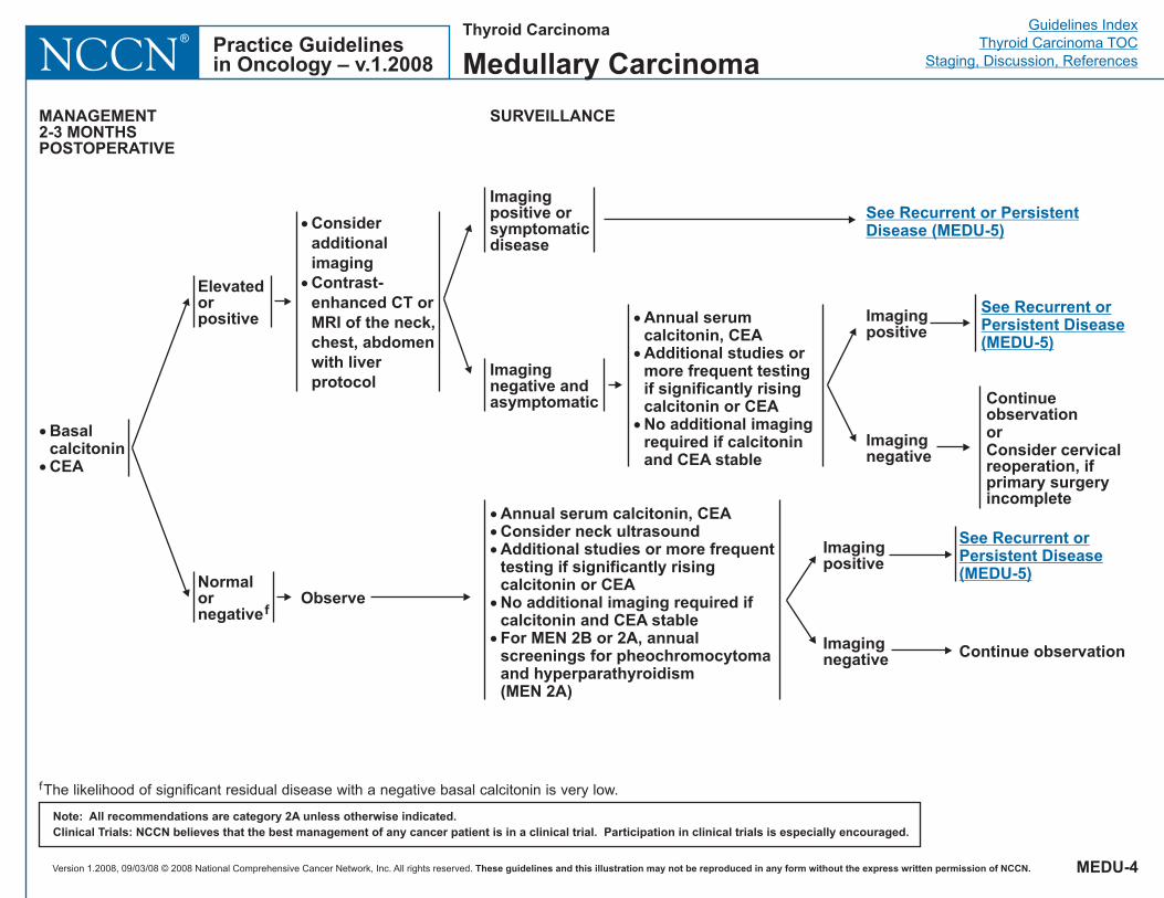
�
�
BasalcalcitoninCEA
Elevatedorpositive
Normalornegativef
�
�
ConsideradditionalimagingContrast-enhanced CT orMRI of the neck,chest, abdomenwith liverprotocol
MANAGEMENT2-3 MONTHSPOSTOPERATIVE
SURVEILLANCE
Observe
Imagingpositive orsymptomaticdisease
Imagingnegative andasymptomatic
See Recurrent or PersistentDisease (MEDU-5)
���
�
�
Annual serum calcitonin, CEAConsider neck ultrasoundAdditional studies or more frequenttesting if significantly risingcalcitonin or CEANo additional imaging required ifcalcitonin and CEA stableFor MEN 2B or 2A, annualscreenings for pheochromocytomaand hyperparathyroidism(MEN 2A)
ContinueobservationorConsider cervicalreoperation, ifprimary surgeryincomplete
Continue observation
�
�
�
Annual serumcalcitonin, CEAAdditional studies ormore frequent testingif significantly risingcalcitonin or CEANo additional imagingrequired if calcitoninand CEA stable
See Recurrent orPersistent Disease(MEDU-5)
Imagingpositive
Imagingnegative
Imagingpositive
Imagingnegative
fThe likelihood of significant residual disease with a negative basal calcitonin is very low.
See Recurrent orPersistent Disease(MEDU-5)
MEDU-4
Thyroid Carcinoma
Medullary Carcinoma

Version 1.2008, 09/03/08 © 2008 National Comprehensive Cancer Network, Inc. All rights reserved. These guidelines and this illustration may not be reproduced in any form without the express written permission of NCCN.
Guidelines IndexThyroid Carcinoma TOC
Staging, Discussion, ReferencesPractice Guidelinesin Oncology – v.1.2008NCCN
®
Note: All recommendations are category 2A unless otherwise indicated.Clinical Trials: NCCN believes that the best management of any cancer patient is in a clinical trial. Participation in clinical trials is especially encouraged.
Locoregional
Symptomatic,distant metastases
Asymptomatic,distant metastases
Surgical resection ±postoperative RTorConsider RT, if symptomaticprogressive disease orunresectable
Consider palliative resection,ablation (eg, radiofrequency[RFA], embolization or otherregional therapy) or otherregional treatment
Disseminatedsymptomaticdisease
�
�
�
Clinical trial (preferred)orRT for focal symptomsorSorafeniborDTIC-based chemotherapyConsider bisphosphonatetherapy for bone metastasesBest Supportive Care
RECURRENT OR PERSISTENT DISEASE
ObserveorConsider resection, if possible,or ablation (eg, RFA,embolization or other regionaltherapy) especially ifprogressive disease
MEDU-5
Thyroid Carcinoma
Medullary Carcinoma
Version 1.2008, 09/03/08 © 2008 National Comprehensive Cancer Network, Inc. All rights reserved. These guidelines and this illustration may not be reproduced in any form without the express written permission of NCCN.
Guidelines IndexThyroid Carcinoma TOC
Staging, Discussion, ReferencesPractice Guidelinesin Oncology – v.1.2008NCCN
®Thyroid Carcinoma
Anaplastic Carcinoma
Note: All recommendations are category 2A unless otherwise indicated.Clinical Trials: NCCN believes that the best management of any cancer patient is in a clinical trial. Participation in clinical trials is especially encouraged.
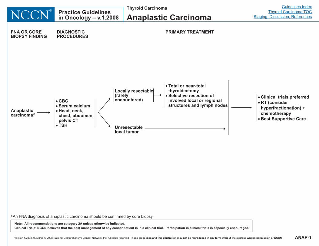
Anaplasticcarcinomaa
FNA OR COREBIOPSY FINDING
DIAGNOSTICPROCEDURES
PRIMARY TREATMENT
���
�
CBCSerum calciumHead, neck,chest, abdomen,pelvis CTTSH
Locally resectable(rarelyencountered)
Unresectablelocal tumor
�
�
Total or near-totalthyroidectomySelective resection ofinvolved local or regionalstructures and lymph nodes
�
�
�
Clinical trials preferredRT (considerhyperfractionation) +chemotherapyBest Supportive Care
ANAP-1
aAn FNA diagnosis of anaplastic carcinoma should be confirmed by core biopsy.
Version 1.2008, 09/03/08 © 2008 National Comprehensive Cancer Network, Inc. All rights reserved. These guidelines and this illustration may not be reproduced in any form without the express written permission of NCCN.
Guidelines IndexThyroid Carcinoma TOC
Staging, Discussion, ReferencesPractice Guidelinesin Oncology – v.1.2008NCCN
®
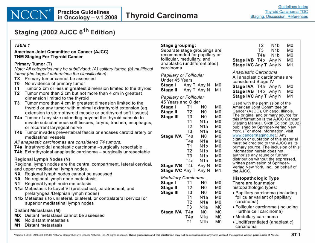
Table 1
American Joint Committee on Cancer (AJCC)TNM Staging For Thyroid Cancer
Primary Tumor (T)Note: All categories may be subdivided: (A) solitary tumor, (b) multifocaltumor (the largest determines the classification).
All anaplastic carcinomas are considered T4 tumors.
TXT0T1T2
T3
T4a
T4b
T4aT4b
NXN0N1N1a
N1b
MXM0M1
Primary tumor cannot be assessedNo evidence of primary tumorTumor 2 cm or less in greatest dimension limited to the thyroidTumor more than 2 cm but not more than 4 cm in greatestdimension limited to the thyroidTumor more than 4 cm in greatest dimension limited to thethyroid or any tumor with minimal extrathyroid extension (eg,extension to sternothyroid muscle or perithyroid soft tissues)Tumor of any size extending beyond the thyroid capsule toinvade subcutaneous soft tissues, larynx, trachea, esophagus,or recurrent laryngeal nerveTumor invades prevertebral fascia or encases carotid artery ormediastinal vessels
Intrathyroidal anaplastic carcinoma –surgically resectableExtrathyroidal anaplastic carcinoma – surgically unresectable
Regional lymph nodes are the central compartment, lateral cervical,and upper mediastinal lymph nodes.
Regional lymph nodes cannot be assessedNo regional lymph node metastasisRegional lymph node metastasisMetastasis to Level VI (pretracheal, paratracheal, andprelaryngeal/Delphian lymph nodes)Metastasis to unilateral, bilateral, or contralateral cervical orsuperior mediastinal lymph nodes
Distant metastasis cannot be assessedNo distant metastasisDistant metastasis
Regional Lymph Nodes (N)
Distant Metastasis (M)
Stage grouping:
Histopathologic Type
Separate stage groupings arerecommended for papillary orfollicular, medullary, andanaplastic (undifferentiated)carcinoma.
Under 45 YearsAny T Any N M0Any T Any N M1
45 Years and OlderT1 N0 M0T2 N0 M0T3 N0 M0T1 N1a M0T2 N1a M0T3 N1a M0
T4a N0 M0T4a N1a M0T1 N1b M0T2 N1b M0T3 N1b M0
T4a N1b M0T4b Any N M0
Any T Any N M1
T1 N0 M0T2 N0 M0T3 N0 M0T1 N1a M0T2 N1a M0T3 N1a M0
T4a N0 M0T4a N1a M0T1 N1b M0
T2 N1b M0T3 N1b M0
T4a N1b M0T4b Any N M0
Any T Any N M1
All anaplastic carcinomas areconsidered Stage IV
T4a Any N M0T4b Any N M0
Any T Any N M1
There are four majorhistopathologic types:
Papillary or Follicular
Papillary or Follicular
Medullary Carcinoma
Anaplastic Carcinoma
Stage IStage II
Stage IStage IIStage III
Stage IVA
Stage IVBStage IVC
Stage IStage IIStage III
Stage IVA
Stage IVBStage IVC
Stage IVAStage IVBStage IVC
Used with the permission of theAmerican Joint Committee onCancer (AJCC), Chicago, Illinois.The original and primary source forthis information is the AJCC CancerStaging Manual, Sixth Edition (2002)published by Springer-Verlag NewYork. (For more information, visit
t.) Anycitation or quotation of this materialmust be credited to the AJCC as itsprimary source. The inclusion of thisinformation herein does notauthorize any reuse or furtherdistribution without the expressed,written permission of Springer-Verlag New York, Inc., on behalf ofthe AJCC.
�
�
�
�
Papillary carcinoma (includingfollicular variant of papillarycarcinoma)Follicular carcinoma (includingHurthle cell carcinoma)Medullary carcinomaUndifferentiated (anaplastic)carcinoma
www.cancerstaging.ne
ST-1
Staging (2002 AJCC 6th Edition)
Thyroid Carcinoma
Version 1.2008, 09/03/08 © 2008 National Comprehensive Cancer Network, Inc. All rights reserved. These guidelines and this illustration may not be reproduced in any form without the express written permission of NCCN. MS-1
Guidelines IndexThyroid Carcinoma TOC
Staging, Discussion, ReferencesPractice Guidelinesin Oncology – v.1.2008 NCCN
®
Thyroid Carcinoma
Discussion
NCCN Categories of Evidence and Consensus
Category 1: The recommendation is based on high-level evidence (e.g. randomized controlled trials) and there is uniform NCCN consensus.
Category 2A: The recommendation is based on lower-level evidence and there is uniform NCCN consensus.
Category 2B: The recommendation is based on lower-level evidence and there is nonuniform NCCN consensus (but no major disagreement).
Category 3: The recommendation is based on any level of evidence but reflects major disagreement.
All recommendations are category 2A unless otherwise noted.
Overview Epidemiology Thyroid nodules are approximately 4 times more common in women than in men. These nodules increase in frequency throughout life, reaching a prevalence of about 5% in the U.S. population aged 50 years and older.1 Nodules are even more prevalent when the thyroid gland is examined at autopsy or surgery, or when using ultrasonography; 50% of the thyroids so studied have nodules, which are almost always benign.1,2 New nodules develop at a rate of about 0.1% per year, beginning in early life, but they develop at a much higher rate (about 2% per year) after exposure to head and neck irradiation.3,4
By contrast, thyroid carcinoma is uncommon. For the U.S. population, the lifetime risk of being diagnosed with thyroid carcinoma is less than
1% (0.84% for women and 0.30% for men).5 Approximately 37,340 new cases of thyroid carcinoma will be diagnosed in the United States in the year 2008.6 As with thyroid nodules, this cancer occurs 2 to 3 times more often in women than in men. With the incidence increasing by 6.2% per year, thyroid cancer is currently the eighth most common malignancy diagnosed in women. Among persons aged 15 to 24 years, thyroid cancer accounts for 7.5% to 10% of all diagnosed malignancies.7 The disease is also diagnosed more often in white North Americans than in African Americans. Although thyroid carcinoma can occur at any age, the peak incidence is around age 45 to 49 years in women and 65 to 69 years in men for the period 2000 to 2004.5
There are 3 main histologic types of thyroid cancer: differentiated (including papillary, follicular, and Hürthle), medullary, and anaplastic. Information from the National Cancer Data Base (NCDB) indicates that of 53,856 patients treated for thyroid carcinoma between 1985 and 1995, 80% had papillary carcinoma, 11% had follicular carcinoma, 3% had Hürthle cell carcinoma, 4% had medullary carcinoma, and 2% had anaplastic thyroid carcinoma.8 In 2008, approximately 1590 cancer deaths will occur among persons living with thyroid carcinoma in the United States.6 Thyroid carcinoma occurs more often in women; however, mortality rates are higher for men, probably because men are usually older at the time of diagnosis.5,9
The incidence of thyroid carcinoma increased almost 310% between 1950 and 2004, but mortality rates decreased more than 44%.5 From 1975 to 2004, thyroid cancer rates in the United States doubled. Because overall mortality has remained stable since 1975, the increasing incidence probably partially reflects earlier detection of subclinical disease (ie, small papillary cancers), although even microcarcinomas can metastasize regionally, thereby increasing eventual recurrence risk.10,11 It is also notable that the stable age- and gender-adjusted mortality rate for thyroid carcinoma contrasts distinctly
Version 1.2008, 09/03/08 © 2008 National Comprehensive Cancer Network, Inc. All rights reserved. These guidelines and this illustration may not be reproduced in any form without the express written permission of NCCN. MS-2
Guidelines IndexThyroid Carcinoma TOC
Staging, Discussion, ReferencesPractice Guidelinesin Oncology – v.1.2008 NCCN
®
Thyroid Carcinoma
with the declining rates being observed with other solid tumors in adults.6
The Challenge of Managing Differentiated Thyroid Carcinoma Managing differentiated (ie, papillary, follicular, and Hürthle) thyroid carcinoma can be a challenge, because no prospective randomized trials of treatment have been done. Results from ongoing randomized trials will not be available for many years, given the typically prolonged course and relative infrequency of these tumors. Most of the information about treatment comes from studies of large patient cohorts in which therapy has not been randomly assigned. This accounts for much of the disagreement about managing differentiated carcinoma. Nonetheless, most patients can be cured of this disease when properly treated by experienced physicians and surgeons.12 The treatment of choice is surgery, whenever possible, followed in many patients by radioiodine (131I) and thyroxine therapy. External-beam radiation therapy (RT) and chemotherapy have less prominent roles in managing these tumors.
Radiation-Induced Thyroid Carcinoma Exposure to ionizing radiation is the only known environmental cause of thyroid carcinoma, usually causing papillary carcinoma. The thyroid glands of children are especially vulnerable to the carcinogenic action of ionizing radiation. A child’s thyroid gland has one of the highest risks of developing cancer of any organ. In fact, the thyroid gland is the only organ linked to risk at about 0.10 Gy by convincing evidence.3 The risk of radiation-induced thyroid carcinoma is greater in females, certain Jewish populations, and patients with a family history of thyroid carcinoma.13 This suggests that genetic factors are also important in its development. Beginning within 5 years of irradiation, new nodules develop at a rate of about 2% annually, reaching a peak incidence within 30 years of irradiation but remaining high at 40 years.3,4
Previously, most studies showed that 131I is less effective than external gamma radiation in inducing thyroid carcinoma.14 However, most of the studies that came to this conclusion involved adults, in whom the risk of developing thyroid carcinoma after exposure to 131I appears to be small or nonexistent.15 After the Chernobyl nuclear reactor accident in 1986, many children developed papillary thyroid carcinoma after being exposed to radioiodine fallout. It became evident that 131I and other short-lived radioiodines were potent thyroid carcinogens in children, particularly those who were younger than 10 years when they were exposed.16 Although radiation-induced papillary thyroid cancer tends to appear more aggressive histologically and to have high recurrence rates, the prognosis for survival is not clearly different from that of spontaneously occurring tumors.17,18
Differentiated Thyroid Carcinoma Clinical Presentation and Diagnosis Differentiated (ie, papillary, follicular, or Hürthle) thyroid carcinoma is usually asymptomatic for long periods and commonly presents as a solitary thyroid nodule. However, evaluating all nodules for malignancy is difficult, because benign nodules are so prevalent and thyroid carcinoma, by contrast, is so uncommon.1 Moreover, both benign and malignant thyroid nodules are usually asymptomatic, giving no clinical clue to their diagnosis. About 50% of the malignant nodules are discovered during a routine physical examination, by serendipity on imaging studies, or during surgery for benign disease. The other 50% are usually first noticed by the patient, usually as an asymptomatic nodule.1 Regrettably, the typically indolent nature of differentiated thyroid carcinoma often leads to long delays in diagnosis that may substantially worsen the course of the disease.9
Factors Affecting Risk of Malignancy Nodule size has a bearing on the risk of malignancy and the clinical evaluation. Thyroid nodules smaller than 1 cm occur with such
Version 1.2008, 09/03/08 © 2008 National Comprehensive Cancer Network, Inc. All rights reserved. These guidelines and this illustration may not be reproduced in any form without the express written permission of NCCN. MS-3
Guidelines IndexThyroid Carcinoma TOC
Staging, Discussion, ReferencesPractice Guidelinesin Oncology – v.1.2008 NCCN
®
Thyroid Carcinoma
frequency in the asymptomatic general population that they are found, in many cases, by serendipity when performing imaging studies for other head or neck problems. Often termed “incidentalomas,” nodules smaller than 1 cm are almost invariably clinically benign lesions and usually do not require biopsy.1,2,19 In selected cases, it may be reasonable to follow these nodules with serial ultrasounds. By contrast, nodules more than 4 cm in diameter are more suggestive and pose a somewhat higher risk of malignancy. Fine-needle aspiration (FNA) is the procedure of choice for evaluating suspicious thyroid nodules.20 The Society of Radiologists in Ultrasound wrote a consensus statement about management of thyroid nodules identified at thyroid ultrasonography. Their recommendations describe which nodules should undergo FNA and which should not based on nodule size and ultrasound characteristics, and on clinical features that might predict risk of morbidity from an undiagnosed malignancy.21 Suspicious criteria by ultrasound include central hypervascularity, microcalcifications, and irregular borders.
Although more than 50% of all malignant nodules are asymptomatic, the pretest probability of malignancy in a nodule increases considerably when signs or symptoms are present (see THYR-1).22 For example, the likelihood that a nodule is malignant increases about 7-fold if it is very firm, fixed to adjacent structures, associated with enlarged regional lymph nodes, causes vocal cord paralysis, or is rapidly growing or if symptoms of invasion into neck structures are present.22,23 If 2 or more of these features are present, the likelihood of thyroid cancer is virtually assured.23
A patient’s age and gender also affect the probability of malignancy. The risk of malignancy is higher in patients younger than 15 years and older than 60 years. In particular, a man older than 60 years with a thyroid nodule has about 4 times the risk of having thyroid carcinoma than does a middle-aged woman with a thyroid nodule.24 Other factors
that increase the suspicion of malignancy include (1) a history of head and neck irradiation; (2) a family history of thyroid carcinoma; (3) the presence of familial syndromes associated with thyroid carcinoma (see “Familial Syndromes”), such as Gardner’s syndrome, familial adenomatous polyposis, Carney complex, Cowden’s syndrome, and multiple endocrine neoplasia (MEN) types 2A or 2B; (4) evidence of other diseases seen with thyroid cancer--associated diseases or syndromes such as hyperparathyroidism, pheochromocytoma, marfanoid habitus, and mucosal neuromas (MEN2B), which make the presence of medullary thyroid cancer, more likely; or (5) the presence of suspicious findings detected by imaging such as focal FDG (18-fluorodeoxyglucose) uptake on positron emission tomography (PET), or central hypervascularity, irregular border, and/or microcalcifications on ultrasound.25
Initial Workup Fine-needle aspiration of the nodule and clinically suspicious lymph nodes is recommended as the first diagnostic test in a clinically euthyroid patient before any imaging studies are done.1 Ideally, the serum thyrotropin (thyroid-stimulating hormone [TSH]) results should be known before FNA is performed. This is often impractical, however, and FNA may be done during the initial office visit.
Some clinicians, especially in Europe,26 recommend obtaining serum calcitonin levels from all patients with thyroid nodules. However, there is controversy surrounding the cost effectiveness of this practice in the United States, especially in the absence of confirmatory pentagastrin stimulation testing, and the assumptions used in cost effective analyses. To date, this practice has not been recommended by the American Thyroid Association.27 A recent study showed that calcitonin screening may be cost effective in the United States.28 However, false-positive calcitonin readings that can result from minimal calcitonin elevations can only be ruled out with pentagastrin testing, and
Version 1.2008, 09/03/08 © 2008 National Comprehensive Cancer Network, Inc. All rights reserved. These guidelines and this illustration may not be reproduced in any form without the express written permission of NCCN. MS-4
Guidelines IndexThyroid Carcinoma TOC
Staging, Discussion, ReferencesPractice Guidelinesin Oncology – v.1.2008 NCCN
®
Thyroid Carcinoma
pentagastrin is not available in the United States. Ultrasound of the thyroid and neck including central and lateral neck compartments is also recommended (category 2B).29
Cytologic examination of an FNA specimen, with sufficient cells recovered to assign a diagnosis, is typically categorized as (1) carcinoma; (2) suspicious for malignancy, including follicular neoplasms; (3) thyroid lymphoma; and (4) benign (such as macrofollicular, colloid adenoma, Hashimoto’s thyroiditis, Hürthle cells in the absence of neoplasm). Both the National Cancer Institute (NCI) (http://thyroidfna.cancer.gov/pages/conclusions/) and the Papanicolaou Society of Cytopathology have guidelines for thyroid FNA (http://www.papsociety.org/guidelines.html).
Pathology and cytopathology slides should be reviewed at the treating institution by a pathologist with expertise in the diagnosis of thyroid disorders. Although FNA is a very sensitive test—particularly for papillary—false-negative results are sometimes obtained; therefore, a reassuring FNA should not override concerns in the presence of worrisome clinical findings.30
Pathology synoptic reports (protocols) are useful for reporting results from examinations of surgical specimens; these reports assist pathologists in providing clinically useful and relevant information. The National Comprehensive Cancer Network (NCCN) thyroid panel is in favor of pathology synoptic reports from the (1) College of American Pathologists (CAP), and (2) the Association of Directors of Anatomic and Surgical Pathology (ADASP). Some pathologists currently use a modified format that is felt to comply with both of these synoptic reports. Although there is no published ADASP checklist for thyroid carcinoma, the CAP protocol information and checklists can be accessed at:
http://www.cap.org/apps/cap.portal?_nfpb=true&cntvwrPtlt_actionOverride=%2Fportlets%2FcontentViewer%2Fshow&_windowLabel=cntvwrPtlt
&cntvwrPtlt%7BactionForm.contentReference%7D=committees%2Fcancer%2Fcancer_protocols%2Fprotocols_index.html&_state=maximized&_pageLabel=cntvwr
On January 1, 2004, the Commission on Cancer (COC) of the American College of Surgeons mandated the use of specific checklist elements of the protocols as part of its Cancer Program Standards for Approved Cancer Programs. Therefore, pathologists should familiarize themselves with these documents. The CAP protocol checklist complies with the COC requirements.
FNA is far less able to discriminate follicular and Hürthle cell carcinomas from benign adenomas, because the diagnostic criterion for these malignancies requires demonstration of vascular or capsular invasion. Thus, follicular and Hürthle cell carcinomas are rarely diagnosed on FNA. Nodules that yield an abundance of follicular cells with little or no colloid are nearly impossible to categorize as benign or malignant on the basis of FNA. Surgical biopsy is advisable, because approximately 20% of these lesions are follicular carcinomas.22 Male gender, older patient age, and larger nodule size may increase the likelihood of a malignant diagnosis at surgery as high as 80%, whereas female gender, younger age, and smaller nodule size may reduce the risk as low as 5%. Repeat FNA will not resolve the diagnostic dilemma. Before thyroidectomy is performed, however, serum TSH level and thyroid 123I or 99m technetium scanning may identify patients with an autonomously functioning or “hot” nodule who often may be spared surgery, because the diagnosis of follicular adenoma is highly likely.31 Clinically euthyroid patients with a low TSH and a hot nodule on thyroid scan should be evaluated and treated for thyrotoxicosis as indicated even when cytology is suspicious for follicular neoplasm; those with a “cold” nodule should proceed to surgery. Those patients with a high or normal TSH and cytology suspicious for follicular or Hürthle cell neoplasm should undergo open biopsy with thyroidectomy. A trial of
Version 1.2008, 09/03/08 © 2008 National Comprehensive Cancer Network, Inc. All rights reserved. These guidelines and this illustration may not be reproduced in any form without the express written permission of NCCN. MS-5
Guidelines IndexThyroid Carcinoma TOC
Staging, Discussion, ReferencesPractice Guidelinesin Oncology – v.1.2008 NCCN
®
Thyroid Carcinoma
thyroxine therapy may be considered for a small, clinically nonsuspicious, follicular neoplasm in a young female patient, although the panel disagreed about this recommendation (category 3). If the patient receives thyroxine therapy, re-aspiration or surgery should be performed if the lesion grows.
Nodules yielding benign cytology do not require repeat FNA unless the nodules show evidence of growth.22 The use of thyroid hormone to suppress benign thyroid nodules is controversial with modest clinical impact at best, and a perceived cost/benefit ratio that is not compelling.32,33
An FNA that yields insufficient cellular material for diagnosis should be repeated, because approximately 50% of subsequent specimens are adequate to assign a diagnosis.22 In patients with serial nondiagnostic aspirates, 5% of women and 30% of men may prove to have malignant nodules.34
When a diagnosis of thyroid carcinoma is promptly established using FNA, the tumor is often confined to the thyroid or has metastasized only to regional nodes, thus providing ample opportunity for cure. However, as many as 5% of patients with papillary carcinoma and up to 10% of those patients with follicular or Hürthle cell carcinoma have tumors that aggressively invade structures in the neck or have produced distant metastases. Such cancers are difficult to cure.
Prognosis and Recurrence of Differentiated Thyroid Carcinoma In the NCDB study, the 10-year relative survival rates for patients with papillary, follicular, and Hürthle cell carcinomas were 93%, 85%, and 76%, respectively.8 Although anaplastic thyroid carcinoma is almost uniformly lethal, most thyroid carcinoma deaths are from papillary, follicular, and Hürthle cell carcinomas, which account for nearly 95% of all thyroid carcinoma cases.
Depending on initial therapy and other prognostic variables, about 30% of patients with differentiated thyroid carcinoma have tumor recurrences during several decades; 66% of these recurrences occur within the first decade after initial therapy.9 Although not usually fatal, a recurrence in the neck is serious and must be regarded as the first sign of a potentially lethal outcome.35,36 In one large study, central neck recurrences were seen most often in the cervical lymph nodes (74%), followed by the thyroid remnant (20%), and then the trachea or muscle (6%). Of the group with local recurrences, 8% eventually died of cancer.9 Distant metastases were the sites of recurrence in 21% of this patient cohort, most often (63%) in the lungs alone. Of the patients with distant metastases, 50% died of cancer.9
Age, Stage, and Sex at Diagnosis Although many factors influence the outcome for patients with papillary and follicular thyroid carcinomas, the 2 most important and consistently demonstrable are patient age at the time of initial therapy and tumor stage.9,37-39 Age is the most important prognostic variable for thyroid cancer mortality. However, thyroid cancer is more aggressive in men. Thyroid carcinoma is more lethal in patients older than 40 years, increasingly so with each subsequent decade of life. The mortality rate increases dramatically after age 60 years (see Figure 1). However, tumor recurrence shows a remarkably different behavior with respect to age. Recurrence frequencies are highest (40%) for those younger than 20 years or older than 60 years; recurrence at other ages ensues in only about 20% of patients.9,37-40 This disparity between cancer-related mortality and the frequency of tumor recurrence probably accounts for most of the profound disparity of opinion among clinicians concerning optimal treatment for patients with differentiated thyroid cancer. How clinicians assess the importance of tumor recurrence (as opposed to cancer-specific survival) accounts for much of the debate surrounding the influence of age on the treatment plan for children and young adults.
Version 1.2008, 09/03/08 © 2008 National Comprehensive Cancer Network, Inc. All rights reserved. These guidelines and this illustration may not be reproduced in any form without the express written permission of NCCN. MS-6
Guidelines IndexThyroid Carcinoma TOC
Staging, Discussion, ReferencesPractice Guidelinesin Oncology – v.1.2008 NCCN
®
Thyroid Carcinoma
Children typically present with more advanced disease and have more tumor recurrences after therapy than adults, yet their prognosis for survival is good.41,42 One study found, however, that although the prognosis of children with thyroid carcinoma is favorable for long-term survival (90% at 20 years), the standardized mortality ratio was 8-fold higher than predicted.43 Some authors believe that young age in a patient imparts such a favorable influence on survival that it overshadows the behavior expected from the characteristics of the tumor. Therefore, they classify most thyroid tumors as low-risk tumors that may be treated with lobectomy alone,44-46 although most physicians treating the disease believe that tumor stage and its histologic features should be as significant as the patient’s age in determining management.9,41,47,48
Prognosis is less favorable in men than in women, but the difference is usually small.9,46 One study found that gender was an independent prognostic variable for survival and that the risk of death from cancer was about twice as high in men as in women.9 Because of this risk factor, men with thyroid carcinoma, especially those who are older than 40 years, may be regarded with special concern.49
Familial Syndromes Familial, nonmedullary thyroid carcinoma accounts for about 5% of papillary carcinomas and, in some cases, may be clinically more aggressive than the sporadic form.50 One study found that microscopic familial papillary thyroid carcinoma tends to be multifocal and bilateral, often with vascular invasion, lymph node metastases, and high rates of recurrence and distant metastases.51 Other familial syndromes associated with papillary thyroid carcinoma are Gardner’s syndrome, familial adenomatous polyposis,52 Carney complex (multiple neoplasia and lentiginosis syndrome which affects endocrine glands),53 and Cowden’s syndrome (multiple hamartomas).54 The prognosis for all of
these syndromes is not different from the prognosis of spontaneously occurring papillary thyroid carcinoma.
Tumor Variables Affecting Prognosis Some tumor features have a profound influence on prognosis.40,55-57 Perhaps the most important features are tumor histology, primary tumor size, local invasion, necrosis, vascular invasion, BRAF mutation status, and metastases. A somatic RET oncogene mutation in sporadic medullary thyroid cancer confers an adverse prognosis.58
Histology Although survival rates with typical papillary carcinoma are quite good, cancer-specific mortality rates vary considerably with certain histologic subsets of tumors.1 A well-defined tumor capsule, which is found in about 10% of papillary thyroid carcinomas, is a particularly favorable prognostic indicator. A worse prognosis is associated with (1) anaplastic tumor transformation; (2) tall-cell papillary variants, which have a 10-year mortality of up to 25%; (3) columnar variant papillary carcinoma (a rapidly growing tumor with a high mortality rate); and (4) diffuse sclerosing variants, which infiltrate the entire gland.59 Follicular-variant papillary carcinoma, which is recognized by its follicular architecture and typical papillary cytology, does not appear to have a worse prognosis than the pure papillary lesions.40,59,60
Follicular carcinoma is typically a solitary encapsulated tumor that may be more aggressive than papillary carcinoma. It usually has a microfollicular histologic pattern. It is identified as cancer by follicular cell invasion of the tumor capsule and/or blood vessels. The latter has a worse prognosis than capsular penetration alone.61 Many follicular carcinomas are minimally invasive tumors, exhibiting only slight tumor capsular penetration without vascular invasion. They closely resemble follicular adenomas and are less likely to produce distant metastases or to cause death.62 FNA or frozen section study cannot differentiate a
Version 1.2008, 09/03/08 © 2008 National Comprehensive Cancer Network, Inc. All rights reserved. These guidelines and this illustration may not be reproduced in any form without the express written permission of NCCN. MS-7
Guidelines IndexThyroid Carcinoma TOC
Staging, Discussion, ReferencesPractice Guidelinesin Oncology – v.1.2008 NCCN
®
Thyroid Carcinoma
minimally invasive follicular carcinoma from a follicular adenoma. Therefore, the tumor is often simply referred to as a “follicular neoplasm” by the cytopathologist (see THYR-2). The diagnosis of cancer may be assigned only after thyroidectomy and indeed only after analysis of the “permanent” histologic sections shows tumor capsule invasion by follicular cells.
Highly invasive follicular carcinomas are much less common; they are sometimes recognized at surgery by their invasion of surrounding tissues and extensive invasion of blood vessels. Up to 80% of these cancers metastasize, causing death in as many as 20% of patients, often within a few years of diagnosis.40 The poor prognosis is closely related to the patient’s older age at the time of diagnosis, advanced tumor stage, and larger tumor size.9
The mortality for papillary and follicular carcinomas is similar in patients of comparable age and disease stage. Both cancers have an excellent prognosis if the tumors are confined to the thyroid, are small, and are minimally invasive. Both papillary and follicular carcinomas have far less favorable outcomes if they are highly invasive or develop distant metastases.9,63 Note that staging for patients with papillary and follicular carcinoma who are older than 45 years has been revised in the 2002 guidelines (6th edition) from the American Joint Commission on Cancer (AJCC) (see Table 1).64 Many studies (including those discussed in this manuscript) have been based on AJCC-TNM staging from earlier editions, such as the 5th edition65 and not the 6th edition.64
When Hürthle (oncocytic) cells constitute most or all of a malignant tumor’s mass, the disease is often classified as Hürthle cell carcinoma, although the World Health Organization classification considers it as a variant of follicular carcinoma.66 Molecular studies suggest, however, that this tumor may be more similar to papillary than follicular carcinomas.67 Benign and malignant Hürthle tumors usually cannot be discriminated by FNA or frozen section examination, although large (>4
cm) tumors are more likely to be malignant than smaller ones.68 Hürthle cell carcinomas may be aggressive, especially when vascular invasion or large tumors occur in older patients.69,70 Some believe these cancers are not much more aggressive than similarly staged follicular carcinomas without Hürthle cells.71 In the NCDB report, the 10-year relative survival rates were 85% for follicular carcinomas and 76% for Hürthle cell carcinoma.8
In 2 large series, pulmonary metastases occurred in 25% and 35% of patients with Hürthle cell carcinoma, about twice the frequency of follicular carcinoma metastases.72,73 Fewer Hürthle cell carcinomas concentrate 131I than do papillary or follicular carcinomas. The University of Texas M.D. Anderson Cancer Center reported that in a series of 100 patients with distant metastases, 131I uptake by pulmonary metastases was seen in more than 50% of the follicular (64%) and papillary (60%) carcinomas but in only 36% of Hürthle cell carcinomas.74
Primary Tumor Size Papillary carcinomas smaller than 1 cm, termed “microcarcinomas,” are typically found incidentally after surgery for benign thyroid conditions. Their recurrence and cancer-specific mortality rates are near zero.75,76
Other small papillary carcinomas become clinically apparent. For example, about 20% of microcarcinomas are multifocal tumors that commonly metastasize to cervical lymph nodes. Some researchers report a 60% rate of nodal metastases from multifocal microcarcinomas,77 which may be the presenting feature and also may be associated with distant metastases.76 Otherwise, small (< 1.5 cm) papillary or follicular carcinomas confined to the thyroid almost never cause distant metastases. Furthermore, rates of recurrence after 30 years are one third of those associated with larger tumors; 30-year cancer-specific mortality is 0.4% compared to 7% (P<.001) for tumors 1.5 cm or larger.9 In fact, the prognosis for papillary and follicular
Version 1.2008, 09/03/08 © 2008 National Comprehensive Cancer Network, Inc. All rights reserved. These guidelines and this illustration may not be reproduced in any form without the express written permission of NCCN. MS-8
Guidelines IndexThyroid Carcinoma TOC
Staging, Discussion, ReferencesPractice Guidelinesin Oncology – v.1.2008 NCCN
®
Thyroid Carcinoma
carcinomas is incrementally poorer as tumors increase in size.63,78 There is a linear relationship between tumor size and recurrence or cancer-specific mortality for both papillary and follicular carcinomas (see Figure 2).9
Local Tumor Invasion Up to 10% of differentiated thyroid carcinomas invade through the outer border of the gland and grow directly into surrounding tissues, increasing both morbidity and mortality. The local invasion may be microscopic or gross; it can occur with both papillary and follicular carcinomas.9,79 Recurrence rates are 2 times higher with locally invasive tumors, and as many as 33% of patients with such tumors die of cancer within a decade.9,80
Lymph Node Metastases In one review, nodal metastases were found in 36% of 8029 adults with papillary carcinoma, in 17% of 1540 patients with follicular carcinoma, and in up to 80% of children with papillary carcinoma.40 An enlarged cervical lymph node may be the only sign of thyroid carcinoma. In these patients, multiple nodal metastases are usually found at surgery.81 The prognostic importance of regional lymph node metastases is controversial. Some studies find that the presence of regional lymph node metastases has no effect on recurrence or survival.44-46 Other studies find that nodal metastases are a risk factor for local tumor recurrence and cancer-specific mortality and that nodal metastases correlate with distant metastases, especially if there are bilateral cervical or mediastinal lymph node metastases or if the tumor invades through the lymph node capsule.9,39,82 In one study, 15% of patients with cervical node metastases died of thyroid carcinoma (P<.02), whereas all patients without cervical node metastases survived.83 Another study of patients with distant metastases from papillary carcinoma reported that 80% had mediastinal node metastases at the time cancer was diagnosed.84 Still another study found that patients with papillary or follicular carcinoma who had cervical or mediastinal
lymph node metastases had a significantly (P<.01) higher 30-year cancer-specific mortality (10%) than patients without metastases (6%).9
Distant Metastases Distant metastases are the principal cause of death from papillary and follicular carcinomas. Almost 10% of patients with papillary carcinoma and up to 25% of those with follicular carcinoma develop distant metastases. About 50% of these metastases are present at the time of diagnosis.40 Distant metastases occur even more often in patients with Hürthle cell cancer (35%) and in those patients diagnosed after age 40 years.72,74 The sites of reported distant metastases among 1231 patients in 13 studies were lung (49%), bone (25%), both lung and bone (15%), and the central nervous system (CNS) or other soft tissues (10%). The main predictors of outcome for patients with distant metastases are patient’s age, the tumor’s metastatic site, ability to concentrate 131I, and morphology on chest radiograph.72,74,85,86
Although some patients, especially younger ones, with distant metastases survive for decades, about 50% die within 5 years regardless of tumor histology.40 Even so, some pulmonary metastases are compatible with long-term survival. For example, one study found that when distant metastases were confined to the lung, more than 50% of the patients were alive and free of disease at 10 years, whereas no patients with skeletal metastases survived that long.87 The survival rates are highest in young patients with diffuse lung metastases seen only on 131I imaging and not on x-ray,86,87 which appears to be the most important feature governing an improved survival rate and prolonged disease-free interval with lung metastases.88 Prognosis is worse with large pulmonary metastases that do not concentrate 131I and is intermediate with small nodular metastases that are seen on radiographs but that do concentrate 131I.72,74,85
Version 1.2008, 09/03/08 © 2008 National Comprehensive Cancer Network, Inc. All rights reserved. These guidelines and this illustration may not be reproduced in any form without the express written permission of NCCN. MS-9
Guidelines IndexThyroid Carcinoma TOC
Staging, Discussion, ReferencesPractice Guidelinesin Oncology – v.1.2008 NCCN
®
Thyroid Carcinoma
Tumor Staging and Prognostic Scoring Strategies Several staging and clinical prognostic scoring strategies use patient age older than 40 years as a major feature to identify cancer mortality risk from differentiated thyroid carcinoma.38,44,64,89 When applied to the papillary carcinoma data from the Mayo Clinic, 4 of the schemes using age (EORTC [European Organization for Research and Treatment of Cancer], TNM 5th edition [tumor, node, metastasis], AMES [Age, Metastases, Extent, and Size], and AGES [Age, tumor Grade, Extent, and Size]) were effective in separating low-risk patients (in whom the 20-year, cancer-specific mortality was 1%) from high-risk patients (in whom the 20-year, cancer-specific mortality was 30% to 40%).78 With incrementally worsening MACIS (Metastasis, Age, Completeness of resection, Invasion, and Size) scores of less than 6, 6 to 6.99, 7 to 7.99, and 8+; however, the 20-year survival rates decreased from 99% to 89%, 56%, and 24%, respectively.44 It is noteworthy that only “Completeness of resection” is subject to intervention, and its contribution to prognosis is small.
Unfortunately, a study that classified 269 patients with papillary carcinoma according to 5 different prognostic paradigms found that some patients in the lowest risk group from each approach died of cancer.47 This is particularly true of classification schemes that simply categorize patients dichotomously as low or high risk.64,90 The AJCC TNM staging approach (see Table 1), which is perhaps the most widely used indicator of prognosis, classifies tumors in all patients younger than 45 years as stage I or stage II, even those with distant metastases. Although it predicts cancer mortality reasonably well,91,92 TNM staging was not established as a predictor of recurrence and therefore does not forecast accurately the recurrences that often occur in patients who develop thyroid cancer when they are young. Currently, no prognostic systems address variants of papillary and follicular carcinoma whose clinical behavior affects outcome. Two studies have
demonstrated the poor predictive value of most staging approaches for thyroid carcinoma, including the TNM system.38,93
Differentiated thyroid cancer staging systems are certainly of value in epidemiology studies and as tools to stratify patients for prospective trials.94 Staging systems, which are designed to segregate patients on the basis of survival, offer gross indications of prognosis for groups of patients but probably are of far less utility in determining treatment for individual patients. When treating differentiated thyroid cancer, where most patients do not succumb to cancer, many clinicians have placed a stronger emphasis on potential morbidity than on mortality.
Systems designed to predict survival provide little guidance with respect to morbidity sustained by patients who are likely to be cured by their treatments. Although the TNM classification of the AJCC and International Union Against Cancer (UICC) is universally available and widely accepted for other disease sites, the NCCN Thyroid Carcinoma Guidelines do not use TNM stages to guide therapy. Instead, many tumor and patient characteristics play important roles in these NCCN guidelines. Many specialists in thyroid cancer also follow this paradigm. Several international surveys, including one by the clinical members of the American Thyroid Association, indicate that most clinicians do not factor age into their therapeutic decisions.95,96 This view is held by most participants in this NCCN Thyroid Carcinoma Panel.
Surgical Management of Differentiated Thyroid Carcinoma Ipsilateral Lobectomy Versus Total or Near-Total Thyroidectomy The continuing debate surrounding the appropriate extent of thyroid resection reflects the limitations of prognostic scoring46 and the morbidity often associated with total thyroidectomy performed outside of referral centers. For example, Hay and colleagues reported in 1987 that patients treated at the Mayo Clinic for low-risk papillary thyroid carcinomas (MACIS score 3.99 or less) had no improvement in survival
Version 1.2008, 09/03/08 © 2008 National Comprehensive Cancer Network, Inc. All rights reserved. These guidelines and this illustration may not be reproduced in any form without the express written permission of NCCN. MS-10
Guidelines IndexThyroid Carcinoma TOC
Staging, Discussion, ReferencesPractice Guidelinesin Oncology – v.1.2008 NCCN
®
Thyroid Carcinoma
rates after undergoing procedures more extensive than ipsilateral lobectomy and, accordingly, concluded that more aggressive surgery was indicated only for those with higher MACIS scores.97 In 1998, however, that center reported the results of a study designed to compare cancer-specific mortality and recurrence rates after unilateral or bilateral lobectomy. The study involved patients with papillary carcinoma considered to be low risk by AMES criteria.98 The investigators found no significant differences in cancer-specific mortality or distant metastasis rates between the 2 groups, but the 20-year frequencies of local recurrence and nodal metastasis after unilateral lobectomy were 14% and 19%, respectively, which were significantly higher (P = .0001) than the frequencies of 2% and 6% seen after bilateral thyroid lobe resection. On the basis of these observations, Hay and colleagues concluded that bilateral thyroid resection is the preferable initial surgical approach for patients with AMES low-risk papillary carcinoma.98
Most NCCN panel members (and other authors) advise total or near-total thyroidectomy for all patients in whom the diagnosis of thyroid carcinoma is assigned preoperatively,12,20,99 because, while they have little influence on deaths from cancer, such procedures are associated with improved disease-free survival, even in children and adults with low-risk tumors.35,48,98,100 Some centers report that patients treated by lobectomy alone have a 5% to 10% recurrence rate in the opposite thyroid lobe40,97 with an overall long-term recurrence rate of more than 30% (versus 1% after total thyroidectomy and 131I therapy)9 and the highest frequency (11%) of subsequent pulmonary metastases.101 Higher recurrence rates are also observed with cervical lymph node metastases and multicentric tumors, providing some additional justification for more complete initial thyroid resection.9 However, some prominent thyroid cancer specialists (including some at NCCN institutions) oppose this view and advocate unilateral lobectomy for most patients with papillary and follicular thyroid carcinoma on the basis
of both the low mortality among those patients categorized as low risk by the AMES and other prognostic classification schemes (ie, most patients) and of the high complication rates reported with more extensive thyroidectomy.45,89,102 The large thyroid remnant, however, may complicate long-term follow-up with serum thyroglobulin (Tg) determinations, and it will frustrate whole-body 131I scans. In most clinical settings, decisions surrounding the extent of thyroidectomy should be individualized and undertaken in consultation with the patient. Circumstances in which unilateral thyroidectomy is inadvisable are detailed in the guidelines.
NCCN panelists believe that total lobectomy alone is adequate treatment for papillary microcarcinomas provided the patient has not been exposed to radiation, has no other risk factors, and has a tumor smaller than 1 cm that is unifocal and confined to the thyroid without vascular invasion.9,75,76 The same is true for minimally invasive follicular cancers (see FOLL-1).
Completion Thyroidectomy This procedure is recommended when remnant ablation is anticipated or if long-term follow-up with serum Tg determinations with or without whole-body 131I scans are planned. Large thyroid remnants are difficult to ablate with 131I.101 Completion thyroidectomy has a comparable net complication rate to that of total thyroidectomy. Some experts recommend completion thyroidectomy for routine treatment of tumors 1 cm or larger, because approximately 50% of patients with cancers this size have additional cancer in the contralateral thyroid lobe.79,103-107 In patients with local or distant tumor recurrence after lobectomy, cancer is found in more than 60% of the resected contralateral lobes.105
Miccoli and colleagues studied irradiated children from Chernobyl who developed thyroid carcinoma and were treated by lobectomy; they found that 61% had unrecognized lung or lymph node metastases that could only be identified after completion thyroidectomy.48 In another
Version 1.2008, 09/03/08 © 2008 National Comprehensive Cancer Network, Inc. All rights reserved. These guidelines and this illustration may not be reproduced in any form without the express written permission of NCCN. MS-11
Guidelines IndexThyroid Carcinoma TOC
Staging, Discussion, ReferencesPractice Guidelinesin Oncology – v.1.2008 NCCN
®
Thyroid Carcinoma
study, patients who underwent completion thyroidectomy within 6 months of their primary operation developed significantly fewer lymph node and hematogenous recurrences, and they survived significantly longer than did those in whom the second operation was delayed for more than 6 months.106
Surgical Complications The most common significant complications of thyroidectomy are hypoparathyroidism and recurrent laryngeal nerve injury. These complications occur with much higher frequency after total thyroidectomy. Transient clinical hypoparathyroidism after surgery is common in adults108 and still more common in children48,109 undergoing total thyroidectomy. However, the rates of persistent hypocalcemia are reported to be much lower, at least in the hands of experienced thyroid surgeons. In a review of 7 published surgical series, the average rates of long-term recurrent laryngeal nerve injury and hypoparathyroidism, respectively, were 3% and 2.6% after total thyroidectomy, and 1.9% and 0.2% after subtotal thyroidectomy.110 One study reported hypocalcemia in 5.4% of patients immediately after total thyroidectomy, persisting in only 0.5% of patients 1 year later.111
When experienced surgeons perform the operations, complications occur at a lower rate. A study of 5860 patients treated in the state of Maryland found that surgeons who performed more than 100 thyroidectomies a year had the lowest overall complication rate (4.3%), whereas surgeons who performed fewer than 10 thyroidectomies a year had 4 times as many complications.112
Radioactive Iodine Adjuvant Radioiodine Therapy Postoperative 131I thyroid remnant ablation is performed when the patient has a tumor with the potential for recurrence.113 Studies demonstrate decreased recurrence and disease-specific mortality when postoperative 131I therapy is administered as part of the initial treatment,
but the supportive data are largely confined to higher risk populations.9,39,47,114,115 In a study assessing outcomes in 1004 patients with differentiated thyroid carcinoma, tumor recurrence was about 3-fold higher in patients either treated with thyroid hormone alone or given no postoperative medical therapy when compared with patients who underwent postoperative thyroid remnant ablation with 131I (P<.001). Moreover, fewer patients developed distant metastases (P<.002) after thyroid remnant 131I ablation than after other forms of postoperative treatment; however, this effect is observed only in patients with primary tumors 1.5 cm or more in diameter.114 Some find that remnant ablation has less of a therapeutic effect, perhaps, because more extensive loco-regional surgery had been done.78
Debate continues about ablating the thyroid bed with 131I after near-total thyroidectomy.78,114 Proposed mechanisms by which remnant ablation may decrease recurrences and disease-specific mortality include the ablation of normal tissue destined to become malignant, ablation of residual microscopic malignancy in the remnant, ablation of residual microscopic malignancy outside the remnant, ablation of residual malignancy outside the remnant obscured by uptake in a large thyroid remnant, and the demonstration of unsuspected residual malignancy on the post-therapy scan, which alters disease stage and promotes further patient management. Other reasons favoring remnant ablation include (1) simplified patient follow-up, because elimination of “thyroid bed” uptake eliminates misinterpretation of it as disease; (2) remnant ablation eliminates normal tissue as a source of Tg production, which facilitates identification of patients who are free of disease and may simplify their care while promoting early identification of those with residual cancer; and (3) elimination of normal tissue may eliminate the nidus for continued confounding anti-Tg antibody production. However, long-term evaluation of recurrence risk after adjuvant radioiodine may be confounded by the accompanying improved specificity of diagnostic testing after elimination of the thyroid remnant and by the possibility
Version 1.2008, 09/03/08 © 2008 National Comprehensive Cancer Network, Inc. All rights reserved. These guidelines and this illustration may not be reproduced in any form without the express written permission of NCCN. MS-12
Guidelines IndexThyroid Carcinoma TOC
Staging, Discussion, ReferencesPractice Guidelinesin Oncology – v.1.2008 NCCN
®
Thyroid Carcinoma
that patients who receive adjuvant therapy may be more likely to undergo more intensive follow-up testing.
Diagnostic Whole-Body Scans and Thyroid Stunning Whole-body 131I scans are often performed after surgery to assess the completeness of thyroidectomy and the presence of residual disease. However, a phenomenon termed “stunning” may occur when scanning doses of 131I induce follicular cell damage. Stunning decreases uptake in the thyroid remnant or metastases, thus impairing the therapeutic efficacy of subsequent 131I.116
The use of 123I or small (2 or 3 mCi) doses of 131I and/or a shortened interval of not more than 72 hours between the diagnostic 131I dose and the therapy dose has been recommended to avoid or reduce the stunning effect; however 123I is more expensive and smaller 131I doses have reduced sensitivity when compared with larger 131I doses.116,117 Some experts recommend that diagnostic 131I scans be avoided completely with decisions based on the combination of tumor stage and serum Tg. Other experts advocate that the whole-body 131I diagnostic scan may alter therapy, for example: (1) when unsuspected metastases are identified, or (2) when an unexpectedly large remnant is identified that requires additional surgery or a reduction in radioiodine dosage to avoid substantial radiation thyroiditis.118
Administration of Radioiodine Therapy Historically, the 3 methods of determining 131I therapy activities (doses) have included: empiric fixed doses, quantitative dosimetry, and upper bound limits that are set by blood dosimetry.119 Recently a fourth method that adjusts the activity to deliver a selected dose to the blood (as a surrogate of the activity available for the remnant or target tissue) has become available using simplified single time point whole body dosimetry (Kloos, personal communication). In the past, hospitalization was required to administer therapeutic doses of 131I larger than 30 mCi (1110 MBq). However, hospitalization is no longer necessary in most
states, because a change in federal regulations permits the use of much larger 131I doses in ambulatory patients.119
Fixed 131I Doses
Administration of a fixed dose of 131I is the most widely used and simplest method. Most clinics use this method regardless of the percentage uptake of 131I in the remnant or metastatic lesion. Patients with uptake in tumor are routinely treated with large, fixed amounts of 131I. Lymph node metastases may be treated with about 100 to 175 mCi (3700 to 6475 MBq) of 131I. Cancer growing through the thyroid capsule and incompletely resected is treated with 150 to 200 mCi (5550 to 7400 MBq). Patients with distant metastases are usually treated with 200 mCi (7400 MBq) of 131I, which typically will not induce radiation sickness or produce serious damage to other structures but may exceed generally accepted safety limits to the blood in the elderly and in those with impaired kidney function.120,121 Diffuse pulmonary metastases that concentrate 50% or more of the diagnostic dose of 131I (which is very uncommon) are treated with 150 mCi of 131I (5550 MBq) or less to avoid lung injury, which may occur when more than 80 mCi remain in the whole body 48 hours after treatment.
Quantitative Tumor 131I Dosimetry A second method is to use quantitative dosimetry methods to estimate the amount of radiation delivered to the lesion per unit of 131I administered. This approach is attractive, because radiation exposure from arbitrarily fixed doses of 131I can vary substantially. If the calculated dose to the tumor is less than 3500 cGy, it is unlikely that the cancer will respond to 131I therapy.119,122 Radioiodine activities that deliver more than 30,000 cGy to the residual normal tissue and more than 8000 cGy to metastatic foci are likely to be effective. It is necessary to serially measure the radiation activity in the target using a tracer dose and to estimate the tumor size to make these calculations,
Version 1.2008, 09/03/08 © 2008 National Comprehensive Cancer Network, Inc. All rights reserved. These guidelines and this illustration may not be reproduced in any form without the express written permission of NCCN. MS-13
Guidelines IndexThyroid Carcinoma TOC
Staging, Discussion, ReferencesPractice Guidelinesin Oncology – v.1.2008 NCCN
®
Thyroid Carcinoma
which is difficult to do and is impossible in the setting of diffuse or microscopic lung metastases.
Blood 131I Dosimetry A third method is to administer a dose calculated to deliver a maximum of 200 cGy to the blood, while keeping the whole-body retention less than 120 mCi (4440 MBq) at 48 hours or less than 80 mCi (2960 MBq) when there is diffuse pulmonary uptake.123 Thyroid cancer dosimetry and radioiodine therapy with doses above 200 mCi are best done in medical centers with experience using these treatments.
Post-Treatment 131I Scans When 131I therapy is given, a whole-body radioiodine scan should be performed several days later to document 131I uptake by the tumor. The post-treatment whole-body radioiodine scan should be done primarily because up to 25% of such scans show lesions that may be clinically important that were not detected by the diagnostic scan.119 In a study of pre-treatment and post-treatment scans, the 2 differed in 27% of the treatment cycles, but only 10% of the post-treatment scans showed clinically significant new foci of metastatic disease.124 Post-treatment scans were most likely to reveal clinically important new information in patients younger than 45 years who had received 131I therapy in the past. Conversely, in older patients and patients who had not previously received 131I therapy, the post-treatment scans rarely yielded new information that might have altered the patient’s prognosis.124 Thus, the NCCN panel only gives a category 2B recommendation for post-treatment radioiodine scanning.
Assessment and Management After Initial Treatment Serum Tg determinations, neck ultrasound, and whole-body 131I imaging detect recurrent or residual disease in most patients who have undergone total thyroid ablation.125 In contrast, neither serum Tg or whole-body radioiodine imaging is specific for thyroid cancer in patients
who have not undergone thyroidectomy and remnant ablation. When initial ablative therapy has been completed, serum Tg should be measured periodically Serum Tg can be measured while the patient is taking thyroxine, but the test is more sensitive when thyroxine has been stopped or when recombinant human TSH (rhTSH) is given to increase the serum TSH.126,127 Using current Tg assays, patients with measurable serum Tg levels during TSH suppression and those with stimulated Tg levels more than 2 ng/mL are likely to have residual/recurrent disease that may be localized in almost 50% promptly and in an additional 30% over the next 3-5 years.128 About 6% of patients with detectable serum Tg levels, which are less than 2 ng/mL after stimulation, have recurrences over the next 3-5 years, while this is true for about 2% of patients with completely undetectable serum Tg after stimulation. Conversely, the long-term clinical significance is uncertain for disease only detected by minimally elevated Tg levels after stimulation.
Recombinant Human TSH During follow-up, periodic withdrawal of thyroid hormone therapy has traditionally been required to increase the serum TSH concentrations sufficiently to stimulate thyroid tissue so that serum Tg measurements (with or without 131I scanning) could be performed to detect residual thyroid tissue or carcinoma. An alternative to thyroid hormone withdrawal is the administration of rhTSH intramuscularly, which stimulates thyroidal 131I uptake and Tg release while the patient continues thyroid hormone suppressive therapy and avoids symptomatic hypothyroidism.129
A second multicenter international study was performed to assess the effects of 2 rhTSH dosing schedules on whole-body 131I scans and serum Tg levels when compared with scans and Tg levels obtained after thyroid hormone withdrawal. The scanning method in this study was more carefully standardized and took into account the fact that 131I
Version 1.2008, 09/03/08 © 2008 National Comprehensive Cancer Network, Inc. All rights reserved. These guidelines and this illustration may not be reproduced in any form without the express written permission of NCCN. MS-14
Guidelines IndexThyroid Carcinoma TOC
Staging, Discussion, ReferencesPractice Guidelinesin Oncology – v.1.2008 NCCN
®
Thyroid Carcinoma
retention was higher in patients rendered hypothyroid than in patients given rhTSH.127 Scans were concordant in 89% of the patients and superior in 4% of the patients after rhTSH and superior in 8% of patients after thyroid hormone withdrawal, but these differences were not statistically significant. The main finding in this study was that the combination of rhTSH–stimulated whole-body scanning and serum Tg measurements detected 100% of metastatic carcinoma.127 In this study, 0.9 mg of rhTSH was given intramuscularly every day for 2 days, followed by a minimum of 4 mCi of 131I on the third day. A whole-body scan and Tg measurements were performed on the fifth day. Whole-body 131I images were acquired after 30 minutes of scanning or after obtaining 140,000 counts, whichever came first. A serum Tg of 2.0 ng/mL or higher obtained 72 hours after the last rhTSH injection indicates that thyroid tissue or thyroid carcinoma is present, regardless of the whole-body scan findings.127,130
Recombinant human TSH is well tolerated. Nausea (10.5%) and transient mild headache (7.3%) are its main adverse effects.127 It is associated with significantly fewer symptoms and dysphoric mood states than hypothyroidism induced by thyroid hormone withdrawal.129
Measuring Serum Tg Serum Tg measurement is the best means of detecting thyroid tissue. Tg should be measured when TSH has been stimulated either by thyroid hormone withdrawal or by rhTSH , when serum Tg has a lower false-negative rate than whole-body 131I scanning.126-128,131,132 Serum Tg levels vary in response to the increase in serum TSH after thyroid hormone withdrawal or rhTSH stimulation. Serum Tg generally does not rise as high after rhTSH administration as after withdrawal of thyroid hormone. The conditions for rhTSH–stimulated whole-body 131I scans stipulate using 4-mCi 131I doses (based on the doses used in the pivotal phase III trial)127 and a scanning time of 30 minutes or until 140,000 counts are obtained.
The sensitivity and specificity of various Tg assays, however, vary widely in different laboratories, even with the use of an international standard (CRM 457).133,134 It is therefore recommended that patients undergo Tg monitoring via the same Tg assay performed in the same laboratory. Ideally, serum is frozen and saved for future analyses if needed, especially should a change in Tg assay be necessary.
Anti-Tg antibodies should be measured in the serum sample taken for Tg assay because these antibodies (which are found in up to 25% of patients with thyroid carcinoma) invalidate serum Tg measurements in most assays.135 These antibodies typically falsely lower the Tg value in immunochemiluminometric assay (ICMA) and immunoradiometric (IRMA) assays, while raising the value in older radioimmunoassay (RIA). Although the clinical importance of these antibodies is unclear, their persistence for more than 1 year or so after thyroidectomy and radioiodine ablation probably indicates the presence of residual thyroid tissue and possibly an increased risk of recurrence.135 In one study, 49% of patients with undetectable serum Tg concentrations and serum anti-Tg antibody concentrations of 100 U/mL or more had a recurrence when compared with only 3% of patients with undetectable serum Tg concentrations and serum anti-Tg antibody concentrations of less than 100 U/mL.136 In patients with co-existent autoimmune thyroid disease at the time of surgery, anti-Tg antibodies may persist far longer. In a study of 116 patients with anti-Tg antibodies before thyroidectomy, antibodies remained detectable for up to 20 years in some patients without detectable thyroid tissue, and the median time to disappearance of antibodies was 3 years.137
Heterophile antibodies may falsely increase or decrease serum Tg measurements in the absence of anti-Tg antibodies. Clues to the presence of a false Tg elevation are the lack of Tg rise with TSH stimulation and the lack of linear results with serum sample dilution. Heterophile blocking tubes may be used to correct this problem.
Version 1.2008, 09/03/08 © 2008 National Comprehensive Cancer Network, Inc. All rights reserved. These guidelines and this illustration may not be reproduced in any form without the express written permission of NCCN. MS-15
Guidelines IndexThyroid Carcinoma TOC
Staging, Discussion, ReferencesPractice Guidelinesin Oncology – v.1.2008 NCCN
®
Thyroid Carcinoma
RNA-based detection strategies (including the sodium-iodine symporter [NIS], TSH receptor, and Tg mRNAs) or DNA-based strategies to detect thyroid oncogenes in peripheral blood, represent current areas of active research that may improve the detection of residual cancer and the monitoring of these patients, especially during thyroxine treatment or when circulating anti-Tg antibodies are present.138,139
Treating Tg-Positive/Scan-Negative Patients Post-treatment 131I scans may yield localizing information when the serum Tg level is increased, but a tumor cannot be found by physical examination or localizing techniques (such as diagnostic 131I scans, neck ultrasonography, computed tomography [CT], magnetic resonance imaging [MRI], or PET). Pulmonary metastases may be found only after administering therapeutic doses of 131I and obtaining a whole-body scan within a few days of treatment.140 In a study of 283 patients treated with 100 mCi (3700 MBq) of 131I, 6.4% had lung and bone metastases detected after treatment that had been suspected on the basis of high serum Tg concentrations alone but had not been detected after 2-mCi (74 MBq) diagnostic scans.141
In another study, all but 1 of 17 patients with increased serum Tg concentrations and negative 5-mCi (185 MBq) diagnostic scans showed 131I uptake after 75 to 140 mCi (2775 to 5180 MBq) of 131I; more than 50% of these patients had lung metastases.142
Unfortunately, most diagnostic scan–negative/Tg-positive patients are not rendered disease free by 131I therapy; however, the tumor burden may be diminished.143 For this reason, most patients with residual or recurrent disease confined to the neck undergo re-operation rather than radioiodine therapy in the hopes of a higher chance for cure. Radioiodine therapy is more commonly considered for those with distant metastases or inoperable local disease. Patients not benefiting from this therapy can be considered for clinical trials, especially those patients with progressive metastatic disease. When a large tumor is not
visible on a diagnostic whole-body scan, this implies that its 131I concentrating ability per gram of tissue is very low and hence a lack of response to 131I therapy may be anticipated.
The Tg level recommended for empiric treatment has been decreasing; it was approximately 30 or 40 ng/mL about a decade ago, but now it is approximately 10 ng/mL.140,144 However, no study has yet demonstrated any decrease in morbidity or mortality in patients treated with 131I on the basis of increased Tg measurements alone. In a long-term follow-up study, no suggestion of a survival advantage was associated with empiric high-dose radioiodine in scan-negative patients.145 Further, potential long-term side effects (such as xerostomia, nasolacrimal duct stenosis, bone marrow and gonadal compromise, and the risk of hematologic and other malignancies) may negate any benefit.113,146
Thyroid Hormone Suppression of TSH Decreased recurrence and cancer-specific mortality rates for differentiated thyroid carcinoma are reported by some authors for patients treated with thyroid hormone suppressive therapy.9,114,147-151 The average dosage needed to attain serum TSH levels in the euthyroid range is higher in patients with thyroid carcinoma (2.11 mcg/kg per day) than in those patients with spontaneously occurring primary hypothyroidism (1.62 mcg/kg per day)150 and still higher doses are required to suppress serum TSH in thyroid carcinoma patients. Still, the optimal TSH level to be achieved in patients with thyroid carcinoma is uncertain. Superior outcomes were associated with aggressive thyroid hormone suppression therapy in high-risk patients but were achieved with modest suppression in stage II patients.151 Excessive TSH suppression (into the undetectable, thyrotoxic range) is not required to prevent disease progression in all patients with differentiated thyroid cancer. As a practical matter, the most appropriate dose of thyroid hormone for most low-risk patients with differentiated thyroid carcinoma is the dose that decreases the serum concentration
Version 1.2008, 09/03/08 © 2008 National Comprehensive Cancer Network, Inc. All rights reserved. These guidelines and this illustration may not be reproduced in any form without the express written permission of NCCN. MS-16
Guidelines IndexThyroid Carcinoma TOC
Staging, Discussion, ReferencesPractice Guidelinesin Oncology – v.1.2008 NCCN
®
Thyroid Carcinoma
to just below the lower limit of the normal range for the assay being used. A greater degree of suppression is generally recommended for higher risk patients.
Adjuvant External-Beam Radiation Therapy No prospective controlled trials have been completed using adjuvant external-beam RT. An attempt to perform such a study encountered marked resistance to randomization among most patients eligible to participate in such a trial.152 One retrospective study demonstrated a benefit of adjuvant external-beam RT after radioactive iodine in patients older than 40 years who have invasive papillary thyroid cancer (T4) and lymph node involvement (N1).153 Local recurrence and locoregional and distant failure were significantly improved. A second study demonstrated improved cause-specific survival and local relapse-free rate in selected patients treated with adjuvant external-beam RT (in addition to total thyroidectomy and TSH-suppressive therapy with thyroid hormone) for papillary thyroid carcinoma with microscopic residuum. Not all patients received radioactive iodine therapy.39 Benefit was not shown in patients with follicular thyroid carcinoma or other subgroups of papillary thyroid carcinoma. Similarly, patients with microscopic residual papillary carcinoma after surgery are more commonly rendered disease free after receiving external RT (90%) than when not receiving it (26%).154 In another study, patients with microscopically invasive follicular carcinoma after surgery were also more often disease free when postoperative external RT is given (53%) than when it is not given (38%).154 However, these patients had not received radioactive iodine. Similar benefit was shown with radioactive iodine alone in comparable patients treated with radioactive iodine after surgery.154
Chemotherapy, External-Beam Radiation, and Surgical Excision of Metastases Focal lesions that do not concentrate 131I adequately and isolated skeletal metastases should be considered for surgical excision or
external irradiation. Brain metastases pose a special problem, because 131I therapy may induce cerebral edema. Once brain metastases are diagnosed, disease-specific mortality is very high (67%), with a reported median survival of 12.4 months in one retrospective study. Survival was significantly improved by surgical resection of one or more tumor foci.155
Life-threatening tumors refractory to all other forms of therapy may be palliatively treated with doxorubicin, although the response rate is poor.40 The experience with chemotherapy in patients with differentiated thyroid carcinoma is limited, because most recurrent tumors respond well to surgery, 131I therapy, or external-beam RT. Chemotherapy’s main use has been for tumors that are not surgically resectable, are not responsive to 131I, and have either been treated with or are not amenable to therapy with external-beam RT. Among 49 patients with metastatic differentiated thyroid carcinoma who were treated with 5 chemotherapy protocols, only 2 (3%) patients had objective responses.156 In a review of published series, 38% of patients had a response (defined as a decrease in tumor mass) to doxorubicin.157 Combination chemotherapy is not clearly superior to doxorubicin therapy alone.40 Overall, traditional cytotoxic systemic chemotherapy (eg, doxorubicin) has minimal efficacy in patients with metastatic differentiated thyroid disease.
Several phase II trials have been initiated to evaluate novel treatments for patients with metastatic differentiated thyroid carcinoma. The first to be completed and published was a phase II study of celecoxib (400 mg twice daily) in patients with progressive, radioiodine-unresponsive disease.158 Although 12-month progression-free survival was only 3%, 38% of the patients had stable disease, representing a possible alteration in their disease course. Currently, other agents are in clinical trials, including (1) multitargeted kinase inhibitors, such as motesanib diphosphate (AMG-706),159,160 sorafenib,161, 162 sunitinib,163 axitinib,164
Version 1.2008, 09/03/08 © 2008 National Comprehensive Cancer Network, Inc. All rights reserved. These guidelines and this illustration may not be reproduced in any form without the express written permission of NCCN. MS-17
Guidelines IndexThyroid Carcinoma TOC
Staging, Discussion, ReferencesPractice Guidelinesin Oncology – v.1.2008 NCCN
®
Thyroid Carcinoma
and vandetanib; (2) the histone deacetylase inhibitors, vorinostat and depsipeptide; (3) the DNA methylation inhibitor, decitabine; (4) the heat-shock protein 90 (HSP-90) inhibitor, 17-allylamino-17-demethoxygeldanamycin (17-AAG); (5) the proteasome inhibitor, bortezomib; and (6) a derivative of thalidomide, lenalidomide.165,166
Papillary Thyroid Carcinoma Surgical Therapy A CT/MRI should be performed if the lesion is fixed or substernal (iodinated contrast should be avoided unless essential). A thyroid ultrasound is recommended if not previously done. A lateral neck ultrasound can also be considered (category 2B). In one report, cervical ultrasound performed before primary surgery for newly diagnosed disease identified metastatic sites not appreciated on physical examination in 20% of patients, and surgical strategy was altered in many patients.167 A chest x-ray can be considered. The recommendation from the guidelines panel to evaluate vocal cord mobility was one of nonuniform consensus (category 2B).
The panel members agreed on the characteristics of patients who require total thyroidectomy and neck dissection (if lymph nodes are palpable or biopsy positive) as the primary treatment (see PAP-1). However, there was not uniform consensus about the preferred primary surgery for patients who are assumed to be at lower risk of cancer-specific mortality. The majority of panel members opted for total thyroidectomy (category 2B) in any patient in whom papillary thyroid carcinoma was identified preoperatively or at the time of surgery. However, a minority of panel members felt strongly that, initially, lobectomy plus isthmusectomy (category 2B) is adequate surgery for patients at lower risk. A study in more than 5000 patients found that survival of patients after partial thyroidectomy was similar to the survival after total thyroidectomy for both low- and high-risk patients.168 For patients who undergo lobectomy plus isthmusectomy (lower risk
patients), completion of thyroidectomy is warranted for aggressive variant disease, macroscopic multifocal disease, positive isthmus margins, cervical lymph node metastases, or extrathyroidal extension. Note that aggressive variant disease is defined as tall cell, columnar cell, insular, oxyphilic, or poorly differentiated features.
The panel agreed that completion of thyroidectomy is appropriate for any large tumor (> 4 cm), positive margins, or gross extrathyroidal invasion (T3 or T4). Incidentally discovered papillary carcinomas 1 cm or more in size may warrant a completion thyroidectomy (category 2B) for a clinically suspicious lymph node, contralateral lesion, or perithyroidal node; an aggressive variant; or macroscopic multifocal disease (see PAP-2); Tg measurement plus anti-Tg antibodies is an alternative option to completion thyroidectomy in these patients. Lobectomy is sufficient for tumors resected with negative margins, no contralateral lesion, no suspicious lymph node, or small (< 1 cm) papillary carcinomas found incidentally on the final pathology sections in the course of thyroid surgery for benign disease; Tg measurement plus anti-Tg antibodies may be considered for surveillance for tumors with these features. Thyroxine therapy to reduce serum TSH to low or low normal concentrations is recommended for these patients.
Radioactive Iodine Postoperative Whole-Body 131I Diagnostic Scans Performing a diagnostic whole-body 131I scan with adequate TSH stimulation (thyroid withdrawal or rhTSH stimulation) before 131I therapy is a category 2B recommendation. The panel advises that this decision should be weighed against the problem of stunning that occurs with diagnostic 131I scans.169 A diagnostic whole-body 123I scan does not carry a risk of stunning. The alternatives to performing a diagnostic 131I scan are to obtain an 123I scan before 131I therapy, obtain a thyroid uptake measurement with microcurie quantities of radioiodine to confirm neck uptake, or forgo the diagnostic scan. If radioiodine is
Version 1.2008, 09/03/08 © 2008 National Comprehensive Cancer Network, Inc. All rights reserved. These guidelines and this illustration may not be reproduced in any form without the express written permission of NCCN. MS-18
Guidelines IndexThyroid Carcinoma TOC
Staging, Discussion, ReferencesPractice Guidelinesin Oncology – v.1.2008 NCCN
®
Thyroid Carcinoma
administered after a diagnostic 131I study, the time interval between radioiodine doses should be minimized. Whenever therapeutic radioiodine is administered, a whole-body scan should be obtained about 5 to 8 days after treatment with 131I, which is termed a “post-treatment 131I scan” in the guidelines.
Thyroid Remnant Ablation With Radioactive Iodine The decision to ablate uptake in the thyroid bed is closely linked to the extent of thyroid surgery and is not recommended for patients who have undergone lobectomy or lobectomy plus isthmusectomy as initial surgery. Panel members debated about the use of 131I to ablate uptake in the thyroid bed after total thyroidectomy. Ablation may be appropriate for patients after thyroidectomy if they have nodal metastases, distant metastases, aggressive histology, or their initial lesion was 1 cm or more in size (see PAP-3). There was nonuniform consensus (category 2B) for adjuvant radioiodine ablation (30-100 mCi) of the thyroid bed for suspected (based on pathology, postoperative Tg, and intraoperative findings) or proven thyroid bed uptake in patients who have had total thyroidectomy (see PAP-4). Empiric administration of radioiodine without a diagnostic scan is not routinely recommended by the NCCN panel.
Radioactive Iodine Treatment Therapy with 131I is advised for patients with tumors found on examination, imaging studies, or by increased serum Tg levels if these tumors are not amenable to surgical removal and if they concentrate 131I. Palpable neck disease should be surgically resected before any radioiodine treatment. A negative pregnancy test is required before the administration of radioiodine in women of child-bearing potential. The panel agrees that radioiodine treatment is not needed for patients with Tg levels less than 1 ng/mL, negative radioiodine scans, and negative anti-Tg antibodies. For patients with suspected or proven radioiodine avid residual tumor, radioiodine treatment can be given at 100 to 200
mCi along with a post-treatment scan or dosimetry can be considered for distant metastases (see PAP-4).
For unresectable locoregional recurrence, radioiodine treatment with RT can be given if the radioiodine scan is positive; RT alone is another option in the absence of radioiodine uptake. When recurrent disease is suspected based on a high serum stimulated Tg values (more than 10 ng/mL) and based on negative imaging studies (including PET scans), radioiodine therapy can be considered (category 3) using an empiric fixed dose of 100 to 150 mCi of 131I (see PAP-5); however, there was major disagreement about this recommendation. For patients with metastatic disease that is not locoregional, the panel recommends individualized treatment based on the tumor location(s) (eg, solitary CNS, bone, or other extracervical sites) (see PAP-6).
Adjuvant External-Beam Radiation Therapy The guidelines recommend that external RT be considered for patients older than 45 years with T4 (surgically evident extra-thyroidal invasion) and without gross residual disease in their neck.
Thyroxine Suppression of TSH Thyroxine therapy is required after total thyroid resection, and it is advisable even after lobectomy and isthmusectomy. The level of TSH suppression is not stipulated, because data are conflicting on this point. As a practical matter, the most appropriate dose of thyroid hormone for most low-risk patients with differentiated thyroid cancer is a dose that decreases the serum TSH concentration to just below the lower limit of the normal range. At a minimum, patients should not be permitted to have increased TSH levels, because this would represent inadequate treatment of both postsurgical hypothyroidism and differentiated thyroid carcinoma. A greater degree of TSH suppression is generally recommended for higher risk patients, including those with metastatic disease.
Version 1.2008, 09/03/08 © 2008 National Comprehensive Cancer Network, Inc. All rights reserved. These guidelines and this illustration may not be reproduced in any form without the express written permission of NCCN. MS-19
Guidelines IndexThyroid Carcinoma TOC
Staging, Discussion, ReferencesPractice Guidelinesin Oncology – v.1.2008 NCCN
®
Thyroid Carcinoma
Surveillance and Maintenance Patients should receive an annual follow-up radioiodine scan until no radioactive iodine avid tumor is evident if they have detectable Tg, distant metastases, or soft tissue invasion on initial staging. Low-risk, stage I and II patients no longer require routine radioiodine scans if they have negative stimulated Tg, negative neck ultrasound, and no longer have clinical disease. A subgroup of very low-risk patients (micropapillary carcinomas entirely confined to the thyroid gland) may only require periodic ultrasound followup, without stimulated Tg or followup whole-body scanning, as long as the basal Tg remains low. The guidelines recommend the following (see PAP-5): (1) long-term surveillance and maintenance with a physical examination, TSH, Tg, and anti-Tg antibodies measurements every 6 to 12 months, then annually if patients remain disease free; (2) periodic neck ultrasound; (3) TSH–stimulated Tg at 12 months (without radioiodine scan) in patients previously treated with radioiodine with recent negative neck ultrasound and with undetectable TSH-suppressed Tg (anti-Tg antibody negative) with T1-2, N0-1, M0 on initial staging; (4) regular diagnostic whole-body 131I scans every 12 months until no response is seen to radioiodine treatment in iodine avid tumors (either withdrawal of thyroid hormone or rhTSH ) for patients with detectable Tg, distant metastases, or soft tissue invasion on initial staging; and (5) consider additional nonradioiodine imaging for patients whose 131I scans are negative and stimulated Tg is more than 2 to 5 ng/mL (eg, FDG PET with or without CT if Tg levels are 10 ng/mL or more). The panel acknowledges that the suggested Tg cutoff levels will continue to evolve as new Tg assays are introduced.
Recurrent and Metastatic Disease The panel agrees that the preferred therapy for recurrent disease is surgery if the tumor can be localized and is resectable (see PAP-5). For unresectable locoregional recurrences, 131I therapy is recommended for tumors that concentrate 131I (that is, radioiodine scan positive), and
external-beam RT alone is recommended for those that do not concentrate 131I (that is, radioiodine scan negative). Unresectable iodine avid locoregional disease that is unlikely to respond to radioiodine therapy alone may additionally be treated with external-beam RT. For extra-cervical metastatic disease, several therapeutic approaches are recommended (see PAP-6), depending on the site and number of tumor foci. For skeletal metastases, surgical palliation is recommended for symptomatic or asymptomatic tumors in weight-bearing extremities; other therapeutic options are 131I treatment (if the whole-body scan is positive) with consideration of dosimetry to maximize dosing and/or external-beam RT. Intravenous bisphosphonate (pamidronate or zoledronic acid) therapy may be considered for symptomatic bone metastases; embolization of metastases can also be considered.170 For metastases to the CNS, neurosurgical resection should be considered for appropriate cases, and/or radioiodine treatment (with rhTSH and steroid prophylaxis) if the radioiodine scan is positive (with consideration of dosimetry to maximize dosing), and/or image-guided RT (see NCCN Central Nervous System Cancers). For solitary CNS lesions, either neurosurgical resection or stereotactic radiosurgery is preferred. For other extracervical sites, surgical resection of selected, enlarging, or symptomatic metastases can be considered; for other disseminated tumors, 131I is recommended if the tumor concentrates the radioisotope (with consideration of dosimetry to maximize the dosing), systemic therapy if the patient is not in a clinical trial, or best supportive care.171 Because chemotherapy has been generally disappointing, the guidelines recommend clinical trials for non-radioiodine avid tumors reserving sorafenib or traditional cytotoxic systemic therapy for progressive or symptomatic disease if a trial is not available.20,161,162 There are several agents in clinical trials (www.thyroidtrials.org, http://www.nci.nih.gov/clinicaltrials).
Version 1.2008, 09/03/08 © 2008 National Comprehensive Cancer Network, Inc. All rights reserved. These guidelines and this illustration may not be reproduced in any form without the express written permission of NCCN. MS-20
Guidelines IndexThyroid Carcinoma TOC
Staging, Discussion, ReferencesPractice Guidelinesin Oncology – v.1.2008 NCCN
®
Thyroid Carcinoma
Follicular Thyroid Carcinoma Because the diagnosis and treatment of papillary and follicular carcinoma are similar, only the important differences in the management of follicular carcinoma are highlighted. FNA is not as specific for follicular thyroid carcinoma as it is for papillary carcinoma and accounts for the main differences in management of the 2 tumor types. The FNA cytologic diagnosis of “follicular neoplasm” will prove to be a benign follicular adenoma in 80% of cases. However, 20% of patients with “follicular neoplasms” are ultimately diagnosed with follicular thyroid carcinoma when the final pathology is assessed. Further diagnostic and treatment decisions for patients who present with follicular neoplasms are based on their TSH levels (see THYR-2). The diagnosis of follicular carcinoma requires evidence for transcapsular nodule invasion or vascular invasion. Because most patients with “follicular neoplasms” have benign disease, total thyroidectomy is recommended only if invasive cancer or metastatic disease is apparent at the time of surgery or if the patient opts for total thyroidectomy to avoid a second procedure if cancer is found at pathologic review. Otherwise, lobectomy plus isthmusectomy is advised as the initial surgery. If invasive follicular carcinoma (extensive vascular invasion) is found on the final histologic sections after lobectomy plus isthmusectomy, prompt completion of thyroidectomy is recommended (see FOLL-1).
Completion thyroidectomy should be considered for tumors that, on final histologic sections after lobectomy plus isthmusectomy, are minimally invasive follicular carcinomas. Minimally invasive cancer is characterized as a well-defined tumor with microscopic capsular and/or a few foci of vascular invasion and often requires examination of at least 10 histologic sections.172 These tumors may also be simply followed carefully, because minimally invasive follicular carcinomas have an excellent prognosis. However, deaths attributed to minimally invasive follicular carcinoma do occasionally occur. For patients who
have a central neck recurrence, preoperative vocal cord assessment should be considered (see FOLL-4).
The other features of management and follow-up for follicular carcinoma are identical to those of papillary carcinoma with the exception that adjuvant RT is not used as an adjuvant measure postoperatively for advanced lesions (ie, T4). However, RT is used for nonresectable growing disease in the neck. As is done for papillary carcinoma, adjuvant radioiodine ablation of the thyroid bed (category 2B) can be used for suspected or proven thyroid bed uptake. Radioiodine treatment and post-treatment scan (with consideration of dosimetry for distant metastasis) may be administered for suspected or proven radioiodine avid residual tumor (see FOLL-3). The decision to perform a diagnostic whole-body 131I scan with adequate TSH stimulation (thyroid withdrawal or rhTSH stimulation) before 131I therapy is administered is a category 2B recommendation for both follicular and papillary carcinoma.
Hürthle Cell Carcinoma This tumor (also known as oxyphilic cell carcinoma) is usually assumed to be a variant of follicular thyroid carcinoma, although the prognosis of Hürthle cell carcinoma is worse.173,174 The Hürthle cell variant of papillary carcinoma is rare and seems to have a prognosis similar to follicular thyroid carcinoma.175 The management of this Hürthle cell carcinoma is almost identical to follicular carcinoma, except that (1) locoregional nodal metastases occur frequently, and therefore therapeutic compartment lymph node dissections may be needed, or prophylactic (category 2B) central neck compartment dissection may be considered; and (2) metastatic Hürthle cell tumors are less likely to concentrate 131I. For patients who have a central neck recurrence, preoperative vocal cord assessment should be considered (see HÜRT-4).
Version 1.2008, 09/03/08 © 2008 National Comprehensive Cancer Network, Inc. All rights reserved. These guidelines and this illustration may not be reproduced in any form without the express written permission of NCCN. MS-21
Guidelines IndexThyroid Carcinoma TOC
Staging, Discussion, ReferencesPractice Guidelinesin Oncology – v.1.2008 NCCN
®
Thyroid Carcinoma
Adjuvant RT can be considered postoperatively for advanced Hürthle lesions (ie, T4) (see HÜRT-3), similar to the management for papillary carcinoma. Nonetheless, adjuvant radioiodine therapy has been reported to decrease the risk of locoregional recurrence and is recommended for unresectable disease with positive radioiodine scans. Radioiodine therapy (100-150 mCi) should be considered (category 2B) after thyroidectomy for patients with stimulated Tg levels of more than 10 ng/mL and scan negative (including FDG-PET) (see HÜRT-4).70 The panel recommends (category 2B) that a diagnostic whole-body 131I scan with adequate TSH stimulation (thyroid withdrawal or rhTSH stimulation) be performed before 131I therapy is administered. Postoperative RT may be used for advanced lesions.
Medullary Thyroid Carcinoma Medullary thyroid carcinoma (MTC) derives from the neuroendocrine parafollicular or C cells of the thyroid.176 Sporadic MTC accounts for 80% of all cases of the disease. The remaining cases consist of inherited tumor syndromes, such as multiple endocrine neoplasia type 2A (MEN 2A), MEN 2B, or familial MTC.177,178 Because the C cells are predominantly located in the upper portion of each thyroid lobe, patients with sporadic disease typically present with upper pole nodules. Metastatic cervical adenopathy appears in about 50% of patients at initial presentation. Symptoms of upper aerodigestive tract compression or invasion are reported by up to 15% of patients with sporadic disease.179
Symptoms from distant metastases in lungs or bones occur in 5% to 10% of patients. The ability of the tumor to secrete measurable quantities of calcitonin, occasionally along with other hormonally active peptides (such as adrenocorticotrophic hormone [ACTH] or calcitonin-gene related peptide [CGRP]), can contribute to the development of diarrhea, Cushing’s syndrome, or facial flushing in many patients with advanced disease. Sporadic disease typically presents in the fifth or
sixth decade. Familial forms of the disease tend to present at earlier ages, and the risk of concomitant or subsequent development of pheochromocytoma and hyperparathyroidism must always be considered.
Nodule Evaluation and Diagnosis Patients with medullary carcinoma can be identified by using pathologic diagnosis or by prospective genetic screening. Separate paths are included in the guidelines algorithm (see MEDU-1) depending on the method of identification used.
Sporadic MTC is usually suspected after FNA of a solitary nodule. Routine measurement of the serum calcitonin concentration is not recommended as a screen for MTC in a patient with a solitary nodule. However, reports suggest that perhaps as many as 3% of patients with nodular thyroid disease will have an increased serum calcitonin level when measured by a sensitive immunometric assay; 40% of these patients will prove to have MTC at thyroidectomy.180-182 Routine measurement of the serum calcitonin concentration is not recommended for evaluating a patient with nodular thyroid disease because of the expense of screening the many existing thyroid nodules to find the few cases of MTC, the lack of confirmatory pentagastrin stimulation testing, and the resulting need for thyroidectomy in some patients who will ultimately be found to have benign thyroid disease.183,184
For patients in known kindreds with inherited MTC, prospective family screening with testing for mutant ret genes can identify disease carriers long before clinical symptoms or signs are noted.185 The traditional approach of stimulating secretion of calcitonin by either pentagastrin or calcium infusion is no longer recommended, because elevated calcitonin is not a specific or adequately sensitive marker for MTC186 and because pentagastrin is no longer available in the United States.
Version 1.2008, 09/03/08 © 2008 National Comprehensive Cancer Network, Inc. All rights reserved. These guidelines and this illustration may not be reproduced in any form without the express written permission of NCCN. MS-22
Guidelines IndexThyroid Carcinoma TOC
Staging, Discussion, ReferencesPractice Guidelinesin Oncology – v.1.2008 NCCN
®
Thyroid Carcinoma
Serum intact parathyroid hormone levels and calcium levels are measured when inherited disease is suspected (see MEDU-3). Compared with sporadic disease, the typical age of presentation for familial disease is the third decade, without gender preference. In MEN 2A, signs or symptoms of hyperparathyroidism or pheochromocytoma rarely present before those of MTC, even in the absence of screening. All familial forms of MTC and MEN 2 are inherited in an autosomal dominant fashion.
Mutations in the RET proto-oncogene are found in at least 95% of kindreds with MEN 2A and 88% of familial MTC.185,187 The RET proto-oncogene codes for a cell membrane-associated tyrosine kinase receptor for a glial, cell line-derived neurotrophic factor. Mutations associated with MEN 2A and familial MTC have been primarily identified in several codons of the cysteine-rich extracellular domains of exons 10, 11, and 13, whereas MEN 2B and some familial MTC mutations are found within the intracellular exons 14-16 (see Table 2). Somatic mutations in exons 11, 13, and 16 have also been found in at least 25% of sporadic MTC tumors, particularly the codon 918 mutation that activates the tyrosine kinase function of the receptor and is associated with poorer patient prognosis. About 6% of patients with clinically sporadic MTC carry a germline mutation in RET, leading to identification of new kindreds with multiple previously undiagnosed affected individuals.188 Genetic testing for RET proto-oncogene mutations should be encouraged for all newly diagnosed patients with clinically apparent sporadic MTC, and for screening children and adults in known kindreds with inherited forms of MTC; genetic counseling should be considered.
Generally accepted approaches to preoperative workup include measurement of serum markers (calcitonin and serum carcinoembryonic antigen [CEA]) and screening patients with germline RET proto-oncogene mutations for pheochromocytoma (MEN 2A and
2B) and for hyperparathyroidism (MEN 2A). Before undertaking surgical therapy for MTC, it is important to diagnose and prospectively treat co-existing pheochromocytoma (using alpha-adrenergic blockade [phenoxybenzamine] with or without alpha-methyltyrosine) to avoid hypertensive crisis during surgery. Contrast-enhanced CT of the chest and mediastinum can be considered. Vocal cord mobility should also be evaluated (category 2B). Preoperative neck ultrasound can be considered in adults with clinical disease to evaluate for locoregional adenopathy, but the panel disagreed regarding its use in young patients identified by prospective genetic screening, in whom the frequency of nodal metastases is quite low.
Staging Compared with differentiated thyroid carcinoma, a smaller set of staging approaches exist for MTC. The TNM criteria for clinicopathologic tumor staging are based on tumor size, the presence or absence of extrathyroidal invasion, locoregional nodal metastases, and distant metastases (see Table 1) (6th edition AJCC staging manual).64 An MTC less than 2 cm in diameter without evidence of disease outside of the thyroid gland is classified as stage I. Any larger tumor (more than 2 cm but 4 cm or less) limited to the thyroid without nodal or distant metastases is classified as stage II. The presence of level 6 nodal metastases, minimal extrathyroidal invasion, or tumor size greater than 4 cm places the patient in stage III. Tumor extending beyond the perithyroid soft tissues, involving lymph nodes beyond level 6, or spreading to distant metastatic sites is classified as stage IV.
Note that all follow-up studies (discussed in this manuscript) reporting on AJCC-TNM staging have referred to the 5th edition65 and not the 6th edition.64 In one study with a median follow-up period of only 4 years, mortality from MTC was 0% for stage I, 13% for stage II, 56% for stage III, and 100% for stage IV disease.189 An alternative staging classification proposed by DeGroot defines stage I disease as localized
Version 1.2008, 09/03/08 © 2008 National Comprehensive Cancer Network, Inc. All rights reserved. These guidelines and this illustration may not be reproduced in any form without the express written permission of NCCN. MS-23
Guidelines IndexThyroid Carcinoma TOC
Staging, Discussion, ReferencesPractice Guidelinesin Oncology – v.1.2008 NCCN
®
Thyroid Carcinoma
to the thyroid and stage II as limited to the thyroid or locoregional nodes.190 Extrathyroidal or extranodal extension characterizes stage III disease, and distant metastases characterize stage IV. Using this approach, survival significantly declines with increasing stage assignment. In particular, the presence of either stage III or stage IV disease increases the risk of death from MTC at least 7-fold and carries a median disease-specific survival of 3 to 5 years.179 A third approach, used by the National Thyroid Cancer Treatment Cooperative Study Group,38 defines stage I disease as the premalignant lesion C-cell hyperplasia, generally only identified as an incidental finding except as a result of familial screening. Stage II disease is a primary tumor less than 1 cm without locoregional or distant metastasis. Stage III is a tumor greater than 1 cm or locoregional nodal metastasis. The presence of distant metastases defines stage IV disease.
However, these staging classifications lack other important prognostic factors. Notably absent is the age at diagnosis. Patients younger than 40 years at diagnosis have a 5- and 10-year disease-specific survival rate of about 95% and 75%, respectively, compared with 65% and 50% for those older than 40 years.179 Controlling for the effect of age at diagnosis, the prognosis of patients with inherited disease (who typically are diagnosed at an earlier age) is probably similar to those with sporadic disease.191,192 Despite an even younger typical age at diagnosis; however, patients with MEN 2B who have MTC are more likely than those with either MEN 2A or familial MTC to have locally aggressive disease.192 Other factors that may be important for predicting a worse prognosis include: (1) the heterogeneity and paucity of calcitonin immunostaining of the tumor;193 (2) a rapidly increasing CEA level, particularly in the setting of a stable calcitonin level;194 and (3) postoperative residual hypercalcitoninemia.189 Improvement in the predictive value of the TNM staging may result from incorporation of disease type (sporadic versus familial) and the presence of bilateral versus unilateral adenopathy.195 With more study, specific germline or
somatic mutations in RET may also be useful predictors of disease outcome. Certainly, presence of an exon 16 mutation, either within a sporadic tumor or associated with MEN 2B, is associated with more aggressive disease.196
Surgical Management Surgery is the main treatment for MTC, because there is no known curative systemic therapy for medullary carcinoma. MTC cells do not concentrate radioactive iodine, and MTC does not respond well to conventional cytotoxic chemotherapy or TSH suppression. Even with patients who have apparently sporadic disease, the possibility of MEN 2 should dictate that a RET proto-oncogene mutation is proven to be absent or that hyperparathyroidism and pheochromocytoma should be excluded preoperatively. Total thyroidectomy and bilateral central neck dissection (level VI) are indicated in all patients with MTC.179 If a patient with inherited disease is diagnosed early enough, the recommendation is generally to perform a prophylactic total thyroidectomy by age 5 years, especially in patients with codon 609, 611, 618, 620, 630, or 634 RET (risk level II) mutations.197 Total thyroidectomy is recommended in the first year of life or at diagnosis for MEN 2B patients or carriers of codon 883, 918, or 922 RET (risk level III) mutations. However, for patients with codon 768, 790, 791, 891, and 804 RET (risk level I) mutations, the lethality of MTC may be lower than with other RET mutations.198 In patients with these RET mutations, annual provocative (calcium) calcitonin testing may be performed; total thyroidectomy and central node dissection may be deferred until tests become abnormal after the age of 5 years.199 Delaying thyroidectomy until age 10 years may also be appropriate for children with risk level I mutations because of the late onset of MTC development.200 A study found no evidence of persistent or recurrent MTC 5 years or more after prophylactic total thyroidectomy in young patients with RET mutations for MEN 2A; longer follow-up is necessary to determine if these patients are cured.201 Patients with pheochromocytomas must be treated
Version 1.2008, 09/03/08 © 2008 National Comprehensive Cancer Network, Inc. All rights reserved. These guidelines and this illustration may not be reproduced in any form without the express written permission of NCCN. MS-24
Guidelines IndexThyroid Carcinoma TOC
Staging, Discussion, ReferencesPractice Guidelinesin Oncology – v.1.2008 NCCN
®
Thyroid Carcinoma
preoperatively with alpha-adrenergic blockade (phenoxybenzamine) or with alpha-methyltyrosine to avoid a hypertensive crisis during surgery on the thyroid. Forced hydration and alpha-blockade are necessary to prevent hypotension after the tumor is removed. After institution of alpha-blockade and hydration, beta-adrenergic blockade may be necessary to treat tachyarrhythmia.
Variations in surgical strategy for MTC depend on the risk for locoregional node metastases and incorporation of simultaneous parathyroid resection for hyperparathyroidism. A bilateral central neck dissection (level VI) is preferred for all patients with pathologically demonstrated MTC and for those with MEN 2B. For those patients with MEN 2A who undergo prophylactic thyroidectomy, bilateral central neck dissection (level VI) should be considered if patients have an increased calcium-stimulated calcitonin test or if ultrasound shows a thyroid or nodal abnormality. Similarly, more extensive lymph node dissection (levels II to V) is considered for these patients with primary tumor(s) 1 cm or larger in diameter (> 0.5 cm for patients with MEN 2B) or for patients with central compartment lymph node metastases (see MEDU-3). With a concurrent diagnosis of hyperparathyroidism in MEN 2A, the surgeon should leave or autotransplant the equivalent mass of one normal parathyroid gland if multiglandular hyperplasia is present. Cryopreservation of resected parathyroid tissue should be considered to allow future implantation in the event of iatrogenic hypoparathyroidism. Disfiguring radical node dissections do not improve prognosis and are not indicated. In the presence of grossly invasive disease, more extended procedures with resection of involved neck structures may be appropriate. Function-preserving approaches are preferred. Postoperative thyroid hormone therapy is indicated; however, TSH suppression is not appropriate, because C cells lack TSH receptors.
Adjuvant Radiation Therapy External-beam RT has not been adequately studied as adjuvant therapy in medullary carcinoma. Slight improvements have been reported in local disease-free survival after external-beam RT for selected patients, such as those with extrathyroidal invasion or extensive locoregional node involvement. However, most centers do not have extensive experience with adjuvant RT for this disease. When external-beam RT is used, 40 Gy is typically administered in 20 fractions to the cervical, supraclavicular, and upper mediastinal lymph nodes over 4 weeks, with subsequent booster doses of 10 Gy in 5 fractions to the thyroid bed.119 Adjuvant RT can be considered for patients with T4 disease whose tumors are 1.0 cm or more in diameter. As for differentiated carcinoma, external-beam RT can also be given to palliate painful or progressing bone metastases.
Persistently Increased Calcitonin Serum concentrations of calcitonin and CEA should be measured 2 or 3 months postoperatively. About 80% of patients with palpable MTC and 50% of those with nonpalpable but macroscopic MTC who undergo supposedly curative resection have serum calcitonin values indicative of residual disease. Those patients with residual disease may benefit from further evaluation to detect either residual resectable disease in the neck or the presence of distant metastases. Patients with a basal serum calcitonin value greater than 1000 pg/mL and with no obvious MTC in the neck and upper mediastinum probably have distant metastases, most likely in the liver. However, occasionally patients have relatively low serum CEA and calcitonin levels but have extensive metastatic disease; initial postoperative staging imaging is therefore not unreasonable despite the absence of very high serum markers.
The prognosis for patients with postoperative hypercalcitoninemia depends primarily on the extent of disease at the time of initial surgery. In a study of 31 patients (10 patients with apparently sporadic disease,
Version 1.2008, 09/03/08 © 2008 National Comprehensive Cancer Network, Inc. All rights reserved. These guidelines and this illustration may not be reproduced in any form without the express written permission of NCCN. MS-25
Guidelines IndexThyroid Carcinoma TOC
Staging, Discussion, ReferencesPractice Guidelinesin Oncology – v.1.2008 NCCN
®
Thyroid Carcinoma
15 patients with MEN 2A, and 6 patients with MEN 2B), the 5- and 10-year survival rates were 90% and 86%, respectively.202 Two studies have reported higher mortality rates for patients with high postoperative serum calcitonin values, with more than 50% of patients having a recurrence during a mean follow-up of 10 years.189,203 Routine lymphadenectomy or excision of palpable tumor generally fails to normalize the serum calcitonin concentrations in such patients; therefore, some have focused attention on detection and eradication of microscopic tumor deposits with a curative intent in patients without distant metastases. Extensive dissection to remove all nodal and perinodal tissue from the neck and upper mediastinum was first reported to normalize the stimulated serum calcitonin levels in 4 of 11 patients at least 2 years postoperatively.204 In subsequent larger studies, 20% to 40% of patients undergoing microdissection of the central and bilateral neck compartments were biochemically cured, with minimal perioperative morbidity.205,206
When repeat surgery is planned for curative intent, preoperative assessment should include locoregional imaging (such as ultrasonography of the neck and upper mediastinum) and attempts to exclude patients with distant metastases, which may include CT of the chest, and CT or MRI of the abdomen. Laparoscopic assessment of the liver may be performed if distant metastases are not detected by this diagnostic approach.206 However, in the absence of long-term outcomes, application of this approach should probably be limited to those centers with experience with these patients. Other imaging procedures occasionally used include bone scintigraphy; 111In-pentetreotide, 6-18F-flourodopamine, and 18F-FDG positron emission tomography; 99mTc pentavalent dimercaptosuccinic acid, 99mTc-sestamibi, or tetrofosmin; and systemic venous sampling by catheterization of the hepatic veins, both internal jugular veins, and the innominate veins, with measurements of serum calcitonin before and after stimulation.
Postoperative Management and Surveillance Measurements of serum calcitonin and CEA levels are the cornerstone of postoperative assessment for residual disease (see MEDU-4). For patients with a negative calcitonin level, neck ultrasound could be considered. Patients with undetectable calcitonin levels can subsequently be followed with annual measurements of serum markers and additional studies or more frequent testing as indicated. Nonetheless, the likelihood of significant residual disease is very low in patients with a negative basal calcitonin level in a sensitive assay. If the patient has MEN 2, annual screening for pheochromocytoma (MEN 2B or 2A) and hyperparathyroidism (MEN 2A) should also be performed. For some RET mutations (eg, codons 768, 790, V804M, or 891), less frequent screening may be appropriate. Patients with abnormal serum markers should be considered for additional diagnostic imaging to identify the location of tumor. The panel recognizes that many different imaging modalities may be used to examine for residual or metastatic tumor, but there is insufficient evidence to recommend any particular choice or combination of tests.
For the asymptomatic patient with abnormal markers in whom imaging fails to identify foci of disease, the panel recommends conservative annual surveillance with repeat measurement of the serum markers. Neck ultrasound scanning may be considered to examine the superior mediastinum, the bilateral central compartment, and the bilateral lateral neck compartments. For asymptomatic patients with abnormal markers and repeated negative imaging, continued observation or consideration of cervical re-operation is recommended if incomplete primary surgery was performed. For the patient with increasing serum markers, more frequent imaging may be considered. Outside of clinical trials, no therapeutic intervention is recommended on the basis of abnormal markers alone.
Version 1.2008, 09/03/08 © 2008 National Comprehensive Cancer Network, Inc. All rights reserved. These guidelines and this illustration may not be reproduced in any form without the express written permission of NCCN. MS-26
Guidelines IndexThyroid Carcinoma TOC
Staging, Discussion, ReferencesPractice Guidelinesin Oncology – v.1.2008 NCCN
®
Thyroid Carcinoma
Recurrent or Persistent Disease When locoregional disease is identified in the absence of distant metastases, surgical resection is recommended with or without RT. If there is symptomatic progressive and unresectable locoregional disease, then RT is recommended. In the presence of distant metastases, palliative resection may be considered. Similarly, distant metastases that are causing symptoms (such as those in bone) could be considered for palliative resection, ablation (for example, radiofrequency, embolization, or other regional therapy) or other regional treatment (see MEDU-5). These interventions may be considered for asymptomatic distant metastases (especially for progressive disease) but observation is acceptable, given the lack of data regarding alteration in outcome. In the setting of disseminated distant symptomatic metastases, the guidelines recommend the following: (1) clinical trial (preferred); (2) RT can be administered in the setting of focal symptoms; (3) sorafenib, which is an oral vascular endothelial growth factor receptor (VEGFR) inhibitor;207 (4) systemic chemotherapy can be administered, using dacarbazine or combinations including dacarbazine.85,208 (4) bisphosphonate therapy can be considered for bone metastases; and/or (5) best supportive care. In patients with metastatic medullary thyroid cancer, sorafenib reduces symptoms due to hypercalcitonemia and metastases.207 Currently, clinical trials are ongoing, studying the effectiveness of novel multi-targeted therapies including motesanib diphosphate (AMG-706),159 vandetanib (in inherited disease only),209 sunitinib,210 sorafenib,211 and XL 184,212 and pazopanib (GW786034). Of interest, calcitonin levels decreased dramatically after vandetanib therapy which did not directly correlate with changes in tumor volume, thus, calcitonin may not be a reliable marker of tumor volume in patients receiving RET inhibitor therapy.213,214 A study in patients with progressive metastatic MTC assessed treatment using pretargeted anti–CEA radioimmunotherapy with 131I;215 overall survival was improved in the subset of patients with calcitonin doubling times less than 2 years.
Anaplastic Thyroid Carcinoma Anaplastic thyroid carcinomas are aggressive undifferentiated tumors, with a disease-specific mortality approaching 100%. Patients with anaplastic carcinoma are older than those with differentiated carcinomas, with a mean age at diagnosis of approximately 65 years. Fewer than 10% of patients are younger than age 50 years, and 60% to 70% of patients are women.37 Approximately 50% of patients with anaplastic carcinoma have either a prior or coexistent differentiated carcinoma. Anaplastic carcinoma develops from more differentiated tumors as a result of one or more dedifferentiating steps, particularly loss of the p53 tumor suppressor protein.216 No precipitating events have been identified, and the mechanisms leading to anaplastic transformation of differentiated carcinomas are uncertain. Differentiated thyroid carcinomas can concentrate iodine, express TSH receptor, and produce thyroglobulin, whereas poorly differentiated or undifferentiated carcinomas typically do not. Therefore, radioiodine scanning cannot be used and radioiodine treatment is not effective in these patients.
Patients with anaplastic carcinoma present with extensive local invasion, and distant metastases are found at initial disease presentation in 15% to 50% of patients.217 The lungs and pleura are the most common site of distant metastases, being present in up to 90% of patients with distant disease. About 5% to 15% of patients have bone metastases; 5% have brain metastases; and a few have metastases to the skin, liver, kidneys, pancreas, heart, and adrenal glands. All anaplastic carcinomas are considered stage IV (A, B, or C) (see Table 1). The T4 category includes: (1) T4a tumors which are intrathyroidal and surgically resectable; (2) T4b tumors which are extrathyroidal and not surgically resectable. However, clinically apparent anaplastic tumors are usually unresectable.
The diagnosis of anaplastic carcinoma is usually established by FNA or surgical biopsy. However, on occasion it can be difficult to discriminate
Version 1.2008, 09/03/08 © 2008 National Comprehensive Cancer Network, Inc. All rights reserved. These guidelines and this illustration may not be reproduced in any form without the express written permission of NCCN. MS-27
Guidelines IndexThyroid Carcinoma TOC
Staging, Discussion, ReferencesPractice Guidelinesin Oncology – v.1.2008 NCCN
®
Thyroid Carcinoma
between anaplastic thyroid cancer and other primary thyroid malignancies (such as MTC, thyroid lymphoma) or poorly differentiated cancer metastatic to the thyroid.218 Diagnostic procedures include a complete blood count, serum calcium, and TSH level. CT scans of the neck can accurately determine the extent of the thyroid tumor and can identify tumor invasion of the great vessels and upper aero-digestive tract structures.219 CT images of the head, chest, abdomen, and pelvis are used to establish the extent of distant metastases. Bone metastases are usually lytic.
Treatment and Prognosis No effective therapy exists for anaplastic carcinoma, and the disease is almost uniformly fatal. The median survival from diagnosis ranges from 3 to 7 months. The 1- and 5-year survival rates are about 25% and 5%, respectively.217 Death is attributable to upper airway obstruction and suffocation (often despite tracheostomy) in 50% of these patients, and to a combination of complications of local and distant disease and/or therapy in the remaining patients. Patients with disease confined to the neck at diagnosis have a mean survival of 8 months compared with 3 months if the disease extends beyond the neck.220 Other variables that may predict a worse prognosis include older age at diagnosis, male sex, and dyspnea as a presenting symptom.
Except for patients whose tumors are small and confined entirely to the thyroid or readily excised structures, total thyroidectomy with attempted complete tumor resection has not been shown to prolong survival.220,221 External-beam RT, administered in conventional doses, also does not prolong survival. Treatment with single-drug chemotherapy also does not improve survival or control of disease in the neck, although perhaps 20% of patients have some response in distant metastases. The introduction of hyperfractionated RT, combined with radiosensitizing doses of doxorubicin, may increase the local response rate to about 80%, with subsequent median survival of 1 year. Distant metastases
then become the leading cause of death.222 Similar improvement in local disease control has been reported with a combination of hyperfractionated RT and doxorubicin, followed by debulking surgery in responsive patients.223 However, the addition of larger doses of other chemotherapeutic drugs has not been associated with improved control of distant disease or with improved survival. Paclitaxel has been tested in newly diagnosed patients and may provide some palliative benefit.224
Once the diagnosis of anaplastic carcinoma is identified pathologically, the panel recognizes the importance of rapidly determining the potential for local resection. If the disease is deemed likely to be resectable, an attempt at total or near-total thyroidectomy should be made, with selective resection of all involved local or regional structures and nodes. The patency of the airway should be considered throughout the patient’s course. Given the poor outcome with current standard therapy, all patients, regardless of surgical resection, should be considered for clinical trials. Currently, ongoing clinical trials include combretastatin A4 phosphate (CA4P) (a vascular disrupting agent), CS-7107 (an oral PPAR gamma [peroxisome proliferator-activated receptors] agonist), and novel multitargeted therapies including sorafenib and imatinib (Gleevec).166,225 A patient with anaplastic thyroid cancer had a durable complete response in a phase I trial with CA4P, and he has been disease free for more than 3 years.226,227
Multimodality therapy should also be considered. Although optimal results have been reported with hyperfractionated RT combined with chemotherapy, the panel acknowledged that considerable toxicity is associated with such treatment and that prolonged remission is uncommonly reported. The guidelines do not recommend particular chemotherapeutic agents, either for radiosensitization or full-dose therapy, because of a lack of clear evidence of efficacy for any particular regimen.
Version 1.2008, 09/03/08 © 2008 National Comprehensive Cancer Network, Inc. All rights reserved. These guidelines and this illustration may not be reproduced in any form without the express written permission of NCCN. MS-28
Guidelines IndexThyroid Carcinoma TOC
Staging, Discussion, ReferencesPractice Guidelinesin Oncology – v.1.2008 NCCN
®
Thyroid Carcinoma
Disclosures for the NCCN Thyroid Carcinoma Guidelines Panel At the beginning of each panel meeting to develop NCCN guidelines, panel members disclosed financial support they have received in the form of research support, advisory committee membership, or speakers' bureau participation. Members of the panel indicated that they have received support from the following: Abbott Pharmaceuticals; American Thyroid Association; Amgen, Inc; AstraZeneca Pharmaceuticals; Bayer Pharmaceuticals Corp., Bristol-Myers Squibb; Eisai Co.; Enzon Pharmaceuticals, Inc.; Exelixis, Inc.; Genzyme Corp.; GlaxoSmithKline; ImClone; International Thyroid Study Group; Isis Pharmaceuticals, Inc.; Pfizer, Inc.; Quality Oncology, Inc.; Sanofi-Aventis and Schering-Plough Corporation. Some panel members do not accept any support from industry. The panel did not regard any potential conflicts of interest as sufficient reason to disallow participation in panel deliberations by any member.
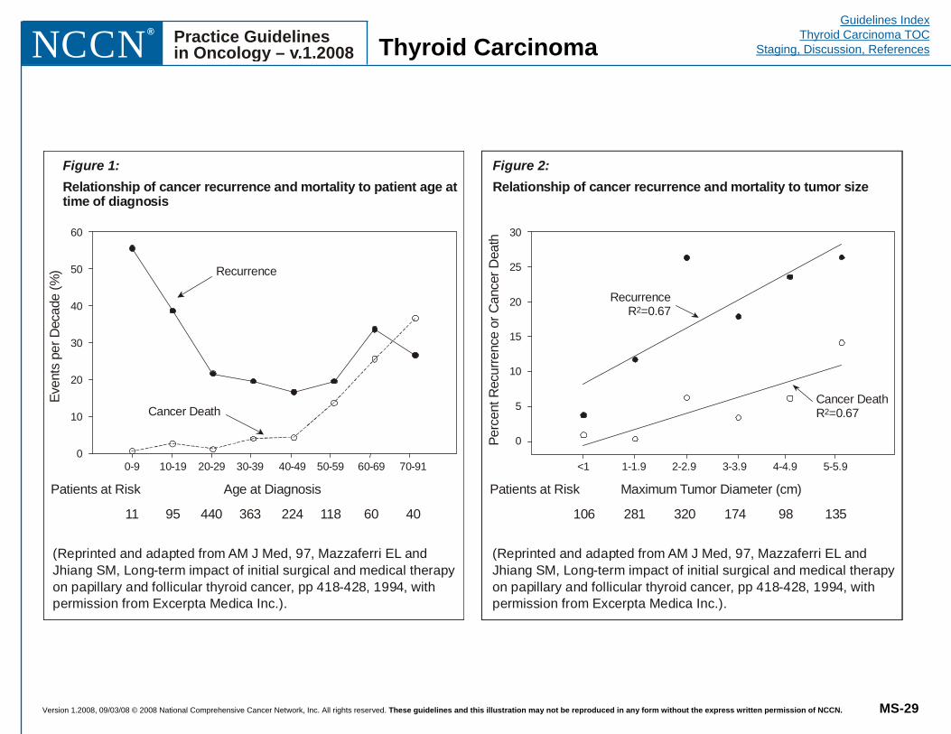
Version 1.2008, 09/03/08 © 2008 National Comprehensive Cancer Network, Inc. All rights reserved. These guidelines and this illustration may not be reproduced in any form without the express written permission of NCCN. MS-29
Guidelines IndexThyroid Carcinoma TOC
Staging, Discussion, ReferencesPractice Guidelinesin Oncology – v.1.2008 NCCN
®
Thyroid Carcinoma
Maximum Tumor Diameter (cm)Patients at RiskPe
rcen
tRec
urre
nce
orC
ance
rDea
th
<1
30
25
20
15
10
5
0
1-1.9 2-2.9 3-3.9 4-4.9 5-5.9
106 281 320 174 98 135
RecurrenceR =0.672
Cancer DeathR =0.672
Age at DiagnosisPatients at Risk
Even
tspe
rDec
ade
(%) Recurrence
Cancer Death
0-9
60
50
40
30
20
10
010-19 20-29 30-39 40-49 50-59 60-69 70-91
11 95 440 363 224 118 60 40
Figure 1:Relationship of cancer recurrence and mortality to patient age attime of diagnosis
(Reprinted and adapted from AM J Med, 97, Mazzaferri EL andJhiang SM, Long-term impact of initial surgical and medical therapyon papillary and follicular thyroid cancer, pp 418-428, 1994, withpermission from Excerpta Medica Inc.).
Figure 2:Relationship of cancer recurrence and mortality to tumor size
(Reprinted and adapted from AM J Med, 97, Mazzaferri EL andJhiang SM, Long-term impact of initial surgical and medical therapyon papillary and follicular thyroid cancer, pp 418-428, 1994, withpermission from Excerpta Medica Inc.).
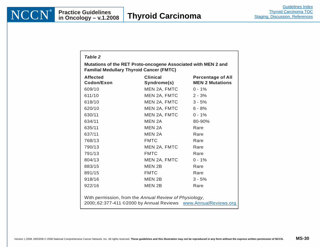
Version 1.2008, 09/03/08 © 2008 National Comprehensive Cancer Network, Inc. All rights reserved. These guidelines and this illustration may not be reproduced in any form without the express written permission of NCCN. MS-30
Guidelines IndexThyroid Carcinoma TOC
Staging, Discussion, ReferencesPractice Guidelinesin Oncology – v.1.2008 NCCN
®
Thyroid Carcinoma
Table 2Mutations of the RET Proto-oncogene Associated with MEN 2 andFamilial Medullary Thyroid Cancer (FMTC)Affected Clinical Percentage of AllCodon/Exon Syndrome(s) MEN 2 Mutations609/10 MEN 2A, FMTC 0 - 1%611/10 MEN 2A, FMTC 2 - 3%618/10 MEN 2A, FMTC 3 - 5%620/10 MEN 2A, FMTC 6 - 8%630/11 MEN 2A, FMTC 0 - 1%634/11 MEN 2A 80-90%635/11 MEN 2A Rare637/11 MEN 2A Rare768/13 FMTC Rare790/13 MEN 2A, FMTC Rare791/13 FMTC Rare804/13 MEN 2A, FMTC 0 - 1%883/15 MEN 2B Rare891/15 FMTC Rare918/16 MEN 2B 3 - 5%922/16 MEN 2B Rare
With permission, from the ,2000;.62:377-411 ©2000 by Annual Reviews
Annual Review of Physiologywww.AnnualReviews.org
Version 1.2008, 09/03/08 © 2008 National Comprehensive Cancer Network, Inc. All rights reserved. These guidelines and this illustration may not be reproduced in any form without the express written permission of NCCN. REF-1
Guidelines IndexThyroid Carcinoma TOC
Staging, Discussion, ReferencesPractice Guidelinesin Oncology – v.1.2008 NCCN
®
Thyroid Carcinoma
References 1. Mazzaferri EL. Thyroid carcinoma: Papillary and follicular. In: Mazzaferri EL, Samaan N, eds. Endocrine Tumors. Cambridge: Blackwell Scientific Publications 1993: 278-333.
2. Ezzat S, Sarti DA, Cain DR, et al. Thyroid incidentalomas: Prevalence by palpation and ultrasonography. Arch Intern Med 1994;154:1838-1840.
3. Ron E, Lubin JH, Shore RE, et al. Thyroid cancer after exposure to external radiation: A pooled analysis of seven studies. Radiat Res 1995;141:259-277.
4. Schneider AB, Bekerman C, Leland J, et al. Thyroid nodules in the follow-up of irradiated individuals: Comparison of thyroid ultrasound with scanning and palpation. J Clin Endocrinol Metab 1997;82:4020-4027.
5. Ries LAG, Melbert D, Krapcho M, et al (eds). SEER Cancer Statistics Review, 1975-2004, National Cancer Institute. Bethesda, MD, http://seer.cancer.gov/csr/1975_2004/ 2007. Accessed July 9, 2008.
6. Jemal A, Siegel R, Ward E, et al. Cancer Statistics, 2008. CA Cancer J Clin 2008;58:71-96.
7. Wu XC, Chen VW, Steele B, et al. Cancer incidence in adolescents and young adults in the United States, 1992-1997. J Adolesc Health 2003;32:405-415.
8. Hundahl SA, Fleming ID, Fremgen AM, et al. A National Cancer Data Base report on 53,856 cases of thyroid carcinoma treated in the United States, 1985-1995. Cancer 1998;83:2638-2648.
9. Mazzaferri EL, Jhiang SM. Long-term impact of initial surgical and medical therapy on papillary and follicular thyroid cancer. Am J Med 1994;97:418-428.
10. Davies L, Welch HG. Increasing incidence of thyroid cancer in the United States, 1973-2002. JAMA 2006;295:2164-2167.
11. Ito Y, Tomoda C, Uruno T, et al. Papillary microcarcinoma of the thyroid: how should it be treated? World J Surg 2004;28(11):1115-1121.
12. Sherman SI. Thyroid carcinoma. The Lancet 2003;361:501-511.
13. Wong FL, Ron E, Gierlowski T, et al. Benign thyroid tumors: General risk factors and their effects on radiation risk estimation. Am J Epidemiol 1996;144:728-733.
14. Hall P, Holm LE. Radiation-associated thyroid cancer—facts and fiction. Acta Oncol 1998;37:325-330.
15. Ron E, Doddy MM, Becker DV, et al. Cancer mortality following treatment for adult hyperthyroidism. JAMA 1998;280:347-355.
16. Jacob P, Goulko G, Heidenreich WF, et al. Thyroid cancer risk to children calculated. Nature 1998;392:31-32.
17. Schneider AB. Radiation-induced thyroid tumors. Endocrinol Metab Clin North Am 1990;19:495-508.
18. Nikiforov YE, Nikiforova M, Fagin JA. Radiation-induced post-Chernobyl pediatric thyroid carcinomas. Oncogene 1998;17:1983-1988.
19. Tan GH, Gharib H. Thyroid incidentalomas: Management approaches to nonpalpable nodules discovered incidentally on thyroid imaging. Ann Intern Med 1997;126:226-231.
20. Cooper DS, Doherty GM, Haugen BR, et al; The American Thyroid Association Guidelines Taskforce. Management guidelines for patients with thyroid nodules and differentiated thyroid cancer. Thyroid 2006;16:109-142.
21. Frates MC, Benson CB, Charboneau JW, et al. Management of thyroid nodules detected at US: Society of Radiologists in Ultrasound Consensus Conference Statement. Radiology 2005;237:794-800.
22. Mazzaferri EL. Thyroid cancer in thyroid nodules: Finding a needle in the haystack (editorial). Am J Med 1992;93:359-362.
Version 1.2008, 09/03/08 © 2008 National Comprehensive Cancer Network, Inc. All rights reserved. These guidelines and this illustration may not be reproduced in any form without the express written permission of NCCN. REF-2
Guidelines IndexThyroid Carcinoma TOC
Staging, Discussion, ReferencesPractice Guidelinesin Oncology – v.1.2008 NCCN
®
Thyroid Carcinoma
23. Hamming JF, Goslings BM, van Steenis GJ, et al. The value of fine-needle aspiration biopsy in patients with nodular thyroid disease divided into groups of suspicion of malignant neoplasms on clinical grounds. Arch Intern Med 1990;150:113-116.
24. Belfiore A, La Rosa GL, LaPorta GA, et al. Cancer risk in patients with cold thyroid nodules: Relevance of iodine intake, sex, age, and multinodularity. Am J Med 1992;93:363-369.
25. Chan BK, Desser TS, McDougall IR, et al. Common and uncommon sonographic features of papillary thyroid carcinoma. J Ultrasound Med 2003;22:1083-1090.
26. Henry JF, Denizot A, Puccini M, et al. Early diagnosis of sporadic medullary cancer of the thyroid: Contribution of routine calcitonin assay. Presse Med 1996;25:1583-1588.
27. Singer PA, Cooper DS, Daniels GH, et al. Treatment guidelines for patients with thyroid nodules and well-differentiated thyroid cancer. Arch Intern Med 1996;156:2165-2172.
28. Cheung K, Roman SA, Wang TS, et al. Calcitonin measurement in the evaluation of thyroid nodules in the United States: a cost-effectiveness and decision analysis. J Clin Endocrinol Metab 2008;93(6):2173-2180. Epub 2008 Mar 25.
29. Koike E, Noguchi S, Yamashita H, et al. Ultrasonographic characteristics of thyroid nodules: prediction of malignancy. Arch Surg 2001;136(3):334-337.
30. Yeh MW, Demircan O, Ituarte P, et al. False-negative fine-needle aspiration cytology results delay treatment and adversely affect outcome in patients with thyroid carcinoma. Thyroid 2004;14(3):207-215.
31. Cersosimo E, Gharib H, Suman VJ, et al. "Suspicious" thyroid cytologic findings: Outcome in patients without immediate surgical treatment. Mayo Clin Proc 1993;68:343-348.
32. Richter B, Neises G, Clar C. Pharmacotherapy for thyroid nodules. A systematic review and meta-analysis. Endocrinol Metab Clin North Am 2002;31:699-722.
33. Castro MR, Caraballo PJ, Morris JC. Effectiveness of thyroid hormone suppressive therapy in benign solitary thyroid nodules: a meta-analysis. J Clin Endocrinol Metab 2002;87:4154-4159.
34. McHenry CR, Walfish PG, Rosen IB. Non-diagnostic fine needle aspiration biopsy: a dilemma in management of nodular thyroid disease. Am Surg 1993;59:415-419.
35. Newman KD, Black T, Heller G, et al. Differentiated thyroid cancer: Determinants of disease progression in patients < 21 years of age at diagnosis–a report from the Surgical Discipline Committee of the Children's Cancer Group. Ann Surg 1998;227:533-541.
36. Robie DK, Dinauer CW, Tuttle RM, et al. The impact of initial surgical management on outcome in young patients with differentiated thyroid cancer. J Pediatr Surg 1998;33:1134-1138.
37. Gilliland FD, Hunt WC, Morris DM, et al. Prognostic factors for thyroid carcinoma: A population-based study of 15,698 cases from the surveillance, epidemiology, and end results (SEER) program, 1973-1991. Cancer 1997;79:564-573.
38. Sherman SI, Brierley JD, Sperling M, et al. Prospective multicenter study of treatment of thyroid carcinoma: Initial analysis of staging and outcome. Cancer 1998;83:1012-1021.
39. Tsang RW, Brierley JD, Simpson WJ, et al. The effects of surgery, radioiodine, and external radiation therapy on the clinical outcome of patients with differentiated thyroid carcinoma. Cancer 1998;82:375-388.
40. Mazzaferri EL. Management of a solitary thyroid nodule. N Engl J Med 1993;328:553-559.
41. Dottorini ME, Vignati A, Mazzucchelli L, et al. Differentiated thyroid carcinoma in children and adolescents: A 37-year experience in 85 patients. J Nucl Med 1997;38:669-675.
Version 1.2008, 09/03/08 © 2008 National Comprehensive Cancer Network, Inc. All rights reserved. These guidelines and this illustration may not be reproduced in any form without the express written permission of NCCN. REF-3
Guidelines IndexThyroid Carcinoma TOC
Staging, Discussion, ReferencesPractice Guidelinesin Oncology – v.1.2008 NCCN
®
Thyroid Carcinoma
42. Samuel AM, Rajashekharrao B, Shah DH. Pulmonary metastases in children and adolescents with well-differentiated thyroid cancer. J Nucl Med 1998;39:1531-1536.
43. Schlumberger M, De Vathaire F, Travagli JP, et al. Differentiated thyroid carcinoma in childhood: Long-term follow-up of 72 patients. J Clin Endocrinol Metab 1987;65:1088-1094.
44. Hay ID, Bergstralh EJ, Goellner JR, et al. Predicting outcome in papillary thyroid carcinoma: Development of a reliable prognostic scoring system in a cohort of 1779 patients surgically treated at one institution during 1940 through 1989. Surgery 1993;114:1050-1058.
45. Shaha AR, Loree TR, Shah JP. Prognostic factors and risk group analysis in follicular carcinoma of the thyroid. Surgery 1995;118:1131-1138.
46. Cady B. Staging in thyroid carcinoma. Cancer 1998;83:844-847.
47. DeGroot LJ, Kaplan EL, Straus FH, et al. Does the method of management of papillary thyroid carcinoma make a difference in outcome? World J Surg 1994;18:123-130.
48. Miccoli P, Antonelli A, Spinelli C, et al. Completion total thyroidectomy in children with thyroid cancer secondary to the Chernobyl accident. Arch Surg 1998;133:89-93.
49. Palme CE, Waseem Z, Raza SN, et al. Management and outcome of recurrent well-differentiated thyroid carcinoma. Arch Otolaryngol Head Neck Surg 2004;130(7):819-824.
50. Frankenthaler RA, Sellin RV, Cangir A, et al. Lymph node metastasis from papillary-follicular thyroid carcinoma in young patients. Am J Surg 1990;160:341-343.
51. Agostini L, Mazzi P, Cavaliere A. Multiple primary malignant tumours: Gemistocytic astrocytoma with leptomeningeal spreading and papillary thyroid carcinoma: A case report. Acta Neurol (Napoli) 1990;12:305-310.
52. Soravia C, Sugg SL, Berk T, et al. Familial adenomatous polyposis-associated thyroid cancer: A clinical, pathological, and molecular genetics study. Am J Pathol 1999;154:127-135.
53. Stratakis CA, Courcoutsakis NA, Abati A, et al. Thyroid gland abnormalities in patients with the syndrome of spotty skin pigmentation, myxomas, endocrine overactivity, and schwannomas (Carney complex). J Clin Endocrinol Metab 1997;82:2037-2043.
54. Marsh DJ, Dahia PLM, Caron S, et al. Germline PTEN mutations in Cowden syndrome-like families. J Med Genet 1998;35:881-885.
55. Mazzaferri EL. Papillary thyroid carcinoma: Factors influencing prognosis and current therapy. Semin Oncol 1987;14:315-332.
56. LiVolsi VA. Follicular lesions of the thyroid. In: LiVolsi VA, ed. Surgical Pathology of the Thyroid. Philadelphia: WB Saunders 1990:173-212.
57. LiVolsi VA. Papillary lesions of the thyroid. In LiVolsi VA, ed. Surgical Pathology of the Thyroid. Philadelphia: WB Saunders 1990:136-172.
58. Elisei R, Cosci B, Romei C, et al. Prognostic significance of somatic RET oncogene mutations in sporadic medullary thyroid cancer: a 10-year follow-up study. J Clin Endocrinol Metab 2008;93(3):682-687. Epub 2007 Dec 11.
59. LiVolsi VA. Unusual variants of papillary thyroid carcinoma. In: Mazzaferri EL, Kreisberg RA, Bar RS, eds. Advances in Endocrinology and Metabolism. St. Louis: Mosby-Year Book 1995:39-54.
60. Tielens ET, Sherman SI, Hruban RH, et al. Follicular variant of papillary thyroid carcinoma: A clinicopathologic study. Cancer 1994;73:424-431.
61. van Heerden JA, Hay ID, Goellner JR, et al. Follicular thyroid carcinoma with capsular invasion alone: A nonthreatening malignancy. Surgery 1992;112:1130-1138.
Version 1.2008, 09/03/08 © 2008 National Comprehensive Cancer Network, Inc. All rights reserved. These guidelines and this illustration may not be reproduced in any form without the express written permission of NCCN. REF-4
Guidelines IndexThyroid Carcinoma TOC
Staging, Discussion, ReferencesPractice Guidelinesin Oncology – v.1.2008 NCCN
®
Thyroid Carcinoma
62. LiVolsi V, Asa SL. The demise of follicular carcinoma of the thyroid gland. Thyroid 1994;4:233-236.
63. Brennan MD, Bergstralh EJ, van Heerden JA, et al. Follicular thyroid cancer treated at the Mayo Clinic, 1946 through 1970: Initial manifestations, pathologic findings, therapy, and outcome. Mayo Clin Proc 1991;66:11-22.
64. Greene FL, Page DL, Fleming ID, et al, eds. AJCC Cancer Staging Manual, 6th ed. New York: Springer-Verlag, 2002:77-87.
65. Fleming ID, Cooper JS, Henson DE, et al, eds. AJCC Cancer Staging Manual, 5th ed. Philadelphia: Lippincott Williams & Wilkins, 1997:59.
66. Hedinger CE. Problems of classification of thyroid tumors and their bearing on prognosis and therapy. Schweiz Med Wochenschr 1993;123:1673-1681.
67. Belchetz G, Cheung CC, Freeman J, et al. Hürthle cell tumors: using molecular techniques to define a novel classification system. Arch Otolaryngol Head Neck Surg 2002;128:237-240.
68. Chen H, Nicol TL, Zeiger MA, et al. Hürthle cell neoplasms of the thyroid: Are there factors predictive of malignancy? Ann Surg 1998;227:542-546.
69. Thompson NW, Dunn EL, Batsakis JG, et al. Hürthle cell lesions of the thyroid gland. Surg Gynecol Obstet 1973;139:555-560.
70. Lopez-Penabad L, Chiu AC, Hoff AO, et al. Prognostic factors in patients with Hürthle cell neoplasms of the thyroid. Cancer 2003;97:1186-1194.
71. Khafif A, Khafif RA, Attie JN. Hürthle cell carcinoma: A malignancy of low-grade potential. Head Neck 1999;21:506-511.
72. Ruegemer JJ, Hay ID, Bergstralh EJ, et al. Distant metastases in differentiated thyroid carcinoma: A multivariate analysis of prognostic variables. J Clin Endocrinol Metab 1988;67:501-558.
73. Samaan NA, Schultz PN, Hickey RC, et al. Well-differentiated thyroid carcinoma and the results of various modalities of treatment: A retrospective review of 1599 patients. J Clin Endocrinol Metab 1992;75:714-720.
74. Samaan NA, Schultz PN, Haynie TP, et al. Pulmonary metastasis of differentiated thyroid carcinoma: Treatment results in 101 patients. J Clin Endocrinol Metab 1985;60:376-380.
75. Moosa M, Mazzaferri EL. Occult thyroid carcinoma. Cancer J 1997;10:180-188.
76. Baudin E, Travagli JP, Ropers J, et al. Microcarcinoma of the thyroid gland: The Gustave-Roussy Institute experience. Cancer 1998;83:553-559.
77. Sugino K, Ito K Jr, Ozaki O, et al. Papillary microcarcinoma of the thyroid. J Endocrinol Invest 1998;21:445-448.
78. Hay ID. Papillary thyroid carcinoma. Endocrinol Metab Clin North Am 1990;19:545-576.
79. Emerick GT, Duh Q-Y, Siperstein AE, et al. Diagnosis, treatment, and outcome of follicular thyroid carcinoma. Cancer 1993;72:3287-3295.
80. Salvesen H, Njolstad PR, Akslen LA, et al. Papillary thyroid carcinoma: A multivariate analysis of prognostic factors including an evaluation of the p-TNM staging system. Eur J Surg 1992;158:583-589.
81. Pingpank JF Jr, Sasson AR, Hanlon AL, et al. Tumor above the spinal accessory nerve in papillary thyroid cancer that involves lateral neck nodes: a common occurrence. Arch Otolaryngol Head Neck Surg 2002;128:1275-1278.
82. Yamashita H, Noguchi S, Murakami N, et al. Extracapsular invasion of lymph node metastasis is an indicator of distant metastasis and poor prognosis in patients with thyroid papillary carcinoma. Cancer 1997;80:2268-2272.
Version 1.2008, 09/03/08 © 2008 National Comprehensive Cancer Network, Inc. All rights reserved. These guidelines and this illustration may not be reproduced in any form without the express written permission of NCCN. REF-5
Guidelines IndexThyroid Carcinoma TOC
Staging, Discussion, ReferencesPractice Guidelinesin Oncology – v.1.2008 NCCN
®
Thyroid Carcinoma
83. Sellers M, Beenken S, Blankenship A, et al. Prognostic significance of cervical lymph node metastases in differentiated thyroid cancer. Am J Surg 1992;164:578-581.
84. Lindegaard MW, Paus E, Hie J, et al. Thyroglobulin radioimmunoassay and 131 I scintigraphy in patients with differentiated thyroid carcinoma. Acta Chir Scand 1988;154:141-145.
85. Schlumberger M, Challeton C, De Vathaire F, et al. Treatment of distant metastases of differentiated thyroid carcinoma. J Endocrinol Invest 1995;18:170-172.
86. Sisson JC, Giordano TJ, Jamadar DA, et al. 131-I treatment of micronodular pulmonary metastases from papillary thyroid carcinoma. Cancer 1996;78:2184-2192.
87. Brown AP, Greening WP, McCready VR, et al. Radioiodine treatment of metastatic thyroid carcinoma: The Royal Marsden Hospital experience. Br J Radiol 1984;57:323-327.
88. Casara D, Rubello D, Saladini G, et al. Different features of pulmonary metastases in differentiated thyroid cancer: Natural history and multivariate statistical analysis of prognostic variables. J Nucl Med 1993;34:1626-1631.
89. Cady B. Our AMES is true: How an old concept still hits the mark, or risk group assignment points the arrow to rational therapy selection in differentiated thyroid cancer. Am J Surg 1997;174:462-468.
90. Cady B, Sedgwick CE, Meissner WA, et al. Risk factor analysis in differentiated thyroid cancer. Cancer 1979;43:810-820.
91. Loh KC, Greenspan FS, Gee L, et al. Pathological tumor-node-metastasis (pTNM) staging for papillary and follicular thyroid carcinomas: A retrospective analysis of 700 patients. J Clin Endocrinol Metab 1997;82:3553-3562.
92. Lin JD, Kao PF, Weng HF, et al. Relative value of thallium-201 and iodine-131 scans in the detection of recurrence or distant metastasis of well differentiated thyroid carcinoma. Eur J Nucl Med 1998;25:695-700.
93. Brierley JD, Panzarella T, Tsang RW, et al. A comparison of different staging systems predictability of patient outcome: Thyroid carcinoma as an example. Cancer 1997;79:2414-2423.
94. Sherman SI. Toward a standard clinicopathologic staging approach for differentiated thyroid carcinoma. Semin Surg Oncol 1999;16:12-15.
95. Van De Velde CJH, Hamming JF, Goslings BM, et al. Report of the consensus development conference on the management of differentiated thyroid cancer in the Netherlands. Eur J Cancer Clin Oncol 1988;24:287-292.
96. Solomon BL, Wartofsky L, Burman KD. Current trends in the management of well-differentiated papillary thyroid carcinoma. J Clin Endocrinol Metab 1996;81:333-339.
97. Hay ID, Grant CS, Taylor WF, et al. Ipsilateral lobectomy vs bilateral lobar resection in papillary thyroid carcinoma: A retrospective analysis of surgical outcome using a novel prognostic scoring system. Surgery 1987;102:1088-1095.
98. Hay ID, Grant CS, Bergstralh EJ, et al. Unilateral total lobectomy: Is it sufficient surgical treatment for patients with AMES low-risk papillary thyroid carcinoma? Surgery 1998;124:958-966.
99. Dackiw AP, Zeiger M. Extent of surgery for differentiated thyroid cancer. Surg Clin North Am 2004;84(3):817-832.
100. Mazzaferri EL. Treating differentiated thyroid carcinoma: Where do we draw the line? Mayo Clin Proc 1991;66:105-111.
101. Massin JP, Savoie JC, Garnier H, et al. Pulmonary metastases in differentiated thyroid carcinoma: Study of 58 cases with implications for the primary tumor treatment. Cancer 1984;53:982-992.
102. Shaha AR. Implications of prognostic factors and risk groups in the management of differentiated thyroid cancer. Laryngoscope 2004;114:393-402.
Version 1.2008, 09/03/08 © 2008 National Comprehensive Cancer Network, Inc. All rights reserved. These guidelines and this illustration may not be reproduced in any form without the express written permission of NCCN. REF-6
Guidelines IndexThyroid Carcinoma TOC
Staging, Discussion, ReferencesPractice Guidelinesin Oncology – v.1.2008 NCCN
®
Thyroid Carcinoma
103. DeGroot LJ, Kaplan EL. Second operations for "completion" of thyroidectomy in treatment of differentiated thyroid cancer. Surgery 1991;110:936-940.
104. Mettlin C, Lee F, Drago J, et al. The American Cancer Society National Prostate Cancer Detection Project. Cancer 1991;67:2949-2958.
105. Pasieka JL, Thompson NW, McLeod MK, et al. The incidence of bilateral well-differentiated thyroid cancer found at completion thyroidectomy. World J Surg 1992;16:711-716.
106. Scheumann GFW, Seeliger H, Musholt TJ, et al. Completion thyroidectomy in 131 patients with differentiated thyroid carcinoma. Eur J Surg 1996;162:677-684.
107. Chao TC, Jeng LB, Lin JD, et al. Completion thyroidectomy for differentiated thyroid carcinoma. Otolaryngol Head Neck Surg 1998;118:896-899.
108. Burge MR, Zeise TM, Johnsen MW, et al. Risks of complication following thyroidectomy. J Gen Intern Med 1998;13:24-31.
109. Dralle H, Gimm O, Simon D, et al. Prophylactic thyroidectomy in 75 children and adolescents with hereditary medullary thyroid carcinoma: German and Austrian experience. World J Surg 1998;22:744-751.
110. Udelsman R, Lakatos E, Ladenson P. Optimal surgery for papillary thyroid carcinoma. World J Surg 1996;20:88-93.
111. Pattou F, Combemale F, Fabre S, et al. Hypocalcemia following thyroid surgery: Incidence and prediction of outcome. World J Surg 1998;22:718-724.
112. Sosa JA, Bowman HM, Tielsch JM, et al. The importance of surgeon experience for clinical and economic outcomes from thyroidectomy. Ann Surg 1998;228:320-328.
113. Mazzaferri EL, Kloos RT. Current approaches to primary therapy for papillary and follicular thyroid cancer. J Clin Endocrinol Metab 2001;86(4):1447-1463.
114. Mazzaferri EL. Thyroid remnant 131-I ablation for papillary and follicular thyroid carcinoma. Thyroid 1997;7:265-271.
115. Taylor T, Specker B, Robbins J, et al. Outcome after treatment of high-risk papillary and non–Hürthle-cell follicular thyroid carcinoma. Ann Intern Med 1998;129:622-627.
116. Leger FA, Izembart M, Dagousset F, et al. Decreased uptake of therapeutic doses of iodine-131 after 185- MBq iodine-131 diagnostic imaging for thyroid remnants in differentiated thyroid carcinoma. Eur J Nucl Med 1998;25:242-246.
117. Muratet JP, Giraud P, Daver A, et al. Predicting the efficacy of first iodine-131 treatment in differentiated thyroid carcinoma. J Nucl Med 1997;38:1362-1368.
118. Mazzaferri EL. Carcinoma of follicular epithelium: Radioiodine and other treatment outcomes. In: Braverman LE, Utiger RD, eds. The Thyroid: A Fundamental and Clinical Text. Philadelphia: Lippincott-Raven, 1996:922-945.
119. Brierley J, Maxon HR. Radioiodine and external radiation therapy in the treatment of thyroid cancer. In: Fagin JA, ed. Thyroid Cancer. Boston/Dordrecht/London: Kluwer Academic 1998:285-317.
120. Tuttle RM, Leboeuf R, Robbins RJ, et al. Empiric radioactive iodine dosing regimens frequently exceed maximum tolerated activity levels in elderly patients with thyroid cancer. J Nucl Med 2006;47(10):1587-1591. Erratum in: J Nucl Med 2007;48(1):7.
121. Van Nostrand D, Wartofsky L. Radioiodine in the treatment of thyroid cancer. Endocrinol Metab Clin North Am 2007;36(3):807-822, vii-viii.
122. Maxon HR, Englaro EE, Thomas SR, et al. Radioiodine-131 therapy for well-differentiated thyroid cancer—a quantitative radiation
Version 1.2008, 09/03/08 © 2008 National Comprehensive Cancer Network, Inc. All rights reserved. These guidelines and this illustration may not be reproduced in any form without the express written permission of NCCN. REF-7
Guidelines IndexThyroid Carcinoma TOC
Staging, Discussion, ReferencesPractice Guidelinesin Oncology – v.1.2008 NCCN
®
Thyroid Carcinoma
dosimetric approach: Outcome and validation in 85 patients. J Nucl Med 1992;33:1132-1136.
123. Benua RS, Cicale NR, Sonenberg M, et al. The relation of radioiodine dosimetry to results and complications in the treatment of metastatic thyroid cancer. Am J Roentgenol 1962;87:171-178.
124. Sherman SI, Tielens ET, Sostre S, et al. Clinical utility of posttreatment radioiodine scans in the management of patients with thyroid carcinoma. J Clin Endocrinol Metab 1994;78:629-634.
125. Pacini F, Molinaro E, Castagna MG, et al. Recombinant human thyrotropin-stimulated serum thyroglobulin combined with neck ultrasonography has the highest sensitivity in monitoring differentiated thyroid carcinoma. J Clin Endocrinol Metab 2003;88(8):3668-3673.
126. Pacini F, Lari R, Mazzeo S, et al. Diagnostic value of a single serum thyroglobulin determination on and off thyroid suppressive therapy in the follow-up of patients with differentiated thyroid cancer. Clin Endocrinol 1985;23:405-411.
127. Haugen B, Pacini F, Reiners C, et al. A comparison of recombinant human thyrotropin and thyroid hormone withdrawal for the detection of thyroid remnant or cancer. J Clin Endocrinol Metab 1999;84:3877-3885.
128. Kloos RT, Mazzaferri EL. A single recombinant human thyrotropin-stimulated serum thyroglobulin measurement predicts differentiated thyroid carcinoma metastases three to five years later. J Clin Endocrinol Metab 2005;90(9):5047-5057. Epub 2005 Jun 21.
129. Ladenson PW, Braverman LE, Mazzaferri EL, et al. Comparison of administration of recombinant human thyrotropin with withdrawal of thyroid hormone for radioactive iodine scanning in patients with thyroid carcinoma. N Engl J Med 1997;337:888-896.
130. Mazzaferri EL, Kloos RT. Is diagnostic iodine-131 scanning with recombinant human TSH (rhTSH) useful in the follow-up of differentiated thyroid cancer after thyroid ablation? J Clin Endocrinol Metab 2002;87(4):1490-1498.
131. Kloos RT, Mazzaferri EL. A single recombinant human thyrotropin-stimulated serum thyroglobulin measurement predicts differentiated thyroid carcinoma metastases three to five years later. J Clin Endocrinol Metab 2005;90(9):5047-5057. Epub 2005 Jun 21.
132. Castagna MG, Brilli L, Pilli T, et al. Limited value of repeat recombinant human thyrotropin (rhTSH)-stimulated thyroglobulin testing in differentiated thyroid carcinoma patients with previous negative rhTSH-stimulated thyroglobulin and undetectable basal serum thyroglobulin levels. J Clin Endocrinol Metab 2008;93(1):76-81. Epub 2007 Oct 30.
133. Spencer CA, Takeuchi M, Kazarosyan M. Current status and performance goals for serum thyroglobulin assays. Clin Chem 1996;42:164-173.
134. Spencer CA, Lopresti JS. Measuring thyroglobulin and thyroglobulin autoantibody in patients with differentiated thyroid cancer. Nat Clin Pract Endocrinol Metab 2008;4(4):223-233. Epub 2008 Feb 12.
135. Spencer CA, Takeuchi M, Kazarosyan M, et al. Serum thyroglobulin autoantibodies: Prevalence, influence on serum thyroglobulin measurement, and prognostic significance in patients with differentiated thyroid carcinoma. J Clin Endocrinol Metab 1998;83:1121-1127.
136. Chung JK, Park YJ, Kim TY, et al. Clinical significance of elevated level of serum antithyroglobulin antibody in patients with differentiated thyroid cancer after thyroid ablation. Clin Endocrinol 2002;57:215-221.
137. Chiovato L, Latrofa F, Braverman LE, et al. Disappearance of humoral thyroid autoimmunity after complete removal of thyroid antigens. Ann Intern Med 2003;139:346-351.
138. Ringel M, Ladenson P, Levine MA. Molecular diagnosis of residual and recurrent thyroid cancer by amplification of thyroglobulin messenger ribonucleic acid in peripheral blood. J Clin Endocrinol Metab 1998;83:4435-4442.
Version 1.2008, 09/03/08 © 2008 National Comprehensive Cancer Network, Inc. All rights reserved. These guidelines and this illustration may not be reproduced in any form without the express written permission of NCCN. REF-8
Guidelines IndexThyroid Carcinoma TOC
Staging, Discussion, ReferencesPractice Guidelinesin Oncology – v.1.2008 NCCN
®
Thyroid Carcinoma
139. Ringel MD. Molecular detection of thyroid cancer: differentiating "signal" and "noise" in clinical assays. J Clin Endocrinol Metab 2004;89:29-32.
140. Schlumberger M, Mancusi F, Baudin E, et al. 131-I therapy for elevated thyroglobulin levels. Thyroid 1997;7:273-276.
141. Schlumberger M, Tubiana M, De Vathaire F, et al. Long-term results of treatment of 283 patients with lung and bone metastases from differentiated thyroid carcinoma. J Clin Endocrinol Metab 1986;63:960-967.
142. Pacini F, Lippi F, Formica N, et al. Therapeutic doses of iodine-131 reveal undiagnosed metastases in thyroid cancer patients with detectable serum thyroglobulin levels. J Nucl Med 1987;28:1888-1891.
143. Pineda JD, Lee T, Ain K, et al. Iodine-131 therapy for thyroid cancer patients with elevated thyroglobulin and negative diagnostic scan. J Clin Endocrinol Metab 1995;80:1488-1492.
144. Mazzaferri EL. Treating high thyroglobulins with radioiodine: A magic bullet or a shot in the dark? J Clin Endocrinol Metab 1995;80:1485-1487.
145. Pacini F, Agate L, Elisei R, et al. Outcome of differentiated thyroid cancer with detectable serum Tg and negative diagnostic 131-I whole body scan: comparison of patients treated with high 131-I activities versus untreated patients. J Clin Endocrinol Metab 2001;86:4092-4097.
146. Burns JA, Morgenstern KE, Cahill KV, et al. Nasolacrimal obstruction secondary to I(131) therapy. Ophthal Plast Reconstr Surg 2004;20(2);126-129.
147. McGriff NJ, Csako G, Gourgiotis L, et al. Effects of thyroid hormone suppression therapy on adverse clinical outcomes in thyroid cancer. Ann Med 2002;34(7-8):554-564.
148. Pujol P, Daures JP, Nsakala N, et al. Degree of thyrotropin suppression as a prognostic determinant in differentiated thyroid cancer. J Clin Endocrinol Metab 1996;81:4318-4323.
149. Cooper DS, Specker B, Ho M, et al. Thyrotropin suppression and disease progression in patients with differentiated thyroid cancer: Results from the National Thyroid Cancer Treatment Cooperative Registry. Thyroid 1998;8:737-744.
150. Burmeister LA, Goumaz MO, Mariash CN, et al. Levothyroxine dose requirements for thyrotropin suppression in the treatment of differentiated thyroid cancer. J Clin Endocrinol Metab 1992;75:344-350.
151. Jonklaas J, Sarlis NJ, Litofsky D, et al. Outcomes of patients with differentiated thyroid carcinoma following initial therapy. Thyroid 2006;16(12):1229-1242.
152. Biermann M, Pixberg MK, Schuck A, et al. Multicenter study differentiated thyroid carcinoma (MSDS). Diminished acceptance of adjuvant external beam radiotherapy. Nuklearmedizin 2003;42:244-250.
153. Farahati J, Reiners C, Stuschke M, et al. Differentiated thyroid cancer: Impact of adjuvant external radiotherapy in patients with perithyroidal tumor infiltration (stage pT4). Cancer 1996;77:172-180.
154. Simpson WJ, Panzarella T, Carruthers JS, et al. Papillary and follicular thyroid cancer: Impact of treatment in 1578 patients. Int J Radiat Oncol Biol Phys 1988;14:1063-1075.
155. Chiu AC, Delpassand ES, Sherman SI. Prognosis and treatment of brain metastases in thyroid carcinoma. J Clin Endocrinol Metab 1997;82:3637-3642.
156. Droz JP, Schlumberger M, Rougier P, et al. Chemotherapy in metastatic nonanaplastic thyroid cancer: Experience at the Institut Gustave-Roussy. Tumori 1990;76:480-483.
157. Ahuja S, Ernst H. Chemotherapy of thyroid carcinoma. J Endocrinol Invest 1987;10:303-310.
158. Mrozek E, Kloos RT, Ringel MD, et al. Phase II study of celecoxib in metastatic differentiated thyroid carcinoma. J Clin Endocrinol Metab 2006;91(6):2201-2204. Epub 2006 Mar 7.
Version 1.2008, 09/03/08 © 2008 National Comprehensive Cancer Network, Inc. All rights reserved. These guidelines and this illustration may not be reproduced in any form without the express written permission of NCCN. REF-9
Guidelines IndexThyroid Carcinoma TOC
Staging, Discussion, ReferencesPractice Guidelinesin Oncology – v.1.2008 NCCN
®
Thyroid Carcinoma
159. Rosen LS, Kurzrock R, Mulay M, et al. Safety, pharmacokinetics, and efficacy of AMG 706, an oral multikinase inhibitor, in patients with advanced solid tumors. J Clin Oncol 2007;25:2369-2376.
160. Sherman SI, Wirth LJ, Droz JP, et al; Motesanib Thyroid Cancer Study Group. Motesanib diphosphate in progressive differentiated thyroid cancer. N Engl J Med 2008;359(1):31-42.
161. Kloos R, Ringel M, Knopp M, et al. Significant clinical and biologic activity of RAF/VEGF-R kinase inhibitor BAY 43-9006 in patients with metastatic papillary thyroid carcinoma (PTC): Updated results of a phase II study. J Clin Oncol (ASCO Annual Meeting Proceedings) 2006:5534.
162. Gupta-Abramson V, Troxel AB, Nellore A, et al. Phase II trial of sorafenib in advanced thyroid cancer. J Clin Oncol 2008 Jun 9. [Epub ahead of print]
163. Cohen EE, Leedles BM, Cullen KJ, et al. Phase 2 study of sunitinib in refractory thyroid cancer [abstract]. J Clin Oncol 2008;26:6025.
164. Cohen EE, Rosen LS, Vokes EE, et al. Axitinib is an active treatment for all histologic subtypes of advanced thyroid cancer: results from a phase II study. J Clin Oncol 2008 Jun 9. [Epub ahead of print]
165. Ain KB, Lee C, Holbrook KM, et al. Phase II study of lenalidomide in distantly metastatic, rapidly progressive, and radioiodine-unresponsive thyroid carcinomas: preliminary results [abstract]. J Clin Oncol 2008;26:6027.
166. Deshpande HA, Gettinger SN, Sosa JA. Novel chemotherapy options for advanced thyroid tumors: small molecules offer great hope. Curr Opin Oncol 2008;20(1):19-24.
167. Kouvaraki MA, Shapiro SE, Fornage BD, et al. Role of preoperative ultrasonography in the surgical management of patients with thyroid cancer. Surgery 2003;134:946-954; discussion 954-955.
168. Haigh PI, Urbach DR, Rotstein LE. Extent of thyroidectomy is not a major determinant of survival in low- or high-risk papillary thyroid cancer. Ann Surg Oncol 2005;12:81-89. Epub 2004 Dec 27.
152. Morris LF, Waxman AD, Braunstein GD. Thyroid stunning. Thyroid 2003;13:333-340.
169. Morris LF, Waxman AD, Braunstein GD. Thyroid stunning. Thyroid 2003;13:333-340.
170. Eustatia-Rutten CF, Romijn JA, Guijt MJ, et al. Outcome of palliative embolization of bone metastases in differentiated thyroid carcinoma. J Clin Endocrinol Metab 2003;88:3184-3189.
171. Van Nostrand D, Atkins F, Yeganeh F, et al. Dosimetrically determined doses of radioiodine for the treatment of metastatic thyroid carcinoma. Thyroid 2002;12:121-134.
172. Thompson LD, Wieneke JA, Paal E, et al. A clinicopathologic study of minimal invasive follicular carcinoma of the thyroid gland with a review of the English literature. Cancer 2001;91:505-524.
173. Sugino K, Ito K, Mimura T, et al. Hürthle cell tumor of the thyroid: analysis of 188 cases. World J Surg 2001;25(9):1160-1163.
174. Lopez-Penabad L, Chiu AC, Hoff AO, et al. Prognostic factors in patients with Hürthle cell neoplasms of the thyroid. Cancer 2003;97(5):1186-1194.
175. Herrera MF, Hay ID, Wu PS, et al. Hurthle cell (oxyphilic) papillary thyroid carcinoma: a variant with more aggressive biologic behavior. World J Surg 1992;16:669-674.
176. Ball DW, Baylin SB, de Bustros AC. Medullary thyroid carcinoma. In: Braverman LE, Utiger RD, eds. Werner and Ingbar's The Thyroid, 7th ed. Philadelphia: Lippincott-Raven 1996:946-960.
177. Gertner ME, Kebebew E. Multiple endocrine neoplasia type 2. Curr Treat Options Oncol 2004;5(4):315-325.
Version 1.2008, 09/03/08 © 2008 National Comprehensive Cancer Network, Inc. All rights reserved. These guidelines and this illustration may not be reproduced in any form without the express written permission of NCCN. REF-10
Guidelines IndexThyroid Carcinoma TOC
Staging, Discussion, ReferencesPractice Guidelinesin Oncology – v.1.2008 NCCN
®
Thyroid Carcinoma
178. Raue F, Frank-Raue K. Multiple endocrine neoplasia type 2: 2007 update. Horm Res 2007;68 Suppl 5:101-104. Epub 2007 Dec 10.
179. Saad MF, Ordonez NG, Rashid RK, et al. Medullary carcinoma of the thyroid. Medicine 1984;63:319-342.
180. Pacini F, Fontanelli M, Fugazzola L, et al. Routine measurement of serum calcitonin in nodular thyroid diseases allows the preoperative diagnosis of unsuspected sporadic medullary thyroid carcinoma. J Clin Endocrinol Metab 1994;78:826-829.
181. Niccoli P, Wion-Barbot N, Caron P, et al. Interest of routine measurement of serum calcitonin: Study in a large series of thyroidectomized patients. J Clin Endocrinol Metab 1997;82:338-341.
182. Ozgen AG, Hamulu F, Bayraktar F, et al. Evaluation of routine basal serum calcitonin measurement for early diagnosis of medullary thyroid carcinoma in seven hundred seventy-three patients with nodular goiter. Thyroid 1999;9:579-582.
183. Horvit PK, Gagel RF. The goitrous patient with an elevated serum calcitonin: What to do? J Clin Endocrinol Metab 1997;82:335-337.
184. Hodak SP, Burman KD. The calcitonin conundrum—is it time for routine measurement of serum calcitonin in patients with thyroid nodules [Editorial]? J Clin Endocrinol Metab 2004;89:511-514.
185. Gagel RF, Cote GJ. Pathogenesis of medullary thyroid carcinoma. In: Fagin JA, ed. Thyroid Cancer Boston/Dordrecht/London: Kluwer Academic 1998:85-103.
186. Papi G, Corsello SM, Cioni K, et al. Value of routine measurement of serum calcitonin concentrations in patients with nodular thyroid disease: A multicenter study. J Endocrinol Invest 2006;29(5):427-437.
187. Kouvaraki MA, Shapiro SE, Perrier ND, et al. RET proto-oncogene: a review and update of genotype-phenotype correlations in hereditary medullary thyroid cancer and associated endocrine tumors. Thyroid 2005;15(6):531-544.
188. Wohllk N, Cote GJ, Bugalho MMJ, et al. Relevance of RET proto-oncogene mutations in sporadic medullary thyroid carcinoma. J Clin Endocrinol Metab 1996;81:3740-3745.
189. Dottorini ME, Assi A, Sironi M, et al. Multivariate analysis of patients with medullary thyroid carcinoma: Prognostic significance and impact on treatment of clinical and pathologic variables. Cancer 1996;77:1556-1565.
190. DeGroot LJ. Thyroid carcinoma. Med Clin North Am 1975;59:1233-1246.
191. Samaan NA, Schultz PN, Hickey RC. Medullary thyroid carcinoma: Prognosis of familial versus sporadic disease and the role of radiotherapy. J Clin Endocrinol Metab 1988;67:801-805.
192. O'Riordain DS, O'Brien T, Weaver AL, et al. Medullary thyroid carcinoma in multiple endocrine neoplasia types 2A and 2B. Surgery 1994;116:1017-1023.
193. Lippman SM, Mendelsohn G, Trump DL, et al. The prognostic and biological significance of cellular heterogeneity in medullary thyroid carcinoma: a study of calcitonin, L-dopa decarboxylase, and histaminase. J Clin Endocrinol Metab 1982;54:233-240.
194. Mendelsohn G, Wells SA, Baylin SB. Relationship of tissue carcinoembryonic antigen and calcitonin to tumor virulence in medullary thyroid carcinoma. Cancer 1984;54:657-662.
195. Maldonado MR, Sherman SI. Predictive value of clinicopathologic staging for medullary thyroid carcinoma. 72nd Annual Meeting of the American Thyroid Association. 1999; Palm Beach, FL.
196. Romei C, Elisei R, Pinchera A, et al. Somatic mutations of the ret protooncogene in sporadic medullary thyroid carcinoma are not restricted to exon 16 and are associated with tumor recurrence. J Clin Endocrinol Metab 1996;81:1619-1622.
197. Ogilvie JB, Kebebew E. Indication and timing of thyroid surgery for patients with hereditary medullary thyroid cancer syndromes. J Natl Compr Canc Netw 2006;4:139-147.
Version 1.2008, 09/03/08 © 2008 National Comprehensive Cancer Network, Inc. All rights reserved. These guidelines and this illustration may not be reproduced in any form without the express written permission of NCCN. REF-11
Guidelines IndexThyroid Carcinoma TOC
Staging, Discussion, ReferencesPractice Guidelinesin Oncology – v.1.2008 NCCN
®
Thyroid Carcinoma
198. Learoyd DL, Gosnell J, Elston MS, et al. Experience of prophylactic thyroidectomy in multiple endocrine neoplasia type 2A kindreds with RET codon 804 mutations. Clin Endocrinol (Oxf). 2005 Dec;63(6):636-41.
199. Brandi ML, Gagel RF, Angeli A, et al. Consensus: Guidelines for diagnosis and therapy of MEN type 1 and type 2. J Clin Endocrinol Metab 2001;86:5658-5671.
200. Machens A, Niccoli-Sire P, Hoegel J, et al. Early malignant progression of hereditary medullary thyroid cancer. N Engl J Med 2003;349(16):1517-1525.
201. Skinner MA, Moley JA, Dilley WG, et al. Prophylactic thyroidectomy in multiple endocrine neoplasia type 2A. N Engl J Med 2005;353:1105-1113.
202. van Heerden JA, Grant CS, Gharib H, et al. Long-term course of patients with persistent hypercalcitoninemia after apparent curative primary surgery for medullary thyroid carcinoma. Ann Surg 1990;212:395-401.
203. Scopsi L, Sampietro G, Boracchi P, et al. Multivariate analysis of prognostic factors in sporadic medullary carcinoma of the thyroid. Cancer 1996;78:2173-2183.
204. Tisell LE, Hansson G, Jansson S, et al. Reoperation in the treatment of asymptomatic metastasizing medullary thyroid carcinoma. Surgery 1986;99:60-66.
205. Moley JF, Debenedetti MK, Dilley WG, et al. Surgical management of patients with persistent or recurrent medullary thyroid cancer. J Intern Med 1998;243:521-526.
206. Fleming JB, Lee JE, Bouvet M, et al. Surgical strategy for the treatment of medullary thyroid carcinoma. Ann Surg 1999;230:697-707.
207. Kober F, Hermann M, Handler A, et al. Effect of sorafenib in symptomatic metastatic medullary thyroid cancer [abstract]. J Clin Oncol 2007;25:14065.
208. Nocera M, Baudin E, Pellegriti G, et al. Treatment of advanced medullary thyroid cancer with an alternating combination of doxorubicin-streptozocin and 5 FU dacarbazine. Groupe d'Etude des Tumeurs a Calcitonine (GETC). Br J Cancer 2000;83:715-718.
209. Haddad RI, Krebs AD, Vasselli J, et al. A phase II open-label study of vandetanib in patients with locally advanced or metastatic hereditary medullary thyroid cancer [abstract]. J Clin Oncol 2008;26:6024.
210. Goulart B, Carr L, Martins RG, et al. Phase II study of sunitinib in iodine refractory, well-differentiated thyroid cancer (WDTC) and metastatic medullary thyroid carcinoma (MTC) [abstract]. J Clin Oncol 2008;26:6062.
211. Hong D, Ye L, Gagel R, et al. Medullary thyroid cancer: targeting the RET kinase pathway with sorafenib/tipifarnib. Mol Cancer Ther 2008 Apr 29 [Epub ahead of print]
212. Salgia R, Sherman S, Hong DS, et al. A phase I study of XL184, a RET, VEGFR2, and MET kinase inhibitor, in patients (pts) with advanced malignancies, including pts with medullary thyroid cancer (MTC) [abstract]. J Clin Oncol 26: 2008;26:3522.
213. Wells SYY, Lakhani V, Hou J. A phase II trial of ZD6474 in patients with hereditary metastatic medullary thyroid cancer [abstract]. J Clin Oncol 2006;24:5553.
214. Wells SA, Gosnell JE, Gagel RF, et al. Vandetanib in metastatic hereditary medullary thyroid cancer: Follow-up results of an open-label phase II trial [abstract]. J Clin Oncol 2007;25(18S):6018.
215. Chatal JF, Campion L, Kraeber-Bodere F, et al. Survival improvement in patients with medullary thyroid carcinoma who undergo pretargeted anti-carcinoembryonic-antigen radioimmunotherapy: a collaborative study with the French Endocrine Tumor Group. J Clin Oncol 2006;24:1705-1711. Epub 2006 Mar 20.
216. Moretti F, Farsetti A, Soddu S, et al. p53 re-expression inhibits proliferation and restores differentiation of human thyroid anaplastic carcinoma cells. Oncogene 1997;14:729-740.
Version 1.2008, 09/03/08 © 2008 National Comprehensive Cancer Network, Inc. All rights reserved. These guidelines and this illustration may not be reproduced in any form without the express written permission of NCCN. REF-12
Guidelines IndexThyroid Carcinoma TOC
Staging, Discussion, ReferencesPractice Guidelinesin Oncology – v.1.2008 NCCN
®
Thyroid Carcinoma
217. Sherman SI. Anaplastic carcinoma: Clinical aspects. In: Wartofsky L, ed. Thyroid Cancer: A Comprehensive Guide to Clinical Management. Totowa, NJ: Humana Press 1999:319-325.
218. Asa SL, Bedard YC. Fine-needle aspiration cytology and histopathology. In: Clark OH, Noguchi S, eds. Thyroid Cancer: Diagnosis and Treatment. St Louis. Quality Medical Publishing, 2000:105-126.
219. Takashima S, Morimoto S, Ikezoe J, et al. CT evaluation of anaplastic thyroid carcinoma. Am J Roentgenol 1990;154:1079-1085.
220. Venkatesh YSS, Ordonez NG, Schultz PN, et al. Anaplastic carcinoma of the thyroid: a clinicopathologic study of 121 cases. Cancer 1990;66:321-330.
221. Junor EJ, Paul J, Reed NS. Anaplastic thyroid carcinoma: 91 patients treated by surgery and radiotherapy. Eur J Surg Oncol 1992;18:83-88.
222. Kim JH, Leeper RD. Treatment of locally advanced thyroid carcinoma with combination doxorubicin and radiation therapy. Cancer 1987;60:2372-2375.
223. Wallin G, Lundell G, Tennvall J. Anaplastic giant cell thyroid carcinoma. Scand J Surg 2004;93(4):272-277.
224. Ain KB. Anaplastic thyroid carcinoma. Behavior, biology, and therapeutic approaches. Thyroid 1998;8:715-726.
225. Thorpe PE. Vascular targeting agents as cancer therapeutics. Clin Cancer Res 2004;10(2):415-427.
226. Dowlati A, Robertson K, Cooney M, et al. A phase I pharmacokinetic and translational study of the novel vascular targeting agent combretastatin a-4 phosphate on a single-dose intravenous schedule in patients with advanced cancer. Cancer Res 2002;62:3408-3416.
227. Cooney MM, Savvides P, Agarwala SS, et al. Phase II study of combretastatin A4 phosphate (CA4P) in patients with advanced
anaplastic thyroid carcinoma [abstract]. J Clin Oncol 2006;24(18S):5580.


![Papillary thyroid carcinoma coexists with undifferentiated ... · Papillary thyroid carcinoma (PTC) is the commonest thyroid carcinoma worldwide [1], while undifferentiated thyroid](https://img.pdfslide.us/doc/110x75/605714f9a806da25134f71a8/papillary-thyroid-carcinoma-coexists-with-undifferentiated-papillary-thyroid.jpg)


![Thyroid carcinoma final [part 2]](https://img.pdfslide.us/doc/110x75/58f9ac081a28ab0f5b8b4659/thyroid-carcinoma-final-part-2.jpg)