Embed Size (px)
Citation preview


Practical Veterinary Urinalysis


Practical Veterinary Urinalysis
Carolyn A. Sink, MS, MT (ASCP)Nicole M. Weinstein, DVM, DACVPIllustrations by Ashley Marlowe
A John Wiley & Sons, Inc., Publication

This edition first published 2012 © 2012 by John Wiley & Sons, Inc.
Wiley-Blackwell is an imprint of John Wiley & Sons, formed by the merger of Wiley’s global Scientific, Technical and Medical business with Blackwell Publishing.
Registered office: John Wiley & Sons Ltd, The Atrium, Southern Gate, Chichester, West Sussex, PO19 8SQ, UK
Editorial offices: 2121 State Avenue, Ames, Iowa 50014-8300, USAThe Atrium, Southern Gate, Chichester, West Sussex, PO19 8SQ, UK9600 Garsington Road, Oxford, OX4 2DQ, UK
For details of our global editorial offices, for customer services and for information about how to apply for permission to reuse the copyright material in this book please see our website at www.wiley.com/wiley-blackwell.
Authorization to photocopy items for internal or personal use, or the internal or personal use of specific clients, is granted by Blackwell Publishing, provided that the base fee is paid directly to the Copyright Clearance Center, 222 Rosewood Drive, Danvers, MA 01923. For those organizations that have been granted a photocopy license by CCC, a separate system of payments has been arranged. The fee codes for users of the Transactional Reporting Service are ISBN-13: 978-0-4709-5824-7/2012.
Designations used by companies to distinguish their products are often claimed as trademarks. All brand names and product names used in this book are trade names, service marks, trademarks or registered trademarks of their respective owners. The publisher is not associated with any product or vendor mentioned in this book. This publication is designed to provide accurate and authoritative information in regard to the subject matter covered. It is sold on the understanding that the publisher is not engaged in rendering professional services. If professional advice or other expert assistance is required, the services of a competent professional should be sought.
Library of Congress Cataloging-in-Publication Data
Sink, Carolyn A. Practical veterinary urinalysis / Carolyn Sink and Nicole Weinstein ; illustrations by Ashley Marlowe. p. ; cm. Includes bibliographical references and index. ISBN 978-0-470-95824-7 (pbk. : alk. paper) I. Weinstein, Nicole. II. Title. [DNLM: 1. Urinalysis–veterinary–Laboratory Manuals. SF 773] LC classification not assigned 636.089'66–dc23 2011035231
A catalogue record for this book is available from the British Library.
Wiley also publishes its books in a variety of electronic formats. Some content that appears in print may not be available in electronic books.
Set in 9 on 12.5 pt Interstate Light by Toppan Best-set Premedia Limited
Disclaimer
The publisher and the author make no representations or warranties with respect to the accuracy or completeness of the contents of this work and specifically disclaim all warranties, including without limitation warranties of fitness for a particular purpose. No warranty may be created or extended by sales or promotional materials. The advice and strategies contained herein may not be suitable for every situation. This work is sold with the understanding that the publisher is not engaged in rendering legal, accounting, or other professional services. If professional assistance is required, the services of a competent professional person should be sought. Neither the publisher nor the author shall be liable for damages arising herefrom. The fact that an organization or Website is referred to in this work as a citation and/or a potential source of further information does not mean that the author or the publisher endorses the information the organization or Website may provide or recommendations it may make. Further, readers should be aware that Internet Websites listed in this work may have changed or disappeared between when this work was written and when it is read.
1 2012

This book is dedicated to veterinary students, clinical laboratory profession-
als, clinical veterinarians, and all those who continue to develop and improve
the veterinary clinical laboratory.


vii
Contents
Preface and Acknowledgments ix
Chapter 1 Functional Renal Physiology and Urine Production 1
Glomerular filtration 1
Tubular reabsorption and secretion 3
Collecting tubules 5
Renal function and measures of renal function 5
Laboratory assessment of renal function 6
Chapter 2 Specimen Procurement 9
Laboratory definitions for collection methods 9
Urine specimen containers 12
Specimen handling and preservation 15
Types of urine specimens 16
Chapter 3 Routine Urinalysis: Physical Properties 19
Solute concentration 19
Urine color 25
Chapter 4 Routine Urinalysis: Chemical Analysis 29
pH 29
Protein 32
Glucose 38
Ketone 40
Blood 42
Bilirubin 45
Urobilinogen 48
Nitrite 49
Leukocyte esterase 50
Specific gravity 51
Chapter 5 Routine Urinalysis: Microscopic Elements 55
Urine sediment preparation 55
Examination of urine sediment 57
Microscopic elements of urine sediment 59

viii Contents
Chapter 6 Proteinuria 113
Protein handling by the kidney 113
Significance of proteinuria 116
Laboratory diagnosis of proteinuria 117
Recommendations regarding diagnosis of proteinuria 126
Additional considerations for proteinuria 126
Chapter 7 Advanced Diagnostics 133
Detection of bacteriuria versus diagnosis of urinary tract
infection 133
Urinary tract cytology 136
Fractional excretion 147
Urinary biomarkers 149
Chapter 8 Laboratory Quality Assurance 155
Physical requirements of the laboratory 155
Laboratory equipment 157
Reagents and supplies for the urinalysis laboratory 160
Laboratory waste 160
Quality control in the urinalysis laboratory 161
Procedure manuals 163
Index 165

ix
Preface
Laboratory evaluation of urine provides a significant amount of information
to the veterinarian, as a variety of disease states may produce abnormal
findings. Routine laboratory tests used to assess urine are quick and inex-
pensive, as well as reliable, when performed by a well-trained laboratory
professional. Practical Veterinary Urinalysis is intended to provide laboratory
diagnosticians—veterinarians, medical technologists, veterinary technicians,
and veterinary students—with a comprehensive study of all aspects of routine
urinalysis, including chemical analysis and microscopy. Those tasked with
performing routine urinalysis will find both consolidated and comprehensive
information needed to produce timely and accurate results.
Practical Veterinary Urinalysis is designed to provide the reader with a
concise, systematic overview of renal physiology and urine production. Both
functional and physiological aspects of the urinary system are reviewed with
disease processes highlighted. When appropriate, discussions are reinforced
by images, tables, and drawings. Specimen collection and handling are
reviewed with emphasis on differences in laboratory findings between three
frequently used methods.
An in-depth discussion of urine physical properties, chemical analysis, and
sediment analysis is covered in separate chapters. Summarized charts are
provided as testing guides when appropriate. Within the microscopic analysis
section, Practical Veterinary Urinalysis contains images of the wet prep along
with cytology (Wright’s stain), and Gram stain if indicated.
Abnormal or suspicious results obtained by routine urinalysis may mandate
additional or confirmatory tests. A chapter dedicated to advanced urine
diagnostics presents newer test methodologies, and associated laboratory
procedures are discussed.
Practical Veterinary Urinalysis also includes sections dedicated to labora-
tory setup, including physical and functional requirements along with
instrument, reagent, and supply preferences. Quality assurance measures
and quality goals are defined, assuring laboratorians that the results they
produce are reliable.
x Preface
Acknowledgments
We are grateful for the support and guidance of Drs. Rachel Cianciolo and
Reema Patel, who provided invaluable insight and editorial commentary. We
would also like to acknowledge veterinary students Rachel Baum and Alicyn
Cross for their laboratory prowess in identifying interesting urine samples
used in this book.
Carolyn Sink and Nicole Weinstein

Ch
apte
r 1
1
Chapter 1
Functional Renal Physiology and Urine Production
Urinalysis can provide insight into hydration status, renal function or dysfunc-
tion, systemic disease, and toxic insults. Accurate interpretation of urinalysis
results requires knowledge of renal physiology and urine formation.
Given the complexity of both, a brief overview is presented below; more
detailed explanations can be found in a variety of references on which this
chapter is based (Gregory 2003; Kaneko 2008; Schrier 2007; Stockham and
Scott 2008; Watson 1998).
Descriptions of renal function and schematics of the kidney typically
portray a single nephron, which is the functional unit of the kidney; each
kidney contains hundreds of thousands of nephrons working in unison (Reece
1993). Each nephron requires:
(1) A blood supply
(2) A functional glomerulus, which filters a portion of the renal blood flow,
to form an ultrafiltrate
(3) Renal tubules that function to reabsorb water, electrolytes, and other
substances from the ultrafiltrate
(4) Collecting tubules and ducts, which further reabsorb or excrete water
and solutes and thus determine final urine concentration.
Urine formation starts as an ultrafiltrate formed by glomerular filtration. The
ultrafiltrate is then further altered by tubular reabsorption and secretion.
Glomerular filtration
The glomerulus is a collection of twisted capillaries that receive blood from the
afferent arteriole of the renal blood supply and exit the kidney via the efferent
arteriole (Figure 1.1). The high pressure within this system results in passage of
fluids and small substances out of the capillaries into a space around the glom-
erulus, known as Bowman’s capsule, which together form a renal corpuscle.
Practical Veterinary Urinalysis, First Edition. Carolyn Sink, Nicole Weinstein.© 2012 John Wiley & Sons, Inc. Published 2012 by John Wiley & Sons, Inc.

2 Practical Veterinary Urinalysis
Ch
apter 1
Glomerular filtration is driven by both blood volume and pressure and is con-
sidered a primarily passive process. Ultrafiltrate is the product of glomerular
filtration of blood following its passage through a glomerular filtration barrier,
the glomerular capillary wall (GCW). The GCW prevents entry of red and white
blood cells, platelets, and larger proteins into the ultrafiltrate. The exact com-
position and function of the GCW is the subject of intense research and debate
currently in the human literature. Additional information is presented about
glomerular filtration and the GCW in Chapter 6, “Proteinuria.”
Concentrations of urea, creatinine, amino acids, glucose, bicarbonate, and
electrolytes are similar between the ultrafiltrate and plasma. Specific gravity
(SG) which ranges from 1.008 to 1.012 and osmolality (300 mOsm/kg with a
range of 280–310 mOsm/kg) will also be similar between the two (Watson
1998). Note the SG range of 1.008–1.012 is identical to the range described
for isosthenuria. For additional information regarding interpretation of urine
specific gravity (USG), see Chapter 3. Knowledge of this range is relevant for
the diagnosis of renal failure as well as the interpretation of urine with an SG
of less than 1.008. Chapter 3, “Routine Urinalysis: Physical Properties,” dis-
cusses USG in greater detail.
Renal blood flow ultimately determines the glomerular filtration rate (GFR)
or rate of blood flow within the glomerulus. GFR can be affected by numerous
factors such as a patient’s blood volume, cardiac output, and total number
of functional glomeruli (Stockham and Scott 2008). GFR and direct and indi-
rect measures of GFR can therefore be influenced by renal disease as well
Figure 1.1 Glomerulus.
Bowman’s Space
Ultrafiltrate
Proximal RenalTubular Epithelial Cells
Glomerular Capillary Wall
Podocyte
Blood Flow
Blood Flow
Efferent Arteriole
Afferent Arteriole Capillaries
Functional Renal Physiology and Urine Production 3
Ch
apte
r 1
as nonrenal factors such as dehydration, severe blood loss, hypotension,
diuretic use, fluid therapy, and hypoalbuminemia to name a few.
Ultrafiltrate exiting Bowman’s capsule enters into the proximal convoluted
tubule (PCT).
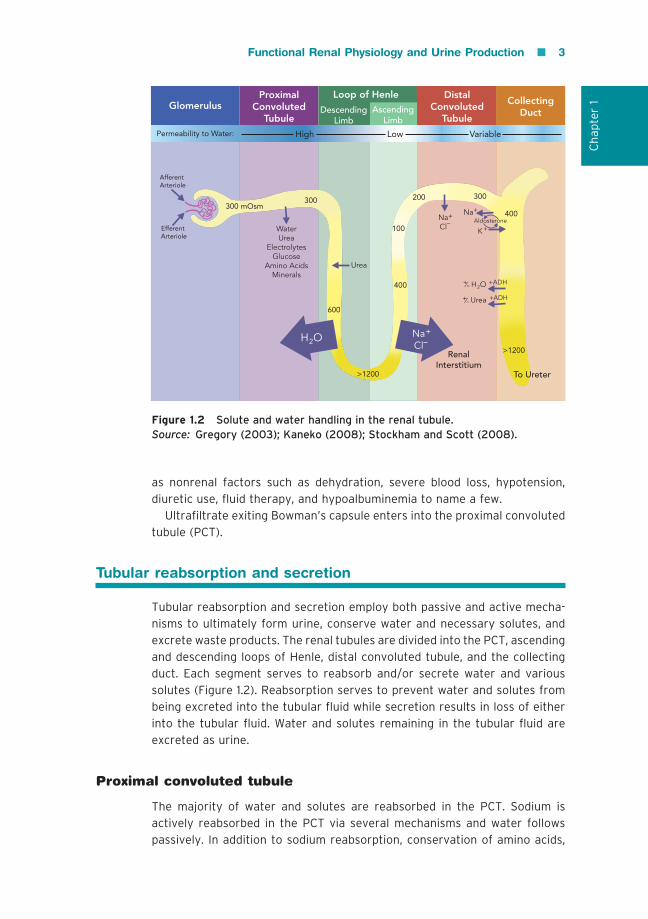
Tubular reabsorption and secretion
Tubular reabsorption and secretion employ both passive and active mecha-
nisms to ultimately form urine, conserve water and necessary solutes, and
excrete waste products. The renal tubules are divided into the PCT, ascending
and descending loops of Henle, distal convoluted tubule, and the collecting
duct. Each segment serves to reabsorb and/or secrete water and various
solutes (Figure 1.2). Reabsorption serves to prevent water and solutes from
being excreted into the tubular fluid while secretion results in loss of either
into the tubular fluid. Water and solutes remaining in the tubular fluid are
excreted as urine.
Proximal convoluted tubule
The majority of water and solutes are reabsorbed in the PCT. Sodium is
actively reabsorbed in the PCT via several mechanisms and water follows
passively. In addition to sodium reabsorption, conservation of amino acids,
Figure 1.2 Solute and water handling in the renal tubule.Source: Gregory (2003); Kaneko (2008); Stockham and Scott (2008).
300300 mOsm
100
400
>1200
300200
>1200
400
600
Efferent Arteriole
Afferent Arteriole
Permeability to Water:
Glomerulus
WaterUrea
ElectrolytesGlucose
Amino AcidsMinerals
High
ProximalConvoluted
Tubule
Urea
RenalInterstitium
Low
AscendingLimb
DescendingLimb
Loop of Henle
NaCl–
+
+ADHUrea+/-
+ADHH O2+/-
Na
+KAldosterone
H O2
+
NaCl–
+
Variable
DistalConvoluted
Tubule
To Ureter
CollectingDuct