Embed Size (px)
Citation preview
PPHN - Persistent Pulmonary Hypertension of the Newborn
4/24/2019
Elizabeth Papp, RN, MSN, CNS

PPHN2
Describe the normal physiologic transition of the cardiopulmonary system from fetal to neonatal circulation
Identify risk factors for development PPHN Identify the clinical presentation of PPHN Describe nursing interventions to minimize progression of
PPHN Identify current treatment strategies Explain significance of PPHN in the neonatal population
Objectives

PPHN3
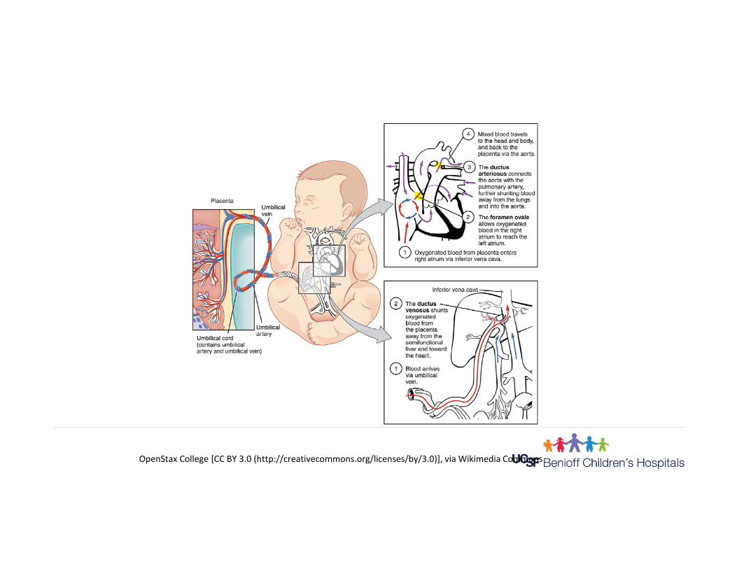
Fetal Circulation
OpenStax College [CC BY 3.0 (http://creativecommons.org/licenses/by/3.0)], via Wikimedia Commons
Fetal Circulation: Quick Overview Organ of respiration is placenta Fetal lungs fluid-filled Pulmonary arteries constricted High right heart and lung pressures Low left heart pressures Open fetal shunts

Placenta Site for gas exchange in fetus Very vascular, large surface area High flow/low resistance circuit Responsible for delivering nutrients to
and carrying waste products away from fetus

Open Fetal Shunts Ductus Venosus Foramen Ovale Ductus Arteriosus Purpose: shunt blood away from
lungs, send well-oxygenated blood from placenta to fetus
Fetal Lungs
Fluid-filled Low-flow, high-resistance circuit Only 10% cardiac output flows to lungs Only need blood flow for growth
OpenStax College [CC BY 3.0 (http://creativecommons.org/licenses/by/3.0)], via Wikimedia Commons
Lung Maturation Highlights
Fetal Breathing Movements- Aid with lung fluid regulation, growth of lung tissue,
strength of diaphragm
Antioxidant Defenses- Scavenge/detoxify oxygen radicals from aerobic
metabolism (toxic to cells) Surfactant
- Reduces surface tension, aids lung expansion with lower pressures
- Stabilizes alveoli, maintains FRC, prevents atelectasis

Lung Maturation
Factors enhancing lung maturity- Stress increased fetal catecholamines,
cortosteroid levels- Chronic maternal hypertension, CV disease- Pre-eclampsia- IUGR- PROM

Lung Maturation
Factors delaying lung maturation
- IDM: high glucose/insulin level delays/interferes with surfactant production
- rH isoimmunization with hydrops
- Androgens delay Type II cell maturation (surfactant production)
Female fetal lung 1 week more mature
Surfactant
Phospholipid Synthesized and secreted by Type II alveolar cells Unique: when layer is laterally compressed it changes physical
nature from liquid to semisolid Forms protective oily layer on lung surface: antimicrobial properties Reduces surface tension, facilitates lung expansion with lower
pressure Stabilizes alveolar surface during expiration so FRC is maintained
Lung Fluid
Approximately 100 ml/kg/day secreted Becomes part of amniotic fluid Volume equivalent to FRC Function
- Cell maturation/development- Formation, shape, size of airspaces
Lung Fluid
Production slows late pregnancy Absorption during early labor
- Active Na transport across epithelium- Liquid from lung lumen to interstitium, vasculature/lymphatics
35% original volume remains at birth Small amount squeezed out during vag delivery Delayed clearance with C-section
Fetal Pulmonary Vessels
Greater amount of smooth muscle compared to adults- Increases tone of vessels, increases resistance to
flow Constrictor response (reactivity) of smooth muscle is
great (hypoxia) Number of vessels increases during fetal life
- Decreases resistance to flow
Pulmonary Vascular Tone
Central role: pulmonary endothelial cells Produce and release mediators that act on smooth
muscle cells Delicate balance between vasoconstrictors and
vasodilators maintain vascular tone

Pulmonary Vascular Tone
Chemical and mechanical factors that decrease PVR include:- Oxygen- Nitric oxide, prostacylines, some prostaglandins- Lung inflation- Structural changes in vessel walls -> thinning muscle
Pulmonary Vascular Tone
Chemical and mechanical factors that increasePVR include:- Hypoxia, acidosis- Over or underinflation of the lungs- Increased chemical mediators: Leukotrienes Thromboxane Endothelin
- Protein: vasoactive peptide
- Promotes vasoconstriction
- Proliferation of SM cells in PA’s
Pulmonary Vascular Tone
Vascular tone increases with gestational age
In late gestation, pulmonary pressures are equivalent to systemic pressures
Major influences in utero favoring vasoconstriction- Low oxygen tension
- High levels of endothelin-1 and leukotrienes
- Low production of prostacyclines and nitric oxide

Pulmonary Vasculature
Pulmonary hypertension with reduced PBF is normal state in fetus
Fetus is physiologically hypoxemic (pO2~25): adequate for lung/cell growth
Fetus is not hypoxic Adequate O2 delivery to tissues in utero
- High cardiac output
- High hemoglobin level in term infant
- Presence of fetal hemoglobin (high affinity for O2)

Fetal Circulation in Detail
Umbilical VeinOne vesselFrom placenta to fetusHighest O2 content (70%, 32-35 mm Hg)Branches into 2 parts inside the fetus

Fetal Circulation in Detail
Ductus VenosusMajority of blood passes through DV From Umbilical Vein to IVCBlood then flows into Right Atrium

Fetal Circulation in Detail
Foramen OvaleMajority of blood (> 50%) enters RA & flows through FO to LA
Bypasses the lungs (2o to high PVR) so oxygenated blood gets to upper body through LV and aorta
Pressure relationships keep FO open
Fetal Circulation in Detail
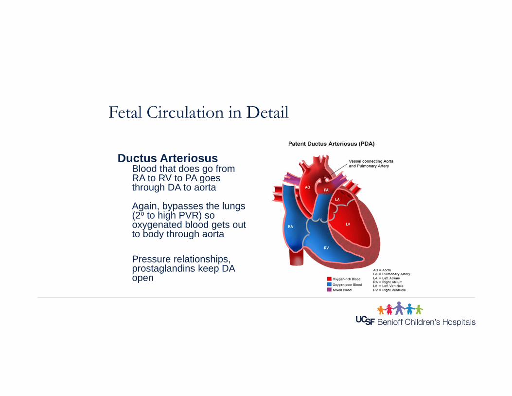
Ductus ArteriosusBlood that does go from RA to RV to PA goes through DA to aorta
Again, bypasses the lungs (2o to high PVR) so oxygenated blood gets out to body through aorta
Pressure relationships, prostaglandins keep DA open

Fetal Circulation in Detail
Umbilical ArteriesTwo vessels
Deoxygenated blood from fetal circulation returns to placenta through umbilical arteries via the descending aorta
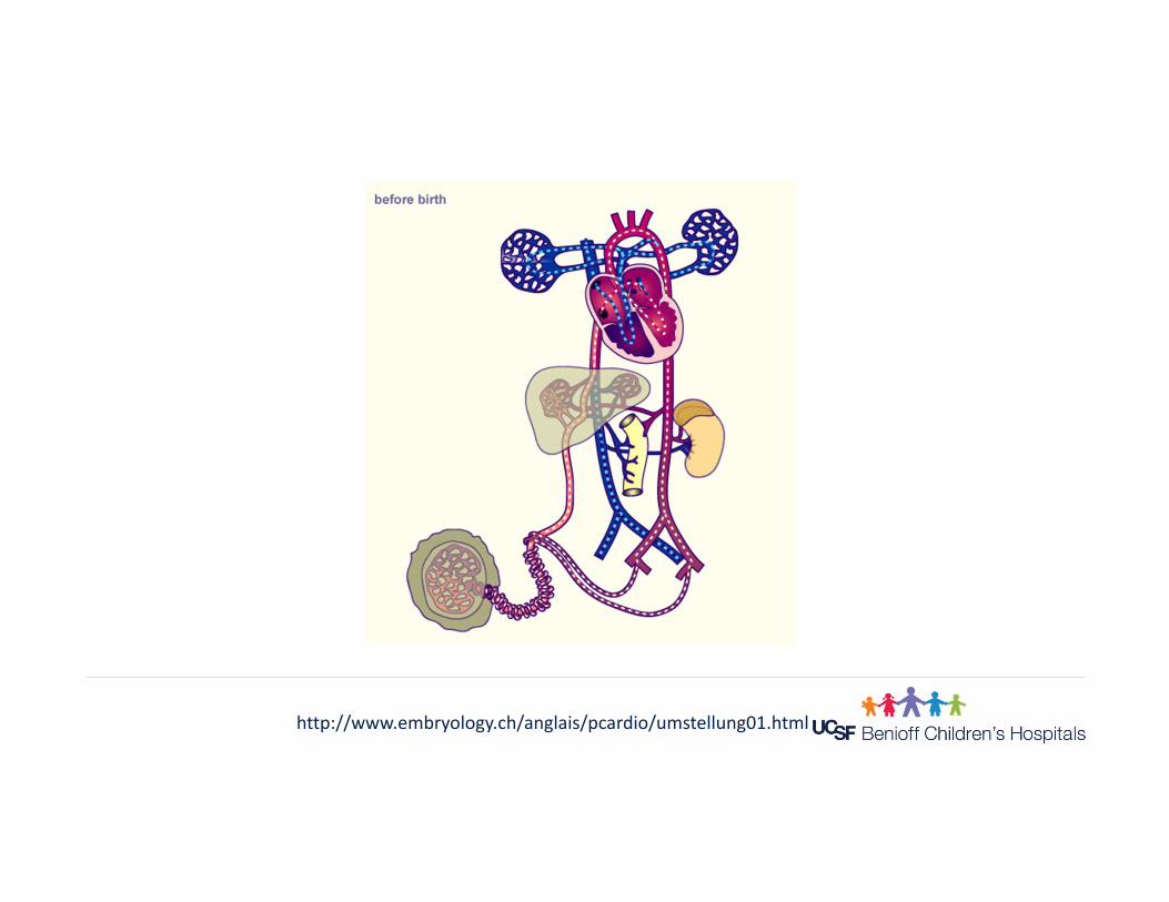
http://www.embryology.ch/anglais/pcardio/umstellung01.html
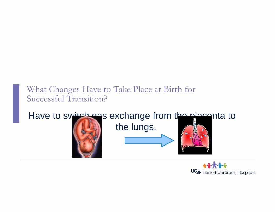
What Changes Have to Take Place at Birth for Successful Transition?
Have to switch gas exchange from the placenta to the lungs.

Transition: Critical Events
Initiate respiratory movements Air into lungs, expansion of alveoli Establish FRC Increase pulmonary blood flow, redistribute
cardiac output

Transition: Critical Events
Oxygenation- Increases oxygen tension: pulm vessels dilate
- Reduces PVR, increases PBF/venous return
- Decreases ductal shunting
Ventilation- Clears lung fluid/creates gas-fluid interface
- Stimulates surfactant secretion
- Stimulates pulm stretch receptors/increase PBF
Cord clamping- Removes low resistance placenta
- Increases SVR
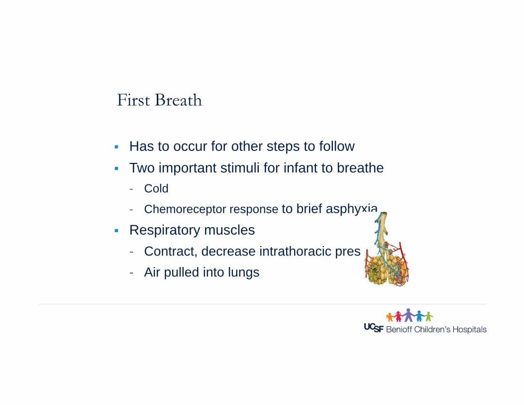
First Breath
Has to occur for other steps to follow Two important stimuli for infant to breathe
- Cold
- Chemoreceptor response to brief asphyxia Respiratory muscles
- Contract, decrease intrathoracic pressures- Air pulled into lungs

Establish FRC
Volume of air retained in lung at end expiration (40% of fully expanded volume)
Initial opening breath requires high pressure for expansion (40 - 60 cm H2O)
Next breath requires much less pressure, better inflation (need surfactant)
Forces opposing air entry- Lung fluid- Surface tension in alveoli
Circulation Changes
First breath causes rise in paO2
Pulmonary vessels dilate PVR, increased flow
- Pulm vasculature becoming high-flow, low-pressure circuit
R heart pressures
Circulation Changes
Removal of low resistance placenta Increases systemic pressure/LV pressure Increases volume (venous return from lungs)
left side of heart L heart pressures > R heart pressures Functional closure of the Foramen Ovale
within minutes to hours after birth (anatomical closure 30 months or longer)
Circulation Changes
Pressure in aorta becomes > pressure in PA’s: reverse flow through Ductus Arteriosus
oxygen level, smooth muscle constricts to close the ductus PGE removed by lungs aids in closure Functional closure in healthy term baby by 96 hrs of age Anatomical closure later by tissue growth (up to 3 months)

Circulation Changes
Ductus venosus functionally closes within hours after cord clamping when there is no longer blood flow from umbilical vein
Anatomical closure by 10 to 14 days after birth

PPHN36
PPHN: Types and Associated Factors

PPHN – The Pathophysiology
PVR does not fall PFO & PDA remain open shunting blood away from
lungs Impaired gas exchange Progressive hypoxia & hypoxemia Increased cardiac afterload (right side) RVH, TI & heart failure

elevated PVR
deoxygenated + oxygenated blood
systemic circulation
hypoxemia
The Three Types of PPHN(What Causes Elevated PVR)
Underdevelopment
Maldevelopment
Maladaptation

A Little Physics(Poiseulle’s law)
Volume Flowrate =
Pressure difference x radius4
Viscostiy x length
(Taken from http://hyperphysic.phy-astr.gsu.edu)

Small changes go a long way
(Taken from http://hyperphysic.phy-astr.gsu.edu)
Causes of Vasoconstriction
Acidosis Hypoxia (acute or chronic) Asphyxia Vasoactive mediators Muscular hyperplasia
Arterial vasodilation
Alkalosis Oxygen Vasoactive mediators Medications
Underdevelopment(Lung Hypoplasia)
Decreased cross sectional area of pulmonary vasculature Examples: CDH, CCAM, renal agenesis, obstructive
uropathy, IUGR Fixed elevated PVR High mortality

Maldevelopment(Abnormal vasculature)
Lung parenchyma develops normally Thick muscular layer around arterioles Vascular mediators are involved Genetic predisposition Excessive fetal perfusion of lungs Examples: post dates, premature closure of the ductus
aterious
Maladaptation(Pulmonary vasoconstriction)
Normal pulmonary vascular bed Also known as secondary PPHN Active vasoconstriction at or after birth Examples: perinatal depression, hemorrhage, aspiration,
asphyxia, lung disease, infections, hypoglycemia, hypothermia
Risk factors
1. Fetal closure of ductus arteriosus 2. Abnormal response to oxygen levels 3.Hypertrophy of pulmonary smooth muscles 4. Lung hypoplasia 5. Segmental alveolar underventilation

Risk factors cont’d…
6. Dysfunction or proliferation of vasoactive mediators 7. Presence of microthrombi in pulmonary vascular bed
(polycythemia) 8. Maternal/perinatal factors 9. Airway obstruction, MSAF

Diagnosis(How do you know it’s there?)
“This baby just doesn’t look good” Severe cyanosis –a medical emergency! Low PaO2 with normal PaCO2 Pre-ductal saturation higher than post-ductal Cardiac murmur (sometimes), CHF/TR
The Work-up
Pre & post ductal saturations Hyperoxygenation test Chest x-ray Echocardiography (most definitive)

Echocardiogram
Excludes congenital heart disease Measures pulmonary artery pressures (TR, TI, shunt
velocites) Defines the presence, degree, and direction of intra-cardiac
shunting Describe ventricular outputs and function
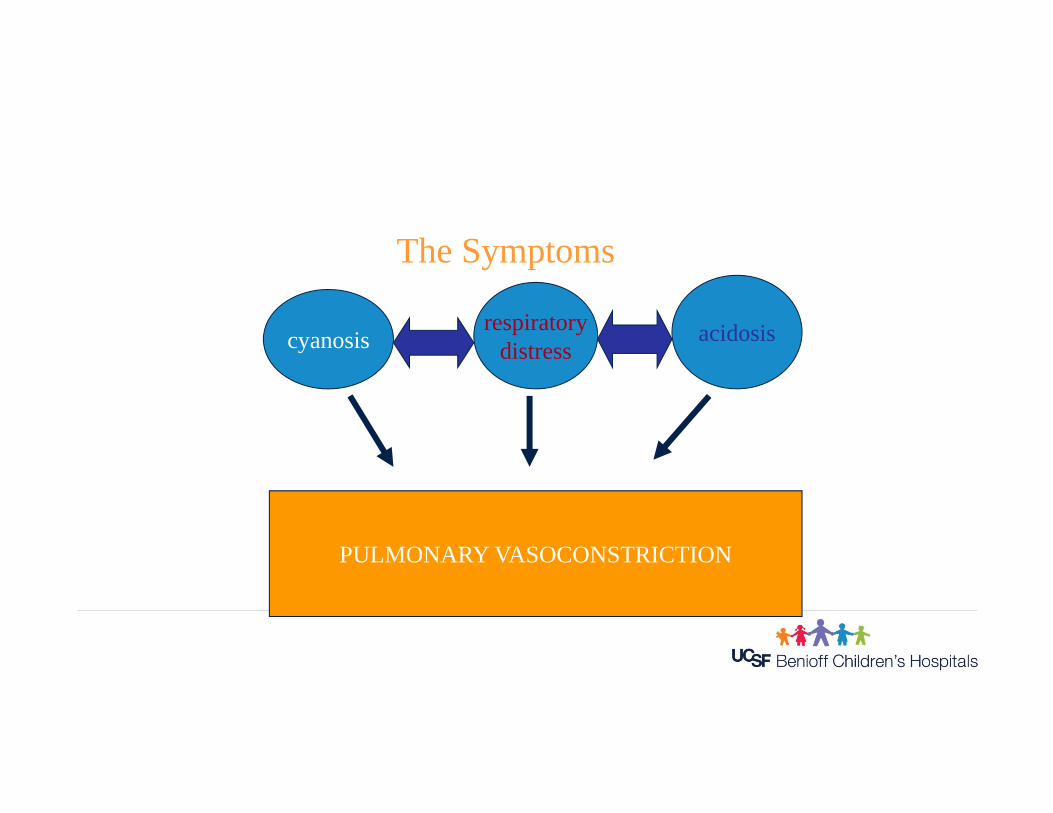
The Symptoms
cyanosisrespiratory
distressacidosis
PULMONARY VASOCONSTRICTION

Nursing Assessments and Management
Antepartum Intrapartum Postpartum
- Crucial nursing assessment/intervention periods
Antepartum
Good prenatal care! Prenatal education
Intrapartum
Close intrapartum observation is key!- Careful fetal monitoring
- Careful documentation!
Postpartum Preparedness
Be prepared for high risk deliveries Suction equipment, Delee suction, ETT & laryngoscope,
meconium aspirator, oxygen, PPV equipment Closely observe infant especially if respiratory distress
present
Nursing Assessments
Monitor HR, color closely If low Apgars or intubated
- Frequent vital signs & respiratory assessments
- Cord and post-natal blood gas analysis
- Arterial stick for blood gas if not intubated
- UAC if intubated or with high O2 requirement
Nursing Interventions
Optimize oxygenation- Keep saturations in high 90’s
- Oxygen challenge test
- Wean very slowly if stable
Correct metabolic acidosis- Fluid boluses
Closely monitor perfusion and blood pressure- Color, CFT, pulses, temperature of skin
- May need to treat hypotension/hypoperfusion with fluid boluses and/or inotropes

Keep calm - May need sedation or paralysis if mechanically ventilated
- Minimize O2 consumption
Keep NPO Provide IVF and antibiotics
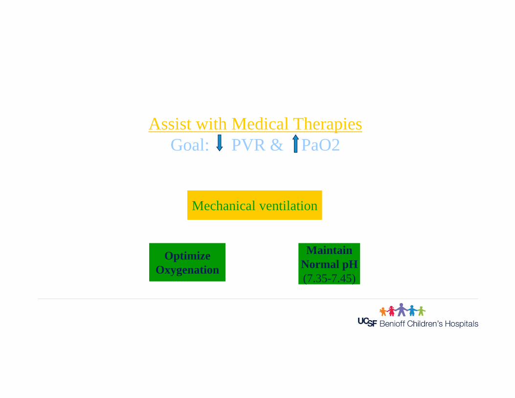
Assist with Medical TherapiesGoal: PVR & PaO2
Mechanical ventilation
OptimizeOxygenation
OptimizeOxygenation
MaintainNormal pH(7.35-7.45)
MaintainNormal pH(7.35-7.45)
Other Medical Therapies & Diagnostic Tests
High frequency ventilation
Inhaled nitric oxide
Surfactant administration
ECMO – previously needed for 40% severe PPHN
Experimental Therapies
Sildenafil Systemic steroids (for MAS) Magnesium sulfate Prostacycline
A Special Word:Nitric Oxide
Produced endogenously Enzyme NO synthetase acts on arginine Increases levels of cGMP Potent smooth muscle relaxant Vasodilation

Inhaled Nitric Oxide
Diffuses through the alveolar membrane Into the blood stream and deactivated No systemic effects
Inhaled NO(Guidelines for Use)
Clinical and echocardiographic signs of PPHN FIO2 1.0 already in use Access to ECMO Start at 20ppm, using a accurate delivery system Wean slowly when clinically stable
Side effects to Watch For
Methemoglobinemia -- NO intereacts with oxyhemoglobin to form methemoglobin and
nitrates
Watch for rebound hypertension –negative feedback?
Provide Parental Support
Explain! Explain! Explain! They can touch their infant, take pictures Prepare them for possible transport even before you know for
sure it will happen
Prepare for Transport
Ensure adequate IV access Suction ETT Sedation and maybe paralysis Continue 100% oxygen & iNO if applicable
Morbidity & Mortality
Primary risks stem from:- 1. Delay in recognition of existence and severity of hypoxemia
- 2. Delay in timely transfer to an ECMO center
- 3. Lack of communication with an ECMO center especially if iNO is initiated
Outcomes of PPHN
Before ECMO, mortality 12 to 50% Since ECMO, survival ~ 85% Significant morbidity still 10 to 45% Hyperventilation – some sensorineuronal hearing loss
reported as high as 53% Higher risk for developmental delay & motor disability but
most survivors are normal
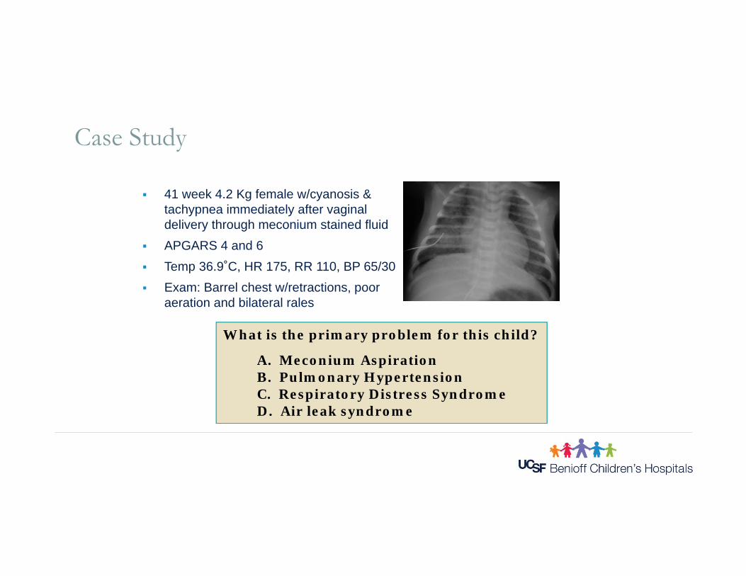
Case Study
41 week 4.2 Kg female w/cyanosis & tachypnea immediately after vaginal delivery through meconium stained fluid
APGARS 4 and 6 Temp 36.9˚C, HR 175, RR 110, BP 65/30 Exam: Barrel chest w/retractions, poor
aeration and bilateral rales
What is the primary problem for this child?
A. Meconium AspirationB. Pulmonary HypertensionC. Respiratory Distress SyndromeD. Air leak syndrome
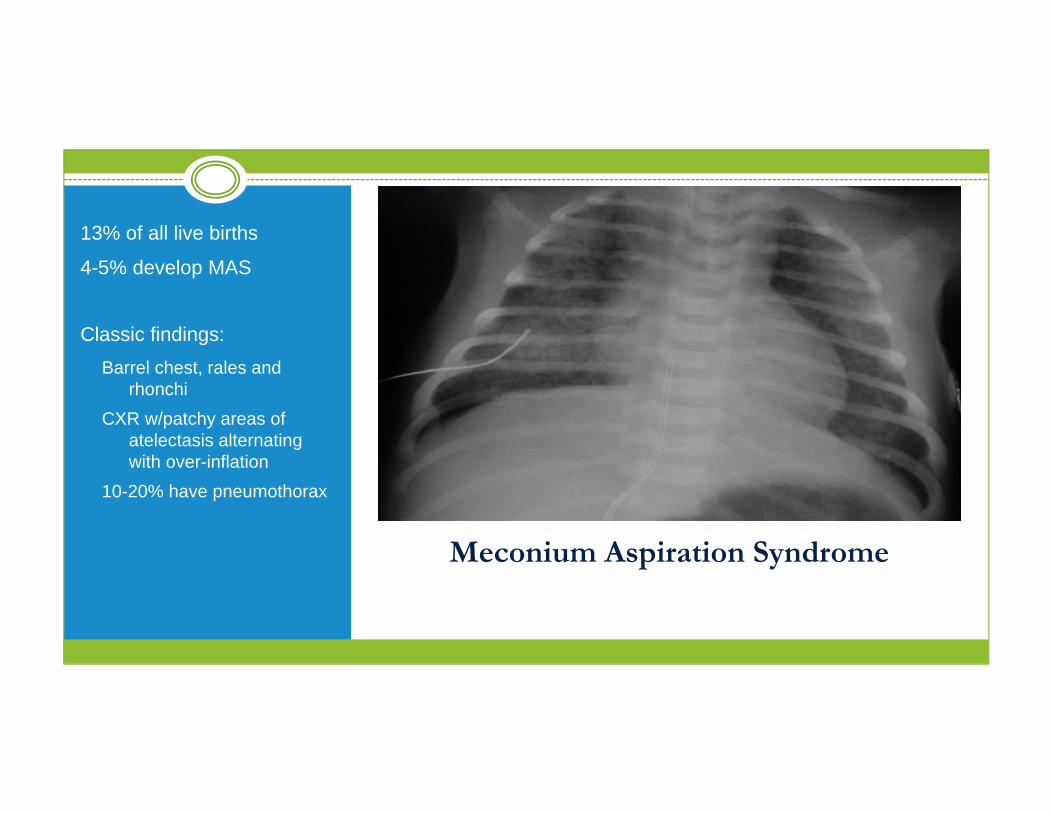
Meconium Aspiration Syndrome
13% of all live births
4-5% develop MAS
Classic findings: Barrel chest, rales and
rhonchiCXR w/patchy areas of
atelectasis alternating with over-inflation
10-20% have pneumothorax
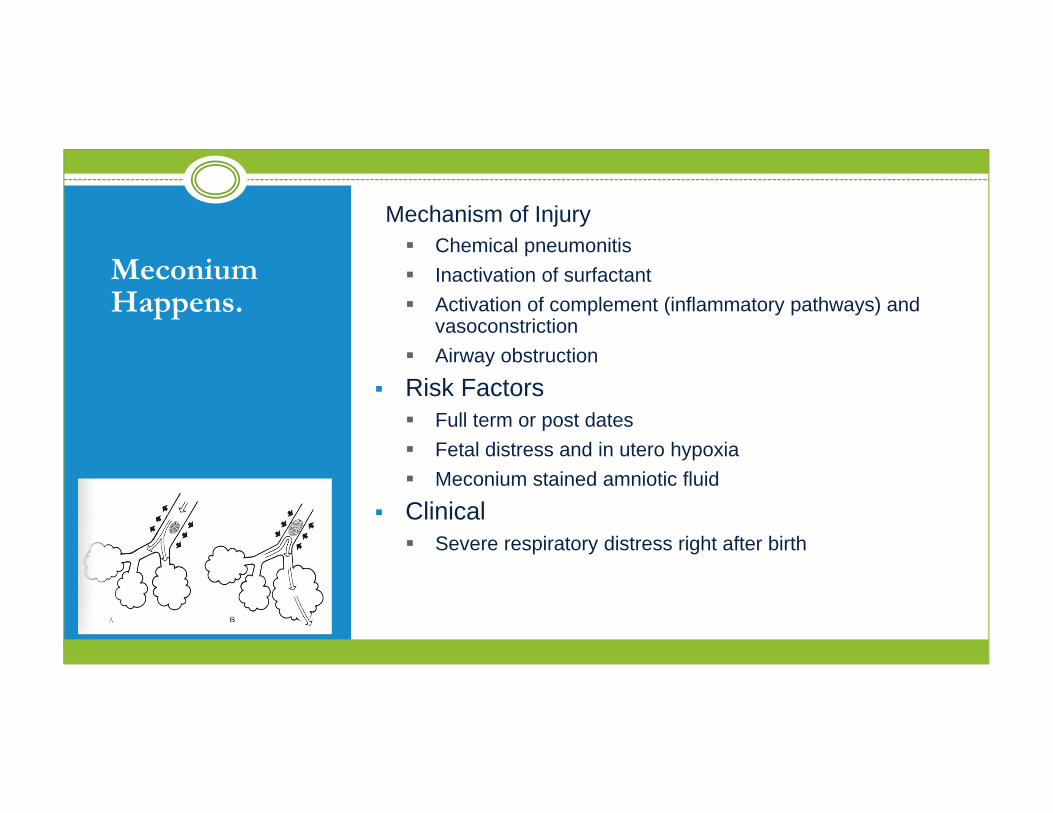
Meconium Happens.
Mechanism of Injury Chemical pneumonitis Inactivation of surfactant Activation of complement (inflammatory pathways) and
vasoconstriction Airway obstruction
Risk Factors Full term or post dates Fetal distress and in utero hypoxia Meconium stained amniotic fluid
Clinical Severe respiratory distress right after birth
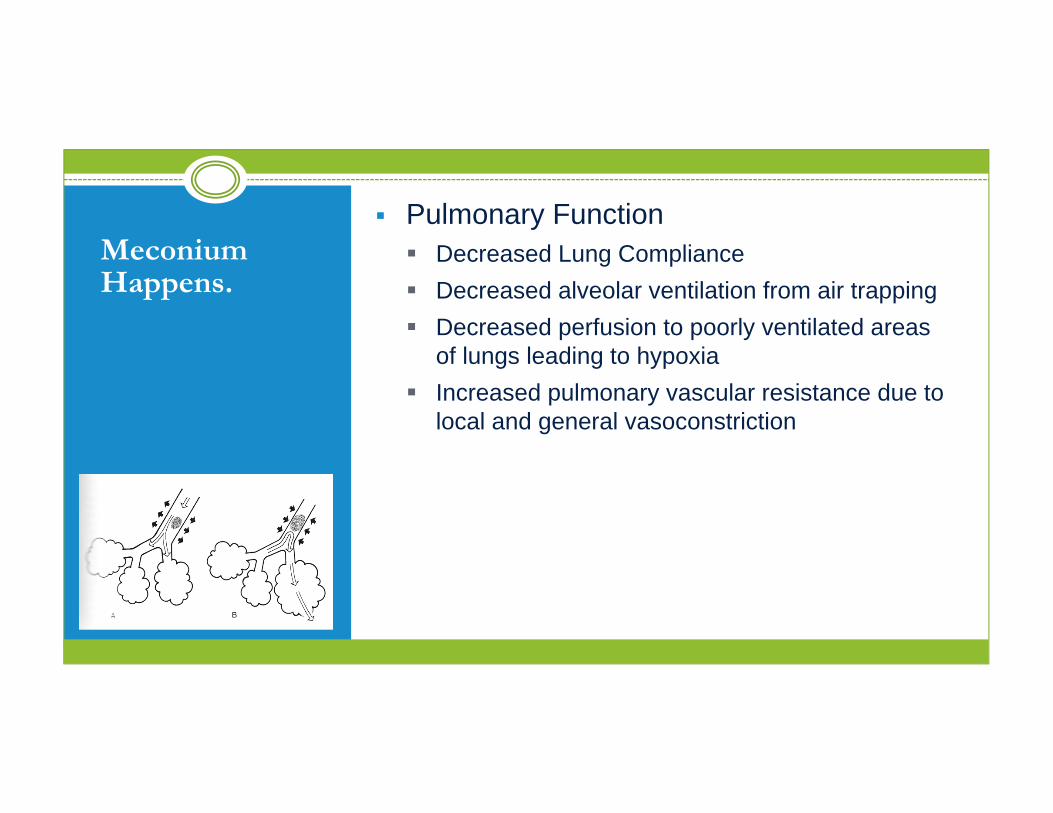
Meconium Happens.
Pulmonary Function Decreased Lung Compliance Decreased alveolar ventilation from air trapping Decreased perfusion to poorly ventilated areas
of lungs leading to hypoxia Increased pulmonary vascular resistance due to
local and general vasoconstriction
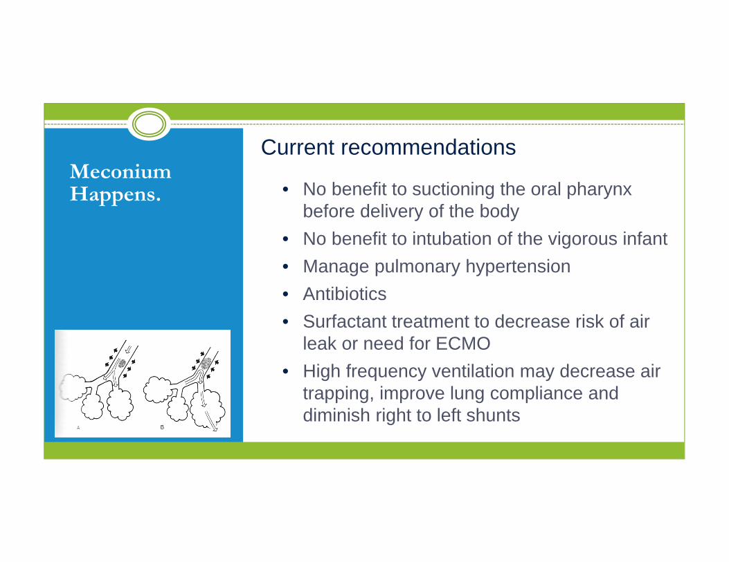
Meconium Happens.
Current recommendations
• No benefit to suctioning the oral pharynx before delivery of the body
• No benefit to intubation of the vigorous infant• Manage pulmonary hypertension• Antibiotics• Surfactant treatment to decrease risk of air
leak or need for ECMO• High frequency ventilation may decrease air
trapping, improve lung compliance and diminish right to left shunts