Embed Size (px)
Citation preview
This is the published version Bomberger, Jennifer M., MacEachran, Daniel P., Coutermarsh, Bonita A., Ye, Siying, O'Toole, George A. and Stanton, Bruce A. 2009, Long-distance delivery of bacterial virulence factors by Pseudomonas aeruginosa outer membrane vesicles, PLoS pathogens, vol. 5, no. 4, pp. 1-13. Available from Deakin Research Online http://hdl.handle.net/10536/DRO/DU:30040949 Reproduced with the kind permission of the copyright owner Copyright: 2009, Public Library of Science
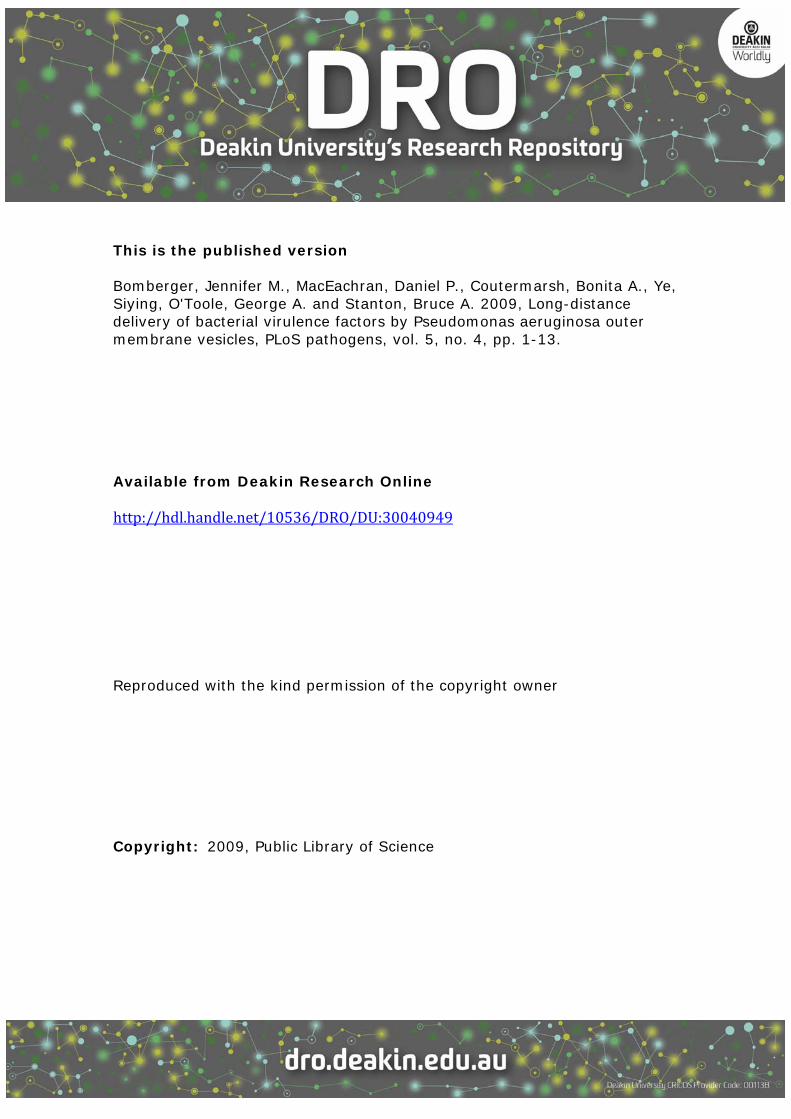
Long-Distance Delivery of Bacterial Virulence Factors byPseudomonas aeruginosa Outer Membrane VesiclesJennifer M. Bomberger1*, Daniel P. MacEachran2, Bonita A. Coutermarsh1, Siying Ye1, George A.
O’Toole2, Bruce A. Stanton1
1 Department of Physiology, Dartmouth Medical School, Hanover, New Hampshire, United States of America, 2 Department of Microbiology and Immunology, Dartmouth
Medical School, Hanover, New Hampshire, United States of America
Abstract
Bacteria use a variety of secreted virulence factors to manipulate host cells, thereby causing significant morbidity andmortality. We report a mechanism for the long-distance delivery of multiple bacterial virulence factors, simultaneously anddirectly into the host cell cytoplasm, thus obviating the need for direct interaction of the pathogen with the host cell tocause cytotoxicity. We show that outer membrane–derived vesicles (OMV) secreted by the opportunistic human pathogenPseudomonas aeruginosa deliver multiple virulence factors, including b-lactamase, alkaline phosphatase, hemolyticphospholipase C, and Cif, directly into the host cytoplasm via fusion of OMV with lipid rafts in the host plasma membrane.These virulence factors enter the cytoplasm of the host cell via N-WASP–mediated actin trafficking, where they rapidlydistribute to specific subcellular locations to affect host cell biology. We propose that secreted virulence factors are notreleased individually as naked proteins into the surrounding milieu where they may randomly contact the surface of thehost cell, but instead bacterial derived OMV deliver multiple virulence factors simultaneously and directly into the host cellcytoplasm in a coordinated manner.
Citation: Bomberger JM, MacEachran DP, Coutermarsh BA, Ye S, O’Toole GA, et al. (2009) Long-Distance Delivery of Bacterial Virulence Factors by Pseudomonasaeruginosa Outer Membrane Vesicles. PLoS Pathog 5(4): e1000382. doi:10.1371/journal.ppat.1000382
Editor: Frederick M. Ausubel, Massachusetts General Hospital, United States of America
Received November 24, 2008; Accepted March 16, 2009; Published April 10, 2009
Copyright: � 2009 Bomberger et al. This is an open-access article distributed under the terms of the Creative Commons Attribution License, which permitsunrestricted use, distribution, and reproduction in any medium, provided the original author and source are credited.
Funding: This work was supported by NIH grants 5T32DK007301-30, 5R01HL074175-04, and 5R01DK045881-14 (BAS), and Cystic Fibrosis Foundation grantsBOMBER08F0 (JMB) and STANTO07R0 (BAS). The funders had no role in study design, data collection and analysis, decision to publish, or preparation of themanuscript.
Competing Interests: The authors have declared that no competing interests exist.
* E-mail: [email protected]
Introduction
Nosocomial infections contribute $4.5 billion to annual
healthcare costs in this country alone, with an estimated 2 million
nosocomial infections occurring in the US annually, resulting in
99,000 deaths [1]. Many of these nosocomial infections are caused
by Gram-negative pathogens, and interaction of these pathogens
with the host is often mediated by secreted virulence factors.
Bacteria have evolved mechanisms for the secretion of virulence
factors into the host cell to alter host cell biology and enable
bacterial colonization, and these mechanisms typically require that
bacteria be in intimate contact with the host. For example, the
Type III secretion system (T3SS) and Type IV secretion system
(T4SS) deliver proteins directly into the host cytoplasm from an
extracellular bacterial pathogen’s cytoplasm [2] utilizing transport
machines that act as macromolecular syringes [3]. Delivery of
extracellular bacteria or bacterial products can also occur via
endocytosis initially into the lumen of the host endocytic compart-
ment, then movement to the host cytoplasm via lysis of the endocytic
compartment or delivery of the proteins across the endocytic
membrane via the Type III Secretion System (T3SS) [3].
For several decades, work by Beveridge’s group has characterized
bacterial-derived outer membrane vesicles (OMV) to be a novel
secretion mechanism employed by bacteria to deliver various
bacterial proteins and lipids into host cells, eliminating the need
for bacterial contact with the host cell [4–7]. OMV are 50–200 nm
proteoliposomes constitutively released from pathogenic and
non-pathogenic species of Gram-negative bacteria [8,9]. Biochem-
ical and proteomic analyses have revealed that OMV are comprised
of lipopolysaccharide, phospholipids, outer membrane proteins, and
soluble periplasmic proteins [8,9]. Many virulence factors that are
periplasmic proteins are enriched in OMV, for example, Escherichia
coli cytolysin A (ClyA), enterotoxigenic E. coli heat labile enterotoxin
(LT), and Actinobacillus actinomycetemcomitans leukotoxin [10–12].
Beveridge’s group and others have reported that some secreted
virulence factors from P. aeruginosa, including b-lactamase, hemolytic
phospholipase C, alkaline phosphatase, pro-elastase, hemolysin, and
quorum sensing molecules, like N-(3-oxo-dodecanoyl) homoserine
lactone and 2-heptyl-3-hydroxy-4-quinolone (PQS) [6,7,13,14], are
also associated with P. aeruginosa OMV [8,9]. Whether these secreted
virulence factors packaged in OMV are eventually delivered to the
host and the mechanism by which this occurs is currently unknown.
A recent study suggested that E. coli OMV fuse with lipid rafts in the
host colonic epithelial cell, but the delivery and intracellular trafficking
of the OMV cargo was not characterized [15]. Thus, we investigated
the possibility that OMV deliver multiple secreted virulence factors
into the host cell through a lipid raft-mediated pathway, eliminating
the need for intimate contact of the pathogen with the host.
Results
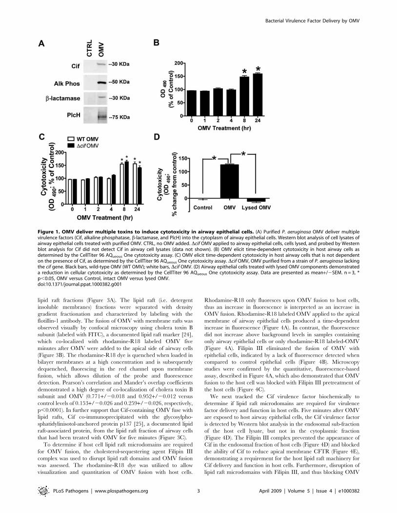
Outer membrane vesicles deliver toxins to airway epithelial cellsBased on reports that multiple virulence factors are packaged in
OMV, we hypothesized that these virulence factors could be
PLoS Pathogens | www.plospathogens.org 1 April 2009 | Volume 5 | Issue 4 | e1000382
simultaneously delivered in a coordinated manner in OMV to the
host cell by the microbe. We tracked four P. aeruginosa secreted
factors, including alkaline phosphatase, b-lactamase, hemolytic
phospholipase C, and Cif, previously reported to be packaged in
OMV [6,7,13,14,16]. We chose these secreted virulence factors
because they play important roles in host colonization, for
example alkaline phosphatase promotes biofilm formation
[17,18], b-lactamase degrades host antimicrobial peptides, hemo-
lytic phospholipase C is cytotoxic and promotes P. aeruginosa
virulence [19], and Cif is a recently characterized toxin that
inhibits CFTR-mediated chloride secretion in the airways [16]
and thereby likely reduces mucociliary clearance. To purify OMV
from bacterial products not packaged in OMV, like pilus, that may
also elicit a host response, we modified a published protocol [14]
utilizing high-speed differential centrifugation and density gradient
fractionation to isolate OMV from an overnight P. aeruginosa
culture supernatant. The Cif protein, as well as a protein present
in the membrane of OMV, Omp85 [20], were identified in
purified bacterial-derived OMV (Figure S1). When airway cells
were treated with isolated and purified OMV for ten minutes all
four OMV proteins examined were detected in host airway
epithelial cell lysate (Figure 1A). By contrast, these virulence
factors were not detected in lysates of control cells treated with
vehicle (Figure 1A). Therefore, OMV deliver multiple virulence
factors to host airway epithelial cells in the absence of bacteria,
thus providing a mechanism for bacteria to alter host cell
physiology without the need for intimate contact with the host.
To explore the significance of OMV in the delivery of virulence
factors into the host cytoplasm, we examined the cytotoxic effect of
P. aeruginosa OMV on host airway cells using the CellTiter 96
AQueous One cytotoxicity assay. OMV were cytotoxic after a delay
of 8 hours (Figure 1B), although virulence factors could be
detected in the cytoplasm of host cells after 10 minutes
(Figure 1A). The time-dependent increase in cytotoxicity induced
by OMV was not dependent on Cif expression in OMV, given
that Dcif OMV did not produce a statistically significant difference
in cytotoxicity compared to wild-type OMV (Figure 1C). To
determine if intact OMV are required for cytotoxicity, purified
OMV were lysed with 0.1 M EDTA and the lysate was applied to
airway epithelial cells for 8 h (Figure S2, Figure 1D). This method
was previously employed by Horstman et al. to effectively lyse E.
coli OMV [21]. The lysed OMV did not have a cytotoxic effect on
airway epithelial cells, demonstrating that cytotoxicity is mediated
by virulence factors delivered into the host cell cytoplasm by
bacterial-derived OMV (Figure 1D).
In the next series of experiments we began to examine the
mechanism whereby OMV deliver virulence factors into the
cytoplasm of the host airway epithelial cell. Previously we reported
that purified, recombinant Cif, a virulence factor secreted in OMV
by P. aeruginosa, is necessary and sufficient to reduce apical
membrane expression of CFTR and P-glycoprotein (Pgp) in
human airway epithelial cells [16,22], thus reducing mucociliary
clearance and xenobiotic resistance of the host cells, respectively.
In the current study we use Cif as a model protein to investigate
how OMV deliver virulence factors into the cytoplasm of human
airway epithelial cells. First, experiments were conducted to
confirm our previous observation that Cif is secreted in purified
OMV and second, to determine if Cif is an intravesicular
component of OMV. Cif was detected in OMV derived from P.
aeruginosa expressing the cif gene, but not in OMV derived from P.
aeruginosa in which the cif gene was deleted (Figure S3). The
inability of Proteinase K (0.1 mg/ml), which does not enter the
lumen of OMV, to degrade OMV-associated Cif indicates that
this virulence factor is an intravesicular component of OMV
(Figure S3). Thus, these studies demonstrate that Cif maintains an
intravesicular localization in purified OMV.
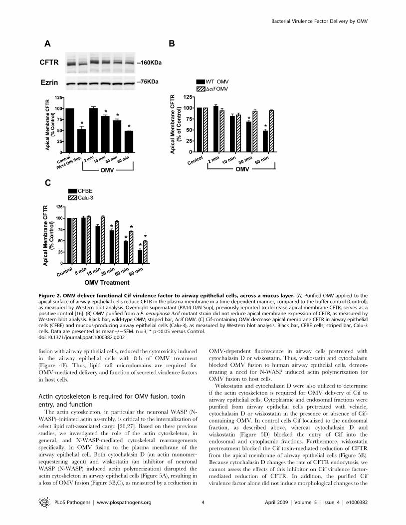
Next, studies were conducted to determine if the Cif virulence
factor packaged in OMV was functional when delivered to airway
epithelial cells. Cif function was measured by examining the ability of
Cif to reduce apical plasma membrane CFTR abundance in airway
epithelial cells. Purified OMV containing Cif reduced apical plasma
membrane CFTR in a time-dependent manner (Figure 2A), whereas
purified OMV from P. aeruginosa deleted for the cif gene had no effect
on CFTR membrane expression (Figure 2B). Taken together these
studies confirm and extend our previous observations that OMV-
packaged Cif reduces plasma membrane CFTR.
If OMV modulate host physiology of lung cells without direct
bacteria-host contact, we would predict that OMV secreted by
bacteria should overcome barriers such as the mucus overlying
human airway cells [23]. OMV containing Cif reduced CFTR in
the apical plasma membrane of airway epithelial cells that have a
thick layer of mucus on the apical surface (Calu-3 cells, Figure 2C).
A delay in the Cif-mediated reduction of apical membrane CFTR
abundance was observed in Calu-3 cells, compared to airway cells
that lack a overlying mucus layer, suggesting the mucus only delays
OMV from diffusing to host airway epithelial cells. Thus, OMV
allow the long distance delivery of secreted bacterial factors to the
host cell in the absence of direct bacteria-host contact.
Host cell detergent-resistant membranes (aka, lipid rafts)are required for OMV fusion and toxin delivery
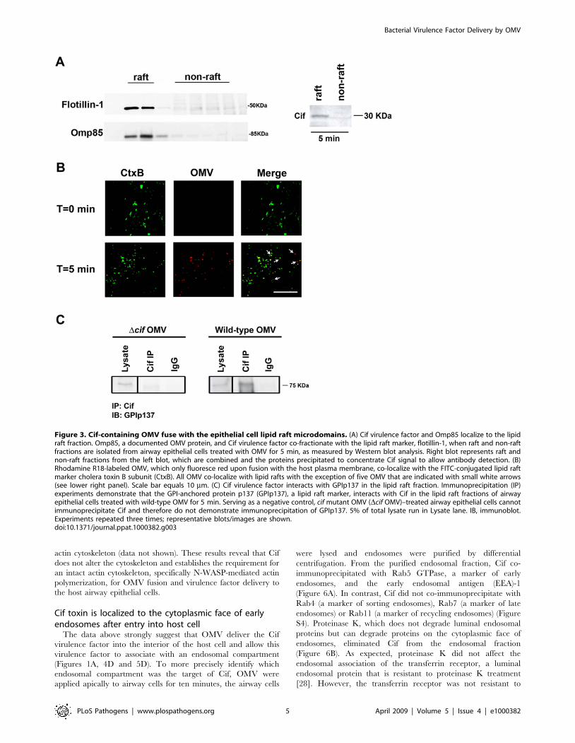
We next explored the mechanism whereby OMV deliver
bacterial proteins into the host cell using the Cif virulence factor as
a model. Based on a recent study showing that Filipin III disrupts
E. coli OMV association with host cells [15], we hypothesized that
OMV deliver secreted bacterial proteins to host cells by fusing
with lipid raft microdomains. Five minutes after addition of OMV
to epithelial cells, Cif, as well as a protein documented to be
associated with OMV, Omp85 [20], were detected in membrane
Author Summary
Gram-negative pathogens are responsible for 2 millionannual hospital-acquired infections, adding tremendouslyto U.S. healthcare costs. Pseudomonas aeruginosa, anopportunistic human pathogen, is commonly associatedwith nosocomial infections, particularly ventilator-associ-ated infections and pseudomonal pneumonia in immuno-compromised patients with cystic fibrosis, chronic ob-structive pulmonary disease, ventilator-associatedpneumonia, community-acquired pneumonia, and bron-chiectasis. We have identified the mechanism for asecretion system that Gram-negative bacteria use tostrategically deliver toxins to the host to promote bacterialvirulence and host colonization, a pathway that we hopeto target to develop new therapies to treat P. aeruginosainfections. Our findings have significant implications forthe study of Gram-negative bacterial pathogenesis. Wepropose that secreted virulence factors are not releasedindividually as naked proteins into the surrounding milieuwhere they may randomly contact the surface of the hostcell, but instead bacterial-derived outer membrane vesicles(OMV) deliver multiple virulence factors simultaneouslyand directly into the host cell cytoplasm in a coordinatedmanner. This long-distance bacterial communication tothe host via OMV is reminiscent of the delivery of signalingproteins and miRNA between eukaryotic cells via exo-somes, and may represent a general protein secretionstrategy used by both pathogen and host.
Bacterial Virulence Factor Delivery by OMV
PLoS Pathogens | www.plospathogens.org 2 April 2009 | Volume 5 | Issue 4 | e1000382
lipid raft fractions (Figure 3A). The lipid raft (i.e. detergent
insoluble membranes) fractions were separated with density
gradient fractionation and characterized by labeling with the
flotillin-1 antibody. The fusion of OMV with membrane rafts was
observed visually by confocal microscopy using cholera toxin B
subunit (labeled with FITC), a documented lipid raft marker [24],
which co-localized with rhodamine-R18 labeled OMV five
minutes after OMV were added to the apical side of airway cells
(Figure 3B). The rhodamine-R18 dye is quenched when loaded in
bilayer membranes at a high concentration and is subsequently
dequenched, fluorescing in the red channel upon membrane
fusion, which allows dilution of the probe and fluorescence
detection. Pearson’s correlation and Mander’s overlap coefficients
demonstrated a high degree of co-localization of cholera toxin B
subunit and OMV (0.771+/20.018 and 0.952+/20.012 versus
control levels of 0.153+/20.026 and 0.259+/20.026, respectively,
p,0.0001). In further support that Cif-containing OMV fuse with
lipid rafts, Cif co-immunoprecipitated with the glycosylpho-
sphatidylinisotol-anchored protein p137 [25], a documented lipid
raft-associated protein, from the lipid raft fraction of airway cells
that had been treated with OMV for five minutes (Figure 3C).
To determine if host cell lipid raft microdomains are required
for OMV fusion, the cholesterol-sequestering agent Filipin III
complex was used to disrupt lipid raft domains and OMV fusion
was assessed. The rhodamine-R18 dye was utilized to allow
visualization and quantitation of OMV fusion with host cells.
Rhodamine-R18 only fluoresces upon OMV fusion to host cells,
thus an increase in fluorescence is interpreted as an increase in
OMV fusion. Rhodamine-R18 labeled OMV applied to the apical
membrane of airway epithelial cells produced a time-dependent
increase in fluorescence (Figure 4A). In contrast, the fluorescence
did not increase above background levels in samples containing
only airway epithelial cells or only rhodamine-R18 labeled-OMV
(Figure 4A). Filipin III eliminated the fusion of OMV with
epithelial cells, indicated by a lack of fluorescence detected when
compared to control epithelial cells (Figure 4B). Microscopy
studies were confirmed by the quantitative, fluorescence-based
assay, described in Figure 4A, which also demonstrated that OMV
fusion to the host cell was blocked with Filipin III pretreatment of
the host cells (Figure 4C).
We next tracked the Cif virulence factor biochemically to
determine if lipid raft microdomains are required for virulence
factor delivery and function in host cells. Five minutes after OMV
are exposed to host airway epithelial cells, the Cif virulence factor
is detected by Western blot analysis in the endosomal sub-fraction
of the host cell lysate, but not in the cytoplasmic fraction
(Figure 4D). The Filipin III complex prevented the appearance of
Cif in the endosomal fraction of host cells (Figure 4D) and blocked
the ability of Cif to reduce apical membrane CFTR (Figure 4E),
demonstrating a requirement for the host lipid raft machinery for
Cif delivery and function in host cells. Furthermore, disruption of
lipid raft microdomains with Filipin III, and thus blocking OMV
Figure 1. OMV deliver multiple toxins to induce cytotoxicity in airway epithelial cells. (A) Purified P. aeruginosa OMV deliver multiplevirulence factors (Cif, alkaline phosphatase, b-lactamase, and PlcH) into the cytoplasm of airway epithelial cells. Western blot analysis of cell lysates ofairway epithelial cells treated with purified OMV. CTRL, no OMV added. Dcif OMV applied to airway epithelial cells, cells lysed, and probed by Westernblot analysis for Cif did not detect Cif in airway cell lysates (data not shown). (B) OMV elicit time-dependent cytotoxicity in host airway cells asdetermined by the CellTiter 96 AQueous One cytotoxicity assay. (C) OMV elicit time-dependent cytotoxicity in host airway cells that is not dependenton the presence of Cif, as determined by the CellTiter 96 AQueous One cytotoxicity assay. Dcif OMV, OMV purified from a strain of P. aeruginosa lackingthe cif gene. Black bars, wild-type OMV (WT OMV); white bars, Dcif OMV. (D) Airway epithelial cells treated with lysed OMV components demonstrateda reduction in cellular cytotoxicity as determined by the CellTiter 96 AQueous One cytotoxicity assay. Data are presented as mean+/2SEM. n = 3, *p,0.05, OMV versus Control, intact OMV versus lysed OMV.doi:10.1371/journal.ppat.1000382.g001
Bacterial Virulence Factor Delivery by OMV
PLoS Pathogens | www.plospathogens.org 3 April 2009 | Volume 5 | Issue 4 | e1000382
fusion with airway epithelial cells, reduced the cytotoxicity induced
in the airway epithelial cells with 8 h of OMV treatment
(Figure 4F). Thus, lipid raft microdomains are required for
OMV-mediated delivery and function of secreted virulence factors
in host cells.
Actin cytoskeleton is required for OMV fusion, toxinentry, and function
The actin cytoskeleton, in particular the neuronal WASP (N-
WASP)–initiated actin assembly, is critical to the internalization of
select lipid raft-associated cargo [26,27]. Based on these previous
studies, we investigated the role of the actin cytoskeleton, in
general, and N-WASP-mediated cytoskeletal rearrangements
specifically, in OMV fusion to the plasma membrane of the
airway epithelial cell. Both cytochalasin D (an actin monomer-
sequestering agent) and wiskostatin (an inhibitor of neuronal
WASP (N-WASP) induced actin polymerization) disrupted the
actin cytoskeleton in airway epithelial cells (Figure 5A), resulting in
a loss of OMV fusion (Figure 5B,C), as measured by a reduction in
OMV-dependent fluorescence in airway cells pretreated with
cytochalasin D or wiskostatin. Thus, wiskostatin and cytochalasin
blocked OMV fusion to human airway epithelial cells, demon-
strating a need for N-WASP induced actin polymerization for
OMV fusion to host cells.
Wiskostatin and cytochalasin D were also utilized to determine
if the actin cytoskeleton is required for OMV delivery of Cif to
airway epithelial cells. Cytoplasmic and endosomal fractions were
purified from airway epithelial cells pretreated with vehicle,
cytochalasin D or wiskostatin in the presence or absence of Cif-
containing OMV. In control cells Cif localized to the endosomal
fraction, as described above, whereas cytochalasin D and
wiskostatin (Figure 5D) blocked the entry of Cif into the
endosomal and cytoplasmic fractions. Furthermore, wiskostatin
pretreatment blocked the Cif toxin-mediated reduction of CFTR
from the apical membrane of airway epithelial cells (Figure 5E).
Because cytochalasin D changes the rate of CFTR endocytosis, we
cannot assess the effects of this inhibitor on Cif virulence factor-
mediated reduction of CFTR. In addition, the purified Cif
virulence factor alone did not induce morphological changes to the
Figure 2. OMV deliver functional Cif virulence factor to airway epithelial cells, across a mucus layer. (A) Purified OMV applied to theapical surface of airway epithelial cells reduce CFTR in the plasma membrane in a time-dependent manner, compared to the buffer control (Control),as measured by Western blot analysis. Overnight supernatant (PA14 O/N Sup), previously reported to decrease apical membrane CFTR, serves as apositive control [16]. (B) OMV purified from a P. aeruginosa Dcif mutant strain did not reduce apical membrane expression of CFTR, as measured byWestern blot analysis. Black bar, wild-type OMV; striped bar, Dcif OMV. (C) Cif-containing OMV decrease apical membrane CFTR in airway epithelialcells (CFBE) and mucous-producing airway epithelial cells (Calu-3), as measured by Western blot analysis. Black bar, CFBE cells; striped bar, Calu-3cells. Data are presented as mean+/2SEM. n = 3, * p,0.05 versus Control.doi:10.1371/journal.ppat.1000382.g002
Bacterial Virulence Factor Delivery by OMV
PLoS Pathogens | www.plospathogens.org 4 April 2009 | Volume 5 | Issue 4 | e1000382
actin cytoskeleton (data not shown). These results reveal that Cif
does not alter the cytoskeleton and establishes the requirement for
an intact actin cytoskeleton, specifically N-WASP-mediated actin
polymerization, for OMV fusion and virulence factor delivery to
the host airway epithelial cells.
Cif toxin is localized to the cytoplasmic face of earlyendosomes after entry into host cell
The data above strongly suggest that OMV deliver the Cif
virulence factor into the interior of the host cell and allow this
virulence factor to associate with an endosomal compartment
(Figures 1A, 4D and 5D). To more precisely identify which
endosomal compartment was the target of Cif, OMV were
applied apically to airway cells for ten minutes, the airway cells
were lysed and endosomes were purified by differential
centrifugation. From the purified endosomal fraction, Cif co-
immunoprecipitated with Rab5 GTPase, a marker of early
endosomes, and the early endosomal antigen (EEA)-1
(Figure 6A). In contrast, Cif did not co-immunoprecipitate with
Rab4 (a marker of sorting endosomes), Rab7 (a marker of late
endosomes) or Rab11 (a marker of recycling endosomes) (Figure
S4). Proteinase K, which does not degrade luminal endosomal
proteins but can degrade proteins on the cytoplasmic face of
endosomes, eliminated Cif from the endosomal fraction
(Figure 6B). As expected, proteinase K did not affect the
endosomal association of the transferrin receptor, a luminal
endosomal protein that is resistant to proteinase K treatment
[28]. However, the transferrin receptor was not resistant to
Figure 3. Cif-containing OMV fuse with the epithelial cell lipid raft microdomains. (A) Cif virulence factor and Omp85 localize to the lipidraft fraction. Omp85, a documented OMV protein, and Cif virulence factor co-fractionate with the lipid raft marker, flotillin-1, when raft and non-raftfractions are isolated from airway epithelial cells treated with OMV for 5 min, as measured by Western blot analysis. Right blot represents raft andnon-raft fractions from the left blot, which are combined and the proteins precipitated to concentrate Cif signal to allow antibody detection. (B)Rhodamine R18-labeled OMV, which only fluoresce red upon fusion with the host plasma membrane, co-localize with the FITC-conjugated lipid raftmarker cholera toxin B subunit (CtxB). All OMV co-localize with lipid rafts with the exception of five OMV that are indicated with small white arrows(see lower right panel). Scale bar equals 10 mm. (C) Cif virulence factor interacts with GPIp137 in the lipid raft fraction. Immunoprecipitation (IP)experiments demonstrate that the GPI-anchored protein p137 (GPIp137), a lipid raft marker, interacts with Cif in the lipid raft fractions of airwayepithelial cells treated with wild-type OMV for 5 min. Serving as a negative control, cif mutant OMV (Dcif OMV)–treated airway epithelial cells cannotimmunoprecipitate Cif and therefore do not demonstrate immunoprecipitation of GPIp137. 5% of total lysate run in Lysate lane. IB, immunoblot.Experiments repeated three times; representative blots/images are shown.doi:10.1371/journal.ppat.1000382.g003
Bacterial Virulence Factor Delivery by OMV
PLoS Pathogens | www.plospathogens.org 5 April 2009 | Volume 5 | Issue 4 | e1000382

Figure 4. Disruption of lipid rafts blocks virulence factor delivery and function in airway epithelial cells. (A) Rhodamine-R18 labeledOMV were applied to the apical side of polarized airway epithelial cells, and fluorescence was measured over time as readout for OMV fusion.Rhodamine-R18 signal is quenched at high concentration in OMV, but fluorescence increases as the rhodamine probe is diluted by fusion of the OMVwith the host plasma membrane. OMV and cells alone serve as negative controls where the fluorescence does not change over time (backgroundfluorescence). Black square, OMV+cells; black triangle, OMV alone; black inverted triangle, cells alone. Data are presented as mean fluorescenceintensity. (B,C) Visualization and quantitative assay demonstrating that disruption of lipid rafts with the cholesterol-sequestering reagent, filipin IIIcomplex (10 mg/ml), inhibits OMV fusion with airway epithelial cells. Rhodamine-R18 labeled OMV were applied to the apical side of polarized airwayepithelial cells in the presence or absence of filipin III, and fluorescence was measured over time. Wheat germ agglutin (WGA-blue) is a marker of hostepithelial cell membranes. Scale bars equal 10 mm. Black square, control; black triangle, filipin III. (D) Filipin III prevents delivery of Cif to host cellendosomes, as measured by cytoplasmic and endosomal fractionation and Western blot analysis. Rab5 GTPase and actin are endosomal andcytoplasmic markers, respectively. Cyto, cytoplasm; Endo, endosome. (E) Disruption of lipid rafts blocks Cif-mediated reduction in apical membraneCFTR expression, compared to buffer control (Control), as measured by Western blot analysis. ‘‘Centrifuge Sup’’ refers to the overnight culturesupernatant of P. aeruginosa PA14 depleted of OMV, which serves as a negative control in this experiment. (F) Filipin III complex disruption of hostcell lipid raft microdomains reduces the cytotoxic effect of OMV on host airway cells with 8 h OMV treatment, as measured with the CellTiter 96AQueous One cytotoxicity assay. Data are presented as mean+/2SEM. n = 3, * p,0.05, OMV versus Control; # p,0.05, OMV versus OMV+Filipin III.doi:10.1371/journal.ppat.1000382.g004
Bacterial Virulence Factor Delivery by OMV
PLoS Pathogens | www.plospathogens.org 6 April 2009 | Volume 5 | Issue 4 | e1000382
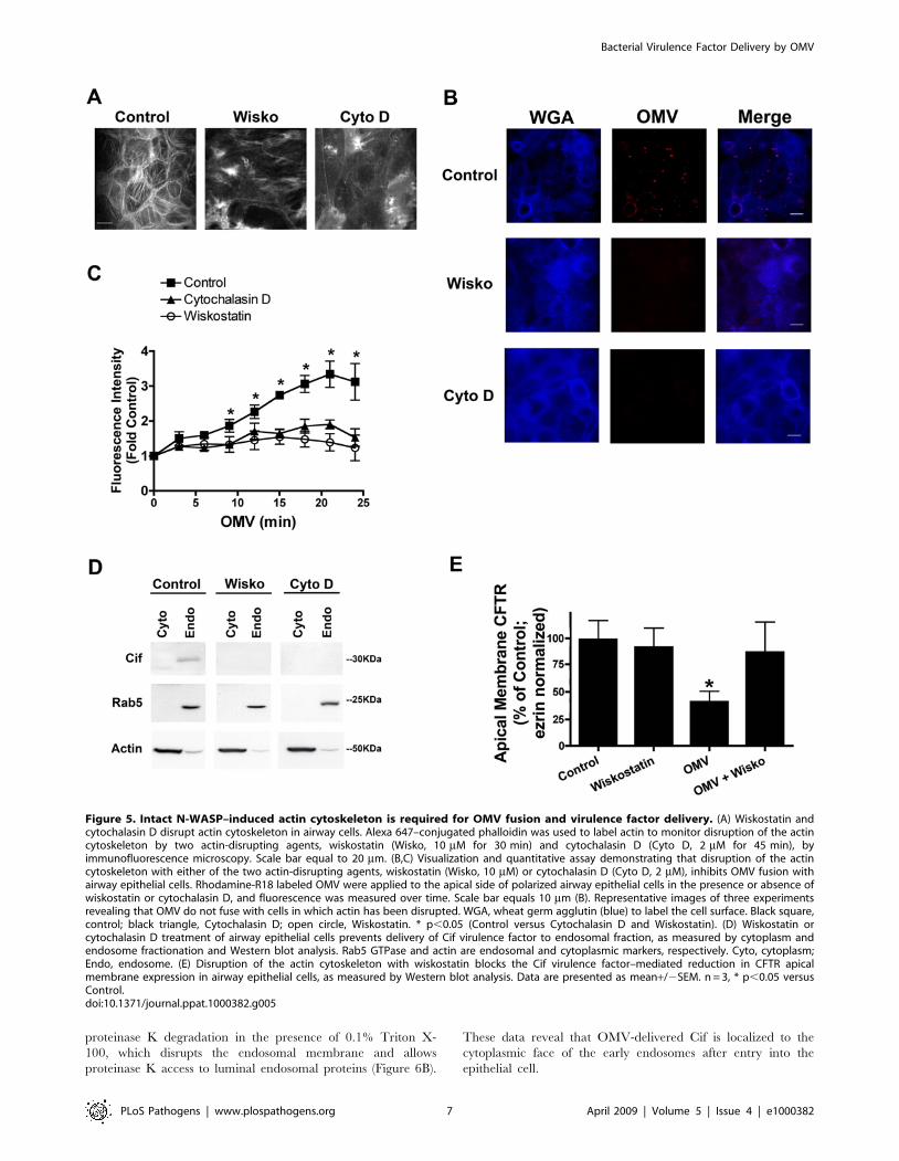
proteinase K degradation in the presence of 0.1% Triton X-
100, which disrupts the endosomal membrane and allows
proteinase K access to luminal endosomal proteins (Figure 6B).
These data reveal that OMV-delivered Cif is localized to the
cytoplasmic face of the early endosomes after entry into the
epithelial cell.
Figure 5. Intact N-WASP–induced actin cytoskeleton is required for OMV fusion and virulence factor delivery. (A) Wiskostatin andcytochalasin D disrupt actin cytoskeleton in airway cells. Alexa 647–conjugated phalloidin was used to label actin to monitor disruption of the actincytoskeleton by two actin-disrupting agents, wiskostatin (Wisko, 10 mM for 30 min) and cytochalasin D (Cyto D, 2 mM for 45 min), byimmunofluorescence microscopy. Scale bar equal to 20 mm. (B,C) Visualization and quantitative assay demonstrating that disruption of the actincytoskeleton with either of the two actin-disrupting agents, wiskostatin (Wisko, 10 mM) or cytochalasin D (Cyto D, 2 mM), inhibits OMV fusion withairway epithelial cells. Rhodamine-R18 labeled OMV were applied to the apical side of polarized airway epithelial cells in the presence or absence ofwiskostatin or cytochalasin D, and fluorescence was measured over time. Scale bar equals 10 mm (B). Representative images of three experimentsrevealing that OMV do not fuse with cells in which actin has been disrupted. WGA, wheat germ agglutin (blue) to label the cell surface. Black square,control; black triangle, Cytochalasin D; open circle, Wiskostatin. * p,0.05 (Control versus Cytochalasin D and Wiskostatin). (D) Wiskostatin orcytochalasin D treatment of airway epithelial cells prevents delivery of Cif virulence factor to endosomal fraction, as measured by cytoplasm andendosome fractionation and Western blot analysis. Rab5 GTPase and actin are endosomal and cytoplasmic markers, respectively. Cyto, cytoplasm;Endo, endosome. (E) Disruption of the actin cytoskeleton with wiskostatin blocks the Cif virulence factor–mediated reduction in CFTR apicalmembrane expression in airway epithelial cells, as measured by Western blot analysis. Data are presented as mean+/2SEM. n = 3, * p,0.05 versusControl.doi:10.1371/journal.ppat.1000382.g005
Bacterial Virulence Factor Delivery by OMV
PLoS Pathogens | www.plospathogens.org 7 April 2009 | Volume 5 | Issue 4 | e1000382

To determine if OMV-delivered Cif enters the host cell
cytoplasm by penetrating the membrane of endosomal vesicles,
cells were treated with ammonium chloride, a lysosomotropic drug
that inhibits vesicle acidification, and thereby inhibits the
movement of virulence factors from endosomal vesicles into the
cytoplasm [3]. Ammonium chloride had no effect on the ability of
Proteinase K to decrease the amount of Cif in the endosomal
fractions (Figure 6C), indicating that the Cif virulence factor does
not reach the cytoplasm via penetrating intracellular vesicular
membranes.
Figure 6. Cif virulence factor is delivered to the cytoplasm and localizes to the cytoplasmic face of early endosomes. (A) Cif localizesto the early endosomal (Rab5 GTPase, early endosomal antigen (EEA)-1 labeled) compartment after entry into airway epithelial cells. Airway epithelialcells were treated with OMV for 10 min, cells lysed, and endosomes were purified. Cif was immunoprecipitated from the endosomal fraction, andWestern blot analysis was performed for Rab5 GTPase and EEA-1, early endosomal markers. IgG IP is a non-immune control immunoprecipitationexperiment. (B) The ability of proteinase K (PK) to degrade Cif from the early endosomal fraction reveals that the Cif virulence factor localizes to thecytoplasmic face of early endosomes, as measured by Western blot analysis. The transferrin receptor serves as a control for a luminal endosomalprotein marker, which is only exposed to proteinase K after treatment with Triton X-100 (TX). (C) Cif entry into airway epithelial cells is not altered bydisruption of endosomal acidification by NH4Cl (5 mM). Rab5 GTPase and actin are endosomal and cytoplasmic markers, respectively. (D) Entry of Cifvirulence factor into airway cells is unaffected by inhibition of retrograde transport with Brefeldin A (1 mM), as measured by Western blot analysis.Rab5 GTPase and actin are endosomal and cytoplasmic markers, respectively. (E) Retrograde transport to the endoplasmic reticulum is not requiredfor Cif to reduce plasma membrane CFTR in airway cells. Airway epithelial cells pretreated with Brefeldin A (1 mM), which inhibits retrogradetransport, were treated with OMV. Brefeldin A had no effect on the ability of Cif to reduce plasma membrane CFTR, as measured by Western blotanalysis. Data are presented as mean+/2SEM. n = 3, * p,0.05 versus Control.doi:10.1371/journal.ppat.1000382.g006
Bacterial Virulence Factor Delivery by OMV
PLoS Pathogens | www.plospathogens.org 8 April 2009 | Volume 5 | Issue 4 | e1000382
Some intracellular bacteria and virulence factors move through
the retrograde pathway from endosomes, to the Golgi apparatus
and endoplasmic reticulum, from which they enter the host
cytoplasm. However, Brefeldin A, a pharmacologic inhibitor of
retrograde transport, had no effect on the entry of the Cif into the
airway cell and the appearance of Cif in the endosomal fraction
(Figure 6D), or the Cif-mediated reduction in apical membrane
CFTR abundance (Figure 6E). Thus, our data demonstrate that
OMV deliver Cif directly to the host cytoplasm rather than
requiring passage across an endosomal membrane or through the
retrograde transport pathway.
Interestingly, PlcH and alkaline phosphatase also localized to the
endosomes after entry into the airway epithelial cells, whereas b-
lactamase was detected in the cytoplasmic fraction, as determined by
subcellular fractionation and Western blot analysis (data not shown).
Thus, virulence factors with differing functions are distributed to
different subcellular locations after entry into the host cytoplasm.
OMVs are a physiological delivery mechanism forsecreted virulence factors
We propose that rather than secretion of virulence factors into
the surrounding medium, OMV are a physiologically- and
clinically-relevant mechanism utilized by Gram-negative bacteri-
um, in particular P. aeruginosa, to deliver secreted products into the
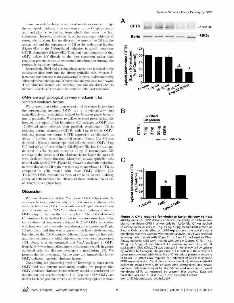
host cell. In support of this hypothesis, Cif packaged in OMV was
17,000-fold more effective than purified, recombinant Cif in
reducing plasma membrane CFTR, with 3 ng of Cif in OMV-
reducing plasma membrane CFTR expression as effectively as
50 mg of purified, recombinant Cif protein (Figure 7A). Cif was
detected in lysates of airway epithelial cells exposed to OMV (3 ng
Cif) and 50 mg of recombinant Cif (Figure 7B), but Cif was not
detected in cells exposed to up to 10 ng of recombinant Cif,
correlating the presence of the virulence factor inside the host cell
with virulence factor function. Moreover, airway epithelial cells
treated with lysed OMV (Figure S2) showed a dramatic reduction
in the ability of the Cif toxin to reduce apical membrane CFTR, as
compared to cells treated with intact OMV (Figure 7C).
Therefore, OMV-mediated delivery of virulence factors to airway
epithelial cells increases the efficacy of these virulence factors in
altering host cell physiology.
Discussion
We have demonstrated that P. aeruginosa OMV deliver multiple
virulence factors, simultaneously, into host airway epithelial cells
via a mechanism of OMV fusion with host cell lipid raft machinery
and trafficking via an N-WASP induced actin pathway to deliver
OMV cargo directly to the host cytoplasm. The OMV-delivered
Cif virulence factor is then localized to the cytoplasmic face of the
early endosomal compartment (Figure 8). E. coli OMV association
with host cells had previously been shown to be sensitive to Filipin
III treatment, and thus was proposed to be lipid raft-dependent,
but whether the OMV actually delivered cargo into the host cells
and the mechanism by which this occurred was not characterized
[15]. Fiocca et al. demonstrated that VacA packaged in OMV
from H. pylori was internalized into a cytoplasmic vacuole in gastric
epithelial cells, but did not investigate a mechanism [29]. We
propose the first mechanism for the entry and intracellular fate of
OMV-delivered bacterial virulence factors.
Considering the pioneering work of Beveridge to characterize
OMV and our current mechanistic studies, we propose that
OMV-mediated virulence factor delivery should be considered for
designation as a secretion system [4–7]. Like the T3SS, OMV can
deliver bacterial proteins directly to the host cell cytoplasm without
Figure 7. OMV required for virulence factor delivery to hostairway cells. (A) OMV delivery enhances the ability of Cif to reduceplasma membrane CFTR in airway cells by 17,000-fold. Cif was appliedto airway epithelial cells as 1 ng, 10 ng, 50 mg recombinant protein or3 ng in OMV, and its effect on CFTR expression at the apical plasmamembrane was measured by Western blot analysis. (B) Cif was detectedin airway cells treated with 50 mg Cif or 3 ng Cif packaged in OMV.Airway epithelial cells were treated with vehicle (Control-CTRL), 1 ng,10 ng, or 50 mg of recombinant Cif protein, or with 3 ng of Cifpackaged in OMV (OMV). Cif was detected in the airway cell cytoplasmby Western blot analysis. The presence of Cif protein in the airway cellcytoplasm correlated with the ability of Cif to reduce plasma membraneCFTR (A). (C) Intact OMV required for reduction of apical membraneCFTR expression (i.e., Cif virulence factor function). Airway epithelialcells were treated with OMV or lysed OMV components, and airwayepithelial cells were assayed for the Cif-mediated reduction in apicalmembrane CFTR, as measured by Western blot analysis. Data arepresented as mean+/2SEM. n = 3, * p,0.05 versus Control.doi:10.1371/journal.ppat.1000382.g007
Bacterial Virulence Factor Delivery by OMV
PLoS Pathogens | www.plospathogens.org 9 April 2009 | Volume 5 | Issue 4 | e1000382
releasing the naked bacterial proteins into the extracellular
environment where they could be degraded by secreted proteases
[30–32]. OMV deliver fully-folded, enzymatically-active secreted
virulence factors into host cells, ready for immediate action upon
delivery. By delivering multiple, active OMV-packaged virulence
factors, the pathogen may be able to impact the host on multiple
levels. For example, simultaneously altering epithelial cell function
by perturbing surfactant abundance or tight junction integrity, and
the innate immune response to bacteria by stimulating pro-
inflammatory cytokine production [14,33–36]. Based on our
studies in P. aeruginosa and published reports of OMV production
by E. coli, H. pylori, A. actinomycetemcomitans, V. cholerae and N.
meningitidis, it is likely that other bacteria package multiple secreted
virulence factors in OMV for efficient transfer to host cells and
thus, the studies proposed here likely represent a general strategy
utilized by Gram-negative bacteria in their interactions with the
host [10–12,37,38].
In contrast to known secretion systems, OMV-mediated direct
delivery of bacterial proteins to the host can occur at a distance,
and in the absence of bacteria, thus obviating the need for the
pathogen to interact directly with the host cell to cause cellular
cytotoxicity and alter host cell biology to promote colonization.
Furthermore, OMV can deliver bacterial factors across host
barriers, such as mucus layers. We believe that our work should
prompt those studying bacterial pathogens to reconsider how
secreted virulence factors impact host cells. That is, our data
suggest that secreted virulence factors are not released individually
into the surrounding milieu where they may randomly contact the
surface of the host cell, but are released in a strategic manner,
packaged with multiple virulence factors in OMV for coordinated
delivery directly into the host cell cytoplasm. It is also possible that
OMV provide a mechanism for delivering a concentrated bolus
of virulence factors to the host, instead of individual toxins being
delivered one at a time to the host cell. Moreover, OMV-
mediated, long distance delivery of virulence factors might help
explain observations such as, bacterial colonization of catheters
causing systemic symptoms in kidney dialysis patients, ocular
keratitis occurring in patients who do not have cultivatable
pathogens, and the significant lung damage in cystic fibrosis,
bronchiectasis, and chronic obstructrive pulmonary disease
patients resulting from chronic infections with P. aeruginosa
suspended in mucus above the airway epithelium. This
mechanism of OMV-mediated protein secretion is reminiscent
of the long distance delivery of signaling proteins between and
among eukaryotic cells via exosomes [39], and may represent a
general protein secretion strategy used by both pathogen and
host.
Materials and Methods
Antibodies and reagentsThe antibodies used were: rabbit anti-Cif antibody (Covance
Research Products, Denver, Pa [16]); rabbit anti-OprF antibody (a
generous gift from Nobuhiko Nomura, Graduate School of Life
and Environmental Sciences, University of Tsukuba); rabbit anti-
pilus antibody (a generous gift from Michael Zegans, Dartmouth
Medical School); goat anti-phospholipase C-H antibody (a
generous gift from Michael Vasil); mouse anti-human CFTR C-
terminus antibody (clone 24-1; R&D systems, Minneapolis, MN);
mouse anti-CFTR antibody (clone M3A7; Upstate Biotechnology,
Lake Placid, NY); mouse anti-EEA1 antibody, mouse anti-ezrin
antibody, mouse anti-flotillin-1 antibody, mouse anti-Rab5
antibody, mouse anti-actin antibody (BD Biosciences, San Jose,
CA); cholera toxin B subunit-FITC (Sigma-Aldrich, St. Louis,
MO); rabbit anti-GPIp137 antibody (Abgent, San Diego, CA);
Alexa 647-conjugated phalloidin (Molecular Probes, Carlsbad,
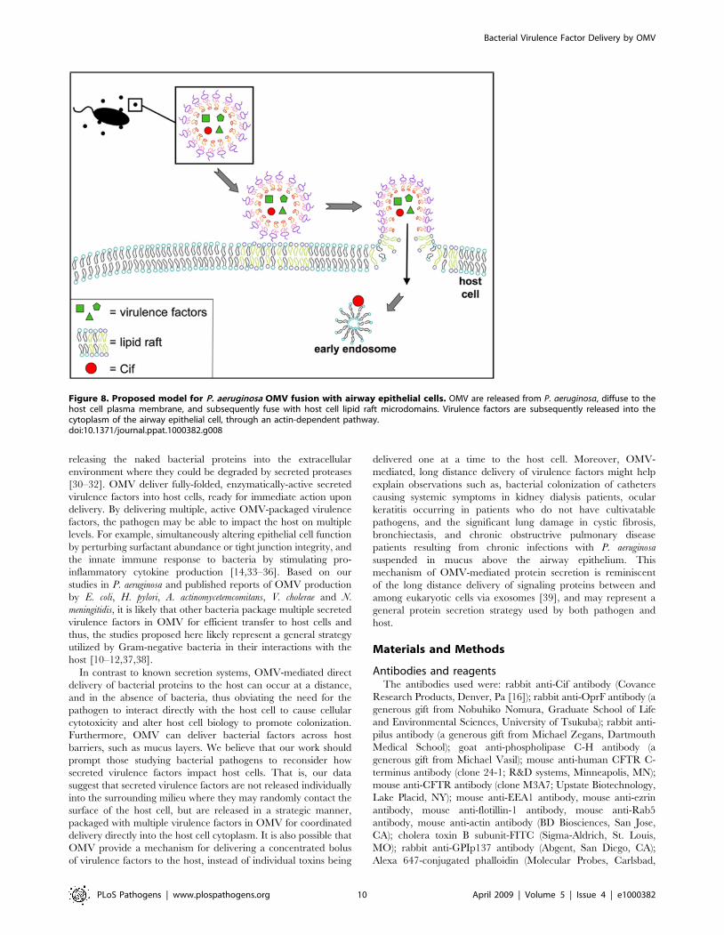
Figure 8. Proposed model for P. aeruginosa OMV fusion with airway epithelial cells. OMV are released from P. aeruginosa, diffuse to thehost cell plasma membrane, and subsequently fuse with host cell lipid raft microdomains. Virulence factors are subsequently released into thecytoplasm of the airway epithelial cell, through an actin-dependent pathway.doi:10.1371/journal.ppat.1000382.g008
Bacterial Virulence Factor Delivery by OMV
PLoS Pathogens | www.plospathogens.org 10 April 2009 | Volume 5 | Issue 4 | e1000382
CA); rabbit anti-Rab4 antibody, rabbit anti-Rab7 antibody (Santa
Cruz Biotechnology, Santa Cruz, CA); mouse anti-Rab11
antibody, mouse anti-transferrin receptor antibody (Zymed, San
Francisco, CA); mouse anti-b lactamase antibody (Novus Biologicals,
Littleton, CO); rabbit anti-alkaline phosphatase antibody (GeneTex,
Inc., San Antonio, TX) and horseradish peroxidase-conjugated goat
anti-mouse and goat anti-rabbit secondary antibodies (Bio-Rad,
Hercules, CA). Other reagents include: Filipin III complex,
ammonium chloride, Optiprep, proteinase K, and cytochalasin D
(Sigma-Aldrich), wiskostatin (Calbiochem, San Diego, CA), Triton
X-100 (Bio-Rad, Hercules, CA). All antibodies and reagents were
used at the concentrations recommended by the manufacturers or as
indicated in the figure legends.
Cell cultureTwo airway epithelial cell lines were studied to examine outer
membrane vesicle fusion and toxin delivery to host epithelial cells.
First, human bronchial epithelial CFBE cells (DF508/DF508) were
stably transduced with WT-CFTR (generous gift from Dr. J. P.
Clancy, University of Alabama at Birmingham, Birmingham, AL;
hereafter referred to as airway epithelial cells) [40]. CFBE WT-
CFTR cells were polarized on 24-mm transwell permeable
supports (0.4-mm-pore size; Corning, Corning, NY) coated with
vitrogen plating medium containing human fibronectin, as
described previously [41]. Second, human airway epithelial cells
(Calu-3) were obtained from the American Type Culture
Collection (Manassas, VA) and polarized on 24-mm transwell
permeable supports, as described previously [42].
Pseudomonas aeruginosa culturesLysogeny broth (LB) was inoculated with P. aeruginosa strain
UCBPP-PA14 (PA14) [43] and cultures were prepared as
previously reported [16].
Outer membrane vesicle purificationOMV were purified using a differential centrifugation and
discontinuous Optiprep gradient protocol adapted from Bauman et
al. [14] OMV were lysed, when noted, with 100 mM EDTA at
37uC for 60 minutes.
Cell compartment fractionationTo study the localization of the Cif toxin after OMV fusion with
the airway epithelial cell, differential centrifugation and fraction-
ation techniques were used to isolate cytosolic and early
endosomal compartments. Early endosomes were isolated using
a protocol adapted from Butterworth et al. [44].
ImmunoprecipitationTo characterize proteins interacting with the Cif toxin in lipid
raft microdomains, Cif was immunoprecipitated from airway
epithelial cell lipid raft fractions by methods described previously
[45].
Detergent-resistant membrane fractionationTo determine if OMV fuse with lipid raft microdomains of the
host, detergent-resistant membranes were purified from airway
epithelial cells that had been exposed to OMV. These studies were
performed using a discontinuous Optiprep gradient in a protocol
adapted from Pike et al. [46].
OMV fusion assayTo monitor the fusion of OMV with airway epithelial cells,
OMV were fluorescently labeled with a probe that fluoresces upon
membrane fusion. OMV purified with the method described
above were resuspended in labeling buffer (50 mM Na2CO3,
100 mM NaCl, pH 9.2). Rhodamine isothiocyanate B-R18
(Molecular Probes), which integrates in the membrane of the
OMV, was added at a concentration of 1 mg/ml for 1 hour at
25uC, followed by ultracentrifugation at 52,0006g for 30 min at
4uC. Rhodamine isothiocyanate B-R18 fluorescence is quenched
at high concentrations in bilayer membranes, and fluorescence is
dequenched when the probe is diluted upon vesicle fusion.
Subsequently, rhodamine labeled-OMV were resuspended in PBS
(0.2 M NaCl) and pelleted at 52,0006g for 30 min a 4uC. After a
final centrifugation step, the labeled-OMV were resuspended in
1 ml PBS (0.2 M NaCl) containing a protease inhibitor cocktail
tablet (Complete Protease Inhibitor Tablet, Roche). Labeled-
OMV were applied to the apical side of airway epithelial cells at
1:4 dilution of labeled-OMV to Earle’s Minimal Medium (MEM,
Invitrogen) and fluorescence was detected over time as indicated
on a fluorescent plate reader (Ex 570 nm; Em 595 nm).
Fluorescence intensity was normalized for fluorescence detected
by labeled-OMV in the absence of airway epithelial cells at the
indicated time points.
Confocal microscopyTo visualize the fusion and localization of OMV with airway
epithelial cells, rhodamine R18-labeled OMV (see OMV Fusion
Assay method) were applied to the apical membrane of cells and
confocal sections were captured over time. Airway epithelial cells
were seeded at 0.16106 on collagen-coated, glass-bottom MatTek
dishes (MatTek, Ashland, MA) and grown for 6–7 days in culture
at 37uC. For wheat germ agglutin (WGA, which labels the plasma
membrane) studies, nonpermeabilized cells were incubated for
5 minutes with Alexa-647 WGA (1 mg/ml, 37uC; Molecular
Probes) following 15-minute vesicle incubation. Z-stacks of all
labeled cells were acquired with a Nikon Sweptfield confocal
microscope (Apo TIRF 606 oil immersion 1.49 NA objective)
fitted with a QuantEM:512sc camera (Photometrics, Tuscon, AZ)
and Elements 2.2 software (Nikon, Inc.). For OMV fusion
experiments, a single confocal section (0.4 mm) at the apical
membrane of the airway epithelial cells is presented. Experiments
were repeated three times, with five fields imaged for each
experiment.
Cell-surface biotinylations and Western blot analysisTo examine the effect of OMV on the apical membrane
expression of CFTR, cell surface biotinylation was performed as
described in detail previously by our laboratory [41]. Protein band
intensity was analyzed as described previously using NIH image
software, version 1.63 (Wayne Rasband, NIH, USA; http://rsb.
info.nih.gov).
Cytotoxicity assayTo determine if P. aeruginosa OMV are cytotoxic to airway cells,
cells were incubated with OMV in serum-free media for the
indicated time points. Cytotoxicity was measured using the
CellTiter 96 AQueous One Solution Reagent (Promega, Madison,
WI), according to the manufacturer’s protocol.
Data analysis and statisticsStatistical analysis of the data was performed using Graphpad
Prism version 4.0 for Macintosh (Graphpad, San Diego, CA).
Means were compared using a Students t-test or one-way
ANOVA, followed by a Tukey-Kramer post hoc test using a
95% confidence interval. Data are expressed as means+/2SEM.
Bacterial Virulence Factor Delivery by OMV
PLoS Pathogens | www.plospathogens.org 11 April 2009 | Volume 5 | Issue 4 | e1000382

Accession numbersCif (PA2934, NP 251624.1); PlcH (PA0844, YP 792433.1);
alkaline phosphatase (PA3296, YP 789857.1); b-lactamase
(PA1797, YP 791446.1); N-WASP (NP 003932); Omp85
(PA3648, YP 789516); GPIp137 (NP 005889).
Supporting Information
Figure S1 The Cif virulence factor is packaged in P. aeruginosa
OMV. From an overnight P. aeruginosa PA14 culture, Optiprep
density gradient centrifugation was utilized to purify OMV from
the bacteria and possible contaminants, including pilus (PilA).
Purified OMVs retrieved from fractions 2 and 3 were pooled for
use in all experiments described. Experiment repeated three times;
representative blot shown.
Found at: doi:10.1371/journal.ppat.1000382.s001 (0.35 MB TIF)
Figure S2 EDTA effectively lyses OMV. EDTA (0.1 M)
disrupted OMV membranes to allow proteinase K (PK)-mediated
degradation of Cif, an intravesicular OMV component, as
measured by Western blot analysis. Experiment repeated three
times; representative blot shown.
Found at: doi:10.1371/journal.ppat.1000382.s002 (0.18 MB TIF)
Figure S3 Cif virulence factor is an intravesicular OMV
component. Isolated OMV treated with Proteinase K (PK:
100 mg/ml) for 1 h at 37uC to degrade proteins on the exterior
of OMV. Dcif: OMV purified from a P. aeruginosa Dcif mutant
strain. Experiment repeated three times; representative blot
shown.
Found at: doi:10.1371/journal.ppat.1000382.s003 (0.35 MB TIF)
Figure S4 Cif does not localize to Rab4, Rab7, or Rab11-
labeled endosomes. Cif does not localize to the sorting endosomal
(Rab4 GTPase-labeled), late endosomal (Rab7 GTPase-labeled),
or recycling endosomal (Rab11 GTPase-labeled) compartments
after entry into airway epithelial cells. Airway epithelial cells were
treated with OMV for 10 min, cells lysed, and endosomes were
purified. Cif was immunoprecipitated from the endosomal fraction
and Western blot analysis was performed for Rab4, 7, and 11
GTPases. IgG IP is a non-immune control immunoprecipitation
experiment.
Found at: doi:10.1371/journal.ppat.1000382.s004 (0.60 MB TIF)
Acknowledgments
We thank Dr. Nobuhiko Nomura and Dr. Michael Zegans for their
generous gifts of Omp85 and PilA antibodies, respectively. We also express
thanks to Dr. Deborah Hogan for her critical analysis of the manuscript.
Author Contributions
Conceived and designed the experiments: JMB DPM GAO BAS.
Performed the experiments: JMB DPM BAC SY. Analyzed the data:
JMB. Contributed reagents/materials/analysis tools: GAO. Wrote the
paper: JMB GAO BAS.
References
1. Weinstein RA (1998) Nosocomial infection update. Emerg Infect Dis 4:
416–420.
2. Ernst JD (2000) Bacterial Inhibition of Phagocytosis. Cell Microbiol 2: 379–386.
3. Blanke SR (2006) Portals and Pathways: Principles of Bacterial Toxin Entry into
Host Cells. Microbe 1: 26–32.
4. Nguyen TT, Saxena A, Beveridge TJ (2003) Effect of surface lipopolysaccharide
on the nature of membrane vesicles liberated from the Gram-negative bacterium
Pseudomonas aeruginosa. J Electron Microsc (Tokyo) 52: 465–469.
5. Kadurugamuwa JL, Beveridge TJ (1997) Natural release of virulence factors in
membrane vesicles by Pseudomonas aeruginosa and the effect of aminoglycoside
antibiotics on their release. J Antimicrob Chemother 40: 615–621.
6. Kadurugamuwa JL, Beveridge TJ (1996) Bacteriolytic effect of membrane
vesicles from Pseudomonas aeruginosa on other bacteria including pathogens:
conceptually new antibiotics. J Bacteriol 178: 2767–2774.
7. Kadurugamuwa JL, Beveridge TJ (1995) Virulence factors are released from
Pseudomonas aeruginosa in association with membrane vesicles during normal
growth and exposure to gentamicin: a novel mechanism of enzyme secretion.
J Bacteriol 177: 3998–4008.
8. Kuehn MJ, Kesty NC (2005) Bacterial outer membrane vesicles and the host-
pathogen interaction. Genes Dev 19: 2645–2655.
9. Mashburn LM, Whiteley M (2005) Membrane vesicles traffic signals and
facilitate group activities in a prokaryote. Nature 437: 422–425.
10. Kato S, Kowashi Y, Demuth DR (2002) Outer membrane-like vesicles secreted
by Actinobacillus actinomycetemcomitans are enriched in leukotoxin. Microb Pathog
32: 1–13.
11. Kesty NC, Kuehn MJ (2004) Incorporation of heterologous outer membrane
and periplasmic proteins into Escherichia coli outer membrane vesicles. J Biol
Chem 279: 2069–2076.
12. Wai SN, Lindmark B, Soderblom T, Takade A, Westermark M, et al. (2003)
Vesicle-mediated export and assembly of pore-forming oligomers of the
enterobacterial ClyA cytotoxin. Cell 115: 25–35.
13. Montes LR, Ibarguren M, Goni FM, Stonehouse M, Vasil ML, et al. (2007)
Leakage-free membrane fusion induced by the hydrolytic activity of PlcHR(2), a
novel phospholipase C/sphingomyelinase from Pseudomonas aeruginosa. Biochim
Biophys Acta 1768: 2365–2372.
14. Bauman SJ, Kuehn MJ (2006) Purification of outer membrane vesicles from
Pseudomonas aeruginosa and their activation of an IL-8 response. Microbes Infect 8:
2400–2408.
15. Kesty NC, Mason KM, Reedy M, Miller SE, Kuehn MJ (2004) Enterotoxigenic
Escherichia coli vesicles target toxin delivery into mammalian cells. Embo J 23:
4538–4549.
16. MacEachran DP, Ye S, Bomberger JM, Hogan DA, Swiatecka-Urban A, et al.
(2007) The Pseudomonas aeruginosa secreted protein PA2934 decreases apical
membrane expression of the cystic fibrosis transmembrane conductance
regulator. Infect Immun 75: 3902–3912.
17. Huang CT, Xu KD, McFeters GA, Stewart PS (1998) Spatial patterns of
alkaline phosphatase expression within bacterial colonies and biofilms in
response to phosphate starvation. Appl Environ Microbiol 64: 1526–1531.
18. Xu KD, Stewart PS, Xia F, Huang CT, McFeters GA (1998) Spatial
physiological heterogeneity in Pseudomonas aeruginosa biofilm is determined by
oxygen availability. Appl Environ Microbiol 64: 4035–4039.
19. Vasil ML, Berka RM, Gray GL, Nakai H (1982) Cloning of a phosphate-
regulated hemolysin gene (phospholipase C) from Pseudomonas aeruginosa.
J Bacteriol 152: 431–440.
20. Vipond C, Wheeler JX, Jones C, Feavers IM, Suker J (2005) Characterization of
the protein content of a meningococcal outer membrane vesicle vaccine by
polyacrylamide gel electrophoresis and mass spectrometry. Hum Vaccin 1: 80–84.
21. Horstman AL, Kuehn MJ (2000) Enterotoxigenic Escherichia coli secretes active heat-
labile enterotoxin via outer membrane vesicles. J Biol Chem 275: 12489–12496.
22. Ye S, Maceachran DP, Hamilton JW, O’Toole GA, Stanton BA (2008)
Chemotoxicity of doxorubicin and surface expression of P-glycoprotein (MDR1)
is regulated by the Pseudomonas aeruginosa toxin Cif. Am J Physiol Cell Physiol 295:
C807–C818.
23. Worlitzsch D, Tarran R, Ulrich M, Schwab U, Cekici A, et al. (2002) Effects of
reduced mucus oxygen concentration in airway Pseudomonas infections of cystic
fibrosis patients. J Clin Invest 109: 317–325.
24. Harder T, Scheiffele P, Verkade P, Simons K (1998) Lipid domain structure of
the plasma membrane revealed by patching of membrane components. J Cell
Biol 141: 929–942.
25. Ellis JA, Luzio JP (1995) Identification and characterization of a novel protein
(p137) which transcytoses bidirectionally in Caco-2 cells. J Biol Chem 270:
20717–20723.
26. Caron E, Crepin VF, Simpson N, Knutton S, Garmendia J, et al. (2006)
Subversion of actin dynamics by EPEC and EHEC. Curr Opin Microbiol 9:
40–45.
27. McGee K, Zettl M, Way M, Fallman M (2001) A role for N-WASP in invasin-
promoted internalisation. FEBS Lett 509: 59–65.
28. Stoorvogel W, Geuze HJ, Griffith JM, Schwartz AL, Strous GJ (1989) Relations
between the intracellular pathways of the receptors for transferrin, asialoglyco-
protein, and mannose 6-phosphate in human hepatoma cells. J Cell Biol 108:
2137–2148.
29. Fiocca R, Necchi V, Sommi P, Ricci V, Telford J, et al. (1999) Release of
Helicobacter pylori vacuolating cytotoxin by both a specific secretion pathway and
budding of outer membrane vesicles. Uptake of released toxin and vesicles by
gastric epithelium. J Pathol 188: 220–226.
30. Lieberman J (2003) The ABCs of granule-mediated cytotoxicity: new weapons in
the arsenal. Nat Rev Immunol 3: 361–370.
31. Shapiro SD (1994) Elastolytic metalloproteinases produced by human
mononuclear phagocytes. Potential roles in destructive lung disease.
Am J Respir Crit Care Med 150: S160–S164.
Bacterial Virulence Factor Delivery by OMV
PLoS Pathogens | www.plospathogens.org 12 April 2009 | Volume 5 | Issue 4 | e1000382
32. Caughey GH (1994) Serine proteinases of mast cell and leukocyte granules. A
league of their own. Am J Respir Crit Care Med 150: S138–S142.
33. Azghani AO (1996) Pseudomonas aeruginosa and epithelial permeability: role of
virulence factors elastase and exotoxin A. Am J Respir Cell Mol Biol 15:
132–140.
34. Azghani AO, Bedinghaus T, Klein R (2000) Detection of elastase from
Pseudomonas aeruginosa in sputum and its potential role in epithelial cell
permeability. Lung 178: 181–189.
35. Luberto C, Stonehouse MJ, Collins EA, Marchesini N, El-Bawab S, et al. (2003)
Purification, characterization, and identification of a sphingomyelin synthase
from Pseudomonas aeruginosa. PlcH is a multifunctional enzyme. J Biol Chem 278:
32733–32743.
36. Stonehouse MJ, Cota-Gomez A, Parker SK, Martin WE, Hankin JA, et al.
(2002) A novel class of microbial phosphocholine-specific phospholipases C. Mol
Microbiol 46: 661–676.
37. Quakyi EK, Hochstein HD, Tsai CM (1997) Modulation of the biological
activities of meningococcal endotoxins by association with outer membrane
proteins is not inevitably linked to toxicity. Infect Immun 65: 1972–1979.
38. Schild S, Nelson EJ, Camilli A (2008) Immunization with Vibrio cholerae outer
membrane vesicles induces protective immunity in mice. Infect Immun 76:
4554–4563.
39. Schorey JS, Bhatnagar S (2008) Exosome function: from tumor immunology to
pathogen biology. Traffic 9: 871–881.
40. Bebok Z, Collawn JF, Wakefield J, Parker W, Li Y, et al. (2005) Failure of cAMP
agonists to activate rescued deltaF508 CFTR in CFBE41o- airway epithelialmonolayers. J Physiol 569: 601–615.
41. Swiatecka-Urban A, Brown A, Moreau-Marquis S, Renuka J, Coutermarsh B, et
al. (2005) The short apical membrane half-life of rescued DF508-cystic fibrosistransmembrane conductance regulator (CFTR) results from accelerated
endocytosis of DF508-CFTR in polarized human airway epithelial cells. J BiolChem 280: 36762–36772.
42. Swiatecka-Urban A, Boyd C, Coutermarsh B, Karlson KH, Barnaby R, et al.
(2004) Myosin VI regulates endocytosis of the cystic fibrosis transmembraneconductance regulator. J Biol Chem 279: 38025–38031.
43. Rahme LG, Ausubel FM, Cao H, Drenkard E, Goumnerov BC, et al. (2000)Plants and animals share functionally common bacterial virulence factors. Proc
Natl Acad Sci U S A 97: 8815–8821.44. Butterworth MB, Edinger RS, Ovaa H, Burg D, Johnson JP, et al. (2007) The
deubiquitinating enzyme UCH-L3 regulates the apical membrane recycling of
the epithelial sodium channel. J Biol Chem 282: 37885–37893.45. Swiatecka-Urban A, Talebian L, Kanno E, Moreau-Marquis S, Coutermarsh B,
et al. (2007) Myosin VB is required for trafficking of CFTR in RAB11A-specificapical recycling endosomes in polarized human airway epithelial cells. J Biol
Chem 282: 23725–23736.
46. Pike LJ, Han X, Gross RW (2005) Epidermal growth factor receptors arelocalized to lipid rafts that contain a balance of inner and outer leaflet lipids: a
shotgun lipidomics study. J Biol Chem 280: 26796–26804.
Bacterial Virulence Factor Delivery by OMV
PLoS Pathogens | www.plospathogens.org 13 April 2009 | Volume 5 | Issue 4 | e1000382


![kuPLoS Pathogens, 13(10), [e1006694]. DOI: 10.1371/journal.ppat.1006694 Download date: 27. okt.. 2018 RESEARCH ARTICLE miRNA independen t hepacivirus variants suggest a strong evolutionary](https://img.pdfslide.us/doc/110x75/607fcc929a7014578728d76d/ku-plos-pathogens-1310-e1006694-doi-101371-download-date-27-okt-2018.jpg)
![Metagenomic analysis of gut microbiome and resistome of ... · De et al. Gut Pathog Page 2 of 48 Background Diarrheaisaleadingcauseofmortalityaccountingfor morethan1.6milliondeathsworldwide[1].Itcauses](https://img.pdfslide.us/doc/110x75/612248476f350412e40c06ba/metagenomic-analysis-of-gut-microbiome-and-resistome-of-de-et-al-gut-pathog.jpg)