Embed Size (px)
Citation preview
Chapter 6: A Tour of the Cell
6.1 To study cells, biologists use
microscopes and the tools of biochemistry
pp. 94 - 97
Light Microscope: Visible light is passed through specimen and then through glass lenses, which refract the light so that the image of the specimen is magnified.◦ Advantages: Does not kill specimen◦ Disadvantages: Cannot resolve detail finer than .2 micrometers; cellular
structures are too small to be resolved by LM
Electron Microscope: Focuses beam of electrons through specimen or onto its surface.◦ Scanning Electron Microscope (SEM) – Electron beam scans the surface
of the sample, which is usually coated with gold◦ Transmission Electron Microscope (TEM) – Aims electron beam through
thin section of the specimen (stained with heavy metals that attach to certain organelles) to produce an image of the cell’s ultrastructure
◦ Disadvantages: Methods used to prepare specimen often kill the cells and may introduce artifacts (illusions of structural features that don’t really exist)
Microscopy
Takes apart and separates major organelles and other subcellular structures from one another.
Uses centrifuge. Spins test tubes holding mixtures of cells at various speeds.
Low speeds produce pellets with larger components.
High speeds produce pellets with smaller components.
Through fractionation, biologists discovered that mitochondria are the sites of cellular respiration.
Cell Fractionation
6.2 Eukaryotic cells have internal membranes that
compartmentalize their functions
pp. 98 - 102
Prokaryotes Both Eukaryotes
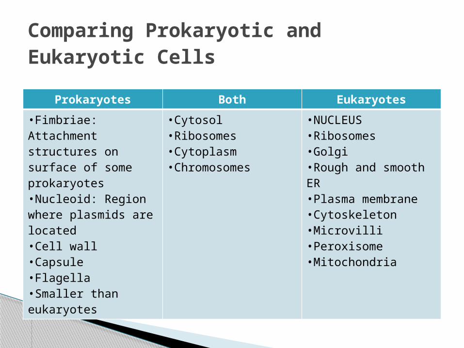
•Fimbriae: Attachment structures on surface of some prokaryotes•Nucleoid: Region where plasmids are located•Cell wall•Capsule•Flagella•Smaller than eukaryotes
•Cytosol•Ribosomes•Cytoplasm•Chromosomes
•NUCLEUS•Ribosomes•Golgi•Rough and smooth ER•Plasma membrane•Cytoskeleton•Microvilli•Peroxisome•Mitochondria
Comparing Prokaryotic and Eukaryotic Cells
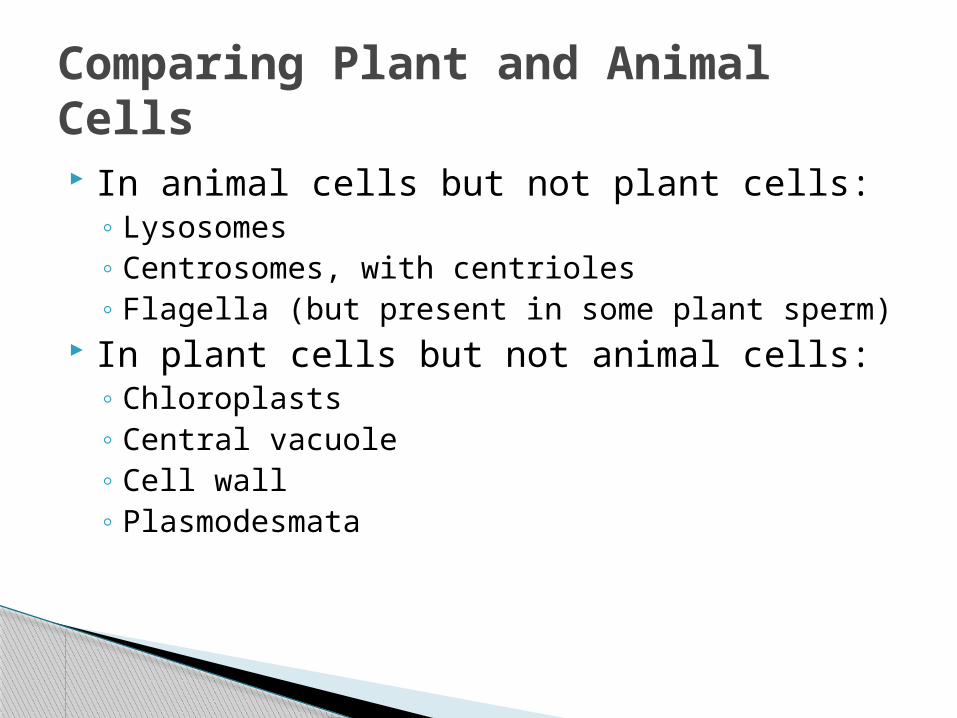
In animal cells but not plant cells:◦ Lysosomes◦ Centrosomes, with centrioles◦ Flagella (but present in some plant sperm)
In plant cells but not animal cells:◦ Chloroplasts◦ Central vacuole◦ Cell wall◦ Plasmodesmata
Comparing Plant and Animal Cells
6.3 The eukaryotic cell’s genetic instructions are
housed in the nucleus and carried out by the
ribosomespp. 102 - 104
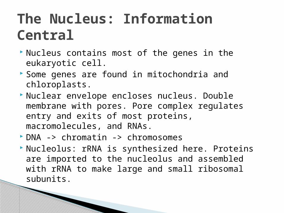
Nucleus contains most of the genes in the eukaryotic cell.
Some genes are found in mitochondria and chloroplasts.
Nuclear envelope encloses nucleus. Double membrane with pores. Pore complex regulates entry and exits of most proteins, macromolecules, and RNAs.
DNA -> chromatin -> chromosomes Nucleolus: rRNA is synthesized here. Proteins are
imported to the nucleolus and assembled with rRNA to make large and small ribosomal subunits.
The Nucleus: Information Central
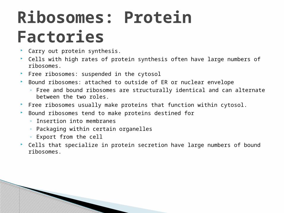
Carry out protein synthesis. Cells with high rates of protein synthesis often have large numbers of ribosomes. Free ribosomes: suspended in the cytosol Bound ribosomes: attached to outside of ER or nuclear envelope
◦ Free and bound ribosomes are structurally identical and can alternate between the two roles.
Free ribosomes usually make proteins that function within cytosol. Bound ribosomes tend to make proteins destined for
◦ Insertion into membranes◦ Packaging within certain organelles◦ Export from the cell
Cells that specialize in protein secretion have large numbers of bound ribosomes.
Ribosomes: Protein Factories
6.4 The endomembrane system regulates
protein traffic and performs metabolic functions in the cell
pp. 104 - 108
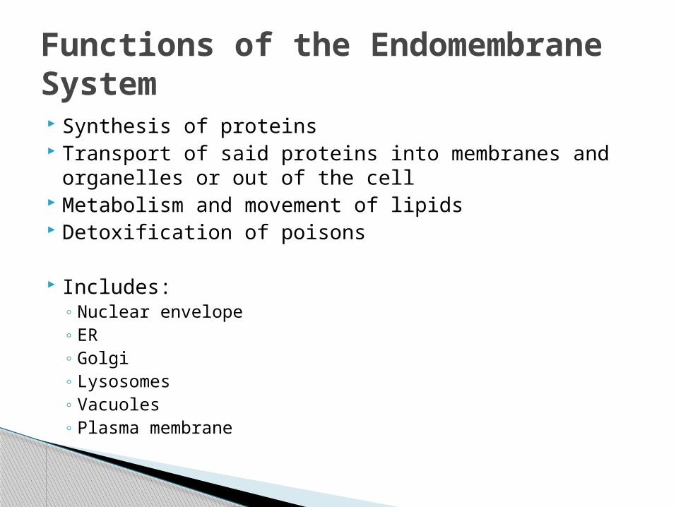
Synthesis of proteins Transport of said proteins into membranes and
organelles or out of the cell Metabolism and movement of lipids Detoxification of poisons
Includes:◦ Nuclear envelope◦ ER◦ Golgi◦ Lysosomes◦ Vacuoles◦ Plasma membrane
Functions of the Endomembrane System
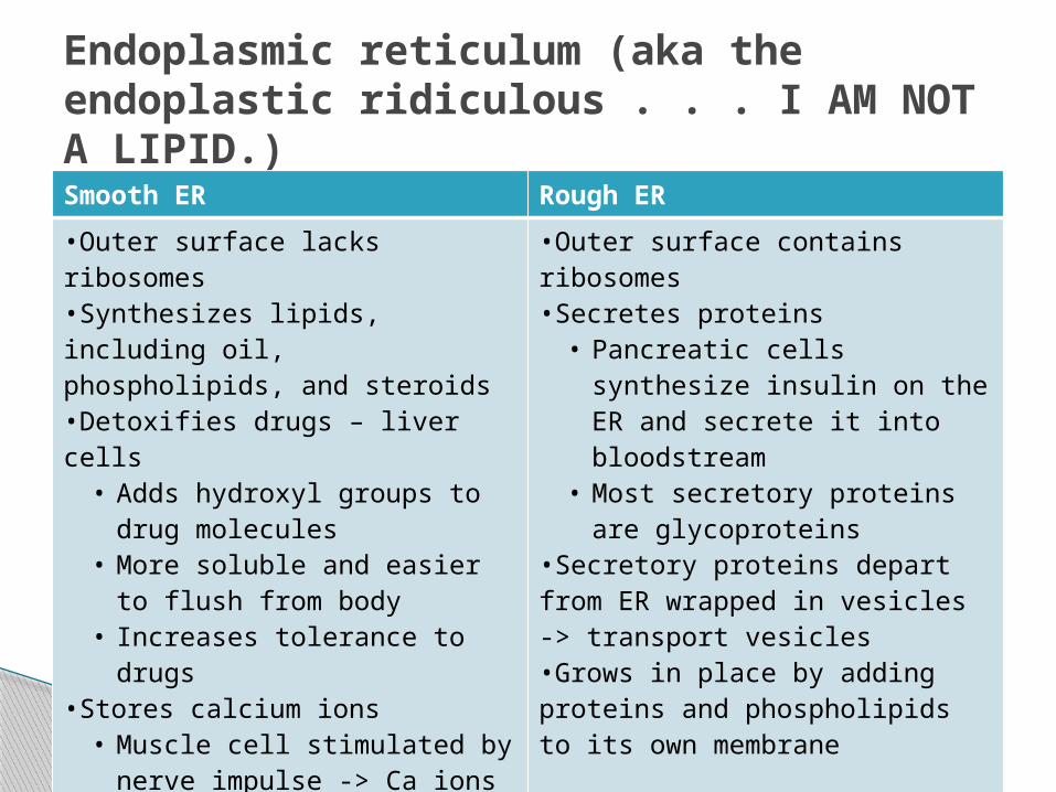
Smooth ER Rough ER
•Outer surface lacks ribosomes•Synthesizes lipids, including oil, phospholipids, and steroids•Detoxifies drugs – liver cells• Adds hydroxyl groups to drug
molecules• More soluble and easier to
flush from body• Increases tolerance to drugs
•Stores calcium ions• Muscle cell stimulated by
nerve impulse -> Ca ions rush back across ER membrane into cytosol and trigger contraction of the muscle cell
•Outer surface contains ribosomes•Secretes proteins• Pancreatic cells synthesize
insulin on the ER and secrete it into bloodstream
• Most secretory proteins are glycoproteins
•Secretory proteins depart from ER wrapped in vesicles -> transport vesicles•Grows in place by adding proteins and phospholipids to its own membrane
Endoplasmic reticulum (aka the endoplastic ridiculous . . . I AM NOT A LIPID.)
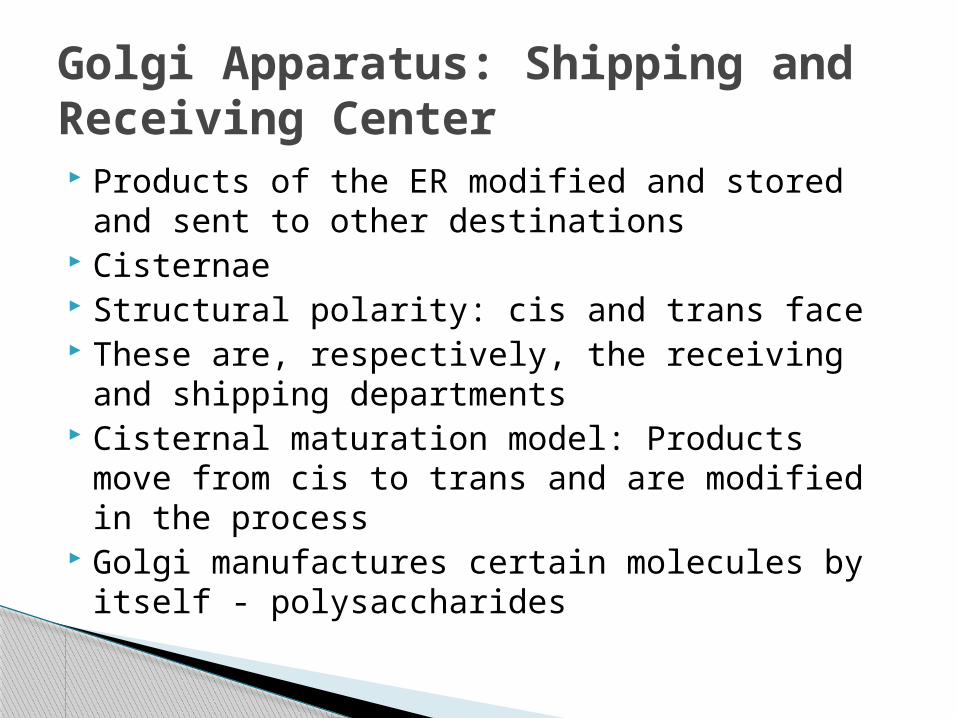
Products of the ER modified and stored and sent to other destinations
Cisternae Structural polarity: cis and trans face These are, respectively, the receiving and
shipping departments Cisternal maturation model: Products move
from cis to trans and are modified in the process
Golgi manufactures certain molecules by itself - polysaccharides
Golgi Apparatus: Shipping and Receiving Center
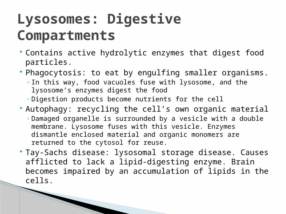
Contains active hydrolytic enzymes that digest food particles.
Phagocytosis: to eat by engulfing smaller organisms. ◦ In this way, food vacuoles fuse with lysosome, and the
lysosome’s enzymes digest the food◦ Digestion products become nutrients for the cell
Autophagy: recycling the cell’s own organic material◦ Damaged organelle is surrounded by a vesicle with a double
membrane. Lysosome fuses with this vesicle. Enzymes dismantle enclosed material and organic monomers are returned to the cytosol for reuse.
Tay-Sachs disease: lysosomal storage disease. Causes afflicted to lack a lipid-digesting enzyme. Brain becomes impaired by an accumulation of lipids in the cells.
Lysosomes: Digestive Compartments
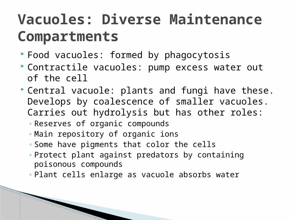
Food vacuoles: formed by phagocytosis Contractile vacuoles: pump excess water out of
the cell Central vacuole: plants and fungi have these.
Develops by coalescence of smaller vacuoles. Carries out hydrolysis but has other roles:◦ Reserves of organic compounds◦ Main repository of organic ions◦ Some have pigments that color the cells◦ Protect plant against predators by containing
poisonous compounds◦ Plant cells enlarge as vacuole absorbs water
Vacuoles: Diverse Maintenance Compartments
6.5 Mitochondria and chloroplasts change energy
from one form to anotherpp. 109 - 111
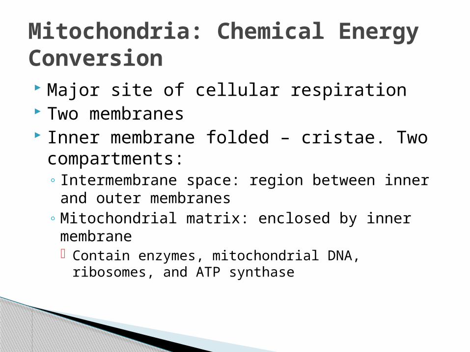
Major site of cellular respiration Two membranes Inner membrane folded – cristae. Two
compartments:◦ Intermembrane space: region between inner and
outer membranes◦ Mitochondrial matrix: enclosed by inner
membrane Contain enzymes, mitochondrial DNA, ribosomes,
and ATP synthase
Mitochondria: Chemical Energy Conversion
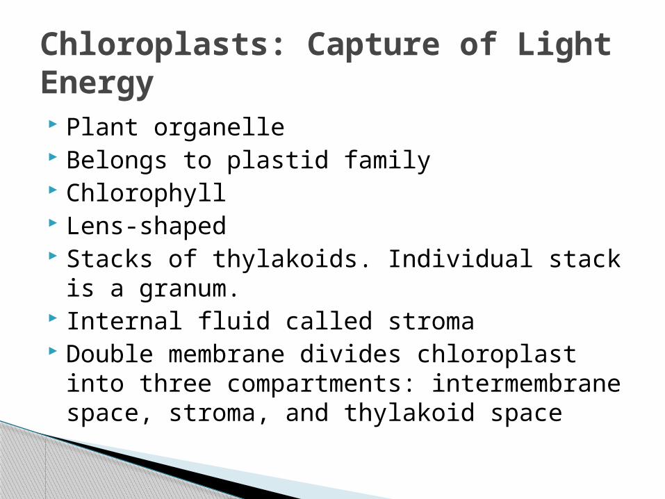
Plant organelle Belongs to plastid family Chlorophyll Lens-shaped Stacks of thylakoids. Individual stack is a
granum. Internal fluid called stroma Double membrane divides chloroplast into
three compartments: intermembrane space, stroma, and thylakoid space
Chloroplasts: Capture of Light Energy
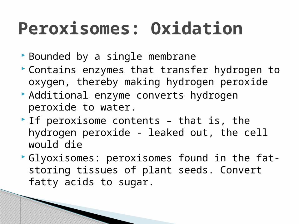
Bounded by a single membrane Contains enzymes that transfer hydrogen to
oxygen, thereby making hydrogen peroxide Additional enzyme converts hydrogen
peroxide to water. If peroxisome contents – that is, the hydrogen
peroxide - leaked out, the cell would die Glyoxisomes: peroxisomes found in the fat-
storing tissues of plant seeds. Convert fatty acids to sugar.
Peroxisomes: Oxidation
6.6 The cytoskeleton is a network of fibers that
organizes structures and activities within the cell
pp. 112 - 118
Support Motility Regulation Provides anchorage for many organelles Motility generally requires interaction
between cytoskeleton and motor proteins Cytoskeleton is composed of microtubules,
microfilaments, and intermediate filaments
Roles of the Cytoskeleton
Hollow tubes; wall consists of 13 columns of tubulin molecules
25 nm diameter with 15-nm lumen (folds) Protein subunit is tubulin, a dimer consisting
of alpha tubulin and beta tubulin. Functions:
◦ Maintenance of cell shape◦ Cell motility (as in cilia or flagella)◦ Chromosome movements in cell division◦ Organelle movements
Microtubules
Two intertwined strands of actin Diameter 7 nm Protein subunit is . . . well, actin Functions:
◦ Maintenance of cell shape◦ Changes in cell shape◦ Muscle contraction◦ Cytoplasmic streaming◦ Cell motility (as in pseudopodia)◦ Cell division (cleavage furrow formation)
Microfilaments
Fibrous proteins supercoiled into thicker cables
8 – 12 nm diameter One of several different proteins of the
keratin family, depending on cell type Functions:
◦ Maintenance of cell shape◦ Anchorage of nucleus and certain other organelles◦ Formation of nuclear lamina
Intermediate Filaments
Centrosome: Located near nucleus. Microtubule organizing center.
Centrioles: Located in the centrosome. One pair composed of nine sets of triplet microtubules arranged in a ring.
Before an animal cell divides, the centrioles replicate
Yeast cells and plant cells lack centrosomes with centrioles, but they still have microtubules.
Centrosomes and Centrioles
Cilia are the hair-like structures on the outside of the cell. Flagella are tail-like structures.
Cilia: ◦ Occur in large numbers on the cell surface◦ .25 micrometers in diameter◦ 2-20 micrometers long◦ Work more like oars – alternating power and recovery
strokes perpendicular to the cilium’s axis Flagella:
◦ Same diameter as cilia but 10-200 micrometers long◦ Usually limited to just one or two per cell◦ Undulating motion that generates force in same direction
as the flagellum’s axis.
Cilia and Flagella

6.7 Extracellular components and
connections between cells help coordinate
cellular activitiespp. 118 - 122
Protects plant cell Maintains its shape Prevents excessive uptake of water Thicker than the plasma membrane Composed of cellulose microfibrils, which
are synthesized by cellulose synthase and secreted to the extracellular space
Perforated by plasmodesmata
Cell Walls of Plants
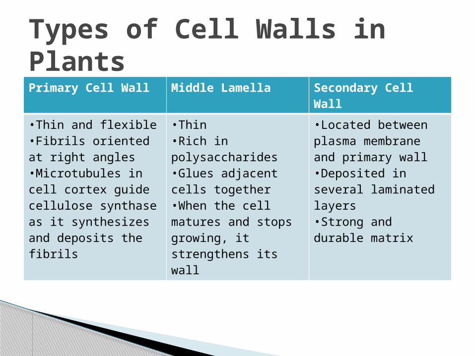
Primary Cell Wall Middle Lamella Secondary Cell Wall
•Thin and flexible•Fibrils oriented at right angles•Microtubules in cell cortex guide cellulose synthase as it synthesizes and deposits the fibrils
•Thin•Rich in polysaccharides•Glues adjacent cells together•When the cell matures and stops growing, it strengthens its wall
•Located between plasma membrane and primary wall•Deposited in several laminated layers•Strong and durable matrix
Types of Cell Walls in Plants
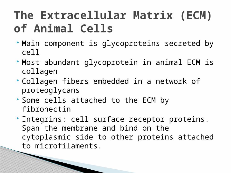
Main component is glycoproteins secreted by cell
Most abundant glycoprotein in animal ECM is collagen
Collagen fibers embedded in a network of proteoglycans
Some cells attached to the ECM by fibronectin Integrins: cell surface receptor proteins. Span
the membrane and bind on the cytoplasmic side to other proteins attached to microfilaments.
The Extracellular Matrix (ECM) of Animal Cells
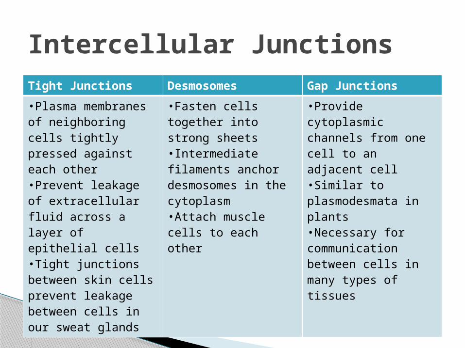
Tight Junctions Desmosomes Gap Junctions
•Plasma membranes of neighboring cells tightly pressed against each other•Prevent leakage of extracellular fluid across a layer of epithelial cells•Tight junctions between skin cells prevent leakage between cells in our sweat glands
•Fasten cells together into strong sheets•Intermediate filaments anchor desmosomes in the cytoplasm•Attach muscle cells to each other
•Provide cytoplasmic channels from one cell to an adjacent cell•Similar to plasmodesmata in plants•Necessary for communication between cells in many types of tissues
Intercellular Junctions


![Lenses Lenses are curved surfaces that refract light as it passes through. [optics/dazle/photos/finished_lenses/lenses_1_to_4_1.jpg]](https://img.pdfslide.us/doc/110x75/56649d8b5503460f94a72b00/lenses-lenses-are-curved-surfaces-that-refract-light-as-it-passes-through.jpg)