Embed Size (px)
Citation preview

1
PowerPoint Slides English Traducción al español
Breast Cancer: Surgical Treatment Approaches Video Transcript
Cáncer de mama: Enfoques de tratamiento quirúrgico Trascripción del video
Professional Oncology Education Breast Cancer: Surgical Treatment Approaches Time: 58:15
Educación Oncológica Profesional Cáncer de mama: Enfoques de tratamiento quirúrgico Duración: 58:15
Kelly K. Hunt, M.D., F.A.C.S. Professor, Surgical Oncology The University of Texas MD Anderson Cancer Center
Dra. Kelly K. Hunt, F.A.C.S. Profesora de Oncología Quirúrgica MD Anderson Cancer Center de la Universidad de Texas
Hello. I’m Dr. Kelly Hunt from the University of Texas MD Anderson Cancer Center. This afternoon, I’m going to talk with you about surgical approaches to the treatment of breast cancer.
Hola. Soy la Dra. Kelly Hunt del MD Anderson Cancer Center de la Universidad de Texas. Esta tarde les hablaré de los enfoques quirúrgicos en el tratamiento del cáncer de mama.
2
The objectives that I want to cover are first, to discuss surgical treatment of the primary tumor and then oncoplastic techniques that we use for partial mastectomy reconstruction. I then want to cover the integration of reconstruction for patients undergoing total mastectomy; and finally, talk about nodal staging for breast cancer.
Los objetivos que quiero abordar son: primero, analizar el tratamiento quirúrgico del tumor primario y las técnicas oncoplásticas que utilizamos para una reconstrucción de mastectomía parcial. Luego analizaremos la integración de la reconstrucción en las pacientes que se someten a una mastectomía total. Y, por último, hablaré sobre las diferentes fases ganglionares del cáncer de mama.
So, the factors that we typically consider in surgical planning are numerous. In fact, we look not only at the tumor size or the clinical stage of the primary breast cancer. But, we also consider the patient’s breast size and try to achieve a reasonable tumor size to breast size ratio for breast conservation. We look at the location of the tumor in the breast and also consider the family history or any suggestion of a familial breast cancer or a predisposition to breast cancer, such as a BRCA1 or BRCA2 mutation. We look at contraindications to the use of radiation therapy. And of course, consider the patient’s preference for treatment of the breast cancer and the overall health status of the individual patient.
Los factores que generalmente consideramos al planear una cirugía son numerosos. De hecho, no sólo observamos el tamaño del tumor o el estadio clínico del cáncer de mama primario, sino que analizamos el tamaño de la mama y buscamos una proporción razonable entre el tamaño del tumor y el de la mama para conservarla. También observamos la ubicación del tumor en la mama y consideramos los antecedentes familiares o cualquier indicio de cáncer de mama familiar o una predisposición a éste, como una mutación BRCA1 o BRCA2. Asimismo, evaluamos las contraindicaciones del empleo de radioterapia y, por supuesto, tomamos en cuenta las preferencias de la paciente respecto del tratamiento del cáncer de mama y su estado de salud en general.

3
In terms of pathologic assessment, there are a number of factors that are important in the individual tumor. One is the histologic subtype, the nuclear grade, and typically, we use a modified Black’s nuclear grade staging I, II or III, estrogen and progesterone receptor status, and the status of the HER2/neu oncogene either by immunohistochemistry or fluorescence in situ hybridization.
En la evaluación patológica, existen varios factores importantes con cada tumor en particular: el subtipo histológico, el grado nuclear —generalmente empleamos el sistema de Black modificado de grados I, II y III—, el estado de receptores estrogénicos y progesterónicos, y el estado del oncogén HER2/neu, detectado por inmunohistoquímica o hibridación in situ con fluorescencia.
The primary objective in approaching the tumor in the breast is to excise the tumor with adequate margins. And, in general, our approach to the margins in both invasive and noninvasive breast cancer is to achieve a 2-mm margin in all directions around the tumor. This helps to minimize local recurrence and, again, we look at the expected cosmetic outcome. And that’s where, again, the tumor size to breast size ratio comes into play.
El objetivo principal del enfoque en el tumor en la mama es extirparlo con márgenes adecuados. En general, nuestro enfoque en los márgenes en el cáncer de mama invasivo y no invasivo consiste en lograr un margen de 2 mm en todo el contorno del tumor, lo que ayuda a minimizar la recidiva local. Además, buscamos el resultado estético esperado y es aquí donde la relación entre el tamaño del tumor y el de la mama vuelve a desempeñar un papel importante.

4
For early stage breast cancer, we generally consider breast-conserving treatment and mastectomy to the equivalent. There are a number of randomized trials that were done several decades ago showing that survival outcomes were equivalent in women with early stage breast cancer who were treated with lumpectomy and radiation versus mastectomy. In terms of the components of breast conservation, not only are we removing the primary breast tumor with a segmental or partial mastectomy, but we’re also including axillary staging as appropriate for the patient. So, those with invasive breast cancer would have nodal staging and those with noninvasive breast cancer would not require any axillary staging. Radiation therapy, of course, is also an important component of breast-conserving surgery. For mastectomy, this generally is a total mastectomy. Again, axillary staging, where appropriate, for those individuals with invasive breast cancer, and then the integration of reconstruction which we’ll cover a little bit later.
En el cáncer de mama incipiente, por lo general consideramos igualmente el tratamiento de conservación de la mama y la mastectomía. Diversos ensayos aleatorizados realizados hace varias décadas mostraron que los resultados de supervivencia eran equivalentes en las mujeres con cáncer de mama incipiente que habían sido tratadas con tumorectomía y radiación en comparación con la mastectomía. Si hablamos de los componentes de la conservación de la mama, con una mastectomía segmentaria o parcial no sólo extirpamos el tumor de mama primario, sino también incluimos una estadificación axilar apropiada para la paciente. Así, a las pacientes con cáncer de mama invasivo se les realizaría una estadificación ganglionar, pero aquéllas con cáncer de mama no invasivo no requerirían una estadificación axilar. Por supuesto, la radioterapia también es un componente importante de la cirugía de conservación de la mama. Las mastectomías, por lo general, son totales. Nuevamente, se realiza una estadificación axilar en las pacientes con cáncer de mama invasivo y luego viene la integración de la reconstrucción, que abordaremos un poco más adelante.
So, the surgical options for a patient with early stage breast cancer generally will be segmental mastectomy. Again, there’re several ways to call a segmental mastectomy. There can be lumpectomy, partial mastectomy. In the past, the term “quadrantectomy” has been used, although typically we do not attempt to excise the entire quadrant when we’re excising the primary breast tumor. A total mastectomy can also be called a simple mastectomy or complete mastectomy. The goal is to remove the entire breast. Skin-sparing mastectomy is essentially a total mastectomy that’s performed with preservation of the entire breast skin envelope. And then finally, there’s nipple-sparing mastectomy where not only are we trying to preserve the entire breast skin envelope, but we’re also trying to preserve the entire nipple-areolar complex. And skin-sparing mastectomy and nipple-sparing mastectomy are always done with immediate breast reconstruction.
Entonces, las opciones quirúrgicas de una paciente con cáncer de mama incipiente suelen ser una mastectomía segmentaria, a la cual se le conoce de varias maneras, como tumorectomía o mastectomía parcial. Antes se empleaba el término “cuadrantectomía”, aunque generalmente no pretendemos extirpar todo el cuadrante cuando extirpamos el tumor de mama primario. Una mastectomía total también se conoce como mastectomía simple o completa, y su objetivo es extirpar toda la mama. En esencia, una mastectomía con conservación de piel es una mastectomía total en la que se conserva todo el recubrimiento dérmico de la mama. Por último, existe una mastectomía con conservación del pezón en la que no sólo se busca preservar todo el recubrimiento dérmico de la mama, sino también todo el complejo pezón-areola. Tanto la mastectomía con conservación de piel como la mastectomía con conservación del pezón se

5
realizan siempre con una reconstrucción inmediata de la mama.
So, the sequencing of treatment is something that comes into play more frequently now as chemotherapy and other systemic therapies are utilized in the management of breast cancer patients. In the past, we would always do surgical treatment as the initial treatment. But, now that we know that systemic chemotherapy or systemic endocrine therapy may be used in an individual patient, we can sometimes use that treatment prior to any surgery in order to improve the surgical outcomes. And so, giving the treatment --- the systemic treatment before surgery is often called neoadjuvant therapy. It can also be called primary systemic therapy or induction therapy. The indications that we typically use for neoadjuvant chemotherapy are to downsize the tumor in the breast to achieve breast conservation. So, in women with relatively large tumors in relationship to the size of the breast, where we know systemic chemotherapy is warranted in the management of that individual, we would consider doing the chemotherapy before surgery to shrink that tumor and require less volume of breast tissue to be excised at surgery. We also look at nodal involvement. So, women who present with metastatic disease in the lymph nodes that’s proven by fine needle aspiration biopsy are also candidates for systemic therapy prior to surgical intervention. And part of this is to assess the response to therapy because we know that after we’ve completed surgery and removed all evidence of the primary tumor and any regional nodal metastases, we can only wait in follow-up to see if there’s any recurrence. We don’t have any direct assessment of the response to that systemic therapy. If the tumor is still intact and we give the treatment prior to any surgery, we can measure the response clinically, radiographically. And then also at pathology, we can see how much residual invasive cancer is left and how much residual noninvasive breast cancer and get the pathologic response rate to that individual regimen. So, the indications for adjuvant chemotherapy are based
La secuencia del tratamiento es algo que con más frecuencia desempeña un papel importante, ya que el control de las pacientes con cáncer de mama requiere quimioterapia y otras terapias sistémicas. En el pasado, solíamos administrar un tratamiento quirúrgico como el tratamiento inicial, pero ahora que sabemos que en pacientes específicas se puede utilizar una quimioterapia sistémica o una terapia endocrina sistémica, a veces podemos emplear ese tratamiento antes de cualquier cirugía para obtener mejores resultados quirúrgicos. Por ello, a la administración de un tratamiento sistémico antes de una cirugía a menudo se le conoce como terapia prequirúrgica. También se le llama terapia sistémica primaria o terapia de inducción. Las indicaciones que generalmente seguimos con la quimioterapia prequirúrgica son disminuir el tamaño del tumor en la mama para su conservación. En las mujeres con tumores relativamente grandes con respecto al tamaño de la mama, donde se sabe que se necesita una quimioterapia sistémica para el control de una paciente en particular, administraríamos quimioterapia antes de la cirugía para encoger el tumor y extirpar un volumen menor de tejido mamario en la cirugía. También observamos la afectación ganglionar. Así, las mujeres con metástasis en los ganglios linfáticos, comprobada con una biopsia espirativa, también son candidatas para recibir terapia sistémica antes de una cirugía. Parte de esto consiste en evaluar la respuesta a la terapia porque sabemos que después de la cirugía y de extirpar toda evidencia del tumor primario y cualquier metástasis ganglionar regional, sólo podemos esperar el seguimiento para ver si hay alguna recidiva. No contamos con ninguna evaluación directa de la respuesta a dicha terapia sistémica. Si el tumor sigue intacto y damos tratamiento antes de cualquier cirugía, podemos medir la respuesta en términos clínicos mediante radiografías. Luego, en patología, podemos ver cuánto cáncer residual existe, invasivo y no invasivo, y evaluar el índice de respuesta patológica a ese tratamiento en particular. Entonces, las

6
on the size of the primary tumor, any evidence of nodal involvement, estrogen receptor status, the age of the patient, and then also, other considerations, such as overexpression of the HER2/neu oncogene. And these are the factors that we look at when considering, if we know the patient is going to be a candidate for adjuvant chemotherapy, we might move that chemotherapy to the preoperative setting and give it prior to any surgical intervention.
indicaciones de una quimioterapia complementaria se basan en el tamaño del tumor primario, cualquier evidencia de afectación ganglionar, el estado de receptores estrogénicos, la edad de la paciente y otros aspectos, como la sobreexpresión del oncogén HER2/neu. Estos son los factores que analizamos. Si sabemos que la paciente puede recibir quimioterapia complementaria, podemos moverla al entorno prequirúrgico y administrarla antes de realizar cualquier intervención quirúrgica.
Now, the other factor to consider in breast conservation, as I mentioned before, is any contraindications to radiation therapy. And so, in some cases where the patient has already had radiation treatment, perhaps for Hodgkin’s disease in --- when they were a teenage or an early or young adult, or radiation treatment to the chest wall for other medical conditions, we might not be able to do breast conservation because of overlapping treatment of the radiation fields. In breast --- the indications for radiation therapy in the management of breast cancer patients are listed here on this slide. In --- in pretty much all cases of breast-conserving surgery, we’re going to recommended postoperative adjuvant radiation therapy when the patient has invasive breast cancer. Some exceptions are those elderly patients with strongly hormone receptor positive tumors where you might be able to avoid radiation. And in some cases of noninvasive breast cancer where we have a low grade tumor on pathologic assessment with widely negative margins, we might consider avoiding radiation. But, in general, when we’re doing a partial mastectomy or segmental mastectomy, radiation will be indicated. If the size of the tumor is a T3 tumor, that being greater than 5 cm in size, radiation therapy is generally indicated even if the patient is planned for a mastectomy for management of the primary tumor. And then these are the other indications for the use of radiation – those patients with T4 disease, nodal involvement where there’s more than four positive axillary lymph nodes, and in cases where there’s positive margins after mastectomy or evidence of extensive lymphovascular space invasion.
Como ya se mencionó, otro factor que tomamos en cuenta en la conservación de la mama es cualquier contraindicación para la radioterapia. En algunos casos en que la paciente ya ha recibido radioterapia, quizá para la enfermedad de Hodgkin cuando era adolescente o durante los primeros años de la adultez; o bien, una radioterapia en el tórax para otras afecciones médicas, es posible que no podamos conservar la mama debido a la yuxtaposición de los tratamientos en los campos de radiación. Las indicaciones para la radioterapia en el control de las pacientes con cáncer de mama se enumeran en esta diapositiva. En casi todos los casos de cirugía de conservación de la mama recomendamos la radioterapia posquirúrgica complementaria cuando la paciente tiene cáncer de mama invasivo. Algunas excepciones son aquellas pacientes de edad avanzada con tumores con receptores hormonales, en las que es posible evitar la radiación. Y en algunos casos de cáncer de mama no invasivo, en los que la evaluación patológica muestra un tumor de bajo grado con márgenes bastante negativos, también podemos evitar la radiación. No obstante, en general, cuando realizamos una mastectomía parcial o segmentaria, se indica la radiación. Si el tamaño del tumor es T3, es decir, excede los 5 cm, generalmente se indica la radioterapia incluso si a la paciente se le programa una mastectomía para controlar el tumor primario. Otros indicadores para el uso de radiación son aquellas pacientes con enfermedad T4, afectación en más de cuatro ganglios axilares, y casos con márgenes positivos luego de una mastectomía o evidencia de una extensa invasión del espacio linfovascular.

7
This is a general algorithm for our approach to the patient presenting with an early stage breast cancer that has a small or medium breast size. In general, if we anticipate a small parenchymal defect because it’s a small tumor in the breast, and the defect --- and the tumor is in a favorable location, that being a superior location in the breast or in the lateral aspect of the breast, we will usually proceed with segmental resection and local tissue rearrangement. In some cases, for a patient who has a very small breast, we might consider a composite flap. If the tumor is in an unfavorable location, and we generally consider this to be anywhere in the inferior aspect of the breast below the nipple-areolar complex, we look at whether there’s significant ptosis or not in the breast. And, so, for the patient who does have ptosis, we would consider doing a vertical reduction mammoplasty or mastopexy at the same time as resection of the primary tumor. For those patients who do not have any significant ptosis and we anticipate needing to resect skin in addition to the primary tumor, we would consider using a latissimus flap or a TAP flap or, in fact, proceeding with a mastectomy because, again, we anticipate that there would be a poor cosmetic outcome with breast-conserving surgery. Also, if the patient initially presents with a very large parenchymal defect or we know that there’s going to be a significant amount of skin resection at the time of surgery, we might consider going directly to mastectomy or doing a partial mastectomy with a latissimus flap or a TAP flap. Or, again, this is a consideration where we might do neoadjuvant or preoperative therapy prior to doing any surgical intervention.
Se trata de un algoritmo general en nuestro enfoque a las pacientes con cáncer de mama incipiente cuyas mamas son pequeñas o medianas. En general, si anticipamos un pequeño defecto parenquimatoso debido a un tumor pequeño en la mama que se encuentra en un lugar favorable, es decir, en la parte superior de la mama o a uno de sus lados, generalmente procedemos con una resección segmentaria y la restructuración del tejido local. En algunos casos, para una paciente con mamas muy pequeñas, podemos considerar el empleo de un colgajo compuesto. Si el tumor está en un lugar poco favorable, lo que para nosotros equivale a cualquier parte de la mama debajo del complejo pezón-areola, evaluamos si existe o no ptosis considerable en la mama. En las pacientes con ptosis realizamos una mamoplastia de reducción vertical o una mastopexia, al mismo tiempo que extirpamos el tumor primario. En aquéllas sin ptosis considerable y en quienes se anticipa la necesidad de extirpar piel además del tumor primario, se analiza el uso de un colgajo dorsal o un colgado perforante toracodorsal (o TAP), o, de hecho, se realiza una mastectomía porque, como ya se dijo, se prevé que la cirugía con conservación de la mama tendrá un resultado estético inaceptable. Además, si la paciente en un principio presenta un defecto parenquimatoso considerable o si sabemos que tendremos que extirpar mucha piel durante una cirugía, podemos considerar realizar directamente una mastectomía o una mastectomía parcial con un colgajo dorsal o un TAP. Es una situación en la que podemos administrar una terapia prequirúrgica antes de cualquier intervención.

8
In the patient who has a very large breast size or is an obese patient, we look at the size of the parenchymal defect. As listed here, we basically assess less than 15 percent of the breast, 15 to 30 percent of the breast, or greater than 30 percent of the breast to resect the primary tumor with a negative margin. If we anticipate a small parenchymal defect, that’s an individual where we would proceed to breast-conserving therapy. But, if it’s a 15 to 30 percent parenchymal defect, we know that this will cause significant deformity in the breast and will also cause asymmetry with the contralateral breast. And, so, this is where we would consider local tissue rearrangement or reduction mammoplasty. And for women with very large ptotic breasts, reduction mammoplasty is done bilaterally and this is a very useful tool for them. And then, for a very large parenchymal defect, we would definitely consider reduction mammoplasty or insertion of a latissimus flap or consider a mastectomy.
En una paciente con mamas muy grandes o con obesidad, analizamos el tamaño del defecto parenquimatoso. Básicamente evaluamos menos del 15% de la mama, del 15% al 30% de la mama, o más del 30% de ella para extirpar el tumor primario con un margen negativo. Si anticipamos un pequeño defecto parenquimatoso, se trata de una paciente a quien administramos una terapia de conservación de la mama, pero, si el defecto parenquimatoso representa de un 15% a 30%, sabemos que podría causar una deformación importante en la mama y asimetría con la otra mama. Entonces, aquí optamos por reestructurar el tejido local o efectuar una mamoplastia de reducción. En las mujeres con mamas con ptosis considerable, la mamoplastia de reducción se realiza de manera bilateral, lo cual resulta bastante útil. Así, cuando el defecto parenquimatoso es muy grande, definitivamente evaluamos una mamoplastia de reducción o la inserción de un colgajo dorsal, o bien, una mastectomía.
So, the primary components, then, of breast conservation…
Luego entonces, los principales componentes de la conservación de la mama…

9
…are removing the primary tumor with a negative margin, but we also look at several other factors. We prefer that this is a unifocal lesion in the breast because we know that historically, data suggests that patients who have multifocal or multicentric disease in the breast have a higher rate of in-breast recurrence. We also look at the mammogram to assure that there’re no extensive microcalcifications that would make it difficult to follow that patient after surgery and radiation therapy. Again, we look at the breast size to tumor size ratio, the tumor location, and, then, look at whether or not preoperative therapy might be indicated. So, if for patients who have a high nuclear grade, estrogen receptor negative tumor and lymphovascular space invasion, those are considerations for chemotherapy. And we might consider the chemotherapy preoperatively. For women who have strongly estrogen receptor positive tumors, we might consider preoperative endocrine therapy in a postmenopausal patient that would typically be with an aromatase inhibitor. And we know from clinical trials that we can achieve breast conservation in a significant portion of those women who have strongly estrogen receptor positive tumors. Finally, we look at the family history and any need for BRCA testing. Those individuals who are BRCA mutation carriers are at much higher risk for developing additional primary breast cancers in their lifetime. That’s in the treated breast and in the contralateral breast. So, in general, we would not consider those individuals to be good candidates for breast-conserving therapy. Although we do discuss the options with them, and some patients may choose to go to go that route, but we are --- we make sure that they are aware that the expected risk of re --- in-breast recurrence is much higher than what we would typically consider to be an acceptable risk. Patients who have a strong family history where they’ve not been tested for BRCA mutation status may also fall in this category and they should be counseled accordingly.
…son la extirpación del tumor primario con un margen negativo, pero también consideramos muchos otros factores. Preferimos que sea una lesión unifocal en la mama, ya que sabemos que, en términos históricos, la información sugiere que las pacientes con una enfermedad multifocal o multicéntrica en la mama tienen un mayor índice de recidiva en esa parte del cuerpo. Asimismo, observamos la mamografía para verificar que no haya microcalcificaciones generalizadas que dificulten el seguimiento de la paciente luego de la cirugía y la radioterapia. También observamos el tamaño de la mama en relación con el del tumor, la ubicación del tumor y si resulta viable o no la terapia prequirúrgica. Así, en las pacientes con un alto grado nuclear, un tumor sin receptores estrogénicos e invasión del espacio linfovascular, se puede indicar el uso de quimioterapia y podríamos considerar su aplicación prequirúrgica. En el caso de las mujeres con tumores con receptores estrogénicos, consideramos la aplicación de una terapia endocrina prequirúrgica, específicamente en las pacientes posmenopáusicas que, por lo general, presentan el inhibidor de la aromatasa. Gracias a los ensayos clínicos sabemos que podemos conservar gran parte de la mama de aquellas mujeres que tienen tumores con receptores estrogénicos. Por último, investigamos los antecedentes familiares y si es necesario realizar pruebas de BRCA. Las pacientes con una mutación de BRCA tienen un riesgo mucho mayor de desarrollar otros tipos de cáncer primario de la mama en su vida, tanto en la mama tratada como en la otra. En general, no consideraríamos a esas mujeres como aptas para una terapia de conservación de la mama. Aunque sí analizamos las opciones con ellas y algunas pueden elegir ese camino, nos aseguramos de que sepan que el riesgo previsto de recidiva en la mama es mucho mayor de lo que normalmente consideraríamos un riesgo aceptable. Las pacientes con antecedentes familiares contundentes y a quienes no se les han realizado pruebas de la mutación de BRCA también pueden caer en esta categoría y se les debe orientar de manera acorde.

10
So, the breast-conserving treatment is a multidisciplinary team effort. The diagnostic imaging specialists are the ones who will define the extent of disease in the breast for us preoperatively, depending on whether there’re calcifications or mass lesion or architectural distortion. They will place localization wires to show the extent of the disease for us so that we can resect the full extent of the disease. And also image the regional nodal basins typically with ultrasound, preoperatively, so that we can approach the appropriate nodal basins as well. The surgeon is responsible for resecting the disease in the breast and assessing the nodal status. And typically, we prefer to do this with one operation. So, for diagnostic purposes, we always prefer a needle core biopsy and --- so that we have the definitive diagnosis prior to proceeding with any surgery. The pathologist then assesses the margins for us intraoperatively and helps us to determine if we need to take additional tissue at that surgical procedure. And they also assess the lymph nodes for us so that we understand whether or not the patient needs a complete node dissection. Plastic surgeons are involved in providing optimal cosmesis for the individual patient and the medical oncologist for systemic therapy, the radiation oncologist for radiation therapy.
El tratamiento de conservación de la mama es un esfuerzo de equipo multidisciplinario. Los especialistas en diagnóstico por imagen son quienes definen el alcance de la enfermedad en la mama en la etapa prequirúrgica, según la presencia de calcificaciones, lesiones masivas o deformaciones estructurales. Para ello, colocan cables de localización a fin de mostrarnos el alcance de la enfermedad y así extirpar todo el tumor. También obtienen imágenes de las regiones ganglionares, por lo general mediante una ecografía prequirúrgica, para que podamos tener acceso a dichas regiones. El cirujano es responsable de extirpar el tumor de la mama y evaluar el estado ganglionar. Preferimos hacer esto mediante una sola operación por lo que, para fines de diagnóstico, siempre optamos por una biopsia con aguja gruesa para realizar un diagnóstico definitivo antes de proceder con cualquier cirugía. El patólogo luego evalúa los márgenes de manera intraoperatoria y nos ayuda a determinar si necesitamos extirpar más tejido durante la cirugía. Asimismo, evalúa los ganglios linfáticos para que podamos decidir si la paciente necesita o no una disección total de ellos. Los cirujanos plásticos se encargan de asegurar un resultado estético óptimo para cada paciente; el oncólogo médico, de la terapia sistémica, y el oncólogo radioterapeuta, de la radioterapia.

11
This is a diagram showing some of the placement of incisions that we consider for the individual patient. Typically, in the upper superior aspect of the breast and the lateral breast, we place the incision overlying the palpable tumor. However, when it’s relatively --- when the tumor is relatively close to the nipple-areolar complex, we do consider using circumareolar incisions as they’re less obvious than the ones placed directly on the breast. In the medial part or the middle aspect of the breast, we tend to use more of a radial type incision, either medially or laterally. And in the inferior breast, we typically use a radial type incision that might be able to be incorporated into a mastopexy incision or a reduction mammoplasty incision.
En este diagrama se muestra la ubicación de algunas incisiones que realizamos en las pacientes. Por lo general, la incisión que se realiza sobre el tumor palpable se localiza en la parte superior de la mama, así como en la lateral. No obstante, cuando el tumor se encuentra relativamente cerca del complejo pezón-areola, analizamos la posibilidad de efectuar incisiones periareolares, ya que son menos evidentes que las realizadas directamente en la mama. En la parte interna o media de la mama solemos realizar incisiones radiales, ya sea internas o laterales. Y en la parte inferior de la mama, por lo general realizamos incisiones radiales que puedan incorporarse a una incisión de mastopexia o de mamoplastia de reducción.
To talk about patients based on the breast size for partial mastectomy and local tissue rearrangement, those individuals that have a C-cup breast size that have a small tumor, no ptosis and minimal skin resection are the best candidates for local tissue rearrangement.
Si hablamos de las pacientes a partir del tamaño de sus mamas y su necesidad de una mastectomía parcial o una restructuración del tejido local, aquéllas cuyo busto es de copa C, con un tumor pequeño sin ptosis y resección dérmica mínima son las más aptas para una restructuración del tejido local.

12
And there’re many different ways to approach local tissue rearrangement. I’m just going to show you a few examples today. One approach that we have used here at the MD Anderson Cancer Center is to make a circumareolar incision and completely release the nipple-areolar complex to raise the breast skin, much as you would do for a skin-sparing mastectomy, up to the level of the primary tumor, then to resect the tumor with a margin of normal tissue, and then re-close the nipple-areolar complex along with the breast skin envelope so that there’s no tension associated with the closure at the primary tumor site. So, it’s important to lift that skin completely off of the breast and do the resection such that they’re not tethered together, creating a defect when the --- when the healing occurs postoperatively.
Existen distintas formas de considerar una restructuración del tejido local, pero hoy sólo mostraré algunos ejemplos. Uno de los enfoques que hemos empleado aquí en el MD Anderson Cancer Center consiste en realizar una incisión periareolar y retirar por completo el complejo pezón-areola para levantar la piel de la mama, tal como se haría en una mastectomía con conservación de piel, hasta el nivel del tumor primario. Enseguida extirpamos el tumor con un margen de tejido normal y luego volvemos a cerrar el complejo junto con el recubrimiento dérmico de la mama, de manera que no haya tensión en el cierre donde se encontraba el tumor primario. Por ello es importante levantar la piel completamente de la mama y realizar la resección de tal forma que no se unan y se origine un defecto con la cicatrización posterior a la cirugía.
Another approach is to use something called a batwing mastopexy. This has been popularized by several surgeons in the U.S. and the U.K., where you might have a tumor just above the nipple-areolar complex and need to perform a small amount of a skin resection with that tumor. And you can see the incision lines that are planned out here based on that. Once the tumor is resected, the nip --- the nipple-areolar complex is elevated into the space and then the closure takes place here. And at the final closure, you can see how the incisions medially and laterally are visible. However, this incision above the nipple-areolar complex heals quite well and is cosmetically much more acceptable.
Otro enfoque consiste en realizar una mastopexia con la técnica “batwing”, la cual han popularizado numerosos cirujanos de los Estados Unidos y el Reino Unido, cuando el tumor se encuentra justo arriba del complejo pezón-areola y se debe retirar una pequeña porción de piel junto con el tumor. Aquí podemos ver las líneas de incisión planeadas para dicho procedimiento. Una vez extirpado el tumor, el complejo pezón-areola se coloca en un lugar superior del espacio y se cierra aquí. En el cierre final podemos ver cómo las incisiones interna y lateral quedan visibles; sin embargo, este tipo de incisiones arriba del complejo sanan muy bien y son mucho más aceptables en términos estéticos.

13
This shows a patient who’s had a partial mastectomy with an incision placed directly over the primary tumor. And in this case, she’s had a good result. Basically, the --- the --- again, one of things that we’re trying to achieve is to be certain that there’s symmetry with the opposite breast. And that there’s not significant deviation of the nipple-areolar complex once the healing has occurred at the primary tumor site and the adjuvant radiation therapy has been completed.
Aquí se muestra a una paciente a quien se le realizó una mastectomía parcial con una incisión directamente sobre el tumor primario. En este caso, ha tenido buenos resultados. Básicamente, una de las cosas que buscamos lograr es que haya simetría entre las dos mamas. También buscamos una mínima desviación del complejo pezón-areola una vez que ocurra la cicatrización en el lugar del tumor primario y que haya finalizado la radioterapia complementaria.
The breast reduction technique is something that we tend to use for patients who have a larger breast size, such as a D-cup breast size. Or those patients with a C-cup breast size where there is a small tumor, but there is a relative amount of ptosis --- significant amount of ptosis in the breast.
La técnica de reducción mamaria es aquélla que solemos emplear con las pacientes cuyas mamas son más grandes, como las que usan una talla de copa C, o bien, con aquellas pacientes con tamaño de copa C y un tumor pequeño, pero con una relativa cantidad de ptosis en la mama.

14
And so this shows many of the pedicles that we might use for the approach to the reduction mammoplasty. You can see that in these diagrams, the tumor is located in different areas of the breast. In this case, behind the nipple-areolar complex, you might even remove the nipple itself and you can do a free nipple graft in some cases. Or completely remove the nipple-areolar complex and then do a nipple reconstruction once the patient has completed radiation therapy. This works for patients with tumors in the inferior aspect of the breast and then, in other locations, as you can see noted here. And so, these are the vascular pedicles that are used for tumors in those different locations of the breast.
Aquí se muestran muchos de los pedículos que podemos utilizar durante la mamoplastia de reducción. Como se puede ver en estos diagramas, el tumor se localiza en distintas áreas de la mama. En este caso, detrás del complejo pezón-areola, se puede incluso extirpar el pezón y en algunos casos realizar un injerto de pezón libre. También, se puede extirpar por completo el complejo pezón-areola y después realizar una reconstrucción del pezón una vez que la paciente haya finalizado la radioterapia. Esto funciona en las pacientes con tumores en la parte inferior de la mama y también en otros lugares, como se puede ver aquí. Estos son los pedículos vasculares empleados para los tumores ubicados en esas distintas partes de la mama.
This shows an example of a patient who has a relatively large breast size, but she has significant ptosis in the breast. And she’s planned for segmental resection. We anticipate that because of the resection in the in --- in the medial aspect of the breast and the use of radiation therapy, that the patient will have significant lifting of that breast, which causes asymmetry with the contralateral breast. So, of course, it’s important to consider both breasts in the treatment. So, in this case, the segmental resection has been completed here. You can see the planned incisions of the Wise pattern that have been marked out by the plastic surgeon. And in doing the segmental resection, we used that planned incision so we don’t make a separate incision overlying the tumor even though it’s located in the upper aspect of the breast. We go through the planned incision. You can see here where the plastic surgeon has de-epithelialized this and raised the nipple-areolar complex and this pedicle up. And it will be rotated into the defect that was created by the partial mastectomy.
Este es un ejemplo de una paciente con un tamaño de mama relativamente grande, pero con ptosis considerable, a quien se le programa una resección segmentaria. Prevemos que, debido a la resección en la parte interna de la mama y el uso de radioterapia, a la paciente se le levantará bastante la mama, lo cual provocará asimetría con la otra mama. Queda claro, pues, que es importante tomar en cuenta ambas mamas durante el tratamiento. Así, en este caso se realizó una resección segmentaria. Podemos ver las incisiones planeadas del patrón de Wise que fueron marcadas por el cirujano plástico. Al realizar la resección segmentaria, empleamos esa incisión planeada para no hacer otra incisión sobre el tumor, aun cuando se encuentre en la parte superior de la mama. Trabajamos con la incisión planeada y, como podemos observar, el cirujano plástico retiró el epitelio y levantó el complejo pezón-areola junto con el pedículo mostrado, para luego rotarlo hacia el defecto originado por la mastectomía parcial.

15
And this shows the final result postoperatively. The patient’s had good healing of her incisions here and you can see that she has good symmetry of the breast. So, this is a very useful procedure for those patients with a larger breast or those that have a significant amount of ptosis.
Aquí se muestra el resultado final posterior a la cirugía. Las incisiones de la paciente cicatrizaron bien y sus mamas tienen buena simetría. Este procedimiento es muy útil en las pacientes con senos más grandes o en aquellas con ptosis considerable.
The key to par --- to successful partial mastectomy is anticipating the cosmetic outcome. Again, as I talked about, the location of the tumor in the breast, the breast to tum --- to tumor size ratio, and also symmetry with the opposite breast. And these are important considerations because after radiation treatment has occurred, it’s much more difficult to correct a cosmetic deformity. And so, we try to anticipate the cosmetic outcome before the radiation and do any repair before the radiation has taken place. This avoids the need for using other myocutaneous flaps or latissimus flaps to repair a defect that has occurred after radiation treatment has already been completed. The repair, again, is based essentially on the breast size and relationship to the tumor size, and in considering other factors, such as the need for chemotherapy. In a patient with a smaller breast, even a very small tumor can sometimes create a significant defect. This is where we might consider neoadjuvant chemotherapy or insetting another flap or another amount of tissue like a latissimus flap. Using breast remodeling technique for the C-cup breast, as I showed you, or for using the breast reduction technique, design the pedicle based on the tumor location.
La clave para una mastectomía parcial exitosa es anticipar el resultado estético y, como ya se mencionó, la ubicación del tumor en la mama, la relación entre el tamaño de la mama y el del tumor, y la simetría con la otra mama. Estas son consideraciones importantes ya que, una vez realizada la radioterapia, es mucho más difícil corregir una deformidad estética. Por ello tratamos de prever el resultado estético y realizar cualquier reparación antes de la radiación, lo cual evita la necesidad de emplear otros colgajos miocutáneos o dorsales para reparar un defecto ocurrido después de concluida la radioterapia. La reparación se basa principalmente en el tamaño de la mama y su relación con el tamaño del tumor, en consideración de otros factores, como la necesidad de quimioterapia. En una paciente con mamas más pequeñas, incluso un tumor muy pequeño a veces puede dar origen a un defecto considerable. En este caso podríamos considerar una quimioterapia prequirúrgica o la colocación de otro colgajo o de tejido adicional mediante un colgajo dorsal. El empleo de la técnica de remodelación de la mama para una talla C, tal como se mostró, o de la técnica de reducción mamaria, determina el diseño del pedículo según la ubicación del tumor.

16
So, again, using preoperative chemotherapy as a strategy that we’ve employed over the past couple of decades here at the MD Anderson Cancer Center. It’s standard practice for those women who present with locally advanced breast cancer. And so, that we don’t take any stage III or stage IV patients directly to surgery until we’ve achieved maximal response with the appropriate systemic chemotherapy or endocrine therapy. Using this approach, we see excellent clinical response rates. And so, in general, 70 to 80 percent of the patients who have preoperative chemotherapy will have either a complete or partial response in the breast, so that does create a lot of room for different surgical options or different approaches to the surgical management of the tumor. We have not found that there’s any increase in surgical complications related to chemotherapy in the preoperative setting. And there’s no delay in patients receiving their adjuvant therapy such as radiation or other adjuvant endocrine therapy and other things such as this --- of this nature. So, it does not create any difficulties, and very few patients will have any progression of their disease while on preoperative thi --- chemotherapy. And in fact, this is a very important thing to discern, if the patient has any progression of disease while on therapy, then we have the opportunity to switch to a different type of therapy and see if we can get improved response. But, it helps us to understand the biology and the nature of that tumor. We know that there’s no significant difference in survival whether we deliver the chemotherapy in the neoadjuvant or the adjuvant setting. And --- and yet, the neoadjuvant setting offers us many more opportunities for improving surgical options.
El uso de quimioterapia prequirúrgica es una estrategia que hemos empleado durante el último par de décadas aquí en el MD Anderson Cancer Center. Es una práctica estándar para las mujeres con cáncer de mama localmente avanzado. Así, no llevamos a las pacientes en estadio III o IV directamente a cirugía sino hasta que hayamos alcanzado una respuesta máxima con la terapia endocrina o la quimioterapia sistémica adecuada. Con este enfoque hemos obtenido excelentes índices de respuesta clínica. De hecho, en general, del 70% al 80% de las pacientes que reciben quimioterapia prequirúrgica muestran una respuesta total o parcial en la mama, lo que permite tener distintas opciones quirúrgicas o distintos enfoques para el control quirúrgico del tumor. No hemos detectado que haya un aumento en las complicaciones quirúrgicas relacionadas con la quimioterapia en el entorno prequirúrgico y a las pacientes no se les retrasa la terapia complementaria, como la radiación u otra terapia endocrina complementaria ni otros elementos de esta naturaleza. De ese modo no se provocan dificultades y muy pocas pacientes presentan evolución de la enfermedad mientras reciben quimioterapia prequirúrgica. De hecho, es muy importante discernir si la paciente presenta evolución de su enfermedad mientras recibe terapia, ya que podemos tener la oportunidad de darle un tipo de terapia distinto y ver si podemos obtener una mejor respuesta; sin embargo, nos ayuda comprender la biología y naturaleza del tumor. Sabemos que no hay ninguna diferencia considerable en el índice de supervivencia si administramos quimioterapia de manera prequirúrgica o complementaria y, aun así, la terapia prequirúrgica nos brinda muchas más oportunidades para mejorar las opciones quirúrgicas.

17
So, the cri --- the patient who is treated with preoperative chemotherapy who starts out with a relatively large tumor in the breast or even a locally advanced breast cancer is somewhat different from that individual with early stage breast cancer. We need to consider some other factors so that we have the best local regional control in that patient. So, what we consider is that if the patient had any skin edema prior to initiating chemotherapy, we want to see resolution of all of that edema before we do breast conservation after chemotherapy. Ideally, the residual tumor size should be less than 4 cm in size. We found that with relatively large tumors after chemotherapy, that there is a higher local failure rate. And we prefer not to see any lymphovascular space invasion, especially not extensive lymphovascular space invasion, no extensive microcalcifications on mammography, and no evidence of multicentric disease on their baseline imaging that they had before chemotherapy.
La paciente que recibe quimioterapia prequirúrgica e inicia con un tumor relativamente grande en la mama o incluso con cáncer de mama localmente avanzado es ligeramente distinta de aquella con cáncer de mama incipiente. Así pues, debemos considerar algunos otros factores para poder tener el mejor control regional local de la paciente. Entonces, analizamos si la paciente tenía algún edema dérmico antes de iniciar la quimioterapia, ya que debemos procurar que dicho edema desaparezca totalmente antes de realizar una cirugía con conservación de la mama luego de la quimioterapia. Lo ideal es que el tamaño del tumor residual sea menor que 4 cm, ya que hemos determinado que cuando los tumores son relativamente grandes después de la quimioterapia, hay un índice de fracaso local más alto. Además, preferimos que no exista invasión del espacio linfovascular, especialmente una invasión generalizada, microcalcificaciones generalizadas en la mamografía ni indicios de una enfermedad multicéntrica en las imágenes iniciales tomadas antes de la quimioterapia.
At the time of the surgical resection, we typically will have the pathologist ink the segmental resection specimen with six different colors to mark the different margins, superior, inferior, medial, lateral, anterior, and posterior. They will then section through the segmental specimen. And you can see that in this case, grossly there is not any significant defect that’s obvious grossly or even by palpation to the radiologist. They will then radiograph those segments of tissue and then what you can see is that we are able to identify areas of calcifications or density that might suggest areas of residual tumor. And that makes it much easier for the pathologist to assess microscopically. And this helps us to re-resect any close margins at the time of their initial surgery.
Al momento de la resección quirúrgica, generalmente le pedimos al patólogo que tiña la muestra de la resección segmentaria con seis colores distintos para marcar los distintos márgenes: superior, inferior, interno, lateral, anterior y posterior. Enseguida secciona la muestra segmentaria. Como podemos ver en este caso, casi no se observa un defecto importante que resulte demasiado obvio o que pueda descubrirse al tacto del radiólogo. Luego se realizan radiografías de dichos segmentos de tejido y lo que puede observarse es que podemos identificar áreas de calcificaciones o densidad que pueden sugerir áreas del tumor residual. Eso facilita que el patólogo pueda realizar una evaluación microscópica y nos ayuda a extirpar cualquier margen cercano al momento de la cirugía inicial.

18
Next, moving to skin-sparing mastectomy and immediate breast reconstruction.
A continuación hablaremos sobre la mastectomía con conservación de piel y la reconstrucción mamaria inmediata.
The use of breast reconstruction has increased significantly over the past two decades. Initially, we --- we would tell patients that they should wait at least one to two years before having any breast reconstruction because we wanted to see if there was any evidence of recurrent disease before proceeding with breast reconstruction. Now, we know that patients, especially those with early stage breast cancer have excellent long-term outcomes. And we expect improvements in survival with our systemic agents that we are using now. And so we would much prefer to do the reconstruction at the initial surgery. We know now from a few decades of exploring the use of skin-sparing mastectomy and immediate breast reconstruction that doing immediate reconstruction does not compromise the primary operation. We’re still able to achieve negative margins, assess the margins of the primary tumor, and also assess the nodal basins. We know it does not alter survival to do immediate reconstruction. And importantly, the reconstructed breast mound does not interfere with detection of any recurrent tumor in the breast or any treatment for recurrent breast cancer.
El uso de la reconstrucción mamaria ha incrementado de manera considerable en las últimas dos décadas. En un inicio, les indicábamos a las pacientes que debían esperar cuando menos uno o dos años antes de realizarse una reconstrucción mamaria para ver si había alguna evidencia de recidiva antes de dicha reconstrucción. Ahora sabemos que las pacientes, especialmente aquellas con cáncer de mama incipiente, tienen resultados excelentes a largo plazo, y con los agentes sistémicos que usamos ahora, esperamos mejoras en el índice de supervivencia, así que preferiríamos realizar la reconstrucción durante la cirugía inicial. Después de varias décadas de explorar el uso de la mastectomía con conservación de piel y la reconstrucción mamaria inmediata, ahora sabemos que esta última no compromete la operación principal, ya que podemos lograr márgenes negativos, evaluar los márgenes del tumor primario y también evaluar las regiones ganglionares. Sabemos también que una reconstrucción inmediata no afecta al índice de supervivencia y, lo que es más importante, es que la masa de la mama reconstruida no interfiere con la detección de cualquier tumor recurrente en la mama u otro tratamiento para cáncer de mama recidivante.
19
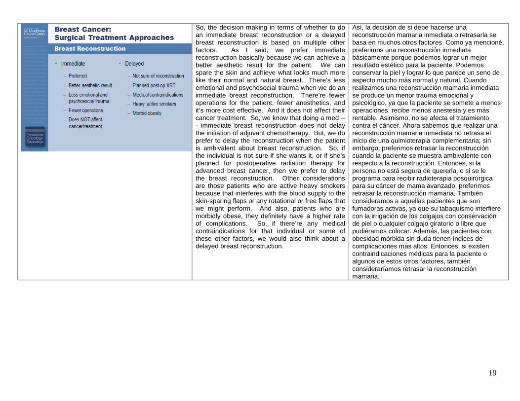
So, the decision making in terms of whether to do an immediate breast reconstruction or a delayed breast reconstruction is based on multiple other factors. As I said, we prefer immediate reconstruction basically because we can achieve a better aesthetic result for the patient. We can spare the skin and achieve what looks much more like their normal and natural breast. There’s less emotional and psychosocial trauma when we do an immediate breast reconstruction. There’re fewer operations for the patient, fewer anesthetics, and it’s more cost effective. And it does not affect their cancer treatment. So, we know that doing a med --- immediate breast reconstruction does not delay the initiation of adjuvant chemotherapy. But, we do prefer to delay the reconstruction when the patient is ambivalent about breast reconstruction. So, if the individual is not sure if she wants it, or if she’s planned for postoperative radiation therapy for advanced breast cancer, then we prefer to delay the breast reconstruction. Other considerations are those patients who are active heavy smokers because that interferes with the blood supply to the skin-sparing flaps or any rotational or free flaps that we might perform. And also, patients who are morbidly obese, they definitely have a higher rate of complications. So, if there’re any medical contraindications for that individual or some of these other factors, we would also think about a delayed breast reconstruction.
Así, la decisión de si debe hacerse una reconstrucción mamaria inmediata o retrasarla se basa en muchos otros factores. Como ya mencioné, preferimos una reconstrucción inmediata básicamente porque podemos lograr un mejor resultado estético para la paciente. Podemos conservar la piel y lograr lo que parece un seno de aspecto mucho más normal y natural. Cuando realizamos una reconstrucción mamaria inmediata se produce un menor trauma emocional y psicológico, ya que la paciente se somete a menos operaciones, recibe menos anestesia y es más rentable. Asimismo, no se afecta el tratamiento contra el cáncer. Ahora sabemos que realizar una reconstrucción mamaria inmediata no retrasa el inicio de una quimioterapia complementaria; sin embargo, preferimos retrasar la reconstrucción cuando la paciente se muestra ambivalente con respecto a la reconstrucción. Entonces, si la persona no está segura de quererla, o si se le programa para recibir radioterapia posquirúrgica para su cáncer de mama avanzado, preferimos retrasar la reconstrucción mamaria. También consideramos a aquellas pacientes que son fumadoras activas, ya que su tabaquismo interfiere con la irrigación de los colgajos con conservación de piel o cualquier colgajo giratorio o libre que pudiéramos colocar. Además, las pacientes con obesidad mórbida sin duda tienen índices de complicaciones más altos. Entonces, si existen contraindicaciones médicas para la paciente o algunos de estos otros factores, también consideraríamos retrasar la reconstrucción mamaria.

20
There’re what --- many different ways to reconstruct the breast. Typically, what’s been used in the past is an implant-based reconstruction. So, a tissue expander is placed behind the pectoralis muscle after the breast has been removed and then either a saline or silicone implant can be placed at a later time once the expansion has been complete. This is obviously less surgery, it’s a shorter recovery time for the patient, and there’s no other incisions anywhere else on the body, so there’s no donor site morbidity. But, typically, with time, patients will get contractures, sometimes infections or other considerations that require removal of the implant and additional surgeries. We generally will consider autologous tissue when appropriate for the patient that has enough of her own tissue to make a breast mound, and that’s typically going to be from the abdominal area or the back, using a latissimus flap. It does provide a more natural shape and feel with the reconstructed breast. It’s more complex surgery and does take a longer recovery time but long-term, there’re fewer complications. And in general, it provides a better aesthetic outcome.
Existen distintas formas de reconstruir la mama. Por lo general, lo que se ha usado en el pasado es una reconstrucción basada en un implante. Una vez extirpada la mama se coloca un extensor de tejidos detrás del músculo pectoral y luego se coloca un implante de solución salina o silicón poco después de que se haya completado la extensión. Por obvias razones es un cirugía menor, el tiempo de recuperación de la paciente es menos y no se realizan otras incisiones en el cuerpo, por lo que no hay morbilidad del sitio del donante. No obstante, con el paso del tiempo, las pacientes sufren contracturas, algunas veces infecciones u otras lesiones que requieren la remoción del implante y otras cirugías. En general, empleamos tejido autólogo cuando es adecuado para la paciente, siempre que tenga suficiente tejido propio como para crear un montículo mamario, el cual suele obtenerse del área abdominal o de la espalda, usando un colgajo dorsal. Este procedimiento permite crear un seno reconstruido con una forma y una textura más naturales. Se trata de una cirugía más compleja y el tiempo de recuperación es mayor, pero con menos complicaciones a largo plazo. Y, en general, produce un resultado estético más aceptable.
For the skin-sparing mastectomy, the definition that we use for this is an en bloc resection of the nipple-areolar complex with any biopsy scars and the underlying breast tissue, and axillary contents as appropriate. It provides a much better cosmetic outcome, it’s oncologically safe, and --- but it is technically more demanding for the individual surgeon.
En el caso de la mastectomía con conservación de piel, la definición que empleamos es de una resección completa del complejo pezón-areola sin cicatrices de biopsia, así como del tejido mamario subyacente y el contenido axilar, si corresponde. Se obtiene un mejor resultado estético y es seguro en términos oncológicos, pero, técnicamente hablando, para los cirujanos es un procedimiento más demandante.

21
This is a diagram showing several different incisions that we use for skin-sparing mastectomies. The most common one is to make an incision around the nipple-areolar complex and remove the breast tissue by lifting up the breast skin and removing any axillary contents through a separate axillary incision. But, as you can see, there’re other approaches. This is sometimes called the tennis racket or lollipop incision. Also, for patients who have significant ptosis in the breast, you can use a Wise pattern incision much as you would if you were doing a reduction mammoplasty. You can also remove a more --- a larger skin ellipse and be --- and close this primarily.
Este diagrama muestra distintas incisiones empleadas en las mastectomías con conservación de piel. La más común consiste en realizar una incisión alrededor del complejo pezón-areola y extirpar el tejido mamario levantando la piel de la mama y extirpando el contenido axilar mediante otra incisión axilar. Sin embargo, como puede verse, existen otros enfoques. A esto se le conoce a veces como incisión periareolar con prolongación lateral en forma de raqueta de tenis o paleta. Para las pacientes con ptosis importante en la mama se puede emplear una incisión en patrón de Wise, tal como se realizaría en una mamoplastia de reducción. También se puede extirpar una elipse más grande de piel y cerrarla en principio.
This shows a patient who’s had a bilateral skin-sparing mastectomy with a nipple reconstruction. You can see her lateral incisions here the --- that lateral approach was used in this case. And you can see the incision from her abdominal flap here. So, the cosmetic outcome is quite good in that this looks much more like her natural breast. It feels much more like a natural breast, and long-term, there are very few complications that would lead to need for further surgery.
Aquí se muestra a una paciente a quien se le realizó una mastectomía bilateral con conservación de piel y reconstrucción de pezón. Se pueden observar las incisiones laterales empleadas en este caso. También se puede ver la incisión del colgajo abdominal. Como resultado, la apariencia estética es muy buena, ya que se asemeja mucho más a un seno natural. También tiene la misma textura que un seno natural y, a largo plazo, se presentan muy pocas complicaciones que podrían dar lugar a otra cirugía.

22
In terms of nipple-areolar preservation, this is something that’s become more popular over the last five to seven years. And this is mo --- most common in women undergoing prophylactic mastectomy, so those patients who are BRCA1 or BRCA2 mutation carriers. There’s some concern that we might be leaving more ductal epithelium behind the nipple-areolar complex and this may increase cancer risk. But, most of the studies that were done a couple of decades ago using subcutaneous mastectomy where there was a significant amount of breast tissue left behind the nipple-areolar complex, those individuals had more than a 90 percent reduction in the development of breast cancer. So, this nipple-areolar preservation is probably a safer approach. There’s left --- less tissue left behind than there is with the subcutaneous mastectomy. In patients who are undergoing mastectomy for cancer, there have been a lot of retrospective studies published in the literature looking at the incidence of occult cancers in the nipple-areolar complex. So, in other words, the mastectomy was performed for a lesion somewhere else in the breast. And then the pathologist looked at the nipple-areolar complex and identified a cancer there. And this ranges anywhere from 8 to 50 percent. So, there’s certainly a lot of variability there. But, now, with improved imaging, it’s very uncommon to find an occult cancer in the nipple-areolar complex.
En términos de la conservación del pezón y la areola, se trata de una técnica que se ha vuelto más popular en los últimos cinco a siete años y es más común en las mujeres a quienes se les realiza una mastectomía profiláctica, como aquellas que son portadoras de una mutación BRCA1 o BRCA2. Existe la preocupación de que podrían quedar más restos del epitelio ductal detrás del complejo pezón-areola, lo que puede aumentar el riesgo de cáncer; no obstante, en la mayoría de los estudios realizados hace un par de décadas con mastectomías subcutáneas en las que se dejó bastante tejido mamario detrás del complejo pezón-areola, las pacientes mostraron una disminución de más del 90% en el índice de desarrollo de cáncer de mama. Entonces, la conservación del pezón y la areola es probablemente el enfoque más seguro debido a que se deja menos tejido que en una mastectomía subcutánea. En las pacientes a quienes se realiza una mastectomía debido al cáncer, se han llevado a cabo muchos estudios retrospectivos publicados para analizar la incidencia de cáncer oculto en el complejo pezón-areola. En otras palabras, la mastectomía se realizó para lesiones ubicadas en otros lugares de la mama. En dicho caso, el patólogo observó el citado complejo e identificó un tipo de cáncer. Esto sucedió entre el 8% y el 50% de los casos, lo que ciertamente indica mucha variabilidad. No obstante, ahora, con la mejora de las imágenes, es muy poco común encontrar un cáncer oculto en el complejo pezón-areola.

23
This shows a patient who’s undergoing a bilateral prophylactic mastectomy. She’s a BRCA mutation carrier. And what you can see is the --- in the lateral projection here, the breast was removed through a radial incision in the lateral aspect of the breast so that the nipple-areolar complex was completely maintained. No incisions are placed around the nipple-areolar complex and she had a bilateral implant-based reconstruction with a good cosmetic outcome.
Aquí se muestra a una paciente a quien se le realizó una mastectomía profiláctica bilateral. Ella es portadora de una mutación de BRCA. En la proyección lateral podemos observar que la mama se extirpó mediante una incisión radial en la parte lateral de la mama, de manera que se pudiera mantener por completo el complejo pezón-areola. Alrededor del complejo no se realizó ninguna incisión, por lo que se llevó a cabo una reconstrucción bilateral con implante, con muy buenos resultados estéticos.
And this just shows another patient who is planned for a bilateral skin spar --- or nipple-areolar sparing mastectomy, showing projection of the nipple.
En esta diapositiva se muestra a otra paciente a quien se le programó una mastectomía bilateral con conservación de piel o de pezón y areola. De hecho, podemos ver la proyección del pezón.

24
And after the mastectomy has been performed, you can see the radial incision here. And she still has quite good projection of the nipple. So, there --- this procedure is possible for many patients to maintain the nipple-areolar complex and have a more natural appearance to the breast.
Y una vez realizada la mastectomía, se puede observar la incisión radial. Como pueden ver, el pezón sigue mostrando una muy buena proyección. Así pues, este procedimiento es posible para que muchas pacientes mantengan el complejo pezón-areola y que su mama tenga una apariencia más natural.
Right now, what we use as eligibility criteria for nipple-areolar sparing is patients who are undergoing prophylactic mastectomy or those with early stage breast cancer, so Stage 0, I, and II breast cancer, who have a primary tumor that’s at least two and a half centimeters away from the nipple-areolar complex. And also, we look for any evidence of microcalcifications extending toward the nipple, such that if there’s any evidence of ductal extension of ductal carcinoma in situ or another process toward the nipple, we would not perform a nipple-sparing mastectomy on these individuals.
En este momento nuestros criterios de idoneidad para la conservación del pezón y la areola incluyen a las pacientes a quienes se les realiza una mastectomía profiláctica o aquellas con cáncer de mama incipiente, es decir, cáncer de mama en estadio 0, I y II, y con un tumor primario que se encuentra al menos a 2.5 centímetros del complejo pezón-areola. También buscamos evidencia de microcalcificaciones que se extienden hacia el pezón, por lo que cuando hay indicios de extensión ductal o carcinoma ductal in situ u otro proceso que se extiende hacia el pezón, no realizamos la mastectomía con conservación del pezón.

25
We do not consider this procedure for smokers. As I mentioned, with skin-sparing mastectomy and immediate breast reconstruction, smokers have more complications. And they definitely have more incidence of nipple necrosis with nipple-areolar sparing mastectomy. We would also not consider this for patients with inflammatory breast cancer or breast skin involvement, those patients with collagen vascular disease, or those with Paget’s disease of the nipple.
Este procedimiento tampoco lo realizamos en fumadoras ya que, como se mencionó anteriormente, ellas tienen más complicaciones cuando se someten a una mastectomía con conservación de piel y a una reconstrucción mamaria inmediata. También tienen mayor incidencia de necrosis del pezón cuando se les realiza una mastectomía con conservación del pezón y la areola. Tampoco realizamos estos procedimientos en las pacientes con cáncer de mama inflamatorio o afectación de la piel mamaria, ni en las pacientes con conectivitis vascular o enfermedad de Paget en el pezón.
And something that --- that we’re starting to explore here more and more at MD Anderson is the use of immediate breast reconstruction even in patients who require post-mastectomy radiation. This has been a very significant multidiscipline --- multidisciplinary effort in that it requires significant coordination between the breast surgeon, the plastic surgeon, and the radiation oncologist. More and more, patients that have Stage II breast cancer, so those with a few positive nodes or a larger primary tumor, might be considered for post-mastectomy radiation. And this is why we’ve incorporated this strategy into our reconstructive algorithm. So, in patients where we think they might need post-mastectomy radiation, we do a skin-sparing mastectomy and place a tissue expander in the subpectoral position and then look at the final pathology, both the assessment of the primary tumor and the nodal disease. And we have a postoperative consultation with the radiation oncologist. If the patient does not require any post-mastectomy radiation therapy, they can proceed to their definitive breast reconstruction, whether that’s an implant-based reconstruction or an autologous tissue reconstruction. But, in those patients where it looks like they will require radiation therapy, we try to get full expansion of the tissue expander within a few weeks and then deflate the expander
Algo que hemos comenzado a explorar con mayor frecuencia en el MD Anderson es la reconstrucción mamaria inmediata incluso en las pacientes que requieren radiación posterior a la mastectomía. Se trata de un esfuerzo multidisciplinario muy importante, ya que requiere la coordinación minuciosa entre el cirujano de mama, el cirujano plástico y el oncólogo radioterapeuta. Cada vez más las pacientes con cáncer de mama en estadio II, es decir, aquellas con afectación en algunos ganglios o un tumor primario más grande, pueden recibir radiación luego de una mastectomía. Este es el motivo por el que hemos incorporado esta estrategia a nuestro algoritmo reconstructivo. En las pacientes que consideramos que podrían necesitar radiación luego de su mastectomía, realizamos una mastectomía con conservación de piel, colocamos un extensor de tejidos en la posición subpectoral y realizamos una evaluación patológica final tanto del tumor primario como de la enfermedad ganglionar. Asimismo, le pedimos al oncólogo radioterapeuta que realice una revisión posquirúrgica. Si la paciente no requiere radioterapia posterior a la mastectomía, se le puede realizar una reconstrucción mamaria definitiva, ya sea que se trate de una reconstrucción con implante o con tejido autólogo. Sin embargo, en aquellas pacientes que al parecer deben recibir radioterapia, tratamos

26
before the radiation takes place. So, that there’s better geometry for the radiation oncologist in treating all of the areas at risk, the chest wall, and any regional nodal basins that need to be treated. We then reinflate the expander immediately after radiation and then, the patient ultimately, a few weeks to months later, will have definitive breast reconstruction. And typically, this is going to be with an autologous reconstruction, either a TRAM flap or a latissimus dorsi flap.
de extender totalmente el extensor de tejidos en el transcurso de algunas semanas y enseguida desinflamos el extensor antes de la radiación. Así, el oncólogo radioterapeuta logra una mejor geometría al tratar todas las áreas en riesgo, la pared torácica y cualquier región ganglionar que requiera tratamiento. Inmediatamente después de la radiación volvemos a inflar el extensor y, por último, unas semanas o meses después, se realiza a la paciente una reconstrucción mamaria definitiva. Por lo general, se trata de una reconstrucción autóloga, con un colgajo de tipo TRAM o un colgajo dorsal.
And this has helped us to save the breast skin for many patients who would otherwise have had a rad --- a modified radical mastectomy with removal of all the skin and then require a significant skin replacement using autologous tissue reconstruction. We look at patients who have clinical Stage I or II disease who we think they might need radiation. Sometimes, it’s difficult to tell if they will, especially in patients who have extensive microcalcifications on mammography or those who have multicentric disease on their imaging where we think that final pathology may lead us to incorporate post-mastectomy radiation. In these patients, they need to be able to withstand two anesthetic procedures, clearly their initial surgery and then their later reconstructive surgery.
Esto nos ha ayudado a conservar la piel mamaria de muchas pacientes que de otra forma hubieran tenido que someterse a una mastectomía radical modificada con extirpación de toda la piel y luego a un reemplazo considerable de piel mediante una reconstrucción de tejido autólogo. Observamos a pacientes con una enfermedad en estadio I o II que, desde nuestra perspectiva, podrían necesitar radiación. Algunas veces es difícil decir si la necesitarán, especialmente aquellas que muestran microcalcificaciones generalizadas en una mamografía, o aquellas con una enfermedad multicéntrica en las imágenes, pues nos hacen pensar que la patología final puede llevarnos a incorporar radiación después de la mastectomía. Esas pacientes deben tener la capacidad para soportar dos procedimientos anestésicos: obviamente una cirugía inicial y, más adelante, una cirugía reconstructiva.

27
This shows a case of a patient with a T2 N1 breast cancer where she had a skin-sparing mastectomy, placement of a tissue expander. And you can see that expander was fully expanded pret --- pretty rapidly in order to save as much of that breast skin as possible. And then, you can see the photo here where it’s been deflated to allow the radiation oncologist to treat all of the areas at risk.
En esta imagen se muestra el caso de una paciente con cáncer de mama T2 N1, a quien se le realizó una mastectomía con conservación de piel y se le colocó un extensor de tejidos. Podemos observar que el extensor se extendió rápidamente para conservar tanta piel mamaria como fuera posible. Enseguida podemos ver la foto en que se desinfló para permitir que el oncólogo radioterapeuta tratara todas las áreas en riesgo.
So, skin-sparing mastectomy is a very oncologically safe tool we’ve studied now for several decades. And recurrence rates are no different than those patients undergoing a standard modified radical mastectomy. And a skin-sparing with immediate reconstruction is generally reserved for those patients who will not require radiation after surgery. But, we are exploring this approach of immediate delayed reconstruction for patients where we think they might need radiation, where we can place a tissue expander, deflate it, and inflate it as needed. And finally the role of nipple-areolar complex preservation is still under investigation. But, we think this is a useful tool especially for those patients undergoing prophylactic mastectomy for risk reduction.
Así pues, la mastectomía con conservación de piel es una herramienta muy segura en términos oncológicos que hemos investigado desde hace varias décadas, y los índices de recidiva no distan muchos de los de las pacientes que se someten a una mastectomía radical modificada estándar. Por lo general, la conservación de piel con reconstrucción inmediata se reserva para las pacientes que no requerirán radiación tras una cirugía; sin embargo, estamos explorando este enfoque de reconstrucción inmediata retrasada para las pacientes que pensamos que podrían necesitar radiación y a quienes se les coloca un extensor de tejidos para luego desinflarlo y volverlo a inflar, según se necesite. Por último, el papel de la conservación del complejo pezón-areola continúa en investigación, aunque pensamos que se trata de una herramienta útil, especialmente para las pacientes que se someten a una mastectomía profiláctica para la reducción de riesgos.

28
The final area that I’m going to talk about, then, is axillary lymph node staging.
Lo último de lo que voy a hablar ahora es de la estadificación de los ganglios linfáticos axilares.
The goals of staging are, of course, to provide an accurate stage for that individual patient to allow for the incorporation of the appropriate systemic and adjuvant therapies. We also want to provide regional nodal control for the individual patient. But, we know there’s no benefit to removing healthy uninvolved lymph nodes. And there’s also significant morbidity associated with the use of a standard axillary lymph node dissection.
Los objetivos de la estadificación son, evidentemente, asignar un estadio preciso a cada paciente para permitir la incorporación de las terapias sistémicas y complementarias adecuadas. Buscamos el control ganglionar regional de cada paciente; no obstante, sabemos que extirpar ganglios sanos y no afectados no ofrece beneficio alguno y que la disección estándar de ganglios axilares se asocia a una morbilidad importante.

29
The complications of an axillary dissection are most commonly lymphedema and this occurs in anywhere to 20 to 25 percent of patients with early stage breast cancer. And the rates are significantly higher in patients with more advanced breast cancer who have both surgery and radiation. There’re also a number of other things that occur after axillary surgery: shoulder dysfunction, paresthesias and pain from interruption of intercostal brachial nerves. And patients can also have persistent seromas and hematomas requiring aspirations and other interventions.
La complicación más frecuente de una disección axilar es el linfedema, que se presenta entre el 20% y el 25% de las pacientes con cáncer de mama incipiente, pero los índices son significativamente más altos en las pacientes con cáncer de mama más avanzado que se someten a cirugía y radiación. Otros eventos que se presentan luego de una cirugía axilar incluyen disfunción del hombro, parestesia y dolor debido a la interrupción de los nervios braquiales intercostales. Asimismo, las pacientes pueden presentar seromas y hematomas persistentes que requieran aspiraciones y otras intervenciones.
The use of lymphatic mapping with sentinel lymph node biopsy was first popularized by Donald Morton in the 1990s for patients with clinically node negative melanoma. And the sentinel node is defined as that first draining lymph node from the primary tumor. And it was found with melanoma and other solid tumors that the sentinel node was predictive of the status of the remaining nodal basin. So, that those patients with a positive node were more likely to require a node dissection. Those patients with a negative sentinel lymph node did not need removal of the remaining lymph nodes. It was David Krag and Armando Giuliano that first reported the use of sentinel node surgery for breast cancer patients in the early 1990s.
El empleo de un la cartografía linfática con biopsia del ganglio centinela fue popularizado inicialmente por Donald Morton en la década de 1990 entre las pacientes con melanoma sin afectación clínica de ganglios. Un ganglio centinela se define como el primer ganglio linfático con drenaje asociado al tumor primario. En los casos de melanoma y otros tumores sólidos, se descubrió que el ganglio centinela predecía el estado de la región ganglionar residual, por lo que las pacientes con afectación ganglionar tenían más probabilidades de requerir una disección ganglionar, mientras que las pacientes sin ganglio centinela no requerían la extirpación de los ganglios linfáticos residuales. Fueron David Krag y Armando Giuliano quienes reportaron por primera vez el empleo de la cirugía del ganglio centinela para las pacientes con cáncer de mama a inicios de la década de 1990.

30
And it’s now become standard of care for patients with early stage clinically node negative breast cancer. Typically, a peritumoral injection of either a blue dye or a radioisotope, or a combination of those are utilized. And this travels through the lymphatic channels to the first draining lymph node, also called the sentinel node.
Hoy en día se ha convertido en el tratamiento de referencia de las pacientes con cáncer de mama incipiente sin afectación clínica de ganglios. Por lo general, se emplea una inyección peritumoral de un tinte azul o un radioisótopo, o una combinación de ambos, que se desplaza a través de los canales linfáticos hasta el primer ganglio linfático con drenaje, también llamado ganglio centinela.
In the operating room, we can identify the blue dye within the axillary contents and when the blue lymphatic --- when the blue dye is passed through the lymphatic channels and accumulates within a lymph node such as this, it’s very easy to discriminate. But, we can also use, as I said, the radioisotope. And we have a handheld probe that registers radioactive counts, in this case, 22,000 counts that are easy to discriminate the uptake from surrounding lymph nodes.
En el quirófano, podemos identificar el tinte azul en el contenido axilar y, cuando el tinte azul pasa a través de los canales linfáticos y se acumula en un ganglio linfático como éste, es muy fácil distinguirlo. Sin embargo, también podemos usar, como ya mencioné, el radioisótopo. Asimismo, contamos con una sonda portátil que registra conteos radioactivos; en este caso, 22,000 conteos que pueden distinguir fácilmente la captación de los ganglios linfáticos de alrededor.

31
Now, there’re several trials that have been performed to assess the safety and utility of sentinel lymph node surgery in breast cancer patients.
Ahora bien, existen numerosos ensayos que se han realizado para evaluar la seguridad y utilidad de la cirugía del ganglio centinela en las pacientes con cáncer de mama.
And the first large trial in the U.S. was the NSABP B-32 trial. In this case, the hypothesis was that sentinel lymph node surgery alone was safe compared with sentinel lymph node surgery with completion axillary dissection. So, patients that had clinically node negative breast cancer were stratified based on age, clinical tumor size, and the planned surgical procedure, and then randomized to either go sentinel lymph node surgery with a completion axillary dissection at the same surgery, or sentinel lymph node surgery alone followed by axillary dissection only in the case when standard H&E staining identified a positive sentinel node. So, those patients who had a negative sentinel node had no specific axillary treatment.
El primer ensayo grande en los Estados Unidos fue el B-23 de NSABP. En este caso, la hipótesis era que la cirugía del ganglio centinela por sí sola era segura en comparación con la cirugía del ganglio centinela con disección axilar de finalización. Entonces, las pacientes con cáncer de mama sin afectación clínica de los ganglios fueron estadificadas con base en su edad, el tamaño del tumor clínico y el procedimiento quirúrgico planificado, y luego eran aleatorizadas a una cirugía del ganglio centinela con disección axilar de finalización en la misma cirugía, o bien, a una cirugía del ganglio centinela sola seguida de una disección axilar únicamente en los casos en que la tinción estándar de H&E identificara la afectación de un ganglio centinela. Así, las pacientes sin afectación del ganglio centinela no recibieron ningún tratamiento axilar específico.

32
This trial was recently reported in Lancet Oncology. The overall accuracy was 97 percent.
Este ensayo fue publicado recientemente en Lancet Oncology y la precisión general fue del 97%.
And you can see the technical success rate here, based on tumor size, overall 97 percent, and basically the --- even in patients --- patients with larger tumors, even those with greater than 5 cm tumors, the success rate was quite good. Now, one thing that was somewhat unexpected with this trial was the relatively high false negative rate. It was almost 10 percent overall. It did not vary based on tumor size so it was stable across even larger tumors. But, what this tells us is that even in --- in very trained hands, all these investigators were trained in the use of sentinel lymph node surgery before the trial. Even in well trained hands, there is a relatively high rate of identifying a negative sentinel lymph node but the axillary nodes still harbored disease within them.
Aquí podemos ver el índice general de éxito técnico del 97% basado en el tamaño del tumor. Incluso en las pacientes con tumores más grandes, incluidas aquellas con tumores de más de 5 cm, el índice de éxito fue muy bueno. Ahora bien, un dato que nos pareció un tanto imprevisto en este ensayo fue el índice relativamente alto de falsos negativos, que fue de casi 10% en total y que no varío según el tamaño del tumor, sino que se mantuvo estable incluso en los tumores más grandes. No obstante, esto nos indica que incluso en manos bien capacitadas —pues todos estos investigadores recibieron formación en el empleo de la cirugía del ganglio centinela antes del ensayo—, incluso en manos bien capacitadas, existe un índice relativamente alto de no afectación del ganglio centinela, pero los ganglios axilares pueden seguir albergando una enfermedad en su interior.

33
The factors that affected the false negative rate were the tumor locations. So, those tumors in the lateral breast were more likely to have a false negative event. Perhaps, because of shine through from the radioactivity in the lateral breast, it was hard to discriminate in the axilla. Patients who had had an excisional biopsy before surgery were more likely to have a false negative sentinel node. And those patients who only had one or two sentinel lymph nodes recovered were more likely to have a false negative sentinel node. This tells us, and the average number of sentinel nodes removed in most trials is 2.5 to 2.7, so this tells us that there is usually more than one sentinel node and that it’s --- it’s best staging when all of the sentinel nodes are recovered.
El factor que afectó el índice de falsos negativos fue la ubicación del tumor. Los tumores en la parte externa de la mama tuvieron más probabilidad de mostrar un falso negativo, quizá debido al brillo que emite la radioactividad en esta zona, por lo que fue más difícil distinguirlo en la axila. Las pacientes a quienes se les realizó una biopsia por escisión antes de una cirugía fueron más propensas a tener un ganglio centinela falso negativo, al igual que aquellas pacientes a quienes se les recuperó uno o dos ganglios centinela. Esto nos indica que la cantidad promedio de ganglios centinela extirpados en la mayoría de los ensayos es de 2.5 a 2.7, es decir, que generalmente hay más de un ganglio centinela y que es más conveniente realizar la estadificación cuando todos los ganglios centinela se han recuperado.
The American College of Surgeons Oncology Group also initiated some sentinel node trials at the same time as the NSABP trial. This was in the late 1990s. These trials all completed in the mid 2000s but were only recently published with long-term follow up. In the ACOSOG trial, patients with T1 and T2 tumors that were planned for breast conservation had sentinel lymph node biopsy. And if the sentinel node was negative, they had no further treatment of the axilla but they all had radiation treatment to the breast. If they had a positive sentinel node, they were eligible for the Z0011 trial. And in the Z0011 trial, patients with one or two positive sentinel nodes were randomized to have a completion axillary dissection as was the standard at the time, or axillary observations, so no further treatment. Again, they all had breast conservation and all had a standard tangential breast radiation to the intact breast without specific radiation to the axilla or the supraclavicular nodes.
El Grupo de Oncología del Colegio Americano de Cirujanos o ACOSOG también inició algunos ensayos sobre ganglios centinela al mismo tiempo que el ensayo NSABP a finales de la década de 1990. Todos estos ensayos finalizaron a mediados de la siguiente década, pero fueron publicados recientemente con un seguimiento a largo plazo. En el ensayo del ACOSOG, las pacientes con tumores T1 y T2 a quienes se les programó una conservación de la mama se sometieron a una biopsia del ganglio centinela. Si éste no estaba afectado, no recibían más tratamiento en la axila, sino que a todas se les radiaba en la mama; pero si estaba afectado, podían participar en el ensayo Z0011. En ese ensayo las pacientes con afectación de uno o más ganglios centinela fueron aleatorizadas para una disección axilar de finalización como era el tratamiento de referencia en ese entonces, o bien se sometían a revisiones axilares sin tratamiento adicional. Todas conservaron la mama y recibieron radiación tangencial estándar en la mama intacta sin radiación específica en la axila o los ganglios supraclaviculares.

34
In the Z0010 trial, there were over 5,000 patients enrolled. Twenty-four percent had a positive sentinel node by standard H&E staining.
En el ensayo Z0010 se inscribieron más de 5,000 pacientes y al 24% se le detectó afectación del ganglio centinela mediante una tinción estándar de H&E.
And you can see the complication rate, relatively low. Only 7.5 percent of patients had a seroma that required any aspiration or other intervention.
Aquí se puede observar el índice de complicaciones, el cual fue relativamente bajo. Sólo el 7.5% de las pacientes tuvo un seroma que requirió una aspiración u otro tipo de intervención.

35
The lymphedema rate, however, was higher than what we expected. As I said, with axillary dissection, it’s reported to be somewhere between 20 to 25 percent. In this trial, sentinel node only, it was about 7 percent and this was based on arm measurements with a 2 cm difference with the ipsilateral treated arm versus the contralateral arm.
No obstante, el índice de linfedema fue más alto que lo esperado. Como ya se mencionó, con la disección axilar se reportó entre un 20% y un 25%. En este ensayo, de ganglios centinela solamente, fue de aproximadamente 7% según las mediciones del brazo con una diferencia de 2 cm con el brazo homolateral tratado respecto del brazo contralateral.
The predictors for those patients that develop lymphedema were those patients of older age. So with increasing decade, there was a higher rate of lymphedema. And also, with increasing BMI, so patients that had increasing BMI had a higher rate of lymphedema. So, this information is very helpful to us in counseling our patients.
Los factores de predicción de aquellas pacientes que desarrollaron linfedema fueron aquellas pacientes de mayor edad. Así, conforme avanzó la década, aumentó el índice de linfedemas. De igual forma, conforme aumentaba el IMC, las pacientes mostraban un mayor índice de linfedemas. Esta información es muy útil al asesorar a nuestras pacientes.

36
In terms of failure to identify a sentinel node, there were both surgeon factors and patient factors. So, the number of cases that the surgeon performed on this trial were important, suggesting that the experience of the surgeon is very critical, and the volume of cases that the surgeon performs is an important factor. And also, increasing age and increasing BMI were also factors that predicted for the failure to identify sentinel node at surgery.
En cuanto a la incapacidad para identificar un ganglio centinela, hubo factores relacionados con el cirujano y otros relacionados con la paciente. El número de casos en los que participó el cirujano en este ensayo fue importante, lo que sugiere que la experiencia del cirujano es fundamental y el volumen de casos en los que participa constituye un factor importante. Asimismo, el aumento de la edad y del IMC fueron también factores que permitieron predecir la incapacidad para identificar un ganglio centinela en la cirugía.
Now, recently, the Z0011 trial was reported and this has created a lot of excitement in the surgical community. And in the breast cancer community, in general, because this suggests this trial --- the results of this trial suggest that axillary lymph node dissection is not needed even in all patients who have a positive sentinel node. So, again, in this trial, small tumors T1 or T2 tumors, if the patient had a sentinel lymph node that was positive, they were eligible to be registered in the trial. And they were randomized to undergo an axillary lymph node dissection or no further surgery. They all received breast radiation as standard for breast conserve --- conservation. And the systemic therapy, either adjuvant chemotherapy or endocrine therapy, was based on the primary tumor factors and individual physician treatment decisions.
Recientemente se informó sobre el ensayo Z0011, lo que ha generado mucha emoción en la comunidad quirúrgica y en la comunidad del cáncer de mama en general, debido a que los resultados sugieren que la disección del ganglio linfático axilar no es necesaria incluso en todas aquellas pacientes con afectación del ganglio centinela. En este ensayo, con los tumores pequeños T1 o T2, si las pacientes tenían afectado el ganglio centinela, se les consideraba aptas para participar en el ensayo y eran aleatorizadas para someterse a una disección del ganglio linfático axilar, o bien para ninguna cirugía adicional. Todas recibieron radiación en la mama como parte del tratamiento de conservación de referencia, y la terapia sistémica, ya sea quimioterapia complementaria o terapia endocrina, se basó en los factores relacionados con el tumor primario y en las decisiones de tratamiento de cada médico.

37
If we look at the patients --- the characteristic patients that were enrolled in this trial, you can see that 65 percent were over age 50. So, this was largely a postmenopausal patient population. The majority had small T1 tumors, 80 percent were estrogen receptor positive, and only 38 percent had any lymphovascular space invasion. Most patients had only one positive sentinel node, although there were some with two or even three positive sentinel nodes. And the majority received syst --- systemic therapy, 60 percent received adjuvant chemotherapy, which is the standard in the U.S. at the time of the --- this trial.
Si analizamos las características de las pacientes que se inscribieron en este ensayo, podemos ver que el 65% tenía más de 50 años, es decir, que se trató en gran parte de una población posmenopáusica. La mayoría tenía tumores T1 pequeños, el 80% con receptores estrogénicos y sólo el 38% con invasión de espacio linfovascular. La mayoría de las pacientes tenía sólo un ganglio centinela afectado, aunque hubo algunas con dos o incluso tres ganglios centinela afectados. La mayoría recibió terapia sistémica y el 60% recibió quimioterapia complementaria, que era el procedimiento de referencia en los Estados Unidos al momento del ensayo.
Now, a completion lymph node dissection, only 27 percent of patients who had the completion node dissection had additional positive nodes in the axilla, and only 14 percent had four or more positive nodes. Now, despite the fact that the patients who did not have a node dissection would be expected to also have this amount of nodal disease in the axilla, there were no differences in local or regional recurrence rates between the two treatment arms. And you can see that they were very, very small numbers in --- in terms of axillary fail --- failures or regional recurrences.
Ahora, en cuanto a la disección del ganglio linfático de finalización, sólo el 27% de las pacientes que se la realizaron tenían otros ganglios afectados en la axila y sólo el 14% tenía cuatro o más ganglios afectados. Ahora bien, a pesar de que se esperaría que las pacientes a quienes no se les realizó una disección ganglionar tuvieran esta cantidad de enfermedad ganglionar en la axila, no hubo diferencias en los índices de recidiva local o regional entre los dos grupos de tratamiento. También se puede ver que el número de fracasos o recidivas regionales en la axila fue muy reducido.

38
So, when we look at the ACOSOG Z0011 trial results, again, all of the patients had positive sentinel nodes. But these are patients who all had breast conservation with whole breast radiation, tangential breast irradiation, and we know that that treats a significant portion of the axilla, based on many different single institution studies. There were no differences in local or regional recurrences. And there were no differences in overall survival between the two treatment arms.
Cuando analizamos los resultados del ensayo Z0011 del ACOSOG, todas las pacientes tuvieron ganglios centinela afectados, pero se trata de pacientes a quienes se les mantuvo intacta la mama con radiación en toda la mama, o radiación tangencial de la misma, las cuales, como sabemos tratan una porción importante de la axila, según varios estudios realizados por instituciones individuales. No se observaron diferencias en la recidiva local o regional, ni tampoco en la supervivencia general entre los dos grupos de tratamiento.
This suggests that axillary node dissection is not needed in all women who have a positive sentinel node. But, axillary dissection is still the gold standard for many cases. So, those patients who present with clinically positive nodes, those individuals should have an axillary node dissection. We still use axillary dissection in patients who have neoadjuvant chemotherapy and have a positive node after chemotherapy. And other considerations where we might still use axillary node dissection are those patients who are very young age. Again, in Z0011, the patients were largely postmenopausal and older patients. And those individuals with triple negative disease, where we know that local regional failure rates are higher in general. But, in patients and in --- and also in patients who wouldn’t otherwise need radiation. So, those women who are undergoing mastectomy who have one or two positive nodes that might not need post-mastectomy radiation therapy, so those low risk Stage II disease. So, there still are indications for the use of axillary dissection, but it’s important to note that many women undergoing breast conserving surgery who have small volume disease in the sentinel nodes probably do not benefit from an axillary lymph node dissection.
Esto sugiere que la disección de ganglios axilares no es necesaria en todas las mujeres con afectación del ganglio centinela; sin embargo, sigue siendo la regla de oro en muchos casos. Por eso las pacientes con afectación clínica de ganglios deben someterse a una disección de ganglios axilares. La disección axilar se sigue empleando en las pacientes que reciben quimioterapia prequirúrgica y con un ganglio afectado luego de la quimioterapia. Otro de los casos en los que se puede emplear la disección de ganglios axilares es en aquellas pacientes que son muy jóvenes. En el ensayo Z0011, las pacientes eran, en su mayoría, posmenopáusicas y de edad avanzada. También en las mujeres con una enfermedad triple negativa, donde sabemos que los índices de fracaso locorregional son más altos en general, así como en las pacientes que, por lo demás, no necesitarían recibir radiación, como en las mujeres a quienes se les realiza una mastectomía y tienen uno o más ganglios afectados que posiblemente no necesitarían radioterapia posterior a la mastectomía, o aquellas con una enfermedad de bajo riesgo en estadio II. Así pues, también existen indicaciones para el empleo de una disección axilar, pero es importante señalar que muchas mujeres que se someten a una cirugía con conservación de la mama y que tienen una enfermedad de bajo

39
volumen en los ganglios centinela probablemente no obtengan ningún beneficio de una disección de los ganglios axilares.
So, at the --- this time, the National Comprehensive Cancer Network Guideli --- Treatment GuidelinesTM suggest that sentinel node biopsy is appropriate for those patients who have a clinically node negative axilla. The team should have documented experience. And in general, axillary node dissection is performed for those with --- when the sentinel node is positive or when the sentinel node cannot be identified.
En este momento, los Lineamientos de Tratamiento de la Red Nacional Integral del Cáncer sugieren que la biopsia del ganglio centinela es adecuada para las pacientes con afectación clínica de un ganglio axilar. El equipo debe contar con experiencia documentada. Y, en general, la disección de ganglios axilares se realiza para aquellas con un ganglio centinela afectado o cuando no es posible identificarlo.
The ASCO Guidelines suggest that axillary dissection is --- should be performed when sentinel node is identified on routine histopathologic exam. I would imagine that we’ll see some modification as these guidelines were published in 2005. And the Z0011 results were only published in just the last year.
Las pautas de la A-S-C-O sugieren que la disección axilar se debe realizar cuando se identifica el ganglio centinela en una exploración histopatológica de rutina. Imagino que veremos algunas modificaciones, ya que estas pautas fueron publicadas en 2005 y los resultados del ensayo Z0011 apenas fueron publicados el año pasado.

40
So, in summary then, what I’ve presented today is --- is significant in terms of breast conservation and mastectomy in the treatment of early stage breast cancer. We know that there’re equivalent in terms of outcome for early stage breast cancer. But, breast conservation is also feasible for those individuals with locally advanced breast cancer or large primary tumors who have a good response to preoperative chemotherapy. Selection criteria are very important to achieve the optimal outcomes in these individuals. For those patients who require a mastectomy or elect to undergo mastectomy, we think that skin-sparing mastectomy provides the most optimal results with immediate reconstruction. And now, we’re considering nipple-sparing mastectomy in selected cases. And, finally, sentinel lymph node biopsy is a tool that is --- provides alternate staging to axillary lymph node dissection. So, it’s accurate, it’s safe, and it has less morbidity than a full axillary lymph node dissection. And in selected patients with a positive sentinel node, we can avoid the use of axillary lymph node dissection, especially those individuals who have breast conserving surgery with whole breast irradiation. I want to thank you very much for your attention and we are certainly happy to hear any feedback from you. Thank you.
En resumen, lo que hoy se presentó es muy importante en términos de la conservación de la mama y la mastectomía en el tratamiento del cáncer de mama incipiente. Sabemos que son equivalentes en cuanto al resultado del cáncer de mama incipiente; sin embargo, la conservación de la mama también es posible para las pacientes con cáncer de mama avanzado localmente o con grandes tumores primarios que muestran una respuesta aceptable a la quimioterapia prequirúrgica. Los criterios de selección son muy importantes para obtener resultados óptimos en estas pacientes. Para aquellas que requieren una mastectomía o que eligen someterse a una, pensamos que la mastectomía con conservación de piel brinda los resultados más óptimos con una reconstrucción inmediata. También analizamos la viabilidad de realizar una mastectomía con conservación de pezón en determinados casos. Y, por último, la biopsia del ganglio centinela es una herramienta que permite la estadificación alterna a la disección de los ganglios axilares. Es precisa, segura y tiene una morbilidad menor que una disección total de los ganglios axilares. Asimismo, en determinadas pacientes con un ganglio centinela afectado, podemos evitar emplear la disección de los ganglios axilares, en especial en aquellas pacientes que se someten a una cirugía con conservación de la mama con radiación total de la misma. Agradezco mucho su atención y con gusto escucharemos cualquier retroalimentación de su parte. Gracias.