Embed Size (px)
Citation preview
28/04/2016
1
I have nothing to declare and no financial
interest or relationship to disclose
Skeletal Anomalies are diverse range of complexities which is NOT
Easy to diagnose.
It is NOT Difficult to detect-Just be systematic and gather signs.
Start with the Femur.
Signs of poor prognosis (Look at the thorax).
Multi-team approach.
Since ancient times, “skeletal deformities” have
aroused great fascination that goes far beyond the
field of medicine, penetrating into areas such as
anthropology and art.

28/04/2016
2
28/04/2016
3
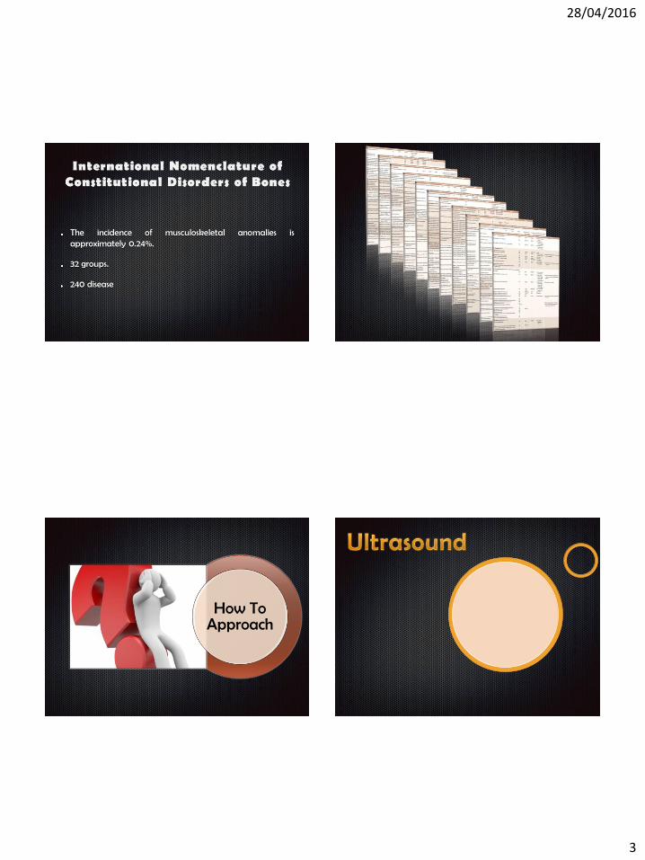
The incidence of musculoskeletal anomalies is approximately 0.24%.
32 groups.
240 disease
How To Approach

28/04/2016
4
Head • Cranial bones
Neck • Nuchal translucency thickness (if accepted • after informed consent and • trained/certified operator available)*
spine • Vertebrae (longitudinal and axial)*
Extremities • Four limbs each with three segments • Hands and feet with normal orientation*
Limb buds are first seen
The Femur /Humerus
Tibia/Fibula &
Radius/Ulna
Digits of the hands/feet
Head • Cranial bones
Face • Median facial profile
Chest • Normal appearing shape/size of chest
Skeletal • No spinal defects or masses (transverse and sagittal views) • Arms and hands present, normal relationships • Legs and feet present, normal relationships
28/04/2016
5
The lengths of the humerus, radius/ulna, femur and tibia/fibula are similar and increase linearly with gestation.
18–23-week the three segments of each extremity should be visualized,
Only necessary to measure the length of one femur.
The relationship of leg and foot should also be assessed to rule out clubfoot.
o Index case:
• Family history of dysplasia,
• Previous affected babies.
o Incidental finding during routine exam
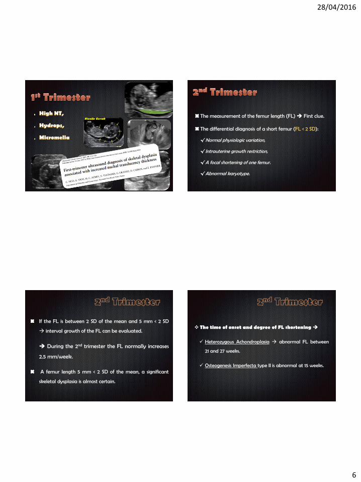
• 1st Trimester High NT, Hydrops, Micromelia .
• 2nd/3rd Trimester:
Micromelia (femur < - 4DS) ,
Abnormality of the shape / echogenicity of the bones
Abnormal Thorax (Tight, Short,…)
28/04/2016
6
Ksenija Gersak The measurement of the femur length (FL) First clue.
The differential diagnosis of a short femur (FL < 2 SD):
√Normal physiologic variation,
√Intrauterine growth restriction,
√A focal shortening of one femur.
√Abnormal karyotype.
If the FL is between 2 SD of the mean and 5 mm < 2 SD
interval growth of the FL can be evaluated.
During the 2nd trimester the FL normally increases
2.5 mm/week.
A femur length 5 mm < 2 SD of the mean, a significant
skeletal dysplasia is almost certain.
The time of onset and degree of FL shortening
Heterozygous Achondroplasia abnormal FL between
21 and 27 weeks.
Osteogenesis Imperfecta type II is abnormal at 15 weeks.
28/04/2016
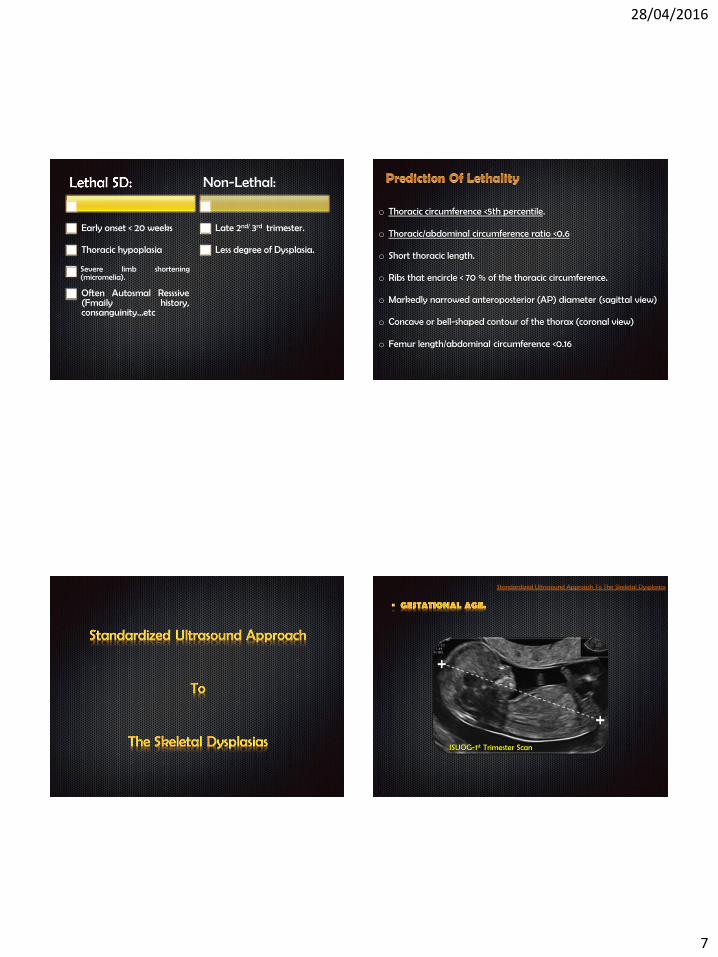
7
Early onset < 20 weeks
Thoracic hypoplasia
Severe limb shortening (micromelia).
Often Autosmal Resssive (Fmaily history, consanguinity…etc
Non-Lethal:
Late 2nd/ 3rd trimester.
Less degree of Dysplasia.
o Thoracic circumference <5th percentile.
o Thoracic/abdominal circumference ratio <0.6
o Short thoracic length.
o Ribs that encircle < 70 % of the thoracic circumference.
o Markedly narrowed anteroposterior (AP) diameter (sagittal view)
o Concave or bell-shaped contour of the thorax (coronal view)
o Femur length/abdominal circumference <0.16
ISUOG-1st Trimester Scan
28/04/2016
8
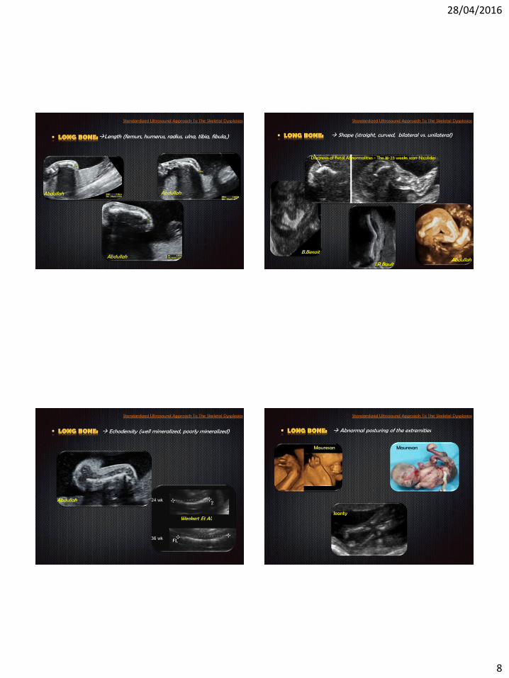
Length (femurs, humerus, radius, ulna, tibia, fibula,)
Abdullah
Abdullah Abdullah
Shape (straight, curved, bilateral vs. unilateral)
B.Benoit
J.P Bault
Diagnosis of Fetal Abnormalities - The 18-23 weeks scan-Nicolides
Abdullah
Echodensity (well mineralized, poorly mineralized)
Abdullah
Wenkert Et Al.
Abnormal posturing of the extremities
Mauresan
Jeanty
Mauresan
28/04/2016
9
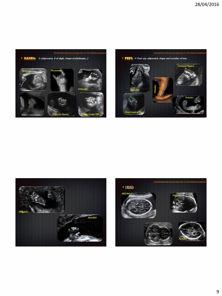
(alignment, # of digits, shape of phalanges…)
D.Moeglin
Françoise Rypens Diagn Image OBS
Nicolaides Nicolaides
Foot size, alignment, shape and number of toes.
Abdullah
Pa
lad
ini
Paladini
Diag imaging Obs
Françoise Rypens
Abdullah
Abdullah
Paladini
Paladini
Abdullah
28/04/2016
10
Abdullah
Diagn Image OBS Paladini
JPB Nicolaides
JPB
Krakow
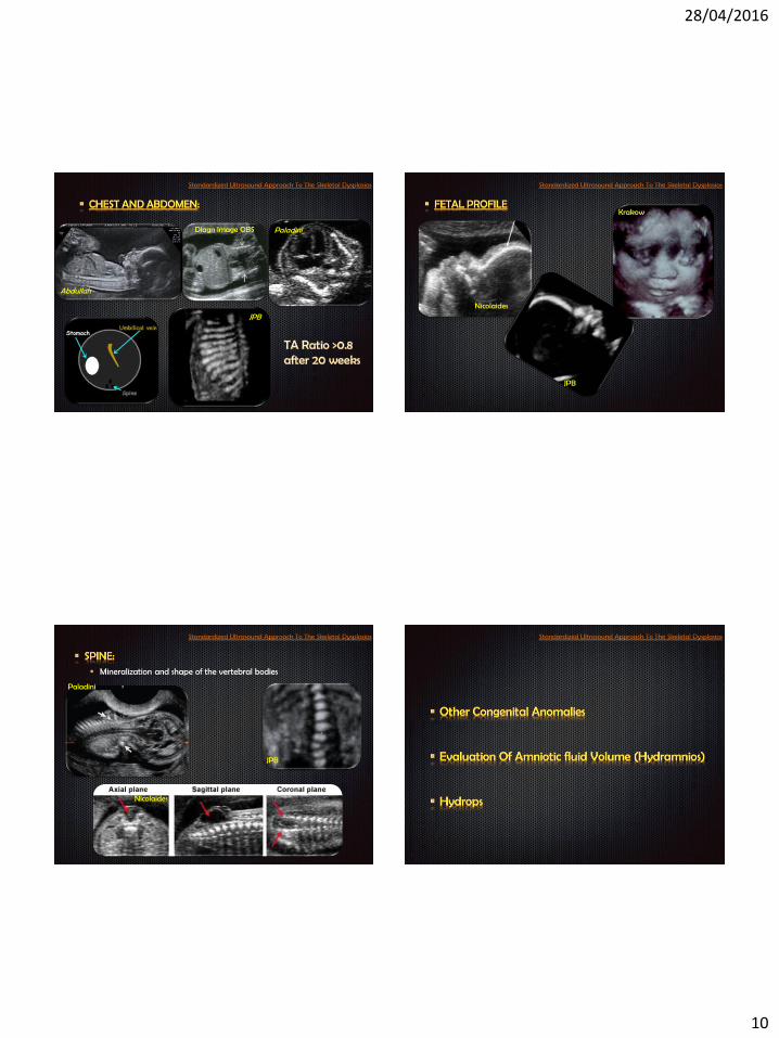
Mineralization and shape of the vertebral bodies
Nicolaides
JPB
Paladini

28/04/2016
11
Cranial sutures.
Spine and ribs Long bones and extremities

28/04/2016
12
Helical CT is a complement to ultrasound, after 26 weeks of
gestation it can discriminate the pathological character of a bone
abnormality that indicates skeletal dysplasia
MRI may be somewhat controversial in view of the
predominant role of prenatal US.

28/04/2016
13
• The molecular defect has been identified in ≈ 50%
of the well-recognized skeletal dysplasias.
• The application of these findings to direct patient
care is not yet possible for many of these disorders.
• Long invasive testing to diagnosis interval.