Embed Size (px)
Citation preview

12/15/20181

Acute Kidney Injury
Prof Brig Gen Mamun MostafiMACP, MRACP, FCPS, FRCP

Epidemiology
INCIDENCE
1-5% of all patients
10-23 % in the ICU
12/15/2018 3

Epidemiology
MORTALITY
20-70% Overall
79% for patients requiring RRT (ICU)
12/15/2018 4

AKI- definition
An abrupt fall in GFR over a period of minutes to days with rapid rise in nitrogenous waste products in blood.
(Rate of production of metabolic waste exceeds the rate of renal excretion)
12/15/2018 5

Definition
AKI is defined as any of the following: Increase in S Creatinine by ≥0.3 mg/dl
(≥26.5 µmol/l) within 48 hours;
or
Increase in S Creatinine to ≥1.5 times baseline, which is known or presumed to have occurred within the prior 7 days;
or
Urine volume <0.5 ml/kg/h for 6 hours.
12/15/2018 6

Acute Kidney Injury Network
(AKIN- 2005)
STAGE I
RISK
(R)
STAGE II
INJURY
(I)
STAGE V
ESRD
(E)
STAGE III
FAILURE
(F)
STAGE IV
LOSS
(L)
Severity Outcome
12/15/2018 7

AKIN
stageSerum Creatinine
Criteria
Urinary Output
CriteriaTime
1 ↑ Cr ≥ 0.3 mg/dl or
≥26.5 µmol/l or
1.5-1.9 times baseline
< 0.5 ml/kg/hr > 6 -12 hrs
2 ↑ Cr 2-2.9 times baseline < 0.5 ml/kg/hr ≥ 12 hrs
3 ↑ Cr ≥ 3 from baseline or
Cr ≥ 4mg/dl (≥353.6 µmol/l)
or
initiation of RRT
< 0.3 ml/kg/hr
or anuria≥ 24 hrs
≥ 12 hrs
AKIN Staging

Acute Kidney Injury
StageIncrease in serum
Creatinine
1 ≥1.5 x previous result
2 ≥2 x previous result
3≥3 x previous result, RRTAnuria ≥ 12 hours
12/15/2018 9

Seru
m C
reati
nin
e
(mg
/dl)
GFR (ml/min per 1.73m2)
1.0
0
2.0
3.0
4.0
5.0
6.0
7.0
8.0
9.0
40 60 80 100120140160180200
Relationship between GFR and serum
creatinine in AKI
12/15/2018 10

Figure: The abrupt drop in GFR but the S.Cr. does not start going up for 24 or 36 hours after the acute insult .
40
80
0
GFR
(mL/min)
0 7 14 21 28
4
Day
s
2
0
6
Serum
Creatinine
(mg/dL)
Urine output starts
to fall
12/15/2018 11

One glass urine in 12 hours

Risk factors of AKI
eGFR <60 ml/min/1.73m2 or history of AKI
Diabetes
Heart failure, liver disease,
Neurological or cognitive impairment
Use of nephrotoxic drugs
Use of iodinated contrast agents within the past week
Symptoms or history of urological obstruction
Sepsis
Age 65years or over12/15/2018 13

To function properly kidneys require:
Normal renal blood flow – Prerenal.
Functioning glomeruli,tubules and interstitium – Intrinsic/Renal.
Clear urinary outflow tract – Postrenal.
12/15/2018 14

Reduction in RBF
HypovolaemiaHaemorrhage,
Vol depletion
( vomit, diarr, diuresis,
burns)
HypotensionCardiogenicshock
Distributive
shock (sepsis,
anaphylaxis)
Oedema statesCardiac failure
Hepatic cirrhosis
Nephr. syndrome
Hypoperfusion
NSAIDs
ACEI / ARBs
RAS /occlusion
Hepatorenal
syndrome
Reduced GFR
PRE-RENAL (Hemodynamic) AKI
Pre Renal AKI12/15/2018 15

Renal / Intrinsic AKI
TubularGlomerular VascularInterstitial
ATN
Ischemia-50%
Toxins -30%
AINDrug: NSAIDs,
antibioticsInfiltrative :
lymphoma
Granulomatous-
Sarcoidosis, TB
Infection : APN
Vascular
occlusions
- Renal artery
occlusion
- Renal vein
thrombosis
- Cholesterol
emboli
AGN
PSGN,
SLE,
ANCA associated,
anti-GBM disease
HSP,
Cryoglobulinemia,
TTP,
HUS
5- 15% 70-80% 8 -20% < 2%
12/15/2018 16

Intra-luminal•Stone,
•Blood clots,
•Papillary
necrosis
•Pelvic
malignancies
•Prolapsed
uterus
•Retroperitoneal
fibrosis
Intrinsic
Intra-mural •Urethral stricture,
•BPH,
•Ca prostate,
• Bladder tumour,
• Radiation fibrosis
Extrinsic
Post-renal Urinary outflow tract
obstruction
12/15/2018 17

12/15/2018 18
Acute Kidney
Injury
PrerenalUosm > 500 mosm/kg
Una < 20 meq/L
FEna < 1%
Microscopy – bland
BUN / S.Cr. Ratio
USG- Normal
Ischemic / Toxic ATNUosm ~ 300 mosm/kg
UNa > 40meq/L
FEna > 2%
Microscopy – dark
pigment cast
Intrinsic/
Renal
Post RenalUosm: variable
UNa: low early, high late
FEna: variable
Microscopy – bland
USG - Diagnostic
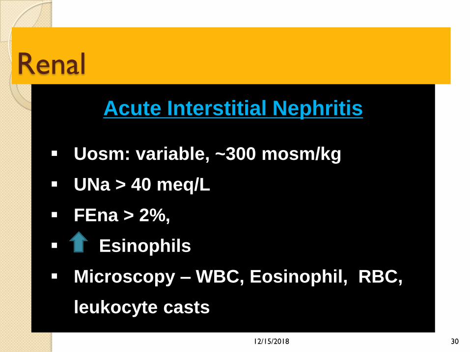
Acute Interstitial Nephritis
Uosm: variable, ~300
mosm/kg
UNa > 40 meq/L
FEna > 2%,
Eosinophils
Microscopy – WBC, RBC,
leukocyte casts
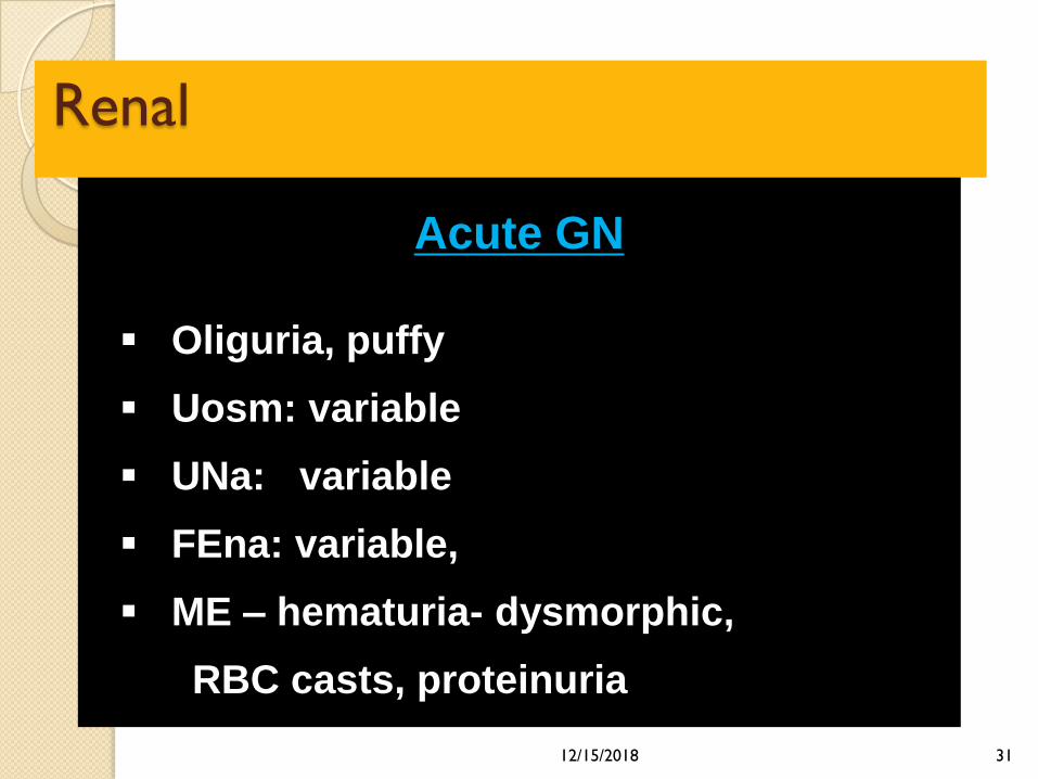
Acute GN
Uosm: variable
UNa: variable
FEna: variable,
ME – hematuria-
dysmorphic, RBC
casts, proteinuria

Pre-renal AKI
History
• Any obvious causes of hypotension, hypovolaemia or hypo perfusion.
a) Haemorrhage/haematoma,
b) GI loss – diarrhoea, vomiting, renal loss, skin loss (burns/exfoliation),
c) Third spacing (pancreatitis).
d) Evidence of cardiac failure
• Sepsis (and if so what is the source?)
12/15/2018 19

Examination
Low BP, rapid pulse.
• Cool peripheries – vascular shut down
• Capillary refill time – greater than 2 seconds
implies volume depletion or poor cardiac
function
• Lying and standing blood pressure – significant
drop implies hypovolaemia
• Warm to touch - sepsis?
• Peripheral pulses - are they bounding
12/15/2018 20

Examination
• Reduced skin turgor, dry lips, mouth and
mucous membranes - systemic hypovolaemia
Face – sunken eyes imply dehydration,
• JVP: may be low if volume depleted
12/15/2018 21
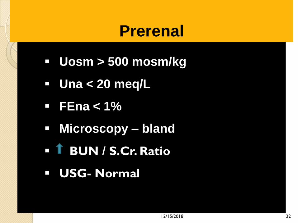
Uosm > 500 mosm/kg
Una < 20 meq/L
FEna < 1%
Microscopy – bland
BUN / S.Cr. Ratio
USG- Normal
Prerenal
12/15/2018 22

Post-renal AKI
• History
• Lower urinary tract symptoms (LUTS) –
frequency, urgency, dysuria, nocturia, poor
stream, hesitancy, terminal dribbling,
strangury.
• Prostatism.
• Haematuria (visible and non-visible)
• Loin pain
12/15/2018 23

Examination
• Look for:
• palpable abdominal masses,
• palpable bladder,
• visible haematuria,
• rectal examination for prostate in
males
12/15/2018 24

Post Renal
Uosm: variable
UNa: low early, high late
FEna: variable
Microscopy – bland/ haematuria
Imaging studies - Diagnostic
12/15/2018 25

Renal
History
◦ Hypovolaemia, hypotension, hypo perfusion,
sepsis or toxin/drugs
◦ Oliguria, haematuria, puffy face, oedema.
◦ Fever, arthritis, rash etc
◦ Headache, nausea, vomiting
◦ SOB
◦ Altered consciousness
◦ Presence or history of a primary
disease/event.
12/15/2018 26

Renal
Examination
• Signs of fluid overload- oedema/anasarca
• JVP: raised if heart failure or AKI causing
significant volume overload
• Heart: Listen for an S3
• Lungs: signs pulmonary oedema.
• Signs of pneumonia / source of sepsis
• Abdomen: Organomegaly, ascites,
evidence of sepsis
• Urine output – catheterize if doubt
12/15/2018 27
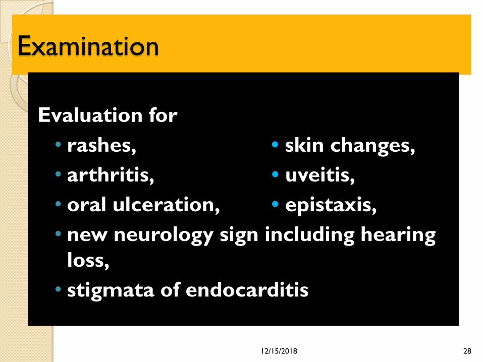
Examination
Evaluation for
• rashes, • skin changes,
• arthritis, • uveitis,
• oral ulceration, • epistaxis,
• new neurology sign including hearing
loss,
• stigmata of endocarditis
12/15/2018 28

Renal
ATN
Uosm ~ 300 mosm/kg
UNa > 40meq/L
FEna > 2%
Microscopy – Muddy brown granular
cast
12/15/2018 29
Acute Interstitial Nephritis
Uosm: variable, ~300 mosm/kg
UNa > 40 meq/L
FEna > 2%,
Esinophils
Microscopy – WBC, Eosinophil, RBC,
leukocyte casts
Renal
12/15/2018 30
Acute GN
Oliguria, puffy
Uosm: variable
UNa: variable
FEna: variable,
ME – hematuria- dysmorphic,
RBC casts, proteinuria
Renal
12/15/2018 31

Renal biopsy
• In specific cases.
• Biopsy will guide the management
12/15/2018 32

PaO2
50 mm of Hg
PaO2
20 mm of Hg
10 mm of Hg
PaO2
12/15/2018 33

12/15/2018 34

Pathophysiology of ATN:Tubular Epithelial Cell Injury and Repair
Loss of polarityNormal Epithelium
Migration , Dedifferentiation of Viable Cells
Differentiation &
Reestablishment of polarity
Sloughing of viable and dead cells
with luminal obstruction
Ischemia/
Reperfusion
ApoptosisNecrosis
Cell death
Proliferation
12/15/2018 35

12/15/2018 36

Acute or Chronic ?
Distinguishing between AKI and chronic
renal impairment is important, as –
◦ The approach to these patients differs
greatly.
◦ This may, save a great deal of unnecessary
investigation.
What to do with a Raised Creatinine ?
12/15/2018 37

◦ History of:
HTN,
DM,
Arthritis and
NSAID,
Stone disease and obstruction,
Congenital diseases .
◦
Factors that suggest chronicity include –
12/15/2018 38
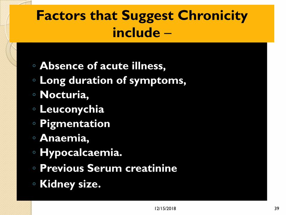
◦ Absence of acute illness,
◦ Long duration of symptoms,
◦ Nocturia,
◦ Leuconychia
◦ Pigmentation
◦ Anaemia,
◦ Hypocalcaemia.
◦ Previous Serum creatinine
◦ Kidney size.
Factors that Suggest Chronicity
include –
12/15/2018 39

What investigations are most useful
in AKI ?
Urinalysis:
Blood,
Protein,
Cells
Casts
UNa, FeNa
Hilton et al, BMJ
2006;333;786-790
12/15/2018 40

RBCs
•Dysmorphic red blood cells suggest glomerular injury.
12/15/2018 41

Red blood cell cast
Marker of glomerular injury
Granular cast
12/15/2018 42

Pigmented granular (“muddy brown”) casts
Marker of acute tubular necrosis
12/15/2018 43

Marker of acute interstitial nephritis.
12/15/2018 44

Haematology
Full blood count, blood film:
◦ Neutrophilia in sepsis
◦ Eosinophilia may be present in acute
interstitial nephritis, cholesterol
embolization, or vasculitis (CSS)
◦ Thrombocytopenia and red cell fragments
suggest thrombotic microangiopathy –TTP,
HUS
12/15/2018 45

Biochemistry
Daily
◦ urea, creatinine,
◦ electrolytes,
◦ PH, serum bicarbonate
◦ Calcium.
12/15/2018 46

Biochem….
CPK, myoglobinuria –
◦ Rhabdomyolysis
Serum immunoglobulins, serum protein
electrophoresis, Bence Jones proteinuria
◦ Myeloma
12/15/2018 47

Haem….
Coagulation studies :
◦ Disseminated intravascular coagulation
associated with sepsis
12/15/2018 48

Immunology
Antinuclear antibody (ANA) , Anti-double
stranded (ds) antibody .
C3 & C4 complement concentrations-
◦ Low in SLE, acute post infectious
glomerulonephritis, Cryoglobulinemia
ANCA
Anti GBM antibodies
ASO and anti-DNAse B titres
◦ High after streptococcal infection
Hepatitis B and C, HIV serology
12/15/2018 49

Imaging
◦ Renal ultrasonography
For renal size, symmetry, evidence of
obstruction
◦ CXR
◦ X-Ray KUB
12/15/2018 50

Clinical Scenario
A 10 year old girl
presented with
S Creatinine of 2.0
mg/dL. She has
oliguria,
haematuria and
puffy face. Her BP
is 150/100 mmHg.
AKI or CKD ?
◦ GN
1. PSGN
2. SLE
3. Vasculitis
History:
Investigations:
12/15/2018 51

Clinical Scenario
S Creatinine of a 21 year old farmer is 2.2 mg/dL.
He reported with severe acute watery diarrhoea and vomiting for two days. He has not passed urine since yesterday.
AKI or CKD ?
◦ Pre renal or
Renal ?
History
Ph Exam
Investigations
12/15/2018 52

Clinical Scenario, What to do ?
S creatinine of a 43 old
man is 4.9mg/dL.
He was having LBP for
last six months along
with irregular fever.
His family physician
advised Naproxen,
which he is taking off
and on for last two
months.
AKI or CKD ?
◦ Prerenal or Renal ?
History
Exam
Investigations
12/15/2018 53

Initial 7 Steps of AKI Management Bundle
• Confirm AKI
• Assess emergency: Pulmonary oedema,
Hyperkalaemia, Acidosis.
• Undertake ABCDE - full clinical examination
• Stop nephrotoxic drugs
• Urine dipstick test and confirm by RME
• Biochemistry - Check & repeat.
• Renal ultrasound and consider urinary
catheter
• Urgent senior review
12/15/2018 54

Management principles…
Identify the source of infection and treat
aggressively keeping dose adjustment.
◦ Minimise indwelling lines
◦ Remove bladder catheter if anuric.
Identify and treat bleeding tendency:
◦ PPI, H2 antagonist, avoid aspirin
◦ transfuse if required
12/15/2018 55

Optimise nutritional support
Maintaining adequate nutrition enhances patient survival
Maintain protein intake about 1gm/Kg/Day
Protein intakes of > 1.2 g/kg/ day can dramatically increase azotaemia.
12/15/2018 56

RRT
Initiate dialysis before uraemic
complications set in.
Early RRT improves mortality and recovery .
Specific types of therapy are available for
critically ill patients.
12/15/2018 57

Conclusions
AKI is increasingly common,
particularly among hospital
inpatients and critically ill
patients.
It carries a high mortality
12/15/2018 58

Conclusions..
Patients at risk are - elderly people;
patients with diabetes, hypertension,
or vascular disease; and those with
pre -existing renal impairment
12/15/2018 59

Conclusions..
AKI is often preventable.
Rapid recognition of incipient AKI
and early treatment of established
AKI is life saving and prevent
irreversible loss of nephrons.
12/15/2018 60

Questions?
12/15/2018 61

.
12/15/2018 62