Embed Size (px)
Citation preview

PowerPoint® Lecture Slides presented by
Dr. Peter Reonisto, Moorpark College,
California
HUMAN ANATOMY
fifth edition
MARIEB | MALLATT | WILHELM
16
Copyright © 2008 Pearson Education, Inc., publishing as Benjamin Cummings
The Special Senses
PART 2

Copyright © 2008 Pearson Education, Inc., publishing as Benjamin Cummings
The Ear: Hearing and Equilibrium
The ear – receptor organ for hearing and equilibrium
Composed of three main regions Outer ear – functions in hearing Middle ear – functions in hearing Inner ear – functions in both hearing and
equilibrium

Copyright © 2008 Pearson Education, Inc., publishing as Benjamin Cummings
The Outer (External) Ear
Composed of The auricle (pinna)
Helps direct sounds External acoustic meatus
Lined with skin Contains hairs, sebaceous glands, and
ceruminous glands
Tympanic membrane Forms the boundary between the external and
middle ear

Copyright © 2008 Pearson Education, Inc., publishing as Benjamin Cummings
Structure of the Ear
Figure 16.16a

Copyright © 2008 Pearson Education, Inc., publishing as Benjamin Cummings
The Middle Ear
The tympanic cavity A small, air-filled space Located within the petrous portion of the temporal
bone
Medial wall is penetrated by Oval window Round window
Pharyngotympanic tube (auditory or eustachian tube) Links the middle ear and pharynx

Copyright © 2008 Pearson Education, Inc., publishing as Benjamin Cummings
Structures of the Middle Ear
Figure 16.16b

Copyright © 2008 Pearson Education, Inc., publishing as Benjamin Cummings Figure 16.17
The Middle Ear
Ear ossicles – smallest bones in the body Malleus – attaches to
the eardrum Incus – between the
malleus and stapes Stapes – vibrates
against the oval window

Copyright © 2008 Pearson Education, Inc., publishing as Benjamin Cummings
The Inner (Internal) Ear
Inner ear – also called the labyrinth Lies within the petrous portion of the temporal
bone Bony labyrinth – a cavity consisting of three parts
Semicircular canals Vestibule Cochlea

Copyright © 2008 Pearson Education, Inc., publishing as Benjamin Cummings
The Inner (Internal) Ear
Figure 16.16b

Copyright © 2008 Pearson Education, Inc., publishing as Benjamin Cummings
The Inner (Internal) Ear
Membranous labyrinth Series of membrane-walled sacs and ducts Fit within the bony labyrinth Consists of three main parts
Semicircular ducts (within semicircular canals)Utricle and saccule (within vestibule)Cochlear duct (within cochlea)

Copyright © 2008 Pearson Education, Inc., publishing as Benjamin Cummings
The Inner (Internal) Ear
Membranous labyrinth (continued) Filled with a clear fluid – endolymph
Confined to the membranous labyrinth
Bony labyrinth is filled with perilymph Continuous with cerebrospinal fluid

Copyright © 2008 Pearson Education, Inc., publishing as Benjamin Cummings
The Membranous Labyrinth
Figure 16.18
Copyright © 2008 Pearson Education, Inc., publishing as Benjamin Cummings
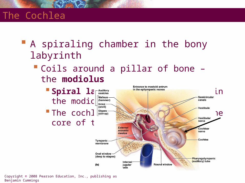
The Cochlea
A spiraling chamber in the bony labyrinth Coils around a pillar of bone – the modiolus
Spiral lamina – a spiral of bone in the modiolusThe cochlear nerve runs through the core of the
modiolus

Copyright © 2008 Pearson Education, Inc., publishing as Benjamin Cummings
The Cochlea
Figure 16.19a, b

Copyright © 2008 Pearson Education, Inc., publishing as Benjamin Cummings
The Cochlea
Figure 16.19b, c
Copyright © 2008 Pearson Education, Inc., publishing as Benjamin Cummings
The Cochlea
The cochlear duct (scala media) – contains receptors for hearing Lies between two chambers
The scala vestibuli The scala tympani
The vestibular membrane – the roof of the cochlear duct
The basilar membrane – the floor of the cochlear duct

Copyright © 2008 Pearson Education, Inc., publishing as Benjamin Cummings
The Cochlea
The cochlear duct (scala media) – contains receptors for hearing Organ of Corti – the receptor epithelium for
hearing Consists of
Supporting cells Inner and outer hair cells (receptor cells)

Copyright © 2008 Pearson Education, Inc., publishing as Benjamin Cummings
The Anatomy of the Cochlea
Figure 16.19a–c

Copyright © 2008 Pearson Education, Inc., publishing as Benjamin Cummings
The Role of the Cochlea in Hearing
Figure 16.20
PLAYPLAY Ear Receptor Complexes

Copyright © 2008 Pearson Education, Inc., publishing as Benjamin Cummings
The Vestibule
The central part of the bony labyrinth
Lies medial to the middle ear Utricle and saccule –
suspended in perilymph Two egg-shaped
parts of the membranous labyrinth
House the macula – a spot of sensory epithelium

Copyright © 2008 Pearson Education, Inc., publishing as Benjamin Cummings
The Vestibule
Macula – contains receptor cells Monitor the position of the head when the head is
still Contains columnar supporting cells Receptor cells – called hair cells
Synapse with the vestibular nerve

Copyright © 2008 Pearson Education, Inc., publishing as Benjamin Cummings
Anatomy and Function of the Maculae
Figure 16.21a

Copyright © 2008 Pearson Education, Inc., publishing as Benjamin Cummings
Anatomy and Function of the Maculae
Figure 16.21b

Copyright © 2008 Pearson Education, Inc., publishing as Benjamin Cummings
The Semicircular Canals
Lie posterior and lateral to the vestibule Anterior and posterior semicircular canals
Lie in the vertical plane at right angles
Lateral semicircular canal Lies in the horizontal plane

Copyright © 2008 Pearson Education, Inc., publishing as Benjamin Cummings
The Semicircular Canals
Figure 16.18

Copyright © 2008 Pearson Education, Inc., publishing as Benjamin Cummings
The Semicircular Canals
Semicircular duct – snakes through each semicircular canal
Membranous ampulla – located within bony ampulla Houses a structure called a crista ampullaris
Cristae contain receptor cells of rotational acceleration Epithelium contains supporting cells and receptor hair
cells

Copyright © 2008 Pearson Education, Inc., publishing as Benjamin Cummings
Structure and Function of the Crista Ampullaris
Figure 16.22a

Copyright © 2008 Pearson Education, Inc., publishing as Benjamin Cummings
Structure and Function of the Crista Ampullaris
Figure 16.22b

Copyright © 2008 Pearson Education, Inc., publishing as Benjamin Cummings
Equilibrium and Auditory Pathways
The equilibrium pathway (macula & crista ampullaris)
Transmits information on the position and movement of the head
Most information goes to lower brain centers (reflex centers)
The ascending auditory pathway (Organ of Corti)
Transmits information from cochlear receptors to the cerebral cortex

Copyright © 2008 Pearson Education, Inc., publishing as Benjamin Cummings
Auditory Pathway from the Organ of Corti
Figure 16.23
Copyright © 2008 Pearson Education, Inc., publishing as Benjamin Cummings
Disorders of Equilibrium and Hearing
Motion sickness – carsickness, seasickness Popular theory for a cause – a mismatch of sensory
inputs
Meniere’s syndrome – equilibrium is greatly disturbed Excessive amounts of endolymph in the
membranous labyrinth

Copyright © 2008 Pearson Education, Inc., publishing as Benjamin Cummings
Disorders of Equilibrium and Hearing
Deafness Conduction deafness
Sound vibrations cannot be conducted to the inner ear Ruptured tympanic membrane, otitis media, otosclerosis
Sensorineural deafness Results from damage to any part of the auditory
pathway

Copyright © 2008 Pearson Education, Inc., publishing as Benjamin Cummings
Embryonic Development of the Ear
Begins in the fourth week of development The inner ear forms from ectoderm The middle ear forms from the first pharyngeal
pouches Ear ossicles develop from cartilage The external ear differentiates from the first
branchial groove

Copyright © 2008 Pearson Education, Inc., publishing as Benjamin Cummings
Embryonic Development of the Ear
Figure 16.24a, b

Copyright © 2008 Pearson Education, Inc., publishing as Benjamin Cummings
Embryonic Development of the Ear
Figure 16.24c, d

Copyright © 2008 Pearson Education, Inc., publishing as Benjamin Cummings
The Special Senses Throughout Life
Smell and taste Sharp in newborns
In the fourth decade of life Ability to taste and smell declines

Copyright © 2008 Pearson Education, Inc., publishing as Benjamin Cummings
The Special Senses Throughout Life
Photoreceptors – fully formed by 25 weeks All newborns are hyperopic By 3 months – image can be focused on the retina By 6 months – depth perception is present

Copyright © 2008 Pearson Education, Inc., publishing as Benjamin Cummings
The Special Senses Throughout Life
With increased age The lens loses its clarity The dilator muscles of the iris become inefficient Visual acuity is dramatically lower in people over
70

Copyright © 2008 Pearson Education, Inc., publishing as Benjamin Cummings
The Special Senses Throughout Life
In the newborn Responses to sounds are reflexive Low-pitched and middle-pitched sounds can be
heard
In the elderly Hair cells are gradually lost Ability to hear high-pitched sounds fades Presbycusis – gradual loss of hearing with age