Embed Size (px)
Citation preview
Linköping University Medical Dissertation No. 1568
Circulating and genetic factors in colorectal cancer
Potential factors for establishing prognosis?
Renate Slind Olsen
Department of Laboratory Medicine Region Jönköping County
Division of Drug Research Department of Medical and Health Sciences
Linköping University, Sweden
Linköping 2017

© Renate Slind Olsen, 2017
Published articles have been reprinted with the permission of the copyright holders Printed by LiU-Tryck, Linköping, Sweden 2017 ISBN 978-91-7685-555-3 ISSN 0345-0082

iii
”Kommer tid, kommer råd”
Jon Slind

iv

v
ABSTRACT
Colorectal cancer (CRC) is defined as a cancer appearing in the colon or in the rectum. In
Sweden, ~ 6300 individuals were diagnosed with the disease in 2014 and ~ 2550 individuals
diagnosed with CRC die each year due to their cancer. Surgery is the main treatment option
of CRC and a survival rate of ~ 10 % is estimated if distant metastases have developed. It is
therefore of importance to find factors that may be useful together with tumour, node,
metastasis (TNM) stage to establish early CRC diagnosis, prognosis and follow-up of CRC
patients. The aim of this thesis was to study the possible association of CD93, PLA2G4C,
PDGF-D and inflammatory cytokines with CRC disease progression.
In a prospective study approach CD93 and PLA2G4C single nucleotide polymorphisms (SNPs)
were of potential importance in CRC prognosis.
The T/T genotype of CD93 was associated with an increased CD93 expression in CRC tissue.
Further, CRC patients carrying this genotype were associated with disseminated CRC at
diagnosis and a lower recurrence-free survival after surgery. The A allele of a SNP of
PLA2G4C was a stronger predictor for CRC-specific mortality than the conventional risk
factors used in the clinic for selection of TNM stage II patients for adjuvant treatment. This
indicates that the T/T genotype of CD93 and the A allele of PLA2G4C may be potential
genetic factors related to disease severity and spread. Furthermore, they distinguish CRC
patients that may benefit from a more comprehensive follow-up and adjuvant treatment.
To study the putative involvement of PDGF-D in CRC the effects of PDGF-D signalling was
studied in vitro. PDGF-D signalling altered the expression of genes of importance in CRC
carcinogenesis and proliferation which was blocked by imatinib, a tyrosine kinase inhibitor.
This indicates that PDGF-D signalling may be an important pathway in CRC progression and a
potential target in CRC treatment.

vi
The analysis of various inflammatory cytokines in plasma at diagnosis showed an association
between high levels and increased total- or CRC-specific mortality two years after surgery.
High levels of CCL1 and CCL24 was the only cytokines strongly correlated with a worse CRC
prognosis after statistical adjustments and may be of interest for further evaluation.
In conclusion, this thesis presents circulating and genetic factors such as CD93, PLA2G4C,
PDGF-D, CCL1 and CCL24 that may be of importance in CRC progression and may be of
clinical value together with TNM stage in establishing prognosis.
vii
SVENSK SAMMANFATTNING
Kolorektal cancer är en tumör i kolon eller rektum. I Sverige diagnosticerades år 2014 ca
6300 individer med denna cancertyp och ca 2550 personer dör årligen till följd av kolorektal
cancer. Operation är det huvudsakliga behandlingsalternativet för kolorektal cancer och vid
fjärrmetastaser är överlevnaden < 10 %. Det är därför viktigt att hitta markörer som
tillsammans med TNM-stadium kan ge tidig information om sjukdomens prognos och lämplig
uppföljning av patienter.
Utveckling av kolorektal cancer sker genom ackumulering av genetiska mutationer och
epigenetisk nedreglering av tumörsuppressorgener. Därutöver spelar interaktionen mellan
tumören och dess närmaste omgivning, innehållande tillväxt- och inflammatoriska faktorer,
en viktig roll i tumörens utveckling och metastasering.
Syftet med avhandlingen var att studera associationen mellan CD93, PLA2G4C, PDGF-D samt
inflammatoriska cytokiner och kolorektal cancer progression.
En prospektiv studie visade att CD93 och PLA2G4C SNP var potentiellt viktiga förbedömning
av kolorektal cancer prognos. T/T genotypen av SNP rs2749817 i CD93 var associerad med
högre uttryck av CD93 i kolorektal cancer vävnad, främst bland patienter i stadium IV.
Därutöver observerades fler återfall efter operation hos patienter med T/T genotypen. A
allelen hos PLA2G4C SNP rs1549637 är en möjligtvis bättre markör för cancerspecifik
överlevnad vid stadium II än faktorer som idag används för att selektera patienter till
adjuvant behandling. Sammantaget antyder detta att T/T genotypen av CD93 och A allelen
av PLA2G4C kan vara genetiska markörer relaterade till allvarlig tumörsjukdom och
spridning. Därutöver kan de eventuellt selektera patienter som kräver tätare uppföljning och
adjuvant behandling.
För att studera den förmodade inblandningen av PDGF-D i kolorektal cancer undersöktes
dess effekt på PDGF-D signalering in vitro. PDGF-D signaleringen förändrade
genexpressionen av gener involverade i tumörutveckling och spridning, vilken kunde
blockeras av tyrosinkinashämmaren imatinib. Det antyder att PDGF-D signalering kan vara en
viktig faktor vid kolorektal cancer progression och ett potentiellt mål för behandling.

viii
Analysen av ett flertal inflammatoriska cytokiner visade en korrelation mellan höga
cytokinnivåer och ökad cancerspecifik och total dödlighet två år efter operation. Höga CCL1
och CCL24 nivåer var de enda faktorerna som förblev signifikant associerade med
cancerspecifik mortalitet vid fördjupad statistisk analys och bör studeras vidare.
Sammanfattningsvis presenterar denna avhandling cirkulerande och genetiska faktorer
såsom CD93, PLA2G4C, PDGF-D, CCL1 and CCL24 som eventuellt är viktiga vid bedömning av
kolorektal cancer progression tillsammans med TNM stadium.
ix
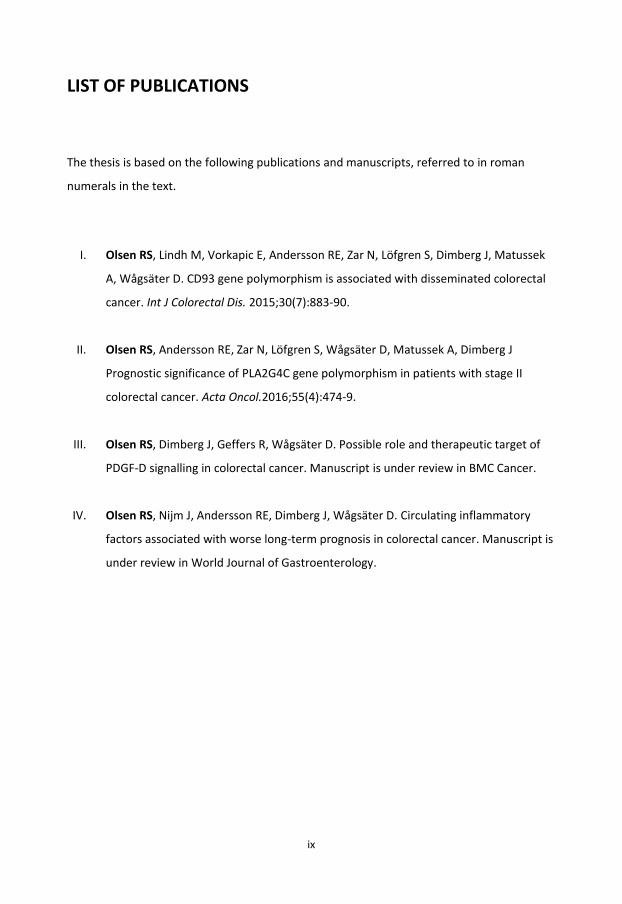
LIST OF PUBLICATIONS
The thesis is based on the following publications and manuscripts, referred to in roman
numerals in the text.
I. Olsen RS, Lindh M, Vorkapic E, Andersson RE, Zar N, Löfgren S, Dimberg J, Matussek
A, Wågsäter D. CD93 gene polymorphism is associated with disseminated colorectal
cancer. Int J Colorectal Dis. 2015;30(7):883-90.
II. Olsen RS, Andersson RE, Zar N, Löfgren S, Wågsäter D, Matussek A, Dimberg J
Prognostic significance of PLA2G4C gene polymorphism in patients with stage II
colorectal cancer. Acta Oncol.2016;55(4):474-9.
III. Olsen RS, Dimberg J, Geffers R, Wågsäter D. Possible role and therapeutic target of
PDGF-D signalling in colorectal cancer. Manuscript is under review in BMC Cancer.
IV. Olsen RS, Nijm J, Andersson RE, Dimberg J, Wågsäter D. Circulating inflammatory
factors associated with worse long-term prognosis in colorectal cancer. Manuscript is
under review in World Journal of Gastroenterology.


ABBREVIATIONS
α Alpha
AA Amino acid
AJCC American Joint Committee on Cancer
APC Adenomatous polyposis coli tumour suppressor gene
ATP Adenosine triphosphate
β Beta
CAM Cell adherence molecule
CCL Chemokine C-C motif ligand
CCR C-C motif chemokine receptor
CD4+ Cluster of differentiation 4-positive
CD93 Cluster of differentiation 93
cDNA Complementary deoxyribonucleic acid
CEA Carcinoembryonic antigen
CEACAM5 Carcinoembryonic antigen related cell adhesion molecule 5
CIS Chromosome instability pathway
COX-2 Cyclooxygenase 2
CRC Colorectal cancer
cRNA Complementary RNA
CVD Cardiovascular disease
CXCL Chemokine C-X-C motif ligand
CX3CL1 C-X3-C motif chemokine ligand 1
DG Dystroglycan
ECM Extracellular matrix
EGF Endothelial growth factor
EGFR Endothelial growth factor receptor
ELISA Enzyme-linked immunosorbent assay
EMT Epithelial to mesenchymal transition
FAP Familial adenomatous polyposis
FGF Fibroblast growth factor
GAPDH Glyceraldehyde-3-phosphate dehydrogenase
GI Gastrointestinal tract
GTP Guanosine triphosphate
HAS2 Hyaluronan synthase 2
HNPCC Hereditary nonpolyposis colorectal cancer
HRP Horseradish peroxidase
IHC Immunohistochemistry
IL Interleukin
IFN-ɣ Interferon gamma
KRAS Kirsten sarcoma viral oncogene homolog proto-oncogene
LD Linkage disequilibrium
LOX Lysyl oxidase
MMP Matrix metalloproteinase
mRNA messenger RNA
MSI Microsatellite instability pathway
PDGF Platelet-derived growth factor
PDGFR Platelet-derived growth factor receptor
phPDGFR Phospho-specific platelet-derived growth factor receptor
PE Phycoerythrin
PGE2 Prostaglandin E2
PLA2 Phospholipase A2
PLA2G2A Phospholipase A2 group II A
PLA2G4 Phospholipase A2 Group IV
PLA2G4A/C Phospholipase A2 Group IV A/C
PPAR Peroxisome proliferator-activated receptor
PVDF Polyvinylidene fluoride
qPCR Real-time polymerase chain reaction
REMARK Recommendations for tumour marker prognostic studies
RPLP0 Ribosomal protein lateral stalk subunit P0
SDS-PAGE Sodium dodecyl sulfate polyacrylamide gel electrophoresis
Serpin E1 Serpin family E member 1
SNP Single nucleotide polymorphism
Taq Thermus aquaticus
TBP TATA-box binding protein
Th T helper
TNF Tumour necrosis factor
TNFSF4 TNF super family member 4

TNM Tumour, node, metastasis
TP53 Tumour suppressor P53
VCAN Versican
VEGF Vascular endothelial growth factor
WB Western Blot

TABLE OF CONTENTS
INTRODUCTION ................................................................................................................. 1
The large intestine: colon and rectum.................................................................................... 1
Colorectal cancer .................................................................................................................... 2
Carcinogenesis of CRC ............................................................................................................ 3
Development of metastasis ................................................................................................ 6
Angiogenesis ....................................................................................................................... 9
Symptoms and risk factors ................................................................................................... 11
Heredity ............................................................................................................................ 11
Lifestyle and age ............................................................................................................... 11
Concomitant diseases ....................................................................................................... 12
Prognosis ............................................................................................................................... 13
Diagnosis and treatment ...................................................................................................... 15
Imatinib ............................................................................................................................. 16
Markers in blood and tissue ................................................................................................. 17
Carcinoembryonic antigen ................................................................................................ 18
Kirsten sarcoma viral oncogene homolog proto-oncogene ............................................. 18
Single nucleotide polymorphisms ..................................................................................... 19
Circulating inflammatory factors and their contribution in CRC .......................................... 20
Cluster of differentiation CD93 ............................................................................................. 22
Phospholipase A2 group IV C ................................................................................................ 24
Platelet-derived growth factor D .......................................................................................... 25

AIMS OF THESIS ........................................................................................................................ 28
MATERIAL AND METHODOLOGICAL CONSIDERATIONS ............................. 30
MATERIAL.............................................................................................................................. 30
Study population................................................................................................................... 30
Clinical CRC covariates used in the statistical analyses ........................................................ 32
METHODS .............................................................................................................................. 33
Tissue homogenization ......................................................................................................... 33
Immunohistochemistry ......................................................................................................... 33
Western Blot ......................................................................................................................... 35
Enzyme-linked immunosorbent assay .................................................................................. 36
Luminex ................................................................................................................................. 37
Caco-2 and HT-29 cell lines ................................................................................................... 40
TACS® MTT cell proliferation assay ...................................................................................... 41
Nucleic acid extraction and reverse transcription ................................................................ 43
Polymerase Chain Reaction .................................................................................................. 43
Affymetrix® GeneChip® Whole Transcript Expression array ................................................ 46
Statistical analyses ................................................................................................................ 48
ETHICS ................................................................................................................................... 49

RESULTS AND DISCUSSION ....................................................................................... 50
CD93 gene polymorphism is associated with disseminated colorectal cancer (Paper I) ..... 50
Prognostic significance of PLA2G4C gene polymorphism in patients with stage II colorectal
cancer (Paper II) .................................................................................................................... 54
Possible role and therapeutic target of PDGF-D signalling in colorectal cancer (Paper III) . 57
Circulating inflammatory factors associated with worse long-term prognosis in colorectal
cancer (Paper IV) ................................................................................................................... 61
CONCLUDING REMARKS AND FUTURE PERSPECTIVES .............................. 65
ACKNOWLEDGEMENT ................................................................................................. 67
REFERENCES ...................................................................................................................... 69
1
INTRODUCTION
The large intestine: colon and rectum
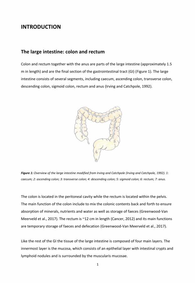
Colon and rectum together with the anus are parts of the large intestine (approximately 1.5
m in length) and are the final section of the gastrointestinal tract (GI) (Figure 1). The large
intestine consists of several segments, including caecum, ascending colon, transverse colon,
descending colon, sigmoid colon, rectum and anus (Irving and Catchpole, 1992).
Figure 1: Overview of the large intestine modified from Irving and Catchpole (Irving and Catchpole, 1992). 1:
caecum; 2: ascending colon; 3: transverse colon; 4: descending colon; 5: sigmoid colon; 6: rectum; 7: anus.
The colon is located in the peritoneal cavity while the rectum is located within the pelvis.
The main function of the colon include to mix the colonic contents back and forth to ensure
absorption of minerals, nutrients and water as well as storage of faeces (Greenwood-Van
Meerveld et al., 2017). The rectum is ~12 cm in length (Cancer, 2012) and its main functions
are temporary storage of faeces and defecation (Greenwood-Van Meerveld et al., 2017).
Like the rest of the GI the tissue of the large intestine is composed of four main layers. The
innermost layer is the mucosa, which consists of an epithelial layer with intestinal crypts and
lymphoid nodules and is surrounded by the muscularis mucosae.
2
The second layer is the submucosa, which contains nerves, blood vessels and connective
tissue, followed by the third layer, the muscularis propria, which consists of two layers of
smooth muscle cells. The fourth layer is the serosa consisting of connective tissue covered
with squamous cell epithelium (Cancer, 2012).
Colorectal cancer
Colorectal cancer (CRC) is defined as a cancer appearing in the colon or in the rectum due to
their adjacent anatomical locations, but the term is still debated. Several studies have shown
genetic, morphological, and clinical differences, as well as differences in survival between
cancers of the colon and rectum (Kornmann et al., 2013, van der Sijp et al., 2016) suggesting
that CRC might consist of two separate entities. As an example Kornmann et al. showed an
improved survival in treated patients with colon cancer but not among patients with rectum
cancer. Also, a higher occurrence of lung metastases among patients with rectal cancer was
found. The conclusion of the study was that there might be differences in chemosensitivity
between the colon and rectum (Kornmann et al., 2013). Another study by van der Sijp et al.
investigated differences in characteristics, prevalence of complications, survival as well as
the rate of recurrence among patients with colon- and rectal cancer. This study showed a
lower survival among colon cancer patients because of a more severe effect of complications
compared with patients with rectal cancer (van der Sijp et al., 2016).
On a world basis about 1.4 million cases of CRC were reported in 2012 and around 700.000
individuals passed away due to CRC (Ferlay et al., 2015). In Sweden, CRC is the third most
common form of malignancy (Norr, 2016) and in 2014 about 6300 individuals were
diagnosed with an equal distribution among men and women (Cancerfonden, 2016).
Approximately 2550 individuals diagnosed with CRC die each year due to their CRC (Norr,
2016).

3
Three categories of CRC can be distinguished by their forms of origin and expression.
Sporadic CRC appears in individuals that carries mutations in the tissue but do not have any
family history of CRC. This category is assumed to include 60-80 % of CRC cases (Watson and
Collins, 2011). The second form is the family type of CRC and constitutes 20-40 % of CRC
cases. This category is based upon population studies that have shown an increased chance
of developing CRC when family members of primary consanguinity have suffered from the
sporadic form of CRC. Nevertheless, it is thought that this category may be a result of both
environmental as well as hereditary factors. The hereditary type is the third category of CRC,
which can be distinguished by the presence of adenomatous polyps (Arvelo et al., 2015).
Carcinogenesis of CRC
Carcinogenesis, also named oncogenesis or tumourigenesis, is a complicated process by
which cancers are generated and is a multistep mechanism occurring due to accumulation of
errors in vital regulatory pathways. The process starts in a single cell which then multiplies
and acquires additional changes that give the cell a survival advantage over its surrounding
cells. This altered cell must then be amplified to generate a large number of cells that
constitute a cancer (King and Robins, 2006b).
CRCs are slowly growing cancers that start developing as a tumour or as a tissue growth on
the inner lining of the colon or rectum. If the growth is abnormal, i.e. a polyp that eventually
becomes cancerous, it can form a tumour on the wall of the colon or rectum, and
subsequently grow into blood vessels or lymph vessels, increasing the risk of developing
metastasis to other sites of the human body (Vogelstein and Kinzler, 1993). In CRC there are
two major pathways in carcinogenesis: the chromosome instability pathway (CIS) and the
microsatellite instability pathway (MSI). The CIS is the pathway of sporadic CRC through
mutations of the adenomatous polyposis coli suppressor gene (APC), the Kirsten sarcoma
viral oncogene homolog proto-oncogene (KRAS) and the tumour suppressor P53 (TP53)
gene. MSI is mainly the pathway for hereditary nonpolyposis colorectal cancer (HNPCC)
through mutations in mismatch repair genes (Raskov et al., 2014).
4
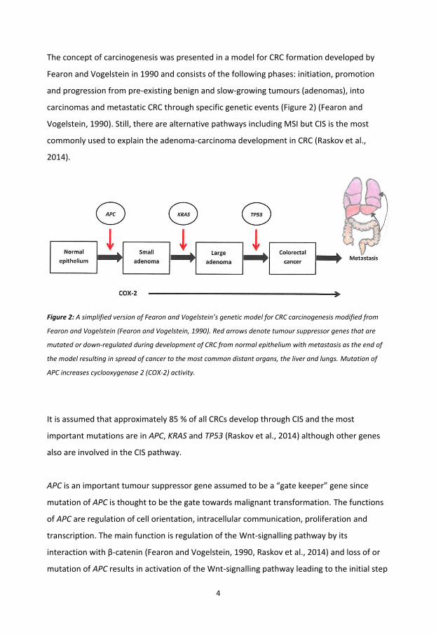
The concept of carcinogenesis was presented in a model for CRC formation developed by
Fearon and Vogelstein in 1990 and consists of the following phases: initiation, promotion
and progression from pre-existing benign and slow-growing tumours (adenomas), into
carcinomas and metastatic CRC through specific genetic events (Figure 2) (Fearon and
Vogelstein, 1990). Still, there are alternative pathways including MSI but CIS is the most
commonly used to explain the adenoma-carcinoma development in CRC (Raskov et al.,
2014).
Figure 2: A simplified version of Fearon and Vogelstein’s genetic model for CRC carcinogenesis modified from
Fearon and Vogelstein (Fearon and Vogelstein, 1990). Red arrows denote tumour suppressor genes that are
mutated or down-regulated during development of CRC from normal epithelium with metastasis as the end of
the model resulting in spread of cancer to the most common distant organs, the liver and lungs. Mutation of
APC increases cyclooxygenase 2 (COX-2) activity.
It is assumed that approximately 85 % of all CRCs develop through CIS and the most
important mutations are in APC, KRAS and TP53 (Raskov et al., 2014) although other genes
also are involved in the CIS pathway.
APC is an important tumour suppressor gene assumed to be a “gate keeper” gene since
mutation of APC is thought to be the gate towards malignant transformation. The functions
of APC are regulation of cell orientation, intracellular communication, proliferation and
transcription. The main function is regulation of the Wnt-signalling pathway by its
interaction with β-catenin (Fearon and Vogelstein, 1990, Raskov et al., 2014) and loss of or
mutation of APC results in activation of the Wnt-signalling pathway leading to the initial step

5
from normal to hyper proliferative epithelium (Fearon and Vogelstein, 1990). The gene
product, the APC protein, ensures the function of the important junctions between
colonocytes ensured by cell adherence molecules (CAMs) the cadherins. To maintain the
proper function of the cadherins the APC protein must bind to the cadherin molecule
together with β-catenin and GSK3-β. This binding to the cadherin molecule ensures the
normal function of the junctions. The binding of β-catenin to the cadherin complex then
ensures low levels of free β-catenin in the cytoplasm and is important since β-catenin
otherwise will translocate into the nucleus and upregulate the Wnt-signalling pathway and
result in an accelerated proliferation, impaired differentiation and apoptosis leading to the
formation of adenomas (Mann et al., 1999, Raskov et al., 2014). Mutation of APC also results
in increased COX-2 activity that is maintained by its production of prostaglandin E2(PGE2)
contributing in promoting cancer growth (Wang and DuBois, 2013, Raskov et al., 2014).
KRAS is an oncogene with its natural form as a proto-oncogene, and is thought to play an
important role in the transition from adenoma to carcinoma (Raskov et al., 2014). KRAS is a
short gene sequence susceptible to point mutations and substitution of single amino acids
(AAs) in a nucleotide that can lead to activation of mutation. KRAS mutations are found in
approximately 30-50 % of CRC cases and gives the colonocytes an advantage in growth as
guanosine triphosphate (GTP) activity is lost with mutation. The increased levels of GTP
result in constant signalling. The gene product of KRAS, KRAS protein, is responsible for the
transduction the mitogenic signals from growth factor receptors on the cell surface to the
cell nucleus (Dobre et al., 2013). In an active form, KRAS can affect several cellular pathways
that control apoptosis, cellular growth, differentiation, organization of the cytoskeleton, cell
motility, inflammation and proliferation (Pino and Chung, 2010). The KRAS mutations are
often seen in early dysplasia and in hyperplastic polyps. If the mutations occur after an APC
mutation the dysplastic lesions are often progress towards cancer (Raskov et al., 2014).
TP53 plays a crucial role in all cells since it controls cell cycle, apoptosis via its protein
product P53, which induces cell cycle arrest and facilitates DNA repair due to replication
errors or mutations, and if the DNA repair is unsuccessful P53 induces apoptosis. In most
tumours the mutated form of TP53 occurs due to missense mutation, which inactivates the
transcriptional activity of P53. TP53 is usually found to be commonly mutated in CRC and is

6
thought to be important in the transition of large adenomas into invasive carcinomas
(Raskov et al., 2014).
Even though CIS is considered the major pathway in the development of sporadic CRC, this
does not explain why the majority of adenomas never progress to invasive carcinomas. This
might be due to other, unknown pathways enabling the cells to overcome CIS (Raskov et al.,
2014).
Development of metastasis
Metastasis is the spread of cancer cells from the primary tumours into the surrounding
tissues and distant organs. The liver and lungs are the most common distant organs for CRC
metastases (Chambers et al., 2002). Of all CRC patients, approximately 25 % have a
metastatic disease at the time of diagnosis, and a high number of patients will carry
undetectable micro-metastases. Moreover, another 25 % of all CRC patients are at the risk of
developing metastatic tumours during the course of the disease (Kindler and Shulman,
2001).
The development of tumour metastasis is a complex process involving a series of events
leading to the spread and growth of cancer cells to secondary sites of the human body
(Figure 3).
7
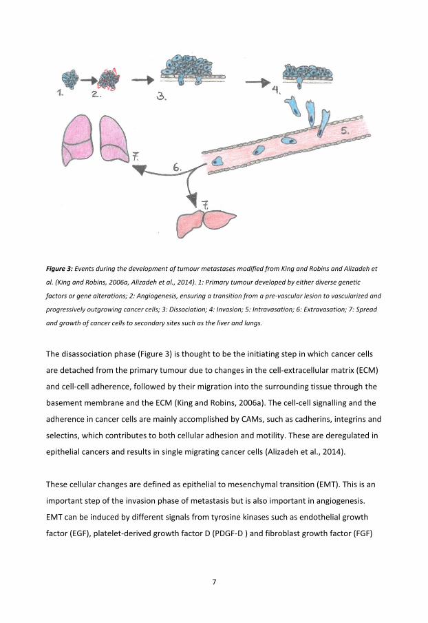
Figure 3: Events during the development of tumour metastases modified from King and Robins and Alizadeh et
al. (King and Robins, 2006a, Alizadeh et al., 2014). 1: Primary tumour developed by either diverse genetic
factors or gene alterations; 2: Angiogenesis, ensuring a transition from a pre-vascular lesion to vascularized and
progressively outgrowing cancer cells; 3: Dissociation; 4: Invasion; 5: Intravasation; 6: Extravasation; 7: Spread
and growth of cancer cells to secondary sites such as the liver and lungs.
The disassociation phase (Figure 3) is thought to be the initiating step in which cancer cells
are detached from the primary tumour due to changes in the cell-extracellular matrix (ECM)
and cell-cell adherence, followed by their migration into the surrounding tissue through the
basement membrane and the ECM (King and Robins, 2006a). The cell-cell signalling and the
adherence in cancer cells are mainly accomplished by CAMs, such as cadherins, integrins and
selectins, which contributes to both cellular adhesion and motility. These are deregulated in
epithelial cancers and results in single migrating cancer cells (Alizadeh et al., 2014).
These cellular changes are defined as epithelial to mesenchymal transition (EMT). This is an
important step of the invasion phase of metastasis but is also important in angiogenesis.
EMT can be induced by different signals from tyrosine kinases such as endothelial growth
factor (EGF), platelet-derived growth factor D (PDGF-D ) and fibroblast growth factor (FGF)
8
leading to a remodeling of the actin cytoskeleton and scattering of cancer cells (Thiery,
2002).
The cancer cells then form a finger-like protrusion called a pseudopod at their leading edge
that establishes a binding to the ECM via integrins. The attachment of integrin to the ECM
ensures an invasion and migration of the cancer cells through the ECM towards the
lymphatic- and vascular circulation (Figure 3). The integrins enables also attachment to
proteases such as serine proteases, matrix metalloproteinases (MMPs). The MMPs are then
activated, resulting in a proteolysis of specific parts of the ECM, creating a path for the
cancer cells through the ECM. The MMPs may also be activated by cytokines. The cancer
cells attach to the vascular endothelial cell wall and by changing their shape they are able to
penetrate into the endothelial cell-cell junctions. Additionally, the modulation of endothelial
cell barrier permeability by macrophage-derived tumour necrosis factor alpha (TNF-α) may
also contribute in this process called intravasation (Figure 3). Cancer cell overexpression of
vascular endothelial growth factor (VEGF) may also contribute by inducing endothelial
barrier disruption (King and Robins, 2006a, Alizadeh et al., 2014).
Inside the bloodstream, single cancer cells are likely to be destroyed by actions of the
surveillance by the host immune system. Cancer cells may avoid this destruction by
travelling together in clusters, interacting with platelets, macrophages, leukocytes and the
vascular endothelium by binding of CAMs (e.g. selectin and integrin) forming an emboli. The
passive displacement of cancer cells is also influenced by cytokines. The emboli can then
attach to the endothelial cell lining of the blood vessel wall by binding of the cancer cells’
CAMs to the glycoproteins (laminin, collagen, fibronectin) expressed on the endothelial cell
surfaces. Once attached the cancer cells can progress their metastatic process by destroying
the basement membrane of the circulation system with the help of proteases, a process
called extravasation (Figure 3) (King and Robins, 2006a, Alizadeh et al., 2014).
After extravasation the cancer cells migrate to their growth site, which requires
decomposition of the ECM, and that the cells reach bigger blood vessels ensuring movement
towards distant organs such as the liver and lungs (Figure 3). Once the cancer cells have

9
reached distant organs and are attached they need to form new blood vessels in a process
called angiogenesis to gain a supply of essential nutrients as well as oxygen to enable further
growth and establishment of metastasis (King and Robins, 2006a).
Angiogenesis
Angiogenesis is a rate-limiting step determining whether or not a metastasis remains in a
dormant state or if it grows further on. Even though angiogenesis is associated with the
establishment of secondary tumours it is also an essential process in the establishment of
primary tumours. The term angiogenesis describes the growth of new capillary blood vessels
by sprouting, in which intestinal tissue columns are inserted into the lumen of pre-existing
vessels and partition the vessel lumen (King and Robins, 2006a) and plays a crucial role in
supplying oxygen and other important nutrients necessary for the growth and progression of
tumours (Kumar et al., 2016).
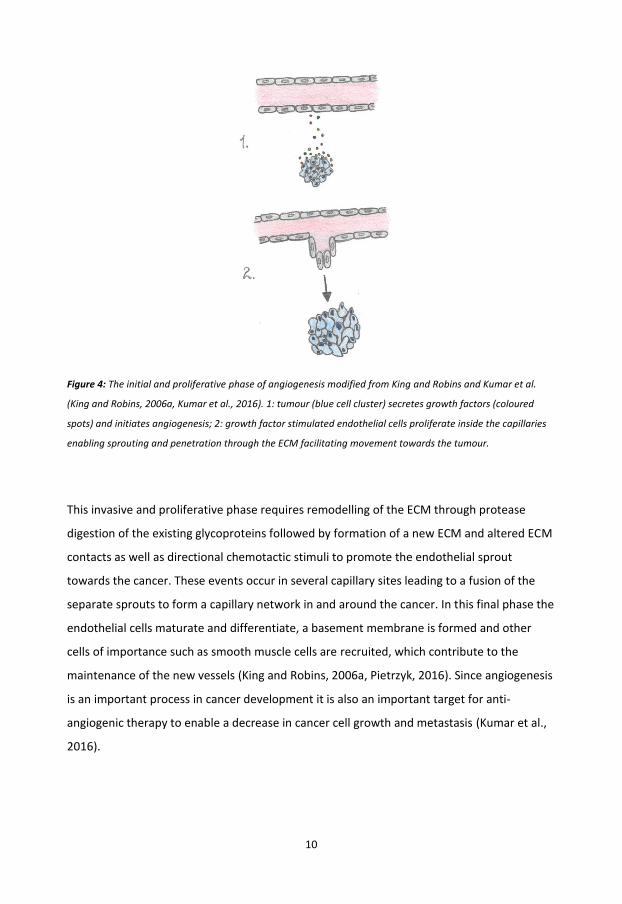
The process of angiogenesis consists mainly of three phases. It is initiated by increased
tumour secretion of tumour-derived growth factors, such as PDGFs, FGFs, transforming
growth factors, EGFs and VEGFs, which stimulate local endothelial proliferation inside the
capillaries enabling them to sprout and penetrate through the ECM and move in the
direction of the tumour (Figure 4) (Kumar et al., 2016, King and Robins, 2006a).
10
Figure 4: The initial and proliferative phase of angiogenesis modified from King and Robins and Kumar et al.
(King and Robins, 2006a, Kumar et al., 2016). 1: tumour (blue cell cluster) secretes growth factors (coloured
spots) and initiates angiogenesis; 2: growth factor stimulated endothelial cells proliferate inside the capillaries
enabling sprouting and penetration through the ECM facilitating movement towards the tumour.
This invasive and proliferative phase requires remodelling of the ECM through protease
digestion of the existing glycoproteins followed by formation of a new ECM and altered ECM
contacts as well as directional chemotactic stimuli to promote the endothelial sprout
towards the cancer. These events occur in several capillary sites leading to a fusion of the
separate sprouts to form a capillary network in and around the cancer. In this final phase the
endothelial cells maturate and differentiate, a basement membrane is formed and other
cells of importance such as smooth muscle cells are recruited, which contribute to the
maintenance of the new vessels (King and Robins, 2006a, Pietrzyk, 2016). Since angiogenesis
is an important process in cancer development it is also an important target for anti-
angiogenic therapy to enable a decrease in cancer cell growth and metastasis (Kumar et al.,
2016).
11
Symptoms and risk factors
The symptoms associated with suspected CRC development are blood in the faeces, hard or
loose faeces, no appetite, weight loss, tiredness and a slow development of anaemia (Norr,
2016, Register, 2013). Even though most cases of CRC arise sporadically (Amersi et al., 2005)
there are several risk factors associated with CRC development and they can be divided into
two groups: controlled risk factors, including environment and life style, and those that
cannot be controlled, such as hereditary factors, previous colonic polyps and age (Haggar
and Boushey, 2009).
Heredity
Heredity of tumour development is thought to be one potential risk factor for contracting
CRC even though most CRCs are assumed to be sporadic. In about 4 % of all CRC cases the
cancer is accounted for by HNPCC, also named Lynch syndrome. It is the most common
hereditary form and patients with HNPCC have an increased risk of developing other cancers
(Hampel et al., 2008). Even though HNPCC-affected individuals can develop colonic
adenomas with a greater frequency than the normal population, polyps rarely develop
(Jasperson et al., 2010). Familial adenomatous polyposis (FAP) is the second most inherited
CRC syndrome and it is characterized by the development of multiple colonic adenomas. It is
a less severe form of CRC disease and the development of FAP starts in early adolescence
(Jasperson et al., 2010, Arvelo et al., 2015). In the absence of preventive surgery FAP may
develop into a malignant form (Arvelo et al., 2015) and the average age at diagnosis if
untreated is 39 years (Jasperson et al., 2010).
Lifestyle and age
The diet is considered as a major risk factor for CRC. High consumption of red meat, food
containing high levels of animal fat and a low level of dietary fiber, as well as processed food
have been associated with increased risk of developing CRC (Haggar and Boushey, 2009,
Watson and Collins, 2011). Other lifestyle-related risk factors for CRC are smoking, high
alcohol consumption, obesity and physical inactivity (Haggar and Boushey, 2009).
12
Age and the development of CRC are considered to go “hand in hand”, indicating that risk of
developing CRC increases with age due to accumulations of DNA damage over long time
periods (Register, 2013). In Sweden, about 80 % of the patient group are 65 years or older
when they get the CRC diagnosis (Cancerfonden, 2016).
Concomitant diseases
Since CRC mostly affects older patients, the risk also increases for development of other
underlying diseases and about 40 % of CRC patients have concomitant diseases such as
cardiovascular disease (CVD), diabetes and other malignancies (Edwards et al., 2014).
CVD and CRC are both diseases that are increasing in incidence and prevalence, in part due
to the aging population. Lifestyle, such as physical inactivity and an unhealthy diet are
common risk factors for both CVD and CRC (Yang et al., 2010) and affect mainly elderly
patients within similar age groups (65 years or older) (Robertson et al., 2010). Also, a higher
risk of abnormal intima/media thickness of carotid arteries among patients with colorectal
adenomas and adenocarcinomas has been seen (Cha et al., 2011).
Inflammatory bowel disease is a chronic inflammatory disorder of the GI divided into Crohn`s
colitis and ulcerative colitis (Qu et al., 2017). Crohn`s colitis has been associated with
increased risk of developing cancer of the colon (von Roon et al., 2007), while the risk of
developing CRC has been shown to increase with duration of ulcerative colitis, 2 % at 10
years and 18 % by 30 years (Eaden et al., 2001). Summarized, the appearance of the colitis
associated inflammation seem to accelerate the progression from chronic colitis to CRC
development (Qu et al., 2017).
13
Prognosis
The medical term ‘prognosis’ is used to indicate the probable direction, outcome, as well as
an estimation of the aggressiveness of a disease. In cancer, prognosis is measured by the
probability of staying alive or without recurrence of the disease for a certain amount of time
after diagnosis, or at the start of adjuvant treatment. Often a threshold of five years is used
for the survival or the cancer re-growth, but depending on the treatment and the disease,
shorter or longer time periods might be considered (Parkin and Hakulinen, 1991).
Pathological stage is the most important estimation of CRC prognosis. The American Joint
Committee on Cancer (AJCC) introduced the tumour-, node- and metastasis (TNM) staging
system, which is more differentiated than the previous Duke`s stage system (Table 1)
(Cancer, 2012). The TNM system is based on local tumour depths of invasion (T1-4), the
presence of and the amount of lymph node metastases (N0-2), as well as the presence of
distant metastases (M0-1) (Hutter, 1987). Today the seventh version of the TNM staging
system published in 2010 is used by clinicians (Table 1) (Cancer, 2012).
The prognosis of CRC is related to the TNM stage at diagnosis with a five-year survival rate of
90 % at early diagnosis resulting in better prognosis and less than 10 % if distant metastases
have developed (Coppede et al., 2014). Among patients with stage II-III CRC that have
undergone intended curative surgery, 20-60 % would be at risk of cancer recurrence
(Mosolits et al., 2005, Tebbutt et al., 2002). Also, if the disease disseminates extensively and
the chance of additional curative surgery is eliminated, only palliative treatment remains for
the patients.

14
Table 1: Classification of CRC based upon Duke`s- and TNM stage classification systems modified from the AJCC
Cancer Staging Atlas (Cancer, 2012) and survival rate modified from the American Cancer Society (The American
Cancer Society, 2017).
Duke`s stage Stage TNM stage Comments 5 year
system grouping system prognosis ▪
0 Tis, N0, M0 Tis: Carcinoma in situ; intraepithelial or invasion of lamina propria
N0: No regional lymph node metastasis
M0: No distant metastasis
A I T1, N0, M0 T1: Tumour invades submucosa T2, N0, M0 T2: Tumour invades muscularis propria ~ 87-92 %
B II A T3, N0, M0 T3: Tumour invades through the muscularis propria into pericolorectal ~ 80-87%
tissues
II B T4a, N0, M0 T4a: Tumour penetrates through the surface of the visceral peritoneum ~ 49-63 %
II C T4b, N0, M0 T4b: Tumour directly invades or is adherent to other organs or structures
C III A T1-T2, N1/N1c, M0 N1: Metastasis in 1-3 regional lymph nodes ~ 84-89 %
or
N1c: Tumour deposite(s) in the subserosa, mesentry, or non-peritonealized
T1, N2a, M0 pericolic or perirectal tissues without regional nodal metastasis
III B T3-T4a, N1/N1c, M0 ~ 69-71 %
or N2a: Metastasis in 4-6 regional lymph nodes
T2-T3, N2a, M0
or
T1-T2, N2b, M0 N2b: Metastasis in ≥7 regional lymp nodes
III C T4a, N2a, M0 ~ 53-58 %
or
T3-T4a, N2b, M0
or
T4b, N1-N2, M0
D IV A Any T, any N, M1a M1a: Metastasis confined to one site or organ ~ 11-12 %
IV B Any T, any N, M1b M1b: Metastasis in more than one site/organ, or the peritoneum
▪ Based upon survival rates (%) for colon and rectum separately.

15
Diagnosis and treatment
Diagnosis of CRC is based upon the results from colonoscopy or sigmoidoscopy and tumour
biopsies from individuals with CRC-associated symptoms (Cunningham et al., 2010). Also,
levels of carcinoembryonic antigen (CEA) may be a preoperative support in preoperative
staging and planning of surgery (Labianca et al., 2010).
The pretreatment phase for newly diagnosed CRC includes physical examination and a
complete colonoscopy to detect any possible metachronous tumours. Computed
tomography of the chest, abdomen and pelvis to localize the tumour and to identify any
metastatic disease is also part of the pretreatment strategy (Norr, 2016). In patients with
rectal cancer, the assessment of local tumour extension is important for optimal treatment.
High resolution magnetic resonance imaging is used to measure the spread in the
surrounding epithelium more accurately and to assess the resection margin between the
edge of the tumour and the surrounding tissue in the rectum (Norr, 2016). Based on the
result of these examinations preoperative treatment with chemo- and/or radiotherapy can
be initiated before surgical removal of the cancer (Norr, 2016).
Surgery is the main treatment option in the treatment phase. During surgery of cancer
located in the colon or rectum a total resection (R0 resection) of the tumour should be
performed with adequate margins and removal of regional lymph nodes. This is defined as
radical surgery. Also, margins of ≥5 cm around the cancer are recommended. At least 12
lymph nodes should be removed and these nodes should be analyzed to allow appropriate
nodal staging since analysis of fewer than 10 lymph nodes may under-stage the tumour
(Cunningham et al., 2010, Norr, 2016). The removal of lymph nodes is essential since spread
of tumour cells through lymph nodes is of importance in development of metastasis. Also,
the removal of lymph nodes may ensure a lower risk of recurrence (Norr, 2016).
16
After surgery and examination of the specimen by a pathologist the TNM stage of the
resected tumour can be decided to determine the treatment strategy and follow-up of the
patient. Decisions on treatment and follow-up of CRC patients are made by a
multidisciplinary group of clinicians. The choice of optimal treatment for the CRC patients
depends on tumour location, differentiation grade of the tumour, TNM stage,
histopathological type, presence of infiltrating blood vessels and lymph nodes, age,
condition of the patient, as well as risk of recurrence. The postoperative treatment option
apart from surgery is adjuvant treatment, which may consist of chemotherapy, radiotherapy,
targeted therapy and liver resection (Labianca et al., 2010, Norr, 2016). The goal with
adjuvant treatment is to decrease tumour growth and ultimately its eradication.
Imatinib
Imatinib (Glivec®, Gleevec®) is a drug that has been approved for treatment of chronic
myeloid leukaemia and gastrointestinal stromal tumours (Druker et al., 2001, Demetri et al.,
2002) and was the first class of agents that acted by inhibition of specific tyrosine kinases,
rather than killing all rapidly dividing cells (Ohno, 2006). Treatment with imatinib show only
mild to moderate levels of side effects such as diarrhoea, rash, fatigue and nausea (Demetri
et al., 2002).
The drug has been shown to inhibit the activity of several tyrosine kinases that play
important roles in cell growth, motility and survival (Popow-Wozniak et al., 2011) such as
breakpoint cluster region-Abelson1 (Schindler et al., 2000), c-Kit, and platelet-derived
growth factor receptors (PDGFRs) such as PDGFR-beta (PDGFR-β) (Moawad, 2015). In short,
imatinib functions by binding to and inducing a blockage of the adenosine triphosphate
(ATP)-binding pocket of the tyrosine kinases preventing access of ATP, which is of
importance for tyrosine activity. Consequently, imatinib shuts down all downstream
signalling from the tyrosine kinases affecting the cell survival (Popow-Wozniak et al., 2011).
In CRC, cell studies have shown that imatinib treatment induces apoptosis and inhibition of

17
cancer cell migration, suggesting that imatinib could be a potential drug in CRC therapy
(Popow-Wozniak et al., 2011).
Markers in blood and tissue
According to the National Institute of Health, the term ‘biomarker’ refers to a biological
molecule found in blood, in other body fluids, or in tissues that is a sign of normal or
abnormal processes, or of a condition or a disease, or even of responses of therapeutic
intervention (De Gruttola et al., 2001).
In cancer such as CRC, it is important to identify biomarkers that allow detection of the
disease at an early stage, which may lead to an early diagnosis, establishment of a
personalized treatment strategy and improvement of prognosis. Additionally, identification
of biomarkers in serum and blood is preferable since sequential blood tests are non-invasive
and more accessible than repeated tissue biopsies in a clinical setting. There are several
suggested biomarkers for CRC available such as the fecal marker faecal occult test and
genetic markers such as KRAS and serum markers such as CEA (Locker et al., 2006, Labianca
et al., 2010, Norr, 2016).
Many studies are focusing on finding new potential biomarkers useful in establishing cancer
prognosis. The reporting recommendations for tumour marker prognostic studies (REMARK)
is a checklist consisting of twenty items that are important to report on when publishing
tumour marker prognostic studies. These recommendations should ensure a more
consistent, high quality reporting of tumour marker studies and applies in general to
prognostic factors e.g. biological molecules, size of tumour, presence of tumour cells in
regional lymph nodes, abnormal features of cells, gender and age. It also applies to other
diseases than cancer. REMARK can be used in any studies involving prognostic factors
regardless if those prognostic factors are biological markers, clinical assessments, imaging
assessments or measures of functional status in activities of daily living (Altman et al., 2012).

18
Carcinoembryonic antigen
CEA is a high molecular weight glycoprotein that belongs to the immunoglobulin superfamily
and is the oldest and most widely used serum marker in CRC. CEA is a recommended soluble
biological marker used in surveillance following curative surgery of the primary CRC tumour,
and measurement of CEA is quite inexpensive and has less disadvantages for the patients
(Duffy et al., 2007).
In the clinic, CEA may be useful in both preoperative staging and planning of surgery. Also,
postoperative CEA levels are recommended to be checked every three months for stage II
and III CRC, for at least three years if the patient is a potential candidate for surgery or
chemotherapy for metastatic CRC. On the other hand, the problem with CEA is its low
predictive value for diagnosis in asymptomatic patients since it has a relatively low sensitivity
and specificity. This explains why elevated CEA levels (>5 mg/mL), which may correlate with
poorer prognosis among CRC patients, is thought to be insufficient for determining whether
or not patients should receive adjuvant treatment (Labianca et al., 2010, Locker et al., 2006).
Kirsten sarcoma viral oncogene homolog proto-oncogene
In CRC metastasis mutation of RAS genes are frequent and mutations of KRAS are most
common (~ 40 %). The RAS proteins effectuates intracellular signals downstream the
endothelial growth factor receptor (EGFR) and specific mutations of the RAS genes results in
modified proteins which induce a constant activation of pathways regulating tumour cell
growth and proliferation through EGFR. This activation cannot be efficiently blocked by
chemotherapy and antibody targeted treatment (Karapetis et al., 2008). In CRC with RAS
mutations a combination of chemotherapy and antibody targeted treatment may give a
prolonged survival of only 1-2 months. However if the RAS mutation is wild-type antibody
targeted treatment or in combination with chemotherapy has shown to be more efficient in
inhibiting EGFR signaling resulting in a prolonged survival of 30 months (Norr, 2016).

19
Single nucleotide polymorphisms
Single nucleotide polymorphisms (SNPs), also named gene polymorphisms or genetic
variants, are single base-pair positions in the genomic DNA that differ from one individual to
another and can easily be analyzed in whole blood samples from individuals. To be defined
as a SNP the least frequent allele should have a frequency of >1 % in the population
(Kruglyak and Nickerson, 2001). Since autosomal regions carry one allele from the maternal
chromosome and one from the paternal chromosome, an individual can exhibit one of three
genotypes: homozygous for the major allele, or heterozygous- or homozygous for the minor
allele. The definitions of the major- and the minor allele refers to the observed frequencies
in a specific population (Crawford and Nickerson, 2005). There are two types of SNPs
influencing the incidence of diseases. If the variation of an SNP is located within or close to
the translated region, any AA substitutions which alter the protein synthesis may be directly
connected to the disease. On the other hand, if a disease-associated SNP is located in a non-
coding region or if there is no other gene near the SNP it is more difficult to determine the
mechanism by which the SNP is associated with (Mimori et al., 2012).
Genetic variations, such as SNPs, are thought to play a role in individual variation in CRC
susceptibility. In cancer, molecular characteristics might predispose tumours to a worse
prognosis and their identification can enable the identification of patients at a higher risk of
recurrence of the disease. Also, suitable predictive markers may also make it easier to
choose the most appropriate therapy (Horvat et al., 2016). In CRC, several studies have
focused on the effects of genetic variants and their association with CRC disease. As an
example Chang et al. found that a polymorphism of stromal derived factor-1α, which can
promote cancer cell migration, was associated with lymph node metastasis in CRC and
suggested that stromal derived factor-1α might be a predictive marker for lymph node
metastasis in CRC patients (Chang et al., 2009). Even though promising results have been
discovered among SNPs most of them need further validation in larger prospective clinical
trials before they can be defined as biomarkers (Horvat et al., 2016).
20
Circulating inflammatory factors and their contribution in CRC
Primary CRC develops progressively over time through the accumulation of genetic
mutations and epigenetic silencing of tumour suppressor genes. Also, evidence suggests that
the development of CRC and CRC metastasis is also promoted by the interaction between
tumour and stroma, the tumour microenvironment (Itatani et al., 2016). The tumour
microenvironment contains several host cells that may suppress or promote aggressiveness
of the cancer. Lymphocytes including macrophages, natural killer cells and regulatory T-cells
are found in the surrounding stroma and will normally eradicate cancer cells but may also
contribute to cancer progression (Mager et al., 2016, Itatani et al., 2016).
The majority of infiltrating lymphocytes are cluster of differentiation 4-positive (CD4+) T
helper (Th) cells and the Th cells are central in development of an immune response by
activating antigen-specific effector cells and in recruitment of cells of the innate immune
system such as macrophages. The CD4+ T helper cells can be divided into Th1 and Th2 cells.
The Th1 cells are primary responsible for activation and regulation of the development and
persistence of cytotoxic T-cell response. The Th2 cells favour a predominantly humoral
response and are responsible for reducing the activation of the cytotoxic T-cell response
(Knutson and Disis, 2005, Murphy et al., 2008). During Th differentiation the cytokine
environment is important. Th1 commitment relies on production of TNF-α and interferon
gamma (IFN-ɣ) while Th2 commitment relies on production of interleukin (IL) 4 (Knutson and
Disis, 2005).
The cytokine network consists of cytokines, chemokines, interferons and interleukins
(Murphy et al., 2008). They are secreted membrane-bound proteins produced from cells of
both the innate- and the adaptive immunity, enabling immune cells to communicate with
each other (Alizadeh et al., 2014, Murphy et al., 2008). Cytokines are structurally and
functionally similar to growth factors which fulfil their functions by binding to their specific
cell membrane receptors present on non-cancerous cell and immune cells such as Th1 and
Th2 cells that induce an inflammatory stimulus. The inflammatory stimuli enable the
infiltration of inflammatory cells that either eradicate cancer cells or participate in functions
facilitating cancer development by affecting the proliferative and invasiveness of the cancers

21
by inducing chemoattraction, inflammation and/or angiogenesis (Itatani et al., 2016, Mager
et al., 2016). The cytokines can be divided into pro-tumourigenic (cytokines that facilitate
cancer progression) and anti-tumourigenic (cytokines that inhibits cancer progression)
cytokines. There are also cytokines that are both pro- and anti-tumourigenic (Figure 5)
(Mager et al., 2016).
Figure 5: Examples of pro- (red circle) and anti-tumourigenic cytokines (blue circle) involved in CRC
carcinogenesis described in Itatani et al. and Mager et al. Cytokines that are both pro- and anti-tumourigenic
are placed in between the circles (Itatani et al., 2016, Mager et al., 2016).
This suggests that it is important to study the specific contribution of cytokines in CRC
development and progression as well as their impact on survival among CRC patients. This
may enable further knowledge in CRC carcinogenesis.
CCL15CCL2
CCL24
CCL20
CX3CL1
CXCL1
CXCL5IL-8/CXCL8
CXCL9
IL-1β
IL-4
IL-6
TNF-αCCL5
CCL19
CCL21
IFN-ɣCXCL10
CXCL12
22
Cluster of differentiation 93
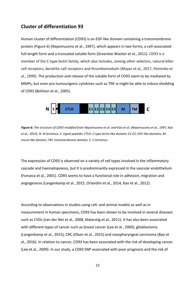
Human cluster of differentiation (CD93) is an EGF-like domain containing a transmembrane
protein (Figure 6) (Nepomuceno et al., 1997), which appears in two forms; a cell-associated
full-length form and a truncated soluble form (Greenlee-Wacker et al., 2011). CD93 is a
member of the C-type lectin family, which also includes, among other selectins, natural killer
cell receptors, dendritic cell receptors and thrombomodulin (Mayer et al., 2017, Petrenko et
al., 1999). The production and release of the soluble form of CD93 seem to be mediated by
MMPs, but even pro-tumourigenic cytokines such as TNF-α might be able to induce shedding
of CD93 (Bohlson et al., 2005).
Figure 6: The structure of CD93 modified from Nepomuceno et al. and Kao et al. (Nepomuceno et al., 1997, Kao
et al., 2012). N: N-terminus; S: signal peptide; CTLD: C-type lectin-like domain; E1-E5: EGF-like domains; M:
mucin-like domain; TM: transmembrane domain; C: C-terminus.
The expression of CD93 is observed on a variety of cell types involved in the inflammatory
cascade and haematopoiesis, but it is predominantly expressed in the vascular endothelium
(Fonseca et al., 2001). CD93 seems to have a functional role in adhesion, migration and
angiogenesis (Langenkamp et al., 2015, Orlandini et al., 2014, Kao et al., 2012).
According to observations in studies using cell- and animal models as well as in
measurement in human specimens, CD93 has been shown to be involved in several diseases
such as CVDs (van der Net et al., 2008, Malarstig et al., 2011). It has also been associated
with different types of cancer such as breast cancer (Lee et al., 2009), glioblastoma
(Langenkamp et al., 2015), CRC (Olsen et al., 2015) and nasopharyngeal carcinoma (Bao et
al., 2016). In relation to cancer, CD93 has been associated with the risk of developing cancer
(Lee et al., 2009). In our study, a CD93 SNP associated with poor prognosis and the risk of
TM CME5E4E3E2E1CTLDSN

23
cancer-specific death (Olsen et al., 2015), which is in agreement with other cancer studies
(Langenkamp et al., 2015, Bao et al., 2016).
To be able to understand the biological mechanisms behind the potential role of CD93 in the
different types of cancers, more functional studies are needed. Several studies on cancer
have implied that CD93 induces angiogenesis due to its increased expression in vascular
endothelial cells and adhesion of endothelial cells (Orlandini et al., 2014, Olsen et al., 2015,
Bao et al., 2016). Also, CD93 seem to be important for proper endothelial cell migration and
vascular function (Langenkamp et al., 2015).
The EGF-like domain of CD93 has been shown to be important in angiogenesis by binding to
EGF receptors, which induces cellular signalling, facilitating the development of angiogenesis
(Kao et al., 2012). Another recent study has identified another signalling pathway of
importance for the function of CD93 in angiogenesis, which was activated due to the
cooperation between CD93 and the important ECM adhesion molecule dystroglycan (DG)
(Galvagni et al., 2016). DG is able to bind to several ECM ligands (Barresi and Campbell,
2006), is expressed on endothelial cells, and is involved in cell adhesion to the ECM
(Hosokawa et al., 2002). In the study by Galvagni et al., cooperative interactions between
CD93 and DG were demonstrated, which promoted migration of endothelial cells and
organization of endothelial cells into capillary structures which is important in the
development of angiogenesis (Galvagni et al., 2016). Since CD93 seems to be an important
regulator of angiogenesis, which is a key process in cancer development and spread, further
studies on its function and regulation in CRC are of importance.
24
Phospholipase A2 group IV C
The phospholipase A2s (PLA2s) is a large group of enzymes that are divided into several
families such as secretory, cytosolic and calcium-independent PLA2s. The enzymes of the
PLA2 family are defined by their ability to catalyze fatty acid from the sn2 position of
glycerophospholipids resulting in free fatty acids, such as arachidonic acid (Yarla et al., 2016).
Arachidonic acid is a substrate for production of inflammatory mediators such as PGE2
(Wang and DuBois, 2013). PGE2 is also produced by COX-2 (Wang and DuBois, 2013, Yarla et
al., 2016), which in high levels has been shown to affect important processes in cancer such
as angiogenesis, apoptosis, cell migration, cell proliferation and tumour invasion (Wang and
DuBois, 2013, Sheng et al., 1998).
Intracellular PLA2s are grouped as cytosolic PLA2s, also called the PLA2 group IV members
(PLA2G4s), and they require increased calcium and phosphatidylinositol phosphate levels to
locate them to the membrane to ensure their activity and the release of arachidonic acid.
PLA2G4 may be regulated by various stimuli from among others cytokines, hormones,
eicosanoids and growth factors. They have shown to be involved in inflammation-related
disorders such as in asthma, sepsis and atherosclerosis and in cancer (Yarla et al., 2016).
In CRC, some PLA2G4s have been suggested to be of relevance in CRC development and
progression. PLA2 group IV A (PLA2G4A) has been shown to be overexpressed in CRC tissue
(Dimberg et al., 1998, Osterstrom et al., 2002). Also, a secretory PLA2, PLA2 group II A
(PLA2G2A), has been suggested to be a prognostic determinant for selecting patients at high
risk for cancer recurrence in stage II CRC who might benefit from adjuvant treatment
(Buhmeida et al., 2009).
Another PLA2G4 member, phospholipase A2 group IV C (PLA2G4C), predominantly
expressed in the human brain, heart and skeletal muscles (Asai et al., 2003) has been shown
to contribute both in the cellular arachidonic acid metabolism and in phospholipid
remodelling (Cummings, 2007, Murakami et al., 2003). It may therefore be linked to the
COX-2 pathway to produce prostaglandins (Murakami et al., 2003). The exact activity of
PLA2G4C expression in tumourigenesis is still unknown but it seems to play a role in breast
25
cancer cell chemotaxis and invasion (Tian et al., 2011). Polymorphic variants of genes may
play a role as factors that mediate inflammatory response and favour CRC progression
(Theodoropoulos et al., 2006). An SNP, rs1549637, of PLA2G4C has been associated with
worse prognosis, especially in stage II CRC, and might be a potential prognostic biomarker in
the planning of individual adjuvant treatment (Olsen et al., 2016). Since PLA2G4s have been
shown to be associated with worse prognosis in stage II CRC, both PLA2G2A and PLA2G4C
should be further investigated in larger CRC patient groups to evaluate their prognostic value
in the clinic.
Platelet-derived growth factor D
The PDGFs act as possible mitogens for cells of mesenchymal origin such as fibroblasts and
monocytes and are some of the many growth factors involved in both cell growth and
division. The PDGF family consist of four monomeric variants, which in active form dimerize
into five different isoforms: PDGF-AA, PDGF-AB, PDGF-BB, PDGF-CC and PDGF-DD. The
PDGFs function by binding to their receptors, PDGFR-α and PDGFR-β, which in active form
exist in three isoforms composed of the homodimers PDGFR-αα, PDGFR-ββ, or as a
heterodimer PDGFR-αβ (Fredriksson et al., 2004). Upon binding of PDGFs to their respective
receptors, the PDGFRs dimerize which leads to auto-phosphorylation and an increased
tyrosine kinase activity as well as binding affinity for signalling molecules. The signal is
transduced into the cell to promote differentiation, migration and survival (Heldin et al.,
1998, Heldin and Westermark, 1999).
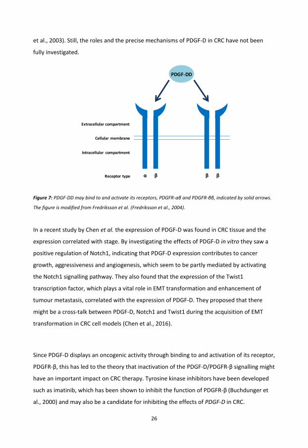
PDGF-D is the latest and the least studied member of the PDGFs, which functions by binding
either to the PDGFR-αβ or the PDGFR-ββ (Figure 7) (Fredriksson et al., 2004) and seems to
play a role in cancers such as breast cancer and CRC (Ahmad et al., 2011, Chen et al., 2016).
In CRC, PDGF-D has been shown to exceed its cellular effects through PDGFR-β, which is
mainly expressed in stromal cells and periocytes (Lindmark et al., 1993, Sundberg et al.,
1993). PDGF-D is involved in regulation of cellular processes such as angiogenesis, apoptosis,
transformation, invasion, metastasis, migration and proliferation (Ustach and Kim, 2005, Li
26
et al., 2003). Still, the roles and the precise mechanisms of PDGF-D in CRC have not been
fully investigated.
Figure 7: PDGF-DD may bind to and activate its receptors, PDGFR-αβ and PDGFR-ββ, indicated by solid arrows.
The figure is modified from Fredriksson et al. (Fredriksson et al., 2004).
In a recent study by Chen et al. the expression of PDGF-D was found in CRC tissue and the
expression correlated with stage. By investigating the effects of PDGF-D in vitro they saw a
positive regulation of Notch1, indicating that PDGF-D expression contributes to cancer
growth, aggressiveness and angiogenesis, which seem to be partly mediated by activating
the Notch1 signalling pathway. They also found that the expression of the Twist1
transcription factor, which plays a vital role in EMT transformation and enhancement of
tumour metastasis, correlated with the expression of PDGF-D. They proposed that there
might be a cross-talk between PDGF-D, Notch1 and Twist1 during the acquisition of EMT
transformation in CRC cell models (Chen et al., 2016).
Since PDGF-D displays an oncogenic activity through binding to and activation of its receptor,
PDGFR-β, this has led to the theory that inactivation of the PDGF-D/PDGFR-β signalling might
have an important impact on CRC therapy. Tyrosine kinase inhibitors have been developed
such as imatinib, which has been shown to inhibit the function of PDGFR-β (Buchdunger et
al., 2000) and may also be a candidate for inhibiting the effects of PDGF-D in CRC.
Extracellular compartment
Receptor type
Cellular membrane
Intracellular compartment
PDGF-DD
α β β β

27

28
AIMS OF THESIS
The overall aim of this thesis was to study the association of circulating and genetic factors
with CRC disease progression.
The specific aims of this thesis were to:
Investigate the expression of CD93 and the association of genetic variations within
the CD93 gene with survival (Paper I).
Investigate the expression of PLA2G4C and the association with a genetic variation
within the PLA2G4C gene with survival (Paper II).
Investigate the expression and localization of PDGF-D in human CRC tissue and to
study the putative involvement of PDGF-D signalling in colorectal carcinogenesis
(Paper III).
Study the association of circulating factors, cytokines, at the time of CRC surgery with
survival (Paper IV).

29

30
MATERIAL AND METHODOLOGICAL CONSIDERATIONS
MATERIAL
Study population
The CRC project at the Department of Laboratory Medicine, Region Jönköping County,
Sweden is an ongoing project established in 1996. The project is a collaboration between the
Department of Laboratory Medicine, the Department of Surgery and the Department of
Clinical Physiology, Region Jönköping County. The external collaborators involved in the
study are the Department of Natural Science and Biomedicine, the School of Health and
Welfare, Jönköping University and the Division of Drug Research, the Department of
Medicine and Health Sciences, Linköping University. The overall aim of the CRC project is to
find risk factors revealing prognosis and survival among CRC patients at an early stage of the
disease. This is done prospectively by comparing the laboratory results obtained in the study
with the clinical covariates found in the patients` medical records.
Today the project consists of samples from >520 CRC patients who have undergone surgical
resections for primary colorectal adenocarcinomas and >500 healthy blood donors (venous
blood samples only) collected from blood bank centers in Region Jönköping County. Blood
samples from blood donors serve as controls in the study. The CRC samples (matched
normal and tumour tissue biopsies and blood samples) are continuously collected from the
Department of Surgery, Region Jönköping County.
31
For each patient included in the project, tumour and adjacent normal tissue biopsies (~5 cm
from the tumour) are excised and collected during surgery, and frozen at -70 ˚C until
analysis. Also two venous blood samples with EDTA are collected. The blood samples are
centrifuged at 1500 g for 10 minutes before plasma and whole blood are separated and
frozen at -70 ˚C until analysis.
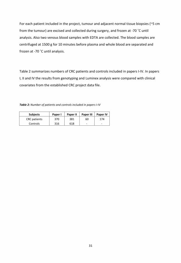
Table 2 summarizes numbers of CRC patients and controls included in papers I-IV. In papers
I, II and IV the results from genotyping and Luminex analysis were compared with clinical
covariates from the established CRC project data file.
Table 2: Number of patients and controls included in papers I-IV
Subjects Paper I Paper II Paper III Paper IV CRC patients 370 381 60 174
Controls 316 618 - -
32
Clinical CRC covariates used in the statistical analyses
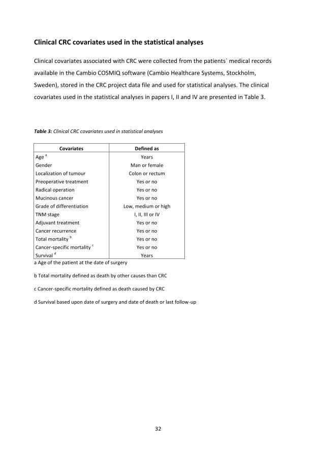
Clinical covariates associated with CRC were collected from the patients` medical records
available in the Cambio COSMIQ software (Cambio Healthcare Systems, Stockholm,
Sweden), stored in the CRC project data file and used for statistical analyses. The clinical
covariates used in the statistical analyses in papers I, II and IV are presented in Table 3.
Table 3: Clinical CRC covariates used in statistical analyses
Covariates Defined as Age a Years Gender Man or female Localization of tumour Colon or rectum Preoperative treatment Yes or no Radical operation Yes or no Mucinous cancer Yes or no Grade of differentiation Low, medium or high TNM stage I, II, III or IV Adjuvant treatment Yes or no Cancer recurrence Yes or no Total mortality b Yes or no Cancer-specific mortality c Yes or no Survival d Years
a Age of the patient at the date of surgery
b Total mortality defined as death by other causes than CRC
c Cancer-specific mortality defined as death caused by CRC
d Survival based upon date of surgery and date of death or last follow-up
33
METHODS
Tissue homogenization
Tissue lysates were obtained from normal and tumour tissue biopsies that were thawed, cut
into sufficient sizes, and put into RIPA buffer (containing 1XTBS, 1 % nonidet P-40, 0.5 %
sodium deoxycholate, 0.1 % SDS, 0.004 % sodium azide and adding PMSF solution, sodium
orthovanadate and protein inhibitor) (Santa Cruz Biotechnology, Inc., Heidelberg, Germany)
in an Eppendorf tube. The tissue biopsies were then homogenized and placed on ice for 30
min and centrifuged at 14000 g for 8 min at 4°C. The protein content of the lysate
supernatant fluid was determined using the Bradford protein assay (Bio-Rad Laboratories,
Ltd., Hertfordshire, UK), according to manufacturer’s recommendations.
Immunohistochemistry
Embedding and sectioning services were performed by co-workers at the Pathology
Laboratory, Department of Laboratory Medicine, Region Jönköping County.
Immunohistochemistry (IHC) was performed in papers I-III to visualize the expression and
localization of the proteins of interest: CD93, PLA2G4C, PDGF-DD, PDGFR-β and phospho-
specific platelet-derived growth factor receptor β (phPDGFR-β) in human matched tumour
and normal tissue sections from CRC patients.
In papers I-III, epitope unmasking was performed using the antigen retrieval solution DIVA,
and background staining was minimized by quenching the endogenous peroxidase activity
with 3 % hydrogen peroxide.

34
Avidin and biotin detection
In papers I-III, IHC was performed using the avidin and biotin detection system. Non-specific
binding between primary antibodies and human tissue samples was blocked either with
normal goat or horse serum prior to incubation with primary antibodies at 4 °C overnight
and secondary antibodies for 1 hour at room temperature. Detection of the antibody-
antigen complex is based on the avidin and biotin complex followed by visualization with
3.3-diaminobenzidine tetrahydrochloride substrate. To give a contrast to the primary stain
the sections were counterstained with hematoxylin before being dehydrated and mounted
for light microscopy visualization.
Protein detection using the IHC method via antibody binding is a widely used method in
clinical applications for diagnosis, prediction and prognosis (Coons, 1951). The formalin-fixed
biological tissue samples are embedded shortly after surgery, which ensures that the tissue
samples may be archived over time without changing their structure. The sectioning of the
samples enables repeated investigations of staining of different proteins of interest. The IHC
method enables identification and visualization of different proteins of interest as well as
their location within the different layers of the tissue due to the binding of a specific
antibody to an antigen. The binding is visualized either directly or indirectly by a fluorophore
or a peroxidase-conjugated antibody.
Monoclonal antibodies are made from identical immune cells cloned from a parent cell,
isolated by chromatography and bound to the same epitope. These antibodies are more
difficult to produce and therefore more expensive than polyclonal antibodies. The polyclonal
antibodies are a mixture of immunoglobulins that can bind to different epitopes of the
target protein antigen of interest, resulting in a stronger signal. The monoclonal antibodies
are more specific than the polyclonal antibodies. (MacPhee, 2010). There are many
commercially available antibodies that are well described but even though the antibodies
have a recommended dilution factor it may be necessary to perform tests of several
dilutions before an optimal dilution factor for the tissue investigated is found. This process
35
may also require changes in the IHC protocols for optimized staining and may be a time-
consuming process.
Western Blot
In papers I and III, Western Blot (WB) was used to reveal protein expression of CD93, PDGFR-
β and phPDGFR-β. In paper I and III, CD93, PDGFR-β were measured in matched tumour and
normal tissue lysates from CRC patients. In paper III, protein expression of phPDGFR-β was
investigated in Caco-2 and HT-29 cell lysates.
The WB method consists of three parts: protein separation, transfer to a solid support, and
visualization of the protein using a specific primary and secondary antibody. After
denaturation of tissue lysates and cell lysates, proteins are separated using sodium dodecyl
sulfate polyacrylamide gel electrophoresis (SDS-PAGE) before they are transferred onto a
polyvinylidene fluoride (PVDF) membrane followed by blocking of unspecific binding sites. A
marker is also loaded onto the gel, which enables size determination of visual protein bands.
A primary antibody specific to the protein of interest is added followed by incubation.
Several steps of washing of the membrane after incubation ensure that only the bound
protein of interest is left. A horseradish peroxidase (HRP) conjugated secondary antibody,
specific to the primary antibody is added, followed by a new incubation period and washing
of the membrane. The protein of interest is then visualized due to the oxidative reaction of
luminol catalyzed by the HRP antibody and captured by a charged coupled device camera
(Mahmood and Yang, 2012).
The WB method gives an opportunity to detect proteins of interest within biological samples
such as tissue- and cell lysates, but since it demands several steps of manual handling it is
time-consuming. The method is a semi-quantitative since it provides a relative comparison
of protein levels and not the absolute measure of quantity. This is due to differences in both
loading and transfer rates between the samples in separate lanes, which differ between
separate blots and should therefore not be used to measure the concentration. Also, the
signal generated during detection is not linear across the concentration range of samples.
36
Coomassie blue staining of the SDS-PAGE gel is an approach useful for control of loading
since it ensure a semi-quantitative measurement of protein bands appearing on the gel. The
PVDF membranes provide a proper mechanical support and allow the blots to be re-probed
and stored minimizing the steps in the protocol and ensuring no unnecessary waste of
samples. A disadvantage is that the background is higher in PVDF membranes compared to
nitrocellulose membranes and careful washing is therefore important. However, leaving the
membrane to wash for a long time may also reduce the signal of the bound antibodies
(Mahmood and Yang, 2012).
Enzyme-linked immunosorbent assay
In papers I and III, commercially available Enzyme-linked immunosorbent assays (ELISAs),
(R&D Systems Europe Ltd., Abingdon, UK) were used to quantify protein levels of CD93 and
PDGF-DD in matched tissue lysates from CRC patients. In paper I protein levels of CD93 were
also measured in plasma from CRC patients and controls. In paper III protein levels of MMP-
1 and serpin family E member 1 (Serpin E1) were measured in cell supernatants from
stimulated Caco-2 cancer cells.
The ELISA technique is a quantitative sandwich enzyme immunoassay that ensures binding
of antigens to specific antibodies enabling detection or quantification of small amounts of
antigens (e.g. proteins) in biological liquids such as plasma, serum, saliva, urine or cell
culture supernatants. A commercial sandwich ELISA is a microtiter plate containing 96 wells
pre-coated with a capture antibody. A sample which may contain the antigen of interest is
added to the wells and if the antigen of interest is present in the sample it get bound by the
immobilized antibody. After washing away of unbound substances, an enzyme-linked
secondary antibody specific for the antigen of interest is added to the wells followed by a
washing step to remove any unbound antibody-enzyme reagent. A substrate solution is then
added to the wells and colour develops in the proportion to the amount of bound antigen of
interest in the initial step. The colour development is stopped and the intensity of the colour
is measured using a spectrophotometer. A standard of known concentration, which is

37
generated for each assay run, is then used to quantify the concentration of the antigen of
interest (Hornbeck, 2015).
There are both advantages and disadvantages with the ELISA method. As mentioned,
different types of biological samples can be analyzed and no radioactive substances are
needed. The intra-assay precision within a commercial ELISA assay is quite accurate but the
inter-assay precision may vary. The main advantage is the high sensitivity of the commercial
ELISAs, and they also have a strong specificity for their coated capture antibodies. A
disadvantage is that ELISAs often requires a large sample volume per well and each sample
should be analyzed in duplicate. Also, only one analyte can be analyzed per sample, per
ELISA assay (Fu et al., 2010) making it costly and time-consuming if more analytes need to be
investigated. Variations in sample collection, processing and storage may cause sample value
differences. The method includes several hands-on steps that may cause varying results due
to poor precision.
Luminex
In paper IV, levels of inflammatory factors were measured in plasma from CRC patients by a
commercially available Luminex assay, Bio-Plex Pro™ Human Chemokine Panel 40-Plex (Bio-
Rad Laboratories, Inc., CA, USA), enabling analysis of 40 cytokines in 50 µl of plasma. The
prognostic association between tertile levels of each cytokine, defined as low, medium or
high levels, was analyzed in relation to survival defined as CRC-specific-, and total mortality.
Luminex is a multiplex bead-based sandwich immunoassay that combines ELISA and flow
cytometry. The method shares the same immunological principle as ELISA with antibodies
and antigens. The Luminex method using the Bio-Plex Pro™ Human Chemokine Panel 40-Plex
involves the use of capture antibodies, which are coated onto magnetic beads (6.5 µm),
filled with a specific mixture of fluorescent dyes. The colour-mixture of each bead is unique,
enabling differently dyed beads coated with different antibodies to be mixed together in the
same well. This permits the detection of multiple cytokines in a single sample.

38
After adding the coated magnetic beads to a 96-well microplate followed by incubation the
magnetic beads are washed using a magnet underneath the plate in the automated washing
station. This results in a magnetic field ensuring that the beads are not removed during the
washing steps. In the next step, diluted samples and standards are added to the microplate
followed by incubation allowing the capture antibody coupled to the beads to interact with
the samples containing the target of interest. After incubation and washing to remove
unbound materials, the biotin-conjugated detection antibodies are added, which are specific
to a different epitope. This is followed by another incubation period creating a sandwich-
complex. The final detection-complex is formed when streptavidin-phycoerythrin
(streptavidin-PE) conjugate is added. PE serves as a fluorescent reporter (Figure 8) (Gupta V.,
2014).
The beads are then read on a flow-based detection instrument with dual lasers. The red
laser (635 nm) detects the bead, determining what cytokine is being measured, and the
green laser (532 nm) assesses the PE fluorescence reporter intensity, which is proportional
to the amount of bound cytokine (Gupta V., 2014). A digital processor manages the data
output and the Bio-Plex Manager™ software presents data as median fluorescence intensity
as well as the concentration (pg/mL). The concentration of analyte bound to each bead is
proportional to the median fluorescence intensity of reporter signal (Bio-Rad Laboratories,
2017).

39
Figure 8: The principle of the Bio-Plex immunoassay modified from Bio-Plex Pro™Human Chemokine assay
instruction manual (Bio-Rad Laboratories, 2017).
An advantage of Luminex is that large numbers of cytokines can be detected simultaneously
in a small volume of a single sample. This is possible due to the magnetic beads with
difference in fluorescent dyes in combination with the wide range of detectable protein
concentrations. When analysing numerous of cytokines the Luminex method is less time
consuming than ELISA and more data can be gathered rapidly. The use of magnetic beads
allows the method to be automated using a washing station and the magnetic separation
ensures better reproducibility compared with vacuum filtration. To achieve proper results it
is of great importance to vortex the magnetic beads properly before adding them to the
plate since insufficient vortexing may result in lower concentrations of the cytokines
measured. An inappropriate incubation time may also result in lower concentrations.
Another disadvantage is that the Luminex analysis requires a specific instrument. For serum
biomarker analysis the Bio-Plex assays together with MULTI ARRAY have been found to have
the best performance, with the lowest limits of detection (Fu et al., 2010).
StreptavidinBiomarker of interestMagnetic bead
Capture antibodyBiotinylated
detection antibodyPE fluorescent
reporter
40
Caco-2 and HT-29 cell lines
In paper III the colon adenocarcinoma cell lines Caco-2 (Sigma-Aldrich, MO, USA) and HT-29
(Sigma-Aldrich) were used to study both the expression and proliferative effects of PDGF-D
in CRC carcinogenesis in vitro. The Caco-2 cell line was also used in the gene array analysis to
study effects of PDGF-D signaling on gene alterations.
The Caco-2- and the HT-29 cell lines are from human Caucasian colon adenocarcinomas
where Caco-2 originates from a 72-year-old male while HT-29 originates from a 44-year-old
female. Both cell lines are developed to emulate the human intestinal epithelium containing
immortalized cells that rapidly can grow into confluent monolayers. Differentiated Caco-2
cells have been shown to express tyrosine kinase receptor of PDGF-D, PDGFR-β and c-Kit
(Kaulfuss et al., 2013). As Caco-2 cells the HT-29 cells also express c-Kit (Bellone et al., 1997)
but seem to express PDGFR-β more poorly compared to Caco-2 (Kaulfuss et al., 2013).
Human CRC model cell lines are common, preclinical model systems of the disease and have
provided important insights into tumour- and cell biology, including the discovery of new
anti-tumour compounds, and have revealed possible drug sensitivity, resistance and toxicity
(Bracht et al., 2010, Weickhardt et al., 2012). The commercially available cancer cell lines are
well defined and have a longer life-time than primary cell lines. It is easier to control
experimental factors, and cell lines are widely used by investigators, thus facilitating
comparison of results (Jones et al., 2008). Also, it is important to stress that cell line models
in general are more efficient than animal models both in time and costs aspects. The Caco-2
cell line has been shown to share the same gene expression pattern involved in proliferation
as seen in human colon cancer in vivo even though it lacks the tumour stroma, which is
thought to affect processes such as proliferation in cancers, making it a suitable cell line for
studying stimulatory effects affecting proliferation (Saaf et al., 2007).
A disadvantage is that cancer cell lines may be exposed to different environments such as
sera, media and additional factors added to the media, as well as varying carbon dioxide
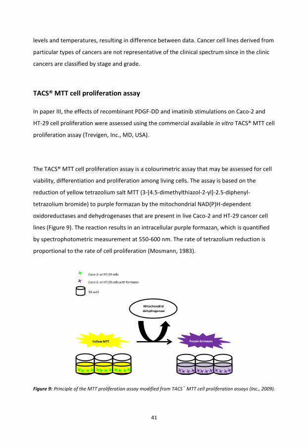
41
levels and temperatures, resulting in difference between data. Cancer cell lines derived from
particular types of cancers are not representative of the clinical spectrum since in the clinic
cancers are classified by stage and grade.
TACS® MTT cell proliferation assay
In paper III, the effects of recombinant PDGF-DD and imatinib stimulations on Caco-2 and
HT-29 cell proliferation were assessed using the commercial available in vitro TACS® MTT cell
proliferation assay (Trevigen, Inc., MD, USA).
The TACS® MTT cell proliferation assay is a colourimetric assay that may be assessed for cell
viability, differentiation and proliferation among living cells. The assay is based on the
reduction of yellow tetrazolium salt MTT (3-[4.5-dimethylthiazol-2-yl]-2.5-diphenyl-
tetrazolium bromide) to purple formazan by the mitochondrial NAD(P)H-dependent
oxidoreductases and dehydrogenases that are present in live Caco-2 and HT-29 cancer cell
lines (Figure 9). The reaction results in an intracellular purple formazan, which is quantified
by spectrophotometric measurement at 550-600 nm. The rate of tetrazolium reduction is
proportional to the rate of cell proliferation (Mosmann, 1983).
Figure 9: Principle of the MTT proliferation assay modified from TACS® MTT cell proliferation assays (Inc., 2009).
Caco-2- or HT-29 cells
Caco-2- or HT-29 cells with formazan
96 well
Purple formazanYellow MTT
Mitochondrial dehydrogenase

42
Tetrazolium salt assays such as MTT are high-throughput and useful in screening of cell lines
for sensitivity to drug treatments and to changes in growth conditions when including
growth factor, hormone and serum supplementation. The assay analysis is easy, does not
include any steps of washing and many samples from different cell lines can be processed at
the same time. The proliferation assay is also quantitative since there is a close correlation
between viable cell numbers and the generated MTT-formazan signal, making it more
efficient than counting viable cell manually with a hemocytometer (Mosmann, 1983). A
limitation of the assay is that the linearity of the results may vary between MTT assays and
between replicates of samples, which also was observed in our study. It has been shown that
the MTT assay is less sensitive at cell numbers lower than 1000 cells per well (van Tonder et
al., 2015). This problem was also observed in our study leading to cultivation of 2.5x10⁴
Caco-2 cells and 1.5x10⁴ HT-29 cells per well.
43
Nucleic acid extraction and reverse transcription
DNA was extracted from whole blood using a QiaAmp DNA blood kit (Qiagen, Hilden,
Germany) and total RNA was extracted from matched tumour and normal tissue biopsies
using the RNeasy kit (Qiagen), according to the manufacturer’s recommendations.
Total RNA (1 µg) from matched tumour and normal tissue biopsies from CRC patients or
from Caco-2 cancer cells were reverse transcribed into complementary deoxyribonucleic
acid (cDNA) with random primers and components of the High-capacity cDNA reverse
transcription kit (Applied Biosystems Inc., CA, USA, according to manufacturer`s
recommendations. Reverse transcription was performed at: 25 °C for 10 min; 37 °C for 1
hour; 85 °C for 5 min; 4 °C for ∞ in a Palm-Cycler™ (Corbett Life Science Pty. Ltd., Mortlake,
Australia).
Polymerase Chain Reaction
In papers I and, II SNP analysis of CD93 and PLA2G4C was done with TaqMan genotyping
assays (Life Technologies, Ltd., Paisley, UK). In short, DNA (10 ng) was mixed with TaqMan
Universal PCR Master mix II (Life Technologies) and TaqMan genotyping assays (Life
Technologies) (Table 4) and amplified using the 7500 Fast Real-Time PCR system (Applied
Biosystems).
Table 4: TaqMan genotyping assays (Life Technologies) used in papers I and II
Gene SNP ID Paper I Paper II CD93 rs2749812 x CD93 rs2749817 x PLA2G4C rs1549637 x
rs: reference ID
In papers I and III, gene expression analysis was done by TaqMan gene expression assays
using TaqMan probes on a 7500 Fast Real-Time PCR system (Applied Biosystems). In paper I,
gene expression of CD93 on cDNA from CRC tissues and matched normal tissues was
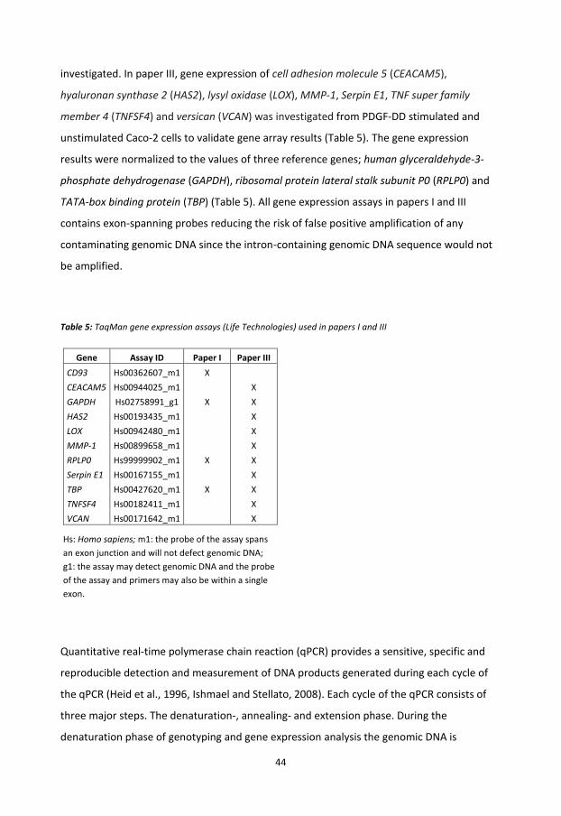
44
investigated. In paper III, gene expression of cell adhesion molecule 5 (CEACAM5),
hyaluronan synthase 2 (HAS2), lysyl oxidase (LOX), MMP-1, Serpin E1, TNF super family
member 4 (TNFSF4) and versican (VCAN) was investigated from PDGF-DD stimulated and
unstimulated Caco-2 cells to validate gene array results (Table 5). The gene expression
results were normalized to the values of three reference genes; human glyceraldehyde-3-
phosphate dehydrogenase (GAPDH), ribosomal protein lateral stalk subunit P0 (RPLP0) and
TATA-box binding protein (TBP) (Table 5). All gene expression assays in papers I and III
contains exon-spanning probes reducing the risk of false positive amplification of any
contaminating genomic DNA since the intron-containing genomic DNA sequence would not
be amplified.
Table 5: TaqMan gene expression assays (Life Technologies) used in papers I and III
Gene Assay ID Paper I Paper III CD93 Hs00362607_m1 X
CEACAM5 Hs00944025_m1 X GAPDH Hs02758991_g1 X X HAS2 Hs00193435_m1 X LOX Hs00942480_m1 X MMP-1 Hs00899658_m1 X RPLP0 Hs99999902_m1 X X Serpin E1 Hs00167155_m1 X TBP Hs00427620_m1 X X TNFSF4 Hs00182411_m1 X VCAN Hs00171642_m1 X
Hs: Homo sapiens; m1: the probe of the assay spans an exon junction and will not defect genomic DNA; g1: the assay may detect genomic DNA and the probe of the assay and primers may also be within a single exon.
Quantitative real-time polymerase chain reaction (qPCR) provides a sensitive, specific and
reproducible detection and measurement of DNA products generated during each cycle of
the qPCR (Heid et al., 1996, Ishmael and Stellato, 2008). Each cycle of the qPCR consists of
three major steps. The denaturation-, annealing- and extension phase. During the
denaturation phase of genotyping and gene expression analysis the genomic DNA is
45
denatured into separate strands. In the annealing phase the probe/primer hybridize to the
complementary target sequence on the template DNA strand. The TaqMan probe have a
fluorescent reporter dye on the 5`-end and a non-fluorescent quencher minor groove binder
dye on the 3`end. During the annealing phase the primer and TaqMan probe will hybridize to
the target sequence if present. As long as the probe is intact the close proximity of the 3`
quencher inhibits the fluorescent signal from the 5` reporter fluorochrome. The fluorescent
signal is emitted when the probe is cleaved. In the extension phase the 5` to 3` exonuclease
activity of the thermostable Ampli Thermus aquatiqus (Taq)-Gold® DNA polymerase cleaves
the hybridized probe, releasing the reporter dye from the quencher. The fluorescence signal
generated by qPCR amplification indicates which alleles are present in the sample in the
genotyping analysis. During each cycle of qPCR the exponential signal is proportional to the
amplified material obtained during gene expression analysis (Scientific, 2017b, Scientific,
2017a, Holland et al., 1991). During the cycles of the qPCR the exponential increase in
fluorescent signal is proportional to the amplified material. This makes it possible to quantify
the amount of starting material by relating it to a standard curve.
The use of TaqMan genotyping and TaqMan gene expression assays ensures an effective
analysis. Since the assays contain minor groove binders a high specificity for the target site is
ensured. TaqMan gene expression analysis by qPCR is a valuable tool for validation of gene
array results (Ishmael and Stellato, 2008). On the other hand, for gene expression analysis
the high sensitivity of qPCR also requires accurate and precise pipetting, high-quality RNA, an
accurate estimation of the RNA concentration as well as an efficient reverse transcription.
Variations in these technical parameters may influence the accuracy and precision of the
results (Remans et al., 2014). Another limitation to gene expression analysis is to choose
proper reference genes for normalization and the need to run standard curves for each gene
of interest with each assay (Ishmael and Stellato, 2008).

46
Affymetrix® GeneChip® Whole Transcript Expression array
In paper III, the Affymetrix® Genechip® Whole Transcript Expression array (Affymetrix, CA,
USA) was used to evaluate the effects of PDGF-D on gene expressions using the Caco-2
cancer cell line as a model. In this paper, employees at the department of Genome Analytics
at the Helmholtz Centre for Infection Research, Braunschweig, Germany, helped to perform
gene array analysis and subsequent interpretation of data.
Gene arrays, also named microarrays, are solid supports where upon a large collection of
gene specific nucleic acids are situated at defined positions. During the gene array analysis
double-stranded cDNA is synthesized from RNA samples. Then an in vitro transcription,
including a biotin-labelled nucleotide, is performed producing biotin labelled complementary
RNA (cRNA) from the cDNA. The cRNA is then purified to remove unincorporated
nucleotides, inorganic phosphates and salt before it is fragmented and hybridized to the
Affymetrix® GeneChip® array. The hybridized gene array is then stained with PE-conjugated
streptavidin and scanned (Figure 10). The emitted light is proportional to the amount of
bound target sequence and hence the gene expression level (Corporation, 2011, Inc., 2009-
2010). The analysis of the obtained gene array data after scanning is performed using
GeneSpring 11.5.1 (Agilent Technologies, Inc., CA, USA), which provides analysis of changes
in gene expression (Agilent Technologies, 2011).
A major advantage of gene array analysis is that the method can provide information on
thousands of targets in a single experiment. The GeneSpring software is helpful in both
visualization of data and in comparing results from different data sets. The Affymetrix®
Genechip® Whole Transcript Expression array ensures expression analysis over the entire
transcript since the probe selection region is defined by exons. This allows identification of
alternatively spliced transcripts. Each spot on the chip contains a certain number of copies
per gene and if a sample contains a higher concentration of certain genes it may lead to a
saturation of the spot leading to measurement of too low RNA levels.
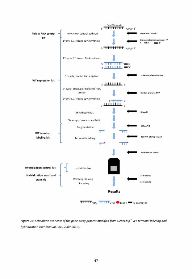
47
Figure 10: Schematic overview of the gene array process modified from GeneChip® WT terminal labeling and
hybridization user manual (Inc., 2009-2010).
DNARNA Biotin T7 promoter
Poly-A RNA controls
5`3` N N NEngineerred random primers + T7
Un-labeled ribonucleotides
Random primers, dUTP
RNase H
TdT DNA labeling reagent
UDG, APE 1
Hybridization controls
Stain coctail 1
Stain coctail 2ScanningWashing/staining
Hybridization
Terminal labelling
Fragmentation
Cleanup of sense strand DNA
aRNA hydrolysis
2st cycle, 1st strand cDNA synthesis
1st cycle, cleanup of antisense RNA (aRNA)
1st cycle, in vitro transcription
1st cycle, 2st strand cDNA synthesis
1st cycle, 1st strand cDNA synthesis
Poly-A RNA control additionPoly-A RNA control kit
WT expression kit
WT terminal labeling kit
Hybridization control kit
Hybridization wash and stain kit
Results
5` AAAAA 3`Total RNA sample
5`3`
3`5`
5`3`
5`3`
3` U 5`
5` AAAAA 3`

48
Statistical analyses
Papers I and II: The Chi squared test was used to test the genotype frequencies for
deviations from the hardy Weinberg equilibrium for all SNPs analyzed.
Paper I, paper II and paper IV: To evaluate differences in the frequencies of the SNPs
analyzed between patients and controls as well as the distribution of clinical covariates
(papers I and II), and to determine the correlation between CRC-specific mortality (paper IV)
and the clinical covariates within the patient group a chi-squared test (Pearson`s chi-squared
test) was performed.
Papers I-IV: Differences in protein expression levels between tumour and matched normal
tissues in relation to clinical covariates (papers I and III), differences in plasma protein levels
between patients and controls in relation to clinical covariates (paper I), differences
between clinical covariates among stage II CRC patients (paper II) and differences in levels of
inflammatory factors between TNM stage combinations in relation to clinical covariates,
(paper IV) were examined by the Wilcoxon signed-rank test (also named the Wilcoxon rank-
sum test or Mann-Whitney U test).
Papers I, II and IV: A Shapiro-Wilk test was used to determine the normal distribution.
Cancer-specific- and disease-free survival were analyzed using the Kaplan-Meier analysis and
log-rank test. The Cox regression analysis was performed for univariate- and multivariate
analysis in association with clinical covariates (papers I and II).
Analysis of CRC-specific- and total mortality was performed with Kaplan-Meier analysis and a
log-rank test. Both a univariate and a multivariate Cox regression analysis were performed to
investigate unadjusted and adjusted effects on the inflammatory tertile levels in relation to
either CRC-specific- or total mortality in association with clinical covariates (paper IV).
The goodness of fit of the multivariable models was tested by Harrell`s C index. (papers I, II
and IV).
49
Paper III: The treatment effects on cancer cell lines in relation to cell proliferation were
examined using a One-Way ANOVA with Post Hoc Multiple Comparisons (Bonferroni) test.
For the gene array results signal intensities (raw data) were log2 transformed and
normalized using the robust multichip average. Gene expression values for each condition
were given as a relative expression to the mean expression value calculated from the
intensities of all conditions (mean centralization, normalized data). To determine
differentially expressed genes after stimulation a statistical t-test followed by a statistical
error correction (Benjamini-Hochberg) were applied to log2 normalized gene expression
data (n = 3). Genes with corrected p-values <0.05 were considered as differentially
expressed.
Statistical analyses were performed in IBM® SPSS® Statistics, 2012 (version 19 and 21, SPSS
Inc., IL, USA) or STATA 12 and 13 software (StataCorp, Stata Statistical Software; release 12
and 13, College Station, TX, USA).
In all studies, p-values < 0.05 were considered statistically significant, where * indicates p <
0.05, ** indicates p <0.01, and *** and indicates p < 0.001.
ETHICS
The CRC project was approved by the Local Ethics Committee of Linköping University,
Sweden, (approval number 98113 and 2013/271-31) approving collection of blood samples,
matched control and tumour biopsies, as well as the collection of clinical covariates
associated with CRC from the patients` medical records available in the Cambio COSMIQ
software (Cambio Healthcare Systems). The patients were de-identified and the samples
were collected together with routine samples not causing the patients an extra burden
during sampling.
50
RESULTS AND DISCUSSION
CD93 gene polymorphism is associated with disseminated
colorectal cancer (Paper I)
CD93 has been studied in various types of diseases, including CVD, as well as in various
cancer forms but not in CRC prior this study. Since the initially aim of the thesis was to study
possible associations of underlying CVDs on CRC disease and prognosis the aim of paper I
was to study the expression and localization of CD93 in normal and adjacent tumour tissues
and CD93 concentrations in plasma from controls and CRC patients. The association of two
SNPs of CD93, rs2749812 and rs2749817 not found to be in linkage disequilibrium (LD) to
another, were also studied (Malarstig et al., 2011). The SNP rs2749812 has previously been
associated with risk of myocardial infarction (Malarstig et al., 2011) and coronary heart
disease (van der Net et al., 2008), while SNP rs2749817 has been associated with the risk of
breast cancer (Lee et al., 2009).
An increased expression of CD93 was seen in vascular endothelial cells around blood vessels
in the cancerous tissues compared to expression observed in the same locations in matched
normal tissues. The same expression and localization patterns have been seen in other types
of cancers such as in glioblastoma (Langenkamp et al., 2015) and nasopharyngeal carcinoma
(Bao et al., 2016). The production of CD93 has been shown to be mediated by inflammatory
factors such as MMPs and TNF-α (Bohlson et al., 2005). The production of CD93 may also be
induced by other unknown inflammatory factors in vascular endothelial cells.
Langenkamp et al. investigated the function of CD93 in tumour angiogenesis and endothelial
cell biology and showed that knock-down of CD93 inhibited adhesion and migration of
endothelial cells as well as tube formation in vitro due to defects in cytoskeletal
rearrangement by the interaction of the C-terminal domain of CD93 with myosin and loss of
endothelial adherence junctions. These results led to the conclusion that CD93 was essential
in establishing endothelial cell-cell and cell-matrix contacts as well as migration and tubular
51
morphogenesis, suggesting that CD93 promotes the formation of well-functioning tumour
vessels (Langenkamp et al., 2015). It is possible to assume that the same actions may also
appear in CRC, which would explain the increased expression of CD93 in vascular endothelial
cells in blood vessels.
Since expression and localization of the CD93 protein was seen in CRC tissue, the protein
levels of CD93 were further investigated in matched tissue lysates as well as in plasma.
Comparing tissue protein levels of total CD93 (cell bound and soluble form of CD93) an 82 %
higher level was seen in CRC tissue lysates compared to matched normal tissue lysates. This
result may be due to an ongoing angiogenesis and increased inflammation in the CRC tissue.
This assumption is strengthened by the findings of Kao et al. showing that the EGF-like
domain of CD93 is important in the angiogenetic process since binding of EGF to its EGF
receptor induces cellular signalling triggering the development of angiogenesis (Kao et al.,
2012).
Even though an increased expression of CD93 was observed in both CRC tissue sections and
CRC tissue lysates the gene expression analysis did not reveal any differences in CD93
expression between CRC tissue and matched normal tissue levels of CD93. In this case it is
important to remember that divergence between protein and messenger RNA (mRNA)
expression is a common finding (Schwanhausser et al., 2011). Also a possible pre-existing
inflammatory condition within the normal tissue distant from the location of the CRC tumour
could explain why there was no difference in CD93 expression levels in CRC tissues
compared to the matched normal tissues. This was suggested as a possible explanation in a
different study by Mounier et al. where similar results in gene expression were observed
(Mounier et al., 2008).
In contrast to the increased levels of total CD93, a 30 % lower level of the soluble form of
CD93 was detected in plasma among CRC patients compared to healthy controls. This result
was quite surprising since the soluble form of CD93 has been shown to be cleaved by MMPs
normally found in higher levels in CRC. TNF-α has also been shown to contribute in the
releasement of CD93 (Bohlson et al., 2005). In a study by Greenlee et al. the soluble CD93
was shown to function as a bridging molecule between apoptotic cells and macrophages
52
ensuring efficient engulfment of apoptotic cells. Also, Greenlee et al. suggested that
proteases such as MMPs responsible for the cleaving and release of CD93 were specific to
activated macrophages, meaning that the MMPs lose their ability to produce the soluble
form of CD93 when the macrophages are quiescent (Greenlee et al., 2009). Endothelial cells
also express soluble CD93 and serve as an additional source of the soluble form (Fonseca et
al., 2001), which might explain the lower levels observed in CRC plasma if macrophages and
their contributing proteases go quiescent due to unknown immunologic imbalances in CRC
patients. The presence of tissue inhibitors of metalloproteinases secreted by cells may also
inhibit the function and decrease the expression of MMPs (King and Robins, 2006a) and may
therefore be another important factor in explaining the lower levels of soluble CD93 in
plasma.
Another observation was that approximately 66 % of the CRC patients had plasma levels of
CD93 below 142 ng/mL, which has been suggested as a threshold associated with increased
risk of myocardial infarction among Swedish patients with coronary artery disease (Malarstig
et al., 2011). Since no clinical information regarding possible underlying CVDs was available
no conclusion on whether low plasma levels of CD93 might indicate a risk of developing both
CRC and CVD, or if the diseases may reflect similar physiological and pathological
mechanisms in the regulation of CD93 could be drawn.
Two SNPs of CD93, rs2749812 and rs2749817, previously associated with risk of CVD and
breast cancer, were investigated to reveal their potential associations with CRC. For both
SNPs no differences in genotype distribution among CRC patients and controls were
observed. This indicates that none of the SNPs are risk factors for contracting CRC. Among
the patients, an adjustment for localization, gender and age did not affect the results.
The genotype distribution of rs2749817 showed an increased frequency of the T/T genotype
in stage IV compared to stage I-III. By studying the T/T genotype in relation to CRC-specific
survival it was shown to be associated with worse prognosis and a lower recurrence-free
survival. It was also associated with an increased risk of disease recurrence among CRC
patients that were tumour-free after surgery compared to patients carrying the C/C- or the
C/T genotype. When comparing the genotype frequencies of rs2749817 to the tissue levels

53
of total CD93, tumour tissue levels among CRC patients carrying the T/T genotype were 20 %
higher compared with the levels in patients carrying the other genotypes. This suggests that
the T/T genotype of rs2749817 may affect the expression of CD93 in CRC tissue.
The association of the T/T genotype of rs2749817 with disseminated cancer at diagnosis and
an increased risk of recurrence after radical operation suggests a lower resistance to the
dissemination of tumour cells as well as a reduced ability to control metastasis. The exact
mechanism of the T/T genotype of rs2749817 is still unknown but it seems to be associated
with increased tissue level of CD93 in CRC which might affect angiogenesis, growth and
spread of CRC. This SNP is situated downstream of the 3` untranslated region of CD93, in an
intergenic region consisting of long non-coding RNAs. Long non-coding RNAs may alter their
activity by folding into structures enabling them to interact with transcription factors
affecting the transcription of nearby genes (Ling et al., 2015). This may influence the
translation and post-translational modifications of CD93. Also, the role of the T/T genotype
of rs2749817 on survival remains to be further investigated, but the results in paper I
suggest that the SNP is a potential risk factor for predicting survival among CRC patients.
Since the CRC patients studied in paper I had a high mean age there might be a risk of other
underlying inflammatory disorders affecting the CD93 levels in plasma or in tissue lysates.
The lack of clinical data regarding possible underlying disorders such as CVDs at the time of
the study is a clear limitation and no conclusions can be made regarding their possible
impact on CD93 levels.
In summary, the results of paper I show that CD93 had an increased expression in CRC tissue,
which may reflect the ability of CD93 to promote formation of well-functioning tumour
vessels as well as an ongoing angiogenesis and inflammation. The lower plasma levels of
CD93 in CRC patients may be a result of the lost ability of MMPs to produce the soluble form
of CD93 when the macrophages are quiescent leaving endothelial cells as the only source
capable of producing soluble CD93. Most importantly, the T/T genotype of SNP rs2749817 of
CD93 may be associated with disseminated CRC at diagnosis and an increased recurrence
rate after radical operation. This indicates that the T/T genotype of SNP rs2749817 may be a
potential marker related to severe tumour disease and spread, even at the time of diagnosis.
54
Prognostic significance of PLA2G4C gene polymorphism in patients
with stage II colorectal cancer (Paper II)
Several studies have shown expression of cytosolic PLA2 enzymes in CRC (Dimberg et al.,
1998, Osterstrom et al., 2002) and the expression of PLA2G4C has been suggested to be
involved in breast cancer cell chemotaxis and invasion (Tian et al., 2011). The cellular source,
localization or possible clinical association of PLA2G4C in CRC has not previously been
described. In paper II, the focus was therefore to study the expression and localization of
PLA2G4C in CRC tissue and to study the association of the PLA2G4C SNP, rs1549637, as a
potential risk factor for CRC and a predictor of disease outcome among CRC patients.
Expression of PLA2G4C was mainly localized in epithelial cells in both tumour and matched
normal tissue from CRC patients with an equal expression and was in line with previous
studies (Panel et al., 2006, Yoo et al., 2011, Osterstrom et al., 2002).
SNP rs1549637 analysis revealed no differences in either the genotypic or in the allelic
frequencies among CRC patients and controls. This observation indicates that the SNP
rs1549637 is not to a risk factor for contracting CRC. Among CRC patients the Kaplan-Meier
analysis showed a significant difference in CRC-specific survival between the genotypes of
rs1549637 among CRC patients of all TNM stages. After combining the A/A and the A/T
genotype due to low prevalence of the A/A genotype, survival analysis revealed that carriers
of the A allele had the highest risk of CRC-specific mortality compared to patients carrying
the T allele. The A allele remained an independent predictor of increased CRC-specific
mortality even after adjustment for the clinical covariates; age, gender, TNM stage, tumour
localization, adjuvant treatment, T4 tumour invasion, histopathological type and
differentiation. Also, compared with T allele carriers the CRC patients carrying the A allele
had a poor prognosis in stage II CRC, with an increased risk of CRC-specific mortality even
after adjustment for the mentioned clinical covariates.
Stage II disease is found in approximately 30 % of all CRC patients (Jemal et al., 2004) and
even though stage II disease has in general a good prognosis 20-25 % of CRC patients will still
experience recurrence of the cancer (Sobrero, 2006, O'Connor et al., 2011). Adjuvant
55
treatment has been shown to be beneficial in stage III CRC (Bockelman et al., 2015), but it
remains controversial in stage II CRC (Sobrero, 2006, O'Connor et al., 2011, Glimelius, 2015).
To be able to identify and select these subgroups of CRC patients for adjuvant treatment the
currently used markers are: elevated CEA levels, lymphovascular or perineural invasion,
perforation, poorly differentiated tumour, T4 growth, and removal of fewer than 12 lymph
nodes from the specimen. However, all these markers are associated with weak prognostic
characteristics (Bockelman et al., 2015) underlining the importance of finding risk factors
with better prognostic characteristics. In a study by Buhmeida et al. another PLA2G4
member, PLA2G4A, was suggested as a prognostic marker for the selection of patients at
high risk of recurrence in stage II CRC that might benefit from adjuvant treatment. They
found that PLA2G4A-positive staining of tumours was associated with lower CRC-specific
survival. On the other hand, no associations with conventional risk factors such as age,
gender, depth of invasion or lymph node status were found (Buhmeida et al., 2009).
The exact function of the A allele of the SNP rs1549637 are currently unknown. The SNP is
situated in the 3` untranslated region of PLA2G4C, which may alter the mRNA level or even
modify the transcriptional activity of the gene (Kao et al., 2000).
There are some limitations to this study that are important to consider in further studies of
PLA2G4C. First, no gene expression analysis was performed, which may have given further
insights on expression of PLA2G4C in matched tumour and normal tissues from CRC patients.
Second, there was no investigation performed whether the SNP rs1549637 was in LD with
another SNP of PLA2G4C, which may be the actual SNP associated with the clinical outcome
among the CRC patients. Third, risk factors such as CEA as well as vascular, lymphatic and
neural tumour invasion were missing among CRC patients in the oldest part of the study
period and were therefore not included in the analyses.
In summary, PLA2G4C was shown to be expressed in epithelial cells in both CRC tissue and
matched normal tissue. The SNP rs1549637 of PLA2G4C seem not to be a risk factor for
contracting CRC. On the other hand, the A allele of this SNP was shown to be a potentially
stronger predictor for CRC-specific mortality in stage II than the available conventional risk

56
factors used in the clinical practice for selection of stage II patients for adjuvant treatment.
The A allele of SNP rs1549637 may be a potential marker for finding stage II CRC patients
that would benefit from a more comprehensive follow-up and adjuvant treatment.

57
Possible role and therapeutic target of PDGF-D signalling in
colorectal cancer (Paper III)
The production of various growth factors and cytokines are important for cancer cells to
enable progressive growth and development of cancer. PDGF-D is expressed in various types
of cancers such as breast cancer and CRC (Ahmad et al., 2011, Chen et al., 2016) and is
shown to be involved in the regulation of cellular processes important in cancer
development (Ustach and Kim, 2005, Li et al., 2003, Chen et al., 2016). PDGF-D induces its
cellular effects by binding to and activating the receptor PDGFR-β (Li et al., 2003, Wang et
al., 2009), a tyrosine kinase receptor, which, due to its phosphorylation is able to trigger a
number of signalling pathways also in CRC (Chen et al., 2016). Still, little is known regarding
PDGF-D’s possible role in CRC. In this study the expression and localization of PDGF-D in CRC
tissue were studied as well as the possible involvement of PDGF-D signalling in colorectal
carcinogenesis in vitro.
The expression and localization of both PDGF-D and its receptor, PDGFR-β, were studied in
sections from CRC tumour tissue and matched normal tissue. PDGF-D was mainly found in
the vascular endothelial cells of blood vessels situated in the cancerous microenvironment
and in the matched normal tissue, which was in line with a previous study (Chen et al.,
2016). In the study by Chen et al. the expression of PDGF-D was associated with
clinicopathological features such as TNM stage, lymph node invasion, and tumour
differentiation (Chen et al., 2016). In this study the correlation between expression levels of
PDGF-D and clinicopathological covariates was not investigated since no differences in tissue
expression levels of the protein were observed by ELISA. The PDGFR-β receptor was mainly
located in both normal epithelial cells and in tumour-associated stromal cells of the
cancerous microenvironment, which was in line with previous reports (Sundberg et al., 1993,
Lindmark et al., 1993). The activated form of the receptor, phPDGFR-β, was observed in
normal epithelial cells and stromal cancerous tissue. The inactivated and the activated form
of PDGFR-β were observed in cell lysates from the CRC cell lines Caco-2 and HT-29. These
observations led to the assumption that both PDGF-D and its receptor, PDGFR-β, may be
58
involved in the carcinogenic progress of CRC and led to further investigation of potential
genetic effects induced by PDGF-D signalling in vitro.
The Caco-2 cell line was used as a model to reveal possible genetic effects after 24-hour
stimulation with PDGF-D followed by a gene array analysis. The PDGF-D stimulation induced
an alteration of target genes, which may potentially modulate the tumourigenic activities of
cancer cells, such as ECM integrity, angiogenesis, migration, metastasis and proliferation to
promote further CRC progression. These results imply that PDGF-D signalling may have an
important role in CRC development and progression.
PDGF-D has previously been shown to be important in the EMT process that is involved in
the development of metastasis, in which epithelial cells are transformed into mesenchymal
cells due to morphologic changes (Wu et al., 2013, Chen et al., 2016). To ensure migration
and invasion the mesenchymal cells may increase the activity of proteases such as serine
proteases and MMPs, which ensures a path for the migration and invasion of the
mesenchymal cells through the ECM (Wu et al., 2013). The gene expression pattern induced
by PDGF-D revealed an increased expression of serine proteases, MMPs, collagens integrins
and selectins that are important for the detachment of and migration of tumour cells during
metastasis. Integrins together with selectins also helps the cancer cells to avoid destruction
of immune cells in the blood stream. Also, collagen, laminin and fibronectin were
upregulated and are involved in the attachment of cancer cells in the blood stream (King and
Robins, 2006a, Alizadeh et al., 2014) strengthening the indication of PDGF-D’s involvement in
EMT but also in the process of angiogenesis and metastasis, which are important processes
in cancer development and progression.
PDGF-D clearly plays an important role in development and progression in several cancers by
deregulation of various cellular processes. Therefore, targeting PDGF-D might be an
important strategy for treatment in CRC. Imatinib, an ATP-competitive tyrosine kinase
inhibitor which is able to inhibit PDGFR-β phosphorylation and activation, has been shown to
59
inhibit both growth and invasion in a CRC cell model (Kitadai et al., 2006). It is also a well-
tolerated drug with mild to moderate clinical side effects (Demetri et al., 2002) and has
shown promising results in management of metastatic CRC (Kelley et al., 2013, Hoehler et
al., 2013, Al-Batran et al., 2007). Due to its promising effects on metastatic CRC, its less
severe side effects and its ability to inhibit PDGFR-β the potential effects of imatinib on CRC
proliferation and PDGF-D signalling were investigated.
Expression of phPDGFR-β was investigated in cell lysates from the Caco-2- and HT-29 cell
line, stimulated with imatinib, imatinib and PDGF-DD or PDGF-DD only. PDGF-DD stimulation
induced phPDGFR-β expression and imatinib attenuated the basal phPDGFR-β expression. An
increased proliferation was induced in of Caco-2 and HT-29 cancer cells stimulated with
PDGF-DD suggesting that the binding of PDGF-D to its PDGFR-β resulted in a phosphorylation
of the receptor and a further downstream signalling triggering the proliferation (Bergsten et
al., 2001). Stimulation with imatinib gave a decreased proliferation in unstimulated and in
PDGF-DD stimulated cells. The observed inhibition on proliferation caused by the tyrosine
kinase inhibitory effects of imatinib may be a result of inhibition of both the PDGFR-β as well
as c-Kit, which also is expressed in both cell lines (Kaulfuss et al., 2013, Bellone et al., 1997),
leading to the anti-proliferative effects (Kitadai et al., 2006).
Cancer cells express peroxisome proliferator-activated receptors (PPARs) initially thought to
have anti-inflammatory and anti-tumourigenic activities (Rohrl et al., 2011). In CRC the
receptor activity has been shown to be lost due to mutation (Sarraf et al., 1999) or the
activation of the receptor is reduced because of down-regulation of its cellular ligands
(Shureiqi et al., 2000) leading to further tumour progression. PPARs have also been shown to
mediate angiogenesis in CRC by inducing VEGF in HT-29 cells indicating that the presence of
PPAR agonists may facilitate angiogenesis (Rohrl et al., 2011). This is still debated in the
literature and need to be further evaluated before any firm conclusions can be drawn.
60
There are some limitations to this study. PDGF-D stimulations in vitro do not reflect the
complete carcinogenic picture appearing in CRC patients because of individual differences. In
vitro studies are suitable for studying possible effects upon carcinogenesis due to stimulation
and may give a hint of how the actual carcinogenic regulation works. In this study any
possible regulation of c-Kit or PPARs influenced by imatinib stimulation were not further
investigated.
In summary, paper III shows that PDGF-D and its receptor, PDGFR-β, are expressed in CRC.
Stimulation of CRC cells with PDGF-D results in expression of genes of importance in CRC
carcinogenesis. Thus, PDGF-D/PDGFR-β signalling may be an important pathway in CRC
progression and a target in CRC treatment.
61
Circulating inflammatory factors associated with worse long-term
prognosis in colorectal cancer (Paper IV)
CRC develops over time through the accumulation of genetic mutations and epigenetic
silencing of tumour suppressor genes but the interaction between a tumour and the tumour
microenvironment is also important (Itatani et al., 2016). Inflammatory factors such as
cytokines are similar to growth factors, which can bind to receptors present on cells
appearing in both the innate- and adaptive immunity, enabling infiltration of inflammatory
cells that may contribute to cancer development (Mager et al., 2016). In this study, a
possible association between the concentrations of several cytokines in plasma at the time
of CRC surgery and CRC prognosis was investigated.
Plasma levels were measured in 174 CRC patients by a commercially available Luminex kit
that detects and quantifies 40 cytokines. Since median plasma levels (pg/mL) of all cytokines
included did not vary between stage combinations (TNM stages I and II vs. TNM stages III
and IV) each factor was divided into three tertile levels: low, medium, or high. Cytokine
levels were then statistically evaluated in relation to the survival time and clinical covariates
of the CRC patients. The clinical covariates were radical surgery, TNM stage, preoperative-
and adjuvant treatment. Since age was highly associated with the increased risk of death in
general this covariate was also included.
In total, 40 of the 174 CRC patients had died due to CRC (mainly stage III or IV patients (~88
%)), while 26 patients died for other reasons, and 108 patients were still alive at follow-up.
Kaplan-Meier analysis showed an increased risk for both total- and CRC-specific mortality for
the highest tertile levels of the cytokines. For most of the cytokines an association between
higher levels and increased total- or CRC-specific mortality appeared two years after surgery.
To study the potential relation between the tertile levels of the cytokines and total mortality
a univariate Cox regression analysis was performed in which single variables, cytokines and

62
clinical covariates separately were taken into account in relation to total mortality. The
highest tertile levels of 14 cytokines including chemokine C-C motif ligand (CCL) 1, CCL3,
CCL15, CCL20, C-X3-C motif chemokine ligand 1 (CX3CL1), Chemokine C-X-C motif ligand
(CXCL) 1, CXCL10, CXCL13, IFN-ɣ, IL-1β, IL-2, IL-4, IL-8/CXCL8 and IL-10 were all statistically
associated with risk of total mortality. Only nine of the cytokines including CCL1, CCL15,
CCL20, CX3CL1, CXCL13, IFN-ɣ, IL-2, IL-4, and IL-10 remained significantly associated with
total mortality after adjustments for age, radical surgery, TNM stage, preoperative- and
adjuvant treatment. Since univariate Cox regression analysis only compares one variable at a
time in relation to total mortality and does not give a clear picture of the effects of potential
interactions between the levels of cytokines, clinical covariates and total mortality a
multivariate Cox regression analysis was also performed. In the multivariate Cox regression
analysis, the levels of all cytokines and clinical covariates were considered in association with
total mortality. None of the nine inflammatory factors remained significant for total
mortality.
The relation between tertile levels of the cytokines and CRC-specific mortality was also
investigated. The univariate Cox regression analysis showed that the highest tertile levels of
eight cytokines including CCL1, CCL3, CCL15, CCL20, CX3CL1, CXCL16, IL-4 and IL-8/CXCL8
were associated with CRC-specific mortality. After adjustment for the clinical covariates, only
four cytokines, CCL1, CCL20, CX3CL1 and IL-4, remained while two new factors, CCL24 and
TNF-α, also became significant for CRC-specific mortality. In previous studies CCL20, CCL24,
CX3CL1, IL-4 and TNF-α have been grouped as pro-tumourigenic cytokines in CRC (Itatani et
al., 2016, Mager et al., 2016). This indicates that these cytokines may play a potential
important role in promoting CRC progression. In the multivariate Cox regression analysis
only CCL1 and CCL24 remained associated with CRC-specific mortality together with TNM
stage IV and radical surgery, which both are strong predictors for diagnosis and treatment in
CRC.
63
CCL1 is secreted from fibroblasts (Yeh et al., 2015) and Th2 cells (Zingoni et al., 1998). It has
been associated with other types of cancers (Yeh et al., 2015) but little is known about its
effects in CRC. The Th2 cells express the C-C motif chemokine receptor (CCR) 8 receptor
(Rivino et al., 2004), which is activated by CCL1 (Roos et al., 1997). CCR8 also mediates the
recruitment of Th2 cells to sites of inflammation (Chensue et al., 2001). The CCL1-CCR8
autocrine loop has been shown to have a protective function in cancer by enabling
lymphoma and T cell leukaemia cells to avoid apoptosis in vitro (Van Snick et al., 1996,
Ruckes et al., 2001) and to be involved in T cell transformation (Tamguney et al., 2004). In
this study, higher tertile levels of CCL1 were associated with a 2.8 fold increased risk of CRC-
specific mortality and it seems likely that the CCL1-CCR8 autocrine loop might help the CRC
cells to progress their development and spread by avoiding destruction by immune cells.
This study indicates that CCL1 may be an important inflammatory factor in CRC.
CCL24 attracts and activates both eosinophils and basophils via its receptor, CCR3, during
Th2 cell responses (Forssmann et al., 1997) and was in this study associated with a 2.5 fold
increased risk for CRC-specific mortality. In a study by Cheadle et al. expression of CCL24 was
especially seen in both primary and metastatic CRC and to a lower degree in the surrounding
non-neoplastic tissue. They also showed that IL-4 and IL-13 could stimulate overexpression
of CCL24 in CRC cells. This expression of CCL24 was strongly associated with poor prognosis
among CRC patients (Cheadle et al., 2007). In this study, high tertile levels of both IL-4 and
CCL24 were observed and associated with CRC-specific mortality but did not remain for IL-4
after the multivariate Cox regression analysis. No conclusions regarding the possible
interaction between IL-4 and CCL24 can be drawn in this study.
Even though cytokines analyzed in this study may be associated with a worse prognosis over
time there are studies showing associations with high levels of inflammatory factors and
lower survival in CRC, but also among CRC patients with good prognosis as well as in healthy
individuals (Mansouri et al., 2016, McSorley et al., 2016). These observations may indicate
that our finding of a worse prognosis in association with increased levels of inflammation is a

64
general phenomenon and not only related to the CRC disease. It may reflect an expression of
a fragile host.
There are limitations that need to be taken into consideration in this investigation. First, a
healthy control group of patients without CRC was not included since the focus of the study
was survival among CRC patients only. Therefore, no conclusions regarding possible
differences or associations between patients and healthy individuals in levels of the
inflammatory factors studied can be drawn. However, there are several previous studies that
investigated differences in cytokine levels among patients and healthy controls (Hillenbrand
et al., 2012, Thorsen et al., 2013). Second, it is important to be aware that the level of
cytokines released during CRC carcinogenesis may also be affected by other underlying
diseases or by systemic inflammation in general. Since no clinical data regarding this matter
was available, these possible associations cannot be conclusively stated.
In summary, a correlation between higher tertile levels of cytokines and an increased
mortality among the CRC patients in general appeared two years after surgery. High tertile
levels of several cytokines at time of surgery were in this study associated with worse long-
term prognosis in CRC patients, both expressed as total mortality and CRC-specific mortality.
This may indicate that the increased inflammatory levels may not only be due to the CRC
disease but inflammation in general that lead to an expression of a fragile host. Higher levels
of CCL1 and CCL24 were the only factors that remained significant for CRC-specific mortality
after multivariate adjustment and may be important inflammatory factors for further
investigation.

65
CONCLUDING REMARKS AND FUTURE PERSPECTIVES
The aim of this thesis was to study associations of both circulating and genetic factors with
CRC disease progression to gain further knowledge on the complex nature of CRC. The
increased knowledge of the factors contribution in CRC progression and prognosis will
hopefully contribute to an early CRC diagnosis, better treatment strategies and follow-up of
CRC patients. The main findings in this thesis and their potential role in CRC progression are
summarized in Figure 11.
Figure 11: Summary of the main findings in paper I-IV.
PDGF-D
Proliferation
Imatinib
Endothelial cells
A allele, rs1549637
PLA2G4C
Arachidonic acid
Epithelial cells
Inflammation
CCL1 CCL24
T/T genotype, rs2749817
Endothelial cells
Inflammation
CD93
Angiogenesis
CRC-specificmortality
Metastasis Stage IV
CRCStage 0
Stage I Stage II Stage III
COX-2PGE2
66
A SNP of CD93 correlated with an increased expression of CD93 in CRC tumour tissue, a risk
of disseminated CRC at diagnosis and a risk of CRC recurrence after surgery. In addition, a
SNP of PLA2G4C was a potentially stronger predictor for CRC-specific mortality in stage II
than the available conventional risk factors used in the clinical practice for selection of stage
II patients for adjuvant treatment. These SNPs should be further validated in larger and
unrelated CRC cohorts to establish their role in clinical practice to predict prognosis.
Furthermore, clinical treatment studies could reveal new insights into their potential to
personalize treatment. The increased expression of CD93 in CRC tissue related to the CD93
SNP should also be further evaluated as a possible histological marker for prediction of
prognosis.
PDGF-D signalling affected the alteration of genes important in CRC progression in vitro and
stimulation with imatinib dampened PDGF-D induced proliferation. To gain further
knowledge on PDGF-D signalling in CRC progression in vivo studies in animal models would
be of importance. As imatinib is an established therapeutic drug on the marked a clinical
study could be taken into consideration to evaluate the therapeutic potential on CRC
progression in man.
High levels of CCL1 and CCL24 in plasma taken at surgery correlated with an increased risk of
CRC-specific mortality. These factors should be of further evaluated in a larger and
independent CRC cohort to reveal their possible importance in prognosis prediction.
Comparing longitudinal levels of CCL1 and CCL24 pre- and post-surgery could reveal insights
into their possible role as markers for recurrence of disease. These factors would possibly
minimize the need for follow-up with methods that are cumbersome, costly or with high
burden for patients such as colonoscopy, computed tomography and high resolution
magnetic resonance imaging.

67
ACKNOWLEDGEMENT
First of all, I would like to thank all the patients and the Department of Surgery for their
contributions to the CRC study.
Many people have in one way or another been involved on my journey and I would like start
with my main supervisor, Dick Wågsäter. Thank you for believing in me and giving me the
opportunity to work with CRC. I look forward to our further studies regarding CVDs. Thanks
to my co-supervisor Roland E. Andersson for patiently learning me how to go through the
patients’ medical records on the search for relevant clinical covariates. I would also like to
thank you for being available for statistical and clinical questions along the road. Thanks to
my co-supervisor Johnny Nijm for all your contribution on my thesis even though it only
contained CRC. I will always remember our “fika/kaffe pauser” in your office while searching
for CVDs among our CRC patients. You are a humble colleague and I look forward for our
future collaborations regarding CVD. Even though you have not been my co-supervisor Jan
Dimberg it still feels like you belong in the row of co-supervisors. You started the CRC project
in 1996 and are still eager on CRC research. I am thankful for being allowed to be a part of
this project and for our fruitful discussions during the last four years and hopefully for the
next X years to come. To all co-authors of the papers included in this thesis, thank you for
your contributions and constructive criticisms.
Thanks to Dag Hvidsten, for introducing me to the research field of ticks, as well as
Laboratory Medicine in Jönköping and for being you. To my dear friend Baldur
Sveinbjörnsson, I am so glad that I got to know you through my time in Tromsø Volley and at
UIT. You are a dear true friend who believed in me and contributed to my decision to leave
“my comfort zone” and fulfilling my dreams. TAKK! To my former colleague Anne Merethe
Hanssen, thank you for being inspiring. I miss our collaborative work with S. aureus and our
personal as well as scientific discussions during our search for staphs in the confocal
microscope.

68
I would like to thank all my colleagues at Utvecklingsenheten, Laboratory Medicine, ni är alla
underbara! A special thanks to Elizabeth “Bettan” Norén and Sara Mernelius for being
honest, cheerful, and humoring. Tack för att NI finns! Thank you, Hanna Odén Poulsen for
believing in me and encouraging me through the last part of my thesis. I would also thank
my co-workers at LiU, Emina Vorkapic and Olivia Forsberg for all support and positive
attitude.
To our dear friends in Jönköping; Ann Hennig, Linda Håkansson, Therese “Tess” Silvander,
Tomas Knutsson and Pia Wikström, Martin and Annica Danielsson, and Niclas and Johanna
Leandersson, you are all fantastic! To my dear friends in “Strand mafiaen” and all around in
Norway, I hope to see you soon. To Kristin Hansen, Hanna Thim och Hanne Brenne. Dere er
riktige venner som man kan regne med og som man vet hvor man har! Thanks to KFUM
Jönköping Volley for lightning up the evenings with sweat and laughs.
To my family, Ragnhild, Knut and Morten. Kjære mamma og pappa, takk for at dere har
stolt på meg og latt meg få sjansen til å utforske verden. Det å ha deres støtte i ryggen på
veien betyr alt. Kjære Morten, du er en fantastisk bror som jeg ser opp til. Takk for våre
diskusjoner om utdanning, politikk og livet generelt. Ser frem imot å kjøre ”døgnvillt” HBO
og Netflix med deg.
Jag vill tacka min svenska familj, Helena, Peter och Jonas. Tack för att ni har accepterad mig
och inkluderad mig i Matussek familjen!
Kjære Andreas og Maja, dere er mine solstråler i livet og jeg ser frem imot å dele fremtiden
med dere. Andreas, jeg vil takke deg for at du både er en fantastisk samboer men fremfor alt
at du er den du er! Du og Maja er alt. Elsker dere!
This thesis was mainly supported by Futurum-the Academy for Healthcare and FORSS, the
Medical Research Council of South-Eastern Sweden.
69
REFERENCES
AGILENT TECHNOLOGIES, I. 2011. Multi-Omic Analysis with Agilents GeneSpring 11.5 Analysis Platform [Online]. Agilent Technologies, Inc. Available: http://hpst.cz/sites/default/files/attachments/5990-7505en-hi.pdf [Accessed 2011-03-25 2011].
AHMAD, A., WANG, Z., KONG, D., et al. 2011. Platelet-derived growth factor-D contributes to aggressiveness of breast cancer cells by up-regulating Notch and NF-kappaB signaling pathways. Breast Cancer Res Treat, 126, 15-25.
AL-BATRAN, S. E., ATMACA, A., SCHLEYER, E., et al. 2007. Imatinib mesylate for targeting the platelet-derived growth factor beta receptor in combination with fluorouracil and leucovorin in patients with refractory pancreatic, bile duct, colorectal, or gastric cancer--a dose-escalation Phase I trial. Cancer, 109, 1897-904.
ALIZADEH, A. M., SHIRI, S. & FARSINEJAD, S. 2014. Metastasis review: from bench to bedside. Tumour Biol, 35, 8483-523.
ALTMAN, D. G., MCSHANE, L. M., SAUERBREI, W., et al. 2012. Reporting Recommendations for Tumor Marker Prognostic Studies (REMARK): explanation and elaboration. PLoS Med, 9, e1001216.
AMERSI, F., AGUSTIN, M. & KO, C. Y. 2005. Colorectal cancer: epidemiology, risk factors, and health services. Clin Colon Rectal Surg, 18, 133-40.
ARVELO, F., SOJO, F. & COTTE, C. 2015. Biology of colorectal cancer. Ecancermedicalscience, 9, 520.
ASAI, K., HIRABAYASHI, T., HOUJOU, T., et al. 2003. Human group IVC phospholipase A2 (cPLA2gamma). Roles in the membrane remodeling and activation induced by oxidative stress. J Biol Chem, 278, 8809-14.
BAO, L., TANG, M., ZHANG, Q., et al. 2016. Elevated expression of CD93 promotes angiogenesis and tumor growth in nasopharyngeal carcinoma. Biochem Biophys Res Commun, 476, 467-74.
BARRESI, R. & CAMPBELL, K. P. 2006. Dystroglycan: from biosynthesis to pathogenesis of human disease. J Cell Sci, 119, 199-207.
BELLONE, G., SILVESTRI, S., ARTUSIO, E., et al. 1997. Growth stimulation of colorectal carcinoma cells via the c-kit receptor is inhibited by TGF-beta 1. J Cell Physiol, 172, 1-11.
BERGSTEN, E., UUTELA, M., LI, X., et al. 2001. PDGF-D is a specific, protease-activated ligand for the PDGF beta-receptor. Nat Cell Biol, 3, 512-6.
BIO-RAD LABORATORIES, I. 2017. Bio-Plex Pro Human Chemokine Assays instruction manual [Online]. Bio-Rad Laboratories, Inc. Available: http://www.bio-rad.com/webroot/web/pdf/lsr/literature/10031990.pdf 2017].
BOCKELMAN, C., ENGELMANN, B. E., KAPRIO, T., et al. 2015. Risk of recurrence in patients with colon cancer stage II and III: a systematic review and meta-analysis of recent literature. Acta Oncol, 54, 5-16.
BOHLSON, S. S., SILVA, R., FONSECA, M. I., et al. 2005. CD93 is rapidly shed from the surface of human myeloid cells and the soluble form is detected in human plasma. J Immunol, 175, 1239-47.
BRACHT, K., NICHOLLS, A. M., LIU, Y., et al. 2010. 5-Fluorouracil response in a large panel of colorectal cancer cell lines is associated with mismatch repair deficiency. Br J Cancer, 103, 340-6.

70
BUCHDUNGER, E., CIOFFI, C. L., LAW, N., et al. 2000. Abl protein-tyrosine kinase inhibitor STI571 inhibits in vitro signal transduction mediated by c-kit and platelet-derived growth factor receptors. J Pharmacol Exp Ther, 295, 139-45.
BUHMEIDA, A., BENDARDAF, R., HILSKA, M., et al. 2009. PLA2 (group IIA phospholipase A2) as a prognostic determinant in stage II colorectal carcinoma. Ann Oncol, 20, 1230-5.
CANCER, A. J. C. O. 2012. AJCC Cancer Staging Atlas. CANCERFONDEN. 2016. Cancerfondsrapporten 2016 [Online]. Cancerfonden. Available:
https://res.cloudinary.com/cancerfonden/image/upload/v1458226058/documents/cancerfondsrapporten-2016.pdf 2016].
CHA, J. M., LEE, J. I., JOO, K. R., et al. 2011. May abnormal carotid intima-media thickness predict colorectal neoplasm? Hepatogastroenterology, 58, 1142-7.
CHAMBERS, A. F., GROOM, A. C. & MACDONALD, I. C. 2002. Dissemination and growth of cancer cells in metastatic sites. Nat Rev Cancer, 2, 563-72.
CHANG, S. C., LIN, P. C., YANG, S. H., et al. 2009. SDF-1alpha G801A polymorphism predicts lymph node metastasis in stage T3 colorectal cancer. Ann Surg Oncol, 16, 2323-30.
CHEADLE, E. J., RIYAD, K., SUBAR, D., et al. 2007. Eotaxin-2 and colorectal cancer: a potential target for immune therapy. Clin Cancer Res, 13, 5719-28.
CHEN, J., YUAN, W., WU, L., et al. 2016. PDGF-D promotes cell growth, aggressiveness, angiogenesis and EMT transformation of colorectal cancer by activation of Notch1/Twist1 pathway. Oncotarget.
CHENSUE, S. W., LUKACS, N. W., YANG, T. Y., et al. 2001. Aberrant in vivo T helper type 2 cell response and impaired eosinophil recruitment in CC chemokine receptor 8 knockout mice. J Exp Med, 193, 573-84.
COONS, A. H. 1951. Fluorescent antibodies as histochemical tools. Fed Proc, 10, 558-9. COPPEDE, F., LOPOMO, A., SPISNI, R., et al. 2014. Genetic and epigenetic biomarkers for
diagnosis, prognosis and treatment of colorectal cancer. World J Gastroenterol, 20, 943-56.
CORPORATION, L. T. 2011. Ambion WT Expression kit user guide [Online]. Life Technologies Corporation. Available: http://tools.thermofisher.com/content/sfs/manuals/cms_064619.pdf 2011].
CRAWFORD, D. C. & NICKERSON, D. A. 2005. Definition and clinical importance of haplotypes. Annu Rev Med, 56, 303-20.
CUMMINGS, B. S. 2007. Phospholipase A2 as targets for anti-cancer drugs. Biochem Pharmacol, 74, 949-59.
CUNNINGHAM, D., ATKIN, W., LENZ, H. J., et al. 2010. Colorectal cancer. Lancet, 375, 1030-47.
DE GRUTTOLA, V. G., CLAX, P., DEMETS, D. L., et al. 2001. Considerations in the evaluation of surrogate endpoints in clinical trials. summary of a National Institutes of Health workshop. Control Clin Trials, 22, 485-502.
DEMETRI, G. D., VON MEHREN, M., BLANKE, C. D., et al. 2002. Efficacy and safety of imatinib mesylate in advanced gastrointestinal stromal tumors. N Engl J Med, 347, 472-80.
DIMBERG, J., SAMUELSSON, A., HUGANDER, A., et al. 1998. Gene expression of cyclooxygenase-2, group II and cytosolic phospholipase A2 in human colorectal cancer. Anticancer Res, 18, 3283-7.
DOBRE, M., COMANESCU, M., ARSENE, D., et al. 2013. K-ras gene mutation status in colorectal cancer: comparative analysis of pyrosequencing and PCR-RFLP. Rom J Morphol Embryol, 54, 567-74.
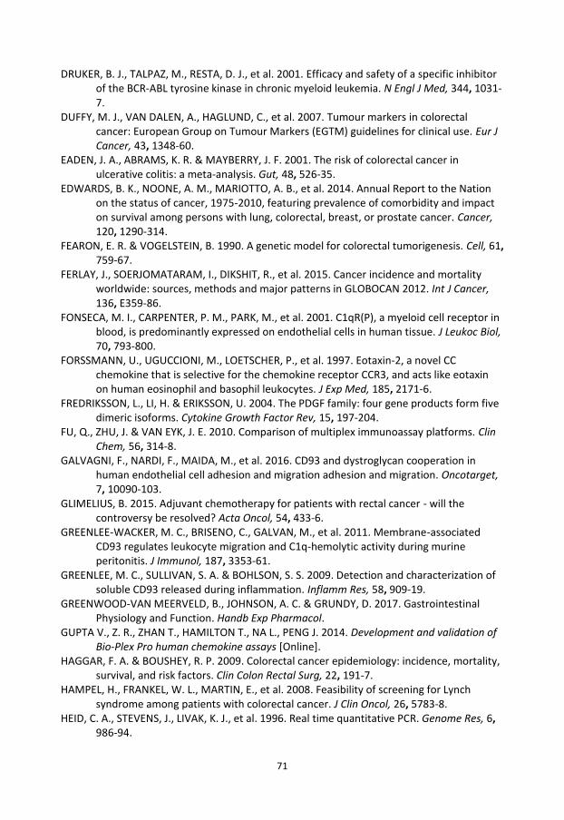
71
DRUKER, B. J., TALPAZ, M., RESTA, D. J., et al. 2001. Efficacy and safety of a specific inhibitor of the BCR-ABL tyrosine kinase in chronic myeloid leukemia. N Engl J Med, 344, 1031-7.
DUFFY, M. J., VAN DALEN, A., HAGLUND, C., et al. 2007. Tumour markers in colorectal cancer: European Group on Tumour Markers (EGTM) guidelines for clinical use. Eur J Cancer, 43, 1348-60.
EADEN, J. A., ABRAMS, K. R. & MAYBERRY, J. F. 2001. The risk of colorectal cancer in ulcerative colitis: a meta-analysis. Gut, 48, 526-35.
EDWARDS, B. K., NOONE, A. M., MARIOTTO, A. B., et al. 2014. Annual Report to the Nation on the status of cancer, 1975-2010, featuring prevalence of comorbidity and impact on survival among persons with lung, colorectal, breast, or prostate cancer. Cancer, 120, 1290-314.
FEARON, E. R. & VOGELSTEIN, B. 1990. A genetic model for colorectal tumorigenesis. Cell, 61, 759-67.
FERLAY, J., SOERJOMATARAM, I., DIKSHIT, R., et al. 2015. Cancer incidence and mortality worldwide: sources, methods and major patterns in GLOBOCAN 2012. Int J Cancer, 136, E359-86.
FONSECA, M. I., CARPENTER, P. M., PARK, M., et al. 2001. C1qR(P), a myeloid cell receptor in blood, is predominantly expressed on endothelial cells in human tissue. J Leukoc Biol, 70, 793-800.
FORSSMANN, U., UGUCCIONI, M., LOETSCHER, P., et al. 1997. Eotaxin-2, a novel CC chemokine that is selective for the chemokine receptor CCR3, and acts like eotaxin on human eosinophil and basophil leukocytes. J Exp Med, 185, 2171-6.
FREDRIKSSON, L., LI, H. & ERIKSSON, U. 2004. The PDGF family: four gene products form five dimeric isoforms. Cytokine Growth Factor Rev, 15, 197-204.
FU, Q., ZHU, J. & VAN EYK, J. E. 2010. Comparison of multiplex immunoassay platforms. Clin Chem, 56, 314-8.
GALVAGNI, F., NARDI, F., MAIDA, M., et al. 2016. CD93 and dystroglycan cooperation in human endothelial cell adhesion and migration adhesion and migration. Oncotarget, 7, 10090-103.
GLIMELIUS, B. 2015. Adjuvant chemotherapy for patients with rectal cancer - will the controversy be resolved? Acta Oncol, 54, 433-6.
GREENLEE-WACKER, M. C., BRISENO, C., GALVAN, M., et al. 2011. Membrane-associated CD93 regulates leukocyte migration and C1q-hemolytic activity during murine peritonitis. J Immunol, 187, 3353-61.
GREENLEE, M. C., SULLIVAN, S. A. & BOHLSON, S. S. 2009. Detection and characterization of soluble CD93 released during inflammation. Inflamm Res, 58, 909-19.
GREENWOOD-VAN MEERVELD, B., JOHNSON, A. C. & GRUNDY, D. 2017. Gastrointestinal Physiology and Function. Handb Exp Pharmacol.
GUPTA V., Z. R., ZHAN T., HAMILTON T., NA L., PENG J. 2014. Development and validation of Bio-Plex Pro human chemokine assays [Online].
HAGGAR, F. A. & BOUSHEY, R. P. 2009. Colorectal cancer epidemiology: incidence, mortality, survival, and risk factors. Clin Colon Rectal Surg, 22, 191-7.
HAMPEL, H., FRANKEL, W. L., MARTIN, E., et al. 2008. Feasibility of screening for Lynch syndrome among patients with colorectal cancer. J Clin Oncol, 26, 5783-8.
HEID, C. A., STEVENS, J., LIVAK, K. J., et al. 1996. Real time quantitative PCR. Genome Res, 6, 986-94.

72
HELDIN, C. H., OSTMAN, A. & RONNSTRAND, L. 1998. Signal transduction via platelet-derived growth factor receptors. Biochim Biophys Acta, 1378, F79-113.
HELDIN, C. H. & WESTERMARK, B. 1999. Mechanism of action and in vivo role of platelet-derived growth factor. Physiol Rev, 79, 1283-316.
HILLENBRAND, A., FASSLER, J., HUBER, N., et al. 2012. Changed adipocytokine concentrations in colorectal tumor patients and morbidly obese patients compared to healthy controls. BMC Cancer, 12, 545.
HOEHLER, T., VON WICHERT, G., SCHIMANSKI, C., et al. 2013. Phase I/II trial of capecitabine and oxaliplatin in combination with bevacizumab and imatinib in patients with metastatic colorectal cancer: AIO KRK 0205. Br J Cancer, 109, 1408-13.
HOLLAND, P. M., ABRAMSON, R. D., WATSON, R., et al. 1991. Detection of specific polymerase chain reaction product by utilizing the 5'----3' exonuclease activity of Thermus aquaticus DNA polymerase. Proc Natl Acad Sci U S A, 88, 7276-80.
HORNBECK, P. V. 2015. Enzyme-Linked Immunosorbent Assays. Curr Protoc Immunol, 110, 2 1 1-23.
HORVAT, M., POTOCNIK, U., REPNIK, K., et al. 2016. Single Nucleotide Polymorphisms as Prognostic and Predictive Factors of Adjuvant Chemotherapy in Colorectal Cancer of Stages I and II. Gastroenterol Res Pract, 2016, 2139489.
HOSOKAWA, H., NINOMIYA, H., KITAMURA, Y., et al. 2002. Vascular endothelial cells that express dystroglycan are involved in angiogenesis. J Cell Sci, 115, 1487-96.
HUTTER, R. V. 1987. At last--worldwide agreement on the staging of cancer. Arch Surg, 122, 1235-9.
INC., A. 2009-2010. GeneChip WT Terminal Labeling and Hybridization user manual for use with the Ambion WT Expression kit [Online]. Affymetrix Inc. Available: http://www.affymetrix.com/estore/catalog/prod110002/AFFY/WT+Terminal+Labeling+and+Controls+Kit#1_3 2013].
INC., T. 2009. TACS MTT Cell Proliferation Assays [Online]. Trevigen Inc. Available: https://trevigen.com/docs/protocol/protocol_4890-025-K.pdf.
IRVING, M. H. & CATCHPOLE, B. 1992. ABC of colorectal diseases. Anatomy and physiology of the colon, rectum, and anus. BMJ, 304, 1106-8.
ISHMAEL, F. T. & STELLATO, C. 2008. Principles and applications of polymerase chain reaction: basic science for the practicing physician. Ann Allergy Asthma Immunol, 101, 437-43.
ITATANI, Y., KAWADA, K., INAMOTO, S., et al. 2016. The Role of Chemokines in Promoting Colorectal Cancer Invasion/Metastasis. Int J Mol Sci, 17.
JASPERSON, K. W., TUOHY, T. M., NEKLASON, D. W., et al. 2010. Hereditary and familial colon cancer. Gastroenterology, 138, 2044-58.
JEMAL, A., TIWARI, R. C., MURRAY, T., et al. 2004. Cancer statistics, 2004. CA Cancer J Clin, 54, 8-29.
JONES, S., CHEN, W. D., PARMIGIANI, G., et al. 2008. Comparative lesion sequencing provides insights into tumor evolution. Proc Natl Acad Sci U S A, 105, 4283-8.
KAO, S. L., CHONG, S. S. & LEE, C. G. 2000. The role of single nucleotide polymorphisms (SNPs) in understanding complex disorders and pharmacogenomics. Ann Acad Med Singapore, 29, 376-82.
KAO, Y. C., JIANG, S. J., PAN, W. A., et al. 2012. The epidermal growth factor-like domain of CD93 is a potent angiogenic factor. PLoS One, 7, e51647.
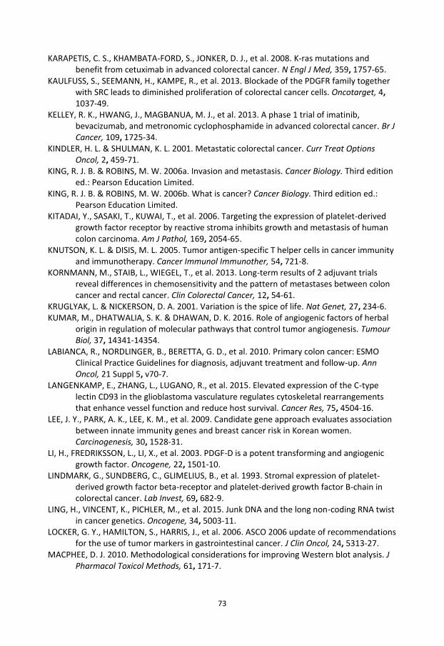
73
KARAPETIS, C. S., KHAMBATA-FORD, S., JONKER, D. J., et al. 2008. K-ras mutations and benefit from cetuximab in advanced colorectal cancer. N Engl J Med, 359, 1757-65.
KAULFUSS, S., SEEMANN, H., KAMPE, R., et al. 2013. Blockade of the PDGFR family together with SRC leads to diminished proliferation of colorectal cancer cells. Oncotarget, 4, 1037-49.
KELLEY, R. K., HWANG, J., MAGBANUA, M. J., et al. 2013. A phase 1 trial of imatinib, bevacizumab, and metronomic cyclophosphamide in advanced colorectal cancer. Br J Cancer, 109, 1725-34.
KINDLER, H. L. & SHULMAN, K. L. 2001. Metastatic colorectal cancer. Curr Treat Options Oncol, 2, 459-71.
KING, R. J. B. & ROBINS, M. W. 2006a. Invasion and metastasis. Cancer Biology. Third edition ed.: Pearson Education Limited.
KING, R. J. B. & ROBINS, M. W. 2006b. What is cancer? Cancer Biology. Third edition ed.: Pearson Education Limited.
KITADAI, Y., SASAKI, T., KUWAI, T., et al. 2006. Targeting the expression of platelet-derived growth factor receptor by reactive stroma inhibits growth and metastasis of human colon carcinoma. Am J Pathol, 169, 2054-65.
KNUTSON, K. L. & DISIS, M. L. 2005. Tumor antigen-specific T helper cells in cancer immunity and immunotherapy. Cancer Immunol Immunother, 54, 721-8.
KORNMANN, M., STAIB, L., WIEGEL, T., et al. 2013. Long-term results of 2 adjuvant trials reveal differences in chemosensitivity and the pattern of metastases between colon cancer and rectal cancer. Clin Colorectal Cancer, 12, 54-61.
KRUGLYAK, L. & NICKERSON, D. A. 2001. Variation is the spice of life. Nat Genet, 27, 234-6. KUMAR, M., DHATWALIA, S. K. & DHAWAN, D. K. 2016. Role of angiogenic factors of herbal
origin in regulation of molecular pathways that control tumor angiogenesis. Tumour Biol, 37, 14341-14354.
LABIANCA, R., NORDLINGER, B., BERETTA, G. D., et al. 2010. Primary colon cancer: ESMO Clinical Practice Guidelines for diagnosis, adjuvant treatment and follow-up. Ann Oncol, 21 Suppl 5, v70-7.
LANGENKAMP, E., ZHANG, L., LUGANO, R., et al. 2015. Elevated expression of the C-type lectin CD93 in the glioblastoma vasculature regulates cytoskeletal rearrangements that enhance vessel function and reduce host survival. Cancer Res, 75, 4504-16.
LEE, J. Y., PARK, A. K., LEE, K. M., et al. 2009. Candidate gene approach evaluates association between innate immunity genes and breast cancer risk in Korean women. Carcinogenesis, 30, 1528-31.
LI, H., FREDRIKSSON, L., LI, X., et al. 2003. PDGF-D is a potent transforming and angiogenic growth factor. Oncogene, 22, 1501-10.
LINDMARK, G., SUNDBERG, C., GLIMELIUS, B., et al. 1993. Stromal expression of platelet-derived growth factor beta-receptor and platelet-derived growth factor B-chain in colorectal cancer. Lab Invest, 69, 682-9.
LING, H., VINCENT, K., PICHLER, M., et al. 2015. Junk DNA and the long non-coding RNA twist in cancer genetics. Oncogene, 34, 5003-11.
LOCKER, G. Y., HAMILTON, S., HARRIS, J., et al. 2006. ASCO 2006 update of recommendations for the use of tumor markers in gastrointestinal cancer. J Clin Oncol, 24, 5313-27.
MACPHEE, D. J. 2010. Methodological considerations for improving Western blot analysis. J Pharmacol Toxicol Methods, 61, 171-7.
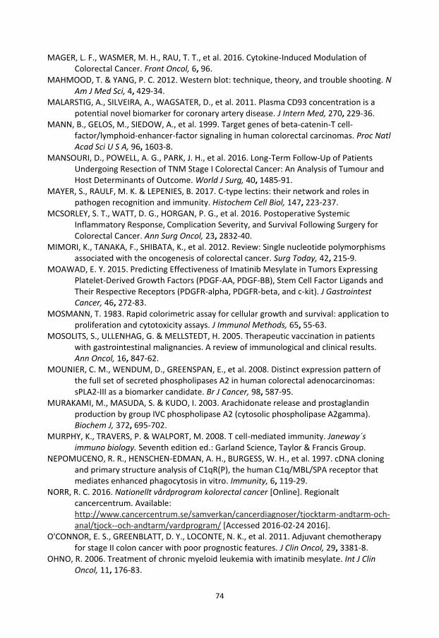
74
MAGER, L. F., WASMER, M. H., RAU, T. T., et al. 2016. Cytokine-Induced Modulation of Colorectal Cancer. Front Oncol, 6, 96.
MAHMOOD, T. & YANG, P. C. 2012. Western blot: technique, theory, and trouble shooting. N Am J Med Sci, 4, 429-34.
MALARSTIG, A., SILVEIRA, A., WAGSATER, D., et al. 2011. Plasma CD93 concentration is a potential novel biomarker for coronary artery disease. J Intern Med, 270, 229-36.
MANN, B., GELOS, M., SIEDOW, A., et al. 1999. Target genes of beta-catenin-T cell-factor/lymphoid-enhancer-factor signaling in human colorectal carcinomas. Proc Natl Acad Sci U S A, 96, 1603-8.
MANSOURI, D., POWELL, A. G., PARK, J. H., et al. 2016. Long-Term Follow-Up of Patients Undergoing Resection of TNM Stage I Colorectal Cancer: An Analysis of Tumour and Host Determinants of Outcome. World J Surg, 40, 1485-91.
MAYER, S., RAULF, M. K. & LEPENIES, B. 2017. C-type lectins: their network and roles in pathogen recognition and immunity. Histochem Cell Biol, 147, 223-237.
MCSORLEY, S. T., WATT, D. G., HORGAN, P. G., et al. 2016. Postoperative Systemic Inflammatory Response, Complication Severity, and Survival Following Surgery for Colorectal Cancer. Ann Surg Oncol, 23, 2832-40.
MIMORI, K., TANAKA, F., SHIBATA, K., et al. 2012. Review: Single nucleotide polymorphisms associated with the oncogenesis of colorectal cancer. Surg Today, 42, 215-9.
MOAWAD, E. Y. 2015. Predicting Effectiveness of Imatinib Mesylate in Tumors Expressing Platelet-Derived Growth Factors (PDGF-AA, PDGF-BB), Stem Cell Factor Ligands and Their Respective Receptors (PDGFR-alpha, PDGFR-beta, and c-kit). J Gastrointest Cancer, 46, 272-83.
MOSMANN, T. 1983. Rapid colorimetric assay for cellular growth and survival: application to proliferation and cytotoxicity assays. J Immunol Methods, 65, 55-63.
MOSOLITS, S., ULLENHAG, G. & MELLSTEDT, H. 2005. Therapeutic vaccination in patients with gastrointestinal malignancies. A review of immunological and clinical results. Ann Oncol, 16, 847-62.
MOUNIER, C. M., WENDUM, D., GREENSPAN, E., et al. 2008. Distinct expression pattern of the full set of secreted phospholipases A2 in human colorectal adenocarcinomas: sPLA2-III as a biomarker candidate. Br J Cancer, 98, 587-95.
MURAKAMI, M., MASUDA, S. & KUDO, I. 2003. Arachidonate release and prostaglandin production by group IVC phospholipase A2 (cytosolic phospholipase A2gamma). Biochem J, 372, 695-702.
MURPHY, K., TRAVERS, P. & WALPORT, M. 2008. T cell-mediated immunity. Janeway´s immuno biology. Seventh edition ed.: Garland Science, Taylor & Francis Group.
NEPOMUCENO, R. R., HENSCHEN-EDMAN, A. H., BURGESS, W. H., et al. 1997. cDNA cloning and primary structure analysis of C1qR(P), the human C1q/MBL/SPA receptor that mediates enhanced phagocytosis in vitro. Immunity, 6, 119-29.
NORR, R. C. 2016. Nationellt vårdprogram kolorectal cancer [Online]. Regionalt cancercentrum. Available: http://www.cancercentrum.se/samverkan/cancerdiagnoser/tjocktarm-andtarm-och-anal/tjock--och-andtarm/vardprogram/ [Accessed 2016-02-24 2016].
O'CONNOR, E. S., GREENBLATT, D. Y., LOCONTE, N. K., et al. 2011. Adjuvant chemotherapy for stage II colon cancer with poor prognostic features. J Clin Oncol, 29, 3381-8.
OHNO, R. 2006. Treatment of chronic myeloid leukemia with imatinib mesylate. Int J Clin Oncol, 11, 176-83.
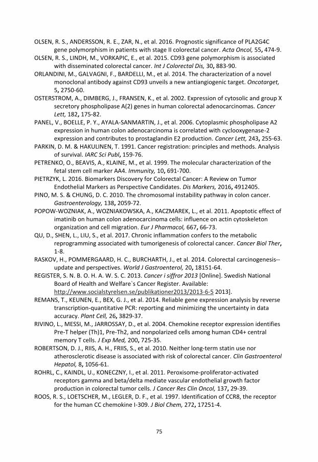
75
OLSEN, R. S., ANDERSSON, R. E., ZAR, N., et al. 2016. Prognostic significance of PLA2G4C gene polymorphism in patients with stage II colorectal cancer. Acta Oncol, 55, 474-9.
OLSEN, R. S., LINDH, M., VORKAPIC, E., et al. 2015. CD93 gene polymorphism is associated with disseminated colorectal cancer. Int J Colorectal Dis, 30, 883-90.
ORLANDINI, M., GALVAGNI, F., BARDELLI, M., et al. 2014. The characterization of a novel monoclonal antibody against CD93 unveils a new antiangiogenic target. Oncotarget, 5, 2750-60.
OSTERSTROM, A., DIMBERG, J., FRANSEN, K., et al. 2002. Expression of cytosolic and group X secretory phospholipase A(2) genes in human colorectal adenocarcinomas. Cancer Lett, 182, 175-82.
PANEL, V., BOELLE, P. Y., AYALA-SANMARTIN, J., et al. 2006. Cytoplasmic phospholipase A2 expression in human colon adenocarcinoma is correlated with cyclooxygenase-2 expression and contributes to prostaglandin E2 production. Cancer Lett, 243, 255-63.
PARKIN, D. M. & HAKULINEN, T. 1991. Cancer registration: principles and methods. Analysis of survival. IARC Sci Publ, 159-76.
PETRENKO, O., BEAVIS, A., KLAINE, M., et al. 1999. The molecular characterization of the fetal stem cell marker AA4. Immunity, 10, 691-700.
PIETRZYK, L. 2016. Biomarkers Discovery for Colorectal Cancer: A Review on Tumor Endothelial Markers as Perspective Candidates. Dis Markers, 2016, 4912405.
PINO, M. S. & CHUNG, D. C. 2010. The chromosomal instability pathway in colon cancer. Gastroenterology, 138, 2059-72.
POPOW-WOZNIAK, A., WOZNIAKOWSKA, A., KACZMAREK, L., et al. 2011. Apoptotic effect of imatinib on human colon adenocarcinoma cells: influence on actin cytoskeleton organization and cell migration. Eur J Pharmacol, 667, 66-73.
QU, D., SHEN, L., LIU, S., et al. 2017. Chronic inflammation confers to the metabolic reprogramming associated with tumorigenesis of colorectal cancer. Cancer Biol Ther, 1-8.
RASKOV, H., POMMERGAARD, H. C., BURCHARTH, J., et al. 2014. Colorectal carcinogenesis--update and perspectives. World J Gastroenterol, 20, 18151-64.
REGISTER, S. N. B. O. H. A. W. S. C. 2013. Cancer i siffror 2013 [Online]. Swedish National Board of Health and Welfare`s Cancer Register. Available: http://www.socialstyrelsen.se/publikationer2013/2013-6-5 2013].
REMANS, T., KEUNEN, E., BEX, G. J., et al. 2014. Reliable gene expression analysis by reverse transcription-quantitative PCR: reporting and minimizing the uncertainty in data accuracy. Plant Cell, 26, 3829-37.
RIVINO, L., MESSI, M., JARROSSAY, D., et al. 2004. Chemokine receptor expression identifies Pre-T helper (Th)1, Pre-Th2, and nonpolarized cells among human CD4+ central memory T cells. J Exp Med, 200, 725-35.
ROBERTSON, D. J., RIIS, A. H., FRIIS, S., et al. 2010. Neither long-term statin use nor atherosclerotic disease is associated with risk of colorectal cancer. Clin Gastroenterol Hepatol, 8, 1056-61.
ROHRL, C., KAINDL, U., KONECZNY, I., et al. 2011. Peroxisome-proliferator-activated receptors gamma and beta/delta mediate vascular endothelial growth factor production in colorectal tumor cells. J Cancer Res Clin Oncol, 137, 29-39.
ROOS, R. S., LOETSCHER, M., LEGLER, D. F., et al. 1997. Identification of CCR8, the receptor for the human CC chemokine I-309. J Biol Chem, 272, 17251-4.
76
RUCKES, T., SAUL, D., VAN SNICK, J., et al. 2001. Autocrine antiapoptotic stimulation of cultured adult T-cell leukemia cells by overexpression of the chemokine I-309. Blood, 98, 1150-9.
SAAF, A. M., HALBLEIB, J. M., CHEN, X., et al. 2007. Parallels between global transcriptional programs of polarizing Caco-2 intestinal epithelial cells in vitro and gene expression programs in normal colon and colon cancer. Mol Biol Cell, 18, 4245-60.
SARRAF, P., MUELLER, E., SMITH, W. M., et al. 1999. Loss-of-function mutations in PPAR gamma associated with human colon cancer. Mol Cell, 3, 799-804.
SCHINDLER, T., BORNMANN, W., PELLICENA, P., et al. 2000. Structural mechanism for STI-571 inhibition of abelson tyrosine kinase. Science, 289, 1938-42.
SCHWANHAUSSER, B., BUSSE, D., LI, N., et al. 2011. Global quantification of mammalian gene expression control. Nature, 473, 337-42.
SCIENTIFIC, T. F. 2017a. Gene expression [Online]. Thermo Fisher Scientific. Available: https://www.thermofisher.com/se/en/home/life-science/pcr/real-time-pcr/qpcr-education/how-taqman-assays-work.html [Accessed March 21 2017].
SCIENTIFIC, T. F. 2017b. Genotyping [Online]. Thermo Fisher Scientific. Available: https://www.thermofisher.com/se/en/home/life-science/pcr/real-time-pcr/qpcr-education/how-taqman-assays-work.html [Accessed March 21 2017].
SHENG, H., SHAO, J., MORROW, J. D., et al. 1998. Modulation of apoptosis and Bcl-2 expression by prostaglandin E2 in human colon cancer cells. Cancer Res, 58, 362-6.
SHUREIQI, I., CHEN, D., LOTAN, R., et al. 2000. 15-Lipoxygenase-1 mediates nonsteroidal anti-inflammatory drug-induced apoptosis independently of cyclooxygenase-2 in colon cancer cells. Cancer Res, 60, 6846-50.
SOBRERO, A. 2006. Should adjuvant chemotherapy become standard treatment for patients with stage II colon cancer? For the proposal. Lancet Oncol, 7, 515-6.
SUNDBERG, C., LJUNGSTROM, M., LINDMARK, G., et al. 1993. Microvascular pericytes express platelet-derived growth factor-beta receptors in human healing wounds and colorectal adenocarcinoma. Am J Pathol, 143, 1377-88.
TAMGUNEY, G., VAN SNICK, J. & FICKENSCHER, H. 2004. Autocrine stimulation of rhadinovirus-transformed T cells by the chemokine CCL1/I-309. Oncogene, 23, 8475-85.
TEBBUTT, N. C., CATTELL, E., MIDGLEY, R., et al. 2002. Systemic treatment of colorectal cancer. Eur J Cancer, 38, 1000-15.
THE AMERICAN CANCER SOCIETY, I. 2017. What are the survival rates for colorectal cancer, by stage? [Online]. The American Cancer Society, Inc. Available: https://www.cancer.org/cancer/colon-rectal-cancer/detection-diagnosis-staging/survival-rates.html [Accessed 2017-03-02 2017].
THEODOROPOULOS, G., PAPACONSTANTINOU, I., FELEKOURAS, E., et al. 2006. Relation between common polymorphisms in genes related to inflammatory response and colorectal cancer. World J Gastroenterol, 12, 5037-43.
THIERY, J. P. 2002. Epithelial-mesenchymal transitions in tumour progression. Nat Rev Cancer, 2, 442-54.
THORSEN, S. B., LUNDBERG, M., VILLABLANCA, A., et al. 2013. Detection of serological biomarkers by proximity extension assay for detection of colorectal neoplasias in symptomatic individuals. J Transl Med, 11, 253.
77
TIAN, G., WANG, X., ZHANG, F., et al. 2011. Downregulation of cPLA2gamma expression inhibits EGF-induced chemotaxis of human breast cancer cells through Akt pathway. Biochem Biophys Res Commun, 409, 506-12.
USTACH, C. V. & KIM, H. R. 2005. Platelet-derived growth factor D is activated by urokinase plasminogen activator in prostate carcinoma cells. Mol Cell Biol, 25, 6279-88.
VAN DER NET, J. B., OOSTERVEER, D. M., VERSMISSEN, J., et al. 2008. Replication study of 10 genetic polymorphisms associated with coronary heart disease in a specific high-risk population with familial hypercholesterolemia. Eur Heart J, 29, 2195-201.
VAN DER SIJP, M. P., BASTIAANNET, E., MESKER, W. E., et al. 2016. Differences between colon and rectal cancer in complications, short-term survival and recurrences. Int J Colorectal Dis, 31, 1683-91.
VAN SNICK, J., HOUSSIAU, F., PROOST, P., et al. 1996. I-309/T cell activation gene-3 chemokine protects murine T cell lymphomas against dexamethasone-induced apoptosis. J Immunol, 157, 2570-6.
VAN TONDER, A., JOUBERT, A. M. & CROMARTY, A. D. 2015. Limitations of the 3-(4,5-dimethylthiazol-2-yl)-2,5-diphenyl-2H-tetrazolium bromide (MTT) assay when compared to three commonly used cell enumeration assays. BMC Res Notes, 8, 47.
WANG, D. & DUBOIS, R. N. 2013. An inflammatory mediator, prostaglandin E2, in colorectal cancer. Cancer J, 19, 502-10.
WANG, Z., KONG, D., LI, Y., et al. 2009. PDGF-D signaling: a novel target in cancer therapy. Curr Drug Targets, 10, 38-41.
WATSON, A. J. & COLLINS, P. D. 2011. Colon cancer: a civilization disorder. Dig Dis, 29, 222-8. WEICKHARDT, A. J., PRICE, T. J., CHONG, G., et al. 2012. Dual targeting of the epidermal
growth factor receptor using the combination of cetuximab and erlotinib: preclinical evaluation and results of the phase II DUX study in chemotherapy-refractory, advanced colorectal cancer. J Clin Oncol, 30, 1505-12.
VOGELSTEIN, B. & KINZLER, K. W. 1993. The multistep nature of cancer. Trends Genet, 9, 138-41.
VON ROON, A. C., REESE, G., TEARE, J., et al. 2007. The risk of cancer in patients with Crohn's disease. Dis Colon Rectum, 50, 839-55.
WU, Q., HOU, X., XIA, J., et al. 2013. Emerging roles of PDGF-D in EMT progression during tumorigenesis. Cancer Treat Rev, 39, 640-6.
YANG, S. Y., KIM, Y. S., CHUNG, S. J., et al. 2010. Association between colorectal adenoma and coronary atherosclerosis detected by CT coronary angiography in Korean men; a cross-sectional study. J Gastroenterol Hepatol, 25, 1795-9.
YARLA, N. S., BISHAYEE, A., VADLAKONDA, L., et al. 2016. Phospholipase A2 Isoforms as Novel Targets for Prevention and Treatment of Inflammatory and Oncologic Diseases. Curr Drug Targets, 17, 1940-1962.
YEH, C. R., HSU, I., SONG, W., et al. 2015. Fibroblast ERalpha promotes bladder cancer invasion via increasing the CCL1 and IL-6 signals in the tumor microenvironment. Am J Cancer Res, 5, 1146-57.
YOO, Y. S., LIM, S. C. & KIM, K. J. 2011. Prognostic significance of cytosolic phospholipase A2 expression in patients with colorectal cancer. J Korean Surg Soc, 80, 397-403.
ZINGONI, A., SOTO, H., HEDRICK, J. A., et al. 1998. The chemokine receptor CCR8 is preferentially expressed in Th2 but not Th1 cells. J Immunol, 161, 547-51.

Papers
The articles associated with this thesis have been removed for copyright
reasons. For more details about these see:
http://urn.kb.se/resolve? urn:nbn:se:liu:diva-136841