Embed Size (px)
Citation preview
RESEARCH ARTICLE
Potential application of novel technology
developed for instant decontamination of
personal protective equipment before the
doffing step
Luıs Alberto Brêda Mascarenhas1☯, Bruna Aparecida Souza MachadoID1,2☯*, Leticia de
Alencar Pereira Rodrigues1‡, Katharine Valeria Saraiva Hodel1‡, Alex Alisson Bandeira
Santos2‡, Paulo Roberto Freitas Neves2‡, Leone Peter Correia da Silva Andrade1‡, Milena
Botelho Soares1,3‡, Jailson Bittencourt de AndradeID1‡, Roberto Badaro1‡
1 SENAI CIMATEC, SENAI Institute of Innovation (ISI) in Health Advanced Systems (CIMATEC ISI SAS),
University Center SENAI/CIMATEC, Salvador, Bahia, Brazil, 2 SENAI CIMATEC, National Service of
Industrial Learning–SENAI, Computational Modeling and Industrial Technology, University Center SENAI/
CIMATEC, Salvador, Bahia, Brazil, 3 Goncalo Moniz Institute, Oswaldo Cruz Foundation (IGM-FIOCRUZ/
BA), Salvador, Bahia, Brazil
☯ These authors contributed equally to this work.
‡ LAPR, KVSH, AABS, PRFN, LPCSA, MBS, JBA and RB also contributed equally to this work.
* [email protected], [email protected]
Abstract
The use of personal protective equipment (PPE) has been considered the most effective
way to avoid the contamination of healthcare workers by different microorganisms, including
SARS-CoV-2. A spray disinfection technology (chamber) was developed, and its efficacy in
instant decontamination of previously contaminated surfaces was evaluated in two expo-
sure times. Seven test microorganisms were prepared and inoculated on the surface of
seven types of PPE (respirator mask, face shield, shoe, glove, cap, safety glasses and lab
coat). The tests were performed on previously contaminated PPE using a manikin with a
motion device for exposure to the chamber with biocidal agent (sodium hypochlorite) for 10
and 30s. In 96.93% of the experimental conditions analyzed, the percentage reduction was
>99% (the number of viable cells found on the surface ranged from 4.3x10⁶ to <10 CFU/
mL). The samples of E. faecalis collected from the glove showed the lowest percentages
reduction, with 86.000 and 86.500% for exposure times of 10 and 30 s, respectively. The
log10 reduction values varied between 0.85 log10 (E. faecalis at 30 s in glove surface) and
9.69 log10 (E. coli at 10 and 30 s in lab coat surface). In general, E. coli, S. aureus, C. freun-
dii, P. mirabilis, C. albicans and C. parapsilosis showed susceptibility to the biocidal agent
under the tested conditions, with >99% reduction after 10 and 30s, while E. faecalis and P.
aeruginosa showed a lower susceptibility. The 30s exposure time was more effective for the
inactivation of the tested microorganisms. The results show that the spray disinfection tech-
nology has the potential for instant decontamination of PPE, which can contribute to an addi-
tional barrier for infection control of healthcare workers in the hospital environment.
PLOS ONE
PLOS ONE | https://doi.org/10.1371/journal.pone.0250854 June 4, 2021 1 / 24
a1111111111
a1111111111
a1111111111
a1111111111
a1111111111
OPEN ACCESS
Citation: Brêda Mascarenhas LA, Machado BAS,
Rodrigues LdAP, Saraiva Hodel KV, Bandeira
Santos AA, Freitas Neves PR, et al. (2021) Potential
application of novel technology developed for
instant decontamination of personal protective
equipment before the doffing step. PLoS ONE
16(6): e0250854. https://doi.org/10.1371/journal.
pone.0250854
Editor: Amitava Mukherjee, VIT University, INDIA
Received: September 27, 2020
Accepted: February 2, 2021
Published: June 4, 2021
Copyright: © 2021 Brêda Mascarenhas et al. This is
an open access article distributed under the terms
of the Creative Commons Attribution License,
which permits unrestricted use, distribution, and
reproduction in any medium, provided the original
author and source are credited.
Data Availability Statement: All relevant data are
within the paper and its Supporting Information
files.
Funding: The author(s) received no specific
funding for this work.
Competing interests: The authors have declared
that no competing interests exist.
Introduction
Contaminated surfaces are a potential source for the spread of many bacterial and fungal path-
ogens [1]. These microorganisms can be considered important vectors for the dissemination
of diseases and, consequently, the increase in mortality and morbidity rates, causing overload
of the health system worldwide [2]. There is currently growing concern that the environment
may be an underestimated source for the spread of emerging viruses, including of the influ-
enza virus [3], Ebola virus [4], and coronaviruses, especially the severe acute respiratory syn-
drome named SARS-CoV-2 [5]. SARS-CoV-2 is the causative agent of novel coronavirus 2019
disease (COVID-19) which was isolated and identified for the first time in humans in the city
of Wuhan, Hubei Province, China [6]. Based on evidence of an increasing incidence of infec-
tions [7] and the possibility of transmission by asymptomatic carriers [8], it was demonstrated
that SARS-CoV-2 can be effectively transmitted between humans through droplets (aerosols)
or direct contact with contaminated surfaces, which facilitated its rapid spread worldwide [9,
10].
Healthcare workers (HCWs) are one of the most vulnerable populations to microbial con-
tamination, mainly because they work in close physical contact with patients [11]. This vulner-
ability was demonstrated at times of emergency in health systems, such as during the outbreak
caused by SARS-CoV [12], Ebola virus [13] and currently with SARS-CoV-2 [14], where a
high rate of infection among HCWs has been reported. The high prevalence of COVID-19
among HCWs is mainly associated with the execution of the procedures involved in airway
management for oxygen supplementation of many patients with severe COVID-19 pneumonia
presenting with pronounced arterial hypoxemia (major generators of aerosol) [12, 15], which
increases the viral load in which these professionals are in contact [16, 17]. The risk of viral
transmission to HCWs has been a concern since the beginning of the outbreak in China,
where more than 3,300 HCWs were infected, with a mortality rate of 1.1% [18]. In Europe,
approximately 20% of HCWs were infected by SARS-CoV-2 in Italy and 26% in Spain, the two
epicenters of the disease in the European continent between March and April [19, 20]. In Bra-
zil, currently considered the epicenter of the disease in Latin America [21], data from the Min-
istry of Health indicate that at least 257,156 HCWs were infected by SARS-CoV-2 by August
of this year [22].
The use of personal protective equipment (PPE) by HCWs has been considered the most
effective way to avoid contamination by different microorganisms of high epidemiological
concern [23–25], including SARS-CoV-2 [26], as they have the ability to act as a barrier to
pathogens [27]. Studies have shown that the use of PPE and actions to decontaminate their
surfaces are crucial to reduce the infection rate among HCWs in direct contact with patients
diagnosed with COVID-19 and other contagious diseases [23, 28]. The step of PPE removal
(doffing) by HCWs can be thus considered critical since there may be contact between the
contaminated surface of the PPE and the HCWs, leading to an increased chance of self-con-
tamination through the mucous membranes of the nose, eyes or mouth [29]. Therefore, this
step should be performed following well-established biosafety protocols [30], It has been dem-
onstrated that doffing PPE is among others an important risk factor associated with HCWs
contamination with SARS-CoV-2 [31].
Several devices with different technologies have been developed for the inactivation or
reduction of bioburden on surfaces and environments [1], and the use of such devices has
gained popularity for presenting a response to the global demand created for the control of
possible environmental surfaces contamination [32]. Examples include devices with ultravio-
let-C (UV-C) or xenon UV light for disinfection of hospital environments [33, 34], portable
equipment with a disinfectant spraying system [35] or hospital air disinfection systems [36]
PLOS ONE Potential application of novel technology developed for instant decontamination
PLOS ONE | https://doi.org/10.1371/journal.pone.0250854 June 4, 2021 2 / 24
and disinfection chambers [37]. Faced with increased production and demand, health regula-
tory agencies recommend that studies be presented to prove the performance of these devices
in the face of decontamination effectiveness [38, 39]. Although there are few studies demon-
strating the efficacy of disinfection chambers [40], their use becomes interesting for the decon-
tamination of surfaces, including personal protective closes and equipment (PPE), since it is
not mandatory that the contaminated material undergoes to manual cleaning mainly at the
moment before the doffing step for previous elimination of the microorganisms [37]. Thus,
disinfection chambers can be a practical alternative for bioburden control in environments
with a high rate of pathogenic microorganisms, such as hospitals. In addition, the disinfection
chambers can help mitigate the possibility of an accident self-contamination of the HCWs dur-
ing the processing of hand manipulation of the disposable PPEs.
Different disinfecting agents have been studied and suggested to act in these devices as bio-
cidal agents against different pathogens of hospital importance (such SARS-CoV-2, multi-
drug-resistant bacteria and fungi). Examples include physical agents such as UV light [41] and
chemical agents such as alcoholic solutions [5, 42], quaternary ammonium compounds [43,
44], ozone gas [45] and sodium hypochlorite [46]. Sodium hypochlorite is one of the most
well-known and used biocidal agents worldwide due to its broad-spectrum microbicidal prop-
erties [47]. Compared with other chlorine-containing biocides, the use of sodium hypochlorite
is characterized by a relatively lower toxicity when in contact with mucous membranes, the
equipment required for its synthesis are simpler, its handling is safer, and its operation and
preparation costs are lower, which makes its use feasible in hospitals [48]. In addition, the use
of sodium hypochlorite is recommended by the World Health Organization (WHO) for the
disinfection of environmental surfaces related to health care in the context of COVID-19, in
concentrations between 0.1 and 0.5% (1000 and 5000 ppm, respectively) [49].
Despite being a promising alternative, especially considering the current situation, there are
still few reports in the literature on the efficacy of disinfection chambers and/or devices using
sodium hypochlorite as a biocidal agent for reducing or inactivating the burden of different
pathogens on contaminated surfaces, more specifically for PPE, and on its potential use in
emergency and public health situations. [38, 50, 51]. The use of disinfection chambers in con-
trolled environments, such as hospitals and health units, could help to reduce the risk of self-
contamination by health workers during the doffing step, since the instantaneously dispersed
solution could significantly reduce the pathogens present on surfaces and contribute to greater
safety of these HCWs self-contamination. Nevertheless, this approach has not yet been
reported by any earlier study.
Through the use of the disinfection chamber it is possible to decontaminate the surfaces of
all PPEs used in clinical practice at the same time, making the doffing step safer for the HCWs.
This can be considered an advantage over the PPE disinfection technologies mentioned in the
literature [52–54], since the proposed decontamination processes do not reduce the risk of
self-contamination. Indeed, the objective of this study was to develop a disinfection chamber
for instantaneous dispersion of a biocidal solution (0.25% sodium hypochlorite) and to deter-
mine its efficacy on previously contaminated surfaces at different exposure times, aiming at its
possible application as an additional barrier against pathogens, such as SARS-CoV-2, to pro-
tect HCWs during the withdraw procedure prior PPE disposal.
Materials and methods
Fig 1 shows the general scheme of the method applied in this study to evaluate the efficacy of
the chamber for instant disinfection of the surfaces of seven PPE previously contaminated
with different microorganisms and subjected to different exposure times to the biocidal agent
PLOS ONE Potential application of novel technology developed for instant decontamination
PLOS ONE | https://doi.org/10.1371/journal.pone.0250854 June 4, 2021 3 / 24
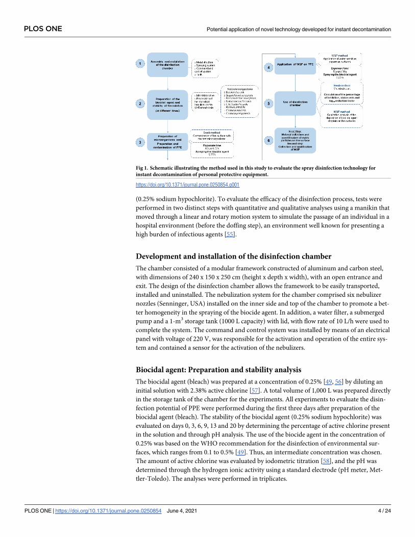
(0.25% sodium hypochlorite). To evaluate the efficacy of the disinfection process, tests were
performed in two distinct steps with quantitative and qualitative analyses using a manikin that
moved through a linear and rotary motion system to simulate the passage of an individual in a
hospital environment (before the doffing step), an environment well known for presenting a
high burden of infectious agents [55].
Development and installation of the disinfection chamber
The chamber consisted of a modular framework constructed of aluminum and carbon steel,
with dimensions of 240 x 150 x 250 cm (height x depth x width), with an open entrance and
exit. The design of the disinfection chamber allows the framework to be easily transported,
installed and uninstalled. The nebulization system for the chamber comprised six nebulizer
nozzles (Senninger, USA) installed on the inner side and top of the chamber to promote a bet-
ter homogeneity in the spraying of the biocide agent. In addition, a water filter, a submerged
pump and a 1-m3 storage tank (1000 L capacity) with lid, with flow rate of 10 L/h were used to
complete the system. The command and control system was installed by means of an electrical
panel with voltage of 220 V, was responsible for the activation and operation of the entire sys-
tem and contained a sensor for the activation of the nebulizers.
Biocidal agent: Preparation and stability analysis
The biocidal agent (bleach) was prepared at a concentration of 0.25% [49, 56] by diluting an
initial solution with 2.38% active chlorine [57]. A total volume of 1,000 L was prepared directly
in the storage tank of the chamber for the experiments. All experiments to evaluate the disin-
fection potential of PPE were performed during the first three days after preparation of the
biocidal agent (bleach). The stability of the biocidal agent (0.25% sodium hypochlorite) was
evaluated on days 0, 3, 6, 9, 13 and 20 by determining the percentage of active chlorine present
in the solution and through pH analysis. The use of the biocide agent in the concentration of
0.25% was based on the WHO recommendation for the disinfection of environmental sur-
faces, which ranges from 0.1 to 0.5% [49]. Thus, an intermediate concentration was chosen.
The amount of active chlorine was evaluated by iodometric titration [58], and the pH was
determined through the hydrogen ionic activity using a standard electrode (pH meter, Met-
tler-Toledo). The analyses were performed in triplicates.
Fig 1. Schematic illustrating the method used in this study to evaluate the spray disinfection technology for
instant decontamination of personal protective equipment.
https://doi.org/10.1371/journal.pone.0250854.g001
PLOS ONE Potential application of novel technology developed for instant decontamination
PLOS ONE | https://doi.org/10.1371/journal.pone.0250854 June 4, 2021 4 / 24
Experimental standard strains
The standard reference strains used in this study were Escherichia coli (ATCC 8739), Staphylo-coccus aureus (ATCC 6538), Pseudomonas aeruginosa (ATCC 27853), Enterococcus faecalis(ATCC 29212), Citrobacter freundii (ATCC 43864), Proteus mirabilis (ATCC 29906), Candidaalbicans (ATCC 18804) and Candida parapsilosis (ATCC 22019), which were obtained from
Microbiologics (St. Cloud, Minnesota) or from the Culture Collection of the Institute of
Health Sciences, Federal University of Bahia (Universidade Federal da Bahia–UFBA), located
in Salvador, Brazil. The selection of test strains was based on studies of microorganisms com-
monly causing nosocomial infections, as well as on the recommendations of regulatory agen-
cies for evaluating the efficacy of chemical disinfectants [57, 59–65]. The suspensions of the
test microorganisms were prepared by transferring cells from the pure culture to plates con-
taining 15–20 mL of plate count agar: agar (9 g/L); dextrose (1 g/L); tryptone (5.0 g/L) and
yeast extract (2.5 g/L). To evaluate the disinfection profile in the chamber against the test
microorganisms, the inocula were prepared by suspending 1–5 colonies in 5 mL of 0.85%
saline solution and the turbidity was adjusted to McFarland No. 0.5 tube [66].
Preparation of study surfaces (PPE)
The PPE items used to evaluate the effectiveness of the disinfection chamber were selected
according to the recommendations for prevention and control of the spread of SARS-CoV-2
and other infectious agents transmitted mainly by aerosols in health services (Table 1) [24, 67,
68]. To ensure the sterility of the surface of the selected items before contamination with the
standard strains, the items were exposed to UV light for 40 minutes using a laminar flow
(model LA2000T, LOGEN) after being sanitized with 70% ethanol [69]. Surface samples from
each item were collected using sterile swabs, and their contents were seeded in nutrient agar
(37˚C for 24 hours) to confirm sterility.
Assay for evaluation and distribution of the biocidal agent for spray
disinfection during exposure in the chamber
The assays for evaluating the disinfection potential of the biocidal agent in the chamber devel-
oped in this study were based on the method used to monitor viable particles on surfaces [70,
71] and qualitative analysis of the biocidal agent distribution on the surfaces [72]. The disinfec-
tion process was performed by spraying the biocidal agent (0.25% sodium hypochlorite) in the
chamber using a suitably dressed manikin with a motion system that allowed the manikin to
pass through the chamber automatically and to perform a 360˚ turn, for 10 and 30 s of expo-
sure. The manikin was chosen so that there would be a simulation closer to what would be the
use of the disinfection chamber by healthcare workers in nosocomial environment. In the first
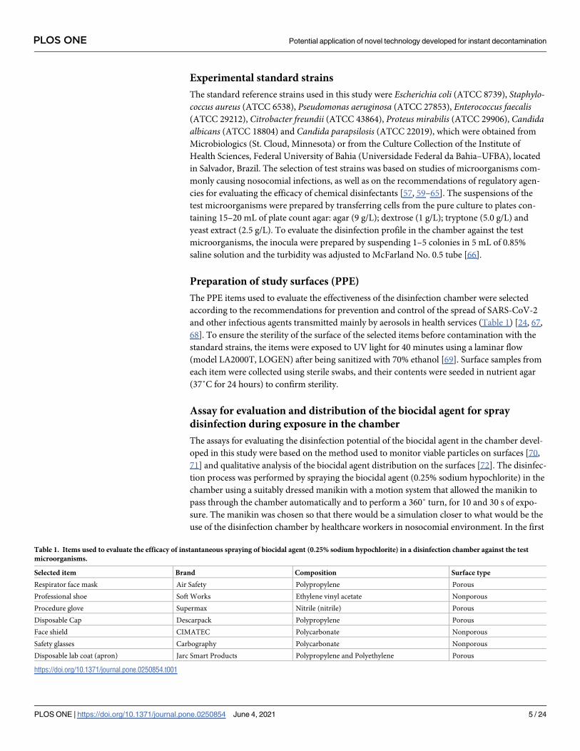
Table 1. Items used to evaluate the efficacy of instantaneous spraying of biocidal agent (0.25% sodium hypochlorite) in a disinfection chamber against the test
microorganisms.
Selected item Brand Composition Surface type
Respirator face mask Air Safety Polypropylene Porous
Professional shoe Soft Works Ethylene vinyl acetate Nonporous
Procedure glove Supermax Nitrile (nitrile) Porous
Disposable Cap Descarpack Polypropylene Porous
Face shield CIMATEC Polycarbonate Nonporous
Safety glasses Carbography Polycarbonate Nonporous
Disposable lab coat (apron) Jarc Smart Products Polypropylene and Polyethylene Porous
https://doi.org/10.1371/journal.pone.0250854.t001
PLOS ONE Potential application of novel technology developed for instant decontamination
PLOS ONE | https://doi.org/10.1371/journal.pone.0250854 June 4, 2021 5 / 24
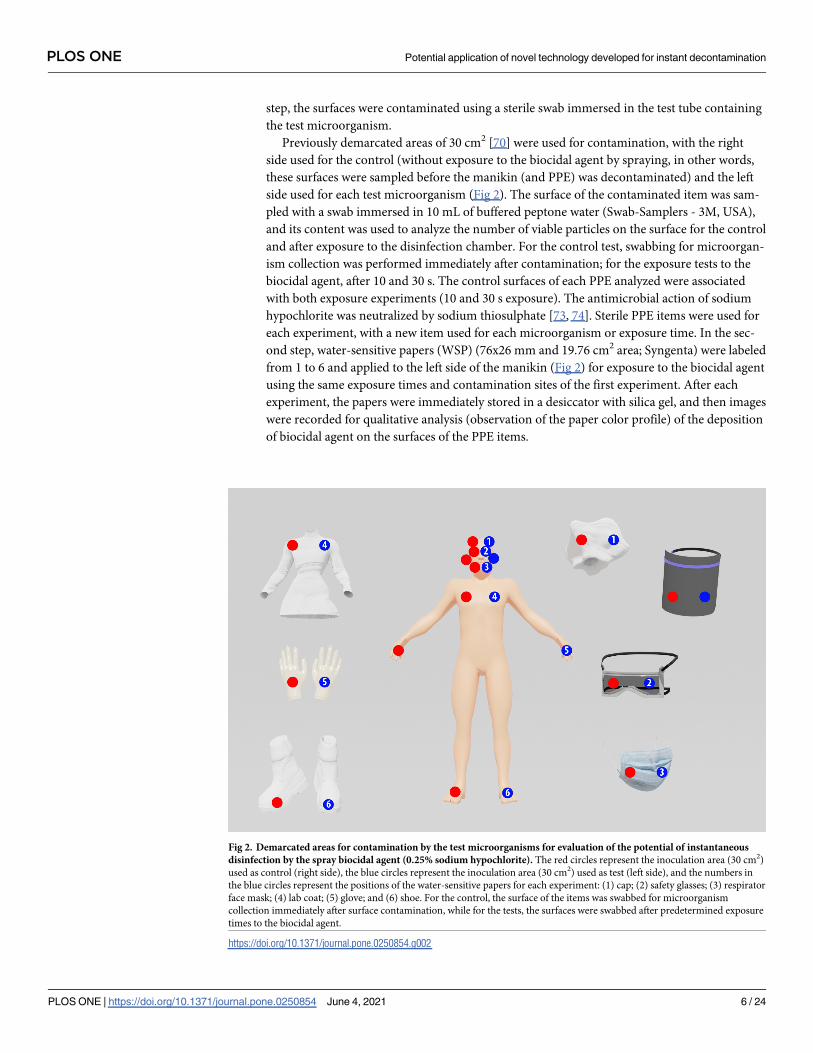
step, the surfaces were contaminated using a sterile swab immersed in the test tube containing
the test microorganism.
Previously demarcated areas of 30 cm2 [70] were used for contamination, with the right
side used for the control (without exposure to the biocidal agent by spraying, in other words,
these surfaces were sampled before the manikin (and PPE) was decontaminated) and the left
side used for each test microorganism (Fig 2). The surface of the contaminated item was sam-
pled with a swab immersed in 10 mL of buffered peptone water (Swab-Samplers - 3M, USA),
and its content was used to analyze the number of viable particles on the surface for the control
and after exposure to the disinfection chamber. For the control test, swabbing for microorgan-
ism collection was performed immediately after contamination; for the exposure tests to the
biocidal agent, after 10 and 30 s. The control surfaces of each PPE analyzed were associated
with both exposure experiments (10 and 30 s exposure). The antimicrobial action of sodium
hypochlorite was neutralized by sodium thiosulphate [73, 74]. Sterile PPE items were used for
each experiment, with a new item used for each microorganism or exposure time. In the sec-
ond step, water-sensitive papers (WSP) (76x26 mm and 19.76 cm2 area; Syngenta) were labeled
from 1 to 6 and applied to the left side of the manikin (Fig 2) for exposure to the biocidal agent
using the same exposure times and contamination sites of the first experiment. After each
experiment, the papers were immediately stored in a desiccator with silica gel, and then images
were recorded for qualitative analysis (observation of the paper color profile) of the deposition
of biocidal agent on the surfaces of the PPE items.
Fig 2. Demarcated areas for contamination by the test microorganisms for evaluation of the potential of instantaneous
disinfection by the spray biocidal agent (0.25% sodium hypochlorite). The red circles represent the inoculation area (30 cm2)
used as control (right side), the blue circles represent the inoculation area (30 cm2) used as test (left side), and the numbers in
the blue circles represent the positions of the water-sensitive papers for each experiment: (1) cap; (2) safety glasses; (3) respirator
face mask; (4) lab coat; (5) glove; and (6) shoe. For the control, the surface of the items was swabbed for microorganism
collection immediately after surface contamination, while for the tests, the surfaces were swabbed after predetermined exposure
times to the biocidal agent.
https://doi.org/10.1371/journal.pone.0250854.g002
PLOS ONE Potential application of novel technology developed for instant decontamination
PLOS ONE | https://doi.org/10.1371/journal.pone.0250854 June 4, 2021 6 / 24
Monitoring of viable particles on the surface
Viable microorganisms in the swabbed samples were determined using a nutrient agar culture
method specific for each type of microorganism, which were quantified from their growth in
the plate [75]. The tests were performed immediately after the swabbed samples were collected.
The samples were vigorously shaken to extract the microorganisms from the swab and release
them into the saline solution so that they could be serially diluted (10−1 to 10−8). The dilutions
were inoculated into the specific culture media and incubated according to the type of method.
For E. coli, P. mirabilis and C. freundii, the VRBA count method was used; for P. aeruginosa, S.
aureus, C. albicans and C. parapsilosis, the count of the total number of mesophilic microor-
ganisms; and E. faecalis were counted by the EPA (US Environmental Protection Agency)
method [70, 76, 77]. After quantification of the colonies under an optical microscope (Nikon
Instruments), the results were expressed as log10 CFU/mL and CFU/cm2. The number of
CFUs was determined after incubation, and the number of CFUs per milliliter was calculated.
The logarithmic scale (log10) reduction factor was calculated using the formula RF = log10 (A)
—log10 (B) (where A is the number of colonies recovered from the unexposed (control) sur-
faces and B is the number of colonies recovered from the exposed (test) surfaces) [56, 78]. The
decimal percentage reduction in CFU/mL was calculated using the formula %R = [(A—B)/A]� 100 [79].
Statistical analysis
Statistical analysis was performed using GraphPad Prism 8 (San Diego, CA, USA), where anal-
ysis of variance and Student’s t-test were used to compare the means of the two groups (10 and
30 s), according to each test condition (microorganism x PPE item), with significance level of
p<0.05. Principal component analysis (PCA) was performed using PAST version 3.26 (Oslo,
Norway) with the means of the logarithmic reductions of each test condition to obtain the cor-
relation between the analyzed variables (PPE–cap, safety glasses, respirator face mask, lab coat,
glove, shoe and face shield–or surface type).
Results
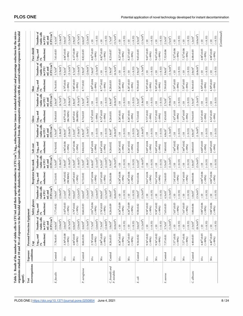
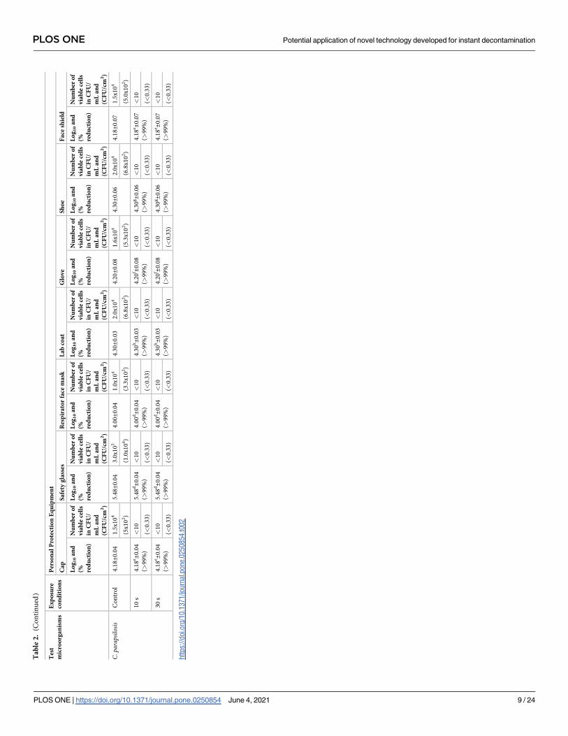
Table 2 shows the results for the number of viable cells (CFU/mL and CFU/cm2), the logarith-
mic reduction factor (log10) of each assay compared to the respective control (without expo-
sure to the biocidal agent), and the percentage reduction (%) after exposure to the biocidal
agent in the disinfection chamber for 10 and 30 s for each test microorganism and for each
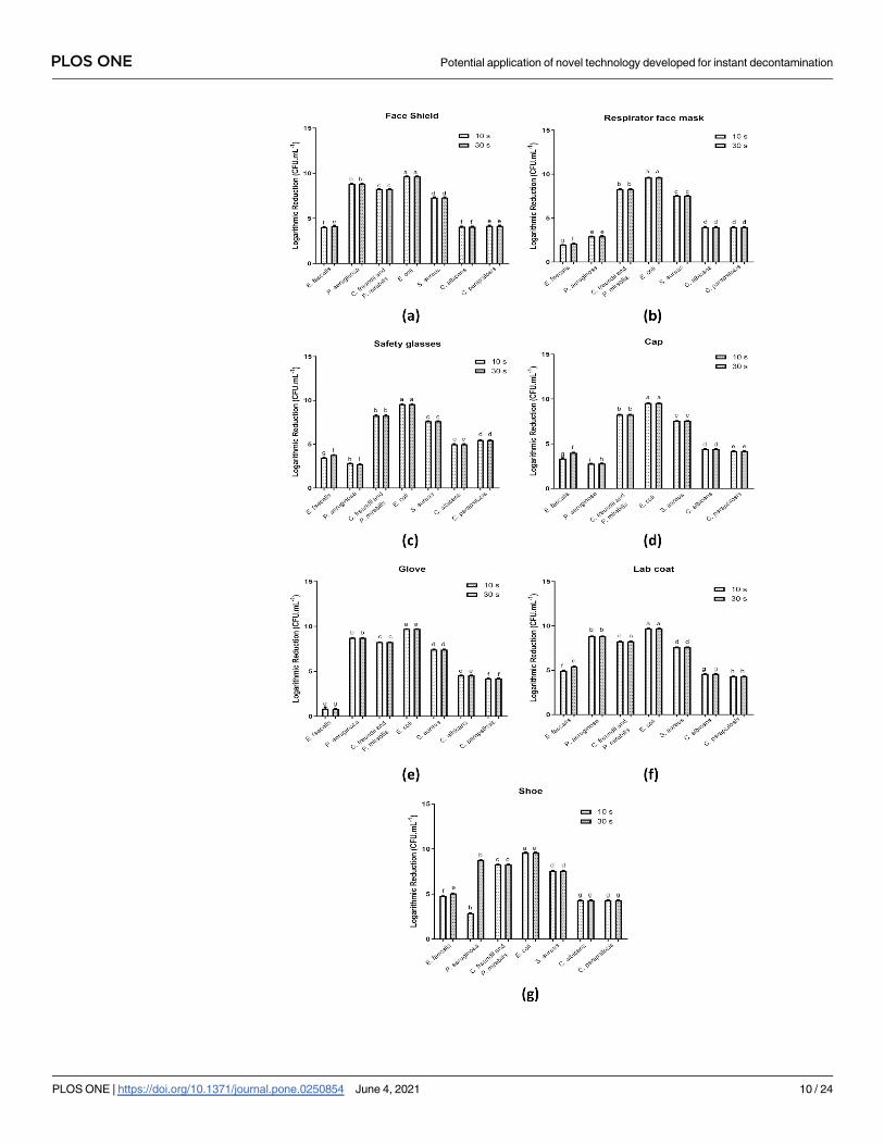
individual PPE item evaluated in this study. Fig 3 shows the graphs of the logarithmic reduc-
tion (log10) of each test microorganism per individual PPE item.
In total, 147 experimental conditions were studied, considering the two exposure times for
each test microorganism (n = 98) and control (n = 49), as well as for the seven different types
of PPE evaluated. In general, there was a significant reduction in the investigated microorgan-
isms after exposure of the previously contaminated items in the disinfection chamber, regard-
less of the item, which demonstrated the efficacy of the biocidal agent spraying system for
instantaneous disinfection of PPE for some of the microorganisms evaluated. In addition, the
results showed that at 10 and 30 s, there was only a significant difference in the microbial load
reduction factor during exposure of the biocidal agent to the microorganisms E. faecalis and P.
aeruginosa (p>0.05). In this case, the exposure time of 30 s was more efficient for the inactiva-
tion of the studied microorganisms in terms of log10 and percentage reduction for all studied
PPE (Table 2 and Fig 3).
A percentage reduction of>99% was determined for 96.93% (n = 95) of the tested condi-
tions when compared to the control, while a percentage reduction of between 86.00–99% was
PLOS ONE Potential application of novel technology developed for instant decontamination
PLOS ONE | https://doi.org/10.1371/journal.pone.0250854 June 4, 2021 7 / 24
Ta
ble
2.
Res
ult
of
the
nu
mb
ero
fv
iab
lece
lls
inC
FU
/mL
an
dth
eir
equ
iva
len
tn
um
ber
in(C
FU
/cm
2),
log
10
red
uct
ion
fact
or
(mea
n±
sta
nd
ard
dev
iati
on
)a
nd
per
cen
tag
ere
du
ctio
nfo
rth
em
icro
-
org
an
ism
sst
ud
ied
at
10
an
d3
0s
of
exp
osu
reto
the
bio
cid
al
ag
ent
inth
ed
isin
fect
ion
cha
mb
er(r
esu
lts
ob
tain
edfr
om
the
com
pa
rati
ve
an
aly
sis
wit
hth
eco
ntr
ol
wit
ho
ut
exp
osu
reto
the
bio
cid
al
ag
ent)
.
Tes
t
mic
roo
rga
nis
ms
Ex
po
sure
con
dit
ion
s
Per
son
al
Pro
tect
ion
Eq
uip
men
t
Ca
pS
afe
tyg
lass
esR
esp
ira
tor
face
ma
skL
ab
coa
tG
lov
eS
ho
eF
ace
shie
ld
Lo
g1
0a
nd
(% red
uct
ion
)
Nu
mb
ero
f
via
ble
cell
s
inC
FU
/
mL
an
d
(CF
U/c
m2)
Lo
g1
0a
nd
(% red
uct
ion
)
Nu
mb
ero
f
via
ble
cell
s
inC
FU
/
mL
an
d
(CF
U/c
m2)
Lo
g1
0a
nd
(% red
uct
ion
)
Nu
mb
ero
f
via
ble
cell
s
inC
FU
/
mL
an
d
(CF
U/c
m2)
Lo
g1
0a
nd
(% red
uct
ion
)
Nu
mb
ero
f
via
ble
cell
s
inC
FU
/
mL
an
d
(CF
U/c
m2)
Lo
g1
0a
nd
(% red
uct
ion
)
Nu
mb
ero
f
via
ble
cell
s
inC
FU
/
mL
an
d
(CF
U/c
m2)
Lo
g1
0a
nd
(% red
uct
ion
)
Nu
mb
ero
f
via
ble
cell
s
inC
FU
/
mL
an
d
(CF
U/c
m2)
Lo
g1
0a
nd
(% red
uct
ion
)
Nu
mb
ero
f
via
ble
cell
s
inC
FU
/
mL
an
d
(CF
U/c
m2)
E.faecalis
Co
ntr
ol
7.7
8±0
.05
6.0
x1
07
7.7
7±0
.02
5.9
x1
07
8.7
2±0
.01
5.3
x1
08
8.7
4±0
.02
5.5
x1
08
4.3
0±0
.03
2.0
x1
04
8.7
6±0
.01
5.7
x1
08
7.6
1±0
.03
4.1
x1
07
(2.0
x1
06)
(2.0
x1
06)
(1.8
x1
07)
(1.8
x1
07)
(6.7
x1
02)
(1.9
x1
07)
(1.4
x1
06)
10
s3
.36
g±0
.08
(>9
9%
)
2.6
x1
04
3.4
5g±0
.03
(>9
9%
)
2.1
x1
04
1.9
6g±0
.02
(98
.90
6%
)
5.8
x1
06
4.9
6f ±
0.0
6
(>9
9%
)
6.0
x1
03
0.8
7g±0
.10
(86
.50
0%
)
2.7
x1
03
4.7
9f ±
0.0
1
(>9
9%
)
9.2
x1
03
4.0
6f ±
0.0
5
(>9
9%
)
3.6
x1
03
(8.7
x1
02)
(7.0
x1
02)
(1.9
x1
06)
(2.0
x1
02)
(9.0
x1
01)
(3.1
x1
02)
(1.2
x1
02)
30
s4
.00
f ±0
.05
(>9
9%
)
6.0
x1
03
3.7
9f ±
0.0
2
(>9
9%
)
9.6
x1
03
2.0
9f ±
0.0
3
(>9
9%
)
4.3
x1
06
5.4
2e±0
.06
(>9
9%
)
2.1
x1
03
0.8
5g±0
.0
(86
.00
0%
)
2.8
x1
03
5.0
8e±0
.03
(>9
9%
)
4.7
x1
03
4.1
5e±0
.09
(>9
9%
)
2.9
x1
03
(2.0
x1
02)
(3,2
x1
02)
(1.4
x1
05)
(7.0
x1
01)
(9.3
x1
01)
(1.6
x1
02)
(9.7
x1
01)
P.aerugino
saC
on
tro
l8
.66±0
.04
4.6
x1
08
8.6
6±0
.03
4.6
x1
08
8.7
2±0
.02
5.3
x1
08
8.8
4±0
.01
6.9
x1
08
8.7
4±0
.03
5.5
x1
08
8.7
7±0
.02
5.9
x1
08
8.8
2±0
.05
6.6
x1
08
(1.5
x1
07)
(1,5
x1
07)
(1.8
x1
07)
(2.3
x1
07)
(1.8
x1
07)
(2.0
x1
07)
(2.2
x1
07)
10
s2
.78
i ±0
.04
(>9
9%
)
7.7
x1
05
2.8
1h±0
.04
(>9
9%
)
7.2
x1
05
2.9
2e±0
.04
(>9
9%
)
6.4
x1
05
8.8
4b±0
.01
(>9
9%
)
<1
08
.74
b±0
.03
<1
02
.89
h±0
.04
(>9
9%
)
7.6
x1
05
8.8
2b±0
.05
(>9
9%
)
<1
0
(2,6
x1
04)
(2,4
x1
04)
(2.1
x1
04)
(<0
.33
)(>
99
%)
(<0
.33
)(2
.5x
10
4)
(<0
.33
)
30
s2
.80
h±0
.05
(>9
9%
)
7.3
x1
05
2.7
6i ±
0.0
3
(>9
9%
)
8.0
x1
05
2.9
3e±0
.05
(>9
9%
)
6.2
x1
05
8.8
4b±0
.01
(>9
9%
)
<1
08
.74
b±0
.03
(>9
9%
)
<1
08
.77
b±0
.02
(>9
9%
)
<1
08
.82
b±0
.05
(>9
9%
)
<1
0
(2.4
x1
04)
(2,7
x1
04)
(2.1
x1
04)
(<0
.33
)(<
0.3
3)
(<0
.33
)(<
0.3
3)
C.fre
undiiand
P.mira
bilis
Co
ntr
ol
8.2
6±0
.03
1.8
x1
08
8.2
6±0
.09
1.8
x1
08
8.2
8±0
.06
1.9
x1
08
8.2
6±0
.02
1.8
x1
08
8.2
6±0
.01
1.8
x1
08
8.2
8±0
.03
1.9
x1
08
8.2
3±0
.07
1.7
x1
08
(6.0
x1
06)
(6,0
x1
06)
(6.3
x1
06)
(6.0
x1
06)
(6.0
x1
06)
(6.3
x1
06)
(5,7
x1
06)
10
s8
.26
b±0
.03
(>9
9%
)
<1
08
.26
b±0
.09
(>9
9%
)
<1
08
.28
b±0
.06
(>9
9%
)
<1
08
.26
c±0
.02
(>9
9%
)
<1
08
.26
c±0
.01
(>9
9%
)
<1
08
.28
c±0
.03
(>9
9%
)
<1
08
.23
c±0
.07
(>9
9%
)
<1
0
(<0
.33
)(<
0.3
3)
(<0
.33
)(<
0.3
3)
(<0
.33
)(<
0.3
3)
(<0
.33
)
30
s8
.26
b±0
.03
(>9
9%
)
<1
08
.26
b±0
.09
(>9
9%
)
<1
08
.28
b±0
.06
(>9
9%
)
<1
08
.26
c±0
.02
(>9
9%
)
<1
08
.26
c±0
.01
(>9
9%
)
<1
08
.28
c±0
.03
(>9
9%
)
<1
08
.23
c±0
.07
(>9
9%
)
<1
0
(<0
.33
)(<
0.3
3)
(<0
.33
)(<
0.3
3)
(<0
.33
)(<
0.3
3)
(<0
.33
)
E.coli
Co
ntr
ol
9.5
6±0
.02
3.6
x1
09
9.5
8±0
.02
3.8
x1
09
9.6
1±0
.04
4.1
x1
09
9.6
9±0
.04
4.9
x1
09
9.7
2±0
.02
5.3
x1
09
9.5
9±0
.05
3.9
x1
09
9.6
5±0
.05
4.5
x1
09
(1.2
x1
08)
(1,3
x1
08)
(1.4
x1
08)
(1.6
x1
08)
(1.8
x1
08)
(1.3
x1
08)
(1.5
x1
08)
10
s9
.56
a±0
.02
(>9
9%
)
<1
09
.58
a±0
.02
(>9
9%
)
<1
09
.61
a±0
.04
(>9
9%
)
<1
09
.69
a±0
.04
(>9
9%
)
<1
09
.72
a±0
.02
(>9
9%
)
<1
09
.59
a±0
.05
(>9
9%
)
<1
09
.65
a±0
.05
(>9
9%
)
<1
0
(<0
.33
)(<
0.3
3)
(<0
.33
)(<
0.3
3)
(<0
.33
)(<
0.3
3)
(<0
.33
)
30
s9
.56
a±0
.02
(>9
9%
)
<1
09
.58
a±0
.02
(>9
9%
)
<1
09
.61
a±0
.04
(>9
9%
)
<1
09
.69
a±0
.04
(>9
9%
)
<1
09
.72
a±0
.02
(>9
9%
)
<1
09
.59
a±0
.05
(>9
9%
)
<1
09
.65
a±0
.05
(>9
9%
)
<1
0
(<0
.33
)(<
0.3
3)
(<0
.33
)(<
0.3
3)
(<0
.33
)(<
0.3
3)
(<0
.33
)
S.au
reus
Co
ntr
ol
7.5
7±0
.01
3.7
x1
07
7.6
3±0
.03
4.3
x1
07
7.5
4±0
.03
3.5
x1
07
7.6
0±0
.02
4.0
x1
07
7.4
3±0
.05
2.7
x1
07
7.5
9±0
.05
3.9
x1
07
7.3
2±0
.06
2.1
x1
07
(1.2
x1
06)
(1.3
x1
06)
(1.2
x1
06)
(1.3
x1
06)
(9.0
x1
05)
(1.3
x1
06)
(7x
10
5)
10
s7
.57
c±0
.01
(>9
9%
)
<1
07
.63
c±0
.03
(>9
9%
)
<1
07
.54
c±0
.03
(>9
9%
)
<1
07
.60
d±0
.02
(>9
9%
)
<1
07
.43
d±0
.05
(>9
9%
)
<1
07
.59
d±0
.05
(>9
9%
)
<1
07
.32
d±0
.06
(>9
9%
)
<1
0
(<0
.33
)(<
0.3
3)
(<0
.33
)(<
0.3
3)
(<0
.33
)(<
0.3
3)
(<0
.33
)
30
s7
.57
c±0
.01
(>9
9%
)
<1
07
.63
c±0
.03
(>9
9%
)
<1
07
.54
c±0
.03
(>9
9%
)
<1
07
.60
d±0
.02
(>9
9%
)
<1
07
.43
d±0
.05
(>9
9%
)
<1
07
.59
d±0
.05
(>9
9%
)
<1
07
.32
d±0
.06
(>9
9%
)
<1
0
(<0
.33
)(<
0.3
3)
(<0
.33
)(<
0.3
3)
(<0
.33
)(<
0.3
3)
(<0
.33
)
C.albicans
Co
ntr
ol
4.4
0±0
.05
2.5
x1
04
5.0
0±0
.06
1.0
x1
05
4.0
0±0
.06
1.0
x1
04
4.6
0±0
.03
4.0
x1
04
4.5
4±0
.05
3.5
x1
04
4.3
0±0
.03
2.0
x1
04
4.0
8±0
.05
1.2
x1
04
(8.3
x1
02)
(3.3
x1
03)
(3.3
x1
02)
(1.3
x1
06)
(1.2
x1
03)
(6.8
x1
02)
(4.0
x1
02)
10
s4
.40
d±0
.05
(>9
9%
)
<1
05
.00
e±0
.06
(>9
9%
)
<1
04
.00
d±0
.06
(>9
9%
)
<1
04
.60
g±0
.03
(>9
9%
)
<1
04
.54
e±0
.05
(>9
9%
)
<1
04
.30
g±0
.03
(>9
9%
)
<1
04
.08
f ±0
.05
(>9
9%
)
<1
0
(<0
.33
)(<
0.3
3)
(<0
.33
)(<
0.3
3)
(<0
.33
)(<
0.3
3)
(<0
.33
)
30
s4
.40
d±0
.05
(>9
9%
)
<1
05
.00
e±0
.06
(>9
9%
)
<1
04
.00
d±0
.04
(>9
9%
)
<1
04
.60
g±0
.03
(>9
9%
)
<1
04
.54
e±0
.05
(>9
9%
)
<1
04
.30
g±0
.03
(>9
9%
)
<1
04
.08
f ±0
.05
(>9
9%
)
<1
0
(<0
.33
)(<
0.3
3)
(<0
.33
)(<
0.3
3)
(<0
.33
)(<
0.3
3)
(<0
.33
)
(Con
tinued)
PLOS ONE Potential application of novel technology developed for instant decontamination
PLOS ONE | https://doi.org/10.1371/journal.pone.0250854 June 4, 2021 8 / 24
Ta
ble
2.
(Co
nti
nu
ed)
Tes
t
mic
roo
rga
nis
ms
Ex
po
sure
con
dit
ion
s
Per
son
al
Pro
tect
ion
Eq
uip
men
t
Ca
pS
afe
tyg
lass
esR
esp
ira
tor
face
ma
skL
ab
coa
tG
lov
eS
ho
eF
ace
shie
ld
Lo
g1
0a
nd
(% red
uct
ion
)
Nu
mb
ero
f
via
ble
cell
s
inC
FU
/
mL
an
d
(CF
U/c
m2)
Lo
g1
0a
nd
(% red
uct
ion
)
Nu
mb
ero
f
via
ble
cell
s
inC
FU
/
mL
an
d
(CF
U/c
m2)
Lo
g1
0a
nd
(% red
uct
ion
)
Nu
mb
ero
f
via
ble
cell
s
inC
FU
/
mL
an
d
(CF
U/c
m2)
Lo
g1
0a
nd
(% red
uct
ion
)
Nu
mb
ero
f
via
ble
cell
s
inC
FU
/
mL
an
d
(CF
U/c
m2)
Lo
g1
0a
nd
(% red
uct
ion
)
Nu
mb
ero
f
via
ble
cell
s
inC
FU
/
mL
an
d
(CF
U/c
m2)
Lo
g1
0a
nd
(% red
uct
ion
)
Nu
mb
ero
f
via
ble
cell
s
inC
FU
/
mL
an
d
(CF
U/c
m2)
Lo
g1
0a
nd
(% red
uct
ion
)
Nu
mb
ero
f
via
ble
cell
s
inC
FU
/
mL
an
d
(CF
U/c
m2)
C.parapsilosis
Co
ntr
ol
4.1
8±0
.04
1.5
x1
04
5.4
8±0
.04
3.0
x1
05
4.0
0±0
.04
1.0
x1
04
4.3
0±0
.03
2.0
x1
04
4.2
0±0
.08
1.6
x1
04
4.3
0±0
.06
2.0
x1
04
4.1
8±0
.07
1.5
x1
04
(5x
10
2)
(1.0
x1
04)
(3.3
x1
02)
(6.8
x1
02)
(5.3
x1
02)
(6.8
x1
02)
(5.0
x1
02)
10
s4
.18
e±0
.04
(>9
9%
)
<1
05
.48
d±0
.04
(>9
9%
)
<1
04
.00
d±0
.04
(>9
9%
)
<1
04
.30
h±0
.03
(>9
9%
)
<1
04
.20
f ±0
.08
(>9
9%
)
<1
04
.30
g±0
.06
(>9
9%
)
<1
04
.18
e±0
.07
(>9
9%
)
<1
0
(<0
.33
)(<
0.3
3)
(<0
.33
)(<
0.3
3)
(<0
.33
)(<
0.3
3)
(<0
.33
)
30
s4
.18
e±0
.04
(>9
9%
)
<1
05
.48
d±0
.04
(>9
9%
)
<1
04
.00
d±0
.04
(>9
9%
)
<1
04
.30
h±0
.03
(>9
9%
)
<1
04
.20
f ±0
.08
(>9
9%
)
<1
04
.30
g±0
.06
(>9
9%
)
<1
04
.18
e±0
.07
(>9
9%
)
<1
0
(<0
.33
)(<
0.3
3)
(<0
.33
)(<
0.3
3)
(<0
.33
)(<
0.3
3)
(<0
.33
)
htt
ps:
//doi.o
rg/1
0.1
371/jo
urn
al.p
one.
0250854.t002
PLOS ONE Potential application of novel technology developed for instant decontamination
PLOS ONE | https://doi.org/10.1371/journal.pone.0250854 June 4, 2021 9 / 24
PLOS ONE Potential application of novel technology developed for instant decontamination
PLOS ONE | https://doi.org/10.1371/journal.pone.0250854 June 4, 2021 10 / 24
found for 3.07% (n = 3) of the tested conditions. The lowest percentage reductions identified
were 86.500 and 86.000% for the test with E. faecalis when the glove was evaluated at exposure
times of 10 and 30 s, respectively. In general, the exposure time of 10 s to 0.25% sodium hypo-
chlorite under the investigated conditions effectively reduced the microbial load by >99% for
all investigated microorganisms, except for E. faecalis. For these microorganism, percentage
reduction >99% at 10 s and 30 s of exposure was identified for all PPE, except for the glove
and respirator face mask. It is noteworthy that the results related to the percentage reduction
for the exposure time of 30 s were similar to the time of 10 s, except for E. faecalis (Table 2).
The percentage reduction is also reflected in the total number of viable cells, where 78.57%
(n = 77) of the analyzed conditions corresponded to<10 CFU/mL or <0.33 CFU/cm2 at 10
and 30 s of exposure to spraying of 0.25% sodium hypochlorite. In general, there was a reduc-
tion in the number of viable cells for all analyzed conditions when compared to the results
found for the control group. The resistance to the biocidal agent under the investigated condi-
tions of the microorganisms E. faecalis and P. aeruginosa was demonstrated in this parameter,
since they were the only ones that showed viable cells in the concentration >10 CFU/mL or
>0.33 CFU/cm2 after the disinfection process in the chamber, with the exception of samples of
P. aeruginosa collected from the face shield, glove and lab coat, at exposure times of 10 and 30
s, in addition to the shoe after 30 s of exposure. The exposure time of 10 s was able to reduce
the number of viable cells for<10 CFU/mL or <0.33 CFU/cm2 in 38 experimental conditions,
while for the time of 30 s, this reduction occurred in 39 experimental conditions. The differ-
ences related to the recovery of microorganisms may be associated with resistance to the bio-
cidal agent under the conditions tested, as well as the inoculum concentration.
Fig 3 shows that E. coli was the microorganism with the highest log10 reduction values,
regardless of the analyzed experimental condition (PPE item/surface and exposure time), with
values>9 log10. The microorganisms S. aureus, C. albicans, C. parapsilosis, C. freundii and P.
mirabilis showed the same log10 reduction at the tested exposure times, without significant dif-
ference regardless of the surface analyzed. It is important to highlight that for Candida species
the log10 reduction value was lower than the bacterial species and this effect may be due to the
lower initial inoculum used. With regard to the PPE items, the lab coat showed the highest
log10 reduction values, varying between 4.30 log10 (C. parapsilosis at 10 and 30 s) and 9.69 log10
(E. coli at 10 and 30 s). The surface of the glove was the only one in which the log10 reduction
of E. faecalis was similar (p>0.05) at the exposure times of 10 and 30 s, with 0.87 and 0.85
log10, respectively, and was the lowest when compared to the reduction in the other analyzed
surface.
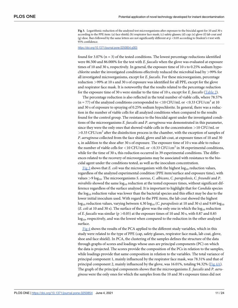
Fig 4 shows the results of the PCA applied to the different study variables, which in this
study were related to the type of PPE (cap, safety glasses, respirator face mask, lab coat, glove,
shoe and face shield). In PCA, the clustering of the samples defines the structure of the data
through graphs of scores and loadings whose axes are principal components (PC) on which
the data is projected. The scores provide the composition of the PCs in relation to the samples,
while loadings provide that same composition in relation to the variables. The total variance of
principal component 1, mainly influenced by the respirator face mask, was 78.31% and that of
principal component 2, mainly influenced by the glove, was 16.01%, totaling 94.32% (Fig 4A).
The graph of the principal components shows that the microorganisms E. faecalis and P. aeru-ginosa were the only ones for which the samples from the 10 and 30 s exposure times did not
Fig 3. Logarithmic reduction of the analyzed test microorganisms after exposure to the biocidal agent for 10 and 30 s
according to the PPE item: (a) face shield; (b) respirator face mask; (c) safety glasses; (d) cap; (e) glove (f) lab coat and
(g) shoe. Bars followed by the same letters are not significantly different at p<0.05 according to Student’s t test with
95% confidence.
https://doi.org/10.1371/journal.pone.0250854.g003
PLOS ONE Potential application of novel technology developed for instant decontamination
PLOS ONE | https://doi.org/10.1371/journal.pone.0250854 June 4, 2021 11 / 24
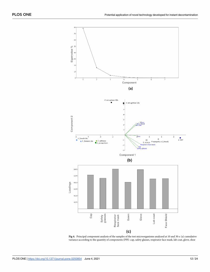
Fig 4. Principal component analysis of the samples of the test microorganisms analyzed at 10 and 30 s: (a) cumulative
variance according to the quantity of components (PPE–cap, safety glasses, respirator face mask, lab coat, glove, shoe
PLOS ONE Potential application of novel technology developed for instant decontamination
PLOS ONE | https://doi.org/10.1371/journal.pone.0250854 June 4, 2021 12 / 24
overlap (Fig 4B). In addition, the graph shows the formation of clusters for all tested microor-
ganisms at different exposure times. Clustering is important because it can indicate similar
behavior among the samples evaluated according to the study variables. Note that the only
analyzed fungi (C. albicans and C. parapsilosis) formed a cluster in the lower left quadrant, as
well as the samples from 10 and 30 s of E. faecalis, having no influence on the type of surface/
item tested. However, the microorganisms S. aureus, E. coli, C. freundii and P. mirabilis clus-
tered together in the lower right quadrant, being influenced by the type of surface/item tested
(safety glasses, cap, respirator face mask and shoe), indicating that these microorganisms had
similar behavior at the evaluated experimental conditions.
P. aeruginosa was the only microorganism whose samples from 10 and 30 s were allocated
in the upper right quadrant, not being influenced by the main analyzed variable, the PPE item.
Note also that there was no negative correlation between the porous and nonporous surface
variables for principal component 1, where all PPE correlated positively with each other (Fig
4c). Thus, from the PCA analysis, it is observed that the type of microorganism analyzed influ-
enced the results, since there was formation of clusters. In addition, even for the microorgan-
isms that did not have overlapping values for the exposure times of 10 and 30 s, the behavior in
response to the variables was similar since they remained in the same quadrant.
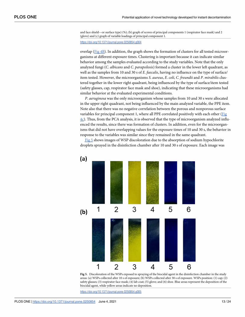
Fig 5 shows images of WSP discoloration due to the absorption of sodium hypochlorite
droplets sprayed in the disinfection chamber after 10 and 30 s of exposure. Each image was
and face shield—or surface type) (%); (b) graph of scores of principal components 1 (respirator face mask) and 2
(glove) and (c) graph of variable loadings of principal component 1.
https://doi.org/10.1371/journal.pone.0250854.g004
Fig 5. Discoloration of the WSPs exposed to spraying of the biocidal agent in the disinfection chamber in the study
areas: (a) WSPs collected after 10 s of exposure; (b) WSPs collected after 30 s of exposure. WSPs position: (1) cap; (2)
safety glasses; (3) respirator face mask; (4) lab coat; (5) glove; and (6) shoe. Blue areas represent the deposition of the
biocidal agent, while yellow areas indicate no deposition.
https://doi.org/10.1371/journal.pone.0250854.g005
PLOS ONE Potential application of novel technology developed for instant decontamination
PLOS ONE | https://doi.org/10.1371/journal.pone.0250854 June 4, 2021 13 / 24
observed ex situ after the WSPs were removed from the study surface areas (positions 1 to 6—
Fig 2). In general, there was good dispersion of the biocidal agent across the study area when
using the disinfection chamber composed of the six nebulizer nozzles. The areas of the WSP
with bluish tones show that there was deposition of the biocidal agent during the passage of
the manikin through the disinfection chamber, while areas with yellowish tones indicate the
absence of deposition of the studied agent.
In general, when comparing the distribution of the biocidal agent at the investigated expo-
sure times, similar good deposition coverage of the agent on the WSPs was observed, which
may be associated with the log10 reduction profile of the studied microorganisms, where the
exposure time to the biocidal agent had no significant influence on the reduction factor (except
for E. faecalis and P. aeruginosa) (Table 2 and Fig 3). Thus, the amount of biocidal agent that
reaches the study areas during the 10 s exposure would be sufficient to inactivate most of the
investigated microorganisms. Qualitatively, greater deposition is observed in some points of
the WSPs for the time of 30 s and this may explain the greater efficacy of the longer exposure
time for the two most resistant microorganisms (E. faecalis and P. aeruginosa). In addition, the
lower deposition of the biocidal agent on the respirator face mask (area 3), indicated by the
yellowish tone, may have interfered with the reduction efficiency of these microorganisms.
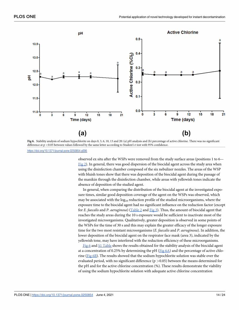
Fig 6 and S1 Table shows the results obtained for the stability analysis of the biocidal agent
at a concentration of 0.25% by determining the pH (Fig 6A) and the percentage of active chlo-
rine (Fig 6B). The results showed that the sodium hypochlorite solution was stable over the
evaluated period, with no significant difference (p>0.05) between the means determined for
the pH and for the active chlorine concentration (%). These results demonstrate the viability
of using the sodium hypochlorite solution with adequate active chlorine concentration
Fig 6. Stability analysis of sodium hypochlorite on days 0, 3, 6, 10, 13 and 20: (a) pH analysis and (b) percentage of active chlorine. There was no significant
difference at p<0.05 between values followed by the same letter according to Student’s t test with 95% confidence.
https://doi.org/10.1371/journal.pone.0250854.g006
PLOS ONE Potential application of novel technology developed for instant decontamination
PLOS ONE | https://doi.org/10.1371/journal.pone.0250854 June 4, 2021 14 / 24
(0.25%) for at least 20 days for application in the disinfection chamber, with maintenance of
its disinfectant capacity.
Discussion
In this study we demonstrated the high rates of microbial load reduction after exposure to the
sodium hypochlorite biocidal agent used in the personal protective closes and equipment
(PPE) disinfection chamber at the two analyzed times (10 and 30 s) regardless of the type of
surface/PPE item investigated. Some studies [80, 81] and standards [79, 82] indicate that disin-
fection methods with�5 log10 CFU reduction are considered effective and, consequently,
appropriate for clinical use, which reinforces the importance of our results for the instant dis-
infection of PPE, especially during the SARS-CoV-2 pandemic. This logarithmic reduction
implies the elimination of 99.999% of the microbial load [79, 82]. Considering these values as a
reference, the microorganisms P. aeruginosa and E. faecalis showed the lowest sensitivity to
sodium hypochlorite under the tested conditions (for some PPE items) when compared to the
other microorganisms, although the results were quite satisfactory in relation to the reduction
factor found for these bacteria under the studied conditions.
There are reports in the literature on the resistance of P. aeruginosa and E. faecalis to
sodium hypochlorite at concentrations <0.3% and<0.22%, respectively [83, 84]. This mecha-
nism may be associated with the bacterial ability to remove or discharge the charge of hypo-
chlorous acid (HClO), which is a strong oxidizing agent that damages the permeability of the
bacterial cell wall and its genetic material [85]. However, Lineback et al. [86] showed that the
use of sodium hypochlorite at a concentration of 1.312% against P. aeruginosa was more effec-
tive than quaternary ammonium, while Yoo et al. [87] reported that this biocidal agent at
0.031% showed activity against clinical isolates of E. faecalis. In this study, the concentration of
0.25% of sodium hypochlorite was effective in reducing the load >99% for P. aeruginosa and
�86.000% for E. faecalis.Regarding the analyzed fungal strains, the percentage reduction value was >99% and the
number of viable cell was <10 CFU/mL or<0.33 CFU/cm2 for all experimental conditions,
indicating that C. albicans and C. parapsilosis are sensitive to sodium hypochlorite under the
tested conditions, which shows that spraying of the biocidal agent may be an effective alterna-
tive for the inactivation of these microorganisms when compared to other methods [62]. Infec-
tions caused by Candida species are classified as one of the main contaminants in the hospital
environment because these pathogens can lead to systemic infection [88]. Although C. albicansis still the species most frequently isolated from nosocomial fungal infections [89], cases associ-
ated with C. parapsilosis have increased significantly in recent years [90] due to the resistance
of Candida species to antifungals and disinfectants [91]. Thus, it is important to note that
spraying systems have been used to control bioburden in nosocomial environments, especially
for combating multidrug-resistant strains [81].
The study by Ishikawa et al. [37] demonstrated that the efficacy of a small disinfection
chamber using a spray system with 5.00% sodium hypochlorite solution for the inactivation of
Bacillus subtilis spores. The authors reported that the disinfection system used is a "test cham-
ber", which does not have the physical structure for the passage of a person, being able only to
perform the sporicidal effect in a small area [37]. However, unlike Ishikawa et al. [37] work,
our study demonstrates the efficacy of a spray system (disinfection chamber) containing
sodium hypochlorite for the instant disinfection of different PPE items (at 10 or 30 s) against
Candida species and Gram-positive and Gram-negative bacteria on different types of surfaces
at the same time. The efficacy demonstrated by the biocide agent in the concentration of
0.25% against the microorganisms tested suggests that new studies can be conducted using a
PLOS ONE Potential application of novel technology developed for instant decontamination
PLOS ONE | https://doi.org/10.1371/journal.pone.0250854 June 4, 2021 15 / 24
lower concentration of the chemical agent, such as 0.1%, which is also in the concentration
range recommended by WHO for the disinfection of environmental surfaces [49].
Hospital infections are caused by factors such as environmental contamination, frequent
handling of contaminated material, and the ability of microorganisms to survive for prolonged
periods on different types of surfaces [1, 92]. Within the hospital environment, sodium hypo-
chlorite is the most widely used disinfectant because it has broad-spectrum antimicrobial
activity, considering Gram-positive and Gram-negative bacteria and fungi [83] as well as dem-
onstrated virucidal activity [93]. Kohler et al. [94] showed that sodium hypochlorite effectively
reduced the concentration of multidrug-resistant Gram-positive bacteria (Pseudomonas, Aci-netobacter and Klebsiella) after exposure times of 1 to 15 min, longer exposure time than those
studied in this work. Regarding the activity against viral agents, It has been demonstrated that
sodium hypochlorite in concentrations between 0.01 and 0.5% is capable of inactivating the
SARS-CoV-1 on stainless steel surfaces [95, 96]. In addition, Ma et al. [97] showed that instant
hand hygiene using disinfecting wipes containing 0.05 or 0.25% active chlorine removed
96.62% and 99.98%, of the influenza virus, respectively, which causes avian influenza. The
authors also point out that, although they have not tested with SARS-CoV-2, hand cleaning
with the disinfecting wipes can help to control the spread of COVID-19. Similarly to the study
by Ma et al. [97], one limitation of our study was not to use SARS-CoV-2 as a test microorgan-
ism. However, based on the promising results identified in this study, which showed that the
biocide capacity of sodium hypochlorite was maintained under the conditions tested, the eval-
uated concentration of 0.25% has the potential for disinfecting surfaces contaminated by dif-
ferent microorganisms and can be extrapolated to enveloped viruses based on results in the
literature, being a potential agent against SARS-CoV-2. The choice of evaluating different bac-
teria and fungi as biological indicators stemmed from the need to accelerate the confirmation
of the disinfectant action of the proposed technology, since some of these microorganisms are
resilient compared to viruses [98], have a relatively faster growth and can be manipulated in
laboratory environments with a lower level of biosafety.
Other studies have already demonstrated the effect of different biocidal agents against the
microorganisms tested in this study, showing that the efficacy of the disinfection process varies
with the type of disinfectant [99, 100] or according to the application method used [101, 102].
There are few reports in the scientific literature on the efficacy of PPE decontamination after
exposure to pathogens, as was analyzed in this study. Among them, Lemmer et al. [103]
showed that disinfection with 2% peracetic acid with 0.2% surfactant through a spray system
was able to inactivate B. thuringiensis spores in high density polyethylene protective coveralls
after 5 min of exposure. Compared to our study, sodium hypochlorite was more effective than
peracetic acid because the exposure time required was shorter to reduce the microbial load on
the surface, considering the polyethylene surface and other analyzed materials. In addition, the
instantaneous decontamination demonstrated by the results obtained in 10 and 30 s exposure
times shows the potential of the disinfection chamber application in places with intense or
moderate people flow, such as the exits of intensive care units and wards in hospitals. In addi-
tion, reducing the microbial load on the surface of PPE can help reduce the risks associated
with handling and exposure to biomedical waste, an important source of environmental con-
tamination [104].
The results also suggest that the use of sodium hypochlorite may be recommended due to
the stability of its solution in terms of pH and concentration of active chlorine over the 20
days, since the exchange or replacement of the biocide agent solution does not need to be per-
formed, for example, on a daily basis. The dissociation of NaOCl into HOCl-, its main active
agent, is pH-dependent [105]. Thus, it is important that the solution remains stable so that the
levels of the active agent do not decrease. The critical issue raised by health authorities’
PLOS ONE Potential application of novel technology developed for instant decontamination
PLOS ONE | https://doi.org/10.1371/journal.pone.0250854 June 4, 2021 16 / 24
agencies are sodium hypochlorite toxicity when in contact with mucous membranes and may
lead to tissue damage or allergic reactions [106, 107]. Therefore, we are aware of the possibility
of episodes of clinical toxicity caused by the sodium hypochlorite, especially in persons that
are known to be allergic to bleaches, and that this can be considered as a limitation of the
study. In addition, we re-emphasized that the disinfection chamber with 0.25% sodium hypo-
chlorite be used only by fully trained workers (considering its installation, the preparation of
the biocide agent in the correct concentration, and for its correct use), thus promoting an
effective disinfection process against bacteria and potential emerging pathogens, such as
SARS-CoV-2, that is safe for users.
The use of the proposed disinfection technology becomes an attractive alternative, espe-
cially for middle-income countries such as Brazil [38]. This occurs due to factors such as the
material used in the chamber framework, which are widely used in the industry for the manu-
facture of different items, its modular design that allows scalable production as well as the low-
cost of sodium hypochlorite. These factors can make production cheaper and facilitate trans-
port and installation in different health facilities. In addition, the results obtained related to
WPS showed that the nebulizer nozzles disposition promoted a satisfactory spraying of the
biocide agent.
Indeed, disinfection chamber could be considered as an interesting disinfection technology
for use in emerging countries, since it can improve the control cases of nosocomial infection
in those places that usually have the most overloaded health systems.
Conclusions
The disinfection chamber proved to be a potential technology for the rapid and effective disin-
fection of the surface of PPE, regardless of the evaluated item, routinely used by HCWs for
protection against infectious agents. The spraying system with the biocidal agent was effective
in reduce the microbial load, where the percentage reduction equal to>99% and, conse-
quently, bringing the number of viable cells to<10 CFU/mL and<0.33 CFU/cm2 after expo-
sure times of 10 and 30 s in 96.93% of the experimental conditions analyzed. The lowest
percentages reduction were found for the sample of E. faecalis collected from the glove, where
the values obtained were 86.000 and 86.500% for the exposition of 10 and 30 s, respectively,
while the highest amount of viable cells was found for P. aeruginosa sample at 10 s in the cap,
with 7.7x10⁵ CFU/mL or 2.6x10⁴ CFU/cm2. The log10 reduction values varied between 0.85
log10 (E. faecalis at 30 s in glove surface) and 9.69 log10 (E. coli at 10 and 30 s in lab coat
surface).
Thus, the bacterial species E. coli, S. aureus, C. freundii and P. mirabilis and the fungi C.
albicans and C. parapsilosis showed susceptibility to 0.25% sodium hypochlorite under the
evaluated experimental conditions independent of the exposure time or PPE item evaluated,
while the microorganism E. faecalis was less susceptible to the biocidal agent under the tested
conditions. In general, a 30-s exposure time was more efficient in reducing the investigated
microbial load.
The results of this study show that the disinfection chamber with 0.25% sodium hypochlo-
rite may be an alternative to control the bioburden in nosocomial environments, especially to
prevent the self-contamination of HCWs in the doffing step. The importance of the use of the
chamber by properly attired HCWs is also emphasized in order to avoid direct contact with
the tested biocidal agent. In addition, because this is a novel study, these results may contribute
to the development and safe use of disinfection equipment in environments where the envi-
ronmental bioburden must be controlled. It is also important to highlight that the experimen-
tal design of the study was carried out in order to have a simulation of how the disinfection
PLOS ONE Potential application of novel technology developed for instant decontamination
PLOS ONE | https://doi.org/10.1371/journal.pone.0250854 June 4, 2021 17 / 24

chamber would be used by HCW in the nosocomial environment. Thus, the manipulation of
viral strains would not be appropriate in the conditions tested, since the handling of these
microorganisms requires a laboratory environment with a higher level of biosafety. However,
although virucidal efficacy was not directly determined, the chamber may be an alternative to
reduce the contamination rates among HCWs in front of different types of emerging microor-
ganisms, reducing the impacts in the area of public health.
Supporting information
S1 Fig. Image of the disinfection chamber: Spray disinfection technology for instant
decontamination of personal protective equipment.
(DOCX)
S2 Fig. Images of the manikin suitably dressed with PPEs used to simulate the use of the
disinfection chamber by healthcare workers in nosocomial environments.
(DOCX)
S1 Table. Raw data of stability of sodium hypochlorite regarding pH and active chlorine
analysis (mean ± standard deviation).
(DOCX)
Acknowledgments
The authors thank Bahia Department of Health (Secretaria de Saude do Estado da Bahia–
SESAB), Directorate of Sanitary and Environmental Surveillance of Bahia (Diretoria de Vigi-
lancia Sanitaria e Ambiental do Estado da Bahia–DIVISA BA) and Hospital Espanhol of Salva-
dor (COVID-19 Campaign Hospital).
Author Contributions
Conceptualization: Luıs Alberto Brêda Mascarenhas, Bruna Aparecida Souza Machado,
Leone Peter Correia da Silva Andrade, Jailson Bittencourt de Andrade, Roberto Badaro.
Data curation: Luıs Alberto Brêda Mascarenhas, Bruna Aparecida Souza Machado, Paulo
Roberto Freitas Neves, Jailson Bittencourt de Andrade, Roberto Badaro.
Formal analysis: Luıs Alberto Brêda Mascarenhas, Bruna Aparecida Souza Machado, Leticia
de Alencar Pereira Rodrigues, Alex Alisson Bandeira Santos, Paulo Roberto Freitas Neves,
Milena Botelho Soares, Roberto Badaro.
Investigation: Luıs Alberto Brêda Mascarenhas, Bruna Aparecida Souza Machado, Leticia de
Alencar Pereira Rodrigues, Alex Alisson Bandeira Santos, Leone Peter Correia da Silva
Andrade, Milena Botelho Soares, Roberto Badaro.
Methodology: Luıs Alberto Brêda Mascarenhas, Bruna Aparecida Souza Machado, Leticia de
Alencar Pereira Rodrigues, Katharine Valeria Saraiva Hodel, Alex Alisson Bandeira Santos,
Paulo Roberto Freitas Neves, Leone Peter Correia da Silva Andrade, Jailson Bittencourt de
Andrade, Roberto Badaro.
Project administration: Luıs Alberto Brêda Mascarenhas, Bruna Aparecida Souza Machado,
Leone Peter Correia da Silva Andrade, Jailson Bittencourt de Andrade, Roberto Badaro.
Software: Katharine Valeria Saraiva Hodel.
Supervision: Luıs Alberto Brêda Mascarenhas, Bruna Aparecida Souza Machado, Leone Peter
Correia da Silva Andrade, Roberto Badaro.
PLOS ONE Potential application of novel technology developed for instant decontamination
PLOS ONE | https://doi.org/10.1371/journal.pone.0250854 June 4, 2021 18 / 24
Validation: Luıs Alberto Brêda Mascarenhas, Bruna Aparecida Souza Machado, Alex Alisson
Bandeira Santos, Paulo Roberto Freitas Neves, Leone Peter Correia da Silva Andrade,
Milena Botelho Soares, Jailson Bittencourt de Andrade, Roberto Badaro.
Visualization: Luıs Alberto Brêda Mascarenhas, Bruna Aparecida Souza Machado, Leone
Peter Correia da Silva Andrade, Roberto Badaro.
Writing – original draft: Luıs Alberto Brêda Mascarenhas, Bruna Aparecida Souza Machado,
Katharine Valeria Saraiva Hodel, Alex Alisson Bandeira Santos, Leone Peter Correia da
Silva Andrade, Milena Botelho Soares, Jailson Bittencourt de Andrade, Roberto Badaro.
References1. Weber DJ, Kanamori H, Rutala WA. “No touch” technologies for environmental decontamination:
Focus on ultraviolet devices and hydrogen peroxide systems. Curr Opin Infect Dis. 2016; 29: 424–
431. https://doi.org/10.1097/QCO.0000000000000284 PMID: 27257798
2. Ferrara P, Albano L. COVID-19 and healthcare systems: What should we do next? Public Health.
2020; 185: 1–2. https://doi.org/10.1016/j.puhe.2020.05.014 PMID: 32502747
3. Chowdhury S, Azziz-Baumgartner E, Kile JC, Hoque MA, Rahman MZ, Hossain ME, et al. Association
of biosecurity and hygiene practices with environmental contamination with influenza a viruses in live
bird markets, Bangladesh. Emerg Infect Dis. 2020; 26: 2087–2096. https://doi.org/10.3201/eid2609.
191029 PMID: 32818393
4. Cutts TA, Robertson C, Theriault SS, Nims RW, Kasloff SB, Rubino JR, et al. Efficacy of microbicides
for inactivation of Ebola–Makona virus on a non-porous surface: a targeted hygiene intervention for
reducing virus spread. Sci Rep. 2020; 10: 1–9. https://doi.org/10.1038/s41598-019-56847-4 PMID:
31913322
5. Kratzel A, Todt D, V’kovski P, Steiner S, Gultom M, Thao TTN, et al. Inactivation of Severe Acute
Respiratory Syndrome Coronavirus 2 by WHO-Recommended Hand Rub Formulations and Alcohols.
Emerg Infect Dis. 2020; 26. https://doi.org/10.3201/eid2607.200915 PMID: 32284092
6. Lu H, Stratton CW, Tang Y. Outbreak of pneumonia of unknown etiology in Wuhan, China: The mys-
tery and the miracle. J Med Virol. 2020; 92: 401–402. https://doi.org/10.1002/jmv.25678 PMID:
31950516
7. Munster VJ, Koopmans M, van Doremalen N, van Riel D, de Wit E. A Novel Coronavirus Emerging in
China—Key Questions for Impact Assessment. N Engl J Med. 2020; 382: 692–694. https://doi.org/10.
1056/NEJMp2000929 PMID: 31978293
8. Yin S, Peng Y, Ren Y, Hu M, Tang L, Xiang Z, et al. The implications of preliminary screening and diag-
nosis: Clinical characteristics of 33 mild patients with SARS-CoV-2 infection in Hunan, China. J Clin
Virol. 2020; 128: 104397. https://doi.org/10.1016/j.jcv.2020.104397 PMID: 32388472
9. Rothe C, Schunk M, Sothmann P, Bretzel G, Froeschl G, Wallrauch C, et al. Transmission of 2019-
nCoV Infection from an Asymptomatic Contact in Germany. N Engl J Med. 2020; 382: 970–971.
https://doi.org/10.1056/NEJMc2001468 PMID: 32003551
10. Liu Y, Ning Z, Chen Y, Guo M, Liu Y, Gali NK, et al. Aerodynamic analysis of SARS-CoV-2 in two
Wuhan hospitals. Nature. 2020; 582: 557–560. https://doi.org/10.1038/s41586-020-2271-3 PMID:
32340022
11. Adams JG, Walls RM. Supporting the Health Care Workforce during the COVID-19 Global Epidemic.
JAMA—J Am Med Assoc. 2020; 323: 1439–1440. https://doi.org/10.1001/jama.2020.3972 PMID:
32163102
12. Tran K, Cimon K, Severn M, Pessoa-Silva CL, Conly J. Aerosol Generating Procedures and Risk of
Transmission of Acute Respiratory Infections to Healthcare Workers: A Systematic Review. Semple
MG, editor. PLoS One. 2012; 7: e35797. https://doi.org/10.1371/journal.pone.0035797 PMID:
22563403
13. James PB, Wardle J, Steel A, Adams J, Bah AJ, Bai P, et al. Providing healthcare to Ebola survivors:
A qualitative exploratory investigation of healthcare providers ‘ views and experiences in Sierra Leone
Providing healthcare to Ebola survivors: A qualitative exploratory investigation of healthcare providers
‘ v. Glob Public Health. 2020; 0: 1–16. https://doi.org/10.1080/17441692.2020.1762105 PMID:
32379008
PLOS ONE Potential application of novel technology developed for instant decontamination
PLOS ONE | https://doi.org/10.1371/journal.pone.0250854 June 4, 2021 19 / 24
14. Wei J-T, Liu Z-D, Fan Z-W, Zhao L, Cao W-C. Epidemiology of and Risk Factors for COVID-19 Infec-
tion among Health Care Workers: A Multi-Centre Comparative Study. Int J Environ Res Public Health.
2020; 17: 7149. https://doi.org/10.3390/ijerph17197149 PMID: 33003634
15. Dhont S, Derom E, Van Braeckel E, Depuydt P, Lambrecht BN. The pathophysiology of “happy” hyp-
oxemia in COVID-19. Respir Res. 2020; 21: 198. https://doi.org/10.1186/s12931-020-01462-5 PMID:
32723327
16. Lai X, Wang M, Qin C, Tan L, Ran L, Chen D, et al. Coronavirus Disease 2019 (COVID-2019) Infection
Among Health Care Workers and Implications for Prevention Measures in a Tertiary Hospital in
Wuhan, China. JAMA Netw Open. 2020; 3: e209666. https://doi.org/10.1001/jamanetworkopen.2020.
9666 PMID: 32437575
17. Nissen K, Krambrich J, Akaberi D, Hoffman T, Ling J, Lundkvist Å, et al. Long-distance airborne dis-
persal of SARS-CoV-2 in COVID-19 wards. Sci Rep. 2020; 10: 19589. https://doi.org/10.1038/
s41598-020-76442-2 PMID: 33177563
18. Wang J, Zhou M, Liu F. Reasons for healthcare workers becoming infected with novel coronavirus dis-
ease 2019 (COVID-19) in China. J Hosp Infect. 2020; 105: 100–101. https://doi.org/10.1016/j.jhin.
2020.03.002 PMID: 32147406
19. Felice C, Luca G, Tanna D, Zanus G, Grossi U. Impact of COVID-19 Outbreak on Healthcare Workers
in Italy: Results from a National E-Survey. J Community Health. 2020 [cited 7 Jun 2020]. https://doi.
org/10.1007/s10900-020-00845-5 PMID: 32440724
20. Kursumovic E, Lennane S, Cook TM. Deaths in healthcare workers due to COVID-19: the need for
robust data and analysis. Anaesthesia. 2020; 5–8. https://doi.org/10.1111/anae.15116 PMID:
32397005
21. Marson FAL, Ortega MM. COVID-19 in Brazil. Pulmonology. 2020. https://doi.org/10.1016/j.pulmoe.
2020.04.008 PMID: 32371054
22. Ministerio da Saude do Brasil. Coronavırus Brasil. 2020 [cited 16 Sep 2020]. Available: https://covid.
saude.gov.br/
23. Suen LKP, Guo YP, Tong DWK, Leung PHM, Lung D, Ng MSP, et al. Self-contamination during doff-
ing of personal protective equipment by healthcare workers to prevent Ebola transmission. Antimicrob
Resist Infect Control. 2018; 7: 157. https://doi.org/10.1186/s13756-018-0433-y PMID: 30607244
24. Tellier R, Li Y, Cowling BJ, Tang JW. Recognition of aerosol transmission of infectious agents: a com-
mentary. BMC Infect Dis. 2019; 19: 1–9. https://doi.org/10.1186/s12879-018-3567-x PMID: 30606108
25. Jones RM, Bleasdale SC, Maita D, Brosseau LM. A systematic risk-based strategy to select personal
protective equipment for infectious diseases. Am J Infect Control. 2020; 48: 46–51. https://doi.org/10.
1016/j.ajic.2019.06.023 PMID: 31358421
26. Cinar P, Kubal T, Freifeld A, Mishra A, Shulman L, Bachman J, et al. Safety at the Time of the COVID-
19 Pandemic: How to Keep our Oncology Patients and Healthcare Workers Safe. J Natl Compr Canc
Netw. 2020; 18: 1–6. https://doi.org/10.6004/jnccn.2020.0003 PMID: 31910382
27. Visnovsky LD, Zhang Y, Leecaster MK, Safdar N, Barko L, Haroldsen C, et al. Effectiveness of a multi-
site personal protective equipment (PPE)-free zone intervention in acute care. Infect Control Hosp Epi-
demiol. 2019; 40: 761–766. https://doi.org/10.1017/ice.2019.111 PMID: 31172904
28. Korth J, Wilde B, Dolff S, Anastasiou OE, Krawczyk A, Jahn M, et al. SARS-CoV-2-specific antibody
detection in healthcare workers in Germany with direct contact to COVID-19 patients. J Clin Virol.
2020; 128: 104437. https://doi.org/10.1016/j.jcv.2020.104437 PMID: 32434708
29. Muñoz-Leyva F, Niazi AU. Common breaches in biosafety during donning and doffing of protective
personal equipment used in the care of COVID-19 patients. Can J Anaesth. 2020; 67: 900–901.
https://doi.org/10.1007/s12630-020-01648-x PMID: 32291630
30. Thadathilankal-Jess, Karin H, Hellmuth W. Donning and doffing of personal protective equipment
(PPE) for angiography during the COVID-19 crisis. Eur Heart J. 2020; 41: 1786–1787. https://doi.org/
10.1093/eurheartj/ehaa283 PMID: 32282025
31. Karim N, Afroj S, Lloyd K, Clarke Oaten L, Andreeva D V, Carr C, et al. Sustainable Personal Protec-
tive Clothing for Healthcare Applications: A Review. ACS Nano. 2020; acsnano.0c05537. https://doi.
org/10.1021/acsnano.0c05537 PMID: 32866368
32. Wickramatillake A, Kurukularatne C. SARS-CoV-2 human disinfection chambers: a critical analysis.
Occup Med (Chic Ill). 2020 [cited 6 Jun 2020]. https://doi.org/10.1093/occmed/kqaa078 PMID:
32372076
33. Gardam M, McGeer A, Mertz D. Portable ultraviolet light surface-disinfecting devices for prevention of
hospital-acquired infections: A health technology assessment. Ont Health Technol Assess Ser. 2018;
18: 1–73.
PLOS ONE Potential application of novel technology developed for instant decontamination
PLOS ONE | https://doi.org/10.1371/journal.pone.0250854 June 4, 2021 20 / 24
34. El Haddad L, Ghantoji SS, Stibich M, Fleming JB, Segal C, Ware KM, et al. Evaluation of a pulsed
xenon ultraviolet disinfection system to decrease bacterial contamination in operating rooms. BMC
Infect Dis. 2017; 17. https://doi.org/10.1186/s12879-017-2792-z PMID: 29017457
35. Cadnum JL, Jencson AL, Livingston SH, Li D, Redmond SN, Pearlmutter B, et al. Evaluation of an
Electrostatic Spray Disinfectant Technology for Rapid Decontamination of Portable Equipment and
Large Open Areas in the Era of SARS-CoV-2. Am J Infect Control. 2020. https://doi.org/10.1016/j.ajic.
2020.06.002 PMID: 32522608
36. Verhougstraete M, Reynolds K. Use of a portable air disinfecting system to remove seeded coliphage
in hospital rooms. Am J Infect Control. 2016; 44: 714–715. https://doi.org/10.1016/j.ajic.2015.12.025
PMID: 26905789
37. Ishikawa S, Ueno S, Mitsui M, Matsumura Y, Hatsuoka T. Construction of its evaluation system in orig-
inally designed test-chamber system and sporicidal activity of aerosolized hypochlorite solution to
bacillus subtilis spores. Biocontrol Sci. 2019; 24: 57–65. https://doi.org/10.4265/bio.24.57 PMID:
30880314
38. Anvisa. NOTA TECNICA No 38/2020/SEI/COSAN/GHCOS/DIRE3/ANVISA. Brasılia; 2020 [cited 5
Aug 2020] pp. 1–5. Available: https://sei.anvisa.gov.br/sei/controlador.php?acao=documento_
imprimir_web&acao_origem=arvore_visualizar&id_documento=1118935&infra_sis. . .1/5
39. FDA. Enforcement Policy for Sterilizers, Disinfectant Devices, and Air Purifiers During the Coronavirus
Disease 2019 (COVID-19) Public Health Emergency Guidance for Industry and Food and Drug
Administration Staff Preface Public Comment. In: FDA [Internet]. Mar 2020 [cited 16 Nov 2020] pp. 1–
14. Available: https://www.fda.gov/regulatory-information/search-fda-guidance-documents.
40. Biswal M, Kanaujia R, Angrup A, Ray P, Mohan Singh S. Disinfection tunnels: potentially counterpro-
ductive in the context of a prolonged pandemic of COVID-19. Public Health. Elsevier B.V.; 2020. pp.
48–49. https://doi.org/10.1016/j.puhe.2020.04.045 PMID: 32422441
41. Keil SD, Ragan I, Yonemura S, Hartson L, Dart NK, Bowen R. Inactivation of severe acute respiratory
syndrome coronavirus 2 in plasma and platelet products using a riboflavin and ultraviolet light-based
photochemical treatment. Vox Sang. 2020; vox.12937. https://doi.org/10.1111/vox.12937 PMID:
32311760
42. Aboualizadeh E, Bumah V V., Masson-Meyers DS, Eells JT, Hirschmugl CJ, Enwemeka CS. Under-
standing the antimicrobial activity of selected disinfectants against methicillin-resistant Staphylococ-
cus aureus (MRSA). Gupta V, editor. PLoS One. 2017; 12: e0186375. https://doi.org/10.1371/journal.
pone.0186375 PMID: 29036196
43. Schrank CL, Minbiole KPC, Wuest WM. Are Quaternary Ammonium Compounds, the Workhorse Dis-
infectants, Effective against Severe Acute Respiratory Syndrome-Coronavirus-2? ACS Infect Dis.
2020; 1–5. https://doi.org/10.1021/acsinfecdis.0c00265 PMID: 32412231
44. Chapuis A, Amoureux L, Bador J, Gavalas A, Siebor E, Chretien M-L, et al. Outbreak of Extended-
Spectrum Beta-Lactamase Producing Enterobacter cloacae with High MICs of Quaternary Ammonium
Compounds in a Hematology Ward Associated with Contaminated Sinks. Front Microbiol. 2016; 7:
1070. https://doi.org/10.3389/fmicb.2016.01070 PMID: 27462306
45. Libonati A, Di Taranto V, Mea A, Montemurro E, Gallusi G, Angotti V, et al. Clinical antibacterial effec-
tiveness Healozone Technology after incomplete caries removal. Eur J Paediatr Dent. 2019; 20: 73–
78. https://doi.org/10.23804/ejpd.2019.20.01.14 PMID: 30919649
46. Henwood AF. Coronavirus disinfection in histopathology. J Histotechnol. 2020; 43: 102–104. https://
doi.org/10.1080/01478885.2020.1734718 PMID: 32116147
47. Rohner E, Jacob B, Bohle S, Rohe S, Loffler B, Matziolis G, et al. Sodium hypochlorite is more effec-
tive than chlorhexidine for eradication of bacterial biofilm of staphylococci and Pseudomonas aerugi-
nosa. Sport Traumatol Arthrosc. 2020 [cited 8 Jun 2020]. https://doi.org/10.1007/s00167-020-05887-9
PMID: 32034427
48. Wang J, Shen J, Ye D, Yan X, Zhang Y, Yang W, et al. Disinfection technology of hospital wastes and
wastewater: Suggestions for disinfection strategy during coronavirus Disease 2019 (COVID-19) pan-
demic in China. Environ Pollut. 2020; 262: 114665. https://doi.org/10.1016/j.envpol.2020.114665
PMID: 32443202
49. WHO. Cleaning and Disinfection of Environmental Surfaces in the context of COVID-19. Geneva;
2020 [cited 30 Aug 2020] p. 7. Available: https://apps.who.int/iris/rest/bitstreams/1277966/retrieve
50. OPAS. O uso de tuneis e outras tecnologias para desinfeccão de humanos usando aspersão de pro-
dutos quımicos ou radiacão UV-C. 2020 [cited 30 Aug 2020] pp. 1–3. Available: https://iris.paho.org/
handle/10665.2/52243
51. WHO. Coronavirus disease (COVID-19) Situation Report-115. In: World Health Organization [Inter-
net]. 14 May 2020 [cited 13 Nov 2020] pp. 1–19. https://doi.org/10.1016/j.ajic.2019.01.014
PLOS ONE Potential application of novel technology developed for instant decontamination
PLOS ONE | https://doi.org/10.1371/journal.pone.0250854 June 4, 2021 21 / 24
52. Yim W, Cheng D, Patel S, Kui R, Meng YS, Jokerst J. Assessment of N95 and K95 respirator decon-
tamination: fiber integrity, filtration efficiency, and dipole charge density. medRxiv. 2020 [cited 11 Nov
2020]. https://doi.org/10.1101/2020.07.07.20148551 PMID: 32676621
53. Mantlo E, Rhodes T, Boutros J, Patterson-Fortin L, Evans A, Paessler S, et al. In vitro efficacy of a
copper iodine complex PPE disinfectant for SARS-CoV-2 inactivation. F1000Research. 2020; 9: 1–
10. https://doi.org/10.12688/f1000research.24651.2 PMID: 33123349
54. Saini V, Sikri K, Dhingra Batra S, Kalra P, Gautam K. Development of a highly effective low-cost vapor-
ized hydrogen peroxide-based method for disinfection of personal protective equipment for their selec-
tive reuse during pandemics. Gut Pathog. 2020; 12: 1–11. https://doi.org/10.1186/s13099-019-0341-6
PMID: 31911822
55. Drohan SE, Levin SA, Grenfell BT, Laxminarayan R. Incentivizing hospital infection control. Proc Natl
Acad Sci U S A. 2019; 116: 6221–6225. https://doi.org/10.1073/pnas.1812231116 PMID: 30858309
56. Suchomel M, Lenhardt A, Kampf G, Grisold A. Enterococcus hirae, Enterococcus faecium and Entero-
coccus faecalis show different sensitivities to typical biocidal agents used for disinfection. J Hosp
Infect. 2019; 103: 435–440. https://doi.org/10.1016/j.jhin.2019.08.014 PMID: 31449920
57. Gottardi W, Nagl M. Chlorine covers on living bacteria: the initial step in antimicrobial action of active
chlorine compounds. J Antimicrob Chemother. 2005; 55: 475–482. https://doi.org/10.1093/jac/dki054
PMID: 15761074
58. Jackson DS, Crockett DF, Wolnik KA. The indirect detection of bleach (sodium hypochlorite) in bever-
ages as evidence of product tampering. J Forensic Sci. 2006; 51: 827–831. https://doi.org/10.1111/j.
1556-4029.2006.00160.x PMID: 16882227
59. Vandini A, Temmerman R, Frabetti A, Caselli E, Antonioli P, Balboni PG, et al. Hard Surface Biocontrol
in Hospitals Using Microbial-Based Cleaning Products. PLoS One. 2014; 9: e108598. https://doi.org/
10.1371/journal.pone.0108598 PMID: 25259528
60. Pereira SSP, de Oliveira HM, Turrini RNT, Lacerda RA. Disinfection with sodium hypochlorite in hospi-
tal environmental surfaces in the reduction of contamination and infection prevention: A systematic
review. Rev da Esc Enferm. 2015; 49: 675–681. https://doi.org/10.1590/S0080-
623420150000400020 PMID: 26353107
61. Choudhury GK, Chitumalla R, Manual L, Rajalbandi SK, Chauhan MS, Talukdar P. Disinfectant effi-
cacy of 0.525% sodium hypochlorite and Epimax on alginate impression material. J Contemp Dent
Pract. 2018; 19: 113–116. https://doi.org/10.5005/jp-journals-10024-2222 PMID: 29358546
62. Cai L, Wang H, Liang L, Wang G, Xu X, Wang H. Response of Formed-Biofilm of Enterobacter cloa-
cae, Klebsiella oxytoca, and Citrobacter freundii to Chlorite-Based Disinfectants. J Food Sci. 2018; 83:
1326–1332. https://doi.org/10.1111/1750-3841.14149 PMID: 29668034
63. Centers for Disease Control and Prevention. Chemical Disinfectants |Guideline for Disinfection and
Sterilization in Healthcare Facilities. 2008 [cited 5 Jun 2020]. Available: https://www.cdc.gov/
infectioncontrol/guidelines/disinfection/disinfection-methods/chemical.html
64. Pires RH, De Fatima Da Silva J, Martins CHG, Almeida AMF, Soares CP, Mendes-Giannini MJS.
Effectiveness of disinfectants used in hemodialysis against both Candida orthopsilosis and C. parapsi-
losis sensu stricto biofilms. Antimicrob Agents Chemother. 2013; 57: 2417–2421. https://doi.org/10.
1128/AAC.01308-12 PMID: 23478969
65. Ba KT, Kang Bsc J, Ba KJ, Kyle AM. Evaluation of the antimicrobial efficacy and skin safety of a novel
color additive in combination with chlorine disinfectants. Am J Infect Control. 2018; 46: 1254–1261.
https://doi.org/10.1016/j.ajic.2018.04.223 PMID: 29803593
66. NCCLS. M44-A2 Method for Antifungal Disk Diffusion Susceptibility Testing of Yeasts; Approved
Guideline-Second Edition. 2009. Available: www.clsi.org.
67. World Health Organization (WHO). Severe Acute Respiratory Infections Treatment Centre. 2020;
1–50.
68. Godoy G. Facial protection for healthcare workers during pandemics: a scoping review. BMJ Glob
Heal. 2020; 5: 2553. https://doi.org/10.1136/bmjgh-2020-002553 PMID: 32371574
69. Andre CB, Santos A Dos, Pfeifer CS, Giannini M, Girotto EM, Ferracane JL. Evaluation of three differ-
ent decontamination techniques on biofilm formation, and on physical and chemical properties of resin
composites. J Biomed Mater Res B Appl Biomater. 2018; 106: 945–953. https://doi.org/10.1002/jbm.
b.33907 PMID: 28440891
70. Anvisa. Brazilian Pharmacopoeia. 2019 [cited 9 Sep 2020] pp. 1–874. Available: http://portal.anvisa.
gov.br/farmacopeia-brasileira
71. WHO. Environmental Monitoring of Clean Rooms in Vaccine Manufacturing Facilities Points to con-
sider for manufacturers of human vaccines. 2012. Available: https://www.who.int/immunization_
PLOS ONE Potential application of novel technology developed for instant decontamination
PLOS ONE | https://doi.org/10.1371/journal.pone.0250854 June 4, 2021 22 / 24
standards/vaccine_quality/env_monitoring/en/#:~:text=Environmental Monitoring of Clean Rooms in
Vaccine Manufacturing Facilities—Points,to submit vaccines for prequalification
72. Nishida K, Ishii M, Tsushima S, Hirai S. Detection of water vapor in cathode gas diffusion layer of poly-
mer electrolyte fuel cell using water sensitive paper. J Power Sources. 2012; 199: 155–160. https://
doi.org/10.1016/j.jpowsour.2011.10.026
73. Bansal N, Sinha I, Virdi JS. Virulence plasmid (pYV)-associated susceptibility of Yersinia enterocolitica
to chlorine and heavy metals. J Appl Microbiol. 2000; 89: 663–667. https://doi.org/10.1046/j.1365-
2672.2000.01164.x PMID: 11054171
74. Bagattini M, Buonocore R, Giannouli M, Mattiacci D, Bellopede R, Grimaldi N, et al. Effect of treatment
with an overheated dry-saturated steam vapour disinfection system on multidrug and extensively
drug-resistant nosocomial pathogens and comparison with sodium hypochlorite activity. BMC Res
Notes. 2015; 8: 551. https://doi.org/10.1186/s13104-015-1534-9 PMID: 26452549
75. Sato-Boku A, Nagano K, Hasegawa Y, Kamimura Y, Sento Y, So MH, et al. Comparison of disinfection
effect between benzalkonium chloride and povidone iodine in nasotracheal intubation: A randomized
trial. BMC Anesthesiol. 2019; 19: 1–8. https://doi.org/10.1186/s12871-018-0673-7 PMID: 30611197
76. Kornacki JL, Gurtler JB, Stawick BA. Enterobacteriaceae, Coliforms, and Escherichia coli as Quality
and Safety Indicators. 5th ed. In: Salfinger Y, Tortorello M Lou, editors. Compendium of Methods for
the Microbiological Examination of Foods. 5th ed. Washington D.C: American Public Health Associa-
tion; 2015. https://doi.org/10.2105/mbef.0222.014
77. U.S. Environmental Protection Agency. Method 1600: Enterococci in Water by Membrane Filtration
Using membrane-Enterococcus Indoxyl-B-D-Glucoside Agar (mEI). EPA 821-R-02–022. Washington
DC; 2002.
78. De Vries T, Hamilton M. Estimating the Antimicrobial Log Reduction: Part 1. Quantitative Assays.
Quant Microbiol. 1999; 1: 29–45. https://doi.org/10.1023/A:1010072226737
79. CEN-European Committee for Standardization. EN 14561:2006: Chemical Disinfectants and Antisep-
tics. Quantitative Carrier Test for the Evaluation of Bactericidal Activity for Instruments Used in the
Medical Area. Test Method and Requirements (Phase 2, Step 2). Brussels:; 2006. Available: https://
www.sis.se/en/produkter/health-care-technology/sterilization-and-disinfection/disinfectants-and-
antiseptics/ssen145612006/
80. Guridi A, Sevillano E, Fuente I de la, Mateo E, Eraso E, Quindos G. Disinfectant activity of a portable
ultraviolet c equipment. Int J Environ Res Public Health. 2019; 16. https://doi.org/10.3390/
ijerph16234747 PMID: 31783593
81. Kenters N, Huijskens EGW, de Wit SCJ, van Rosmalen J, Voss A. Effectiveness of cleaning-disinfec-
tion wipes and sprays against multidrug-resistant outbreak strains. Am J Infect Control. 2017; 45:
e69–e73. https://doi.org/10.1016/j.ajic.2017.04.290 PMID: 28571980
82. CEN–European Committee for Standardization. EN 14562:2006—Chemical disinfectants and anti-
septics. Quantitative carrier test for the evaluation of fungicidal or yeasticidal activity for instruments
used in the medical area. Test method and requirements (phase 2, step 2)–BSI British Standards.
Brussels; 2006. Available: https://shop.bsigroup.com/ProductDetail/?pid=000000000030137934
83. Kawamura M, Fujimura S, Tokuda K, Aoyagi T, Endo S, Kanamori H, et al. Mutant selection window of
disinfectants for Staphylococcus aureus and Pseudomonas aeruginosa. J Glob Antimicrob Resist.
2019; 17: 316–320. https://doi.org/10.1016/j.jgar.2019.01.015 PMID: 30684653
84. Rizzotti L, Rossi F, Torriani S. Biocide and antibiotic resistance of Enterococcus faecalis and Entero-
coccus faecium isolated from the swine meat chain. Food Microbiol. 2016; 60: 160–164. https://doi.
org/10.1016/j.fm.2016.07.009 PMID: 27554158
85. Fukuzaki S. Mechanisms of Actions of Sodium Hypochlorite in Cleaning and Disinfection Processes.
Biocontrol Sci. 2006; 11: 147–157. https://doi.org/10.4265/bio.11.147 PMID: 17190269
86. Lineback CB, Nkemngong CA, Wu ST, Li X, Teska PJ, Oliver HF. Hydrogen peroxide and sodium
hypochlorite disinfectants are more effective against Staphylococcus aureus and Pseudomonas aeru-
ginosa biofilms than quaternary ammonium compounds. Antimicrob Resist Infect Control. 2018; 7: 1–
7. https://doi.org/10.1186/s13756-017-0291-z PMID: 29312658
87. Yoo A, Rossi-Fedele G, Kidd SP, Rogers AH, Zilm PS. Association between Extracellular Material and
Biofilm Formation in Response to Sodium Hypochlorite by Clinical Isolates of Enterococcus faecalis. J
Endod. 2018; 44: 269–273. https://doi.org/10.1016/j.joen.2017.08.025 PMID: 29208399
88. Dadar M, Tiwari R, Karthik K, Chakraborty S, Shahali Y, Dhama K. Candida albicans—Biology, molec-
ular characterization, pathogenicity, and advances in diagnosis and control–An update. Microb
Pathog. 2018; 117: 128–138. https://doi.org/10.1016/j.micpath.2018.02.028 PMID: 29454824
89. Medeiros MAP, de Melo APV, de Oliveira Bento A, de Souza LBFC, de Assis Bezerra Neto F, Garcia
JBL, et al. Epidemiology and prognostic factors of nosocomial candidemia in Northeast Brazil: A six-
PLOS ONE Potential application of novel technology developed for instant decontamination
PLOS ONE | https://doi.org/10.1371/journal.pone.0250854 June 4, 2021 23 / 24
year retrospective study. PLoS One. 2019; 14. https://doi.org/10.1371/journal.pone.0221033 PMID:
31437188
90. Toth R, Nosek J, Mora-Montes HM, Gabaldon T, Bliss JM, Nosanchuk JD, et al. Candida parapsilosis:
From genes to the bedside. Clin Microbiol Rev. 2019; 32: 1–38. https://doi.org/10.1128/CMR.00111-
18 PMID: 30814115
91. Kumar JA, Eilertson B, Cadnum JL, Whitlow CS, Jencson AL, Safdar N, et al. Environmental Contami-
nation with Candida Species in Multiple Hospitals Including a Tertiary Care Hospital with a Candida
auris Outbreak. Pathog Immun. 2019; 4: 260–270. https://doi.org/10.20411/pai.v4i2.291 PMID:
31768483
92. Adams CE, Dancer SJ. Dynamic Transmission of Staphylococcus Aureus in the Intensive Care Unit.
Int J Environ Res Public Health. 2020; 17: 2109. https://doi.org/10.3390/ijerph17062109 PMID:
32235764
93. Jeong MI, Park SY, Ha S Do. Effects of sodium hypochlorite and peroxyacetic acid on the inactivation
of murine norovirus-1 in Chinese cabbage and green onion. Lwt. 2018; 96: 663–670. https://doi.org/
10.1016/j.lwt.2018.06.019
94. Kohler AT, Rodloff AC, Labahn M, Reinhardt M, Truyen U, Speck S. Efficacy of sodium hypochlorite
against multidrug-resistant Gram-negative bacteria. J Hosp Infect. 2018; 100: e40–e46. https://doi.
org/10.1016/j.jhin.2018.07.017 PMID: 30026008
95. Sattar SA, Springthorpe VS, Karim Y, Loro P. Chemical disinfection of non-porous inanimate surfaces
experimentally contaminated with four human pathogenic viruses. Epidemiol Infect. 1989; 102: 493–
505. https://doi.org/10.1017/s0950268800030211 PMID: 2737256
96. Kampf G, Todt D, Pfaender S, Steinmann E. Persistence of coronaviruses on inanimate surfaces and
their inactivation with biocidal agents. J Hosp Infect. 2020; 104: 246–251. https://doi.org/10.1016/j.
jhin.2020.01.022 PMID: 32035997
97. Ma Q-X, Shan H, Zhang H-L, Li G-M, Yang R-M, Chen J-M. Potential utilities of mask-wearing and
instant hand hygiene for fighting SARS-CoV-2. J Med Virol. 2020. https://doi.org/10.1002/jmv.25805
PMID: 32232986
98. Wolfe MK, Gallandat K, Daniels K, Desmarais AM, Scheinman P, Lantagne D. Handwashing and
Ebola virus disease outbreaks: A randomized comparison of soap, hand sanitizer, and 0.05% chlorine
solutions on the inactivation and removal of model organisms Phi6 and E. coli from hands and persis-
tence in rinse water. PLoS One. 2017; 12: e0172734. https://doi.org/10.1371/journal.pone.0172734
PMID: 28231311
99. Cadnum JL, Shaikh AA, Piedrahita CT, Sankar T, Jencson AL, Larkin EL, et al. Effectiveness of disin-
fectants against candida auris and other candida species. Infect Control Hosp Epidemiol. 2017; 38:
1240–1243. https://doi.org/10.1017/ice.2017.162 PMID: 28793937
100. Montagna MT, Triggiano F, Barbuti G, Bartolomeo N, De Giglio O, Diella G, et al. Study on the in vitro
activity of five disinfectants against Nosocomial bacteria. Int J Environ Res Public Health. 2019; 16.
https://doi.org/10.3390/ijerph16111895 PMID: 31146343
101. Bonyadi Z, Mirzaee M, Ejtehadi MM, Mokhtari M. The bactericidal effect of simultaneous titanium
oxide on common hospital bacteria. Environ Monit Assess. 2017; 189: 1–4. https://doi.org/10.1007/
s10661-017-6049-5 PMID: 28623574
102. Fu L, Le T, Liu Z, Wang L, Guo H, Yang J, et al. Different efficacies of common disinfection methods
against candida auris and other candida species. J Infect Public Health. 2020; 13: 730–736. https://
doi.org/10.1016/j.jiph.2020.01.008 PMID: 32005617
103. Lemmer K, Pauli G, Howaldt S, Schwebke I, Mielke M, Grunow R. Decontamination of personal pro-
tective equipment. Heal Secur. 2019; 17: 200–212. https://doi.org/10.1089/hs.2019.0005 PMID:
31173501
104. Ilyas S, Srivastava RR, Kim H. Disinfection technology and strategies for COVID-19 hospital and bio-
medical waste management. Sci Total Environ. 2020; 749. https://doi.org/10.1016/j.scitotenv.2020.
141652 PMID: 32822917
105. Diomedi Pacheco A, Chacon E, Delpiano L, Herve B, Jemenao MI, Medel M, et al. Antiseptics and dis-
infectants: Aiming at rational use. recommendations of the advisory committee on healthcare associ-
ated infections. Sociedad Chilena de infectologıa. Rev Chil Infectol. 2017; 34: 156–174. https://doi.
org/10.4067/S0716-10182017000200010 PMID: 28632831
106. Slaughter RJ, Watts M, Vale JA, Grieve JR, Schep LJ. The clinical toxicology of sodium hypochlorite.
Clin Toxicol. 2019; 57: 303–311. https://doi.org/10.1080/15563650.2018.1543889 PMID: 30689457
107. Nikpour S, Masoumi-Moghaddam E, Pazoki S, Hassanian-Moghaddam H, Zamani N. Upper Gastroin-
testinal Endoscopic Evaluation Following Household Sodium Hypochlorite Ingestion. J Burn Care
Res. 2017; 39: 1. https://doi.org/10.1097/BCR.0000000000000608 PMID: 28661987
PLOS ONE Potential application of novel technology developed for instant decontamination
PLOS ONE | https://doi.org/10.1371/journal.pone.0250854 June 4, 2021 24 / 24


![Novel Rhein Analogues as Potential Anicancer Agents and a ...Novel Rhein Analogues as Potential Anicancer Agents and a Novel Metal Free Synthesis of 6H-ISOINDOLO[2,1-A]INDOL-6-ONE](https://img.pdfslide.us/doc/110x75/60c620c9e4ad4f70564b9969/novel-rhein-analogues-as-potential-anicancer-agents-and-a-novel-rhein-analogues.jpg)