Embed Size (px)
Citation preview
REVIEW Open Access
Potential application of mesenchymal stemcells and their exosomes in lung injury: anemerging therapeutic option for COVID-19patientsSara Al-Khawaga1,2 and Essam M. Abdelalim3,4*
Abstract
The COVID-19 pandemic has negatively impacted the global public health and the international economy; therefore,there is an urgent need for an effective therapy to treat COVID-19 patients. Mesenchymal stem cells (MSCs) have beenproposed as an emerging therapeutic option for the SARS-CoV-2 infection. Recently, numerous clinical trials have beenregistered to examine the safety and efficacy of different types of MSCs and their exosomes for treating COVID-19patients, with less published data on the mechanism of action. Although there is no approved effective therapy forCOVID-19 as of yet, MSC therapies showed an improvement in the treatment of some COVID-19 patients. MSC’stherapeutic effect is displayed in their ability to reduce the cytokine storm, enhance alveolar fluid clearance, andpromote epithelial and endothelial recovery; however, the safest and most effective route of MSC delivery remainsunclear. The use of poorly characterized MSC products remains one of the most significant drawbacks of MSC-basedtherapy, which could theoretically promote the risk for thromboembolism. Optimizing the clinical-grade production ofMSCs and establishing a consensus on registered clinical trials based on cell-product characterization and mode ofdelivery would aid in laying the foundation for a safe and effective therapy in COVID-19. In this review, we shed lighton the mechanistic view of MSC therapeutic role based on preclinical and clinical studies on acute lung injury andARDS; therefore, offering a unique correlation and applicability in COVID-19 patients. We further highlight thechallenges and opportunities in the use of MSC-based therapy.
Keywords: Stem cells, MSCs, SARS-CoV-2, ARDS, Exosome, Treatment, Clinical trials, Pneumonia
BackgroundIn December 2019, the severe acute respiratory syn-drome coronavirus 2 (SARS-CoV-2) has been identifiedas the cause of a respiratory illness coronavirus disease2019 (COVID-19) [1]. The most common treatment forCOVID-19 patients remains to be supportive care.
Despite the emerging therapeutic agents have beenassessed for the treatment of COVID-19, none has yetbeen shown to be efficacious [2, 3]. To date, no dedi-cated therapeutic agent has been implemented yet, nor avaccination strategy that has been confirmed to preventCOVID-19. The case fatality rate (CFR) has been esti-mated by the WHO to range from 0.3 to 1%, higher thanthat of influenza A [4].Immune-mediated lung injury and acute respiratory
distress syndrome (ARDS) are associated with poorprognosis in COVID-19 patients [5]. Symptoms ofCOVID-19 usually range from mild upper respiratory
© The Author(s). 2020 Open Access This article is licensed under a Creative Commons Attribution 4.0 International License,which permits use, sharing, adaptation, distribution and reproduction in any medium or format, as long as you giveappropriate credit to the original author(s) and the source, provide a link to the Creative Commons licence, and indicate ifchanges were made. The images or other third party material in this article are included in the article's Creative Commonslicence, unless indicated otherwise in a credit line to the material. If material is not included in the article's Creative Commonslicence and your intended use is not permitted by statutory regulation or exceeds the permitted use, you will need to obtainpermission directly from the copyright holder. To view a copy of this licence, visit http://creativecommons.org/licenses/by/4.0/.The Creative Commons Public Domain Dedication waiver (http://creativecommons.org/publicdomain/zero/1.0/) applies to thedata made available in this article, unless otherwise stated in a credit line to the data.
* Correspondence: [email protected] Research Center, Qatar Biomedical Research Institute (QBRI),Hamad Bin Khalifa University (HBKU), Qatar Foundation (QF), PO Box 34110,Doha, Qatar4College of Health and Life Sciences, Hamad Bin Khalifa University (HBKU),Qatar Foundation, Education City, Doha, QatarFull list of author information is available at the end of the article
Al-Khawaga and Abdelalim Stem Cell Research & Therapy (2020) 11:437 https://doi.org/10.1186/s13287-020-01963-6
tract symptoms to progressive life-threatening viralpneumonia and progressive hypoxemia requiring mech-anical ventilatory support. The leading cause of mortalityin COVID-19 patients is hypoxemic respiratory failuremost frequently resulting in ARDS, characterized by dif-fuse lung damage with edema, hemorrhage, and intra-alveolar fibrin deposition [6, 7]. More interestingly, la-boratory findings indicate a hyperactivated nature of theimmune system, specifically high levels of circulatingCD4+ and CD8+ lymphocytes. Looking at the hyper-active immune response detected in COVID-19 patients,several potential treatments relating to key immunoregu-lators have been proposed. Another important factor in-fluencing the prognosis of COVID-19 patients is havinga state of hyperinflammation, where several immunosup-pression modalities have provided a tool to decrease themortality in patients with severe condition [8]. Under-standing the pathogenesis of SARS-CoV-2 in associationwith the host immune response will help elucidate somekey targeted treatment options. Repurposing of previ-ously approved medications, such as the anti-malarialdrug hydroxychloroquine, anti-rheumatic drugs, such astocilizumab (interleukin [IL]-6 receptor inhibitor), bari-citinib (Janus kinase [JAK] inhibitor), and anakinra (IL-1receptor antagonist), have been employed to treatCOVID-19, largely attributed to their known pharmaco-kinetic and safety profiles [9].Mesenchymal stromal/stem cells (MSCs) offer a promis-
ing emerging therapeutic approach toward modifying theadverse effects of the infection in SARS-CoV-2 patients.This therapy has been found to decrease the cytokinestorm and exert anti-inflammatory, immunomodulatory,and regenerative functions by altering the expression ofpro-inflammatory cytokines, and aid in repairing the dam-aged tissues in COVID-19 patients. Several clinical trialshave already provided a proof of concept showing thatintravenous (IV) infusion of MSCs is a safe option andcould lead to clinical and immunological improvement insome patients with severe COVID-19 pneumonia [10].Such findings support employing phase 2 randomizedcontrolled trial, where other randomized trials with a con-trol arm consisting of standard treatment, will help toelucidate the mechanistic potential of MSC-based thera-peutic strategy. This review summarizes the immuno-pathogenesis of the SARS-CoV-2 and the therapeuticpotentials of MSCs for treating lung injuries associatedwith COVID-19. Furthermore, we highlight the currentclinical trials using MSCs for treating COVID-19 patientsand discuss limitations of the existing MSC-based treat-ment strategies.
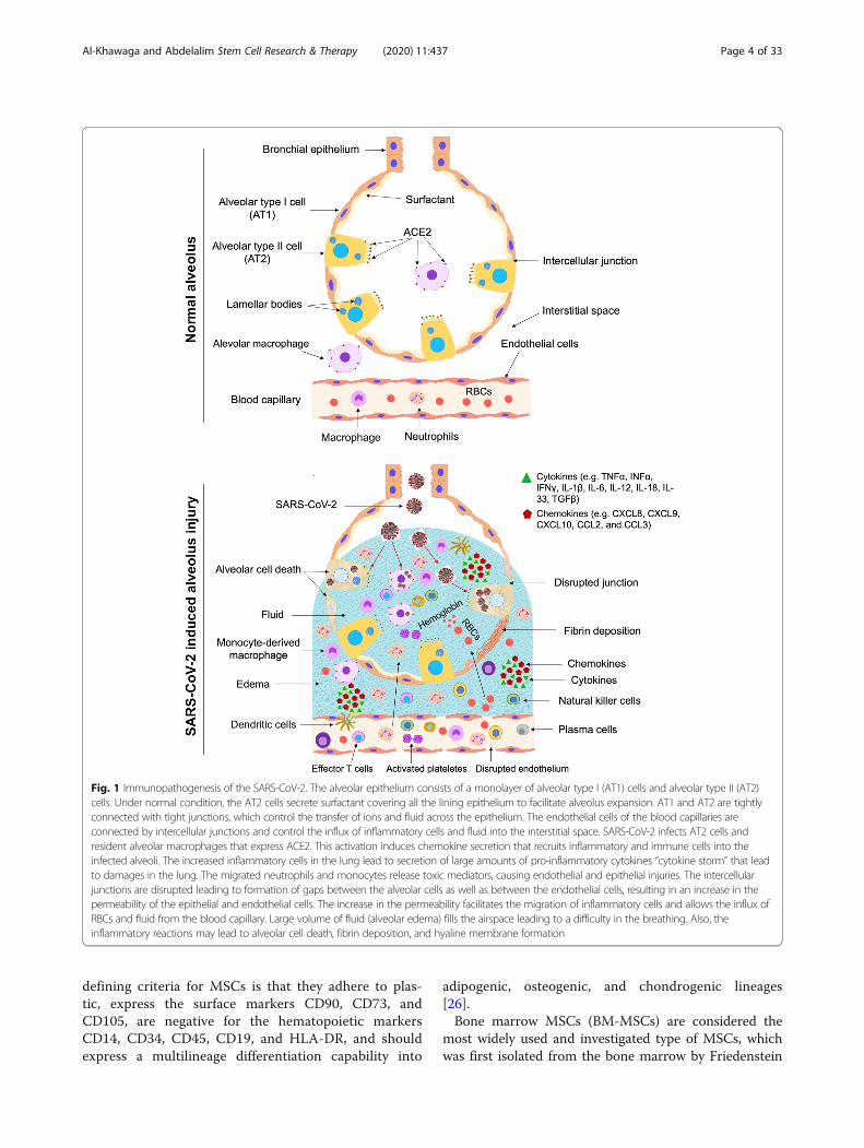
Pathogenesis of SARS-COV-2The lung alveoli are lined with the alveolar epitheliumconsisting of a monolayer of alveolar type I (AT1) cells
and alveolar type II (AT2) cells. Under normal condi-tion, the AT2 cells secrete surfactant covering all the lin-ing epithelium to facilitate alveolus expansion. AT1 andAT2 are tightly connected with tight junctions, whichcontrol the transfer of ions and fluid across the epithe-lium. The endothelial cells of the blood capillaries areconnected by intercellular junctions and control the in-flux of inflammatory cells and fluid into the interstitialspace between the aveoli. Initially, the spike glycoprotein(S protein) expressed on viral envelopes binds to theangiotensin-converting enzyme 2 (ACE2) receptor [11],a very similar structure to that of SARS; however, with a10–20 times much higher binding affinity when com-pared to the SARS S protein [12]. This binding capabilitypartially explains the high transmission of SARS-CoV-2[12]. The main target cells for SARS-CoV-2 infection areAT2 cells and resident alveolar macrophages, becausethey are expressing ACE2. SARS-CoV-2 utilizes ACE2for entry and the serine protease TMPRSS2, which isalso expressed by the alveolar cells, for S protein priming[13]. This activation induces chemokine and cytokine se-cretion that recruits inflammatory and immune cells intothe infected alveoli, followed by other waves of cytokinerelease. Activated macrophages have a significant role inhemophagocytic lymphohistiocytosis (HLH)-like cyto-kine storm during COVID-19 [14]. Secondary HLHcould be precipitated by a genetic defect in cytolyticpathways or observed in during infection, malignancy,and rheumatic disease. HLH is characterized by a pre-dominance of inflammatory cytokines and expansion oftissue macrophages displaying hemophagocytic activity[15]. Cytopenias, a state of elevated inflammatory cyto-kines or hypercytokinaemia, unremitting fever, elevatedferritin level, and multi-organ damage, are among thekey characteristics of HLH seen in seriously ill COVID-19 patients [8]. Type I interferons (IFN) and naturalkiller (NK) cells result in cytolytic immune responses,following a successful recognition of pathogen-associated molecular pattern. This serves as a firstline of defense against SARS-CoV-2 infection throughthe innate immune system. Activated cytotoxic T cellsand B cells are key players of the adaptive immunityhelping with viral clearance via destruction of virus-infected cells and antibody production, respectively.However, when the anti-viral immune response re-mains active, an aberrant and uncontrolled productionof inflammatory cytokines occurs, causing what isknown as the “cytokine storm”, leading to damage inthe pulmonary tissue [16, 17].Severely ill COVID-19 patients, especially the ones
with pneumonia, show disproportionate immune profile,with considerably lower lymphocyte counts (lymphocy-topenia) and increased concentrations of inflammatorycytokines. Among the significant inflammatory
Al-Khawaga and Abdelalim Stem Cell Research & Therapy (2020) 11:437 Page 2 of 33
interleukins (ILs) are IL2, IL-6, IL-7, IL-10 (Th2), IL-1βand IFNγ (Th1), and tumor necrosis factor (TNF) [6].Furthermore, in patients with severe symptoms, an ele-vation in granulocyte-colony stimulating factor (G-CSF),IFNγ-induced protein-10 (IP-10), macrophage inflamma-tory protein 1α (MIP-1α/CCL3), and macrophagechemoattractant protein-1 (MPC-1/CCL2) are noticed[18]. A recent study has performed a screen for 48 cyto-kines in 53 COVID-19 patients with moderate and severesymptoms recorded a dramatic increase of 14 cytokines inCOVID-19 patients in comparison to healthy individuals[19]. Of those cytokines, the increased hepatocyte growthfactor (HGF), MCP3, IP-10, monokine induced gammainterferon (MIG), and MIP1α are associated with the se-verity of the symptoms [19]. Key cells in the adaptive im-munity, such as CD4+ T cells, CD8+ T cells, and NK cellsare also decreased in severely ill patients [5]. On the otherhand, an elevation of CD14+ CD16+ monocytes, IL-17-producing CCR4+ CCR6+ CD4+ (T-helper 17/Th17) cells,perforin and granulysin-expressing cytotoxic T cells arereported. These constitute the pro-inflammatory subsetsof T cells responsible for the severe immune injury in thelungs [5].Among the histological profiles of COVID-19 are the
significant alterations in the morphology of the endothe-lial cells, which also express ACE2. These changes in-clude damage of the intercellular junctions, a loss ofattachment to the basement membrane, and cell swelling[20] (Fig. 1). The migrated neutrophils and monocyte-derived macrophages release toxic mediators, causingendothelial and epithelial injuries (Figs. 1 and 2). Theintercellular junctions are disrupted leading to formationof spaces between the alveolar cells as well as betweenthe endothelial cells, resulting in an increase in the per-meability of the epithelial and endothelial cells (Figs. 1,2, and 3). The increase in the permeability facilitates themigration of inflammatory cells and allows the influx ofRBCs and fluid from the blood capillary. Large volumeof fluid (alveolar edema) fills the airspace leading to adifficulty in the breathing. Also, the inflammatory reac-tions may lead to alveolar cell death, fibrin deposition,and hyaline membrane formation. These findings sup-port an important role of endothelial cells in the vascu-lar phase of COVID-19. Furthermore, pulmonaryintussusceptive angiogenesis and other pulmonary vas-cular lesions have been observed in autopsy specimen ofCOVID-19 patients [20].Severe respiratory illness could be a major symptom of
SARS-CoV-2 infection, because the ACE2 receptor isexpressed in the lung AT2 cells, alveolar macrophage,and capillary endothelial cells [11] (Fig. 1). The expres-sion of the ACE2 has been detected in other tissues,such as the cardiovascular, hepatic, renal, pancreatic,and the gastrointestinal tissues. This expression profile
partially explains why some infected patients not onlydevelop ARDS, but also develop other complications,such as myocardial injury (MI), arrhythmia, acute kidneyinjury (AKI), shock, multi organ failure, diabetes, and ul-timately death [21].IL-6 has an essential part in inflammatory cytokine
storm in COVID-19. IL-6-producing CD14+ CD16+ in-flammatory monocytes are significantly high [22]; there-fore, the rationale for using tocilizumab has been used inCOVID-19 patients. Tocilizumab, which is a recombin-ant humanized monoclonal antibody against the IL-6 re-ceptor, is likely to induce its antagonistic effect on IL-6-producing monocytes following activated Th1 cells inthe lung. Tocilizumab is a first drug for the treatment ofcytokine storm in COVID-19, especially in patients withmultiple comorbidities. Despite the numerous ongoingtrials assessing the safety and efficacy of tocilizumab inCOVID-19 patients, IL-6 play a role in controlling thelung inflammation and is important for the clearance ofviruses [23]. Therefore, inhibiting IL-6 raises the possi-bility of impaired viral clearance or exacerbation of lunginflammation [9].Interestingly, an abnormal coagulation profile has been
shown in COVID-19 patients during the late stage of thedisease; specifically, increased concentrations of D-dimerand other fibrin degradation products are mainly associ-ated with poor prognosis [24]. The HScore is a recom-mended evaluation as well as prognostic tool used inpatients with secondary HLH at high risk of hyperin-flammation. The score combines both critical laboratoryas well as clinical parameters, assessing for an underlyingof immunosuppression and cytopenias, measuring serumaspartate aminotransferase (AST), triglycerides, fibrino-gen, ferritin, body temperature, organomegaly, andhemophagocytosis on bone marrow aspirate [8]. TheHScores generate a probability for the presence of sec-ondary HLH; a score more than 169 is 93% sensitive and86% specific for HLH [8].Finally, since the anti-viral immunity is needed to re-
cover from COVID-19, the use of immunosuppressantson these patients should be used with caution. Onestrategy to avoid the inhibition of anti-viral immunity isto use targeted instead of broad immunosuppressivemedications. Unfortunately, we still lack consensus onthe optimal timing of treatment administration to de-crease the harmful effects of immunosuppression, as wellas the routes of their administration.
Mesenchymal stem cells (MSCs): characteristicsand typesMSCs are a heterogeneous cell population propagat-ing in vitro as plastic-adherent cells, have fibroblast-like morphology, and form colonies in vitro [25]. TheInternational Society for Cellular Therapy (ISCT)
Al-Khawaga and Abdelalim Stem Cell Research & Therapy (2020) 11:437 Page 3 of 33
defining criteria for MSCs is that they adhere to plas-tic, express the surface markers CD90, CD73, andCD105, are negative for the hematopoietic markersCD14, CD34, CD45, CD19, and HLA-DR, and shouldexpress a multilineage differentiation capability into
adipogenic, osteogenic, and chondrogenic lineages[26].Bone marrow MSCs (BM-MSCs) are considered the
most widely used and investigated type of MSCs, whichwas first isolated from the bone marrow by Friedenstein
Fig. 1 Immunopathogenesis of the SARS-CoV-2. The alveolar epithelium consists of a monolayer of alveolar type I (AT1) cells and alveolar type II (AT2)cells. Under normal condition, the AT2 cells secrete surfactant covering all the lining epithelium to facilitate alveolus expansion. AT1 and AT2 are tightlyconnected with tight junctions, which control the transfer of ions and fluid across the epithelium. The endothelial cells of the blood capillaries areconnected by intercellular junctions and control the influx of inflammatory cells and fluid into the interstitial space. SARS-CoV-2 infects AT2 cells andresident alveolar macrophages that express ACE2. This activation induces chemokine secretion that recruits inflammatory and immune cells into theinfected alveoli. The increased inflammatory cells in the lung lead to secretion of large amounts of pro-inflammatory cytokines “cytokine storm” that leadto damages in the lung. The migrated neutrophils and monocytes release toxic mediators, causing endothelial and epithelial injuries. The intercellularjunctions are disrupted leading to formation of gaps between the alveolar cells as well as between the endothelial cells, resulting in an increase in thepermeability of the epithelial and endothelial cells. The increase in the permeability facilitates the migration of inflammatory cells and allows the influx ofRBCs and fluid from the blood capillary. Large volume of fluid (alveolar edema) fills the airspace leading to a difficulty in the breathing. Also, theinflammatory reactions may lead to alveolar cell death, fibrin deposition, and hyaline membrane formation
Al-Khawaga and Abdelalim Stem Cell Research & Therapy (2020) 11:437 Page 4 of 33
and colleagues in 1974 [27]. Later, MSCs were identifiedand successfully produced from other sources, such asthe perivasculature [28], adipose [29], dental pulp [30],muscle [31], dermis [31], and fetal tissue [32]. The abun-dance of adipose-derived stem cells (ASCs), their ease ofisolation using a minimally invasive procedure [33], andtheir expansion as well as their differentiation ability intomultiple lineages make ASCs a promising less-invasivealternative to BM-MSCs for therapeutic applications [34,35]. The most commonly used adult sources for human
MSCs are bone marrow [36] and the adipose tissue stro-mal vascular fraction [29, 34, 35]. The highly harvestablebone marrow or unwanted/waste product of adiposesources forms the foundation for most of the data in thefield of MSC-based therapeutics. The umbilical cord(UC) tissue [37] and the placenta [38, 39] and their asso-ciated tissue Wharton jelly (WJ), and amniotic fluid(AF), are among the other young “adult” tissues, that arealso considered good sources of human MSCs, wherethey are normally discarded after birth.
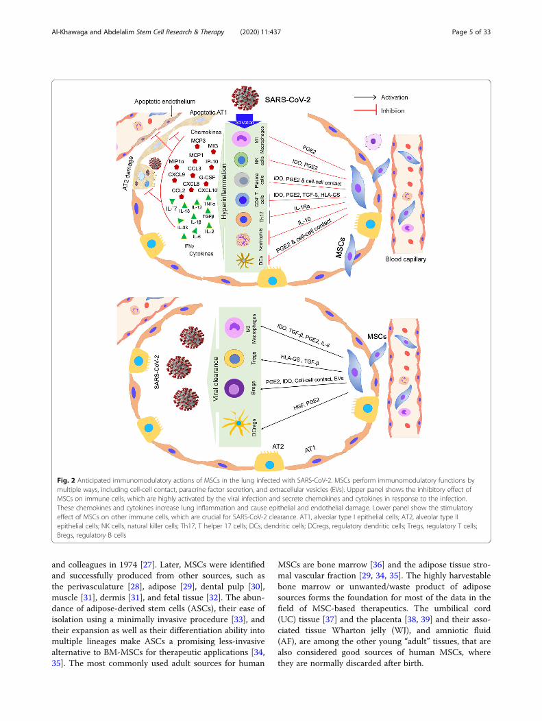
Fig. 2 Anticipated immunomodulatory actions of MSCs in the lung infected with SARS-CoV-2. MSCs perform immunomodulatory functions bymultiple ways, including cell-cell contact, paracrine factor secretion, and extracellular vesicles (EVs). Upper panel shows the inhibitory effect ofMSCs on immune cells, which are highly activated by the viral infection and secrete chemokines and cytokines in response to the infection.These chemokines and cytokines increase lung inflammation and cause epithelial and endothelial damage. Lower panel show the stimulatoryeffect of MSCs on other immune cells, which are crucial for SARS-CoV-2 clearance. AT1, alveolar type I epithelial cells; AT2, alveolar type IIepithelial cells; NK cells, natural killer cells; Th17, T helper 17 cells; DCs, dendritic cells; DCregs, regulatory dendritic cells; Tregs, regulatory T cells;Bregs, regulatory B cells
Al-Khawaga and Abdelalim Stem Cell Research & Therapy (2020) 11:437 Page 5 of 33
MSCs are generally recognized as immune evasivemaking them safe when used in allogeneic settings [40].Allogeneic MSCs are able to bypass the immune systemdue to low expression of the major histocompatibilitycomplex-1 (MHC-I) and -II proteins. MSCs are often re-ferred to as being “immunoprivileged” due to lack of theT cell costimulatory molecules, CD80 and CD86 [41].Previous studies reported that fetal MSCs, adult BM-MSCs, and ASCs express HLA-I and do not expressHLA-II [42–46]; however, these MSCs start to expressHLA-II after stimulation with IFN-γ [44, 45, 47, 48]. Arecent study demonstrated that iPSC-derived MSCs donot express HLA-II and costimulatory molecules [49].Interestingly, induced pluripotent stem cell (iPSC)-de-rived MSCs express a very low level of HLA-II in com-parison to MSCs derived from fetuses and adult sourcesafter their stimulation with interferon-γ (IFN-γ) [49].These findings present iPSC-MSCs as an efficient sourcefor allogenic transplantation without the risk of immunerejection due to the lower immunogenicity compared toadult MSCs.
Therapeutic potentials of MSCsMSCs have been extensively studied over the past ~ 30years for their wide clinical applications and regenerativecapacity. MSCs have made their way over the past 25years into now over 950 registered clinical trials listedwith the FDA, exhibiting an excellent safety profile.With over a 10,000 patients treated with MSCs in a con-trolled clinical setting, and upon successful completionof phase 1 or phase 2 trials, several tens of MSCs-basedstudies have advanced to phase 3 clinical trials (www.clinicaltrials.gov).A fundamental clinical decision remains to choose
among whether to use autologous vs allogeneic sourcesof MSCs, where both have displayed successful produc-tion of large numbers of MSCs [50, 51]. MSC replace-ment in the large numbers is needed to treat significanttissue injury, a process that further requires orchestratedsteps involving successful engraftment and cell differen-tiation [52]. A target dose of 100–150 million MSCs canbe obtained from cell culturing and expansion of 25 mlof BM-MSC aspirate. In about 3 weeks duration, a
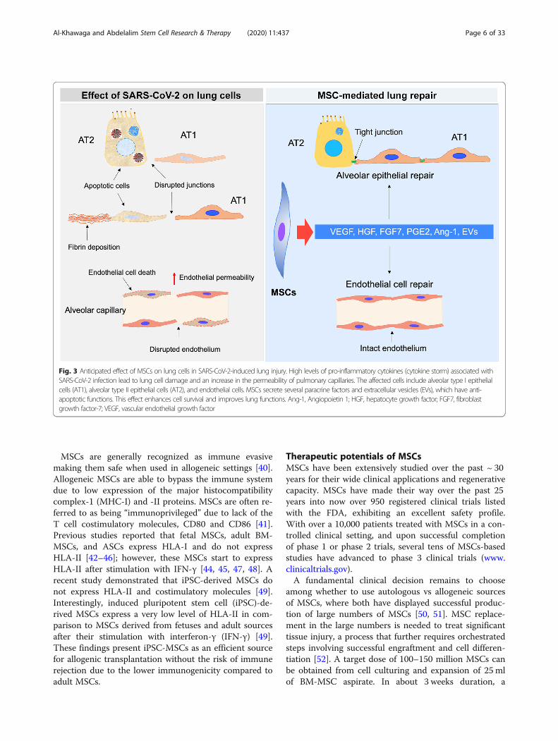
Fig. 3 Anticipated effect of MSCs on lung cells in SARS-CoV-2-induced lung injury. High levels of pro-inflammatory cytokines (cytokine storm) associated withSARS-CoV-2 infection lead to lung cell damage and an increase in the permeability of pulmonary capillaries. The affected cells include alveolar type I epithelialcells (AT1), alveolar type II epithelial cells (AT2), and endothelial cells. MSCs secrete several paracrine factors and extracellular vesicles (EVs), which have anti-apoptotic functions. This effect enhances cell survival and improves lung functions. Ang-1, Angiopoietin 1; HGF, hepatocyte growth factor; FGF7, fibroblastgrowth factor-7; VEGF, vascular endothelial growth factor
Al-Khawaga and Abdelalim Stem Cell Research & Therapy (2020) 11:437 Page 6 of 33
volume of about 0.4–0.5 ml of packed cells can be gener-ated [53]. The MSC isolation from different tissues, suchas BM and adipose tissue and their re-implantation atother sites highlight their ability to repair tissues in vivo.However, this process clearly diminishes in aging popu-lation compared to younger adults [54]. In addition,MSCs could be generated in vitro in large number fromhuman pluripotent stem cells (hPSCs) [55, 56], whichshowed a lower immunogenicity in comparison to adultsources [49].MSCs have been extensively examined for their thera-
peutic capacity in regenerative medicine, because of theirability to home to sites of inflammation and damagedtissue, ultimately serving as a source of growth andtrophic factors and regenerative molecules. The potentialtherapeutic effect of MSCs is based on their low im-munogenicity, their immunomodulatory characteristics,and their ability to secrete growth factors, as well asanti-microbial peptides [57]. MSCs administered system-ically tend to migrate to the injury region to promotefunctional recovery [58]. MSCs can also extravasate fromthe blood vessels, just like immune cells, via the expres-sion of cell surface adhesion molecules. Migration ofMSCs occur in response to chemokines binding to cog-nate receptors present on their cell surface [59] and re-sult in the stimulation of matrix metalloproteinasesdegrading the basal membrane and allowing subsequentextravasation [60]. By displaying a coordinated rolling,MSCs contact the endothelial cells in a P-selectin- andvascular cell-adhesion molecule 1 (VCAM1)-dependentmanner [61]. Guided by chemotactic signals, MSCs mi-grate through the interstitium to the injured area. An in-crease in the MSC migration capacity towardchemokines is achieved via the upregulation of their re-ceptors, CCR2, CCR3, and CCR4. Also, interleukin (IL)-8, an inflammatory chemokine, may induce migration ofMSCs to injured areas [62, 63].
Immunoregulatory functions of MSCsOne of the major therapeutic characteristics of MSCsis their immunomodulatory role, including a networkof cytokines and cell-cell interactions. Interestingly,MSCs only exert its immunoregulatory capacity afterreceiving the activation signals from the inflammatorymilieu; therefore, MSC’s immunoregulatory capacity isnot constitutive, rather is driven by “licensing”process [64]. Previous studies showed that the macro-phages play an essential role during wound healing;thus, they have emerged as key candidate targets intherapeutic tissue regeneration approaches [64, 65].Macrophages exhibit functional repolarization as tis-sue repair progresses, shifting from the pro-inflammatory or M1-phenotype to an anti-inflammatory or M2-phenotype. M1 macrophages
secrete high levels of pro-inflammatory cytokines,while M2 macrophages secrete lower levels of pro-inflammatory cytokines, exhibit tissue repair, and en-hance the resolution of inflammation [66]. Imbalancebetween M1- and M2- activities can lead to continu-ous inflammation and hinders the normal repairprocess, both contributing to impaired tissue repair[67]. MSCs enhance tissue repair and regeneration bymodulating the immune response, acting as sensorsand switchers of inflammation, rather than by re-placing damaged cells. This is largely attributed to thesecretion of growth factors; among the immunoregu-latory factors are prostaglandin E2 (PGE2) and IL-6that help in transitioning macrophages toward M2phenotype [68, 69] (Fig. 2). Further, the classical pro-inflammatory cytokines produced at the acute stage ofinflammation, such as IFN-γ, TNF-α, or IL-1β en-hances the paracrine effects of MSCs exerted on mac-rophages [70, 71].In order to stimulate the MSC immunosuppressive ef-
fect, threshold levels of inflammatory factors are re-quired. Insufficient MSC activation can lead to anincrease in the inflammation [72]. Recently, it has beenshown that IL-10 alone is insufficient to enhance MSCimmunomodulation, rather enhances the priming influ-ence of TNF-α, indicating that MSC activation by IL-10is dependent on TNF-α [64]. MSCs further decreaseTNF-α secretion via PGE2 but not IL-6, supporting theconcept that MSC immunomodulatory potential ishighly correlated to the release of PGE2 [64].Among the other MSC-derived molecules shown to
exert an immunoregulatory functions are transforminggrowth factor beta (TGF-β), hepatocyte growth factor(HGF), and indoleamine 2,3-dioxygenase (IDO) [73](Fig. 2). TGF-β secreted by MSCs could shiftlipopolysaccharide-activated macrophage polarization to-ward the M2-phenotype, decrease inflammatory reac-tions, and enhance the phagocytic activity through theAkt/FoxO1 pathway [74], while HGFs modulate IL-10production in monocytes via the ERK1/2 pathway [75].MSC IDO activity is involved in the differentiation ofmonocytes into IL-10-secreting M2 immunosuppressivemacrophages (CD14+/CD206+) [71]. These processes de-crease immune cell maturation and activation, inaddition to enhancing the differentiation of T cells intoregulatory T cells (Tregs) [52].The immunoregulatory effects of MSCs is highlighted
by the ability of BM-MSCs to suppress T cell prolifera-tion [76, 77] and suppress the conversion of monocytesand CD34+ hematopoietic progenitor cells into dendriticcells (DCs) in vitro [78–81]. Mature DCs cultured withMSCs have reduced production of IL-12 and MHC classII molecules, CD11c, CD83, though hindering the DCantigen-presenting function [78–81].
Al-Khawaga and Abdelalim Stem Cell Research & Therapy (2020) 11:437 Page 7 of 33
Anti-inflammatory and antiproliferative effects of MSCsMSCs reduce the pro-inflammatory effect of DCs by sup-pressing their secretion of TNF [82]. Also, plasmacytoidDCs (pDCs), a set of specific cells for the secretion of highlevels of type I IFN, increase the production of IL-10 fol-lowing the incubation with MSCs [82]. MSCs can furtherinhibit the cytotoxic activity of resting NK cells by redu-cing the production of natural cytotoxicity receptor 3(NKp30) and natural-killer group 2, member D (NKG2D),involved in the activation of NK cells and target cell killing[83]. Therefore, MSCs inhibit NK cell proliferation andIFN production [84, 85]. Also, neutrophils are importantcells of innate immunity, undergoing a process known asthe respiratory burst when binding to an antigen. MSCshave been reported to eliminate the respiratory burst andto prevent the neutrophil cell death by an IL-6-dependentmechanism [86]. Also, MSCs play a key role in the adap-tive immune system, where it inhibits the proliferation ofT cells activated with antigens [76]. This leads to a reduc-tion in the IFN production and an increase in IL-4 pro-duction by T helper 2 (T2) cells, indicating a change in Tcells from a pro-inflammatory (IFN-producing) to an anti-inflammatory (IL-4-producing) state [82].Furthermore, MSCs have been shown to downregulate
CD8+ cytotoxic T lymphocytes (CTL)-mediated cytotox-icity [87] and further inhibit B cell expansion in vitro.Also, MSCs can suppress B cell differentiation and theconstitutive secretion of chemokine receptors, affectedby the MSC-mediated suppression of T cell functions[88]. Furthermore, MSC-derived IDO has been shown tobe required in the inhibition of the expansion of IFN-secreting Th1 cells and, together with PGE2, to stop NKcell activity [89].
Anti-apoptotic and protective functions of MSCsSeveral pro-inflammatory molecules modulate the im-munosuppressive, trafficking, and paracrine potential ofMSCs. Enhanced paracrine potential of MSCs inducedby TNF-α, IL-1b, and nitric oxide (NO), ultimatelyincreases MSC secretions of regenerative, immunomod-ulatory, and trafficking molecules, including the key fac-tor, insulin-like growth factor 1 (IGF-1) [90]. Hemeoxygenase-1 (HO-1) is upregulated by TNF-α, IL-1α, orNO in endothelial cells or alveolar cells, where MSCsoverexpressing HO-1 showed an increase in the anti-inflammatory, anti-apoptotic, and vascular remodelingproperties [91]. Upregulation of HO-1 increases produc-tion of trophic molecules, such as FGF2, and IGF-1, andVEGF [90]. Fibroblast growth factor-10 (FGF-10), kera-tinocyte growth factor-2 (KGF-2), has been found toregulate epithelial-mesenchymal interactions that arecrucial for the development of lung [92]. FGF-10 exertsa role in lung resident-MSC propagation, mobilization,and the protective effects against acute lung injury [93].
MSCs can affect on the endothelial differentiation ofendothelial progenitor cells in vitro, mainly dependenton VEGF [94]. Human leukocyte antigen-G5 (HLA-G5)is another soluble factor secreted by MSCs and its secre-tion is IL-10-dependent. HLA-G5 is required to suppressthe function of T lymphocytes and NK cells and to acti-vate regulatory T cells [95]. Galectin-1 and 3 (Gal-1 andGal-3) as well as Semaphorin-3A (Sema-3A) are othersecreted MSC immune regulators, known for their in-hibitory activities. Gal-1 and Sema-3A are two solublefactors that can suppress T cell proliferation vianeuropilin-1 (NP-1) binding [96, 97]. MSC-derived Gal-1 significantly regulates the release of TNFα, IFNγ, IL-2,and IL-10 [97].Finally, interleukin-1 receptor antagonist (IL-1Ra) and
programmed death-1 (PDL1) are among the other se-creted regulators of MSCs. IL-1Ra is among the anti-inflammatory cytokine produced by MSCs, which can in-hibit Th17 polarization. IL-1Ra expression tends to in-crease in MSCs exposed to IL-1β, TNF-α, and IFN-γ.Th17 cells induce the upregulation of PDL1, playing amajor role in activating the MSC immunosuppressive ef-fect [98]. PDL1 further support the cell-cell contactthrough MSC-mediated inhibition on Th17 cells [98].MSC enhanced PDL1 ligand secretion suppress the activa-tion of CD4+ T cells and downregulate IL-2 secretion [99].
MSC-derived exosomesExtracellular vesicle (EV) is a term including both exo-somes and microvesicles (MVs). The exosome diameteris less than 200 nm, while MV diameter can reach up to1000 nm. The secretomes of MSCs and their vesiclesoffer a powerful tool for cell-free therapy due to theirparacrine and/or endocrine effects [100]. This strategybypasses most of the safety concerns related to cell-based therapy, such as contamination with oncogeniccells and continuous cell proliferation [101]. The keyfeatures of MSC-derived EVs are (1) non-proliferative,which reduce the risk of tumor formation; (2) negativefor HLA-I and HLA-II, which can be induced, andtherefore, they can be used from other individuals with-out any risk of immune response; (3) small in size allow-ing them to pass from the small blood capillaries; and(4) stored without using DMSO, which may change theircharacteristics [102]. EVs bind to a receptor on the cellmembrane of the targeted cells, where they merge withthe membrane to secrete the EV contents inside the cellor enter into the cytoplasm in the form of endocytic ves-icles [103].EVs have proposed as an effective vehicle for deliver-
ing miRNAs, which control above 60% of the mRNAs;therefore, transferring them in EVs is of clinical signifi-cance [102]. MSC engineering is one way miRNA couldbe loaded into EVs and still exert its therapeutic effects
Al-Khawaga and Abdelalim Stem Cell Research & Therapy (2020) 11:437 Page 8 of 33
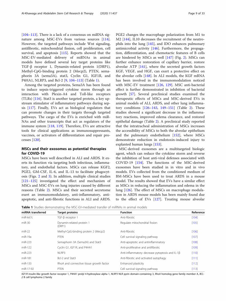
[104–113]. There is a lack of a consensus on miRNA sig-nature among MSC-EVs from various sources [114].However, the targeted pathways include Wnt signaling,antifibrotic, mitochondrial fission, cell proliferation, cellsurvival, and apoptosis [115]. Reports showed that theMSC-EV-mediated delivery of miRNAs in animalmodels have defined several key target proteins likeTGF-β receptor 1, Dynamin-related protein 1(DRP1),Methyl-CpG-binding protein 2 (Mecp2), PTEN, sema-phorin 3A (sema3A), stat3, Cyclin G1, IGF1R, andP4HA1, NLRP3, and Bcl-2 [9, 104–113] (Table 1).Among the targeted proteins, Sema3A has been found
to induce sepsis-triggered cytokine storm through aninteraction with Plexin-A4 and Toll-like receptors(TLRs) [116]. Stat3 is another targeted protein, a key up-stream stimulator of inflammatory pathways during sep-sis [117]. Finally, EVs act as biological regulators thatcan promote changes in their targets through targetedpathways. The cargo of the EVs is enriched with miR-NAs and other transcripts that act as regulators of theimmune system [118, 119]. Therefore, EVs are attractivetools for clinical applications as immunosuppressants,vaccines, or activators of differentiation and repair pro-cesses [120].
MSCs and their exosomes as potential therapiesfor COVID-19MSCs have been well described in ALI and ARDS. It ex-erts its function via targeting both infectious, inflamma-tory, and endothelial factors. MSCs can release KGF2,PGE2, GM-CSF, IL-6, and IL-13 to facilitate phagocyt-osis (Figs. 2 and 3). In addition, multiple clinical studies[121–125] investigated the effect and mechanism ofMSCs and MSC-EVs on lung injuries caused by differentreasons (Table 2). MSCs and their secreted secretomeexert an immunomodulatory, anti-inflammatory, anti-apoptotic, and anti-fibrotic functions in ALI and ARDS.
PGE2 changes the macrophage polarization from M1 toM2 [144], IL10 decreases the recruitment of the neutro-phils into the lung [145], and IDO enhances pulmonaryantimicrobial activity [146]. Furthermore, the propaga-tion, differentiation, and chemotactic features of B cellsare hindered by MSCs as well [147] (Fig. 2). MSCs canfurther enhance restoration of capillary barrier, restorealveolar ATP [141], where the secreted growth factorsKGF, VEGF, and HGF, can exert a protective effect onthe alveolar cells [148]. In ALI models, the KGF mRNAhas been involved in the immunomodulation noticedwith MSC-EV treatment [126, 129]. MSC anti-bacterialeffect is further demonstrated in inhibition of bacterialgrowth [57]. Several preclinical studies examined thetherapeutic effects of MSCs and MSC-derived EVs inanimal models of ALI, ARDS, and other lung inflamma-tory conditions [126–143, 149–151] (Table 2). Thesestudies showed a significant decrease in the inflamma-tory reactions, improved edema clearance, and restoredepithelial damage (Table 2). A preclinical study reportedthat the intratracheal administration of MSCs increasesthe accessibility of MSCs to both the alveolar epitheliumand the pulmonary endothelium [152], where MSCsdemonstrate reduction in endotoxin-induced injury toexplanted human lungs [153].MSC-derived exosomes are a multitargeted biologic
agent, which can reduce the cytokine storm and reversethe inhibition of host anti-viral defenses associated withCOVID-19 [154]. The functions of the MSC-derivedexosomes have been studied in in vitro and in vivomodels. EVs collected from the conditioned medium ofBM-MSCs have been used to treat ARDS in a mousemodel. The results showed that EVs have a similar effectas MSCs in reducing the inflammation and edema in thelung [126]. The effect of MSCs on macrophage modula-tion in ARDS mouse models has been mainly found dueto the effect of EVs [127]. Treating mouse alveolar
Table 1 Studies demonstrating the MSC-EV-mediated transfer of miRNAs in animal models
miRNA transferred Target proteins Function Reference
miR-let7c TGF-β receptor 1 Anti-fibrotic [104]
miR-30 Dynamin-related protein 1(DRP1)
Regulate mitochondrial fission [105]
miR-22 Methyl-CpG-binding protein 2 (Mecp2) Anti-fibrotic [106]
miR-19a PTEN Cell survival signaling pathway [107]
miR-223 Semaphorin 3A (Sema3A) and Stat3 Anti-apoptotic and antiinflammatory [108]
miR-122 Cyclin G1, IGF1R, and P4HA1 Anti-proliferative and antifibrotic [109]
miR-223 NLRP3 Anti-inflammatory: decrease pytoptosis and IL-1β [110]
miR-181 Bcl-2 and Stat3 Anti-fibrotic and activated autophagy [111]
miR-133 RhoA and connective tissue growth factor Enhanced plasticity [112]
miR-17-92 PTEN Cell survival signaling pathway [113]
IGF1R insulin-like growth factor receptor 1, P4HA1 prolyl 4-hydroxylase alpha 1, NLRP3 NLR pyrin domain-containing 3, RhoA homolog gene family member A, BCL-2 B cell lymphoma 2 family
Al-Khawaga and Abdelalim Stem Cell Research & Therapy (2020) 11:437 Page 9 of 33
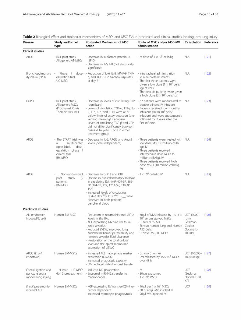
Table 2 Biological effect and molecular mechanisms of MSCs and MSC-EVs in preclinical and clinical studies looking into lung injury
Disease Study and/or celltype
Postulated Mechanism of MSCaction
Route of MSC and/or MSC-MVadministration
EV isolation Reference
Clinical studies
ARDS - RCT pilot study- Allogeneic AT-MSCs
- Decrease in surfactant protein D(SP-D)
- Decrease in Il-6, Il-8 (not statisticallysignificant)
- IV dose of 1 × 10⁶ cells/kg N.A. [121]
Bronchopulmonarydysplasia (BPD)
- Phase I dose-escalation trial
- UC-MSCs
- Reduction of IL-6, IL-8, MMP-9, TNF-α, and TGF-β1 in tracheal aspiratesat day 7
- Intratracheal administration- In nine preterm infants.- The first three patients weregiven a low dose (1 × 10⁷ cells/kg) of cells
- The next six patients were givena high dose (2 × 10⁷ cells/kg)
N.A. [122]
COPD - RCT pilot study- Allogeneic MSCs(Prochymal; OsirisTherapeutics Inc.)
- Decrease in levels of circulating CRP(significant)
- Levels of circulating TNF-α, IFN-γ, IL-2, IL-4, IL-5, and IL-10 were at orbelow limits of assay detection (pre-venting meaningful analysis)
- Levels of circulating TGF-β and CRPdid not differ significantly betweenbaseline to years 1 or 2 in eithertreatment group
- 62 patients were randomized todouble-blinded IV infusions
- Patients received four monthlyinfusions (100 × 106 cells/infusion) and were subsequentlyfollowed for 2 years after thefirst infusion
N.A. [123]
ARDS - The START trial wasa multi-center,open-label, dose-escalation phase 1clinical trial
- BM-MSCs
- Decrease in IL-6, RAGE, and Ang-2levels (dose-independent)
- Three patients were treated withlow dose MSCs (1million cells/kg), IV
- Three patients receivedintermediate dose MSCs (5million cells/kg), IV
- Three patients received highdose MSCs (10 million cells/kg,IV)
N.A. [124]
ARDS - Non-randomized,pilot study (2patients)
- BM-MSCs
- Decrease in ccK18 and K18- Decline in pro-inflammatory miRNAsin circulating EVs (miR-409-3P, 886-5P, 324-3P, 222, 125A-5P, 339-3P,155)
- Increased levels of circulatingCD4+CD25highCD127low TRegs wereobserved in both patients’peripheral blood
- 2 × 10⁶ cells/kg IV N.A. [125]
Preclinical studies
ALI (endotoxininduced/E. coli)
Human BM-MSC - Reduction in neutrophils and MIP-2levels in the BAL
- KGF-expressing MV transfer to in-jured alveolus
- Reduced EVLW, improved lungendothelial barrier permeability andrestored alveolar fluid clearance
- -Restoration of the total cellularlevel and the apical membraneexpression of αENaC
- 30 μl of MVs released by 1.5–3 ×10⁶ serum starved MSCs
- IT and IV routes- Ex vivo human lung and HumanAT2 Cells.
- IT dose: 750,000 MSCs
UCF (3000rpm/BeckmanCoulterOptima L-100XP)
[126]
ARDS (E. coliendotoxin)
Human BM-MSCs - Increased M2 macrophage markerexpression (CD206)
- increased phagocytic capacity- EV-mediated mitochondrial transfer
- Ex vivo (murine)- EVs released by 15 × 106 MSCsover 48 h
UCF (10,000–100,000 xg)
[127]
Caecal ligation andpuncture sepsismodel (lung injury)
- Human UC-MSCs(IL-1β pretreatment)
- Induced M2 polarization- Exosomal miR-146a transfer tomacrophages
- IV- 30 μg exosomes- 1 × 10⁶ MSCs
UCF(BeckmanOptima L-80XP)
[128]
E. coli pneumonia-induced ALI
Human BM-MSCs - KGF-expressing EV transfer/CD44 re-ceptor dependent
- Increased monocyte phagocytosis
- 10 μl per 1 × 10⁶ MSCs- 30 or 60 μl MV, instilled IT- 90 μl MV, injected IV
UCF [129]
Al-Khawaga and Abdelalim Stem Cell Research & Therapy (2020) 11:437 Page 10 of 33
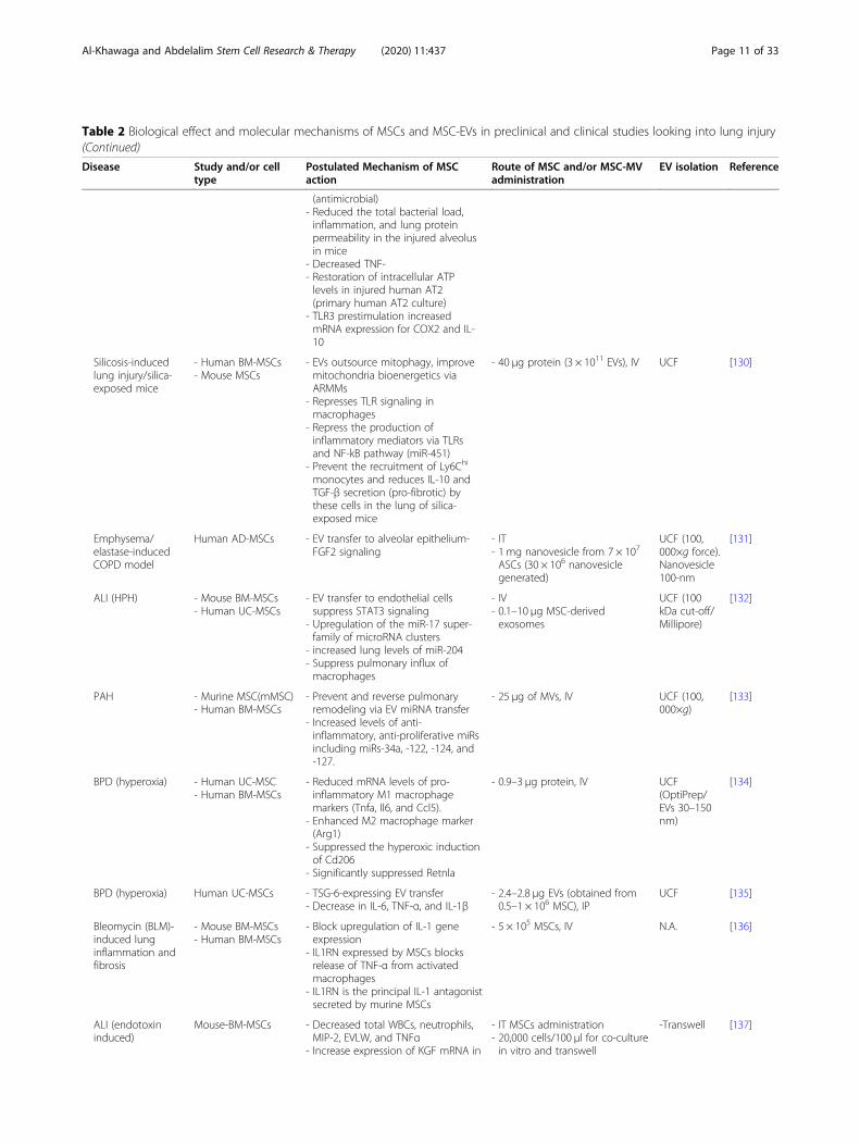
Table 2 Biological effect and molecular mechanisms of MSCs and MSC-EVs in preclinical and clinical studies looking into lung injury(Continued)
Disease Study and/or celltype
Postulated Mechanism of MSCaction
Route of MSC and/or MSC-MVadministration
EV isolation Reference
(antimicrobial)- Reduced the total bacterial load,inflammation, and lung proteinpermeability in the injured alveolusin mice
- Decreased TNF-- Restoration of intracellular ATPlevels in injured human AT2(primary human AT2 culture)
- TLR3 prestimulation increasedmRNA expression for COX2 and IL-10
Silicosis-inducedlung injury/silica-exposed mice
- Human BM-MSCs- Mouse MSCs
- EVs outsource mitophagy, improvemitochondria bioenergetics viaARMMs
- Represses TLR signaling inmacrophages
- Repress the production ofinflammatory mediators via TLRsand NF-kB pathway (miR-451)
- Prevent the recruitment of Ly6Chi
monocytes and reduces IL-10 andTGF-β secretion (pro-fibrotic) bythese cells in the lung of silica-exposed mice
- 40 μg protein (3 × 10¹¹ EVs), IV UCF [130]
Emphysema/elastase-inducedCOPD model
Human AD-MSCs - EV transfer to alveolar epithelium-FGF2 signaling
- IT- 1 mg nanovesicle from 7 × 10⁷
ASCs (30 × 106 nanovesiclegenerated)
UCF (100,000×g force).Nanovesicle100-nm
[131]
ALI (HPH) - Mouse BM-MSCs- Human UC-MSCs
- EV transfer to endothelial cellssuppress STAT3 signaling
- Upregulation of the miR-17 super-family of microRNA clusters
- increased lung levels of miR-204- Suppress pulmonary influx ofmacrophages
- IV- 0.1–10 μg MSC-derivedexosomes
UCF (100kDa cut-off/Millipore)
[132]
PAH - Murine MSC(mMSC)- Human BM-MSCs
- Prevent and reverse pulmonaryremodeling via EV miRNA transfer
- Increased levels of anti-inflammatory, anti-proliferative miRsincluding miRs-34a, -122, -124, and-127.
- 25 μg of MVs, IV UCF (100,000×g)
[133]
BPD (hyperoxia) - Human UC-MSC- Human BM-MSCs
- Reduced mRNA levels of pro-inflammatory M1 macrophagemarkers (Tnfa, Il6, and Ccl5).
- Enhanced M2 macrophage marker(Arg1)
- Suppressed the hyperoxic inductionof Cd206
- Significantly suppressed Retnla
- 0.9–3 μg protein, IV UCF(OptiPrep/EVs 30–150nm)
[134]
BPD (hyperoxia) Human UC-MSCs - TSG-6-expressing EV transfer- Decrease in IL-6, TNF-α, and IL-1β
- 2.4–2.8 μg EVs (obtained from0.5–1 × 10⁶ MSC), IP
UCF [135]
Bleomycin (BLM)-induced lunginflammation andfibrosis
- Mouse BM-MSCs- Human BM-MSCs
- Block upregulation of IL-1 geneexpression
- IL1RN expressed by MSCs blocksrelease of TNF-α from activatedmacrophages
- IL1RN is the principal IL-1 antagonistsecreted by murine MSCs
- 5 × 10⁵ MSCs, IV N.A. [136]
ALI (endotoxininduced)
Mouse-BM-MSCs - Decreased total WBCs, neutrophils,MIP-2, EVLW, and TNFα
- Increase expression of KGF mRNA in
- IT MSCs administration- 20,000 cells/100 μl for co-culturein vitro and transwell
-Transwell [137]
Al-Khawaga and Abdelalim Stem Cell Research & Therapy (2020) 11:437 Page 11 of 33
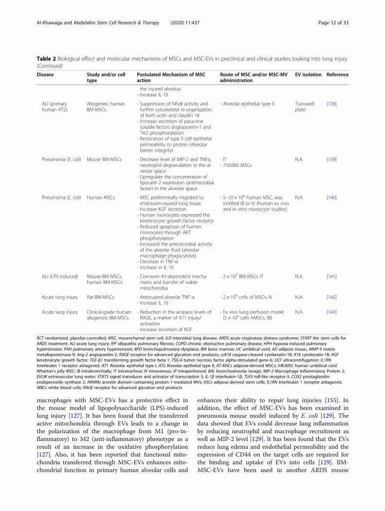
macrophages with MSC-EVs has a protective effect inthe mouse model of lipopolysaccharide (LPS)-inducedlung injury [127]. It has been found that the transferredactive mitochondria through EVs leads to a change inthe polarization of the macrophage from M1 (pro-in-flammatory) to M2 (anti-inflammatory) phenotype as aresult of an increase in the oxidative phosphorylation[127]. Also, it has been reported that functional mito-chondria transferred through MSC-EVs enhances mito-chondrial function in primary human alveolar cells and
enhances their ability to repair lung injuries [155]. Inaddition, the effect of MSC-EVs has been examined inpneumonia mouse model induced by E. coli [129]. Thedata showed that EVs could decrease lung inflammationby reducing neutrophil and macrophage recruitment aswell as MIP-2 level [129]. It has been found that the EVsreduce lung edema and endothelial permeability and theexpression of CD44 on the target cells are required forthe binding and uptake of EVs into cells [129]. BM-MSC-EVs have been used in another ARDS mouse
Table 2 Biological effect and molecular mechanisms of MSCs and MSC-EVs in preclinical and clinical studies looking into lung injury(Continued)
Disease Study and/or celltype
Postulated Mechanism of MSCaction
Route of MSC and/or MSC-MVadministration
EV isolation Reference
the injured alveolus- Increase IL-10
ALI (primaryhuman AT2)
Allogeneic humanBM-MSCs
- Suppression of NFκB activity andfurther cytoskeletal re-organizationof both actin and claudin 18
- Increase secretion of paracrinesoluble factors angiopoietin-1 andTie2 phosphorylation
- Restoration of type II cell epithelialpermeability to protein (Alveolarbarrier integrity)
- Alveolar epithelial type II Transwellplate
[138]
Pneumonia (E. coli) Mouse BM-MSCs - Decrease level of MIP-2 and TNFα,neutrophil degranulation in the al-veolar space
- Upregulate the concentration oflipocalin 2 expression (antimicrobialfactor) in the alveolar space
- IT- 750,000 MSCs
N.A. [139]
Pneumonia (E. coli) Human MSCs - MSC preferentially migrated toendotoxin-injured lung tissue
- Increase KGF secretion- Human monocytes expressed thekeratinocyte growth factor receptor
- Reduced apoptosis of humanmonocytes through AKTphosphorylation
- Increased the antimicrobial activityof the alveolar fluid (alveolarmacrophage phagocytosis).
- Decrease in TNF-α- Increase in IL-10
- 5–10 × 106 human MSC, wasinstilled IB or IV (human ex vivoand in vitro monocyte studies)
N.A. [140]
ALI (LPS-induced) Mouse-BM-MSCs,human BM-MSCs
- Connexin 43-dependent mecha-nisms and transfer of viablemitochondria
- 2 × 10⁵ BM-MSCs IT N.A. [141]
Acute lung injury Rat-BM-MSCs - Attenuated alveolar TNF α- Increase IL 10
- 2 × 10⁶ cells of MSCs, IV N.A. [142]
Acute lung injury Clinical-grade humanallogeneic-BM-MSCs
- Reduction in the airspace levels ofRAGE, a marker of AT1 injury/activation
- Increase secretion of KGF
- Ex vivo lung perfusion model(5 × 10⁶ cells hMSCs, IB)
N.A. [143]
RCT randomized, placebo-controlled; MSC, mesenchymal stem cell; ILD interstitial lung disease; ARDS acute respiratory distress syndrome; START the stem cells forARDS treatment; ALI acute lung injury; IPF idiopathic pulmonary fibrosis; COPD chronic obstructive pulmonary disease; HPH hypoxia-induced pulmonaryhypertension; PAH pulmonary artery hypertension; BPD bronchopulmonary dysplasia; BM bone marrow; UC umbilical cord; AD adipose tissue;, MMP-9 matrixmetalloproteinase-9; Ang-2 angiopoeitin-2; RAGE receptor for advanced glycation end products; ccK18 caspase-cleaved cytokeratin-18; K18 cytokeratin-18; KGFkeratinocyte growth factor; TGF-β1 transforming growth factor beta 1; TSG-6 tumor necrosis factor alpha-stimulated gene-6; UCF ultracentrifugation; IL1RNinterleukin 1 receptor antagonist; AT1 Alveolar epithelial type I; AT2 Alveolar epithelial type II; AT-MSCs adipose-derived MSCs; hWJMSC human umbilical cordWharton’s jelly MSC; IB intrabronchially; IT intratracheal; IV intravenous; IP intraperitoneal; BAL bronchoalveolar lavage; MIP-2 Macrophage Inflammatory Protein 2;EVLW extravascular lung water; STAT3 signal transducer and activator of transcription 3; IL-1β interleukin-1β; TLR3 toll-like receptor-3; COX2 prostaglandin-endoperoxide synthase 2; ARMMs arrestin domain-containing protein 1-mediated MVs; ASCs adipose-derived stem cells; IL1RN interleukin 1 receptor antagonist;WBCs white blood cells; RAGE receptor for advanced glycation end products
Al-Khawaga and Abdelalim Stem Cell Research & Therapy (2020) 11:437 Page 12 of 33
model induced by LPS from Pseudomonas aeruginosa[151]. Tang et al. reported that the EV-mediated transferof angiopoietin-1 (Ang1) mRNA is important for inflam-mation reduction, endothelial cell protection, and barrierrepair through decreasing neutrophil influx and MIP-2level [151]. Furthermore, EVs exert an immunomodula-tory function in the macrophage by inhibiting the secre-tion of TNF-α and enhancing the secretion of IL-10[151]. In a pig model, the influence of MSC-EVs on in-fluenza virus-induced ARDS has been investigated [155].Administration of EVs has been found to decrease theinfluenza virus replication, pro-inflammatory cytokines,and alveolar cell death in pigs through the transfer ofRNA [155]. UC-MSC-EVs have been also used in a ro-dent model. The study found that the UC-EVs are ef-fective in reducing ALI and the EVs primed with INF-γare more efficient than normal EVs in improving ALI[156]. This indicates that EVs isolated from differentsource could be used for lung injury. Interestingly, theprimed EVs have been found to be larger in size thannormal EVs; however, the mechanism of this size in-crease remains unclear [156]. Previous studies usedin vitro human injured lungs to investigate the effect ofMSC-EVs. MSC-EVs restore fluid clearance and reduceedema in human injured lungs in vitro [143, 157]. An-other study examined the effect of EVs on human lungswith pneumonia induced by E. coli found that EVs re-duce the permeability of lung protein and enhance al-veolar fluid clearance [158]. Barrier properties of the ofhuman lung endothelial cells injured with TNF-α, IFN-γ,and IL-1β are restored with EVs. This improvement isdue to an increase in the levels of Ang-1 in the injuredendothelium, treated with EVs [159]. Although there arepromising results obtained from using MSC-EVs in lunginjury, more mechanistic studies are needed to improveour understanding on the molecular mechanisms in-volved in EV effect.MSCs could act upon two ways in the novel COVID19
treatment, namely via its immunomodulatory effects anddifferentiation ability. MSCs display numerous advan-tages of relevance to ALI and ARDS. Although progressin the management of ALI/ARDS depends on improve-ments in supportive measures, ultimately decreasing themortality rates [160], the failure of pharmacologic treat-ments indicate the need to consider new strategies forALI/ARDS. MSC possible therapeutic potential is attrib-uted to their accessible derivation from several adult tis-sues, their low immunogenicity, indicating that theycould be given allogeneically [161], and their relativeease of isolation and expansion ability in culture. In caseof COVID-19 patients, autologous and allogenic MSCtransplantation could be applied, because MSCs do notexpress ACE2 and TMPRSS2; therefore, patient’s ownMSCs cannot be infected by SARS-CoV2 [162].
However, the negative effects caused by the SARS-CoV2infection on the blood cells and different organs may in-fluence the ability to isolate autologous MSCs of highquality and sufficient number to treat the same patient.Taken together with the low immunogenicity of MSCsand the complications associated with the SARS-CoV2infection, using allogenic MSCs is the method of choicefor COVID-19 patients.Resolution of ALI/ARDS in COVID19 is hindered by
the disruption of the epithelial barrier that suppresses al-veolar fluid clearance and depletes surfactant [163].MSC capacity to aid in restoring epithelial and endothe-lial function by differentiating MSCs into these cell typesor by secreting paracrine and trophic factors to increaserestoration of the lung tissue offers a promise for treat-ment of ALI/ARDS in COVID19. MSCs have beenwidely studies in other inflammatory conditions, wherethey demonstrated a reduction in injury and/or en-hanced restoration of function in the kidney [164, 165],liver [166], and heart [167]. MSC immunomodulatoryproperties exhibit a promise for treating ALI/ARDS inCOVID19 via their ability to ‘reprogramme’ the immuneresponse to decrease the destructive inflammatory com-ponents, while maintaining the host response to infec-tions, in addition to enhancing the repair and resolutionof lung injury by acting an effector for tissueregeneration.
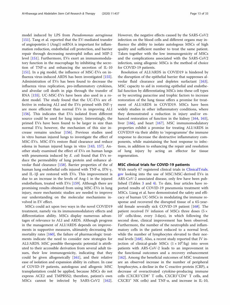
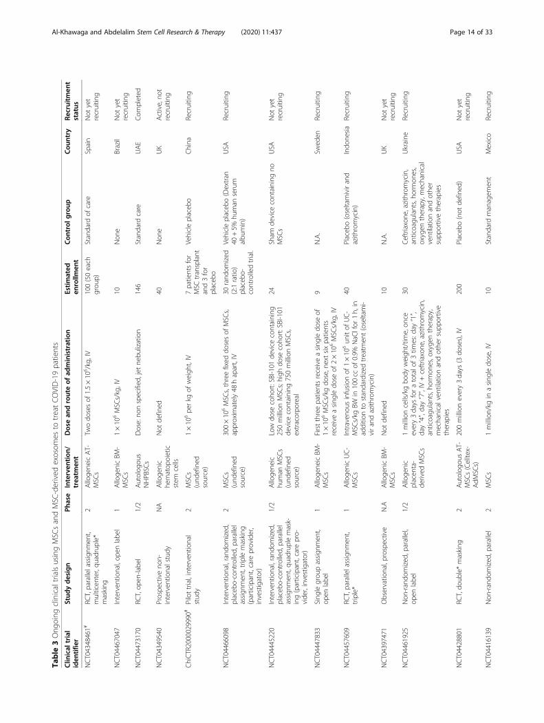
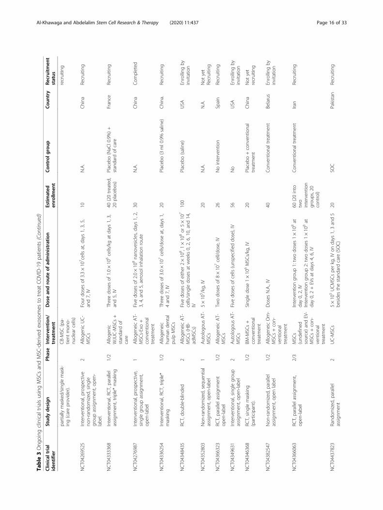
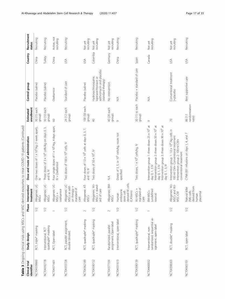
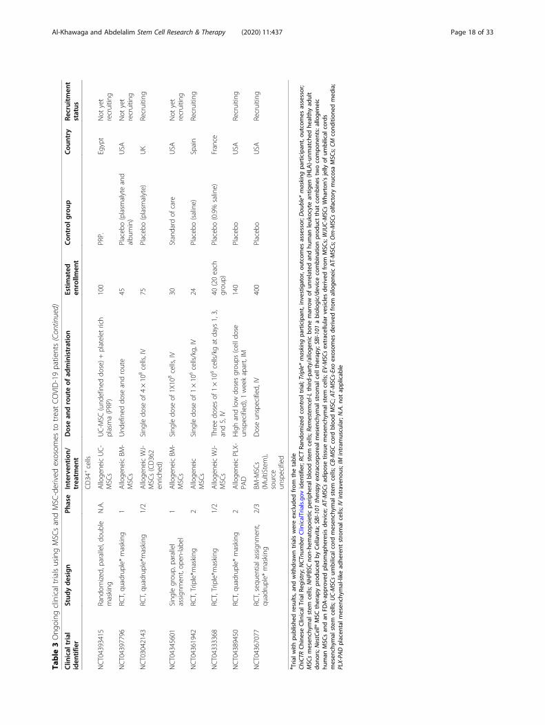
MSC clinical trials for COVID-19 patientsWith nearly 67 registered clinical trials in ClinicalTrials.gov looking into the use of MSC/MSC-derived EVs inARS-CoV-2 associated disease, only few have been pub-lished (Tables 3 and 4). To date, four articles have re-ported results of COVID-19 pneumonia treatment withMSCs. Liang et al. have demonstrated the safety and effi-cacy of human UC-MSCs in modulating the immune re-sponse and recovered the disrupted tissue of a 65-year-old female severally sick COVID-19 patient [168]. Thepatient received IV infusion of MSCs three doses (5 ×10⁷ cells/dose, every 3 days), in which following thesecond dose, clinical improvement has been observed.Furthermore, the number of the neutrophils and inflam-matory cells in the patient reduced to a normal level,while the number of lymphocytes elevated to their nor-mal levels [168]. Also, a recent study reported that IV in-jection of clinical-grade MSCs (1 × 10⁶/kg) into sevenpatients with ARS-CoV-2 leads to an improvement inthe functional outcomes and a recovery enhancement[162]. Among the beneficial outcomes of MSC treatmentare an observed increase in the number of peripherallymphocytes, a decline in the C-reactive protein (CRP), adecrease of overactivated cytokine-producing immunecells (CXCR3+CD8+ T cells, CXCR3+CD4+ T cells, andCXCR3+ NK cells) and TNF-α, and increase in IL-10,
Al-Khawaga and Abdelalim Stem Cell Research & Therapy (2020) 11:437 Page 13 of 33
Table
3Ong
oing
clinicaltrialsusingMSC
sandMSC
-derived
exosom
esto
treatCOVID-19patients
Clin
ical
trial
iden
tifie
rStud
ydesign
Phase
Interven
tion
/trea
tmen
tDosean
drouteof
administration
Estimated
enrollm
ent
Con
trol
group
Cou
ntry
Recruitm
ent
status
NCT04348461#
RCT,parallelassignm
ent,
multicen
ter,qu
adruple*
masking
2Alloge
neicAT-
MSC
sTw
odo
sesof
1.5×10
6 /kg,IV
100(50each
grou
p)Standard
ofcare
Spain
Not
yet
recruitin
g
NCT04467047
Interven
tional,op
enlabe
l1
Alloge
nicBM
-MSC
s1×10
6MSC
s/kg,IV
10Non
eBrazil
Not
yet
recruitin
g
NCT04473170
RCT,op
en-labe
l1/2
Autolog
ous
NHPBSC
sDose:no
nspecified
,jet
nebu
lization
146
Standard
care
UAE
Com
pleted
NCT04349540
Prospe
ctiveno
n-interven
tionalstudy
NA
Alloge
nic
hematop
oietic
stem
cells
Not
defined
40Non
eUK
Active,no
trecruitin
g
ChiCT
R2000029990#
Pilottrial,interven
tional
stud
y2
MSC
s(und
efined
source)
1×10
6pe
rkg
ofweigh
t,IV
7patientsfor
MSC
transplant
and3for
placeb
o
Vehicleplaceb
oChina
Recruitin
g
NCT04466098
Interven
tional,rand
omized
,placeb
o-controlled,
parallel
assign
men
t,triplemasking
(participant,care
provider,
investigator)
2MSC
s(und
efined
source)
300×10
6MSC
s,threefixed
dosesof
MSC
s,approxim
ately48
hapart,IV
30rand
omized
(2:1ratio
)placeb
o-controlledtrial.
Vehicleplaceb
o(Dextran
40+5%
human
serum
albu
min)
USA
Recruitin
g
NCT04445220
Interven
tional,rand
omized
,placeb
o-controlled,
parallel
assign
men
t,qu
adruplemask-
ing(participant,care
pro-
vide
r,investigator)
1/2
Alloge
neic
human
MSC
s(und
efined
source)
Low
dose
coho
rt:SBI-101
device
containing
250millionMSC
s;high
dose
coho
rt:SBI-101
device
containing
750millionMSC
s,extracorpo
real
24Sham
device
containing
noMSC
sUSA
Not
yet
recruitin
g
NCT04447833
Sing
legrou
passign
men
t,op
enlabe
l1
Alloge
neicBM
-MSC
sFirstthreepatientsreceiveasing
ledo
seof
1×10
6MSC
s/kg
dose,nextsixpatients
receiveasing
ledo
seof
2×10
6MSC
s/kg,IV
9N.A.
Swed
enRecruitin
g
NCT04457609
RCT,parallelassignm
ent,
triple*
1Alloge
nicUC-
MSC
sIntraven
ousinfusion
of1×10
6un
itof
UC-
MSC
s/kg
BWin
100cc
of0.9%
NaC
lfor
1h,in
additio
nto
standardized
treatm
ent(oseltami-
virandazith
romycin)
40Placeb
o(oseltamivirand
azith
romycin)
Indo
nesia
Recruitin
g
NCT04397471
Observatio
nal,prospe
ctive
N.A
Alloge
nicBM
-MSC
sNot
defined
10N.A.
UK
Not
yet
recruitin
g
NCT04461925
Non
-rando
mized
,parallel,
open
labe
l1/2
Alloge
nic
placen
ta-
derived
MSC
s
1millioncells/kgbo
dyweigh
t/tim
e,on
ceevery3days
foratotalo
f3tim
es:d
ay“1”,
day“4”,day“7”,IV
+ceftriaxone
,azithromycin,
anticoagu
lants,ho
rmon
es,oxyge
ntherapy,
mechanicalven
tilationandothe
rsupp
ortive
therapies
30Ceftriaxone
,azithromycin,
anticoagu
lants,ho
rmon
es,
oxygen
therapy,mechanical
ventilatio
nandothe
rsupp
ortivetherapies
Ukraine
Recruitin
g
NCT04428801
RCT,do
uble*masking
2Autolog
ousAT-
MSC
s(Celltex-
AdM
SCs)
200millionevery3days
(3do
ses),IV
200
Placeb
o(not
defined
)USA
Not
yet
recruitin
g
NCT04416139
Non
-rando
mized
,parallel
2MSC
s1million/kg
inasing
ledo
se,IV
10Standard
managem
ent
Mexico
Recruitin
g
Al-Khawaga and Abdelalim Stem Cell Research & Therapy (2020) 11:437 Page 14 of 33
Table
3Ong
oing
clinicaltrialsusingMSC
sandMSC
-derived
exosom
esto
treatCOVID-19patients(Con
tinued)
Clin
ical
trial
iden
tifie
rStud
ydesign
Phase
Interven
tion
/trea
tmen
tDosean
drouteof
administration
Estimated
enrollm
ent
Con
trol
group
Cou
ntry
Recruitm
ent
status
assign
men
t,op
enlabe
l(und
efined
source/from
bank)
measures
NCT04429763
RCT,paralleld
esign,triple*
masking
2Alloge
nicUC-
MSC
s1×10
6cells/kgsing
ledo
se,IV
30Placeb
o(not
defined
)Colom
bia
Not
yet
recruitin
g
NCT04444271
Interven
tional,RC
T,parallel
design
,ope
nlabe
l2
Autolog
ous
BM-M
SCs
2×10
6cells/kgon
days
1and7(if
need
ed),
IV20 (20each
grou
p)
Placeb
o(100
mln
ormal
salineIV)
Pakistan
Recruitin
g
NCT04456361
Interven
tional,sing
legrou
passign
men
t,op
enlabe
l1
WJUC-M
SCs
Sing
le-doseof
1×10
8cells,IV
9N.A.
Mexico
Active,no
trecruitin
g
NCT04366271
Rand
omized
,interventional,
paralleld
esign,op
enlabe
l2
Alloge
neicUC-
MSC
sSing
leIV
infusion
MSC
s(doseun
specified
)+standard
ofcare
106
N.A.
Spain
Recruitin
g
NCT04371393
RCT,paralleld
esign,triple*
masking
3Alloge
nicBM
-MSC
s(Rem
es-
temcel-L)+
standard
ofcare
Twodo
sesof
2×10
6MSC
/kg(doses
4days
apart±1day),IV+
standard
ofcare
300(150
each
grou
p)Placeb
o(Plasm
a-Lyte
+standard
ofcare)
USA
Recruitin
g
NCT04313322
Interven
tional,prospe
ctive,
sing
legrou
p,op
en-labe
l.1
Alloge
nicWJ-
MSC
sThreedo
sesof
1×10
6 /kg,3
days
apart,IV
5N.A.
Jordan
Recruitin
g
NCT04452097
Interven
tional,prospe
ctive,
sing
legrou
p,op
en-labe
l.1
UC-M
SC0.5millioncells/kg,
IV,p
lusstandard
treatm
ent
9N.A
NA
Not
yet
recruitin
g
NCT04315987
RCT,qu
adruple*
masking
2NestCell®+
standard
ofcare
Four
dosesof
2×10
6 /kg,atdays
1,3,5,and
7,IV
90(45each
grou
p)Placeb
o(und
efined
)Brazil
Not
yet
recruitin
g
NCT04252118
(prelim
inaryfor
NCT04288102)
Interven
tional,prospe
ctive,
non-rand
omized
,parallelas-
sign
men
t,op
en-labe
l
1Alloge
nicUC-
MSC
s+con-
ventional
treatm
ent
Threedo
sesof
3.0×10
7at
days
0,3,and6
20(10patients
ineach
arm)
Con
ventionaltreatmen
tChina
Recruitin
g
NCT04288102
RCT,qu
adruple*
masking
2Alloge
nicUC-
MSC
s+con-
ventional
treatm
ent
Threedo
sesof
4×10
7 ,at
days
0,3,6,IV+
standard
ofcare
90(60patients
assign
edto
treatm
entand
30to
control
grou
p)
Placeb
o(salinecontaining
1%hu
man
serum
albu
min)
China
Com
pleted
NCT04302519
Interven
tional,prospe
ctive,
non-rand
omized
,single
grou
p,op
en-labe
l
1Den
talp
ulp
MSC
s+
conven
tional
treatm
ent
Threedo
sesof
1.0×10
6cells/kg,
atdays
1,3,
and7,IV
24N.A.
Shangh
aiNot
yet
recruitin
g
NCT04273646
RCT,parallelassignm
ent,
open
labe
lN.A.
Alloge
nicUC-
MSC
s+con-
ventional
treatm
ent
Four
dosesof
5.0×10
6cells/kgat,d
ays1,3,
5,and7,IV+conven
tionaltreatmen
t48
(24patients
ineach
arm)
Placeb
o+conven
tional
treatm
ent
China
Not
yet
recruitin
g
NCT04299152
Prospe
ctive,tw
o-arm,
2Precon
ditio
ned
N.A.
20Con
ventionaltreatmen
tChina
Not
yet
Al-Khawaga and Abdelalim Stem Cell Research & Therapy (2020) 11:437 Page 15 of 33
Table
3Ong
oing
clinicaltrialsusingMSC
sandMSC
-derived
exosom
esto
treatCOVID-19patients(Con
tinued)
Clin
ical
trial
iden
tifie
rStud
ydesign
Phase
Interven
tion
/trea
tmen
tDosean
drouteof
administration
Estimated
enrollm
ent
Con
trol
group
Cou
ntry
Recruitm
ent
status
partially
masked/sing
lemask-
ing(careprovider).
CB-MSC
(pa-
tient
mon
o-nu
clearcells)
recruitin
g
NCT04269525
Interven
tional,prospe
ctive
non-rand
omized
,single
grou
passign
men
t,op
en-
labe
l.
2Alloge
nicUC-
MSC
sFour
dosesof
3.3×10
7 cellsat,d
ays1,3,5,
and7,IV
10N.A.
China
Recruitin
g
NCT04333368
Interven
tional,RC
T,parallel
assign
men
t,triple*masking
1/2
Alloge
nic
WJUC-M
SCs+
standard
ofcare
Threedo
sesof
1.0×10
6cells/kgat
days
1,3,
and5,IV
40(20treated,
20placeb
os)
Placeb
o(NaC
l0.9%)+
standard
ofcare
France
Recruitin
g
NCT04276987
Interven
tional,prospe
ctive,
sing
legrou
passign
men
t,op
en-labe
l
1Alloge
neicAT-
MSC
s-Exo+
conven
tional
treatm
ent
Five
dosesof
2.0×10
8nano
vesicles,d
ays1,2,
3,4,and5,aerosolinh
alationroute
30N.A.
China
Com
pleted
NCT04336254
Interven
tional,RC
T,triple*
masking
1/2
Alloge
neic
human
dental
pulp
MSC
s
Threedo
sesof
3.0×10
7cells/doseat,d
ays1,
4and7,IV
20Placeb
o(3ml0.9%
saline)
China.
Recruitin
g
NCT04348435
RCT,do
uble-blinde
d2
Alloge
neicAT-
MSC
s(HB-
adMSC
s)
Five
dosesof
either
2×10
8 ,1×10
8or
5×10
7
cells/singledo
sesat
weeks
0,2,6,10,and
14,
IV
100
Placeb
o(saline)
USA
Enrolling
byinvitatio
n
NCT04352803
Non
-rando
mized
,seq
uential
assign
men
t,op
en-labe
l1
Autolog
ousAT-
MSC
s5×10
5 /kg,IV
20N.A.
N.A.
Not
yet
Recruitin
g
NCT04366323
RCT,parallelassignm
ent
open
-labe
l1/2
Alloge
neicAT-
MSC
sTw
odo
sesof
8×10
7cell/do
se,IV
26Nointerven
tion
Spain
Recruitin
g
NCT04349631
Interven
tional,sing
legrou
passign
men
t,op
en-labe
l2
Autolog
ousAT-
MSC
sFive
dosesof
cells
(unspe
cifieddo
se),IV
56No
USA
Enrolling
byinvitatio
n
NCT04346368
RCT,sing
lemasking
(participant).
1/2
BM-M
SCs+
conven
tional
treatm
ent
Sing
ledo
se1×10
6MSC
s/kg,IV
20Placeb
o+conven
tional
treatm
ent
China
Not
yet
recruitin
g
NCT04382547
Non
-rando
mized
,parallel
assign
men
t,op
enlabe
l1/2
Alloge
neicOm-
MSC
s+con-
ventional
treatm
ent
Doses
N.A.,IV
40Con
ventionaltreatmen
tBelarus
Enrolling
byinvitatio
n
NCT04366063
RCT,parallelassignm
ent,
open
-labe
l2/3
MSC
s(und
efined
source)andEV-
MSC
s+con-
ventional
treatm
ent
Interven
tiongrou
p1:tw
odo
ses1×10
8at
day0,2,IV
Interven
tiongrou
p2:tw
odo
ses1×10
8at
day0,2+EVsat
days
4,6,IV
60(20into
two
interven
tion
grou
ps,20
control)
Con
ventionaltreatmen
tIran
Recruitin
g
NCT04437823
Rand
omized
,parallel
assign
men
t2
UC-M
SCs
5×10
5UCMSC
spe
rkg,IVon
days
1,3and5
beside
sthestandard
care
(SOC)
20SO
CPakistan
Recruitin
g
Al-Khawaga and Abdelalim Stem Cell Research & Therapy (2020) 11:437 Page 16 of 33
Table
3Ong
oing
clinicaltrialsusingMSC
sandMSC
-derived
exosom
esto
treatCOVID-19patients(Con
tinued)
Clin
ical
trial
iden
tifie
rStud
ydesign
Phase
Interven
tion
/trea
tmen
tDosean
drouteof
administration
Estimated
enrollm
ent
Con
trol
group
Cou
ntry
Recruitm
ent
status
NCT04339660
RCT,triple*masking
1/2
Alloge
neicUC-
MSC
sOne
-twodo
sesof
1×10
6 /kg
(1weekapart),
IV30
(15each
grou
p)Placeb
o(saline)
China
Recruitin
g
NCT04392778
Interven
tional,RC
Tqu
adruple*
masking
1/2
Alloge
neicUC-
MSC
sThreedo
sesof
3×10
6cells/kgon
days
0,3,
and6,IV
30(10each
grou
p)Placeb
o(saline)
Turkey
Recruitin
g
NCT04371601
RCT,Ope
n-labe
l1
Alloge
neicUC-
MSC
s+
oseltamivir
Four
sing
ledo
sesof
1×10
6 /kg,4
days
apart,
IV+Oseltamivir
60Oseltamivir
China
Active,no
trecruitin
g
NCT04355728
RCT,parallelassignm
ent,
doub
leblinde
d,1/2
Alloge
neicUC-
MSC
sas
add-
ontherapy+
standard
ofcare
Twodo
sesof
100×10
6cells,IV
24(12each
grou
p)Standard
ofcare
USA
Recruitin
g
NCT04362189
RCT,qu
adruple*
masking
2Alloge
neicAT-
MSC
sFour
dosesof
1.0×10
8cells
atdays
0,3,7,
and10,IV
100(50each
grou
p)Placeb
o(saline)
USA
Not
yet
recruitin
g
NCT04390152
RCT,qu
adruple*
masking
1/2
Alloge
neicWJ-
MSC
s+stand-
ardof
care
Twodo
sesof
50×10
6 ,IV
40Hydroxychloroqu
ine,
lopinavir/riton
aviror
azith
romycin
andplaceb
o(stand
ardtherapy)
Colom
bia
Not
yet
recruitin
g
NCT04377334
Rand
omized
,parallel
assign
men
t,Ope
n-labe
l2
Alloge
neicBM
-MSC
sN.A.
40(20each
grou
p).
Nointerven
tion.
Germany
Not
yet
recruitin
g
NCT04331613
Interven
tional,op
en-labe
l1/2
Hum
anem
bryonic
stem
cell-
derived
Mcells
(CAStem
)
Doses
of3,5,or
106cells/kg,
routeno
tspecified
9N.A
China
Recruitin
g
NCT04390139
RCT,qu
adruple*
masking
1/2
WJ-MSC
s+
standard
ofcare
Twodo
ses,1×10
6cells/kg,
IV30
(15to
each
grou
p)Placeb
o+standard
ofcare
Spain
Recruitin
g
NCT04400032
Interven
tional,no
n-rand
omized
,seq
uentialas-
sign
men
t,op
en-labe
l
1BM
-MSC
s(und
efined
source)
Interven
tiongrou
p1:threedo
ses25
×10
6at
day0,1,3IV
Interven
tiongrou
p2:threedo
ses50
×10
6at
day0,1,3IV
Interven
tiongrou
p3:threedo
ses90
×10
6at
day0,1,3,IV
9N.A.
Canada
Not
yet
recruitin
g
NCT04398303
RCT,do
uble*masking
1/2
Alloge
neicWJ-
MSC
sandWJ-
MSC
-CM
Interven
tiongrou
p1:1.0×10
6//kgcells
in100mlC
M+Con
ventionaltreatmen
tInterven
tiongrou
p2:100mlC
M+
conven
tionaltreatmen
t
70Con
ventionaltreatmen
t|+placeb
oUSA
Not
yet
recruitin
g
NCT04365101
RCT,op
en-labe
l1/2
Naturalkiller
(NK)
cells
derived
from
human
placen
tal
CYN
K-001infusion
son
days
1,4,and7
86(1:1
rand
omization
ratio
)
Bestsupp
ortivecare
USA
Recruitin
g
Al-Khawaga and Abdelalim Stem Cell Research & Therapy (2020) 11:437 Page 17 of 33
Table
3Ong
oing
clinicaltrialsusingMSC
sandMSC
-derived
exosom
esto
treatCOVID-19patients(Con
tinued)
Clin
ical
trial
iden
tifie
rStud
ydesign
Phase
Interven
tion
/trea
tmen
tDosean
drouteof
administration
Estimated
enrollm
ent
Con
trol
group
Cou
ntry
Recruitm
ent
status
CD34
+cells
NCT04393415
Rand
omized
,parallel,do
uble
masking
N.A.
Alloge
neicUC-
MSC
sUC-M
SC(und
efined
dose)+platelet
rich
plasma(PRP)
100
PRP.
Egypt
Not
yet
recruitin
g
NCT04397796
RCT,qu
adruple*
masking
1Alloge
neicBM
-MSC
sUnd
efined
dose
androute
45Placeb
o(plasm
alyteand
albu
min)
USA
Not
yet
recruitin
g
NCT03042143
RCT,qu
adruple*masking
1/2
Alloge
neicWJ-
MSC
s(CD362
enriche
d)
Sing
ledo
seof
4×10
8cells,IV
75Placeb
o(plasm
alyte)
UK
Recruitin
g
NCT04345601
Sing
legrou
p,parallel
assign
men
t,op
en-labe
l1
Alloge
neicBM
-MSC
sSing
ledo
seof
1X10
8cells,IV
30Standard
ofcare
USA
Not
yet
recruitin
g
NCT04361942
RCT,Triple*m
asking
2Alloge
neic
MSC
sSing
ledo
seof
1×10
6cells/kg,
IV24
Placeb
o(saline)
Spain
Recruitin
g
NCT04333368
RCT,Triple*m
asking
1/2
Alloge
neicWJ-
MSC
sThreedo
sesof
1×10
6cells/kgat
days
1,3,
and5,IV
40(20each
grou
p)Placeb
o(0.9%
saline)
France
NCT04389450
RCT,qu
adruple*
masking
2Alloge
neicPLX-
PAD
Highandlow
dosesgrou
ps(celld
ose
unspecified
),1weekapart,IM
140
Placeb
oUSA
Recruitin
g
NCT04367077
RCT,sequ
entialassignm
ent,
quadruple*
masking
2/3
BM-M
SCs
(MultiStem),
source
unspecified
Doseun
specified
,IV
400
Placeb
oUSA
Recruitin
g
# Tria
lwith
publishe
dresults,and
with
draw
ntrialswereexclud
edfrom
thetable
ChiCTR
Chine
seClin
ical
TrialR
egistry;NCT
numberClin
icalTrials.gov
iden
tifier;RC
TRa
ndom
ized
controltria
l;Triple*masking
participan
t,investigator,o
utcomes
assessor;D
ouble*
masking
participan
t,ou
tcom
esassessor;
MSC
smesen
chym
alstem
cells;N
HPB
SCno
n-he
matop
oieticpe
riphe
ralb
lood
stem
cells;R
emestemcel-L
third
-party/alloge
nicbo
nemarrow
ofun
relatedan
dhu
man
leuk
ocytean
tigen
(HLA
)-un
matched
healthyad
ult
dono
rs;N
estCell®MSC
therap
yprod
uced
byCellavita;SBI-101
therap
yextracorpo
real
mesen
chym
alstromal
celltherap
y;SBI-1
01abiolog
ic/devicecombina
tionprod
uctthat
combine
stw
ocompo
nents:alloge
neic
human
MSC
san
dan
FDA-app
rovedplasmap
heresisde
vice;A
T-MSC
sad
iposetissuemesen
chym
alstem
cells;EV-MSC
sextracellularvesicles
deriv
edfrom
MSC
s;WJUC-MSC
sWha
rton
’sjelly
ofum
bilical
cords
mesen
chym
alstem
cells;U
C-MSC
sum
bilical
cord
mesen
chym
alstem
cells;C
B-MSC
cord
bloo
dMSC
;AT-MSC
s-Exoexosom
esde
rived
from
alloge
neicAT-MSC
s;Om-M
SCsolfactorymucosaMSC
s;CM
cond
ition
edmed
ia;
PLX-PA
Dplacen
talm
esen
chym
al-like
adhe
rent
stromal
cells;IVintraven
ous;IM
intram
uscular;N.A.n
otap
plicab
le
Al-Khawaga and Abdelalim Stem Cell Research & Therapy (2020) 11:437 Page 18 of 33
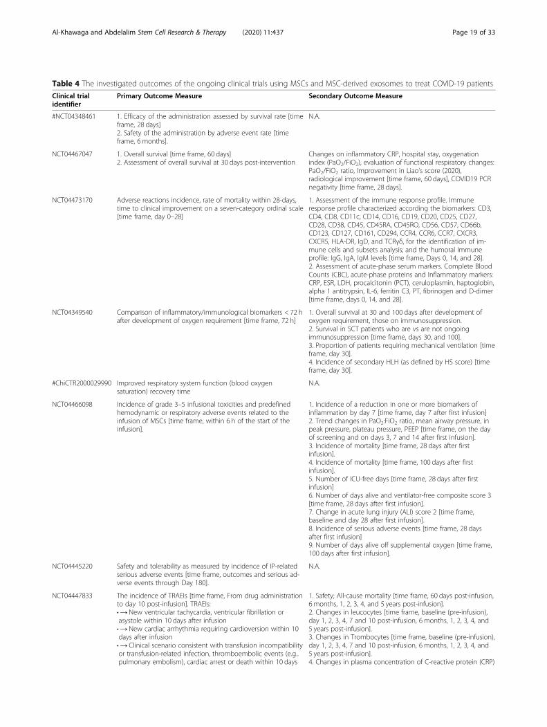
Table 4 The investigated outcomes of the ongoing clinical trials using MSCs and MSC-derived exosomes to treat COVID-19 patients
Clinical trialidentifier
Primary Outcome Measure Secondary Outcome Measure
#NCT04348461 1. Efficacy of the administration assessed by survival rate [timeframe, 28 days]2. Safety of the administration by adverse event rate [timeframe, 6 months].
N.A.
NCT04467047 1. Overall survival [time frame, 60 days]2. Assessment of overall survival at 30 days post-intervention
Changes on inflammatory CRP, hospital stay, oxygenationindex (PaO2/FiO2), evaluation of functional respiratory changes:PaO2/FiO2 ratio, Improvement in Liao’s score (2020),radiological improvement [time frame, 60 days], COVID19 PCRnegativity [time frame, 28 days].
NCT04473170 Adverse reactions incidence, rate of mortality within 28-days,time to clinical improvement on a seven-category ordinal scale[time frame, day 0–28]
1. Assessment of the immune response profile. Immuneresponse profile characterized according the biomarkers: CD3,CD4, CD8, CD11c, CD14, CD16, CD19, CD20, CD25, CD27,CD28, CD38, CD45, CD45RA, CD45RO, CD56, CD57, CD66b,CD123, CD127, CD161, CD294, CCR4, CCR6, CCR7, CXCR3,CXCR5, HLA-DR, IgD, and TCRγδ, for the identification of im-mune cells and subsets analysis; and the humoral Immuneprofile: IgG, IgA, IgM levels [time frame, Days 0, 14, and 28].2. Assessment of acute-phase serum markers. Complete BloodCounts (CBC), acute-phase proteins and Inflammatory markers:CRP, ESR, LDH, procalcitonin (PCT), ceruloplasmin, haptoglobin,alpha 1 antitrypsin, IL-6, ferritin C3, PT, fibrinogen and D-dimer[time frame, days 0, 14, and 28].
NCT04349540 Comparison of inflammatory/immunological biomarkers < 72 hafter development of oxygen requirement [time frame, 72 h]
1. Overall survival at 30 and 100 days after development ofoxygen requirement, those on immunosuppression.2. Survival in SCT patients who are vs are not ongoingimmunosuppression [time frame, days 30, and 100].3. Proportion of patients requiring mechanical ventilation [timeframe, day 30].4. Incidence of secondary HLH (as defined by HS score) [timeframe, day 30].
#ChiCTR2000029990 Improved respiratory system function (blood oxygensaturation) recovery time
N.A.
NCT04466098 Incidence of grade 3–5 infusional toxicities and predefinedhemodynamic or respiratory adverse events related to theinfusion of MSCs [time frame, within 6 h of the start of theinfusion].
1. Incidence of a reduction in one or more biomarkers ofinflammation by day 7 [time frame, day 7 after first infusion]2. Trend changes in PaO2:FiO2 ratio, mean airway pressure, inpeak pressure, plateau pressure, PEEP [time frame, on the dayof screening and on days 3, 7 and 14 after first infusion].3. Incidence of mortality [time frame, 28 days after firstinfusion].4. Incidence of mortality [time frame, 100 days after firstinfusion].5. Number of ICU-free days [time frame, 28 days after firstinfusion]6. Number of days alive and ventilator-free composite score 3[time frame, 28 days after first infusion].7. Change in acute lung injury (ALI) score 2 [time frame,baseline and day 28 after first infusion].8. Incidence of serious adverse events [time frame, 28 daysafter first infusion]9. Number of days alive off supplemental oxygen [time frame,100 days after first infusion].
NCT04445220 Safety and tolerability as measured by incidence of IP-relatedserious adverse events [time frame, outcomes and serious ad-verse events through Day 180].
N.A.
NCT04447833 The incidence of TRAEIs [time frame, From drug administrationto day 10 post-infusion]. TRAEIs:•→ New ventricular tachycardia, ventricular fibrillation orasystole within 10 days after infusion•→ New cardiac arrhythmia requiring cardioversion within 10days after infusion•→ Clinical scenario consistent with transfusion incompatibilityor transfusion-related infection, thromboembolic events (e.g..pulmonary embolism), cardiac arrest or death within 10 days
1. Safety; All-cause mortality [time frame, 60 days post-infusion,6 months, 1, 2, 3, 4, and 5 years post-infusion].2. Changes in leucocytes [time frame, baseline (pre-infusion),day 1, 2, 3, 4, 7 and 10 post-infusion, 6 months, 1, 2, 3, 4, and5 years post-infusion].3. Changes in Trombocytes [time frame, baseline (pre-infusion),day 1, 2, 3, 4, 7 and 10 post-infusion, 6 months, 1, 2, 3, 4, and5 years post-infusion].4. Changes in plasma concentration of C-reactive protein (CRP)
Al-Khawaga and Abdelalim Stem Cell Research & Therapy (2020) 11:437 Page 19 of 33
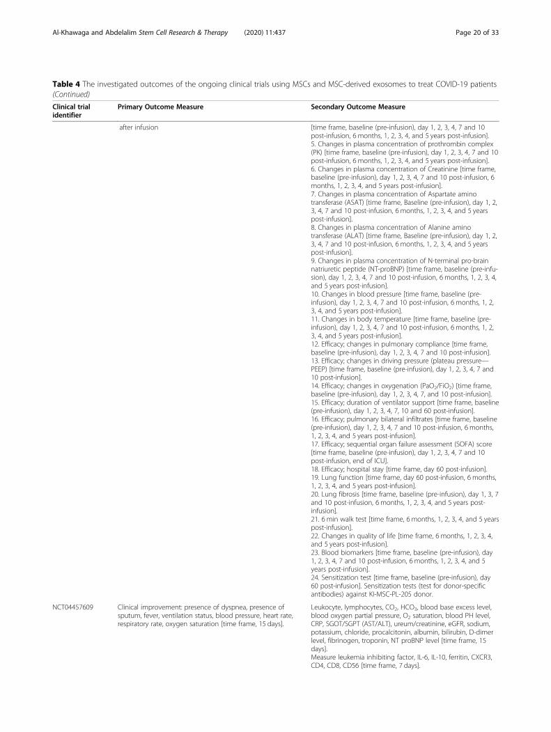
Table 4 The investigated outcomes of the ongoing clinical trials using MSCs and MSC-derived exosomes to treat COVID-19 patients(Continued)
Clinical trialidentifier
Primary Outcome Measure Secondary Outcome Measure
after infusion [time frame, baseline (pre-infusion), day 1, 2, 3, 4, 7 and 10post-infusion, 6 months, 1, 2, 3, 4, and 5 years post-infusion].5. Changes in plasma concentration of prothrombin complex(PK) [time frame, baseline (pre-infusion), day 1, 2, 3, 4, 7 and 10post-infusion, 6 months, 1, 2, 3, 4, and 5 years post-infusion].6. Changes in plasma concentration of Creatinine [time frame,baseline (pre-infusion), day 1, 2, 3, 4, 7 and 10 post-infusion, 6months, 1, 2, 3, 4, and 5 years post-infusion].7. Changes in plasma concentration of Aspartate aminotransferase (ASAT) [time frame, Baseline (pre-infusion), day 1, 2,3, 4, 7 and 10 post-infusion, 6 months, 1, 2, 3, 4, and 5 yearspost-infusion].8. Changes in plasma concentration of Alanine aminotransferase (ALAT) [time frame, Baseline (pre-infusion), day 1, 2,3, 4, 7 and 10 post-infusion, 6 months, 1, 2, 3, 4, and 5 yearspost-infusion].9. Changes in plasma concentration of N-terminal pro-brainnatriuretic peptide (NT-proBNP) [time frame, baseline (pre-infu-sion), day 1, 2, 3, 4, 7 and 10 post-infusion, 6 months, 1, 2, 3, 4,and 5 years post-infusion].10. Changes in blood pressure [time frame, baseline (pre-infusion), day 1, 2, 3, 4, 7 and 10 post-infusion, 6 months, 1, 2,3, 4, and 5 years post-infusion].11. Changes in body temperature [time frame, baseline (pre-infusion), day 1, 2, 3, 4, 7 and 10 post-infusion, 6 months, 1, 2,3, 4, and 5 years post-infusion].12. Efficacy; changes in pulmonary compliance [time frame,baseline (pre-infusion), day 1, 2, 3, 4, 7 and 10 post-infusion].13. Efficacy; changes in driving pressure (plateau pressure—PEEP) [time frame, baseline (pre-infusion), day 1, 2, 3, 4, 7 and10 post-infusion].14. Efficacy; changes in oxygenation (PaO2/FiO2) [time frame,baseline (pre-infusion), day 1, 2, 3, 4, 7, and 10 post-infusion].15. Efficacy; duration of ventilator support [time frame, baseline(pre-infusion), day 1, 2, 3, 4, 7, 10 and 60 post-infusion].16. Efficacy; pulmonary bilateral infiltrates [time frame, baseline(pre-infusion), day 1, 2, 3, 4, 7 and 10 post-infusion, 6 months,1, 2, 3, 4, and 5 years post-infusion].17. Efficacy; sequential organ failure assessment (SOFA) score[time frame, baseline (pre-infusion), day 1, 2, 3, 4, 7 and 10post-infusion, end of ICU].18. Efficacy; hospital stay [time frame, day 60 post-infusion].19. Lung function [time frame, day 60 post-infusion, 6 months,1, 2, 3, 4, and 5 years post-infusion].20. Lung fibrosis [time frame, baseline (pre-infusion), day 1, 3, 7and 10 post-infusion, 6 months, 1, 2, 3, 4, and 5 years post-infusion].21. 6 min walk test [time frame, 6 months, 1, 2, 3, 4, and 5 yearspost-infusion].22. Changes in quality of life [time frame, 6 months, 1, 2, 3, 4,and 5 years post-infusion].23. Blood biomarkers [time frame, baseline (pre-infusion), day1, 2, 3, 4, 7 and 10 post-infusion, 6 months, 1, 2, 3, 4, and 5years post-infusion].24. Sensitization test [time frame, baseline (pre-infusion), day60 post-infusion]. Sensitization tests (test for donor-specificantibodies) against KI-MSC-PL-205 donor.
NCT04457609 Clinical improvement: presence of dyspnea, presence ofsputum, fever, ventilation status, blood pressure, heart rate,respiratory rate, oxygen saturation [time frame, 15 days].
Leukocyte, lymphocytes, CO2, HCO3, blood base excess level,blood oxygen partial pressure, O2 saturation, blood PH level,CRP, SGOT/SGPT (AST/ALT), ureum/creatinine, eGFR, sodium,potassium, chloride, procalcitonin, albumin, bilirubin, D-dimerlevel, fibrinogen, troponin, NT proBNP level [time frame, 15days].Measure leukemia inhibiting factor, IL-6, IL-10, ferritin, CXCR3,CD4, CD8, CD56 [time frame, 7 days].
Al-Khawaga and Abdelalim Stem Cell Research & Therapy (2020) 11:437 Page 20 of 33
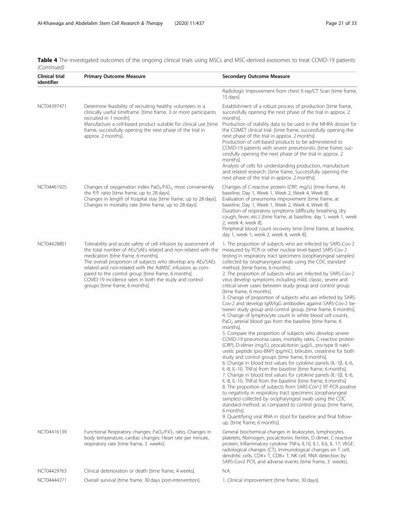
Table 4 The investigated outcomes of the ongoing clinical trials using MSCs and MSC-derived exosomes to treat COVID-19 patients(Continued)
Clinical trialidentifier
Primary Outcome Measure Secondary Outcome Measure
Radiologic Improvement from chest X-ray/CT Scan [time frame,15 days].
NCT04397471 Determine feasibility of recruiting healthy volunteers in aclinically useful timeframe. [time frame, 3 or more participantsrecruited in 1 month].Manufacture a cell-based product suitable for clinical use [timeframe, successfully opening the next phase of the trial inapprox. 2 months].
Establishment of a robust process of production [time frame,successfully opening the next phase of the trial in approx. 2months].Production of stability data to be used in the MHRA dossier forthe COMET clinical trial. [time frame, successfully opening thenext phase of the trial in approx. 2 months]Production of cell-based products to be administered toCOVID-19 patients with severe pneumonitis. [time frame, suc-cessfully opening the next phase of the trial in approx. 2months].Analysis of cells for understanding production, manufactureand related research. [time frame, Successfully opening thenext phase of the trial in approx. 2 months].
NCT04461925 Changes of oxygenation index PaO2/FiO2, most convenientlythe P/F ratio [time frame, up to 28 days].Changes in length of hospital stay [time frame, up to 28 days].Changes in mortality rate [time frame, up to 28 days].
Changes of С-reactive protein (CRP, mg/L) [time frame, Atbaseline, Day 1, Week 1, Week 2, Week 4, Week 8].Evaluation of pneumonia improvement [time frame, atbaseline, Day 1, Week 1, Week 2, Week 4, Week 8].Duration of respiratory symptoms (difficulty breathing, drycough, fever, etc.) [time frame, at baseline, day 1, week 1, week2, week 4, week 8].Peripheral blood count recovery time [time frame, at baseline,day 1, week 1, week 2, week 4, week 8].
NCT04428801 Tolerability and acute safety of cell infusion by assessment ofthe total number of AEs/SAEs related and non-related with themedication [time frame, 6 months].The overall proportion of subjects who develop any AEs/SAEsrelated and non-related with the AdMSC infusions as com-pared to the control group [time frame, 6 months].COVID-19 incidence rates in both the study and controlgroups [time frame, 6 months].
1. The proportion of subjects who are infected by SARS-Cov-2measured by PCR or other nuclear level-based SARS-Cov-2testing in respiratory tract specimens (oropharyngeal samples)collected by oropharyngeal swab using the CDC standardmethod. [time frame, 6 months].2. The proportion of subjects who are infected by SARS-Cov-2virus develop symptoms including mild, classic, severe andcritical sever cases between study group and control group.[time frame, 6 months].3. Change of proportion of subjects who are infected by SARS-Cov-2 and develop IgM/IgG antibodies against SARS-Cov-2 be-tween study group and control group. [time frame, 6 months].4. Change of lymphocyte count in white blood cell counts,PaO2 arterial blood gas from the baseline [time frame, 6months].5. Compare the proportion of subjects who develop severeCOVID-19 pneumonia cases, mortality rates, C-reactive protein(CRP), D-dimer (mg/L), procalcitonin (μg)/L, pro-type B natri-uretic peptide (pro-BNP) (pg/mL), bilirubin, creatinine for bothstudy and control groups [time frame, 6 months].6. Change in blood test values for cytokine panels (IL-1β, IL-6,IL-8, IL-10, TNFα) from the baseline [time frame, 6 months].7. Change in blood test values for cytokine panels (IL-1β, IL-6,IL-8, IL-10, TNFα) from the baseline [time frame, 6 months]8. The proportion of subjects from SARS-CoV-2 RT-PCR positiveto negativity in respiratory tract specimens (oropharyngealsamples) collected by oropharyngeal swab using the CDCstandard method. as compared to control group [time frame,6 months].9. Quantifying viral RNA in stool for baseline and final follow-up. [time frame, 6 months].
NCT04416139 Functional Respiratory changes: PaO2/FiO2 ratio, Changes inbody temperature, cardiac changes: Heart rate per minute,respiratory rate [time frame, 3 weeks].
General biochemical changes in leukocytes, lymphocytes,platelets, fibrinogen, pocalcitonin, ferritin, D-dimer, C-reactiveprotein, Inflammatory cytokine TNFa, IL10, IL1, IL6, IL 17, VEGF,radiological changes (CT), immunological changes on T cell,dendritic cells, CD4+ T, CD8+ T, NK cell, RNA detection bySARS-Cov2 PCR, and adverse events [time frame, 3 weeks].
NCT04429763 Clinical deterioration or death [time frame, 4 weeks]. N.A.
NCT04444271 Overall survival [time frame, 30 days post-intervention]. 1. Clinical improvement [time frame, 30 days].
Al-Khawaga and Abdelalim Stem Cell Research & Therapy (2020) 11:437 Page 21 of 33
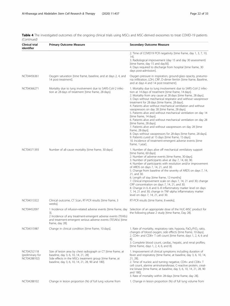
Table 4 The investigated outcomes of the ongoing clinical trials using MSCs and MSC-derived exosomes to treat COVID-19 patients(Continued)
Clinical trialidentifier
Primary Outcome Measure Secondary Outcome Measure
2. Time of COVID19 PCR negativity [time frame, day 1, 3, 7, 10,14].3. Radiological improvement (day 15 and day 30 assessment)[time frame, day 15 and day30].4. Days required to discharge from hospital [time frame, 30days post-admission].
NCT04456361 Oxygen saturation [time frame, baseline, and at days 2, 4, and14 post-treatment].
Oxygen pressure in inspiration, ground-glass opacity, pneumo-nia infiltration, LDH, CRP, D-dimer ferritin [time frame, Baseline,and at days 4 and 14 post-treatment].
NCT04366271 Mortality due to lung involvement due to SARS-CoV-2 infec-tion at 28 days of treatment [time frame, 28 days].
1. Mortality due to lung involvement due to SARS-CoV-2 infec-tion at 14 days of treatment [time frame, 14 days].2. Mortality from any cause at 28 days [time frame, 28 days].3. Days without mechanical respirator and without vasopressortreatment for 28 days [time frame, 28 days].4. Patients alive without mechanical ventilation and withoutvasopressors on day 28 [time frame, 28 days].5. Patients alive and without mechanical ventilation on day 14[time frame, 14 days].6. Patients alive and without mechanical ventilation on day 28[time frame, 28 days].7. Patients alive and without vasopressors on day 28 [timeframe, 28 days].8. Days without vasopressors for 28 days [time frame, 28 days].9. Patients cured at 15 days [time frame, 15 days].10. Incidence of treatment-emergent adverse events [timeframe, 1 year].
NCT04371393 Number of all-cause mortality [time frame, 30 days]. 1. Number of days alive off mechanical ventilatory support[time frame, 60 days].2. Number of adverse events [time frame, 30 days].3. Number of participants alive at day 7, 14, 60, 90.4. Number of participants with resolution and/or improvementof ARDS on days 7, 14, 21, and 30.5. Change from baseline of the severity of ARDS on days 7, 14,21, and 30.6. Length of stay [time frame, 12 months]7. Clinical improvement scale on days 7, 14, 21 and 30; changeCRP concentration on days 7, 14, 21, and 30.8. Change in IL-6 and IL-8 inflammatory marker level on days7, 14, 21 and 30; change in TNF-alpha inflammatory markerlevel on days 7, 14, 21, and 30.
NCT04313322 Clinical outcome, CT Scan, RT-PCR results [time frame, 3weeks].
RT-PCR results [time frame, 8 weeks].
NCT04452097 1 Incidence of infusion-related adverse events [time frame, day3].2 Incidence of any treatment-emergent adverse events (TEAEs)and treatment-emergent serious adverse events (TESAEs) [timeframe, day 28].
Selection of an appropriate dose of the hUC-MSC product forthe following phase 2 study [time frame, Day 28].
NCT04315987 Change in clinical condition [time frame, 10 days]. 1. Rate of mortality, respiratory rate, hypoxia, PaO2/FiO2 ratio,changes of blood oxygen, side effects [time frame, 10 days].2. CD4+ and CD8+ T cell count [time frame, days 1, 2, 4, 6 and8].3. Complete blood count, cardiac, hepatic, and renal profiles;[time frame, days 1, 2, 4, 6, and 8].
NCT04252118(preliminary forNCT04288102)
Size of lesion area by chest radiograph or CT [time frame, atbaseline, day 3, 6, 10, 14, 21, 28]Side effects in the MSCs treatment group [time frame, atbaseline, day 3, 6, 10, 14, 21, 28, 90 and 180].
1. Improvement of clinical symptoms including duration offever and respiratory [time frame, at baseline, day 3, 6, 10, 14,21, 28].2. Time of nucleic acid turning negative, CD4+ and CD8+ Tcell count, alanine aminotransferase, C-reactive protein, creat-ine kinase [time frame, at baseline, day 3, 6, 10, 14, 21, 28, 90and 180].3. Rate of mortality within 28-days [time frame, day 28].
NCT04288102 Change in lesion proportion (%) of full lung volume from 1. Change in lesion proportion (%) of full lung volume from
Al-Khawaga and Abdelalim Stem Cell Research & Therapy (2020) 11:437 Page 22 of 33
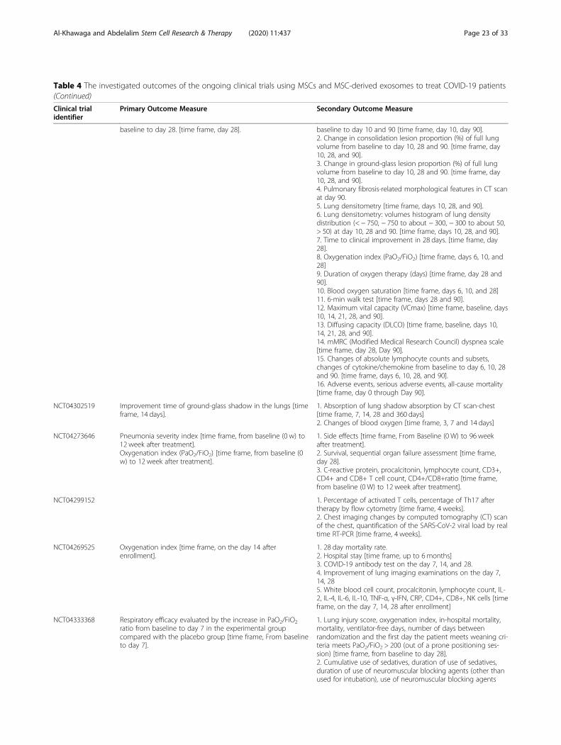
Table 4 The investigated outcomes of the ongoing clinical trials using MSCs and MSC-derived exosomes to treat COVID-19 patients(Continued)
Clinical trialidentifier
Primary Outcome Measure Secondary Outcome Measure
baseline to day 28. [time frame, day 28]. baseline to day 10 and 90 [time frame, day 10, day 90].2. Change in consolidation lesion proportion (%) of full lungvolume from baseline to day 10, 28 and 90. [time frame, day10, 28, and 90].3. Change in ground-glass lesion proportion (%) of full lungvolume from baseline to day 10, 28 and 90. [time frame, day10, 28, and 90].4. Pulmonary fibrosis-related morphological features in CT scanat day 90.5. Lung densitometry [time frame, days 10, 28, and 90].6. Lung densitometry: volumes histogram of lung densitydistribution (< − 750, − 750 to about − 300, − 300 to about 50,> 50) at day 10, 28 and 90. [time frame, days 10, 28, and 90].7. Time to clinical improvement in 28 days. [time frame, day28].8. Oxygenation index (PaO2/FiO2) [time frame, days 6, 10, and28]9. Duration of oxygen therapy (days) [time frame, day 28 and90].10. Blood oxygen saturation [time frame, days 6, 10, and 28]11. 6-min walk test [time frame, days 28 and 90].12. Maximum vital capacity (VCmax) [time frame, baseline, days10, 14, 21, 28, and 90].13. Diffusing capacity (DLCO) [time frame, baseline, days 10,14, 21, 28, and 90].14. mMRC (Modified Medical Research Council) dyspnea scale[time frame, day 28, Day 90].15. Changes of absolute lymphocyte counts and subsets,changes of cytokine/chemokine from baseline to day 6, 10, 28and 90. [time frame, days 6, 10, 28, and 90].16. Adverse events, serious adverse events, all-cause mortality[time frame, day 0 through Day 90].
NCT04302519 Improvement time of ground-glass shadow in the lungs [timeframe, 14 days].
1. Absorption of lung shadow absorption by CT scan-chest[time frame, 7, 14, 28 and 360 days]2. Changes of blood oxygen [time frame, 3, 7 and 14 days]
NCT04273646 Pneumonia severity index [time frame, from baseline (0 w) to12 week after treatment].Oxygenation index (PaO2/FiO2) [time frame, from baseline (0w) to 12 week after treatment].
1. Side effects [time frame, From Baseline (0 W) to 96 weekafter treatment].2. Survival, sequential organ failure assessment [time frame,day 28].3. C-reactive protein, procalcitonin, lymphocyte count, CD3+,CD4+ and CD8+ T cell count, CD4+/CD8+ratio [time frame,from baseline (0 W) to 12 week after treatment].
NCT04299152 1. Percentage of activated T cells, percentage of Th17 aftertherapy by flow cytometry [time frame, 4 weeks].2. Chest imaging changes by computed tomography (CT) scanof the chest, quantification of the SARS-CoV-2 viral load by realtime RT-PCR [time frame, 4 weeks].
NCT04269525 Oxygenation index [time frame, on the day 14 afterenrollment].
1. 28 day mortality rate.2. Hospital stay [time frame, up to 6 months]3. COVID-19 antibody test on the day 7, 14, and 28.4. Improvement of lung imaging examinations on the day 7,14, 285. White blood cell count, procalcitonin, lymphocyte count, IL-2, IL-4, IL-6, IL-10, TNF-α, γ-IFN, CRP, CD4+, CD8+, NK cells [timeframe, on the day 7, 14, 28 after enrollment]
NCT04333368 Respiratory efficacy evaluated by the increase in PaO2/FiO2
ratio from baseline to day 7 in the experimental groupcompared with the placebo group [time frame, From baselineto day 7].
1. Lung injury score, oxygenation index, in-hospital mortality,mortality, ventilator-free days, number of days betweenrandomization and the first day the patient meets weaning cri-teria meets PaO2/FiO2 > 200 (out of a prone positioning ses-sion) [time frame, from baseline to day 28].2. Cumulative use of sedatives, duration of use of sedatives,duration of use of neuromuscular blocking agents (other thanused for intubation), use of neuromuscular blocking agents
Al-Khawaga and Abdelalim Stem Cell Research & Therapy (2020) 11:437 Page 23 of 33
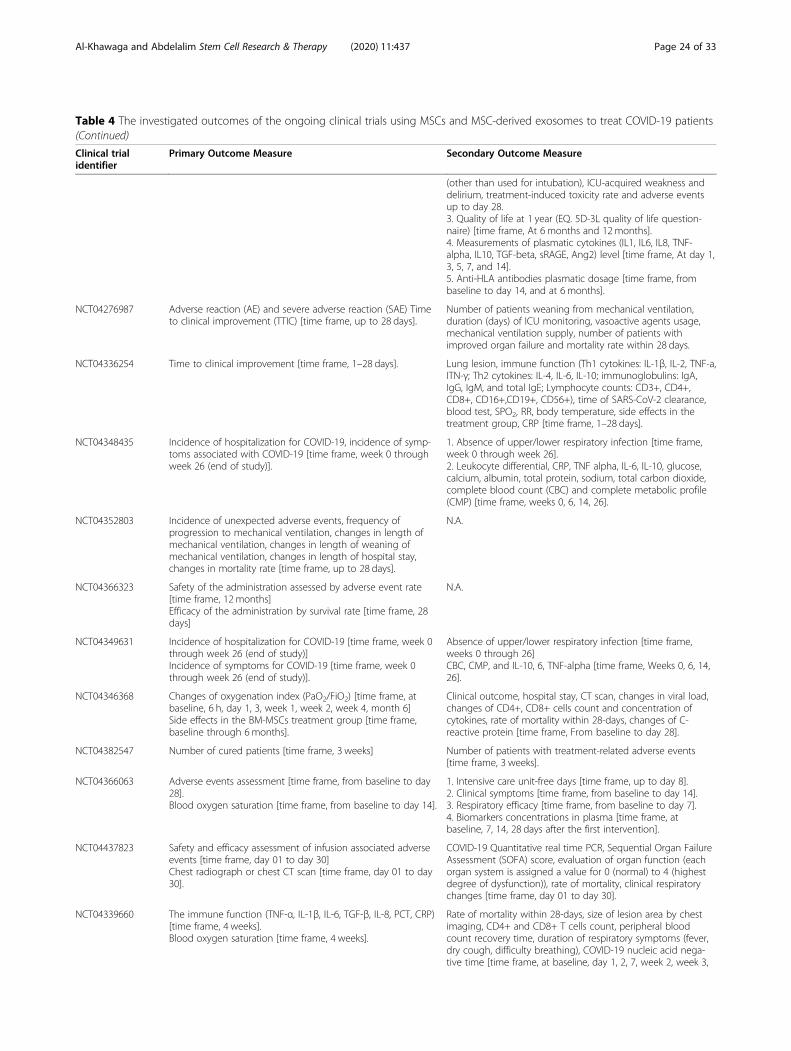
Table 4 The investigated outcomes of the ongoing clinical trials using MSCs and MSC-derived exosomes to treat COVID-19 patients(Continued)
Clinical trialidentifier
Primary Outcome Measure Secondary Outcome Measure
(other than used for intubation), ICU-acquired weakness anddelirium, treatment-induced toxicity rate and adverse eventsup to day 28.3. Quality of life at 1 year (EQ. 5D-3L quality of life question-naire) [time frame, At 6 months and 12months].4. Measurements of plasmatic cytokines (IL1, IL6, IL8, TNF-alpha, IL10, TGF-beta, sRAGE, Ang2) level [time frame, At day 1,3, 5, 7, and 14].5. Anti-HLA antibodies plasmatic dosage [time frame, frombaseline to day 14, and at 6 months].
NCT04276987 Adverse reaction (AE) and severe adverse reaction (SAE) Timeto clinical improvement (TTIC) [time frame, up to 28 days].
Number of patients weaning from mechanical ventilation,duration (days) of ICU monitoring, vasoactive agents usage,mechanical ventilation supply, number of patients withimproved organ failure and mortality rate within 28 days.
NCT04336254 Time to clinical improvement [time frame, 1–28 days]. Lung lesion, immune function (Th1 cytokines: IL-1β, IL-2, TNF-a,ITN-γ; Th2 cytokines: IL-4, IL-6, IL-10; immunoglobulins: IgA,IgG, IgM, and total IgE; Lymphocyte counts: CD3+, CD4+,CD8+, CD16+,CD19+, CD56+), time of SARS-CoV-2 clearance,blood test, SPO2, RR, body temperature, side effects in thetreatment group, CRP [time frame, 1–28 days].
NCT04348435 Incidence of hospitalization for COVID-19, incidence of symp-toms associated with COVID-19 [time frame, week 0 throughweek 26 (end of study)].
1. Absence of upper/lower respiratory infection [time frame,week 0 through week 26].2. Leukocyte differential, CRP, TNF alpha, IL-6, IL-10, glucose,calcium, albumin, total protein, sodium, total carbon dioxide,complete blood count (CBC) and complete metabolic profile(CMP) [time frame, weeks 0, 6, 14, 26].
NCT04352803 Incidence of unexpected adverse events, frequency ofprogression to mechanical ventilation, changes in length ofmechanical ventilation, changes in length of weaning ofmechanical ventilation, changes in length of hospital stay,changes in mortality rate [time frame, up to 28 days].
N.A.
NCT04366323 Safety of the administration assessed by adverse event rate[time frame, 12 months]Efficacy of the administration by survival rate [time frame, 28days]
N.A.
NCT04349631 Incidence of hospitalization for COVID-19 [time frame, week 0through week 26 (end of study)]Incidence of symptoms for COVID-19 [time frame, week 0through week 26 (end of study)].
Absence of upper/lower respiratory infection [time frame,weeks 0 through 26]CBC, CMP, and IL-10, 6, TNF-alpha [time frame, Weeks 0, 6, 14,26].
NCT04346368 Changes of oxygenation index (PaO2/FiO2) [time frame, atbaseline, 6 h, day 1, 3, week 1, week 2, week 4, month 6]Side effects in the BM-MSCs treatment group [time frame,baseline through 6months].
Clinical outcome, hospital stay, CT scan, changes in viral load,changes of CD4+, CD8+ cells count and concentration ofcytokines, rate of mortality within 28-days, changes of C-reactive protein [time frame, From baseline to day 28].
NCT04382547 Number of cured patients [time frame, 3 weeks] Number of patients with treatment-related adverse events[time frame, 3 weeks].
NCT04366063 Adverse events assessment [time frame, from baseline to day28].Blood oxygen saturation [time frame, from baseline to day 14].
1. Intensive care unit-free days [time frame, up to day 8].2. Clinical symptoms [time frame, from baseline to day 14].3. Respiratory efficacy [time frame, from baseline to day 7].4. Biomarkers concentrations in plasma [time frame, atbaseline, 7, 14, 28 days after the first intervention].
NCT04437823 Safety and efficacy assessment of infusion associated adverseevents [time frame, day 01 to day 30]Chest radiograph or chest CT scan [time frame, day 01 to day30].
COVID-19 Quantitative real time PCR, Sequential Organ FailureAssessment (SOFA) score, evaluation of organ function (eachorgan system is assigned a value for 0 (normal) to 4 (highestdegree of dysfunction)), rate of mortality, clinical respiratorychanges [time frame, day 01 to day 30].
NCT04339660 The immune function (TNF-α, IL-1β, IL-6, TGF-β, IL-8, PCT, CRP)[time frame, 4 weeks].Blood oxygen saturation [time frame, 4 weeks].
Rate of mortality within 28-days, size of lesion area by chestimaging, CD4+ and CD8+ T cells count, peripheral bloodcount recovery time, duration of respiratory symptoms (fever,dry cough, difficulty breathing), COVID-19 nucleic acid nega-tive time [time frame, at baseline, day 1, 2, 7, week 2, week 3,
Al-Khawaga and Abdelalim Stem Cell Research & Therapy (2020) 11:437 Page 24 of 33
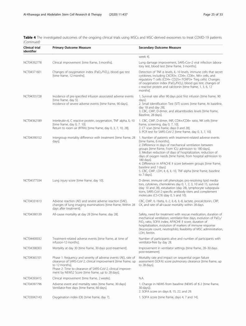
Table 4 The investigated outcomes of the ongoing clinical trials using MSCs and MSC-derived exosomes to treat COVID-19 patients(Continued)
Clinical trialidentifier
Primary Outcome Measure Secondary Outcome Measure
week 4].
NCT04392778 Clinical improvement [time frame, 3 months]. Lung damage improvement, SARS-Cov-2 viral infection labora-tory test, blood test [time frame, 3 months].
NCT04371601 Changes of oxygenation index (PaO2/FiO2), blood gas test[time frame, 12 months].
Detection of TNF-α levels, IL-10 levels, immune cells that secretcytokines, including CXCR3+, CD4+, CD8+, NK+ cells, andregulatory T cells (CD4+ CD25+ FOXP3+ Treg cells). Changesof oxygenation index (PaO2/FiO2), blood gas test, changes ofc-reactive protein and calcitonin [time frame, 1, 3, 6, 12months].
NCT04355728 Incidence of pre-specified infusion associated adverse events[time frame, day 5].Incidence of severe adverse events [time frame, 90 days].
1. Survival rate after 90 days post first infusion [time frame, 90days].2. Small Identification Test (SIT) scores [time frame, At baseline,day 18 and day 28].3. CBC, CMP, D-dimer, and alloantibodies levels [time frame,Baseline, 28 days].
NCT04362189 Interleukin-6, C reactive protein, oxygenation, TNF alpha, IL-10[time frame, day 0, 7. 10].Return to room air (RTRA) [time frame, day 0, 3, 7, 10, 28].
1. CBC, CMP, D-dimer, INR, CD4+/CD8+ ratio, NK cells [timeframe, screening, day 0, 7, 10].2. CT scan [time frame, days 0 and 28].3. PCR test for SARS-CoV-2 [time frame, day 0, 3, 7, 10].
NCT04390152 Intergroup mortality difference with treatment [time frame, 28days].
1. Number of patients with treatment-related adverse events[time frame, 6 months].2. Difference in days of mechanical ventilation betweengroups [time frame, From ICU admission to 180 days].3. Median reduction of days of hospitalization, reduction ofdays of oxygen needs [time frame, from hospital admission to180 days].4. Difference in APACHE II score between groups [time frame,baseline and 7 days]5. CBC, CMP, LDH, IL-6, IL-10, TNF-alpha [time frame, baselineto 7 days].
NCT04377334 Lung injury score [time frame, day 10]. D-dimer, immune cell phenotype, pro-resolving lipid media-tors, cytokines, chemokines day 0, 1, 2, 3, 10 and 15, survival(day 10 and 28), extubation (day 28), lymphocyte subpopula-tions, SARS-CoV-2-specific antibody titers and complementmolecules (C5-C9) (day 0, 5 and 10).
NCT04331613 Adverse reaction (AE) and severe adverse reaction (SAE),changes of lung imaging examinations [time frame, Within 28days after treatment].
CBC, CMP, IL-1beta, IL-2, IL-6, IL-8, lactate, procalcitonin, CRP,CK, and rate of all-cause mortality within 28 days.
NCT04390139 All-cause mortality at day 28 [time frame, day 28]. Safety, need for treatment with rescue medication, duration ofmechanical ventilation, ventilator-free days, evolution of PaO2/FiO2 ratio, SOFA index, APACHE II score, duration ofhospitalization, evolution of markers of immune response(leucocyte count, neutrophils), feasibility of MSC administration,LDH, ferritin.
NCT04400032 Treatment-related adverse events [time frame, at time ofinfusion-12 months].
Number of participants alive and number of participants withventilator-free by day 28.
NCT04398303 Mortality at day 30 [time frame, 30 days post-treatment]. Improvement in ventilator settings [time frame, 28–30 dayspost-treatment].
NCT04365101 Phase 1: frequency and severity of adverse events (AE), rate ofclearance of SARS-CoV-2, clinical improvement [time frame, upto 12 months].Phase 2: Time to clearance of SARS-CoV-2, clinical improve-ment by NEWS2 Score [time frame, up to 28 days].
Mortality rate and impact on sequential organ failureassessment (SOFA) score pulmonary clearance [time frame, upto 28 days].
NCT04393415 Clinical improvement [time frame, 2 weeks]. N.A.
NCT04397796 Adverse event and mortality rates [time frame, 30 days]Ventilator-free days [time frame, 60 days].
1. Change in NEWS from baseline (NEWS of ≤ 2 [time frame,30 days]).2. SOFA score on days 8, 15, 22, and 29.
NCT03042143 Oxygenation index (OI) [time frame, day 7]. 1. SOFA score [time frame, days 4, 7 and 14].
Al-Khawaga and Abdelalim Stem Cell Research & Therapy (2020) 11:437 Page 25 of 33
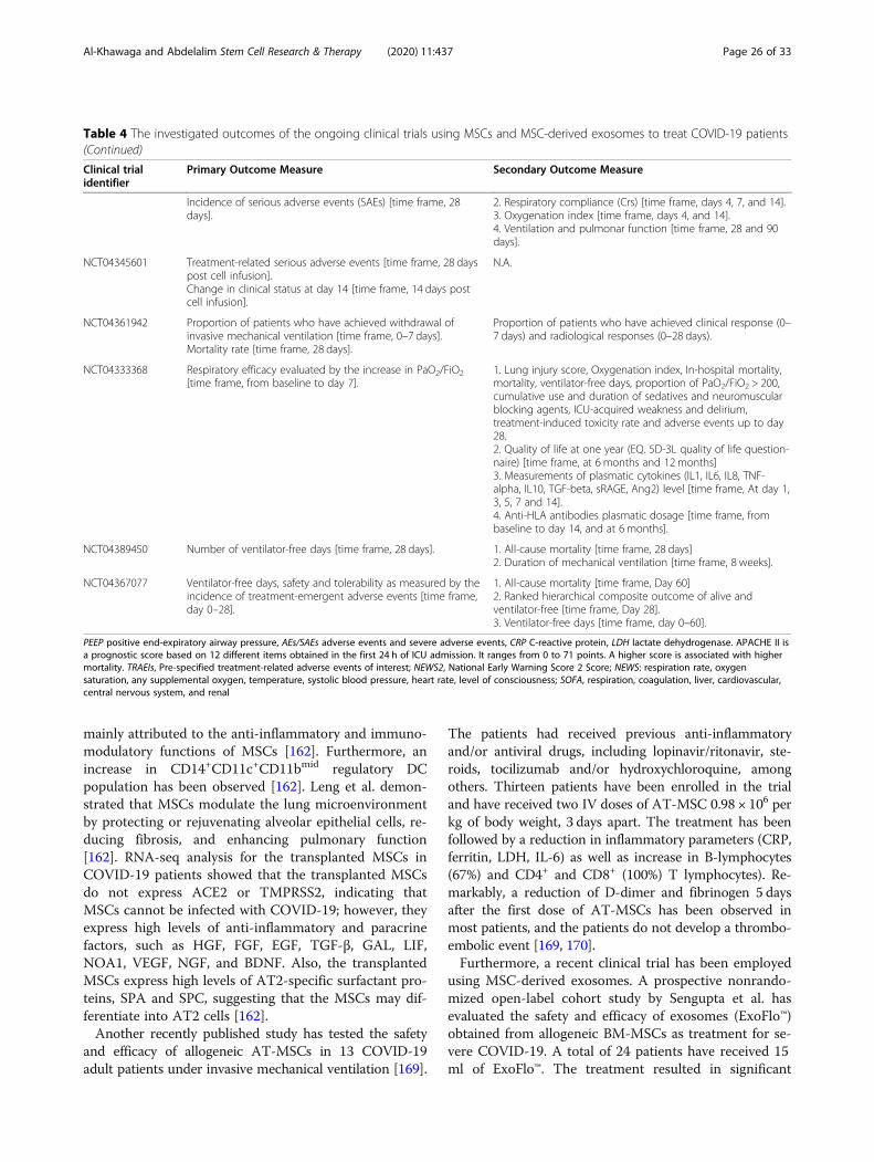
mainly attributed to the anti-inflammatory and immuno-modulatory functions of MSCs [162]. Furthermore, anincrease in CD14+CD11c+CD11bmid regulatory DCpopulation has been observed [162]. Leng et al. demon-strated that MSCs modulate the lung microenvironmentby protecting or rejuvenating alveolar epithelial cells, re-ducing fibrosis, and enhancing pulmonary function[162]. RNA-seq analysis for the transplanted MSCs inCOVID-19 patients showed that the transplanted MSCsdo not express ACE2 or TMPRSS2, indicating thatMSCs cannot be infected with COVID-19; however, theyexpress high levels of anti-inflammatory and paracrinefactors, such as HGF, FGF, EGF, TGF-β, GAL, LIF,NOA1, VEGF, NGF, and BDNF. Also, the transplantedMSCs express high levels of AT2-specific surfactant pro-teins, SPA and SPC, suggesting that the MSCs may dif-ferentiate into AT2 cells [162].Another recently published study has tested the safety
and efficacy of allogeneic AT-MSCs in 13 COVID-19adult patients under invasive mechanical ventilation [169].
The patients had received previous anti-inflammatoryand/or antiviral drugs, including lopinavir/ritonavir, ste-roids, tocilizumab and/or hydroxychloroquine, amongothers. Thirteen patients have been enrolled in the trialand have received two IV doses of AT-MSC 0.98 × 10⁶ perkg of body weight, 3 days apart. The treatment has beenfollowed by a reduction in inflammatory parameters (CRP,ferritin, LDH, IL-6) as well as increase in B-lymphocytes(67%) and CD4+ and CD8+ (100%) T lymphocytes). Re-markably, a reduction of D-dimer and fibrinogen 5 daysafter the first dose of AT-MSCs has been observed inmost patients, and the patients do not develop a thrombo-embolic event [169, 170].Furthermore, a recent clinical trial has been employed
using MSC-derived exosomes. A prospective nonrando-mized open-label cohort study by Sengupta et al. hasevaluated the safety and efficacy of exosomes (ExoFlo™)obtained from allogeneic BM-MSCs as treatment for se-vere COVID-19. A total of 24 patients have received 15ml of ExoFlo™. The treatment resulted in significant
Table 4 The investigated outcomes of the ongoing clinical trials using MSCs and MSC-derived exosomes to treat COVID-19 patients(Continued)
Clinical trialidentifier
Primary Outcome Measure Secondary Outcome Measure
Incidence of serious adverse events (SAEs) [time frame, 28days].
2. Respiratory compliance (Crs) [time frame, days 4, 7, and 14].3. Oxygenation index [time frame, days 4, and 14].4. Ventilation and pulmonar function [time frame, 28 and 90days].
NCT04345601 Treatment-related serious adverse events [time frame, 28 dayspost cell infusion].Change in clinical status at day 14 [time frame, 14 days postcell infusion].
N.A.
NCT04361942 Proportion of patients who have achieved withdrawal ofinvasive mechanical ventilation [time frame, 0–7 days].Mortality rate [time frame, 28 days].
Proportion of patients who have achieved clinical response (0–7 days) and radiological responses (0–28 days).
NCT04333368 Respiratory efficacy evaluated by the increase in PaO2/FiO2
[time frame, from baseline to day 7].1. Lung injury score, Oxygenation index, In-hospital mortality,mortality, ventilator-free days, proportion of PaO2/FiO2 > 200,cumulative use and duration of sedatives and neuromuscularblocking agents, ICU-acquired weakness and delirium,treatment-induced toxicity rate and adverse events up to day28.2. Quality of life at one year (EQ. 5D-3L quality of life question-naire) [time frame, at 6 months and 12months]3. Measurements of plasmatic cytokines (IL1, IL6, IL8, TNF-alpha, IL10, TGF-beta, sRAGE, Ang2) level [time frame, At day 1,3, 5, 7 and 14].4. Anti-HLA antibodies plasmatic dosage [time frame, frombaseline to day 14, and at 6 months].
NCT04389450 Number of ventilator-free days [time frame, 28 days]. 1. All-cause mortality [time frame, 28 days]2. Duration of mechanical ventilation [time frame, 8 weeks].
NCT04367077 Ventilator-free days, safety and tolerability as measured by theincidence of treatment-emergent adverse events [time frame,day 0–28].
1. All-cause mortality [time frame, Day 60]2. Ranked hierarchical composite outcome of alive andventilator-free [time frame, Day 28].3. Ventilator-free days [time frame, day 0–60].
PEEP positive end-expiratory airway pressure, AEs/SAEs adverse events and severe adverse events, CRP C-reactive protein, LDH lactate dehydrogenase. APACHE II isa prognostic score based on 12 different items obtained in the first 24 h of ICU admission. It ranges from 0 to 71 points. A higher score is associated with highermortality. TRAEIs, Pre-specified treatment-related adverse events of interest; NEWS2, National Early Warning Score 2 Score; NEWS: respiration rate, oxygensaturation, any supplemental oxygen, temperature, systolic blood pressure, heart rate, level of consciousness; SOFA, respiration, coagulation, liver, cardiovascular,central nervous system, and renal
Al-Khawaga and Abdelalim Stem Cell Research & Therapy (2020) 11:437 Page 26 of 33
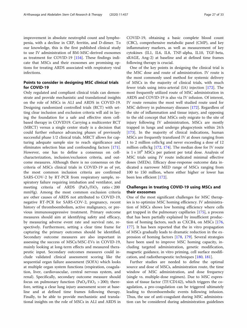
improvement in absolute neutrophil count and lympho-penia, with a decline in CRP, ferritin, and D-dimer. Toour knowledge, this is the first published clinical studyto use IV administration of BM-MSC-derived exosomesas treatment for COVID-19 [154]. These findings indi-cate that MSCs and their exosomes are promising op-tions for treating ARDS associated with respiratory viralinfections.
Points to consider in designing MSC clinical trialsfor COVID-19Only regulated and compliant clinical trials can demon-strate and provide mechanistic and translational insightson the role of MSCs in ALI and ARDS in COVID-19.Designing randomized controlled trials (RCT) with set-ting clear inclusion and exclusion criteria will aid in lay-ing the foundation for a safe and effective stem cell-based therapy in COVID19. Carrying a multicenter RCT(MRCT) versus a single center study is a decision thatcould further enhance advancing phases of previouslysuccessful phase 1/2 clinical trials. MRCT allows for cap-turing adequate sample size to reach significance andeliminates selection bias and confounding factors [171].However, one has to define a consensus on cell-characterization, inclusion/exclusion criteria, and out-come measures. Although there is no consensus on thecriteria of MSCs clinical trials in COVID-19 as of yet,the most common inclusion criteria are confirmedSARS-COV-2 by RT-PCR from respiratory sample, re-spiratory failure requiring intubation and ventilator, andmeeting criteria of ARDS (PaO2/FiO2 ratio < 200mmHg). Among the most common exclusion criteriaare other causes of ARDS not attributed to COVID-19,negative RT-PCR for SARS-COV-2, pregnancy, recenthistory of thromboembolism, active malignancy, or pre-vious immunosuppressive treatment. Primary outcomemeasures should aim at identifying safety and efficacy,by measuring adverse event rate and survival rate, re-spectively. Furthermore, setting a clear time frame forcapturing the primary outcomes should be identified.Secondary outcome measures are also important inassessing the success of MSCs/MSC-EVs in COVID-19,mainly looking at long-term effects and measured thera-peutic input. Secondary outcomes measures could in-clude validated clinical assessment scoring like thesequential organ failure assessment (SOFA) which looksat multiple organ system functions (respiration, coagula-tion, liver, cardiovascular, central nervous system, andrenal). Specifically, secondary outcome measure shouldfocus on pulmonary function (PaO2/FiO2 > 200); there-fore, setting a clear lung injury assessment score at base-line and at defined time frames following therapy.Finally, to be able to provide mechanistic and transla-tional insights on the role of MSCs in ALI and ARDS in
COVID-19, obtaining a basic complete blood count(CBC), comprehensive metabolic panel (CMP), and keyinflammatory markers, as well as measurement of keycytokines (IL1, IL6, IL8, TNF-alpha, IL10, TGF-beta,sRAGE, Ang-2) at baseline and at defined time framesfollowing therapy is crucial.One of the key points in designing the clinical trial is
the MSC dose and route of administration. IV route isthe most commonly used method for systemic deliveryof MSCs in the majority of clinical trials, with muchfewer trials using intra-arterial (IA) injection [172]. Themost frequently utilized route of MSC administration inARDS and COVID-19 is also via IV infusion. Of interest,IV route remains the most well studied route used forMSC delivery in pulmonary diseases [172]. Regardless ofthe site of inflammation and tissue injury, and opposingto the old concept that MSCs only migrate to the site ofinjury following IV administration, MSCs are mostlytrapped in lungs and undergo phagocytosis within 24 h[173]. In the majority of clinical indications, humanMSCs are frequently transfused IV at doses ranging from1 to 2 million cells/kg and never exceeding a dose of 12million cells/kg [172, 174]. The median dose for IV routeis 1 × 10⁸ MSCs per patient per total dose. Analysis ofMSC trials using IV route indicated minimal effectivedoses (MEDs). Efficacy dose-response outcome data in-dicated a narrower MED range of MSCs ranging from100 to 150 million, where either higher or lower hasbeen less efficient [172].
Challenges in treating COVID-19 using MSCs andtheir exosomesOne of the most significant challenges for MSC therap-ies is to optimize MSC homing efficiency. IV administra-tion of MSCs shows low homing efficiency where cellsget trapped in the pulmonary capillaries [175], a processthat has been partially explained by insufficient produc-tion of homing factors, such as CXCR4, on MSCs [176,177]. It has been reported that the in vitro propagationof MSCs gradually leads to dramatic reduction in the ex-pression of homing factors [178, 179]. Several strategieshave been used to improve MSC homing capacity, in-cluding targeted administration, genetic modification,magnetic guidance, in vitro priming, cell surface modifi-cation, and radiotherapeutic techniques [180, 181].Further studies are needed to define the optimal
source and dose of MSCs, administration route, the timewindow of MSC administration, and dose frequency(single vs. multiple-dose regimen). Due to MSC expres-sion of tissue factor (TF/CD142), which triggers the co-agulation, a pro-coagulation can be triggered ultimatelyleading to thromboembolic events following infusion.Thus, the use of anti-coagulant during MSC administra-tion can be considered during administration guidelines
Al-Khawaga and Abdelalim Stem Cell Research & Therapy (2020) 11:437 Page 27 of 33
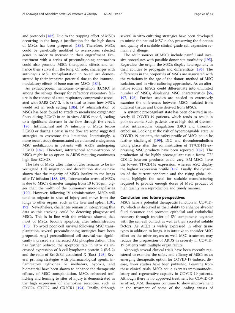
and protocols [182]. Due to the trapping effect of MSCsoccurring in the lung, a justification for the high dosesof MSCs has been proposed [183]. Therefore, MSCscould be genetically modified to overexpress selectedgenes in order to increase in their engraftment. Pre-treatment with a series of preconditioning approachescould also promote MSCs therapeutic effects and en-hance their survival in the lung. Of note, challenges withautologous MSC transplantation in ARDS are demon-strated by their impaired potential due to the immuno-modulatory effects of bone marrow MSCs [184].As extracorporeal membrane oxygenation (ECMO) is
among the salvage therapy for refractory respiratory fail-ure in the context of acute respiratory compromise associ-ated with SARS-CoV-2, it is critical to learn how MSCswould act in such setting [185]. IV administration ofMSCs has been found to attach to membrane oxygenatorfibers during ECMO in an in vitro ARDS model, leadingto a significant decrease in the flow through the circuit[186]. Intratracheal and IV infusions of MSCs beforeECMO or during a pause in the flow are some suggestedstrategies to overcome this limitation. Interestingly, amore recent study demonstrated an enhanced endogenousMSC mobilization in patients with ARDS undergoingECMO [187]. Therefore, intratracheal administration ofMSCs might be an option in ARDS requiring continuoushigh-flow ECMO.The fate of MSCs after infusion also remains to be in-
vestigated. Cell migration and distribution studies haveshown that the majority of MSCs localize to the lungsafter IV infusion [188, 189]. Intravascular arrest of MSCsis due to MSC’s diameter ranging from 10 to 20 μm, big-ger than the width of the pulmonary micro-capillaries[190]. However, following IV administration, MSCs stilltend to migrate to sites of injury and move from thelungs to other organs, such as the liver and spleen [191,192]. Nevertheless, challenges remain in interpreting thisdata as this tracking could be detecting phagocytosedMSCs. This is in line with the evidence showed thatmost of MSCs become apoptotic after administration[193]. To avoid poor cell survival following MSC trans-plantation, several preconditioning strategies have beenproposed. Ang1-preconditioned cell survival was signifi-cantly increased via increased Akt phosphorylation. Thishas further reduced the apoptotic rate in vitro via in-creased expression of B cell lymphoma protein 2 (Bcl-2)and the ratio of Bcl-2/Bcl-associated X (Bax) [193]. Sev-eral priming strategies with pharmacological agents, in-flammatory cytokines or mediators, hypoxia, andbiomaterial have been shown to enhance the therapeuticefficacy of MSC transplantation. MSCs enhanced traf-ficking and homing to sites of injury is demonstrated inthe high expression of chemokine receptors, such asCXCR4, CXCR7, and CX3CR1 [194]. Finally, although
several in vitro culturing strategies have been developedto mimic the natural MSC niche, preserving the functionand quality of a scalable clinical-grade cell expansion re-main a challenge.The adult sources of MSCs include painful and inva-
sive procedures with possible donor site morbidity [195].Regardless the origin, the MSCs display heterogeneity intheir abilities to propagate and differentiate [196]. Thedifferences in the properties of MSCs are associated withthe variations in the age of the donor, method of MSCisolation, and in vitro culturing approaches. As an alter-native source, hPSCs could differentiate into unlimitednumber of MSCs, displaying MSC characteristics [55,197, 198]. Further studies are needed to extensivelyexamine the differences between MSCs isolated fromdifferent tissues and those derived from hPSCs.A systemic procoagulant state has been observed in se-
verely ill COVID-19 patients, which tends to result inpoor outcome. Such patients are at high risk of dissemi-nated intravascular coagulation (DIC) and thrombo-embolism. Looking at the risk of hypercoaguable state inCOVID-19 patients, the safety profile of MSCs could befurther challenged [199]. DIC and thromboembolismtaking place after the administration of TF/CD142-ex-pressing MSC products have been reported [182]. Theproduction of the highly procoagulant tissue factor TF/CD142 between products could vary. BM-MSCs havethe lowest TF/CD142 expression, whereas ASC displaythe highest expression profile [182]. Finally, the dynam-ics of the current pandemic and the rising global de-mand highlight the need for scalable manufacturingrequired to provide enough doses of MSC product ofhigh quality in a reproducible and timely manner.
Conclusion and future perspectivesMSCs have a potential therapeutic function in COVID-19, which is displayed in their ability to enhance alveolarfluid clearance and promote epithelial and endothelialrecovery through transfer of EV components togetherwith the cell-cell contact as well as their secreted solublefactors. As ACE2 is widely expressed in other tissuetypes in addition to lungs, it is intuitive to consider MSCeffect on the other organs as well. MSC treatment mayreduce the progression of ARDS in severely ill COVID-19 patients with multiple organ failure.Although several clinical trials have been recently reg-
istered to examine the safety and efficacy of MSCs as anemerging therapeutic option for COVID-19-induced dis-ease, fewer studies have been published. Learning fromthese clinical trials, MSCs could exert its immunomodu-latory and regenerative capacity in COVID-19 patients.Although there is no approved treatment for COVID-19as of yet, MSC therapies continue to show improvementin the treatment of some of the leading causes of
Al-Khawaga and Abdelalim Stem Cell Research & Therapy (2020) 11:437 Page 28 of 33
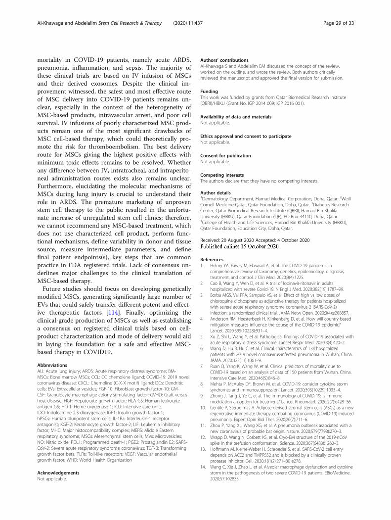
mortality in COVID-19 patients, namely acute ARDS,pneumonia, inflammation, and sepsis. The majority ofthese clinical trials are based on IV infusion of MSCsand their derived exosomes. Despite the clinical im-provement witnessed, the safest and most effective routeof MSC delivery into COVID-19 patients remains un-clear, especially in the context of the heterogeneity ofMSC-based products, intravascular arrest, and poor cellsurvival. IV infusions of poorly characterized MSC prod-ucts remain one of the most significant drawbacks ofMSC cell-based therapy, which could theoretically pro-mote the risk for thromboembolism. The best deliveryroute for MSCs giving the highest positive effects withminimum toxic effects remains to be resolved. Whetherany difference between IV, intratracheal, and intraperito-neal administration routes exists also remains unclear.Furthermore, elucidating the molecular mechanisms ofMSCs during lung injury is crucial to understand theirrole in ARDS. The premature marketing of unprovenstem cell therapy to the public resulted in the unfortu-nate increase of unregulated stem cell clinics; therefore,we cannot recommend any MSC-based treatment, whichdoes not use characterized cell product, perform func-tional mechanisms, define variability in donor and tissuesource, measure intermediate parameters, and definefinal patient endpoints(s), key steps that are commonpractice in FDA registered trials. Lack of consensus un-derlines major challenges to the clinical translation ofMSC-based therapy.Future studies should focus on developing genetically
modified MSCs, generating significantly large number ofEVs that could safely transfer different potent and effect-ive therapeutic factors [114]. Finally, optimizing theclinical-grade production of MSCs as well as establishinga consensus on registered clinical trials based on cell-product characterization and mode of delivery would aidin laying the foundation for a safe and effective MSC-based therapy in COVID19.
AbbreviationsALI: Acute lung injury; ARDS: Acute respiratory distress syndrome; BM-MSCs: Bone marrow MSCs; CCL: CC chemokine ligand; COVID-19: 2019 novelcoronavirus disease; CXCL: Chemoline (C-X-X motif) ligand; DCs: Dendriticcells; EVs: Extracellular vesicles; FGF-10: Fibroblast growth factor-10; GM-CSF: Granulocyte-macrophage colony stimulating factor; GVHD: Graft-versus-host-disease; HGF: Hepatocyte growth factor; HLA-GS: Human leukocyteantigen-G5; HO-1: Heme oxygenase-1; ICU: Intensive care unit;IDO: Indoleamine 2,3-dioxygenase; IGF1: Insulin growth factor 1;hPSCs: Human pluripotent stem cells; IL-1Ra: Interleukin-1 receptorantagonist; KGF-2: Keratinocyte growth factor-2; LIF: Leukemia inhibitoryfactor; MHC: Major histocompatibility complex; MERS: Middle Easternrespiratory syndrome; MSCs: Mesenchymal stem cells; MVs: Microvesicles;NO: Nitric oxide; PDL1: Programmed death-1; PGE2: Prostaglandin E2; SARS-CoV-2: Severe acute respiratory syndrome coronavirus; TGF-β: Transforminggrowth factor beta; TLRs: Toll-like receptors; VEGF: Vascular endothelialgrowth factor; WHO: World Health Organization
AcknowledgementsNot applicable.
Authors’ contributionsAl-Khawaga S and Abdelalim EM discussed the concept of the review,worked on the outline, and wrote the review. Both authors criticallyreviewed the manuscript and approved the final version for submission.
FundingThis work was funded by grants from Qatar Biomedical Research Institute(QBRI)/HBKU (Grant No. IGP 2014 009; IGP 2016 001).
Availability of data and materialsNot applicable.
Ethics approval and consent to participateNot applicable.
Consent for publicationNot applicable.
Competing interestsThe authors declare that they have no competing interests.
Author details1Dermatology Department, Hamad Medical Corporation, Doha, Qatar. 2WeillCornell Medicine-Qatar, Qatar Foundation, Doha, Qatar. 3Diabetes ResearchCenter, Qatar Biomedical Research Institute (QBRI), Hamad Bin KhalifaUniversity (HBKU), Qatar Foundation (QF), PO Box 34110, Doha, Qatar.4College of Health and Life Sciences, Hamad Bin Khalifa University (HBKU),Qatar Foundation, Education City, Doha, Qatar.
Received: 20 August 2020 Accepted: 4 October 2020
References1. Helmy YA, Fawzy M, Elaswad A, et al. The COVID-19 pandemic: a
comprehensive review of taxonomy, genetics, epidemiology, diagnosis,treatment, and control. J Clin Med. 2020;9(4):1225.
2. Cao B, Wang Y, Wen D, et al. A trial of lopinavir-ritonavir in adultshospitalized with severe Covid-19. N Engl J Med. 2020;382(19):1787–99.
3. Borba MGS, Val FFA, Sampaio VS, et al. Effect of high vs low doses ofchloroquine diphosphate as adjunctive therapy for patients hospitalizedwith severe acute respiratory syndrome coronavirus 2 (SARS-CoV-2)infection: a randomized clinical trial. JAMA Netw Open. 2020;3(4):e208857.
4. Anderson RM, Heesterbeek H, Klinkenberg D, et al. How will country-basedmitigation measures influence the course of the COVID-19 epidemic?Lancet. 2020;395(10228):931–4.
5. Xu Z, Shi L, Wang Y, et al. Pathological findings of COVID-19 associated withacute respiratory distress syndrome. Lancet Respir Med. 2020;8(4):420–2.
6. Wang D, Hu B, Hu C, et al. Clinical characteristics of 138 hospitalizedpatients with 2019 novel coronavirus-infected pneumonia in Wuhan, China.JAMA. 2020;323(11):1061–9.
7. Ruan Q, Yang K, Wang W, et al. Clinical predictors of mortality due toCOVID-19 based on an analysis of data of 150 patients from Wuhan, China.Intensive Care Med. 2020;46(5):846–8.
8. Mehta P, McAuley DF, Brown M, et al. COVID-19: consider cytokine stormsyndromes and immunosuppression. Lancet. 2020;395(10229):1033–4.
9. Zhong J, Tang J, Ye C, et al. The immunology of COVID-19: is immunemodulation an option for treatment? Lancet Rheumatol. 2020;2(7):e428–36.
10. Gentile P, Sterodimas A. Adipose-derived stromal stem cells (ASCs) as a newregenerative immediate therapy combating coronavirus (COVID-19)-inducedpneumonia. Expert Opin Biol Ther. 2020;20(7):711–6.
11. Zhou P, Yang XL, Wang XG, et al. A pneumonia outbreak associated with anew coronavirus of probable bat origin. Nature. 2020;579(7798):270–3.
12. Wrapp D, Wang N, Corbett KS, et al. Cryo-EM structure of the 2019-nCoVspike in the prefusion conformation. Science. 2020;367(6483):1260–3.
13. Hoffmann M, Kleine-Weber H, Schroeder S, et al. SARS-CoV-2 cell entrydepends on ACE2 and TMPRSS2 and is blocked by a clinically provenprotease inhibitor. Cell. 2020;181(2):271–80 e278.
14. Wang C, Xie J, Zhao L, et al. Alveolar macrophage dysfunction and cytokinestorm in the pathogenesis of two severe COVID-19 patients. EBioMedicine.2020;57:102833.
Al-Khawaga and Abdelalim Stem Cell Research & Therapy (2020) 11:437 Page 29 of 33
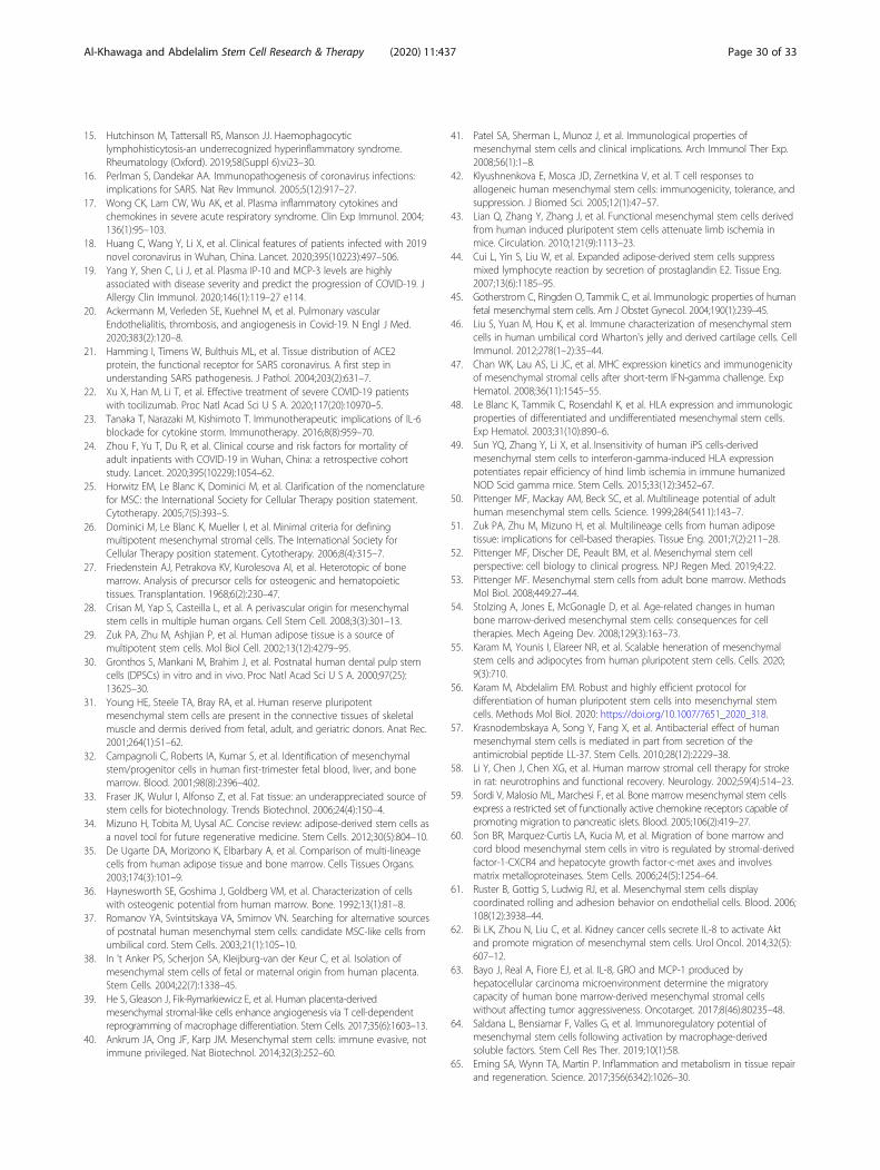
15. Hutchinson M, Tattersall RS, Manson JJ. Haemophagocyticlymphohisticytosis-an underrecognized hyperinflammatory syndrome.Rheumatology (Oxford). 2019;58(Suppl 6):vi23–30.
16. Perlman S, Dandekar AA. Immunopathogenesis of coronavirus infections:implications for SARS. Nat Rev Immunol. 2005;5(12):917–27.
17. Wong CK, Lam CW, Wu AK, et al. Plasma inflammatory cytokines andchemokines in severe acute respiratory syndrome. Clin Exp Immunol. 2004;136(1):95–103.
18. Huang C, Wang Y, Li X, et al. Clinical features of patients infected with 2019novel coronavirus in Wuhan, China. Lancet. 2020;395(10223):497–506.
19. Yang Y, Shen C, Li J, et al. Plasma IP-10 and MCP-3 levels are highlyassociated with disease severity and predict the progression of COVID-19. JAllergy Clin Immunol. 2020;146(1):119–27 e114.
20. Ackermann M, Verleden SE, Kuehnel M, et al. Pulmonary vascularEndothelialitis, thrombosis, and angiogenesis in Covid-19. N Engl J Med.2020;383(2):120–8.
21. Hamming I, Timens W, Bulthuis ML, et al. Tissue distribution of ACE2protein, the functional receptor for SARS coronavirus. A first step inunderstanding SARS pathogenesis. J Pathol. 2004;203(2):631–7.
22. Xu X, Han M, Li T, et al. Effective treatment of severe COVID-19 patientswith tocilizumab. Proc Natl Acad Sci U S A. 2020;117(20):10970–5.
23. Tanaka T, Narazaki M, Kishimoto T. Immunotherapeutic implications of IL-6blockade for cytokine storm. Immunotherapy. 2016;8(8):959–70.
24. Zhou F, Yu T, Du R, et al. Clinical course and risk factors for mortality ofadult inpatients with COVID-19 in Wuhan, China: a retrospective cohortstudy. Lancet. 2020;395(10229):1054–62.
25. Horwitz EM, Le Blanc K, Dominici M, et al. Clarification of the nomenclaturefor MSC: the International Society for Cellular Therapy position statement.Cytotherapy. 2005;7(5):393–5.
26. Dominici M, Le Blanc K, Mueller I, et al. Minimal criteria for definingmultipotent mesenchymal stromal cells. The International Society forCellular Therapy position statement. Cytotherapy. 2006;8(4):315–7.
27. Friedenstein AJ, Petrakova KV, Kurolesova AI, et al. Heterotopic of bonemarrow. Analysis of precursor cells for osteogenic and hematopoietictissues. Transplantation. 1968;6(2):230–47.
28. Crisan M, Yap S, Casteilla L, et al. A perivascular origin for mesenchymalstem cells in multiple human organs. Cell Stem Cell. 2008;3(3):301–13.
29. Zuk PA, Zhu M, Ashjian P, et al. Human adipose tissue is a source ofmultipotent stem cells. Mol Biol Cell. 2002;13(12):4279–95.
30. Gronthos S, Mankani M, Brahim J, et al. Postnatal human dental pulp stemcells (DPSCs) in vitro and in vivo. Proc Natl Acad Sci U S A. 2000;97(25):13625–30.
31. Young HE, Steele TA, Bray RA, et al. Human reserve pluripotentmesenchymal stem cells are present in the connective tissues of skeletalmuscle and dermis derived from fetal, adult, and geriatric donors. Anat Rec.2001;264(1):51–62.
32. Campagnoli C, Roberts IA, Kumar S, et al. Identification of mesenchymalstem/progenitor cells in human first-trimester fetal blood, liver, and bonemarrow. Blood. 2001;98(8):2396–402.
33. Fraser JK, Wulur I, Alfonso Z, et al. Fat tissue: an underappreciated source ofstem cells for biotechnology. Trends Biotechnol. 2006;24(4):150–4.
34. Mizuno H, Tobita M, Uysal AC. Concise review: adipose-derived stem cells asa novel tool for future regenerative medicine. Stem Cells. 2012;30(5):804–10.
35. De Ugarte DA, Morizono K, Elbarbary A, et al. Comparison of multi-lineagecells from human adipose tissue and bone marrow. Cells Tissues Organs.2003;174(3):101–9.
36. Haynesworth SE, Goshima J, Goldberg VM, et al. Characterization of cellswith osteogenic potential from human marrow. Bone. 1992;13(1):81–8.
37. Romanov YA, Svintsitskaya VA, Smirnov VN. Searching for alternative sourcesof postnatal human mesenchymal stem cells: candidate MSC-like cells fromumbilical cord. Stem Cells. 2003;21(1):105–10.
38. In 't Anker PS, Scherjon SA, Kleijburg-van der Keur C, et al. Isolation ofmesenchymal stem cells of fetal or maternal origin from human placenta.Stem Cells. 2004;22(7):1338–45.
39. He S, Gleason J, Fik-Rymarkiewicz E, et al. Human placenta-derivedmesenchymal stromal-like cells enhance angiogenesis via T cell-dependentreprogramming of macrophage differentiation. Stem Cells. 2017;35(6):1603–13.
40. Ankrum JA, Ong JF, Karp JM. Mesenchymal stem cells: immune evasive, notimmune privileged. Nat Biotechnol. 2014;32(3):252–60.
41. Patel SA, Sherman L, Munoz J, et al. Immunological properties ofmesenchymal stem cells and clinical implications. Arch Immunol Ther Exp.2008;56(1):1–8.
42. Klyushnenkova E, Mosca JD, Zernetkina V, et al. T cell responses toallogeneic human mesenchymal stem cells: immunogenicity, tolerance, andsuppression. J Biomed Sci. 2005;12(1):47–57.
43. Lian Q, Zhang Y, Zhang J, et al. Functional mesenchymal stem cells derivedfrom human induced pluripotent stem cells attenuate limb ischemia inmice. Circulation. 2010;121(9):1113–23.
44. Cui L, Yin S, Liu W, et al. Expanded adipose-derived stem cells suppressmixed lymphocyte reaction by secretion of prostaglandin E2. Tissue Eng.2007;13(6):1185–95.
45. Gotherstrom C, Ringden O, Tammik C, et al. Immunologic properties of humanfetal mesenchymal stem cells. Am J Obstet Gynecol. 2004;190(1):239–45.
46. Liu S, Yuan M, Hou K, et al. Immune characterization of mesenchymal stemcells in human umbilical cord Wharton's jelly and derived cartilage cells. CellImmunol. 2012;278(1–2):35–44.
47. Chan WK, Lau AS, Li JC, et al. MHC expression kinetics and immunogenicityof mesenchymal stromal cells after short-term IFN-gamma challenge. ExpHematol. 2008;36(11):1545–55.
48. Le Blanc K, Tammik C, Rosendahl K, et al. HLA expression and immunologicproperties of differentiated and undifferentiated mesenchymal stem cells.Exp Hematol. 2003;31(10):890–6.
49. Sun YQ, Zhang Y, Li X, et al. Insensitivity of human iPS cells-derivedmesenchymal stem cells to interferon-gamma-induced HLA expressionpotentiates repair efficiency of hind limb ischemia in immune humanizedNOD Scid gamma mice. Stem Cells. 2015;33(12):3452–67.
50. Pittenger MF, Mackay AM, Beck SC, et al. Multilineage potential of adulthuman mesenchymal stem cells. Science. 1999;284(5411):143–7.
51. Zuk PA, Zhu M, Mizuno H, et al. Multilineage cells from human adiposetissue: implications for cell-based therapies. Tissue Eng. 2001;7(2):211–28.
52. Pittenger MF, Discher DE, Peault BM, et al. Mesenchymal stem cellperspective: cell biology to clinical progress. NPJ Regen Med. 2019;4:22.
53. Pittenger MF. Mesenchymal stem cells from adult bone marrow. MethodsMol Biol. 2008;449:27–44.
54. Stolzing A, Jones E, McGonagle D, et al. Age-related changes in humanbone marrow-derived mesenchymal stem cells: consequences for celltherapies. Mech Ageing Dev. 2008;129(3):163–73.
55. Karam M, Younis I, Elareer NR, et al. Scalable heneration of mesenchymalstem cells and adipocytes from human pluripotent stem cells. Cells. 2020;9(3):710.
56. Karam M, Abdelalim EM. Robust and highly efficient protocol fordifferentiation of human pluripotent stem cells into mesenchymal stemcells. Methods Mol Biol. 2020: https://doi.org/10.1007/7651_2020_318.
57. Krasnodembskaya A, Song Y, Fang X, et al. Antibacterial effect of humanmesenchymal stem cells is mediated in part from secretion of theantimicrobial peptide LL-37. Stem Cells. 2010;28(12):2229–38.
58. Li Y, Chen J, Chen XG, et al. Human marrow stromal cell therapy for strokein rat: neurotrophins and functional recovery. Neurology. 2002;59(4):514–23.
59. Sordi V, Malosio ML, Marchesi F, et al. Bone marrow mesenchymal stem cellsexpress a restricted set of functionally active chemokine receptors capable ofpromoting migration to pancreatic islets. Blood. 2005;106(2):419–27.
60. Son BR, Marquez-Curtis LA, Kucia M, et al. Migration of bone marrow andcord blood mesenchymal stem cells in vitro is regulated by stromal-derivedfactor-1-CXCR4 and hepatocyte growth factor-c-met axes and involvesmatrix metalloproteinases. Stem Cells. 2006;24(5):1254–64.
61. Ruster B, Gottig S, Ludwig RJ, et al. Mesenchymal stem cells displaycoordinated rolling and adhesion behavior on endothelial cells. Blood. 2006;108(12):3938–44.
62. Bi LK, Zhou N, Liu C, et al. Kidney cancer cells secrete IL-8 to activate Aktand promote migration of mesenchymal stem cells. Urol Oncol. 2014;32(5):607–12.
63. Bayo J, Real A, Fiore EJ, et al. IL-8, GRO and MCP-1 produced byhepatocellular carcinoma microenvironment determine the migratorycapacity of human bone marrow-derived mesenchymal stromal cellswithout affecting tumor aggressiveness. Oncotarget. 2017;8(46):80235–48.
64. Saldana L, Bensiamar F, Valles G, et al. Immunoregulatory potential ofmesenchymal stem cells following activation by macrophage-derivedsoluble factors. Stem Cell Res Ther. 2019;10(1):58.
65. Eming SA, Wynn TA, Martin P. Inflammation and metabolism in tissue repairand regeneration. Science. 2017;356(6342):1026–30.
Al-Khawaga and Abdelalim Stem Cell Research & Therapy (2020) 11:437 Page 30 of 33
66. Galli SJ, Borregaard N, Wynn TA. Phenotypic and functional plasticity of cellsof innate immunity: macrophages, mast cells and neutrophils. Nat Immunol.2011;12(11):1035–44.
67. Sindrilaru A, Peters T, Wieschalka S, et al. An unrestrained proinflammatoryM1 macrophage population induced by iron impairs wound healing inhumans and mice. J Clin Invest. 2011;121(3):985–97.
68. Bernardo ME, Fibbe WE. Mesenchymal stromal cells: sensors and switchersof inflammation. Cell Stem Cell. 2013;13(4):392–402.
69. Nemeth K, Leelahavanichkul A, Yuen PS, et al. Bone marrow stromal cellsattenuate sepsis via prostaglandin E (2)-dependent reprogramming of hostmacrophages to increase their interleukin-10 production. Nat Med. 2009;15(1):42–9.
70. Kim DS, Jang IK, Lee MW, et al. Enhanced immunosuppressive properties ofhuman mesenchymal stem cells primed by interferon-gamma.EBioMedicine. 2018;28:261–73.
71. Francois M, Romieu-Mourez R, Li M, et al. Human MSC suppressioncorrelates with cytokine induction of indoleamine 2,3-dioxygenase andbystander M2 macrophage differentiation. Mol Ther. 2012;20(1):187–95.
72. Li W, Ren G, Huang Y, et al. Mesenchymal stem cells: a double-edged swordin regulating immune responses. Cell Death Differ. 2012;19(9):1505–13.
73. Meisel R, Zibert A, Laryea M, et al. Human bone marrow stromal cells inhibitallogeneic T-cell responses by indoleamine 2,3-dioxygenase-mediatedtryptophan degradation. Blood. 2004;103(12):4619–21.
74. Liu F, Qiu H, Xue M, et al. MSC-secreted TGF-beta regulateslipopolysaccharide-stimulated macrophage M2-like polarization via the Akt/FoxO1 pathway. Stem Cell Res Ther. 2019;10(1):345.
75. Chen PM, Liu KJ, Hsu PJ, et al. Induction of immunomodulatory monocytesby human mesenchymal stem cell-derived hepatocyte growth factorthrough ERK1/2. J Leukoc Biol. 2014;96(2):295–303.
76. Di Nicola M, Carlo-Stella C, Magni M, et al. Human bone marrow stromalcells suppress T-lymphocyte proliferation induced by cellular or nonspecificmitogenic stimuli. Blood. 2002;99(10):3838–43.
77. Bartholomew A, Sturgeon C, Siatskas M, et al. Mesenchymal stem cellssuppress lymphocyte proliferation in vitro and prolong skin graft survivalin vivo. Exp Hematol. 2002;30(1):42–8.
78. Jiang XX, Zhang Y, Liu B, et al. Human mesenchymal stem cells inhibitdifferentiation and function of monocyte-derived dendritic cells. Blood.2005;105(10):4120–6.
79. Nauta AJ, Kruisselbrink AB, Lurvink E, et al. Mesenchymal stem cells inhibitgeneration and function of both CD34+-derived and monocyte-deriveddendritic cells. J Immunol. 2006;177(4):2080–7.
80. Ramasamy R, Fazekasova H, Lam EW, et al. Mesenchymal stem cells inhibitdendritic cell differentiation and function by preventing entry into the cellcycle. Transplantation. 2007;83(1):71–6.
81. Li YP, Paczesny S, Lauret E, et al. Human mesenchymal stem cells licenseadult CD34+ hemopoietic progenitor cells to differentiate into regulatorydendritic cells through activation of the notch pathway. J Immunol. 2008;180(3):1598–608.
82. Aggarwal S, Pittenger MF. Human mesenchymal stem cells modulateallogeneic immune cell responses. Blood. 2005;105(4):1815–22.
83. Moretta A, Bottino C, Vitale M, et al. Activating receptors and coreceptorsinvolved in human natural killer cell-mediated cytolysis. Annu Rev Immunol.2001;19:197–223.
84. Spaggiari GM, Capobianco A, Becchetti S, et al. Mesenchymal stem cell-natural killer cell interactions: evidence that activated NK cells are capableof killing MSCs, whereas MSCs can inhibit IL-2-induced NK-cell proliferation.Blood. 2006;107(4):1484–90.
85. Spaggiari GM, Capobianco A, Abdelrazik H, et al. Mesenchymal stem cellsinhibit natural killer-cell proliferation, cytotoxicity, and cytokine production:role of indoleamine 2,3-dioxygenase and prostaglandin E2. Blood. 2008;111(3):1327–33.
86. Raffaghello L, Bianchi G, Bertolotto M, et al. Human mesenchymal stem cellsinhibit neutrophil apoptosis: a model for neutrophil preservation in thebone marrow niche. Stem Cells. 2008;26(1):151–62.
87. Rasmusson I, Ringden O, Sundberg B, et al. Mesenchymal stem cells inhibitthe formation of cytotoxic T lymphocytes, but not activated cytotoxic Tlymphocytes or natural killer cells. Transplantation. 2003;76(8):1208–13.
88. Gerdoni E, Gallo B, Casazza S, et al. Mesenchymal stem cells effectivelymodulate pathogenic immune response in experimental autoimmuneencephalomyelitis. Ann Neurol. 2007;61(3):219–27.
89. Krampera M, Cosmi L, Angeli R, et al. Role for interferon-gamma in theimmunomodulatory activity of human bone marrow mesenchymal stemcells. Stem Cells. 2006;24(2):386–98.
90. Chen H, Min XH, Wang QY, et al. Pre-activation of mesenchymal stem cellswith TNF-alpha, IL-1beta and nitric oxide enhances its paracrine effects onradiation-induced intestinal injury. Sci Rep. 2015;5:8718.
91. Liang OD, Mitsialis SA, Chang MS, et al. Mesenchymal stromal cellsexpressing heme oxygenase-1 reverse pulmonary hypertension. Stem Cells.2011;29(1):99–107.
92. Ware LB, Matthay MA. Keratinocyte and hepatocyte growth factors in thelung: roles in lung development, inflammation, and repair. Am J PhysiolLung Cell Mol Physiol. 2002;282(5):L924–40.
93. Tong L, Zhou J, Rong L, et al. Fibroblast growth factor-10 (FGF-10) mobilizeslung-resident mesenchymal stem cells and protects against acute lunginjury. Sci Rep. 2016;6:21642.
94. Ge Q, Zhang H, Hou J, et al. VEGF secreted by mesenchymal stem cellsmediates the differentiation of endothelial progenitor cells into endothelialcells via paracrine mechanisms. Mol Med Rep. 2018;17(1):1667–75.
95. Selmani Z, Naji A, Zidi I, et al. Human leukocyte antigen-G5 secretion byhuman mesenchymal stem cells is required to suppress T lymphocyte andnatural killer function and to induce CD4+CD25highFOXP3+ regulatory Tcells. Stem Cells. 2008;26(1):212–22.
96. Sioud M, Mobergslien A, Boudabous A, et al. Mesenchymal stem cell-mediated T cell suppression occurs through secreted galectins. Int J Oncol.2011;38(2):385–90.
97. Gieseke F, Bohringer J, Bussolari R, et al. Human multipotent mesenchymalstromal cells use galectin-1 to inhibit immune effector cells. Blood. 2010;116(19):3770–9.
98. Lee K, Park N, Jung H, et al. Mesenchymal stem cells ameliorateexperimental arthritis via expression of interleukin-1 receptor antagonist.PLoS One. 2018;13(2):e0193086.
99. Davies LC, Heldring N, Kadri N, et al. Mesenchymal stromal cell secretion ofprogrammed death-1 ligands regulates T cell mediatedimmunosuppression. Stem Cells. 2017;35(3):766–76.
100. Meirelles Lda S, Fontes AM, Covas DT, et al. Mechanisms involved in thetherapeutic properties of mesenchymal stem cells. Cytokine Growth FactorRev. 2009;20(5–6):419–27.
101. Togel F, Weiss K, Yang Y, et al. Vasculotropic, paracrine actions of infusedmesenchymal stem cells are important to the recovery from acute kidneyinjury. Am J Physiol Renal Physiol. 2007;292(5):F1626–35.
102. Abraham A, Krasnodembskaya A. Mesenchymal stem cell-derivedextracellular vesicles for the treatment of acute respiratory distresssyndrome. Stem Cells Transl Med. 2020;9(1):28–38.
103. Cocucci E, Racchetti G, Meldolesi J. Shedding microvesicles: artefacts nomore. Trends Cell Biol. 2009;19(2):43–51.
104. Wang B, Yao K, Huuskes BM, et al. Mesenchymal stem cells deliverexogenous MicroRNA-let7c via exosomes to attenuate renal fibrosis. MolTher. 2016;24(7):1290–301.
105. Gu D, Zou X, Ju G, et al. Mesenchymal stromal cells derived extracellularvesicles ameliorate acute renal ischemia reperfusion injury by inhibition ofmitochondrial fission through miR-30. Stem Cells Int. 2016;2016:2093940.
106. Feng Y, Huang W, Wani M, et al. Ischemic preconditioning potentiates theprotective effect of stem cells through secretion of exosomes by targetingMecp2 via miR-22. PLoS One. 2014;9(2):e88685.
107. Yu B, Kim HW, Gong M, et al. Exosomes secreted from GATA-4overexpressing mesenchymal stem cells serve as a reservoir of anti-apoptotic microRNAs for cardioprotection. Int J Cardiol. 2015;182:349–60.
108. Wang X, Gu H, Qin D, et al. Exosomal miR-223 contributes to mesenchymalstem cell-elicited cardioprotection in polymicrobial sepsis. Sci Rep. 2015;5:13721.
109. Lou G, Yang Y, Liu F, et al. MiR-122 modification enhances the therapeuticefficacy of adipose tissue-derived mesenchymal stem cells against liverfibrosis. J Cell Mol Med. 2017;21(11):2963–73.
110. Chen L, Lu FB, Chen DZ, et al. BMSCs-derived miR-223-containing exosomescontribute to liver protection in experimental autoimmune hepatitis. MolImmunol. 2018;93:38–46.
111. Qu Y, Zhang Q, Cai X, et al. Exosomes derived from miR-181-5p-modifiedadipose-derived mesenchymal stem cells prevent liver fibrosis viaautophagy activation. J Cell Mol Med. 2017;21(10):2491–502.
112. Xin H, Li Y, Liu Z, et al. MiR-133b promotes neural plasticity and functionalrecovery after treatment of stroke with multipotent mesenchymal stromal
Al-Khawaga and Abdelalim Stem Cell Research & Therapy (2020) 11:437 Page 31 of 33
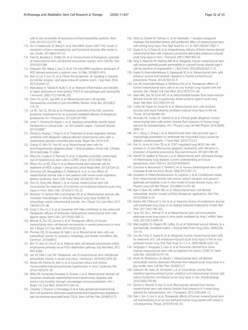
cells in rats via transfer of exosome-enriched extracellular particles. StemCells. 2013;31(12):2737–46.
113. Xin H, Katakowski M, Wang F, et al. MicroRNA cluster miR-17-92 cluster inexosomes enhance neuroplasticity and functional recovery after stroke inrats. Stroke. 2017;48(3):747–53.
114. Park KS, Bandeira E, Shelke GV, et al. Enhancement of therapeutic potentialof mesenchymal stem cell-derived extracellular vesicles. Stem Cell Res Ther.2019;10(1):288.
115. Ferguson SW, Wang J, Lee CJ, et al. The microRNA regulatory landscape ofMSC-derived exosomes: a systems view. Sci Rep. 2018;8(1):1419.
116. Wen H, Lei Y, Eun SY, et al. Plexin-A4-semaphorin 3A signaling is requiredfor toll-like receptor- and sepsis-induced cytokine storm. J Exp Med. 2010;207(13):2943–57.
117. Matsukawa A, Takeda K, Kudo S, et al. Aberrant inflammation and lethalityto septic peritonitis in mice lacking STAT3 in macrophages and neutrophils.J Immunol. 2003;171(11):6198–205.
118. Chen TS, Lai RC, Lee MM, et al. Mesenchymal stem cell secretesmicroparticles enriched in pre-microRNAs. Nucleic Acids Res. 2010;38(1):215–24.
119. Lai RC, Tan SS, Teh BJ, et al. Proteolytic potential of the MSC exosomeproteome: implications for an exosome-mediated delivery of therapeuticproteasome. Int J Proteomics. 2012;2012:971907.
120. Lener T, Gimona M, Aigner L, et al. Applying extracellular vesicles basedtherapeutics in clinical trials - an ISEV position paper. J Extracell Vesicles.2015;4:30087.
121. Zheng G, Huang L, Tong H, et al. Treatment of acute respiratory distresssyndrome with allogeneic adipose-derived mesenchymal stem cells: arandomized, placebo-controlled pilot study. Respir Res. 2014;15:39.
122. Chang YS, Ahn SY, Yoo HS, et al. Mesenchymal stem cells forbronchopulmonary dysplasia: phase 1 dose-escalation clinical trial. J Pediatr.2014;164(5):966–72 e966.
123. Weiss DJ, Casaburi R, Flannery R, et al. A placebo-controlled, randomizedtrial of mesenchymal stem cells in COPD. Chest. 2013;143(6):1590–8.
124. Wilson JG, Liu KD, Zhuo H, et al. Mesenchymal stem (stromal) cells fortreatment of ARDS: a phase 1 clinical trial. Lancet Respir Med. 2015;3(1):24–32.
125. Simonson OE, Mougiakakos D, Heldring N, et al. In vivo effects ofmesenchymal stromal cells in two patients with severe acute respiratorydistress syndrome. Stem Cells Transl Med. 2015;4(10):1199–213.
126. Zhu YG, Feng XM, Abbott J, et al. Human mesenchymal stem cellmicrovesicles for treatment of Escherichia coli endotoxin-induced acute lunginjury in mice. Stem Cells. 2014;32(1):116–25.
127. Morrison TJ, Jackson MV, Cunningham EK, et al. Mesenchymal stromal cellsmodulate macrophages in clinically relevant lung injury models byextracellular vesicle mitochondrial transfer. Am J Respir Crit Care Med. 2017;196(10):1275–86.
128. Song Y, Dou H, Li X, et al. Exosomal miR-146a contributes to the enhancedtherapeutic efficacy of interleukin-1beta-primed mesenchymal stem cellsagainst sepsis. Stem Cells. 2017;35(5):1208–21.
129. Monsel A, Zhu YG, Gennai S, et al. Therapeutic effects of humanmesenchymal stem cell-derived microvesicles in severe pneumonia in mice.Am J Respir Crit Care Med. 2015;192(3):324–36.
130. Phinney DG, Di Giuseppe M, Njah J, et al. Mesenchymal stem cells useextracellular vesicles to outsource mitophagy and shuttle microRNAs. NatCommun. 2015;6:8472.
131. Kim YS, Kim JY, Cho R, et al. Adipose stem cell-derived nanovesicles inhibitemphysema primarily via an FGF2-dependent pathway. Exp Mol Med. 2017;49(1):e284.
132. Lee JH, Park J, Lee JW. Therapeutic use of mesenchymal stem cell-derivedextracellular vesicles in acute lung injury. Transfusion. 2019;59(S1):876–83.
133. Aliotta JM, Pereira M, Wen S, et al. Exosomes induce and reversemonocrotaline-induced pulmonary hypertension in mice. Cardiovasc Res.2016;110(3):319–30.
134. Willis GR, Fernandez-Gonzalez A, Anastas J, et al. Mesenchymal stromal cellexosomes ameliorate experimental bronchopulmonary dysplasia andrestore lung function through macrophage immunomodulation. Am JRespir Crit Care Med. 2018;197(1):104–16.
135. Chaubey S, Thueson S, Ponnalagu D, et al. Early gestational mesenchymalstem cell secretome attenuates experimental bronchopulmonary dysplasia inpart via exosome-associated factor TSG-6. Stem Cell Res Ther. 2018;9(1):173.
136. Ortiz LA, Dutreil M, Fattman C, et al. Interleukin 1 receptor antagonistmediates the antiinflammatory and antifibrotic effect of mesenchymal stemcells during lung injury. Proc Natl Acad Sci U S A. 2007;104(26):11002–7.
137. Gupta N, Su X, Popov B, et al. Intrapulmonary delivery of bone marrow-derivedmesenchymal stem cells improves survival and attenuates endotoxin-inducedacute lung injury in mice. J Immunol. 2007;179(3):1855–63.
138. Fang X, Neyrinck AP, Matthay MA, et al. Allogeneic human mesenchymal stemcells restore epithelial protein permeability in cultured human alveolar type IIcells by secretion of angiopoietin-1. J Biol Chem. 2010;285(34):26211–22.
139. Gupta N, Krasnodembskaya A, Kapetanaki M, et al. Mesenchymal stem cellsenhance survival and bacterial clearance in murine Escherichia colipneumonia. Thorax. 2012;67(6):533–9.
140. Lee JW, Krasnodembskaya A, McKenna DH, et al. Therapeutic effects ofhuman mesenchymal stem cells in ex vivo human lungs injured with livebacteria. Am J Respir Crit Care Med. 2013;187(7):751–60.
141. Islam MN, Das SR, Emin MT, et al. Mitochondrial transfer from bone-marrow-derived stromal cells to pulmonary alveoli protects against acute lunginjury. Nat Med. 2012;18(5):759–65.
142. Curley GF, Hayes M, Ansari B, et al. Mesenchymal stem cells enhancerecovery and repair following ventilator-induced lung injury in the rat.Thorax. 2012;67(6):496–501.
143. McAuley DF, Curley GF, Hamid UI, et al. Clinical grade allogeneic humanmesenchymal stem cells restore alveolar fluid clearance in human lungsrejected for transplantation. Am J Physiol Lung Cell Mol Physiol. 2014;306(9):L809–15.
144. Jin L, Deng Z, Zhang J, et al. Mesenchymal stem cells promote type 2macrophage polarization to ameliorate the myocardial injury caused bydiabetic cardiomyopathy. J Transl Med. 2019;17(1):251.
145. Poe SL, Arora M, Oriss TB, et al. STAT1-regulated lung MDSC-like cellsproduce IL-10 and efferocytose apoptotic neutrophils with relevance inresolution of bacterial pneumonia. Mucosal Immunol. 2013;6(1):189–99.
146. Harrell CR, Sadikot R, Pascual J, et al. Mesenchymal stem cell-based therapyof inflammatory lung diseases: current understanding and futureperspectives. Stem Cells Int. 2019;2019:4236973.
147. Corcione A, Benvenuto F, Ferretti E, et al. Human mesenchymal stem cellsmodulate B-cell functions. Blood. 2006;107(1):367–72.
148. Goolaerts A, Pellan-Randrianarison N, Larghero J, et al. Conditioned mediafrom mesenchymal stromal cells restore sodium transport and preserveepithelial permeability in an in vitro model of acute alveolar injury. Am JPhysiol Lung Cell Mol Physiol. 2014;306(11):L975–85.
149. Rani S, Ryan AE, Griffin MD, et al. Mesenchymal stem cell-derivedextracellular vesicles: toward cell-free therapeutic applications. Mol Ther.2015;23(5):812–23.
150. Katsha AM, Ohkouchi S, Xin H, et al. Paracrine factors of multipotent stromalcells ameliorate lung injury in an elastase-induced emphysema model. MolTher. 2011;19(1):196–203.
151. Tang XD, Shi L, Monsel A, et al. Mesenchymal stem cell microvesiclesattenuate acute lung injury in mice partly mediated by Ang-1 mRNA. StemCells. 2017;35(7):1849–59.
152. MacLoughlin RJ, Higgins BD, Laffey JG, et al. Optimized aerosol delivery to amechanically ventilated rodent. J Aerosol Med Pulm Drug Deliv. 2009;22(4):323–32.
153. Lee JW, Fang X, Gupta N, et al. Allogeneic human mesenchymal stem cellsfor treatment of E. coli endotoxin-induced acute lung injury in the ex vivoperfused human lung. Proc Natl Acad Sci U S A. 2009;106(38):16357–62.
154. Sengupta V, Sengupta S, Lazo A, et al. Exosomes derived from bonemarrow mesenchymal stem cells as treatment for severe COVID-19. StemCells Dev. 2020;29(12):747–54.
155. Khatri M, Richardson LA, Meulia T. Mesenchymal stem cell-derivedextracellular vesicles attenuate influenza virus-induced acute lung injury in apig model. Stem Cell Res Ther. 2018;9(1):17.
156. Varkouhi AK, Jerkic M, Ormesher L, et al. Extracellular vesicles frominterferon-gamma-primed human umbilical cord mesenchymal stromal cellsreduce Escherichia coli-induced acute lung injury in rats. Anesthesiology.2019;130(5):778–90.
157. Gennai S, Monsel A, Hao Q, et al. Microvesicles derived from humanmesenchymal stem cells restore alveolar fluid clearance in human lungsrejected for transplantation. Am J Transplant. 2015;15(9):2404–12.
158. Park J, Kim S, Lim H, et al. Therapeutic effects of human mesenchymal stemcell microvesicles in an ex vivo perfused human lung injured with severe E.coli pneumonia. Thorax. 2019;74(1):43–50.
Al-Khawaga and Abdelalim Stem Cell Research & Therapy (2020) 11:437 Page 32 of 33
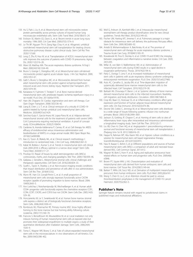
159. Hu S, Park J, Liu A, et al. Mesenchymal stem cell microvesicles restoreprotein permeability across primary cultures of injured human lungmicrovascular endothelial cells. Stem Cells Transl Med. 2018;7(8):615–24.
160. Erickson SE, Martin GS, Davis JL, et al. Recent trends in acute lung injurymortality: 1996-2005. Crit Care Med. 2009;37(5):1574–9.
161. Le Thi BP, Nguyen Thi H, Dang Ngo Chau H, et al. Allogeneic umbilicalcord-derived mesenchymal stem cell transplantation for treating chronicobstructive pulmonary disease: a pilot clinical study. Stem Cell Res Ther.2020;11(1):60.
162. Leng Z, Zhu R, Hou W, et al. Transplantation of ACE2(-) mesenchymal stemcells improves the outcome of patients with COVID-19 pneumonia. AgingDis. 2020;11(2):216–28.
163. Ware LB, Matthay MA. The acute respiratory distress syndrome. N Engl JMed. 2000;342(18):1334–49.
164. Bruno S, Grange C, Deregibus MC, et al. Mesenchymal stem cell-derivedmicrovesicles protect against acute tubular injury. J Am Soc Nephrol. 2009;20(5):1053–67.
165. Gatti S, Bruno S, Deregibus MC, et al. Microvesicles derived from humanadult mesenchymal stem cells protect against ischaemia-reperfusion-induced acute and chronic kidney injury. Nephrol Dial Transplant. 2011;26(5):1474–83.
166. Kanazawa H, Fujimoto Y, Teratani T, et al. Bone marrow-derivedmesenchymal stem cells ameliorate hepatic ischemia reperfusion injury in arat model. PLoS One. 2011;6(4):e19195.
167. Hare JM, Chaparro SV. Cardiac regeneration and stem cell therapy. CurrOpin Organ Transplant. 2008;13(5):536–42.
168. Liang B, Chen J, Li T, et al. Clinical remission of a critically ill COVID-19patient treated by human umbilical cord mesenchymal stem cells.Medicine. 2020;99(31):e21429.
169. Sanchez-Guijo F, García-Arranz M, Lopez-Parra M, et al. Adipose-derivedmesenchymal stromal cells for the treatment of patients with severe SARS-CoV-2 pneumonia requiring mechanical ventilation. A proof of conceptstudy. EClinicalMedicine. 2020;25:100454.
170. Cardenes N, Aranda-Valderrama P, Carney JP, et al. Cell therapy for ARDS:efficacy of endobronchial versus intravenous administration andbiodistribution of MAPCs in a large animal model. BMJ Open Respir Res.2019;6(1):e000308.
171. Kao LS, Tyson JE, Blakely ML, et al. Clinical research methodology I:introduction to randomized trials. J Am Coll Surg. 2008;206(2):361–9.
172. Kabat M, Bobkov I, Kumar S, et al. Trends in mesenchymal stem cell clinicaltrials 2004-2018: is efficacy optimal in a narrow dose range? Stem CellsTransl Med. 2020;9(1):17–27.
173. Prockop DJ. Repair of tissues by adult stem/progenitor cells (MSCs):controversies, myths, and changing paradigms. Mol Ther. 2009;17(6):939–46.
174. Galipeau J, Sensebe L. Mesenchymal stromal cells: clinical challenges andtherapeutic opportunities. Cell Stem Cell. 2018;22(6):824–33.
175. Scarfe L, Taylor A, Sharkey J, et al. Non-invasive imaging reveals conditionsthat impact distribution and persistence of cells after in vivo administration.Stem Cell Res Ther. 2018;9(1):332.
176. Wynn RF, Hart CA, Corradi-Perini C, et al. A small proportion ofmesenchymal stem cells strongly expresses functionally active CXCR4receptor capable of promoting migration to bone marrow. Blood. 2004;104(9):2643–5.
177. Von Luttichau I, Notohamiprodjo M, Wechselberger A, et al. Human adultCD34- progenitor cells functionally express the chemokine receptors CCR1,CCR4, CCR7, CXCR5, and CCR10 but not CXCR4. Stem Cells Dev. 2005;14(3):329–36.
178. Honczarenko M, Le Y, Swierkowski M, et al. Human bone marrow stromalcells express a distinct set of biologically functional chemokine receptors.Stem Cells. 2006;24(4):1030–41.
179. Rombouts WJ, Ploemacher RE. Primary murine MSC show highly efficienthoming to the bone marrow but lose homing ability following culture.Leukemia. 2003;17(1):160–70.
180. Francois S, Bensidhoum M, Mouiseddine M, et al. Local irradiation not onlyinduces homing of human mesenchymal stem cells at exposed sites butpromotes their widespread engraftment to multiple organs: a study of theirquantitative distribution after irradiation damage. Stem Cells. 2006;24(4):1020–9.
181. Toma C, Wagner WR, Bowry S, et al. Fate of culture-expanded mesenchymalstem cells in the microvasculature: in vivo observations of cell kinetics. CircRes. 2009;104(3):398–402.
182. Moll G, Ankrum JA, Kamhieh-Milz J, et al. Intravascular mesenchymalstromal/stem cell therapy product diversification: time for new clinicalguidelines. Trends Mol Med. 2019;25(2):149–63.
183. Fischer UM, Harting MT, Jimenez F, et al. Pulmonary passage is a majorobstacle for intravenous stem cell delivery: the pulmonary first-pass effect.Stem Cells Dev. 2009;18(5):683–92.
184. Antebi B, Mohammadipoor A, Batchinsky AI, et al. The promise ofmesenchymal stem cell therapy for acute respiratory distress syndrome. JTrauma Acute Care Surg. 2018;84(1):183–91.
185. Kowalewski M, Fina D, Slomka A, et al. COVID-19 and ECMO: the interplaybetween coagulation and inflammation-a narrative review. Crit Care. 2020;24(1):205.
186. Millar JE, von Bahr V, Malfertheiner MV, et al. Administration ofmesenchymal stem cells during ECMO results in a rapid decline inoxygenator performance. Thorax. 2019;74(2):194–6.
187. Patry C, Doniga T, Lenz F, et al. Increased mobilization of mesenchymalstem cells in patients with acute respiratory distress syndrome undergoingextracorporeal membrane oxygenation. PLoS One. 2020;15(1):e0227460.
188. Assis AC, Carvalho JL, Jacoby BA, et al. Time-dependent migration ofsystemically delivered bone marrow mesenchymal stem cells to theinfarcted heart. Cell Transplant. 2010;19(2):219–30.
189. Barbash IM, Chouraqui P, Baron J, et al. Systemic delivery of bone marrow-derived mesenchymal stem cells to the infarcted myocardium: feasibility,cell migration, and body distribution. Circulation. 2003;108(7):863–8.
190. Crop MJ, Baan CC, Korevaar SS, et al. Inflammatory conditions affect geneexpression and function of human adipose tissue-derived mesenchymalstem cells. Clin Exp Immunol. 2010;162(3):474–86.
191. Devine SM, Cobbs C, Jennings M, et al. Mesenchymal stem cells distributeto a wide range of tissues following systemic infusion into nonhumanprimates. Blood. 2003;101(8):2999–3001.
192. Jackson JS, Golding JP, Chapon C, et al. Homing of stem cells to sites ofinflammatory brain injury after intracerebral and intravenous administration:a longitudinal imaging study. Stem Cell Res Ther. 2010;1(2):17.
193. Liu XB, Chen H, Chen HQ, et al. Angiopoietin-1 preconditioning enhancessurvival and functional recovery of mesenchymal stem cell transplantation. JZhejiang Univ Sci B. 2012;13(8):616–23.
194. Haque N, Rahman MT, Abu Kasim NH, et al. Hypoxic culture conditions as asolution for mesenchymal stem cell based regenerative therapy.ScientificWorldJournal. 2013;2013:632972.
195. Hass R, Kasper C, Bohm S, et al. Different populations and sources of humanmesenchymal stem cells (MSC): a comparison of adult and neonatal tissue-derived MSC. Cell Commun Signal. 2011;9:12.
196. Wagner W, Bork S, Horn P, et al. Aging and replicative senescence haverelated effects on human stem and progenitor cells. PLoS One. 2009;4(6):e5846.
197. Brown PT, Squire MW, Li WJ. Characterization and evaluation ofmesenchymal stem cells derived from human embryonic stem cells andbone marrow. Cell Tissue Res. 2014;358(1):149–64.
198. Barberi T, Willis LM, Socci ND, et al. Derivation of multipotent mesenchymalprecursors from human embryonic stem cells. PLoS Med. 2005;2(6):e161.
199. Wang T, Chen R, Liu C, et al. Attention should be paid to venousthromboembolism prophylaxis in the management of COVID-19. LancetHaematol. 2020;7(5):e362–3.
Publisher’s NoteSpringer Nature remains neutral with regard to jurisdictional claims inpublished maps and institutional affiliations.
Al-Khawaga and Abdelalim Stem Cell Research & Therapy (2020) 11:437 Page 33 of 33