Embed Size (px)
Citation preview
OPEN ACCESS Journal of Biological Sciences
ISSN 1727-3048DOI: 10.3923/jbs.2017.157.170
Research ArticlePotential Anticryptococcal Compound from Marine Nocardiopsissynnemataformans
Sudarshan Singh Rathore, Aishwarya Mathivanan, Abirami Ravindran, Ponnusami Venkatachalam and Jayapradha Ramakrishnan
Centre for Research in Infectious Diseases, School of Chemical and Biotechnology, SASTRA University, Tirumalaisamudram, Thanjavur,613401 Tamilnadu, India
AbstractBackground and Objective: Cryptococcus neoformans (C. neoformans) is an emerging opportunistic fungal pathogen, which usuallycauses infection in immunocompromised hosts. Limited antifungal drugs are available to manage cryptococcal meningitis, which requiresnew effective class of drugs with less toxicity. The aims of the study were to enhance the production of antifungal compound from a newstrain of marine Nocardiopsis synnemataformans AF1 against C. neoformans using statistical approach, to determine the molecularweight of the purified compound and to evaluate the effect of antifungal compound against the major virulence factors of C. neoformans, namely capsule and melanin. Materials and Methods: A new strain was screened and isolated from marine sediments based onantagonistic assay against C. neoformans. The antifungal compound was produced using Oyster shells as the substrate. The solid statecultural conditions were selected by one factor, time method and optimized by using response surface methodology. The antifungalcompound was extracted using xylene and purified using HPLC. The purified fraction showing inhibitory action against the C. neoformans capsule growth and melanized cells were studied using MALDI-TOF MS. The minimum inhibitory concentration was determined usingdilution method. Cytotoxicity was observed by HepG2 cell line. One-way ANOVA and t-test performed to test statistical significance formultiple comparisons. Results: The optimized condition to produce the antifungal compound from the new strain AF1 are found to be,50% initial moisture content, 2% yeast extract, Oyster shells with the particle size of 16 and temperature at 40EC. The antifungalcompound exhibits a significant reduction in C. neoformans cells, capsule size (30.18%) and melanized cells (99.3%). The MIC for thepurified compound is estimated to be 200 µg mLG1. The MALDI-TOF MS estimated the molecular weight as 242 Da. Conclusion: Theresults of this study show that the new strain N. synnemataformans AF1 isolated from marine environment exhibited potentialantagonistic activity against C. neoformans.
Key words: Antifungal compound, Cryptococcus neoformans, capsule, melanin, optimization, oyster shells, actinomycetes Received: January 05, 2017 Accepted: March 17, 2017 Published: April 15, 2017
Citation: Sudarshan Singh Rathore, Aishwarya Mathivanan, Abirami Ravindran, Ponnusami Venkatachalam and Jayapradha Ramakrishnan, 2017. Potentialanticryptococcal compound from marine Nocardiopsis synnemataformans. J. Biol. Sci., 17: 157-170.
Corresponding Author: Jayapradha Ramakrishnan, Centre for Research in Infectious Diseases (CRID), School of Chemical and Biotechnology, SASTRA University, Tirumalaisamudram, Thanjavur, 613401 Tamilnadu, India Tel: +91-9952538211
Copyright: © 2017 Sudarshan Singh Rathore et al. This is an open access article distributed under the terms of the creative commons attribution License,which permits unrestricted use, distribution and reproduction in any medium, provided the original author and source are credited.
Competing Interest: The authors have declared that no competing interest exists.
Data Availability: All relevant data are within the paper and its supporting information files.
J. Biol. Sci., 17 (4): 157-170, 2017
INTRODUCTION
Cryptococcus meningitis is an opportunistic neurologicaldisease in immunocompromised individuals. It has emergedas a leading cause of morbidity and mortality in HIV patients1.The etiologic agent, Cryptococcus neoformans, is anencapsulated opportunistic fungal pathogen emerging as amajor threat to immunocompromised patients. However itrarely affects healthy individuals2. Lungs serve as a primaryroute for C. neoformans infections and cause cryptococcalmeningitis when left untreated. Compared with other types ofmeningitis, cryptococcal meningitis accounts for 45% ofdeaths3. Recently, Centre for Disease Control estimatedapproximately one million new cases of cryptococcalmeningitis occurring each year, resulting in 625,000 deathsworldwide. Advances in medical techniques such asorgan transplantation, cancer therapies with the use ofimmunosuppressive drugs, diabetes mellitus and immaturebirth increases the incidence of cryptococcal infections 4,5. Theseverity of infection is due to the presence of multiplevirulence factors such as an outer coat capsule, melanization,production of phospholipase, mannitol, urease andproteinases6. Among these, capsule and melanin are mostimportant virulence factors. Encapsulated C. neoformansresists phagocytosis and causes destructive immuneresponses7. Many of the resistance factors studied inCryptococcus sp., are concerned with preventing damage byReactive Oxygen Species (ROS). The capsule appears to beprotective against ROS and other antimicrobials andprotection is proportional to the size of capsule8. Similarly,melaninzed C. neoformans can potentially bind andneutralize cationic antimicrobial peptides and are mostresistant to amphotericine-B and caspofungin9,10.
The current treatment strategy for cryptococcalmeningitis includes amphotericin B in combination withflucytocine for a period of 2 weeks, followed by fluconozole fora minimum of 8-10 weeks. However a long term therapy leadsto fluconozole resistance in C. neoformans, which is anotheremerging problem11. Furthermore, fluconozole intake for aprolonged period of time can lead to other yeast infectionsuch as candidiasis. Towards the end of these antibioticstherapy, protein abnormalities may persist for years, whichwould cause adverse effects including nephrotoxicity12.
Although the lipid based formulations of amphotericin Bhas excellent efficiency in reducing nephrotoxicity, its cost andinability to pass through the blood brain barrier are majorsetbacks13. Thus, the primary motive of this present studywas to find an alternative antifungal and there are no reportsto study marine bioactive compounds specifically to treat
cryptococcal infections14,15. Present study approach in findinga specific compound that targets capsule and melaninsynthesis is of great importance in treating the cryptococcalinfections effectively.
Bioactive compounds are produced by both solidstate and submerged fermentation. However, solid statefermentation is easily controlled and it is preferred sinceexpenditure on media, scale up and downstream processingare less16. Response Surface Methodology is an efficient toolfor media optimization and analysis of complexity during theinteraction of various factors17. In the present study, usedoyster shell (major marine waste product) as a novel substratefor antifungal production. This is the first report on utilizingoyster shells as substrate for the production of antifungalcompounds.
This study describes the isolation and taxonomy ofanticryptococcal producing strain, optimization by RSMapproach, purification of bioactive molecule anddetermination of molecular weight by MALDI ToF MS. Thecompound was then evaluated for its activity against thetwo major virulence factors of C. neoformans (Capsule andmelanin).
MATERIALS AND METHODS
All the studies were carried out during the 2015-2016academic session in SASTRA University campus. Allreagents were purchased commercial grade from suppliers(Himedia and SRL) and used without further purification.
Test culture: Cryptococcus neoformans 14116 waspurchased from Microbial Culture Collection Centre,Chandigarh. The strain was maintained on potato dextroseagar slants at 4EC and 15% glycerol stocks at -80EC.
Isolation of marine actinomycetes: The marine sedimentsamples were collected from Pamban near Rameshwaram atGulf of Mannar in the month of January. Actinomycetes sp.,were isolated using zobell marine agar, actinomycetesisolation agar, starch agar and starch casein nitrate agarsupplemented with fluconozole (50 µg mLG1) and gentamycin(10 µg mLG1) to inhibit the growth of fungi and bacteria,respectively18. The plates were incubated for 21 days at 28EC.The isolates obtained from each media were stored in 15%glycerol at -80EC.
Screening of antagonistic activity against C. neoformans:The antifungal activity was determined using the protocol ofRamakrishnan et al.19 to select a potential strain. Briefly, the
158
J. Biol. Sci., 17 (4): 157-170, 2017
spore suspensions of individual isolates were spot inoculatedon Muller Hinton agar plates and incubated at 30EC for 3 days.The cells were killed by chloroform vapors. The plates weresubsequently over laid with peptone yeast extract agarswabbed with C. neoformans. The resulting clear Zone ofInhibition (ZOI) was measured after 2 days of incubation.The experiment was repeated thrice. Mean diameter of ZOIand standard deviations were calculated. The strain AF1which exhibited the maximum antagonistic activity againstC. neoformans was selected for taxonomical investigation.
Morphology and taxonomy of antifungal producing strain:The spore morphology of AF1 was studied using ScanningElectron Microscope. The morphology was studied byexamining gold-coated dehydrated specimen using the Japanmake JEOL (JSM 5610 LV).
For 16S rRNA gene sequencing, the genomic DNA wasisolated using the procedure described by Kimura20. The genefragments were amplified by using PCR Kit (GENEI Pvt, Ltd,India) and 517 F (5’-CCA GCA GCC GCG GTA AT-3’) andAct704R (5’-TCT GCG CATTTC ACC GCT AC-3’)21. PCR wasperformed using Eppendorf Mastercycler pro thermal cycler230 V/50-60 Hz with the following profile: initial denaturationat 95EC for 4 min, 30 amplification cycles of (95EC for 1 min,annealing temperature at 50EC for 60 sec, 72EC for 1 min) anda final extension step at 72EC for 4 min. The PCR product wasrun and excised from 1.5% agarose gel, purified with theQIAquick PCR purification kit (QIAGEN) and sequenced usingthe primers 8F and U1492R as previously mentioned.Sequencing was done in Chromous Biotech, Bengaluru,India using ABI 3100 sequencer (Applied Biosystems). Thesequence was edited using FinchTV (Geospiza Inc.) and BioEdit(Ibis Biosciences, Abbott Labs).
Phylogenetic analysis: Sequence similarity search of 16S rRNAsequence from AF1 strain was carried out using BLAST (NCBI).Evolutionary tree was inferred by using neighbour-joiningmethod22. The Clustal X program was used for multiplealignments and phylogenetic23.
Media optimizationSubstrate: Oyster shells were collected from Pamban seashore and used as substrate for solid state fermentation. Theshells were washed thoroughly with distilled water and driedat room temperature. The shells were then broken into piecesand passed through ISI meshes and fractions of mesh 4, 6, 8,10, 12, 14, 16 were collected. Oyster shells of different particlesizes were then autoclaved at 121EC for 15 min for furtherexperiments.
Lassical screeningOne Factor A Time Method (OFAT): The process variableswere selected like temperature, initial moisture level, particlesize and effect of additional nutrients for antifungalproduction from AF1. Among the four variables, effect ofadditional nutrients and initial moisture level alone wereoptimized by OFAT method, remaining two variables aredirectly chosen for interaction studies by RSM approach24.
Effect of moisture content and additional nutrients: Tostudy the effect of initial moisture content, various moisturelevels ranging from 20-70% were employed to fermentationmedium by adjusting with sea water. The fermentationprocess was carried out at 30EC for 13 days. In addition, effectof additional nutrients such as fermentation yeast extract(1%), beef extract (1%), (NH4)2SO4 (0.05%), NH4Cl (0.05%) wereevaluated for their effect on antifungal production from AF1strain.
Antifungal activity was monitored by well diffusionmethod using C. neoformans. The yield was expressed asunits of activity/milliliter of crude dissolved in phosphatebuffer (pH 7), where one U was defined as one mm annularclear ring around the antibiotic disc24.
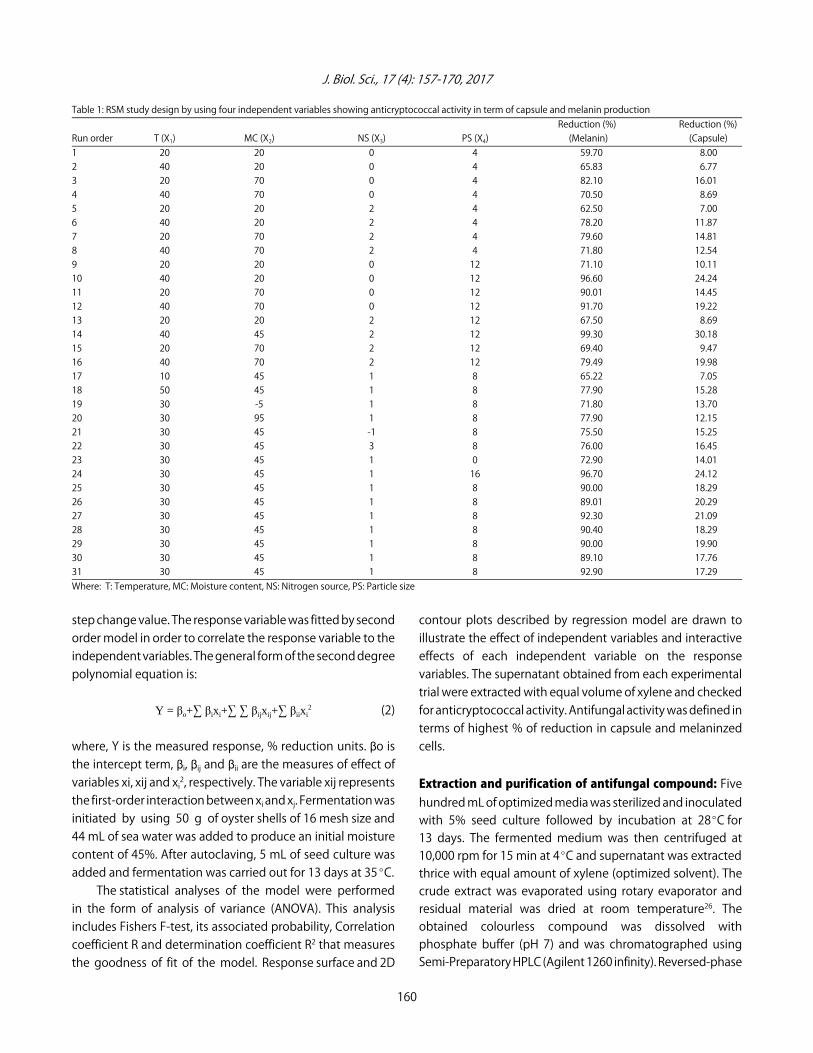
Central Composite Design (CCD): In solid state fermentation,optimization of substrate particle size, initial moisture leveland temperature are crucial factors to increase the yield ofbioactive compound. The effects of yeast extract at differentconcentrations were also considered for experimental design.Based on OFAT method, parameters that had significant effect(p<0.05) on antibiotic production were selected for furtherexperiments of Central Composite Design (CCD) and RSM. Thesoftware Minitab was used for experimental design, dataanalyses and quadratic model building. Experiments werecarried out in 32 trials and duplicates were maintained foreach run. The student’s t-test and p-values were used toidentify the effect of each factor on bioactive compoundproduction. A 24 factorial composite (for 4 factors)experimental design resulting in 32 experiments as shown inTable 1. Minitab statistical software was used to optimize thescreened variables grouped as temperature (X1), moisture(X2), nitrogen source (X3), particle size (X4). In developingregression equation, the test variables were coded accordingto the equation25:
(1)i 0X -Xxi =
X
where, xi is independent variable coded value, Xi isindependent variable real value on the centre point and )X is
159
J. Biol. Sci., 17 (4): 157-170, 2017
Table 1: RSM study design by using four independent variables showing anticryptococcal activity in term of capsule and melanin productionReduction (%) Reduction (%)
Run order T (X1) MC (X2) NS (X3) PS (X4) (Melanin) (Capsule)1 20 20 0 4 59.70 8.002 40 20 0 4 65.83 6.773 20 70 0 4 82.10 16.014 40 70 0 4 70.50 8.695 20 20 2 4 62.50 7.006 40 20 2 4 78.20 11.877 20 70 2 4 79.60 14.818 40 70 2 4 71.80 12.549 20 20 0 12 71.10 10.1110 40 20 0 12 96.60 24.2411 20 70 0 12 90.01 14.4512 40 70 0 12 91.70 19.2213 20 20 2 12 67.50 8.6914 40 45 2 12 99.30 30.1815 20 70 2 12 69.40 9.4716 40 70 2 12 79.49 19.9817 10 45 1 8 65.22 7.0518 50 45 1 8 77.90 15.2819 30 -5 1 8 71.80 13.7020 30 95 1 8 77.90 12.1521 30 45 -1 8 75.50 15.2522 30 45 3 8 76.00 16.4523 30 45 1 0 72.90 14.0124 30 45 1 16 96.70 24.1225 30 45 1 8 90.00 18.2926 30 45 1 8 89.01 20.2927 30 45 1 8 92.30 21.0928 30 45 1 8 90.40 18.2929 30 45 1 8 90.00 19.9030 30 45 1 8 89.10 17.7631 30 45 1 8 92.90 17.29Where: T: Temperature, MC: Moisture content, NS: Nitrogen source, PS: Particle size
step change value. The response variable was fitted by secondorder model in order to correlate the response variable to theindependent variables. The general form of the second degreepolynomial equation is:
Y = βo+3 βixi+3 3 βijxij+3 βiixi2 (2)
where, Y is the measured response, % reduction units. $o isthe intercept term, $i, $ij and $ii are the measures of effect ofvariables xi, xij and xi2, respectively. The variable xij representsthe first-order interaction between xi and xj. Fermentation wasinitiated by using 50 g of oyster shells of 16 mesh size and44 mL of sea water was added to produce an initial moisturecontent of 45%. After autoclaving, 5 mL of seed culture wasadded and fermentation was carried out for 13 days at 35EC.
The statistical analyses of the model were performedin the form of analysis of variance (ANOVA). This analysisincludes Fishers F-test, its associated probability, Correlationcoefficient R and determination coefficient R2 that measuresthe goodness of fit of the model. Response surface and 2D
contour plots described by regression model are drawn toillustrate the effect of independent variables and interactiveeffects of each independent variable on the responsevariables. The supernatant obtained from each experimentaltrial were extracted with equal volume of xylene and checkedfor anticryptococcal activity. Antifungal activity was defined interms of highest % of reduction in capsule and melaninzedcells.
Extraction and purification of antifungal compound: Fivehundred mL of optimized media was sterilized and inoculatedwith 5% seed culture followed by incubation at 28EC for13 days. The fermented medium was then centrifuged at10,000 rpm for 15 min at 4EC and supernatant was extractedthrice with equal amount of xylene (optimized solvent). Thecrude extract was evaporated using rotary evaporator andresidual material was dried at room temperature26. Theobtained colourless compound was dissolved withphosphate buffer (pH 7) and was chromatographed usingSemi-Preparatory HPLC (Agilent 1260 infinity). Reversed-phase
160
J. Biol. Sci., 17 (4): 157-170, 2017
C18 column (10×250 mm) was used with a linear gradientfrom 1-60% MeOH and 9-40% water was performed over25 min at a flow rate of 1 mL minG1. The elution pattern wasmonitored at 225 nm. Fractions were collected and antifungalactivity was performed by broth dilution method in 96 wellplate.
Determination of Minimum Inhibitory Concentration (MIC):MIC of crude and purified compound was estimated bybroth dilution method in 96 well plate and test tubes27.Five mL of PDB media added with 50 µL of C. neoformans culture (106 cells/mL). Culture was added with crude and purified bioactive compound concentration between0-2 mg mLG1 with 200 µg mLG1 interval and 0-500 µg mLG1
with 50 µg mLG1 interval respectively, incubated for 48 h at37EC on a rotary shaker at 120 rpm. After 24 h, 96 well plateobserved in ELISA plate reader (Sunrise, Tecan, Austria GmbH)at 620 nm. Cells were plated on potato dextrose agar platesfrom tubes and number of cells was measured. The lowestcompound concentration with more than 50% fungalinhibition was estimated to be MIC.
Cell line toxicity: The mammalian cell lines HepG2 humanhepatocyte cell line exposed to bioactive compound and timeinterval. The cell lines maintained at 37EC in 5% CO2 and 95%humidity incubator in DMEM media (Himedia) supplementedwith 10 % FBS (Himedia) and 1X antibiotic (Himedia). Cell lineswere incubated with antifungal bioactive compoundconcentration between 0-500 µg mLG1 with the interval of50 µg mLG1 and maximum incubation period for 24 h. Afterincubation cell viability was measured using MTT assay kit(CCK003, EZcount MTT cell assay kit, Himedia) following themanufacturer’s protocol. The assay was performed in 96-wellplates in triplicates and differences were tested for statisticalsignificance by student’s t-test28.
Assay of antifungal compound on major virulence factorsEffect on melanin inhibition: Fifty µL of C. neoformans(106 CFU mLG1) fresh culture was inoculated with crude(1 mg mLG1) and pure (200 µg mLG1) bioactive compound andincubated at 24 h at 30EC in minimal broth media. Afterincubation, 10 µL of treated culture was plated on minimalagar medium containing glucose (15 mM), MgSO4 (10 mM),KH2PO4 (29.4 mM), glycine (13 mM), thiamine (3 :M), agar (2%)with the addition of L-DOPA (1 mM) for induction ofmelanization29. Plates were incubated at 30EC for 4-7 days indark and the number of melanized cells was counted30. Theeffect of compound on the melanised cells and non melanisedcell were compared.
Effect on capsule growth: As the capsule size ofC. neoformansis directly proportional to its virulence, thecapsule was induced in Artificial cerebral spinal fluidmedium31. C. neoformans with maximum capsule was usedto study the effect of bioactive compound on capsule. After48 h of induction period, 50 µL inoculated C. neoformans wasadded with 1 mg mLG1 and 200 µg mLG1 of compound andre-incubated for 24 h at 30EC. Effect of compound on capsulewas evaluated by performing negative staining with India inkusing Trinocular microscope (Eclipse Ci-L, Nikon). Images weretaken with a camera (SLR, Nikon D5100, Camera). To calculaterelative size of capsule, diameters of whole cell, includingcapsule (Dwc) and cell body limited by cell wall (Dcb), weremeasured using Image J software. The size of the capsulerelative to that of the whole cell was defined, as a percentageas [(Dwc - Dcb)/Dwc]100. Fifteen cells were measured for eachdetermination and average was calculated32.
Mass spectroscopy analysis: The purified compoundwas subjected to Nano Spray matrix assisted laserdesorption/ionization time of flight mass spectrometry(MALDI-ToF MS) and the molecular weight was determinedfrom the time of flight.
Statistical analysis: All experiments were performed induplicates and the data analysis was done using Minitab 16software and GraphPad Prism 6. One-way ANOVA and t-testperformed to test statistical significance for multiplecomparisons (p<0.05)31. All graphs were prepared withGraphPad Prism 6 and were expressed as Mean±Experimentsdone in duplicates.
RESULTS AND DISCUSSION
Isolation and taxonomy of potential strain: In this study33 actinomycetes were isolated from marine sediments. Thestrain AF1 isolated using starch agar showed the maximumactivity against C. neoformans. It displayed maximum activityagainst the test organism with 15 mm mean diameter of zoneof inhibition.
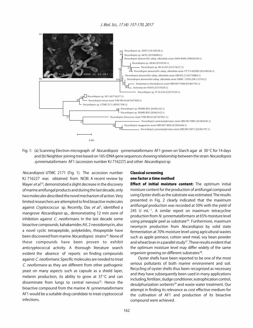
The electron micrographs reveals the spores have smoothsurface in oval shapes around 4-6 spores/round (Fig. 1). The16S rRNA gene sequence of the strain AF1 and its comparisonwith the gene sequences against the GenBank databaserevealed that the organism form a distinct phylogenetic linein the N. synnemataformans. The isolate was closely relatedto the type strain of Nocardiopsis strain UTMC 2171, sharinga homology of 99%. The 16S rRNA sequence analysissupport the classification of the isolate AF1 as a new strain of
161
J. Biol. Sci., 17 (4): 157-170, 2017
Nocardiopsis sp. 20057 (AY336518.1)
Nocardiopsis sp. 64/93 (AY036004.1)
Nocardiopsis dassonvillei subsp. albirubida strain DSM 40465 (NR026339.1)
Nocardiopsis sp. AE46 (JF319150.1)
Nocardiopsis sp. 06-St-023 (GU574127.1)
Nocardiopsis dassonvillei subsp. albirubida strain VTT E-062983 (EU430536.1)
Nocardiopsis dassonvillei subsp. albirubida strain ZBGKL15 (KJ734884.1)
Nocardiopsis dassonvillei subsp. albirubida strain NBRC 13392 (NR 112743.1)
Streptomyces f lavidofuscus strain HBUM173408 (EU841705.1)
Actinomycete Nd105 (FJ379329.1)
Nocardiopsis sp. 07-St-010 (GS574130.1)
Nocardiopsis sp. AF1 (KJ716227.1)
Nocardiopsis terrae strain YIM 98134 (KY427828.1)
Nocardiopsis sp. UTMC2171 (KF917196.1)
Nocardiopsis sp. HSMR-BS1 (KJ461142.1)
Nocardiopsis sp. HSMR-BS2 (KJ461143.1)
Nocardiopsis f lavescens strain YIM 98142 (KY427821.1)
Nocardiopsis synnemataformans strain HBUM174881 (EU841645.1)
Nocardiopsis tangguensis strain HBUM174826 (EU841644.1)
Nocardiopsis synnemataformans strain HBUM174071 (EU841707.1)
20 34
33
6142
42
100 50
1090
53 38
99
23
37
9885
0.001
(a)
(b)
Fig. 1: (a) Scanning Electron micrograph of Nocardiopsis synnemataformans AF1 grown on Starch agar at 30EC for 14 daysand (b) Neighbor-joining tree based on 16S rDNA gene sequences showing relationship between the strain Nocardiopsissynnemataformans AF1 (accession number KJ 716227) and other Nocardiopsis sp
Nocardiopsis UTMC 2171 (Fig. 1). The accession numberKJ 716227 was obtained from NCBI. A recent review byMayer et al.33, demonstrated a slight decrease in the discoveryof marine antifungal products and during the last decade, onlytwo molecules described the novel mechanism of action. Verylimited researchers are attempted to find bioactive moleculesagainst Cryptococcus sp. Recently, Das et al.5, identified amangrove Nocardiopsis sp., demonstrating 12 mm zone ofinhibition against C. neoformans. In the last decade somebioactive compounds, kahakamides AV, 2 neosidomycin, alsoa novel cyclic tetrapeptide, polyketides, thiopeptide havebeen discovered from marine Nocardiopsis strains34. None ofthese compounds have been proven to exhibitanticryptococcal activity. A thorough literature searchevident the absence of reports on finding compoundsagainst C. neoformans. Specific molecules are needed to treatC. neoformans as they are different from other pathogenicyeast on many aspects such as capsule as a shield layer,melanin production, its ability to grow at 37EC and candisseminate from lungs to central nervous35. Hence thebioactive compound from the marine N. synnemataformansAF1 would be a suitable drug candidate to treat cryptococcalinfections.
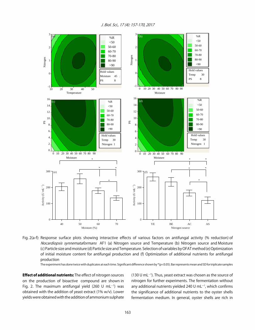
Classical screeningone factor a time methodEffect of initial moisture content: The optimum initialmoisture content for the production of antifungal compoundusing Oyster shells as the substrate was estimated. The resultspresented in Fig. 2 clearly indicated that the maximumantifungal production was recorded at 50% with the yield of245 U mLG1. A similar report on maximum tetracyclineproduction from N. synnemataformans at 65% moisture levelusing pineapple peel as substrate36. Furthermore, maximumneomycin production from Nocardiopsis by solid statefermentation at 70% moisture level using agricultural wastessuch as apple pomace, cotton seed meal, soy bean powderand wheat bran in a parallel study37. These results evident thatthe optimum moisture level may differ widely of the sameorganism growing on different substrates38.
Oyster shells have been reported to be one of the mostserious pollutants of both marine environment and soil.Recycling of oyster shells thus been recognised as necessaryand they have subsequently been used in many applicationsincluding, fertilizer, sludge conditioner, eutrophication control,desulphurization sorbents39 and waste water treatment. Ourattempt in finding its relevance as cost effective medium forthe cultivation of AF1 and production of its bioactivecompound were achieved.
162
J. Biol. Sci., 17 (4): 157-170, 2017
300
200
100
0
Act
ivit
y (U
mL
)G1
40 7050 60Moisture (%)
*
* *
*
(e) 300
200
100
0
Act
ivit
y (U
mL
)G1
YE ASBE ACNitrogen source
*
* *
*
(f)
*
Fig. 2(a-f): Response surface plots showing interactive effects of various factors on antifungal activity (% reduction) ofNocardiopsis synnemataformans AF1 (a) Nitrogen source and Temperature (b) Nitrogen source and Moisture(c) Particle size and moisture (d) Particle size and Temperature. Selection of variables by OFAT method (e) Optimizationof initial moisture content for antifungal production and (f) Optimization of additional nutrients for antifungalproductionThe experiment has done twice with duplicates at each time. Significant difference shown by *(p<0.05). Bar represents mean and SD for triplicate samples
Effect of additional nutrients: The effect of nitrogen sourceson the production of bioactive compound are shown inFig. 2. The maximum antifungal yield (260 U mLG1) wasobtained with the addition of yeast extract (1% w/v). Loweryields were obtained with the addition of ammonium sulphate
(130 U mLG1). Thus, yeast extract was chosen as the source ofnitrogen for further experiments. The fermentation withoutany additional nutrients yielded 240 U mLG1, which confirmsthe significance of additional nutrients to the oyster shellsfermentation medium. In general, oyster shells are rich in
163
Moisture
16
14
12
10
8
6
4
2
0
PS
0 10 20 30 40 50 60 70 80 90
Moisture
%R
<50
50-60
60-70
70-80
80-90
>90
Hold values
Temp 30
Nitrogen 1
(c)
3
2
1
0
-1
Nit
roge
n
%R
<50
50-60
60-70
70-80
80-90
>90
Hold values
Temp 30
PS 8
(b)
0 10 20 30 40 50 60 70 80 90
3
2
1
0
-1
Nit
roge
n
10 20 30 40 50 Temperature
(a)
Hold values
Moisture 45
PS 8
%R <50
50-60 60-70 70-80 80-90 >90
16
14
12
10
8
6
4
2
0
PS
(d) %R
<50
50-60
60-70
70-80
80-90
>90
Hold values
Temp 30
Nitrogen 1
0 10 20 30 40 50 60 70 80 90 Moisture
J. Biol. Sci., 17 (4): 157-170, 2017
calcium carbonate, other essential minerals and organicmatter. For more than 3 decades till date, researchers exploitthe calcium carbonate enriched medium for isolation ofactinomycetes40. From these results, oyster shell mediumenriched with yeast extract would be a suitable cost effectivemedium for the production of bioactive compound fromN. synnemataformans. Moreover it can be an appropriatecultivation and production media for other actinomycetesalso.
Antifungal activity of compound produced by OFATmethod: While screening for the anticryptococcal activity ofthe crude compound obtained by the classical optimization(yeast extract -1%, moisture -50%, oyster shells -16 mesh size,temperature 35EC), the yield obtained was 260 U mLG1. Theeffect of the compound on the Crytococcous capsule growthand on melanized cells shows the reduction of 8 and 48%,respectively.
Central composite regression designStatistical approach: While analyzing the effects of variousfactors (temperature, moisture, particle size and yeast extract) and their interactions in enhancing the antibioticproduction. The goodness of fit of the model based onRSM checked by applying multiple regression analysis onthe experimental data, experimental results of the CCDdesign were fitted with second-order polynomial equation.The results of regression analyses are shown in Table 2. The student’s t-test and p-values were used as a tool to
check the significance of each coefficient that alsoindicated the interaction strength between each independentvariable. The larger magnitude of t-value and smaller thep-value, more significant is the corresponding coefficient41. Itcan be seen from the degree of significance.
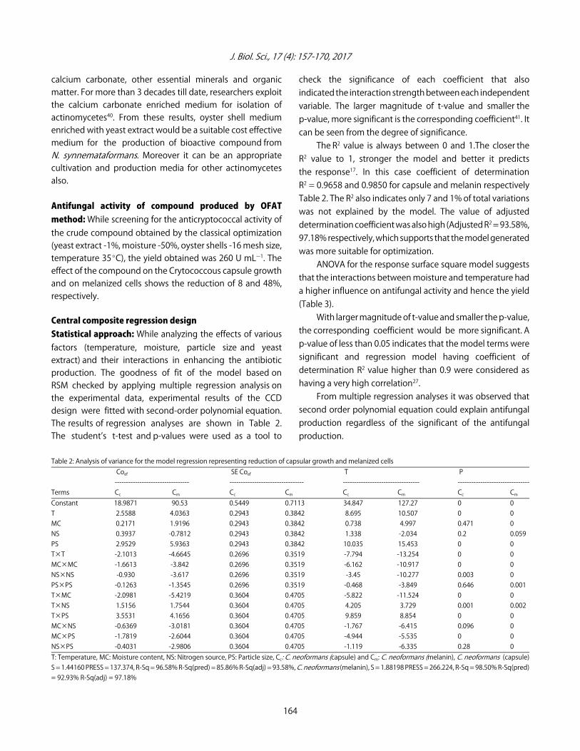
The R2 value is always between 0 and 1.The closer theR2 value to 1, stronger the model and better it predictsthe response17. In this case coefficient of determinationR2 = 0.9658 and 0.9850 for capsule and melanin respectivelyTable 2. The R2 also indicates only 7 and 1% of total variationswas not explained by the model. The value of adjusteddetermination coefficient was also high (Adjusted R2 = 93.58%,97.18% respectively, which supports that the model generatedwas more suitable for optimization.
ANOVA for the response surface square model suggeststhat the interactions between moisture and temperature hada higher influence on antifungal activity and hence the yield(Table 3).
With larger magnitude of t-value and smaller the p-value,the corresponding coefficient would be more significant. Ap-value of less than 0.05 indicates that the model terms weresignificant and regression model having coefficient ofdetermination R2 value higher than 0.9 were considered ashaving a very high correlation27.
From multiple regression analyses it was observed thatsecond order polynomial equation could explain antifungalproduction regardless of the significant of the antifungalproduction.
Table 2: Analysis of variance for the model regression representing reduction of capsular growth and melanized cells Coef SE Coef T P--------------------------------- --------------------------------- ---------------------------------- --------------------------------
Terms Cc Cm Cc Cm Cc Cm Cc CmConstant 18.9871 90.53 0.5449 0.7113 34.847 127.27 0 0T 2.5588 4.0363 0.2943 0.3842 8.695 10.507 0 0MC 0.2171 1.9196 0.2943 0.3842 0.738 4.997 0.471 0NS 0.3937 -0.7812 0.2943 0.3842 1.338 -2.034 0.2 0.059PS 2.9529 5.9363 0.2943 0.3842 10.035 15.453 0 0T×T -2.1013 -4.6645 0.2696 0.3519 -7.794 -13.254 0 0MC×MC -1.6613 -3.842 0.2696 0.3519 -6.162 -10.917 0 0NS×NS -0.930 -3.617 0.2696 0.3519 -3.45 -10.277 0.003 0PS×PS -0.1263 -1.3545 0.2696 0.3519 -0.468 -3.849 0.646 0.001T×MC -2.0981 -5.4219 0.3604 0.4705 -5.822 -11.524 0 0T×NS 1.5156 1.7544 0.3604 0.4705 4.205 3.729 0.001 0.002T×PS 3.5531 4.1656 0.3604 0.4705 9.859 8.854 0 0MC×NS -0.6369 -3.0181 0.3604 0.4705 -1.767 -6.415 0.096 0MC×PS -1.7819 -2.6044 0.3604 0.4705 -4.944 -5.535 0 0NS×PS -0.4031 -2.9806 0.3604 0.4705 -1.119 -6.335 0.28 0T: Temperature, MC: Moisture content, NS: Nitrogen source, PS: Particle size, Cc: C. neoformans (capsule) and Cm: C. neoformans (melanin), C. neoformans (capsule)S = 1.44160 PRESS = 137.374, R-Sq = 96.58% R-Sq(pred) = 85.86% R-Sq(adj) = 93.58%, C. neoformans (melanin), S = 1.88198 PRESS = 266.224, R-Sq = 98.50% R-Sq(pred)= 92.93% R-Sq(adj) = 97.18%
164
J. Biol. Sci., 17 (4): 157-170, 2017
Table 3: ANOVA for Significant Interactions between moisture and temperature Seq SS Adj SS Adj MS F P----------------------------- ---------------------------- ----------------------------- -------------------------- ------------------------------
Sources Cc Cm Cc Cm Cc Cm Cc Cm Cc CmRegression 938.199 3710.61 938.199 3710.61 67.014 265.043 32.25 74.83 0 0Linear 371.258 1339.81 371.258 1339.81 92.814 334.953 44.66 94.57 0 0T 157.133 390.99 157.133 390.99 157.133 390.992 75.61 110.39 0 0MC 1.131 88.44 1.131 88.44 1.131 88.435 0.54 24.97 0.471 0NS 3.721 14.65 3.721 14.65 3.721 14.648 1.79 4.14 0.2 0.059PS 209.273 845.74 209.273 845.74 209.273 845.738 100.70 238.78 0 0Square 197.867 1177.15 197.867 1177.15 49.467 294.286 23.8 83.09 0 0T×T 102.095 445.49 126.259 622.17 126.259 622.168 60.75 175.66 0 0MC×MC 71.017 330.35 78.919 422.10 78.919 422.096 37.97 119.17 0 0NS×NS 24.299 348.84 24.733 374.10 24.733 374.105 11.90 105.62 0.003 0PS×PS 0.456 52.46 0.456 52.46 0.456 52.462 0.22 14.81 0.646 0.001Interaction 369.074 1193.65 369.074 1193.65 61.512 198.941 29.60 56.17 0 0T×MC 70.434 470.35 70.434 470.35 70.434 470.348 33.89 132.80 0 0T×NS 36.754 49.25 36.754 49.25 36.754 49.245 17.69 13.90 0.001 0.002T×PS 201.995 277.64 201.995 277.64 201.995 277.639 97.20 78.39 0 0MC×NS 6.490 145.75 6.490 145.75 6.490 145.745 3.12 41.15 0.096 0MC×PS 50.801 108.52 50.801 108.52 50.801 108.524 24.44 30.64 0 0NS×PS 2.600 142.15 2.600 142.15 2.600 142.146 1.25 40.13 0.28 0Residual error 33.251 56.67 33.251 56.67 2.078 3.542Lack-of-fit 20.941 42.99 20.941 42.99 2.094 4.299 1.02 1.88 0.514 0.226Pure error 12.311 13.68 12.311 13.68 2.052 2.281Total 971.450 3767.27
(3)
1 2 3 4
1 2 1 3 1 4 22 2c
3 2 4 3 4 1 22 23 4
17.6338+3.1856X +0.1444X +0.6894X +3.4956X -1.8381X *X +1.2556X *X +3.2931X *X -0.3769X *
YX -1.52192X *X -0.6631X *X -4.8898X -3.1298X -0.2048X 32-4.0102X
(4)
1 2 3 4
1 2 1 3 1 4 2 32m
2 4 3 4 2 22 23 4
90.53+4.0363X +4.0363X -0.7812X +5.9363X - 5.4219X *X +1.7544X *X +4.1656X *X -3.0181X *X -
Y = 2.6044X *X -2.9806X *X -4.6645X1 -3.8420X -3.6170X -1.3545X
where, Yc and Ym correspond to the effects on the capsule andmelanized cells, respectively.The interaction studies from 2D contour plot shown in
Fig. 2 reveal that all factors (moisture, temperature, yeastextract, particle size), had significant effects (p<0.05) on theantibiotic production. The best interactions are betweenmoisture of 40% temperature of 40EC, yeast extract of 2% andOyster shells with the particle size of 16. The smaller particlesize of oyster shell provides large surface area which helpsto mix the substrate with the microorganisms, other nutrients and uniform distribution of temperature thatsupports the microbes to enhance the production ofantibiotics. Ellaiah et al.42 convey that the increase in highersubstrate moisture in solid state fermentation results in suboptimal product formation due to reduced mass transferprocess such as diffusion of solutes and gas to cell duringfermentation. Also, the decrease in moisture results in reducedsolubility minimizes heat exchange, oxygen transfer and low
availability of nutrients to the culture, leading to decrease inproductivity42. This supports our results at 45% initial moisturelevel using oyster shell.Further validation experiments were also carried out to
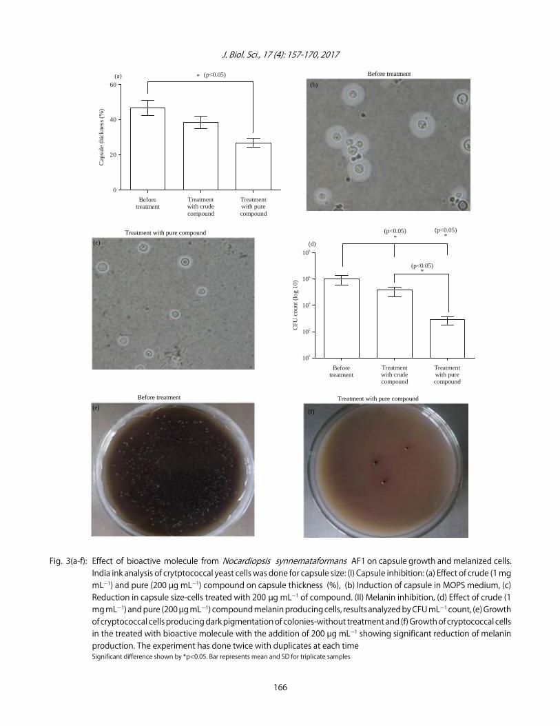
verify the adequacy and accuracy of the model on the resultsuggested that the predicted value agreed with experimentalvalues. The enhanced antifungal activity was obtained withstatistically optimized production medium exhibiting 20.18and 99.3% reduction of capsule growth and melanized cells,respectively. Figure 3 clearly reveals the significant reduction(p<0.05) in capsule growth and melanized cells, respectively.The antifungal compound production yield was increased by60 U mLG1 in statistically optimized condition when comparedto conventional method.
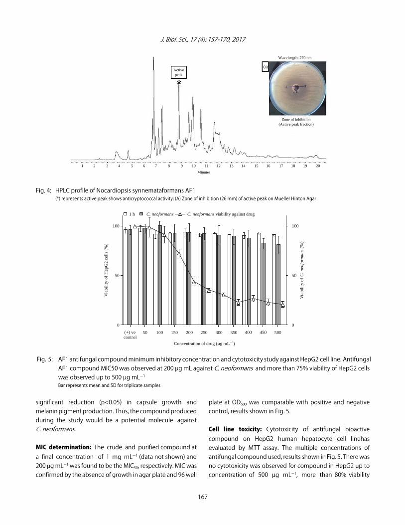
Extraction and Purification of antifungal compound: Thesupernatant which were obtained by growing the strain AF1in the optimized medium for a period of 13 days wereextracted with xylene and the extract was evaporated todryness resulting in a yellow oily residue (2.6 g). The crudeextract was purified by using semi preparatory HPLC. TheHPLC analysis yield 20 fractions and the compoundscorresponding to the first peak starts from 3.5-14.4 min(Fig. 4). The active peak was identified by performingantifungal assay by disc diffusion method. A zone of clearanceof 26 mm for HPLC peak 10th with a RT of 8.7-8.9 min wasobserved on assay plates. However, the other peaks showsno antifungal activity. Also the purified compound showed
165
J. Biol. Sci., 17 (4): 157-170, 2017
60
40
20
0
Cap
sule
thic
knes
s (%
)
Beforetreatment
Treatmentwith purecompound
Treatment with crudecompound
* (p<0.05)(a)
108
106
104
102
100
CFU
cou
nt (
log
10)
Beforetreatment
Treatmentwith purecompound
Treatment with crudecompound
*(p<0.05)
(d)*
(p<0.05)
*(p<0.05)
Fig. 3(a-f): Effect of bioactive molecule from Nocardiopsis synnemataformans AF1 on capsule growth and melanized cells.India ink analysis of crytptococcal yeast cells was done for capsule size: (I) Capsule inhibition: (a) Effect of crude (1 mgmLG1) and pure (200 µg mLG1) compound on capsule thickness (%), (b) Induction of capsule in MOPS medium, (c)Reduction in capsule size-cells treated with 200 µg mLG1 of compound. (II) Melanin inhibition, (d) Effect of crude (1mg mLG1) and pure (200 µg mLG1) compound melanin producing cells, results analyzed by CFU mLG1 count, (e) Growthof cryptococcal cells producing dark pigmentation of colonies-without treatment and (f) Growth of cryptococcal cellsin the treated with bioactive molecule with the addition of 200 µg mLG1 showing significant reduction of melaninproduction. The experiment has done twice with duplicates at each timeSignificant difference shown by *p<0.05. Bar represents mean and SD for triplicate samples
166
Treatment with pure compound
(c)
Before treatment Treatment with pure compound (e)
(f)
Before treatment
(b)
J. Biol. Sci., 17 (4): 157-170, 2017
100
50
0
Via
bilit
y of
Hep
G2
cell
s (%
)
(+) vecontrol
50045040035030025020015010050
Concentration of drug (µg mL ) G1
100
50
0
Via
bili
ty o
f (
%)
C. n
eofo
rman
s
1 h viability against drug C. neoformans C. neoformans
Fig. 4: HPLC profile of Nocardiopsis synnemataformans AF1(*) represents active peak shows anticryptococcal activity; (A) Zone of inhibition (26 mm) of active peak on Mueller Hinton Agar
Fig. 5: AF1 antifungal compound minimum inhibitory concentration and cytotoxicity study against HepG2 cell line. AntifungalAF1 compound MIC50 was observed at 200 µg mL against C. neoformans and more than 75% viability of HepG2 cellswas observed up to 500 µg mLG1Bar represents mean and SD for triplicate samples
significant reduction (p<0.05) in capsule growth andmelanin pigment production. Thus, the compound producedduring the study would be a potential molecule againstC. neoformans.
MIC determination: The crude and purified compound ata final concentration of 1 mg mLG1 (data not shown) and200 :g mLG1 was found to be the MIC50, respectively. MIC wasconfirmed by the absence of growth in agar plate and 96 well
plate at OD600 was comparable with positive and negativecontrol, results shown in Fig. 5.
Cell line toxicity: Cytotoxicity of antifungal bioactivecompound on HepG2 human hepatocyte cell linehasevaluated by MTT assay. The multiple concentrations ofantifungal compound used, results shown in Fig. 5. There wasno cytotoxicity was observed for compound in HepG2 up toconcentration of 500 µg mLG1, more than 80% viability
167
1 2 3 4 5 6 7 8 9 10 11 12 13 14 15 16 17 18 19 20
Minutes
Active peak
(a)
Wavelength: 270 nm
Zone of inhibition (Active peak fraction)
J. Biol. Sci., 17 (4): 157-170, 2017
Fig. 6: Mass spectroscopy analysis of HPLC purified antifungal compound from Nocardiopsis synnemataformans AF1
observed with maximum of 24 h incubation for the cell lineswith antifungal compound. As per test parametric statisticalanalysis, no significant difference was observed for 1 and 24 hcompound treatment to HepG2 cells.
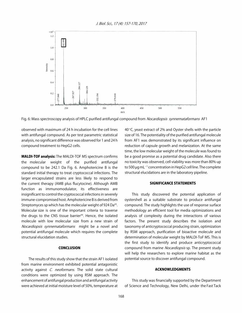
MALDI-TOF analysis: The MALDI-TOF MS spectrum confirmsthe molecular weight of the purified antifungal compound to be 242.1 Da Fig. 6. Amphotericine B is thestandard initial therapy to treat cryptococcal infections. Thelarger encapsulated strains are less likely to respond tothe current therapy (AMB plus flucytocine). Although AMBfunction as immunomodulator, its effectiveness areinsignificant to control the cryptococcal infections in severelyimmune-compromised host. Amphotericine B is derived fromStreptomyces sp which has the molecular weight of 924 Da43.Molecular size is one of the important criteria to traversethe drugs to the CNS tissue barrier44. Hence, the isolatedmolecule with low molecular size from a new strain ofNocardiopsis synnemataformans might be a novel andpotential antifungal molecule which requires the completestructural elucidation studies.
CONCLUSION
The results of this study show that the strain AF1 isolatedfrom marine environment exhibited potential antagonisticactivity against C. neoformans. The solid state culturalconditions were optimized by using RSM approach. Theenhancement of antifungal production and antifungal activitywere achieved at initial moisture level of 50%, temperature at
40EC, yeast extract of 2% and Oyster shells with the particlesize of 16. The potentiality of the purified antifungal moleculefrom AF1 was demonstrated by its significant influence onreduction of capsule growth and melanization. At the sametime, the low molecular weight of the molecule was found tobe a good promise as a potential drug candidate. Also thereno toxicity was observed, cell viability was more than 80% upto 500 µg mLG1 concentration in HepG2 cell line. The completestructural elucidations are in the laboratory pipeline.
SIGNIFICANCE STATEMENTS
This study discovered the potential application ofoystershell as a suitable substrate to produce antifungalcompound. The study highlights the use of response surfacemethodology an efficient tool for media optimizations andanalysis of complexity during the interactions of variousfactors. The present study describes the isolation andtaxonomy of anticryptococcal producing strain, optimizationby RSM approach, purification of bioactive molecule anddetermination of molecular weight by MALDI-ToF MS. This isthe first study to identify and produce anticryptococcalcompound from marine Nocardiopsis sp. The present studywill help the researchers to explore marine habitat as thepotential source to discover antifungal compound.
ACKNOWLEDGMENTS
This study was financially supported by the Departmentof Science and Technology, New Delhi, under the Fast Tack
168
×105
1.2
1.0
0.8
0.6
0.4
0.2
0.0
Inte
nsit
y
250 300 350 400 450 500 550
298.
022
379.
018 30
4.12
9
284.
127
267.
927
277.
840
332.
194
242.
009
m/z
J. Biol. Sci., 17 (4): 157-170, 2017
Young Scientist Scheme. (No: SB/FT/LS-249/2012) andHPLC facility EMR scheme SR/S0/HS-0073/2012 to JP. Wethank SASTRA University for providing us the infrastructure tocarry out our research work. We thank to Dr. GunasekarVaradarajan for his valuable suggestion for RSM study.
REFERENCES
1. Muzoora, C.K., T. Kabanda, G. Ortu, J. Ssentamu andP. Hearn et al., 2012. Short course amphotericin B withhigh dose fluconazole for HIV-associated cryptococcal meningitis. J. Infect., 64: 76-81.
2. Chang, W.C., C. Tzao, H.H. Hsu, S.C. Lee, K.L. Huang, H.J. Tungand C.Y. Chen, 2006. Pulmonary cryptococcosis: Comparisonof clinical and radiographic characteristics inimmunocompetent and immunocompromised patients.Chest, 129: 333-340.
3. Antinori, S., 2013. New insights into HIV/AIDS-associatedcryptococcosis. ISRN AIDS, Vol. 2013. 10.1155/2013/471363.
4. Karkowska-Kuleta, J., M. Rapala-Kozik and A. Kozik, 2009.Fungi pathogenic to humans: Molecular bases of virulence ofCandida albicans, Cryptococcus neoformans and Aspergillusfumigatus. Acta Biochim. Pol., 56: 211-224.
5. Das, A., S. Bhattacharya, A.Y.H. Mohammed and S.S. Rajan,2014. In vitro antimicrobial activity and characterization ofmangrove isolates of streptomycetes effective againstbacteria and fungi of nosocomial origin. Braz. Arch. Biol.Technol., 57: 349-356.
6. Steenbergen, J.N. and A. Casadevall, 2003. The origin andmaintenance of virulence for the human pathogenic fungusCryptococcus neoformans. Microbes Infect., 5: 667-675.
7. Maxson, M.E., E. Dadachova, A. Casadevall and O. Zaragoza,2007. Radial mass density, charge and epitope distribution in the Cryptococcus neoformans capsule.Eukaryotic Cell, 6: 95-109.
8. Johnston, S.A. and R.C. May, 2013. Cryptococcus interactionswith macrophages: Evasion and manipulation of thephagosome by a fungal pathogen. Cell. Microbiol.,15: 403-411.
9. Nosanchuk, J.D. and A. Casadevall, 2006. Impact of melaninon microbial virulence and clinical resistance to antimicrobialcompounds. Antimicrob. Agents Chemother, 50: 3519-3528.
10. Liu, G.Y. and V. Nizet, 2009. Color me bad: Microbial pigmentsas virulence factors. Trends Microbiol., 17: 406-413.
11. Yamazumi, T., M.A. Pfaller, S.A. Messer, A.K. Houston andL. Boyken et al., 2003. Characterization of heteroresistanceto fluconazole among clinical isolates of Cryptococcusneoformans. J. Clin. Microbiol., 41: 267-272.
12. Sloan, D.J., M.J. Dedicoat and D.G. Lalloo, 2009. Treatment ofcryptococcal meningitis in resource limited settings. Curr.Opin. Infect. Dis., 22: 455-463.
13. Kim, S.K., 2012. Marine Medicinal Foods: Implications andApplications-Animals and Microbes. Academic Press, USA.,ISBN: 9780124160033, Pages: 523.
14. Subramani, R. and W. Aalbersberg, 2012. Marineactinomycetes: An ongoing source of novel bioactivemetabolites. Microbiol. Res., 167: 571-580.
15. Manivasagan, P., K.H. Kang, K. Sivakumar, E.C. Li-Chan,H.M. Oh and S.K. Kim, 2014. Marine actinobacteria: Animportant source of bioactive natural products. Environ.Toxicol. Pharmacol., 38: 172-188.
16. Robinson, T., D. Singh and P. Nigam, 2001. Solid-statefermentation: A promising microbial technology forsecondary metabolite production. Applied Microbiol.Biotechnol., 55: 284-289.
17. Vijayabharathi, R., P. Brunthadevi, S. Sathyabama, P. Bruheimand V.B. Priyadarisini, 2012. Optimization of resistomycinproduction purified from Streptomyces aurantiacus AAA5using response surface methodology. J. Biochem. Technol.,3: 402-408.
18. Abraham, J. and R. Chauhan, 2015. Bioactivity and in-silicoanalysis of R-alpine borane produced by Streptomycestoxytricini Jar4. Int. J. Pharm. Bio Sci., 6: 265-276.
19. Ramakrishnan, J., H. Balakrishnan, S.T.K. Raja,N. Sundararamakrishnan, S. Renganathan and V.N. Radha,2011. Formulation of economical microbial feed usingdegraded chicken feathers by a novel Streptomyces sp:Mitigation of environmental pollution. Braz. J. Microbiol.,42: 825-834.
20. Kimura, M., 1980. A simple method for estimatingevolutionary rates of base substitutions throughcomparative studies of nucleotide sequences. J. Mol. Evol.,16: 111-120.
21. Singh, A.K., M. Singh and S.K. Dubey, 2013. Changes inActinomycetes community structure under the influence ofBt transgenic brinjal crop in a tropical agroecosystem. BMCMicrobiol., Vol. 13. 10.1186/1471-2180-13-122
22. Saitou, N. and M. Nei, 1987. The neighbor-joining method:A new method for reconstructing phylogenetic trees.Mol. Biol. Evol., 4: 406-425.
23. Thompson, J.D., T.J. Gibson, F. Plewniak, F. Jeanmougin andD.G. Higgins, 1997. The CLUSTAL_X windows interface:Flexible strategies for multiple sequence alignment aided byquality analysis tools. Nucleic Acids Res., 25: 4876-4882.
24. Wang, Q., Y. Hou, Z. Xu, J. Miao and G. Li, 2008. Optimizationof cold-active protease production by the psychrophilicbacterium Colwellia sp. NJ341 with response surfacemethodology. Bioresour. Technol., 99: 1926-1931.
25. Ponnusamy, S.K. and R. Subramaniam, 2013. Processoptimization studies of Congo red dye adsorption ontocashew nut shell using response surface methodology. Int.J. Ind. Chem., Vol. 4. 10.1186/2228-5547-4-17.
169
J. Biol. Sci., 17 (4): 157-170, 2017
26. Lavermicocca, P., F. Valerio, A. Evidente, S. Lazzaroni,A. Corsetti and M. Gobetti, 2000. Purification andcharacterization of novel antifungal compounds from thesourdough Lactobacillus plantarum strain 21B. AppliedEnviron. Microbiol., 66: 4084-4090.
27. Sharma, D. and R.K. Manhas, 2013. Application ofPlackett-Burman experimental design and box and wilsondesign to improve broad-spectrum antimicrobial compound.Ind. J. Biotechnol., 12: 386-394.
28. Rismanchian, M., N. Khodaeian, L. Bahramian, M. Fathi andH. Sadeghi-Aliabadi, 2013. In-vitro comparison of cytotoxicityof two bioactive glasses in micropowder and nanopowderforms. Iran. J. Pharm. Res., 12: 437-443.
29. Van Duin, D., A. Casadevall and J.D. Nosanchuk, 2002.Melanization of Cryptococcus neoformans and Histoplasmacapsulatum reduces their susceptibilities to amphotericinB and caspofungin. Antimicrob. Agents Chemother.,46: 3394-3400.
30. Ikeda, R., T. Sugita, E.S. Jacobson and T. Shinoda, 2003. Effectsof melanin upon susceptibility of Cryptococcus to antifungals.Microbiol. Immunol., 47: 271-277.
31. Rathore, S.S., T. Raman and J. Ramakrishnan, 2016.Magnesium ion acts as a signal for capsule induction inCryptococcus neoformans. Front. Microbiol., Vol. 7.10.3389/fmicb.2016.00325.
32. Van Duin, D., W. Cleare, O. Zaragoza, A. Casadevall andJ.D. Nosanchuk, 2004. Effects of voriconazole onCryptococcus neoformans. Antimicrob. Agents Chemother.,48: 2014-2020.
33. Mayer, A.M.S., A.D. Rodriguez, O. Taglialatela-Scafati andN. Fusetani, 2013. Marine pharmacology in 2009-2011:Marine compounds with antibacterial, antidiabetic,antifungal, anti-inflammatory, antiprotozoal, antituberculosisand antiviral activities; affecting the immune and nervoussystems and other miscellaneous mechanisms of action. Mar.Drugs, 11: 2510-2573.
34. Raju, R., A.M. Piggott, L.X.B. Diaz, Z. Khalil and R.J. Capon,2010. Heronapyrroles A-C: Farnesylated 2-nitropyrrolesfrom an Australian marine-derived Streptomyces sp. Org.Lett., 12: 5158-5161.
35. Dyavaiah, S.M. and G.N. Prashanth, 2013. Yeasts: Candida andCryptococcus. In: Bacterial and Mycotic Infections inImmunocompromised Hosts: Clinical and MicrobiologicalAspects, Mascellino, M.T. (Ed.). Omics Group Incorporation,Nevada, USA., pp: 1-14.
36. Basavaraj, M., Vastrad and S.E. Neelagund, 2011.Production and optimisation of tetracycline by variousstrains of streptomyces under solid state fermentation usingpineapple peel as a novel substrate. Rec. Res. Sci. Technol.,3: 1-8.
37. Vastrad, B.M. and S.E. Neelagund, 2011. Optimization andproduction of neomycin from different agro industrialwastes in solid state fermentation. Int. J. Pharm. Sci. Drug Res.,3: 104-111.
38. Manpreet, S., S. Sawraj, D. Sachin, S. Pankaj and U.C. Banerjee,2005. Influence of process parameters on the production ofmetabolites in solid-state fermentation. Malay. J. Microbiol.,1: 1-9.
39. Jung, J.H., J.J. Lee, G.W. Lee, K.S. Yoo and B.H. Shon, 2012.Reuse of Waste Shells as a SO2/NOx Removal Sorbent. In:Material Recycling-Trends and Perspectives, Achilias, D.S.(Ed.). InTech, USA., pp: 301-322.
40. Khanna, M., R. Solanki and R. Lal, 2011. Selective isolation ofrare actinomycetes producing novel antimicrobialcompounds. Int. J. Adv. Biotechnol. Res., 2: 357-375.
41. Gebreyohannes, G., F. Moges, S. Sahile and N. Raja,2013. Isolation and characterization of potentialantibiotic producing actinomycetes from water andsediments of Lake Tana, Ethiopia. Asian Pac. J. Trop. Biomed.,3: 426-435.
42. Ellaiah, P., B. Srinivasulu and K. Adinarayana, 2004.Optimisation studies on neomycin production by a mutantstrain of Streptomyces marinensis in solid statefermentation. Process Biochem., 39: 529-534.
43. Kethireddy, S. and D. Andes, 2007. CNS pharmacokinetics ofantifungal agents. Exp. Opin. Drug Metab. Toxicol., 3: 573-581.
44. Pardridge, W.M., 2005. The blood-brain barrier: Bottleneck inbrain drug development. NeuroRx, 2: 3-14.
170