Embed Size (px)
Citation preview
Posttreatment followup study of abdominal cystic echinococcosis in Tibetan communities of northwest
Sichuan Province, ChinaLi, T, Ito, A, Pengcuo, R, Sako, Y, Chen, X, Qiu, D, Xiao, N and Craig, PS
http://dx.doi.org/10.1371/journal.pntd.0001364
Title Posttreatment followup study of abdominal cystic echinococcosis in Tibetan communities of northwest Sichuan Province, China
Authors Li, T, Ito, A, Pengcuo, R, Sako, Y, Chen, X, Qiu, D, Xiao, N and Craig, PS
Type Article
URL This version is available at: http://usir.salford.ac.uk/19238/
Published Date 2011
USIR is a digital collection of the research output of the University of Salford. Where copyright permits, full text material held in the repository is made freely available online and can be read, downloaded and copied for noncommercial private study or research purposes. Please check the manuscript for any further copyright restrictions.
For more information, including our policy and submission procedure, pleasecontact the Repository Team at: [email protected].
Post-Treatment Follow-Up Study of Abdominal CysticEchinococcosis in Tibetan Communities of NorthwestSichuan Province, ChinaTiaoying Li1,2*, Akira Ito3*, Renqing Pengcuo4, Yasuhito Sako3, Xingwang Chen1, Dongchuan Qiu1, Ning
Xiao1, Philip S. Craig2
1 Institute of Parasitic Diseases, Sichuan Centers for Disease Control and Prevention, Chengdu, Sichuan Province, People’s Republic of China, 2 Cestode Zoonoses Research
Group, School of Environment and Life Sciences, University of Salford, Salford, United Kingdom, 3 Department of Parasitology, Asahikawa Medical University, Asahikawa,
Japan, 4 Shiqu County Centers for Disease Control and Prevention, Shiqu, Ganzi Tibetan Prefecture, Sichuan Province, People’s Republic of China
Abstract
Background: Human cystic echinococcosis (CE), caused by the larval stage of Echinococcus granulosus, with the liver as themost frequently affected organ, is known to be highly endemic in Tibetan communities of northwest Sichuan Province.Antiparasitic treatment with albendazole remains the primary choice for the great majority of patients in this resource-poorremote area, though surgery is the most common approach for CE therapy that has the potential to remove cysts and leadto complete cure. The current prospective study aimed to assess the effectiveness of community based use of cyclicalbendazole treatment in Tibetan CE cases, and concurrently monitor the changes of serum specific antibody levels duringtreatment.
Methodology/Principal Findings: Ultrasonography was applied for diagnosis and follow-up of CE cases after cyclicalbendazole treatment in Tibetan communities of Sichuan Province during 2006 to 2008, and serum specific IgG antibodylevels against Echinococcus granulosus recombinant antigen B in ELISA was concurrently monitored in these cases. A total of196 CE cases were identified by ultrasound, of which 37 (18.9%) showed evidence of spontaneous healing/involution ofhepatic cyst(s) with CE4 or CE5 presentations. Of 49 enrolled CE cases for treatment follow-up, 32.7% (16) were consideredto be cured based on B-ultrasound after 6 months to 30 months regular albendazole treatment, 49.0% (24) were improved,14.3% (7) remained unchanged, and 4.1% (2) became aggravated. In general, patients with CE2 type cysts (daughter cystspresent) needed a longer treatment course for cure (26.4 months), compared to cases with CE1 (univesicular cysts) (20.4months) or CE3 type (detached cyst membrane or partial degeneration of daughter cysts) (9 months). In addition, thecurative duration was longer in patients with large (.10 cm) cysts (22.3 months), compared to cases with medium (5–10 cm) cysts (17.3 months) or patients with small (,5 cm) cysts (6 months). At diagnosis, seven (53.8%) of 13 cases with CE1type cysts without any previous intervention showed negative specific IgG antibody response to E. granulosus recombinantantigen B (rAgB). However, following 3 months to 18 months albendazole therapy, six of these 7 initially seronegative CE1cases sero-converted to be specific IgG antibody positive, and concurrently ultrasound scan showed that cysts changed toCE3a from CE1 type in all the six CE cases. Two major profiles of serum specific IgG antibody dynamics during albendazoletreatment were apparent in CE cases: (i) presenting as initial elevation followed by subsequent decline, or (ii) a persistentdecline. Despite a decline, however, specific antibody levels remained positive in most improved or cured CE cases.
Conclusions: This was the first attempt to follow up community-screened cystic echinococcosis patients after albendazoletherapy using ultrasonography and serology in an endemic Tibetan region. Cyclic albendazole treatment proved to beeffective in the great majority of CE cases in this resource-poor area, but periodic abdominal ultrasound examination wasnecessary to guide appropriate treatment. Oral albendazole for over 18 months was more likely to result in CE cure. Poordrug compliance resulted in less good outcomes. Serology with recombinant antigen B could provide additional limitedinformation about the effectiveness of albendazole in CE cases. Post-treatment positive specific IgG antibodyseroconversion, in initially seronegative, CE1 patients was considered a good indication for positive therapeutic efficacyof albendazole.
Citation: Li T, Ito A, Pengcuo R, Sako Y, Chen X, et al. (2011) Post-Treatment Follow-Up Study of Abdominal Cystic Echinococcosis in Tibetan Communities ofNorthwest Sichuan Province, China. PLoS Negl Trop Dis 5(10): e1364. doi:10.1371/journal.pntd.0001364
Editor: Hector H. Garcia, Universidad Peruana Cayetano Heredia, Peru
Received December 30, 2010; Accepted September 2, 2011; Published October 25, 2011
Copyright: � 2011 Li et al. This is an open-access article distributed under the terms of the Creative Commons Attribution License, which permits unrestricteduse, distribution, and reproduction in any medium, provided the original author and source are credited.
Funding: The study was supported by grant number R01 TW001565 from the Fogarty International Center of National Institutes of Health (NIH). This study wasalso supported in part by a PhD split-site studentship (to TL) between the University of Salford, UK and SIPD/Sichuan CDC, China, Grants-in-Aid for InternationalScientific Research (No. 21256003) from Japan Society for the Promotion of Science (JSPS), JSPS-Asia/Africa Scientific Platform Fund (2006–2011), JSPS-CAMSMedical Cooperation Fund between Japan and China (2009–2010) and by the special fund for International Leadership in Science and Technology from Ministryof Education, Japan (2010–2012). The funders had no role in the study design, data collection and analysis, decision to publish, or preparation of the manuscript.
Competing Interests: The authors have declared that no competing interests exist.
* E-mail: [email protected] (TL); [email protected] (AI)
www.plosntds.org 1 October 2011 | Volume 5 | Issue 10 | e1364
Introduction
Human cystic echinococcosis (CE), caused by the metacestode
stage of Echinococcus granulosus, is a complex, chronic disease with a
cosmopolitan distribution, and the liver is the most frequently
affected organ [1]. Clinical manifestation of this disease ranges
from asymptomatic infection to severe, or rarely even fatal disease.
Diagnosis of CE remains highly dependent on imaging techniques,
due to the fact that immunodiagnosis frequently lacks sensitivity
[2], with about 20% of clinically or surgically confirmed CE cases,
and up to 50% of community-detected patients presenting
negative serology [3–6]. The most common applied imaging
techiniques include magnetic resonance imaging (MRI), ultraso-
nography (US) or radiography, for detection of characteristic
space-occupying cysts [7,8]. MRI is able to show highly specific
features of CE, but it is prohibitively expensive and not available in
rural areas of many endemic countries. In contrast, US is
accessible, much less expensive, and can identify hydatid cyst
pathological type (CE1-CE5) [9].
Approaches in clinical management for CE include surgery,
percutaneous techniques and antiparasitic treatment for active
cysts, and the so-called watch and wait approach for inactive cysts
[9]. Currently, surgery remains the most common approach for
CE treatment that has the potential to remove cysts and lead to
complete cure, but it involves risks including those associated with
any surgical intervention, anaphylactic reactions, and secondary
CE owing to spillage of viable parasite (protoscoleces) material
[10–12]. Drug therapy with benzimidazoles (albendazole or
mebendazole) has increasingly been used to treat CE, and proved
to have efficacy against the parasite in humans, with about 30% of
patients cured and 30%–50% of cases improved after 12 months
follow-up [10]. However, the response to drug therapy is
unpredictable, and the optimum duration has not been definitively
determined [11,13]. Moreover, risk of recurrence remains the
major problem in surgical or medical treatment [10,12,13].
Therefore, post-treatment or post-surgical follow-up of CE
patients for several years is usually indicated.
Imaging techniques such as MRI, X-ray or ultrasonography,
are useful tools for follow-up of CE patients. However, these
techniques are sometimes difficult to detect the newly growing
small cyst and also to discriminate between dead and viable cysts
[14]. Therefore, efforts have been directed at applying immuno-
logical tests of significantly diagnostic and prognostic values.
ELISA and immunoblotting for serum antibody detection using
various antigen preparations, including crude hydatid cyst fluid,
purified fractions of antigen 5 or B, and E. granulosus protoscolex
soluble extract, have been applied to follow up CE patients [15–
20]. However, all of these tests exhibited problems mainly related
to temporally delayed reactions to clinical changes [18,20].
Recombinant antigen B (rAgB) proved to have similar diagnostic
value to native antigen B in CE patients [21,22]. However, there
has been little or no application of rAgB for post-treatment follow-
up of CE patients.
In Tibetan regions of China, human cystic echinococcosis is
highly endemic [23]. Albendazole therapy is the primary choice of
treatment in the majority of patients owing to remote communi-
ties, poor socioeconomics and basic hospital facilities in Tibetan
Autonomous Prefectures/communities. The current prospective
study was designed to assess the effectiveness of cyclic albendazole
treatment in community detected CE patients using ultrasonog-
raphy as well as ELISA with rAgB as diagnostic/follow-up tools,
and also to monitor the changes of serum specific IgG antibody
levels against rAgB in these patients during treatment.
Materials and Methods
Ethics statementThe study protocol was approved by the Ethical Committee of
Sichuan Centers for Disease Control and Prevention (Sichuan
CDC). Clearance to carry out the study was obtained from Shiqu
County CDC. Information about the purpose of the post-
treatment follow-up study was spread to the villagers. Persons
with confirmative ultrasound images of CE were voluntarily self-
selected to be involved in this study by written informed consent
and were assured free medical treatment with cyclic albendazole
therapy if necessary. Recommendations were also provided for
possible surgical intervention (cyst removal). At confirmed
ultrasound diagnosis, each patient was requested to complete a
questionnaire which was designed to get information on
demographics. Questions were mainly designed to identify clinical
manifestations, history of any previous treatment with albendazole
(regular or irregular, duration), as well as history of surgery. At
each follow-up, another questionnaire was completed to obtained
information on administration of albendazole, surgery associated
with echinococcosis, improvement of symptoms if any, adverse
effects such as gastrointestinal disturbances, alopecia, jaundice,
skin itch, hepatic pain/sting, dizziness etc. Chinese-Tibetan
translators were employed when necessary.
Criteria for diagnosis and classification of cysticechinococcosis
Diagnosis and classification of cystic echinococcosis (CE) was
made using portable ultrasound according to the criteria proposed
by the World Health Organization Informal Working Group on
Echinococcosis for CE [12,24]. On the basis of patho-morpho-
logical features of cysts, CE lesions were differentiated into six
types: CL, CE1, CE2, CE3, CE4 and CE5 (Figure 1). Briefly, the
CL type cyst refers to a cystic lesion of parasite origin without a
clear rim indicating a very early stage of parasite development,
while the presence of CE1 (unilocular cyst with thick endo
membrane) or CE2 (daughter cysts present) is suggestive of active
Author Summary
Cystic echinococcosis is a serious public health problem inTibetan communities of northwest Sichuan Province, China.Antiparasitic treatment with albendazole remains the onlychoice in most cases, due to the poor socio-economy andinadequate hospital facilities in this area. A post-treatmentfollow-up study was carried out in community-detected 49CE cases by application of abdominal ultrasound andserology with recombinant antigen B (rAgB) in a Tibetanregion of Sichuan from 2006 to 2008. Following 6 to 30months regular albendazole therapy, 32.7% of CE caseswere considered cured at ultrasound, 49.0% were classed asimproved, 14.3% remained unchanged or static, and 4.1% ofcases became aggravated. The treatment course for curewas longer in patients with CE2 type cyst pathologycompared to cases with CE1, CE3a or CE3b type cysts. Inaddition, patients with large cysts ($10 cm) had a longercurative duration compared to those with medium cysts (5–10 cm) or small cysts (,5c m). The changes of serumspecific IgG antibody levels against rAgB were not stronglyassociated with the viability of cystic echinococcal lesions,however, post-treatment specific IgG antibody positivesero-conversion in initially seronegative CE1 patients, wasan indicator for the albendazole efficacy in specific CEpatients.
Follow-Up Study of Tibetan Cystic Echinococcosis
www.plosntds.org 2 October 2011 | Volume 5 | Issue 10 | e1364

stages of the disease. While CE3 is broken into CE3a and CE3b
characterized by detached cyst membrane and partial degenera-
tion of daughter cysts, respectively, indicating the parasite is at a
transitional stage, and CE4 and CE5 implies cyst involution,
necrosis, partially calcified or inactive parasite [1,12,24].
Application of chest X-ray for diagnosis of lung CE was not
carried out in this study.
Origin of cystic echinococcosis (CE) patientsDuring May 2006 to November 2008, mass ultrasound
examination was carried out in eight Tibetan townships of Shiqu
County (Ganzi Prefecture, Sichuan Province) for detection of
individuals with abdominal cystic echinococcosis infection.
Patients with CE1/CE2/CE3a/CE3b type cysts were invited to
enroll in the current prospective follow-up study. All CE patients,
whether enrolled or not, were offered free albendazole treatment.
Albendazole therapyCyclic treatment with albendazole was provided freely to each
patient as 100-mg tablet at a daily dose of 10–15 mg/kg body
weight (in two divided doses, together with fat-rich meal). Cyclic
treatment of 30 days was followed by a ‘wash out’ period of 7–10
days without albendazole [1]. Albendazole tablets sufficient for six
months application were delivered to patients at each follow-up, to
whom possible adverse effects were explained. In addition,
albendazole was also available freely in the local county CDC
clinic.
Follow-up was carried out at six months intervals. Once a cystic
lesion changed to CE4 type, the patient was requested to cease
albendazole, but further regular ultrasound examination was
necessary to understand if the cyst remained inactive. According to
the questionnaire investigation, patients who took albendazole as
requested during follow-up period were included in the regular-
treated group, whereas others who did not take albendazole as
requested due to poor compliance belonged to the irregular-
treated group.
Responses to albendazole therapyThe effectiveness of albendazole in CE patients assessed by
ultrasound was described as follows: cured, improved, unchanged/
static or aggravated. ‘‘Cured’’ was defined as disappearance of
cysts, or degeneration of cyst contents. In other words, ‘cured’
referred to a cyst changing to a CE4 or CE5 type from a CE1,
CE2, CE3a or CE3b type cyst. ‘Improved’ was determined as
detachment of cyst membrane, partial degeneration of cyst
contents (or daughter cysts) and/or reduction of cyst size,
indicative of the cyst converting to CE3a/CE3b type from a
CE1 or CE2 cyst. A ‘static’ or unchanged cyst showed no
morphological and/or size changes. ‘Aggravated’ CE disease was
defined as enlargement of the cyst and/or recurrence of daughter
cysts.
Collection of serum samples and image dataApproximately 3 ml of venous blood was taken voluntarily from
patients at diagnosis (during mass ultrasound screening) as well as
at each follow-up, and then centrifuged on the same day. Sera
were aliquoted and stored at 220uC for later serological analysis.
Blood transaminase levels were not monitored in the current
study, due to the difficulty of doing liver function tests in the field.
Information about the characteristics of hydatid cysts for new
CE cases and follow-up CE patients was documented in detail,
including the cyst type (CE1-5), the number of cysts (single or
multiple), location (the lobe of the liver, abdominal cavity, pelvic
cavity, spleen or kidney), and the size (cm).
SerologyELISA with recombinant antigen B (rAgB) based on previous
description [22] was performed on each serum sample for
determination of Echinococcus specific IgG. Samples from the same
patient were analyzed concurrently. The cut-off point was
determined as the mean optical density plus 3 times standard
deviation for a panel of serum samples obtained from healthy
donors (n = 30).
In these assays, 100-ml volume was applied throughout unless
otherwise stated. 96-well microtiter plates (MaxiSorp; Nalge Nunc
International, Roskilde, Denmark) were coated with diluted rAgB
at 0.5 mg/ml in PBS overnight at 4uC. Plates were rinsed 3 times
with PBST and blocked with 300 ml of 1% casein buffer at 37uCfor 1 hr. Sera were diluted 1:100 in 1% casein buffer. Plates with
diluted sera were incubated in duplicate wells at 37uC for 1 hr and
then washed five times with PBST. Rabbit anti-human horserad-
ish peroxidase-conjugated protein G (Zymed Laboratories, Inc.,
South San Francisco, Calif.) was diluted at 1:4000 in 1% casein
buffer and incubated at 37uC for 1 hour. Plates were washed five
times with PBST. For colour development, substrate solution
(0.4 mM 2,29-azino-bis[3-ethybenzthiazoline-6-sulfonic acid] in
0.1 M citric acid buffer and 0.2 M Na2HPO4) was added into
each well and incubated at room temperature for 30 min. Colour
reaction was then stopped by application of 1% SDS in each well.
The optical density at 405 nm was evaluated with an ELISA
reader.
Statistical analysisChi-square test was used to compare the occurrence rate of
spontaneous involution between males and females, and the cure
rate between the patient groups with albendazole course #6
months and those .6 months, #12 months and .12 months,
#18 months and .18 months, and #24 months and .24
months. Significance was set at P#0.05.
Results
A total of 196 persons with CE infection were registered in this
study (male = 83, female = 113), with a mean age of 37.5 years at
diagnosis (range 4–80 years). Of these 196 cases, 55 (28.1%) had
Figure 1. Ultrasound classification of cystic echinococcosis based on WHO expert consensus. CL: as a potentially parasitic cyst, indicatesa very early stage of parasite development. CE1 or CE2: suggests active parasite. CE3a: is characterized by detachment cyst membrane and/or partialdegeneration of cyst content, without daughter cysts, indicates a transitional stage. CE3b: suggests a transitional stage of parasite, partialdegeneration of daughter cysts. CE4 or CE5: indicates an inactive parasite.doi:10.1371/journal.pntd.0001364.g001
Follow-Up Study of Tibetan Cystic Echinococcosis
www.plosntds.org 3 October 2011 | Volume 5 | Issue 10 | e1364
received prior regular albendazole therapy, while an additional 15
(8.2%) had prior surgery. However, only 4 of 15 operated cases
received regular albendazole treatment following surgery.
Totally 98 of 108 CE patients without any previous albendazole
treatment at diagnosis were investigated about clinical symptoms
and signs, 55.1% (54) reported various degrees of discomfort, while
44.9% (44) were asymptomatic. The most common discomfort was
hepatic or epigastric pain in 49.0% of patients, other complaints
included abdominal distention, palpable abdominal mass, etc. All
the 32 patients with cysts of CL, CE4 or CE5 type were
asymptomatic, while 74.2% (49/66) of patients with cysts of CE1,
CE2 or CE3 presented various degrees of symptoms.
Of these 196 cases, 37 (18.9%) were observed at first
examination, to have evidence of spontaneous involution of cystic
lesions without any interventional procedures, presenting inactive
cysts (CE4 or CE5) in the liver. Persons with CE4 cysts had a
mean age of 39.2 years (n = 27), while individuals with CE5 cysts
had an average age of 61.3 years (n = 10). Of the 37 patients with
evidence of spontaneous involution, 23 were male and 14 were
female. In other words, spontaneous cure of cystic echinococcosis
occurred more frequently in male (27.7%) than in female (12.4%),
and the difference was significant (x2 = 7.3, P,0.01).
A total of 49 CE patients received regular albendazole
treatment for 6 to 30 months, including 19 males and 30 females.
The youngest CE case was 4 years old and the oldest was 80 years,
with a mean age of 37.7 years. Cystic lesions were confined in the
liver in 43 cases, and the remaining 6 cases had lesions not only to
the liver, but also in the abdominal cavity. Of these 49 patients, 16
had CE1 type cysts, 17 had CE2 cysts, 10 had CE3a type cysts,
and the remaining 6 had CE3b cysts (Table 1). The cyst measured
$10 cm in 25 patients, the cyst varied in size 5 cm–10 cm in 20
cases, whereas the remaining 4 CE cases had cysts less than 5 cm
(Table 1).
Following 6 to 30 months regular therapy, 16 (32.7%) of 49
patients were observed to have cysts that changed to CE4 type (ie.
considered cured), 24 (49.0%) were observed to have cysts
converted to CE3a or CE3b from CE1 or CE2 type (ie. improved)
(mean duration = 14 months), cysts remained unchanged in the
other 7 (14.3%) patients (mean = 10.3 months), and enlargement
of hydatid cysts was observed in the remaining 2 (4.1%) patients
(Figure 2.1; Table 2). The cure rate was 15.4% (2/13) and 38.9%
(14/36) in the patient groups with albendazole course #6 months
and those .6 months, 21.4% (6/28) and 47.6% (10/21)) for the
group with treatment duration #12 months and those .12
months, 22.9% (8/35) and 51.7% (8/14) for cases with treatment
course #18 months and those .18 months, and 26.8% (11/41)
and 62.5% (5/8) for patients with albendazole course #24 months
and those .24 months. Further statistical analysis revealed that
the cure rate was significantly different only between the patient
group with treatment course #18 months and those .18 months
(x2 = 5.24, P,0.05). The 16 ‘cured’ patients were composed of 5
CE1 cases, 5 CE2, 4 CE3a and 2 cases with CE3b cyst. The
treatment course for cure varied in patients with cysts at different
stages, that is, the mean curative course was 26.4 months in CE2
patients, 20.4 months in CE1 cases and 9 months in CE3a/CE3b
patients. Moreover, the curative duration also differed in cases
with cysts at different size. Patients with large hydatid cysts
($10 cm) needed 22.3 months treatment (n = 7), whereas cure was
achieved following 17.3 months therapy in cases with medium
cysts (5 cm to 10 cm) (n = 8) and 6 months in patients with small
cysts (#5 cm) (n = 1). In addition, 5 of these 16 cured CE patients
were further followed up with ultrasound for 6 to 24 months, in
whom the cysts remained inactive (CE4 type), indicative of no
recurrence.
In contrast, 12 CE patients who poorly complied with
albendazole treatment were observed to have much poorer
prognosis during 6 to 30 months follow-up observation (mean
= 17.0 months). Of these 12 patients, cysts remained unchanged in
8 cases, while enlargement of the cyst or recurrence of daughter
cysts was observed in the remaining 4 patients (Figure 2.2).
Of 13 CE1 patients without any previous albendazole
treatment, 7 (53.8%) were sero- negative for a specific IgG
response to rAgB (Figure 2.3). However, following 3 to 18 months
albendzole therapy, positive IgG seroconversion was observed in 6
(no. p1-p6) of these 7 initially seronegative cases (Figure 2.3 and
figure 2.4). Concurrently, ultrasound scan detected detachment of
cyst membrane and/or partial degeneration of cyst content (i.e.
CE1 type changed to CE3a type) in all these six patients (no. p1-
p6) (Figure 2.4). In another patient (no. p7) in whom serum specific
IgG antibody remained negative during 6 months follow-up
period (Figure 2.4), ultrasound scan did not detect any changes of
the cyst (the image was not shown). A questionnaire investigation
revealed that this patient had reported a poor compliance with
albendazole therapy.
Sequential serum samples (n = 36) were obtained from 8 CE
patients (CE1 = 4, CE2 = 3 and CE3b = 1), who did not receive
any previous chemotherapy at diagnosis and were considered to be
cured according to ultrasound images following albendazole
therapy in the current study. At least 3 serum samples were
monitored in each patient. The follow-up period ranged from 24
months to 78 months (mean 39.8 months), and an average 4.5
serum samples were taken from each patient. Longitudinal
assessment of specific IgG antibody against rAgB in ELISA
revealed that serum antibody levels of IgG were initially elevated,
and subsequently decreased in five (4 CE1 and 1 CE2) of these 8
patients (Figure 2.5). In another two patients (1 CE2 and 1 CE3b),
specific IgG antibody levels decreased but with a minor fluctuation
during albendazole administration. In the remaining patient with
a CE2 type cyst, a cured CE end-point was achieved following
treatment, but there was no significant change of specific IgG
antibody levels during the period of treatment (Figure 2.6A).
Serum specific IgG antibody in fact remained positive in all the 8
patients where CE was considered ‘cured’ even 24 months after
cure in one CE patient (ie. P2) (Figure 2.5A).
Consecutively collected 25 sera from 6 patients (CE1 = 2 and
CE2 = 4) with improved CE following albendazole treatment were
also assessed for specific IgG antibody against rAgB. Of these 6
cases, four (1 CE1 and 3 CE2) did not receive any previous
albendaozle therapy at diagnosis, and initial elevation and
subsequent decline of specific IgG antibody levels occurred in all
these 4 patients (Figure 2.6B). For the other 2 cases with previous
albendazole treatment at diagnosis, all antibody levels were
Table 1. The cyst stage and size in 49 CE cases.
Size No. cases Total
CE1 CE2 CE3a CE3b
Small 2 1 1 0 4
Medium 8 4 6 2 20
Large 6 12 3 4 25
Total 16 17 10 6 49
Small: the cyst measured ,5 cm at maximum diameter;Medium: the cyst measured 5–10 cm at maximum diameter;Large: the cyst measured $10 cm at maximum diameter.doi:10.1371/journal.pntd.0001364.t001
Follow-Up Study of Tibetan Cystic Echinococcosis
www.plosntds.org 4 October 2011 | Volume 5 | Issue 10 | e1364

Follow-Up Study of Tibetan Cystic Echinococcosis
www.plosntds.org 5 October 2011 | Volume 5 | Issue 10 | e1364
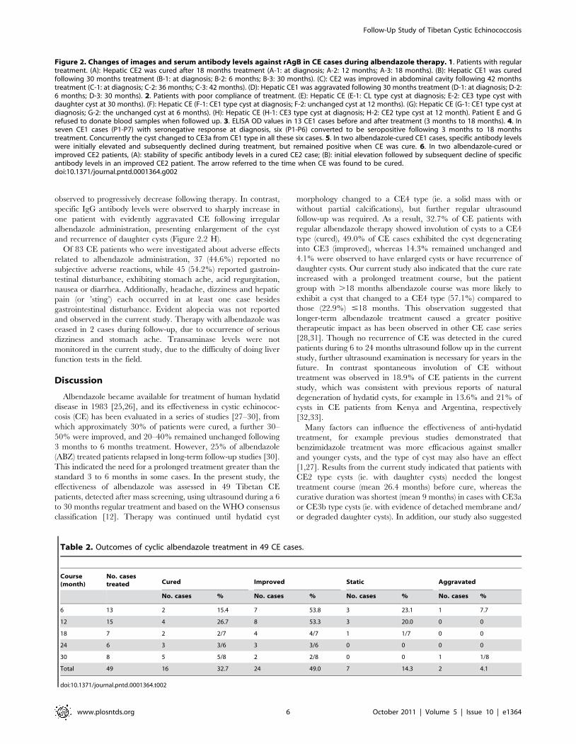
observed to progressively decrease following therapy. In contrast,
specific IgG antibody levels were observed to sharply increase in
one patient with evidently aggravated CE following irregular
albendazole administration, presenting enlargement of the cyst
and recurrence of daughter cysts (Figure 2.2 H).
Of 83 CE patients who were investigated about adverse effects
related to albendazole administration, 37 (44.6%) reported no
subjective adverse reactions, while 45 (54.2%) reported gastroin-
testinal disturbance, exhibiting stomach ache, acid regurgitation,
nausea or diarrhea. Additionally, headache, dizziness and hepatic
pain (or ’sting’) each occurred in at least one case besides
gastrointestinal disturbance. Evident alopecia was not reported
and observed in the current study. Therapy with albendazole was
ceased in 2 cases during follow-up, due to occurrence of serious
dizziness and stomach ache. Transaminase levels were not
monitored in the current study, due to the difficulty of doing liver
function tests in the field.
Discussion
Albendazole became available for treatment of human hydatid
disease in 1983 [25,26], and its effectiveness in cystic echinococ-
cosis (CE) has been evaluated in a series of studies [27–30], from
which approximately 30% of patients were cured, a further 30–
50% were improved, and 20–40% remained unchanged following
3 months to 6 months treatment. However, 25% of albendazole
(ABZ) treated patients relapsed in long-term follow-up studies [30].
This indicated the need for a prolonged treatment greater than the
standard 3 to 6 months in some cases. In the present study, the
effectiveness of albendazole was assessed in 49 Tibetan CE
patients, detected after mass screening, using ultrasound during a 6
to 30 months regular treatment and based on the WHO consensus
classification [12]. Therapy was continued until hydatid cyst
morphology changed to a CE4 type (ie. a solid mass with or
without partial calcifications), but further regular ultrasound
follow-up was required. As a result, 32.7% of CE patients with
regular albendazole therapy showed involution of cysts to a CE4
type (cured), 49.0% of CE cases exhibited the cyst degenerating
into CE3 (improved), whereas 14.3% remained unchanged and
4.1% were observed to have enlarged cysts or have recurrence of
daughter cysts. Our current study also indicated that the cure rate
increased with a prolonged treatment course, but the patient
group with .18 months albendazole course was more likely to
exhibit a cyst that changed to a CE4 type (57.1%) compared to
those (22.9%) #18 months. This observation suggested that
longer-term albendazole treatment caused a greater positive
therapeutic impact as has been observed in other CE case series
[28,31]. Though no recurrence of CE was detected in the cured
patients during 6 to 24 months ultrasound follow up in the current
study, further ultrasound examination is necessary for years in the
future. In contrast spontaneous involution of CE without
treatment was observed in 18.9% of CE patients in the current
study, which was consistent with previous reports of natural
degeneration of hydatid cysts, for example in 13.6% and 21% of
cysts in CE patients from Kenya and Argentina, respectively
[32,33].
Many factors can influence the effectiveness of anti-hydatid
treatment, for example previous studies demonstrated that
benzimidazole treatment was more efficacious against smaller
and younger cysts, and the type of cyst may also have an effect
[1,27]. Results from the current study indicated that patients with
CE2 type cysts (ie. with daughter cysts) needed the longest
treatment course (mean 26.4 months) before cure, whereas the
curative duration was shortest (mean 9 months) in cases with CE3a
or CE3b type cysts (ie. with evidence of detached membrane and/
or degraded daughter cysts). In addition, our study also suggested
Figure 2. Changes of images and serum antibody levels against rAgB in CE cases during albendazole therapy. 1. Patients with regulartreatment. (A): Hepatic CE2 was cured after 18 months treatment (A-1: at diagnosis; A-2: 12 months; A-3: 18 months). (B): Hepatic CE1 was curedfollowing 30 months treatment (B-1: at diagnosis; B-2: 6 months; B-3: 30 months). (C): CE2 was improved in abdominal cavity following 42 monthstreatment (C-1: at diagnosis; C-2: 36 months; C-3: 42 months). (D): Hepatic CE1 was aggravated following 30 months treatment (D-1: at diagnosis; D-2:6 months; D-3: 30 months). 2. Patients with poor compliance of treatment. (E): Hepatic CE (E-1: CL type cyst at diagnosis; E-2: CE3 type cyst withdaughter cyst at 30 months). (F): Hepatic CE (F-1: CE1 type cyst at diagnosis; F-2: unchanged cyst at 12 months). (G): Hepatic CE (G-1: CE1 type cyst atdiagnosis; G-2: the unchanged cyst at 6 months). (H): Hepatic CE (H-1: CE3 type cyst at diagnosis; H-2: CE2 type cyst at 12 month). Patient E and Grefused to donate blood samples when followed up. 3. ELISA OD values in 13 CE1 cases before and after treatment (3 months to 18 months). 4. Inseven CE1 cases (P1-P7) with seronegative response at diagnosis, six (P1-P6) converted to be seropositive following 3 months to 18 monthstreatment. Concurrently the cyst changed to CE3a from CE1 type in all these six cases. 5. In two albendazole-cured CE1 cases, specific antibody levelswere initially elevated and subsequently declined during treatment, but remained positive when CE was cure. 6. In two albendazole-cured orimproved CE2 patients, (A): stability of specific antibody levels in a cured CE2 case; (B): initial elevation followed by subsequent decline of specificantibody levels in an improved CE2 patient. The arrow referred to the time when CE was found to be cured.doi:10.1371/journal.pntd.0001364.g002
Table 2. Outcomes of cyclic albendazole treatment in 49 CE cases.
Course(month)
No. casestreated Cured Improved Static Aggravated
No. cases % No. cases % No. cases % No. cases %
6 13 2 15.4 7 53.8 3 23.1 1 7.7
12 15 4 26.7 8 53.3 3 20.0 0 0
18 7 2 2/7 4 4/7 1 1/7 0 0
24 6 3 3/6 3 3/6 0 0 0 0
30 8 5 5/8 2 2/8 0 0 1 1/8
Total 49 16 32.7 24 49.0 7 14.3 2 4.1
doi:10.1371/journal.pntd.0001364.t002
Follow-Up Study of Tibetan Cystic Echinococcosis
www.plosntds.org 6 October 2011 | Volume 5 | Issue 10 | e1364
that CE patients with large cysts ($10 cm) needed a longer
curative treatment period (mean 22.3 months), compared to
patients with medium size (5–10 cm) cysts (mean 17.3 months) or
those presenting with small (,5 cm) cysts (mean 6 months).
Therefore, the putative duration of albendazole curative treatment
may differ greatly in individual CE patients. Thus, it is strongly
recommended that regular imaging monitoring, even in remote
communities, be continued during the period of albendazole
treatment to guide the appropriate medical treatment.
Antigen B (AgB), as a major component of E. granulosus hydatid
cyst fluid, has been proved to have a high diagnostic value in the
serological diagnosis of cystic echinococcosis in humans [34–37].
Our most recent serological study indicated that recombinant
antigen B (rAgB) positive serum samples in ELISA were
significantly lower in patients with CE1 type cysts, compared to
other patients with CE2 or CE3 type cysts [38]. Similarly, in the
current follow-up study only 46.2% (6/13) of patients with CE1
cyst showed positive antibody responses against rAgB before ABZ
treatment. Interestingly, serum specific IgG antibody levels were
observed to convert in six of the seven initially sero-negative CE1
patients following albendazole therapy. Concurrently, ultrasound
scan revealed that hydatid cysts progressed from a CE1 type to a
CE3a type in all these 6 CE patients with positive IgG antibody
seroconversion. In the remaining one case (P7), specific serum
antibody still remained negative by the end of follow-up, and
ultrasound scan could not detect any improvement of the cyst.
Subsequent questionnaire investigation showed that this patient
failed to take albendazole as requested. This finding suggests that
serum specific IgG antibody (against rAgB) was initially absent in a
great proportion (53.8%) of CE1 patients, probably due to the
typical intact cyst wall in this type of cyst which limits the release of
antigenic cyst fluid into the circulation. Therefore, occurrence of
serum specific IgG antibody in initially sero-negative CE1 hydatid
patients after albendazole administration, not only conversely
verified the clinical diagnosis, but importantly also acted as a
positive indicator for the potential therapeutic effectiveness of
antiparasitic drugs.
The present study on community detected Tibetan CE cases
disclosed two broad profiles of specific IgG antibody dynamics in a
majority of improved or cured CE patients during albendazole
therapy. One profile was typified by specific antibody levels that
were initially elevated then subsequently decreased, whereas the
second antibody profile was characterized by progressive decline
in serum IgG antibody levels. However, all cured CE patients still
remained seropositive at the end of medical treatment. Therefore,
assessment of serum rAgB-specific IgG antibody levels in this
group of Tibetan CE patients could only provide limited
information about the effectiveness of albendazole. Nevertheless,
occurrence of post-chemotherapeutic IgG antibody seroconver-
sion in CE1 patients appeared to be an important indicator for
effectiveness of albendazole in such cases.
The most frequent adverse reactions associated with long-term
albendazole treatment have been reported as gastrointestinal
disturbances, reversible alopecia, elevation of liver transaminases,
and pancytopenia [29,39,40]. In general, significant reversible
abnormalities of liver function occur in about 20% of cases,
reversible alopecia appears in about 5% of cases, while fatalities
due to pancytopenia have so far been reported in only 3 cases of
echinococcosis [29,41]. In the current study, gastrointestinal upset
was commonly reported by 54.2% of Tibetan CE patients taking
oral ABZ therapy as out-patients, but was generally well tolerated
by the majority. However, regular monitoring of parameters
indicative of liver function were not carried out in the current
community based study, as nomadic lifestyle and absence of
medical facilities in this exceptional area limited the ability to use
liver function testing. Importantly albendazole has proved to be
relatively safe with normally only mild side-effects when they
occurred [29,42,43].
In conclusion, this was the first attempt to assess the
effectiveness of cyclic albendazole treatment for Tibetan CE
patients treated as out-patients in their own communities. Though
the sample size was small (n = 49), results from this study indicated
that albendazole treatment showed beneficial efficacy in over 80%
of CE patients, and the treatment course for a curative indication
was strongly associated with hydatid cyst pathological type as well
as the cyst size. In addition, the changes of specific serum IgG
antibody levels against rAgB in CE patients provided a degree of
limited but useful information about the effectiveness of albenda-
zole during treatment. It should also be noted that nearly 19% of
community diagnosed hepatic CE cases showed evidence of
spontaneous cure. Further community based post-treatment
follow-up studies of human CE needs to be continued in resource
poor Tibetan areas of western China.
Accession NumberThe accession number in GenBank for recombinant antigen B
applied in the current study is Z26336.
Acknowledgments
We would like to thank the director Guangqing Li of Shiqu County CDC
for his contribution to organization of the field work. We also acknowledge
Zhaxicuo from Shiqu County CDC for her contributions to Tibetan-
Chinese translation work in the field.
Author Contributions
Conceived and designed the experiments: TL AI PSC. Performed the
experiments: TL RP YS XC DQ NX. Analyzed the data: TL. Contributed
reagents/materials/analysis tools: YS AI. Wrote the paper: TL PSC.
References
1. Pawlowski ZS, Eckert J, Vuitton DA, Ammann RW, Kern P, et al. (2001)Echinococcosis in humans: clinical aspects, diagnosis and treatment. In: WHO/
OIE manual on echinococcosis in humans and animals: a public health problem
of global concern (Eckert J, Gemmell M, Meslin F-X, Pawlowski Z, eds. )Paris:World Organisation for Animal Health. pp 20–59.
2. Zhang W, McManus DP (2006) Recent advances in the immunology anddiagnosis of echinococcosis. FEMS Immunol Med Microbiol 47: 24–41.
3. Verastegui M, Moro P, Guevara A, Rodriguez T, Miranda E, et al. (1992)Enzyme-linked immunoelectrotransfer blot test for diagnosis of human hydatid
disease. J Clin Microbiol 30: 1557–1561.
4. Cohen H, Paolillo E, Bonifacino R, Botta B, Parada L, et al. (1998) Humancystic echinococcosis in a Uruguayan community: a sonographic, serologic, and
epidemiologic study. Am J Trop Med Hyg 59: 620–627.
5. Ortona E, Rigano R, Margutti P, Notargiacomo S, Ioppolo S, et al. (2000)
Native and recombinant antigens in the immunodiagnosis of human cystic
echinococcosis. Parasite Immunol 22: 553–559.
6. Moro P, Garcia HH (2005) Screening for cystic echinococcosis in an endemicregion of Peru using portable ultrasonography and the enzyme-linked
immunoelectrotransfer blot (EITB) assay. Parasitol Res 96: 242–246.
7. Eckert J, Deplazes P (2004) Biological, epidemiological, and clinical aspects of
echinococcosis, a zoonosis of increasing concern. Clin Microbiol 17: 107–135.
8. McManus DP, Zhang W, Li J, Bartley PB (2003) Echinococcosis. Lancet 362:1295–1304.
9. Brunetti E, Junghanss T (2007) Update on cystic hydatid disease. Curr OpinInfect Dis 22: 497–502.
10. World Health Organzition (1996) Guidelines for treatment of cystic and alveolarechinococcosis. WHO Informal Working Group on Echinococcosis. Bull World
Health Organzition 74: 231–242.
11. Junghanss T, Da Silva AM, Horton J, Chiodini PL, Brunetti E (2008) Clinical
management of cystic echinococcosis: state of the art, problems, and perspectives.
Am J Trop Med Hyg 79: 301–311.
Follow-Up Study of Tibetan Cystic Echinococcosis
www.plosntds.org 7 October 2011 | Volume 5 | Issue 10 | e1364
12. Brunetti E, Kern P, Vuitton DA, Writing Panel for the WHO-IWGE (2010)
Expert consensus for the diagnosis and treatment of cystic and alveolarechinococcosis in humans. Acta Trop 114: 1–16.
13. Stojkovic M, Zwahlen M, Teggi A, Vutova K, Cretu CM (2009) Treatment
response of cystic echinococcosis to benzimidazole: a systematic review. PloSNegl Trop Dis 3: 1–10.
14. Wen H, New RR, Craig PS (1993) Diagnosis and treatment of humanhydatidosis. Br J Clin Pharmacol 35: 565–574.
15. Sbihi Y, Janssen D, Osuna A (1996) Serologic recognition of hydatid cyst
antigens using different purification methods. Diagn Microbiol Infect Dis 24:205–211.
16. Gadea I, Ayala G, Diago MT, Cunat A, Garcia De Lomas J (2000)Immunological diagnosis of human hydatid cyst relapse: utility of the inzyme-
linked immunoeclectrotransfer blot and discriminant analysis. Clin Diagn LabImmunol 7: 549–552.
17. Doiz O, Benito R, Gil J, Rojas A, Rubio MC, et al. (2002) Pre- and post-surgical
detection of IgG, IgM, and IgA specific to hydatidosis by ELISA with purifiedantigen enriched with the 5/B antigen complex. J Clin Lab Anal 16: 295–298.
18. Rigano R, Ioppolo S, Ortona E, Margutti P, Profumo E, et al. (2002) Long-termserological evaluation of patients with cystic echinococcosis treated with
benzimidazole carbamates. Clin Exp Immunol 129: 485–492.
19. Lawn SD, Bligh J, Craig PS, Chiodini PL (2004) Human cystic echinococcosis:evaluation of post-treatment serologic follow-up by IgG subclass antibody
detection. Am J Trop Med Hyg 70: 329–335.20. Ben Nouir N, Nunez S, Gianinazzi C, Gorcii M, Muller N, et al. (2008)
Assessment of Echinococcus granulosus somatic protoscolex antigens for serologicalfollow-up of young patients surgically treated for cystic echinococcosis. J Clin
Microbiol 46: 1631–1640.
21. McVie A, Ersfeld K, Rogan MT, Craig PS (1997) Expression andimmunological characterization of Echinococcus granulosus recombinant antigen
B for IgG4 subclass detection in human cystic echinococcosis. Acta Trop 67:19–35.
22. Mamuti W, Yamasaki H, Sako Y, Nakao M, Xiao N, et al. (2004) Molecular
cloning, expression, and serological evaluation of an 8-kilodalton subunit ofantigen B from Echinococcus multilocularis. J Clin Microbiol 42: 1082–1088.
23. Li T, Chen X, Ren Z, Qiu J, Qiu D, et al. (2010) Widespread co-endemicity ofhuman cystic and alveolar echinococcosis on the eastern Tibetan Plateau,
northwest Sichuan/ southeast Qinghai, China. Act Tropica 113: 248–256.24. World Health Organization Informal Working Group (2003) International
classification of ultrasound images in cystic echinococcosis for application in
clinical and field epidemiological settings. Acta Trop 85: 253–261.25. Morris DL, Dykes PW, Dickson B (1983) Albendazole in hydatid disease. Brit
Med J 286: 103–204.26. Saimot AG, Couland JP (1985) Treatment of echinococcosis (E.granulosus) with
albendazole. Bull Soc Pathol Exot Filiales 78(Suppl): 718–722.
27. Todorov T, Mechkov G, Vutova K, Georgiev P, Lazarova I, et al. (1992) Factors
influencing the response to chemotherapy in human echinococcosis. Bull WorldHealth Organzition 70: 347–358.
28. Gil-Grande LA, Rodriguez-Cabeiro F, Prieto JG, Sanchez-Ruano JJ, Brasa C,
et al. (1993) Randomised controlled trial of efficacy of albendazole in intra-abdominal hydatid disease. Lancet 342: 1269–1272.
29. Horton RJ (1997) Albendazole in treatment of human cystic echinococcosis: 12years of experience. Acta Trop 64: 79–93.
30. Franchi C, Di Vico B, Teggi A (1999) Long-term evaluation of patients with
hydatidosis treated with benzimidazole carbamates. Clin Infect Dis 29: 304–309.31. Vutova k, Medhkov G, Vachkov P, Petkov R, Georgiev P, et al. (1999) Effect of
mebendazole on human cystic echinococcosis: the role of dosage and treatmentduration. Ann Trop Med Parasitol 93: 357–365.
32. Romig T, Zeyhle E, Macpherson CN, Rees PH, Were JB (1986) Cyst grow andspontaneous cure in hydatid disease. Lancet 1: 861.
33. Larrieu E, Carpio MD, Salvitti JC, Mercapide C, Sustersic J, et al. (2004)
Ultrasonographic diagnosis and medical treatment of human cystic echinococ-cosis in asymptomatic school age carriers: 5 year of follow-up. Acta Trop 91:
5–13.34. Lightowlers MW, Liu D, Haralambous A, Rickard MD (1989) Subunit
composition and specificity of major cyst fluid antigens of Echinococcus granulosus.
Mol Biochem Parasitol 37: 171–182.35. Leggatt GR, Yang W, McManus DP (1992) Serological evaluation of the 12 kDa
subunit of antigen B in Echinococcus granulosus cyst fluid by immunoblot analysis.Trans R Soc Trop Med Hyg 86: 1–4.
36. Ioppolo S, Notargiacomo S, Profumo E, Franchi C, Ortona E, et al. (1996)Immunological responses to antigen B from Echinococcus granulosus cyst fluid in
hydatid patients. Parasite Immunol 18: 571–578.
37. Ito A, Ma L, Schantz PM, Gottstein B, Liu YH, et al. (1999) Differentialserodiagnosis for cystic and alveolar echinococcosis using fractions of Echinococcus
granulosus cyst fluid (antigen B) and Echinococcus multilocularis protoscolex (Em18).Am J Trop Med Hyg 60: 188–192.
38. Li T, Ito A, Chen X, Sako Y, Qiu J, et al. (2010) Specific IgG responses to
recombinant antigen B and Em18 in cystic and alveolar echinococcosis inChina. Clin Vaccine Immunol 17: 470–476.
39. Horton J (2003) Albendazole for the treatment of echinococcosis. Fundam ClinPharmacol 17: 205–212.
40. Venkatesan P (1998) Albendazole. J Antimicrob Chemother 41: 145–147.41. Opatrny L, Prichard R, Snell L, Maclean JD (2005) Death related to
albendazole-induced pancytopenia: case report and review. Am J Trop Med
Hyg 72: 291–294.42. Horton RJ (1989) Chemotherapy of Echinococcus infection in man with
albendazole. Trans R Soc Trop Med Hyg 83: 97–102.43. Kern P (2003) Echinococcus granulosus infection: clinical presentation, medical
treatment and outcome. Langenbecks Arch Surg 388: 413–20.
Follow-Up Study of Tibetan Cystic Echinococcosis
www.plosntds.org 8 October 2011 | Volume 5 | Issue 10 | e1364