Embed Size (px)
Citation preview

© 2013 Bohaty et al, publisher and licensee Dove Medical Press Ltd. This is an Open Access article which permits unrestricted noncommercial use, provided the original work is properly cited.
Clinical, Cosmetic and Investigational Dentistry 2013:5 33–42
Clinical, Cosmetic and Investigational Dentistry
Posterior composite restoration update: focus on factors influencing form and function
Brenda S Bohaty1,2
Qiang Ye3
Anil Misra3,4
Fabio Sene6
Paulette Spencer3,5
1Department of Pediatric Dentistry, University of Missouri-Kansas City School of Dentistry, Kansas City, MO, USA; 2Department of Pediatric Dentistry, Children’s Mercy Hospital, Kansas City, MO, USA; 3Bioengineering Research Center, 4Department of Civil, Environmental, and Architectural Engineering, 5Department of Mechanical Engineering, University of Kansas, Lawrence, KS, USA; 6Department of Restorative Dentistry, State University of Londrina, School of Dentistry, Londrina, Brazil
Correspondence: Brenda Bohaty University of Missouri School of Dentistry, 650 East 25th Street, Kansas City, MO 64108, USA Tel +1 816 235 2036 Fax +1 816 235 2157 Email [email protected]
Abstract: Restoring posterior teeth with resin-based composite materials continues to gain
popularity among clinicians, and the demand for such aesthetic restorations is increasing. Indeed,
the most common aesthetic alternative to dental amalgam is resin composite. Moderate to large
posterior composite restorations, however, have higher failure rates, more recurrent caries,
and increased frequency of replacement. Investigators across the globe are researching new
materials and techniques that will improve the clinical performance, handling characteristics,
and mechanical and physical properties of composite resin restorative materials. Despite such
attention, large to moderate posterior composite restorations continue to have a clinical lifetime
that is approximately one-half that of the dental amalgam. While there are numerous recom-
mendations regarding preparation design, restoration placement, and polymerization technique,
current research indicates that restoration longevity depends on several variables that may be
difficult for the dentist to control. These variables include the patient’s caries risk, tooth posi-
tion, patient habits, number of restored surfaces, the quality of the tooth–restoration bond, and
the ability of the restorative material to produce a sealed tooth–restoration interface. Although
clinicians tend to focus on tooth form when evaluating the success and failure of posterior
composite restorations, the emphasis must remain on advancing our understanding of the clini-
cal variables that impact the formation of a durable seal at the restoration–tooth interface. This
paper presents an update of existing technology and underscores the mechanisms that negatively
impact the durability of posterior composite restorations in permanent teeth.
Keywords: composites, bonding, dental restorations
Clinical performance of composite versus dental amalgam restorationsIn the United States, 166 million dental restorations were placed in 2005,1 and clinical
studies suggest that more than half were replacements for failed restorations.2 It is
anticipated that the emphasis on replacement therapy will increase with the phasing
out of dental amalgam. Global concerns regarding mercury in the environment are the
primary driver for the discontinuation of dental amalgam. Identified as one of the top
five mercury-added products, dental amalgam is ranked fifth behind batteries, measur-
ing devices, electrical switches and relays, and mercury-containing light bulbs.3
Resin composite is the most common alternative to dental amalgam,4 but
numerous studies report that composite restorations have more recurrent caries,
higher failure rates, and increased frequency of replacement.2,4–10 Simecek et al
reviewed the dental records of more than 3000 patients and concluded that there
was a significantly higher risk of replacement for posterior composite restorations
Dovepress
submit your manuscript | www.dovepress.com
Dovepress 33
R E v I E w
open access to scientific and medical research
Open Access Full Text Article
http://dx.doi.org/10.2147/CCIDE.S42044

Clinical, Cosmetic and Investigational Dentistry 2013:5
as compared to amalgam.4 In a study of posterior restora-
tions placed by 243 Norwegian dentists, failed amalgam
restorations had a mean age of about 11 years, while the
mean age of failed composite restorations was statistically
significantly lower at 6 years.8 A study of composite and
amalgam restorations in the pediatric population indicated
that the need for additional treatment was 50% greater in
children receiving composite restorations.11 Depending on
factors, including the size of the restoration, tooth location,
and patient type, the lifetime of large to moderate posterior
composite restorations is approximately one-half that of
dental amalgam.12
The use of composite to restore form and function for
posterior teeth damaged by disease, age, or trauma is gain-
ing wide acceptance by the dental community. A myriad of
factors can influence the clinical success of class II com-
posite restorations. Clinical parameters, including patient
characteristics, tooth preparation, matrix utilization, and
composite composition–dentin bonding will be the focus of
this review article.
Patient selectionThe popularity and demand for resin-based posterior resto-
rations has been increasing steadily since the introduction
of these materials in the mid-1950s. The societal focus on
aesthetics as well as the worldwide move toward eliminat-
ing amalgam restorative materials has contributed to this
phenomenon.3 Unfortunately the success and/or failure of
resin-based composite restorations is dependent upon vari-
ables that may be difficult for the operator to control. For
example, restorations placed in patients with high caries risk
have restoration failure rates two times those of patients with
low caries risk.13 These findings have been documented in
the adult as well as the pediatric dental patient population.14
Clinical data indicate that regardless of which preparation
design is adopted or the type of posterior resin-based res-
toration that is utilized, the practitioner must give careful
consideration to the caries status of the patient and adjust
recommendations for restorative materials accordingly.
Tooth preparationPosterior resin restorations have been indicated for
various types of tooth preparations. In particular, resins are
utilized to maximize aesthetics and minimize the loss of
tooth structure during preparation. Due to the location of
the caries and thus the need to restore proximal surfaces in
class II restorations, a number of tooth preparation designs
have been advocated. The underlying goal of all of these
tooth preparation designs is a reduction in the loss of sound
tooth structure.
The “tunnel” technique, as reported by Hunt15 and
Knight,16 has been used to remove proximal caries while leav-
ing the marginal ridge intact. Although potentially promising,
the lack of long-term clinical studies limits wide adoption
of this technique.17 The ability to access and restore a proxi-
mal carious lesion directly represents the most conservative
proximal restorative technique available.17 This technique
is relatively successful in preserving intact tooth structure
(Figures 1 and 2).
The ability to access proximal carious lesions directly is
usually limited. Minibox or “slot” preparations for the res-
toration of proximal lesions in posterior teeth have also been
recommended by clinicians and researchers. These prepara-
tion designs have been described as minimally invasive and
relatively successful with a reported 70% success rate over
an average of 7 years.18
The aforementioned tooth preparation designs success-
fully limit the removal of sound tooth structure and take
advantage of appropriate etching techniques in bonding to
intact enamel and dentin. However, depending upon the
location and extent of the caries, traditional preparation
designs, which involve access through the carious marginal
ridge and the removal of infected occlusal enamel and den-
tin, may be required. These more invasive preparations are
indicated in this clinical situation (Figure 3) and are well
documented in the literature.19 Whenever possible, conser-
vative structure-sparing preparation techniques should be
used. When restoring proximal surfaces with resin-based
composite.
Considerable attention has been devoted to the relation-
ship between cavity type, cavity size, number of surfaces
restored, and the risk of restoration failure. As the number
of restored surfaces increases, the risk of restoration
Figure 1 Proximal carious lesion with direct access.
submit your manuscript | www.dovepress.com
Dovepress
Dovepress
34
Bohaty et al

Clinical, Cosmetic and Investigational Dentistry 2013:5
Figure 2 Tooth preparation with direct proximal access.
Figure 3 Traditional class II tooth preparation. Figure 4 Pre-operative class II amalgam restoration.
failure also increases.20–22 For example, as reported in
the 2012 review by Demarco et al,23 single-surface and
class I restorations are less likely to fail as compared to
multisurface restorations, and class II restorations. To
minimize restoration failure and mitigate the effects of
bonding multiple tooth surfaces, most clinical strategies
have focused on methods to decrease the ratio between
the bonded surface area to the nonbonded surface area,
also described as the cavity configuration or C-factor. The
higher the C-factor the less chance for relaxation of polym-
erization shrinkage. Some studies have indicated that the
increase in C-factor is also associated with decreased bond
strength.24,25 However, recent investigations have suggested
that this finding may not be valid for the newer low-shrink
resin-based composites.26
Along with preparation design and extent of tissue
removal, the position of the tooth in the mouth directly
influences the overall clinical performance and longevity
of the restoration. Studies suggest that restorations placed
in premolars fail less often than similar restorations placed
in molars.20,21 Intuitively this finding makes sense in that
the masticatory forces and stresses placed on restorations
in molar teeth are higher than those placed in premolars.
Nonetheless, the findings in terms of tooth position and
number of restored surfaces indicate that clinicians should
utilize posterior resin composites in areas where aesthetics is
deemed essential and should maintain as much tooth structure
as possible. Figures 4 and 5 illustrate the aesthetic results
obtained when replacing a proximal amalgam restoration
with a resin-based composite restoration.
Polymerization and matricesThe techniques used to fill and cure resin-based compos-
ites, particularly in areas of high masticatory stresses,
have received considerable attention. The debate among
researchers as well as practitioners regarding bulk cure
versus incremental cure continues. Incremental filling tech-
niques (Figure 6) have long been recommended due to the
polymerization shrinkage associated with dental composites.
Reducing the volume of composite that is polymerized at
each stage of the restorative procedure minimizes shrinkage
and maximizes the conversion of monomers to polymer.
This is achieved, in part, by decreasing the attenuation of
the curing light.27 While incremental filling techniques have
been taught and utilized for decades, some studies indicate
that incremental filling of resin-based composites produces
higher shrinkage stress.27,28 In direct contrast, more recent
studies report that incremental filling produces lower shrink-
age stress when compared to bulk filling techniques.29,30
These diverse and contradictory conclusions are likely due
to different testing methods.31 Currently, manufacturers are
striving to produce resin-based composite systems that have
submit your manuscript | www.dovepress.com
Dovepress
Dovepress
35
Posterior composite restoration update
Clinical, Cosmetic and Investigational Dentistry 2013:5
filling and curing of posterior composites may no longer be
recommended. However, until the long-term clinical success
of the lower shrinking composite resin systems is confirmed,
using an incremental filling technique in deep cavity prepara-
tions is recommended.26
The influence of matrix type on the quality of the proximal
contact and the ease of placement of class II resin restora-
tions has also been evaluated. The ability to reproduce an
appropriate, functional, proximal contact with a class II resin
restoration is important to minimize food impaction and thus
maintain healthy periodontal tissues. In addition, a poorly
adapted and finished proximal restoration may have an “open
margin” through which oral fluids, eg, saliva, enzymes, water,
and cariogenic bacteria, may penetrate. This marginal leak-
age can lead to recurrent caries, which is the most often cited
reason for composite restoration failure.2,4–10
Manufacturers have introduced various types of matri-
ces into the dental market with the goal of affecting or
influencing the direction of composite shrinkage during
polymerization.32 The literature no longer supports the
concept of “directional polymerization,33 but these matri-
ces still exist. Although there are a myriad of different
shapes and sizes, the majority of matrices fall into one of
two basic types: (1) metal matrices, which are straight or
circumferential/precontoured and (2) transparent matrices
which are either straight or circumferential/precontoured.
Despite the theory that transparent matrices will enhance
polymerization at the gingival margin, the recent literature
suggests that the choice of matrix does not influence the
clinical success of class II posterior resins.32
In addition to matrix type, there are numerous tooth
separation (wedging) products and techniques. These include
wooden wedges and separation rings. The literature suggests
that the type of matrix material/wedge does not influence
the clinical performance of class II composite restorations.34
However, the literature does indicate that no matrix/wedge
combination can accurately reproduce an intact proximal
surface contact at the precise location of the natural intact
tooth.35
Composite restoration failuresResearchers and industry continue their efforts to modify
composite resin restorative materials in order to improve their
handling characteristics, mechanical and physical properties,
and clinical performance. The majority of the current resin
composites have mechanical properties that make them
suitable for use in all areas of the mouth. The functionality
of these restorations, however, in areas of high masticatory
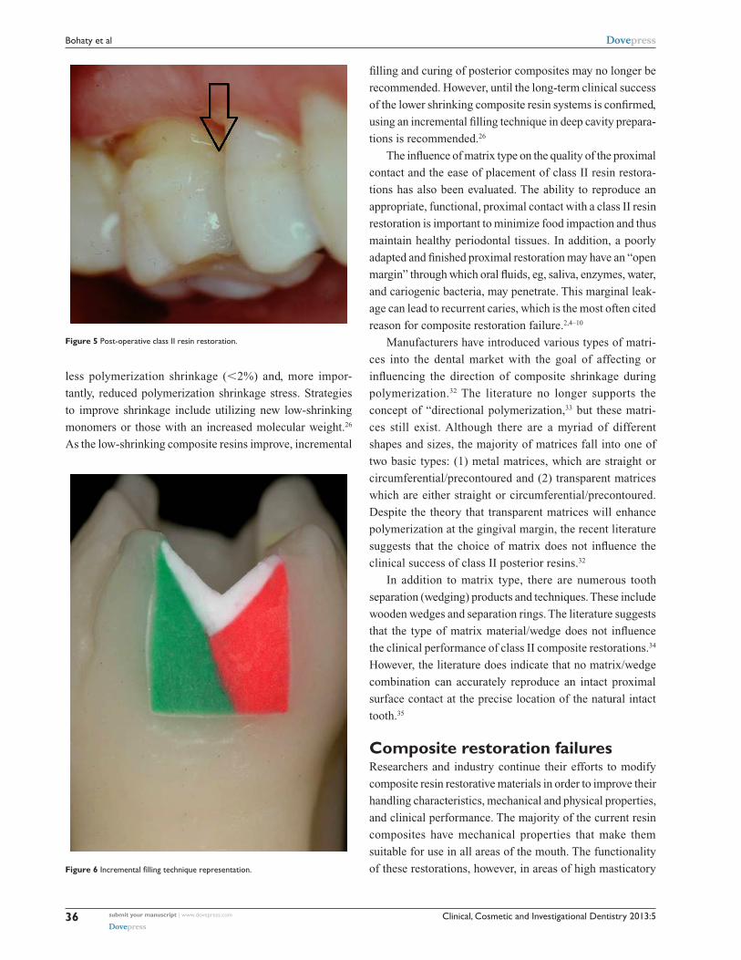
Figure 5 Post-operative class II resin restoration.
Figure 6 Incremental filling technique representation.
less polymerization shrinkage (,2%) and, more impor-
tantly, reduced polymerization shrinkage stress. Strategies
to improve shrinkage include utilizing new low-shrinking
monomers or those with an increased molecular weight.26
As the low-shrinking composite resins improve, incremental
submit your manuscript | www.dovepress.com
Dovepress
Dovepress
36
Bohaty et al

Clinical, Cosmetic and Investigational Dentistry 2013:5
stress is still a concern. Resin restorations that are placed in
areas of high function are more prone to exhibit excessive
wear and/or marginal fracture despite the advances in the
current materials. Clinicians must exercise caution when
placing large resin-based composite restorations in areas of
high function. The longevity of posterior resin restorations
placed in patients who have a history of clenching or grinding
may be particularly limited.35
While resin composition, tooth preparation design, and
matrix systems may influence the lifetime of posterior com-
posite restorations, the primary factor in the clinical failure
of moderate to large composite restorations is secondary
caries at the margins of the restorations.8 As an example, in
a study of radiographs from 459 adults, age 18–19 years, the
investigators reported that, among interproximal restorations,
the failure rate as a result of secondary or recurrent caries was
43% for composite as compared to 8% for amalgam.7 In a
separate study of amalgam and composite restorations placed
in 8–12-year-old children, the primary reason for failure of
both materials was secondary caries, but secondary caries
was 3.5 times higher in composite restorations.5
An increase in secondary caries at the margins of
composite restorations suggests that the seal at the com-
posite–tooth interface is not adequate to resist the physical,
chemical, and mechanical stresses that are present in the
mouth. The failure of moderate to large composite restora-
tions has been linked to the degradation of the bond at the
tooth surface–composite material interface12,36–41 and an
increase in the concentration of the cariogenic bacterium
Streptococcus mutans at the perimeter of these materi-
als.42–46 Degradation of the bond at the interface between the
tooth and composite has been associated with the failure of
adhesives to form an impervious seal with the dentin.2,41–50
Failure of the adhesive/dentin (a/d) bond leads to open pores
at the composite–tooth interface and bacterial enzymes, oral
fluids, and even bacteria can penetrate these open pores.51
Data from in vivo and in vitro studies indicate that the
infiltration of these agents into the voids between the tooth
and composite will lead to recurrent caries, hypersensitiv-
ity, and pulpal inflammation.41,47,52,53 Results from clinical
studies indicate loss of retention, poor marginal adapta-
tion, and marginal discoloration when the a/d interface is
exposed to the oral cavity.54 Effective mechanical bonding
between the composite restoration and treated enamel has
been achieved using appropriate acid-etching protocols,
but failure of the bond at the a/d interface threatens the
long-term clinical survival of moderate to large posterior
composite restorations.39,41,43,52,55–57
Bonding failures have been commonly tracked to the
gingival margin of class II composite restorations.58 A sepa-
ration between the composite material and tooth surface has
been noted at the gingival margin.55 In class II composite
restorations, there is generally little enamel available for
bonding at the gingival margin; therefore, the bond at this
site depends on the integrity of the seal formed with dentin.
Gaps at the gingival margin have been attributed to unreliable
dentin bonding.55,57 In a study comparing the microtensile
a/d bond strength of gingival and proximal walls of class II
composite restorations, the adhesive bond to the gingival wall
was significantly weaker.59 A complementary spectroscopic
study reported a twofold difference in the extent of dentin
demineralization at the proximal and gingival margins.50
The difference in demineralization suggests less mineralized
dentin at the gingival margin. The cumulative effect of less
mineral, increased density, and size of the tubules60 would
mean faster and deeper etching at the gingival margin as
compared to the proximal wall. Although the etch was deeper
at the gingival margin, there was considerably less adhesive
infiltration of the demineralized dentin matrix at the gingi-
val margin.50 The discrepancy between etching depth and
adhesive infiltration led to a large area of exposed collagen
at the gingival margin.
Yoshiyama et al suggested that the increased number of
tubules per unit area at the gingival margin would promote
efficient adhesive infiltration at this margin.61 However, other
variables, including water content, interfere with efficient
adhesive infiltration at the gingival margin. Water content
is higher in dentin at the gingival margin as compared to
the proximal wall. Water content is increased because of
the water present within the demineralized dentin matrix
and patent tubules that contain a great deal of dentinal fluid.
The presence of this fluid contributes to the contamination of
the prepared surface.62 The increased water leads to reduced
adhesive infiltration and lower monomer/polymer conversion
of the adhesive at the gingival margin as compared to the
proximal wall.50 The impact of water upon the effectiveness
of bonding is further supported by in vitro investigations that
indicate that adhesive monomers or oligomers and unpro-
tected collagen at the gingival margin of class II composite
restorations undergo hydrolytic degradation after 90 days of
aqueous storage.49
Wet bonding techniques were introduced in the early
1990s to counteract the problems noted with collagen col-
lapse following drying of the demineralized dentin matrix.63–66
Wet bonding means that the demineralized dentin matrix is
fully hydrated throughout the bonding protocol. Using this
submit your manuscript | www.dovepress.com
Dovepress
Dovepress
37
Posterior composite restoration update

Clinical, Cosmetic and Investigational Dentistry 2013:5
procedure, the channels between the demineralized dentin
collagen fibrils are filled with water, solvent, conditioner,
and/or oral fluids.67,68 Adhesive must diffuse into the fluid-
filled spaces of the substrate and along the collagen fibrils.
Ideally, the solvent in combination with hydrophilic mono-
mers, eg, hydroxyethyl methacrylate (HEMA) conditions
the collagen to remain expanded during adhesive infiltration.
However, HEMA, a primary component in many single-
bottle, commercial, dentin adhesives, can dramatically reduce
the evaporation of water.69 Hydrophobic monomers, such as
2,2-bis[4(2-hydroxy-3-methacryloyloxy-propyloxy)-phenyl]
propane (BisGMA), would resist diffusing into these sites
where there is residual water.70–72
In the in vivo situation, there may be little control over the
amount of water left on the tooth. Thus, it is possible to leave
the dentin surface so wet that the adhesive physically sepa-
rates into hydrophobic- and hydrophilic-rich phases.71,73,74
Indeed, results from laboratory investigations indicate that
excess moisture prohibited the formation of an impervious
structurally integrated a/d bond at the gingival margin of
class II composite restorations.49,50
Under clinical conditions, dentists must routinely attempt
to bond to naturally wet substrates, eg, caries-affected dentin75
or deep dentin.76–79 The water content of caries-affected dentin
is 2.7 times greater than that of normal dentin.75 Exposed
tubules account for 22% of the surface area in deep dentin.
In contrast, exposed tubules account for 1% of the surface
area of dentin close to the dentino-enamel junction.80 The
large increase in exposed tubules in deep dentin means that
pulpal fluid will contribute additional moisture to that already
present within the demineralized dentin matrix. With the
sensitivity of our current adhesives to excess moisture, it is
obvious that bonding to these clinically relevant substrates
is a formidable challenge.79,81–83 This difficulty highlights the
potential limitations in utilizing resin-based composites to
restore large, deep, carious lesions.
Sensitivity of adhesive to wet bonding conditionsWater blisters that form in adhesives placed on overly wet
surfaces84–86 and adhesive phase separation that leads to very
limited infiltration of the critical but hydrophobic dimethacry-
late component71,87,88 are two examples of the sensitivity of our
current adhesives to excess moisture. The optimum amount
of wetness varies as a function of the adhesive system.89 It is
impossible to simultaneously achieve uniform wetness on all
of the walls of the cavity preparation.90 In short, wet bonding
is a very technique-sensitive procedure. Optimum bonding
with our current commercial dentin adhesives occurs over a
very narrow range of conditions, eg, water content.78
Strategies to promote bonding of the resinous materials to
intrinsically wet dentin substrates include the incorporation
of ionic and hydrophilic monomers into the adhesive.91
These adhesives etch and prime simultaneously, thus
addressing the problems of collagen collapse and simplifying
the bonding protocol. The hydrophilicity of these adhesives
enhances water sorption, which can lead to hydrolytic break-
down in the mouth.85,90,92 With these systems, the bonded
interface lacks a nonsolvated hydrophobic resin coating. The
hybrid layers made with these adhesive systems behave as
semi-permeable membranes; water is transferred throughout
the bonded interface even after adhesive polymerization.54 The
increase in the concentration of hydrophilic monomers in these
systems has been associated with decreased structural integrity
at the a/d interface.54,93 Deterioration of the a/d bond formed
with these systems was noted after 1 year of in vivo aging.94
These results suggest that hydrophilicity and hydrolytic stabil-
ity of resin monomers are generally antagonistic.90
Effects of function, fatigue, and degradationWhen measured immediately, dentin-composite bonds are
generally considered adequate to tolerate conditions in the
mouth, but these bonds deteriorate with time. The two major
mechanisms of deterioration are fatigue and hydrolysis.95
Fatigue has been linked to stresses transmitted to the bond
by occlusal forces, thermal expansion and contraction, and
polymerization shrinkage of the composite. Chronic dete-
rioration of the dentin-composite bond is also related to
hydrolysis and leaching of the adhesive that has infiltrated
the tooth structure.70,79
Fatigue investigations have indicated that the overall
time-dependent behavior of the composite–tooth interface
is a complex function of the individual material phases. For
example, microfinite element analyses have shown that each
material phase at the a/d interface experiences different stress
concentrations at functional loads.96,97 The overall failure
behavior of the bond at the a/d interface is not determined by
the weakest component but by the component whose stress
concentration is closest to its failure strength. Similarly, the
overall fatigue life of the a/d interface is governed by the
material component with the shortest fatigue life under a
given loading condition.98
Under masticatory function the material components at
the composite–tooth interface are subjected to both chemi-
cal and mechanical stresses. The interplay between these
submit your manuscript | www.dovepress.com
Dovepress
Dovepress
38
Bohaty et al

Clinical, Cosmetic and Investigational Dentistry 2013:5
stresses can result in a deterioration of the properties of
the material over time. The breaking of covalent bonds by
addition of water to ester bonds is considered one of the
primary reasons for deterioration of the adhesive at the
interface between the composite and tooth.89,90 Interest-
ingly, degradation of methacrylate ester groups produces
carboxylic acids – the same functional group that is the
culprit in lactic acid-induced dental caries. The change in
mechanical properties of the materials can be contributed to
a variety of mechanisms that include proliferation of surface
and subsurface flaws.95,97,99–101 These flaws in combination
with the chemical and biochemical stresses that are present
in the mouth can then lead to restoration failure.
In conclusion, the a/d bond can be the first defense against
substances that may penetrate and ultimately undermine the
gingival margin in composite restorations in vivo. It has been
hypothesized that the in vivo degradation of the bond at the
a/d interface follows a cascade of events that begins when
the dentin is acid etched102,103 Disruption of the tooth structure
by acid etching exposes and activates proteolytic enzymes,
eg, matrix metalloproteinases (MMPs), that can degrade
the exposed collagen component of the hybrid layer.104,105
The following factors inhibit the formation of a durable a/d
bond: (1) water sorption and hydrolysis of the adhesive resin;
(2) inadequate monomer/polymer conversion of the infiltrat-
ing adhesive; (3) incomplete resin infiltration of the demin-
eralized dentin matrix; (4) incomplete solvent evaporation;
and (5) enzymatic challenges within the cavity preparation
through exposure to oral fluids.49,71,83,104–115 Although durable
a/d bonds are critical for maintaining a seal at the tooth–
composite interface, the properties of the materials are only
one part of an extremely complex problem.116
SummaryRestoring posterior teeth with resin-based composite mate-
rials continues to gain popularity among clinicians, and
the demand for such aesthetic restorations is increasing.
Manufacturers are working aggressively to improve resin
composite materials by modifying components to decrease
polymerization shrinkage, to improve mechanical and physi-
cal properties, and to enhance handling characteristics. The
two main causes of posterior composite restoration failure
are secondary caries and fracture (restoration or tooth).35
A review and update of posterior resin composites in terms
of preparation design, matrix choice, and resin systems
demonstrate the limited extent to which these factors influ-
ence the overall clinical lifetime of resins placed in posterior
teeth. Clinical and patient factors, including caries risk, cavity
size, cavity type, number of restored surfaces, and position
of the tooth in the mouth must be given careful attention in
the selection of any restorative material including composite
resins.
While clinicians tend to focus on tooth form and func-
tion when evaluating the success and failure of posterior
resins, the emphasis must remain in advancing our under-
standing and knowledge of the intricate and complicated
characteristics of the restoration–tooth interface. This paper
presents an update in existing technology and underscores the
mechanisms that negatively impact the durability of posterior
composites in permanent teeth.
AcknowledgmentsThe authors gratefully acknowledge partial support for this
work by the National Institute of Health and the National
Institute of Dental and Craniofacial Research (R01DE014392
and R01 DE022054).
DisclosureThe authors report no conflicts of interest in this work.
References 1. Beazoglou T, Eklund S, Heffley D, Meiers J, Brown LJ, Bailit H.
Economic impact of regulating the use of amalgam restorations. Public Health Rep. 2007;122(5):657–663.
2. Murray PE, Windsor LJ, Smyth TW, Hafez AA, Cox CF. Analysis of pulpal reactions to restorative procedures, materials, pulp capping, and future therapies. Crit Rev Oral Biol Med. 2002;13(6):509–520.
3. Palmer C. Good progress reported in mercury treaty talks. ADA News. 2011;42(21):1–2.
4. Simecek JW, Diefenderfer KE, Cohen ME. An evaluation of replace-ment rates for posterior resin-based composite and amalgam restorations in US. Navy and marine corps recruits. J Am Dent Assoc. 2009;140(2): 200–209.
5. Bernardo M, Luis H, Martin MD, et al. Survival and reasons for failure of amalgam versus composite posterior restorations placed in a random-ized clinical trial. J Am Dent Assoc. 2007;138(6):775–783.
6. Malhotra N, Mala K, Acharya S. Resin-based composite as a direct esthetic restorative material. Compend Contin Educ Dent. 2011;32(5): 14–23.
7. Levin L, Coval M, Geiger SB. Cross-sectional radiographic survey of amalgam and resin-based composite posterior restorations. Quintessence Int. 2007;38(6):511–514.
8. Mjor IA, Dahl JE, Moorhead JE. Age of restorations at replacement in permanent teeth in general dental practice. Acta Odontol Scand. 2000;58(3):97–101.
9. Opdam NJ, Bronkhorst EM, Loomans BA, Huysmans MC. 12-year survival of composite vs amalgam restorations. J Dent Res. 2010;89(10): 1063–1067.
10. Soncini JA, Maserejian NN, Trachtenberg F, Tavares M, Hayes C. The longevity of amalgam versus compomer/composite restorations in posterior primary and permanent teeth: findings From the New England Children’s Amalgam Trial. J Am Dent Assoc. 2007;138(6): 763–772.
11. DeRouen TA, Martin MD, Leroux BG, et al. Neurobehavioral effects of dental amalgam in children: a randomized clinical trial. JAMA. 2006;295(15):1784–1792.
submit your manuscript | www.dovepress.com
Dovepress
Dovepress
39
Posterior composite restoration update

Clinical, Cosmetic and Investigational Dentistry 2013:5
12. Van Nieuwenhuysen JP, D’Hoore W, Carvalho J, Qvist V. Long-term evaluation of extensive restorations in permanent teeth. J Dent. 2003; 31(6):395–405.
13. Kohler B, Rasmusson CG, Odman P. A five-year clinical evaluation of Class II composite resin restorations. J Dent. 2000;28(2):111–116.
14. Trachtenberg F, Maserejian NN, Tavares M, Soncini JA, Hayes C. Extent of tooth decay in the mouth and increased need for replacement of dental restorations: the New England Children’s Amalgam Trial. Pediatr Dent. 2008;30(5):388–392.
15. Hunt PR. A modified class II cavity preparation for glass ionomer restorative materials. Quintessence Int Dent Dig. 1984;15(10): 1011–1018.
16. Knight GM. The tunnel restoration – nine years of clinical experience using capsulated glass ionomer cements. Case report. Aust Dent J. 1992;37(4):245–251.
17. Wiegand A, Attin T. Treatment of proximal caries lesions by tunnel restorations. Dent Mater. 2007;23(12):1461–1467.
18. Tyas MJ, Anusavice KJ, Frencken JE, Mount GJ. Minimal intervention dentistry – a review. FDI Commission Project 1–97. Int Dent J. 2000; 50(1):1–12.
19. Lopes GC, Vieira LC, Araujo E. Direct composite resin restorations: a review of some clinical procedures to achieve predictable results in posterior teeth. J Esthet Restor Dent. 2004;16(1):19–31.
20. Da Rosa Rodolpho PA, Donassollo TA, Cenci MS, et al. 22-Year clinical evaluation of the performance of two posterior composites with different filler characteristics. Dent Mater. 2011;27(10):955–963.
21. Da Rosa Rodolpho PA, Cenci MS, Donassollo TA, Loguercio AD, Demarco FF. A clinical evaluation of posterior composite restorations: 17-year findings. J Dent. 2006;34(7):427–435.
22. Plasmans PJ, Creugers NH, Mulder J. Long-term survival of extensive amalgam restorations. J Dent Res. 1998;77(3):453–460.
23. Demarco FF, Correa MB, Cenci MS, Moraes RR, Opdam NJ. Longevity of posterior composite restorations: not only a matter of materials. Dent Mater. 2012;28(1):87–101.
24. El-Sahn NA, El-Kassas DW, El-Damanhoury HM, Fahmy OM, Gomaa H, Platt JA. Effect of C-factor on microtensile bond strengths of low-shrinkage composites. Oper Dent. 2011;36(3):281–292.
25. Shirai K, De Munck J, Yoshida Y, et al. Effect of cavity configuration and aging on the bonding effectiveness of six adhesives to dentin. Dent Mater. 2005;21(2):110–124.
26. Van Ende A, Mine A, De Munck J, Poitevin A, Van Meerbeek B. Bonding of low-shrinking composites in high C-factor cavities. J Dent. 2012;40(4):295–303.
27. Kuijs RH, Fennis WM, Kreulen CM, Barink M, Verdonschot N. Does layering minimize shrinkage stresses in composite restorations? J Dent Res. 2003;82(12):967–971.
28. Abbas G, Fleming GJ, Harrington E, Shortall AC, Burke FJ. Cuspal movement and microleakage in premolar teeth restored with a pack-able composite cured in bulk or in increments. J Dent. 2003;31(6): 437–444.
29. Lee MR, Cho BH, Son HH, Um CM, Lee IB. Influence of cavity dimension and restoration methods on the cusp deflection of premolars in composite restoration. Dent Mater. 2007;23(3):288–295.
30. Park J, Chang J, Ferracane J, Lee IB. How should composite be layered to reduce shrinkage stress: incremental or bulk filling? Dent Mater. 2008;24(11):1501–1505.
31. Kwon Y, Ferracane J, Lee IB. Effect of layering methods, composite type, and flowable liner on the polymerization shrinkage stress of light cured composites. Dent Mater. 2012;28(7):801–809.
32. Cenci MS, Demarco FF, Pereira CL, Lund RG, de Carvalho RM. One-year comparison of metallic and translucent matrices in Class II composite resin restorations. Am J Dent. 2007;20(1):41–45.
33. Versluis A, Tantbirojn D, Douglas WH. Do dental composites always shrink toward the light? J Dent Res. 1998;77(6):1435–1445.
34. Cenci MS, Lund RG, Pereira CL, de Carvalho RM, Demarco FF. In vivo and in vitro evaluation of Class II composite resin restorations with different matrix systems. J Adhes Dent. 2006;8(2):127–132.
35. Kampouropoulos D, Paximada C, Loukidis M, Kakaboura A. The influ-ence of matrix type on the proximal contact in Class II resin composite restorations. Oper Dent J. 2010;35(4):454–462.
36. Collins CJ, BR, Hodge KLV. A clinical evaluation of posterior com-posite resin restorations: 8-year findings. J Dent. 1998;26:311–317.
37. Mair LH. Ten-year clinical assessment of three posterior resin com-posites and two amalgams. Quintessence Int. 1998;29(8):483–490.
38. Mjor IA, Dahl JE, Moorhead JE. Placement and replacement of restorations in primary teeth. Acta Odontologica Scandinavica. 2002;60:25–28.
39. Nordbo H, LJ, von der Fehr FR. Saucer-shaped cavity preparations for posterior aproximal resin composite restorations: Observations up to 10 years. Quintessence Int. 1988;29(1):5–11.
40. Owens BM, Johnson WW. Effect of insertion technique and adhesive system on microleakage of Class V resin composite restorations. J Adhes Dent. 2005;7:303–308.
41. Van Meerbeek B, Van Landuyt K, De Munck J, et al. Technique- sensitivity of contemporary adhesives. Dent Mater J. 2005;24(1): 1–13.
42. Anusavice KJ. Management of dental caries as a chronic infectious disease. J Dent Educ. 1998;62:791–802.
43. Dunne SM, Gainsford ID, Wilson NHF. Current materials and tech-niques for direct restorations in posterior teeth. Part 1: silver amalgam. Int Dent J. 1997;47:123–136.
44. Hansel C, Leyhausen G, Mai UE, Geurtsen W. Effects of various resin composite (co)monomers and extracts on two caries-associated micro-organisms in vitro. J Dent Res. 1998;77:60–67.
45. Santerre JP, Shajii L, Leung BW. Relation of dental composite formu-lations to their degradation and the release of hydrolyzed polymeric-resin-derived products. Crit Rev Oral Biol Med. 2001;12:136–151.
46. Svanberg M, Mjor IA, Orstavik D. Mutans Streptococci in plaque from margins of Amalgam composite, and glass-ionomer restorations. J Dent Res. 1990;69(3):861–864.
47. Hashimoto M, Ohno H, Kaga M, Endo K, Sano H, Oguchi H. Resin-tooth adhesive interfaces after long-term function. Am J Dent. 2001;14(4):211–215.
48. Meiers JC, Kresin J. Cavity disinfectants and dentin bonding. Oper Dent. 1996;21:153–159.
49. Spencer P, Wang Y, Bohaty B. Interfacial chemistry of moisture-aged class II composite restorations. J Biomed Mater Res B Appl Biomater. 2006;77(2):234–240.
50. Wang Y, Spencer P. Interfacial chemistry of class II composite restoration: structure analysis. J Biomed Mater Res A. 2005;75(3): 580–587.
51. Gallo LM, Nickel JC, Iwasaki LR, Palla S. Stress-field translation in the healthy human temporomandibular joint. J Dent Res. 2000;79(10): 1740–1746.
52. Andersson-Wenckert IE, van Dijken JW, Kieri C. Durability of exten-sive class II open-sandwich restorations with a resin-modified glass ionomer cement after 6 years. Am J Dent. 2004;17:43–50.
53. Brannstrom M. Communication between the oral cavity and the dental pulp associated with restorative treatment. Oper Dent. 1984;9:57–68.
54. Breschi L, Mazzoni A, Ruggeri A, Cadenaro M, Di Lenarda R, De Stefano Dorigo E. Dental adhesion review: aging and stability of the bonded interface. Dent Mater. 2008;24(1):90–101.
55. Roulet JF. Benefits and disadvantages of tooth-coloured alternatives to amalgam. J Dent. 1997;25:459–473.
56. Van Dijken JW. Direct resin composite inlays/onlays: an 11 year follow-up. J Dent. 2000;28:299–306.
57. Kleverlaan CJ, Feilzer AJ. Polymerization shrinkage and contraction stress of dental resin composites. Dent Mater. 2005;21:1150–1157.
58. Bouillaguet S, Ciucchi B, Jacoby T, Wataha JC, Pashley D. Bonding characteristics to dentin walls of class II cavities, in vitro. Dent Mater. 2001;17:316–321.
59. Purk JH, Dusevich V, Glaros AG, Spencer P, Eick JD. In vivo versus in vitro microtensile bond strength of axial versus gingival cavity prepa-ration walls in class II resin-based composite restorations. J Am Dent Assoc. 2004;135:185–193.
submit your manuscript | www.dovepress.com
Dovepress
Dovepress
40
Bohaty et al

Clinical, Cosmetic and Investigational Dentistry 2013:5
60. Garberoglio P. The ratio of the densities of dentinal tubules on the cervical and axial wall in cavities. Quintessence Int. 1994;25: 49–52.
61. Yoshiyama M, Carvalho R, Sano H, Horner J, Brewer PD, Pashley DH. Interfacial morphology and strength of bonds made to superficial versus deep dentin. Am J Dent. 1995;8:297–302.
62. Pashley DH. Clinical correlations of dentin structure and function. J Prosthet Dent. 1991;66:777–781.
63. Gwinnett AJ. Chemically conditioned dentin: a comparison of conven-tional and environmental scanning electron microscopy findings. Dent Mater. 1994;10:150–155.
64. Gwinnett AJ. Dentin bond strength after air drying and rewetting. Am J Dent. 1994;7:144–148.
65. Gwinnett AJ. Altered tissue contribution to interfacial bond strength with acid conditioned dentin. Am J Dent. 1994;7:243–246.
66. Kanca J. Improved bond strength through acid etching of dentin and bond-ing to wet dentin surfaces. J Am Dent Assoc. 1992;123:235–243.
67. Pashley DH, Ciucchi B, Sano H, Horner JA. Permeability of dentin to adhesive agents. Quintessence Int. 1993;24:618–631.
68. Nakabayashi N, Watanabe A, Arao T. A tensile test to facilitate identification of defects in resin-bonded dentin specimens. J Dent. 1998;26:379–385.
69. Pashley EL, Zhang Y, Lockwood PE, Rueggeberg FA, Pashley DH. Effects of HEMA on water evaporation from water-HEMA mixtures. Dent Mater. 1998;14(1):6–10.
70. Wang Y, Spencer P. Hybridization efficiency of the adhesive dentin interface with wet bonding. J Dent Res. 2003;82:141–145.
71. Spencer P, Wang Y. Adhesive phase separation at the dentin interface under wet Bonding conditions. J Biomed Mater Res. 2002;62(3):447–456.
72. Spencer P, Wang Y, Walker MP, Wieliczka DM, Swafford JR. Interfacial chemistry of the dentin/adhesive bond. J Dent Res. 2000; 79(7):1458–1463.
73. Ye Q, Park J, Laurence JS, Parthasarathy R, Misra A, Spencer P. Ternary phase diagram of model dentin adhesive exposed to over-wet environments. J Dent Res. 2011;90(12):1434–1438.
74. Ye Q, Park JG, Parthasarathy R, et al. Quantitative analysis of aque-ous phase composition of model dentin adhesives experiencing phase separation. J Biomed Mater Res Part B: Appl Biomaterials. 2012;100B:1086–1092.
75. Ito S, Hashimoto M, Wadgaonkar B, et al. Effects of resin hydrophilicity on water sorption and changes in modulus of elasticity. Biomaterials. 2005;26(33):6449–6459.
76. Marshall GW, Marshall SJ, Kinney JH, Balooch M. The dentin substrate:structure and properties related to bonding. J Dent. 1997;25: 441–458.
77. Pereira PNR, Okuda M, Sano H, Yoshikawa T, Burrow MF, Tagami J. Effect of intrinsic wetness and regional difference on dentin bond strength. Dent Mater. 1999;15:46–53.
78. Roulet JF, Degrange M, editors. Adhesion: The Silent Revolution in Dentistry, 1st ed: Quintessence Publishing Co, Inc; Berlin, Germany; 1999:263.
79. Wang Y, Spencer P, Hager C, Bohaty B. Comparison of interfacial characteristics of adhesive bonding to superficial versus deep dentin using SEM and staining techniques. J Dent. 2006;34:26–34.
80. Pashley DH. Dentin: A dynamic substrate in dentistry. Scanning Microsc. 1989;3:161–176.
81. Spencer P, Wang Y, Katz JL, Misra A. Physicochemical interactions at the dentin/adhesive interface using FTIR chemical imaging. J Biomed Opt. 2005;10(3):031104.
82. Wang Y, Spencer P. Continuing etching of an all-in-one adhesive in wet dentin tubules. J Dent Res. 2005;84:350–354.
83. Wang Y, Spencer P, Yao X, Brenda B. Effect of solvent content on resin hybridization in wet dentin bonding. J Biomed Mater Res A. 2007;82(4): 975–983.
84. Tay FR, Gwinnett AJ, Pang KM, Wei SHY. An optical, micromor-phological study of surface moisture in the total etched resin-dentin interface. Am J Dent. 1996;9:43–48.
85. Tay FR, Gwinnett AJ, Wei SHY. The overwet phenomenon: a trans-mission electron microscopic study of surface moisture in the acid-conditioned, resin-dentin interface. Am J Dent. 1996;9:161–166.
86. Tay FR, Gwinnett AJ, Wei SHY. Micromophological spectrum from overdrying to overwetting acid-conditioned dentin in water-free, acetone-based, single-bottle primer/adhesives. Dent Mater. 1996;12:236–244.
87. Gwinnett AJ, Tay FR, Pang KM, Wei SHY. Quantitative Contribution of the Collagen Network in Dentin Hybridization. Am J Dent. 1996;9: 140–144.
88. Wang Y, Spencer P, Yao X. Micro-raman imaging analysis of monomer/ mineral distribution in intertubular region of adhesive/dentin interfaces. J Biomed Optics. 2006;11:024005-1–024005-7.
89. Tay FR, Pashley DH. Water treeing – a potential mechanism for degradation of dentin adhesives. Am J Dent. 2003;16(1):6–12.
90. Tay FR, Pashley DH. Have dentin adhesives become too hydrophilic? J Can Dent Assoc. 2003;69(11):726–731.
91. Hebling J, Pashley DH, Tjaderhane L, Tay FR. Chlorhexidine arrests subclinical degradation of dentin hydbrid layers in vivo. J Dent Res. 2005;84(8):741–746.
92. Parthasarathy R, Misra A, Park J, Ye Q, Spencer P. Diffusion coef-ficients of water and leachables in methacrylate-based crosslinked polymers using absorption experiments. J Mater Sci Mater Med. 2012;23(5):1157–1172.
93. Peumans M, Kanumilli P, De Munck J, Van Landuyt K, Lambrechts P, Van Meerbeek B. Clinical effectiveness of contem-porary adhesives: a systematic review of current clinical trials. Dent Mater. 2005;21(9):864–881.
94. Donmez N, Belli S, Pashley DH, Tay FR. Ultrastructural correlates of in vivo/in vitro bond degradation in self-etch adhesives. J Dent Res. 2005;84(4):355–359.
95. Staninec M, Kim P, Marshall GW, Ritchie RO, Marshall SJ. Fatigue of dentin-composite interfaces with four-point bend. Dent Mater. 2008;24(6):799–803.
96. Misra A, Spencer P, Marangos O, Wang Y, Katz JL. Micromechanical analysis of dentin/adhesive interface by the finite element method. J Biomed Mater Res B Appl Biomater. 2004;70(1):56–65.
97. Misra A, Spencer P, Marangos O, Wang Y, Katz JL. Parametric study of the effect of phase anisotropy on the micromechanical behaviour of dentin-adhesive interfaces. J R Soc Interface. 2005;2(3):145–157.
98. Singh V, Misra A, Marangos O, et al. Fatigue life prediction of dentin-adhesive interface using micromechanical stress analysis. Dent Mater. 2011;27(9):e187–e195.
99. Katz JL, Misra A, Spencer P, et al. Multiscale mechanics of hierarchical structure/property relationships in calcified tissues and tissue/material interfaces. Mater Sci Eng A Struct Mater. 2007;27(3):450–468.
100. Singh V, Misra A, Marangos O, et al. Viscoelastic and fatigue proper-ties of model methacrylate-based dentin adhesives. J Biomed Mater Res B Appl Biomater. 2010;95(2):283–290.
101. Belli R, Baratieri LN, Braem M, Petschelt A, Lohbauer U. Tensile and bending fatigue of the adhesive interface to dentin. Dent Mater. 2010;26(12):1157–1165.
102. Pashley DH, Tay FR, Yiu C, et al. Collagen degradation by host-derived enzymes during aging. J Dent Res. 2004;83(3):216–221.
103. Sano H. Microtensile testing, nanoleakage, and biodegradation of resin-dentin bonds. J Dent Res. 2006;85(1):11–14.
104. Mazzoni A, Pashley DH, Nishitani Y, et al. Reactivation of quenched endogenous proteolytic activities in phosphoric acid-etched den-tine by etch-and-rinse adhesives. Biomaterials. 2006;27(25): 4470–4476.
105. Tay FR, Pashley DH, Loushine RJ, Weller RN, Monticelli F, Osorio R. Self-etching adhesives increase collagenolytic activity in radicular dentin. J Endodo. 2006;32(9):862–868.
106. Spencer P, Swafford JR. Unprotected protein at the dentin-adhesive interface. Quintessence Int. 1999;30(7):501–507.
107. Spencer P, Ye Q, Park J, Topp EM, et al. Adhesive/dentin interface: the weak link in the composite restoration. Ann Biomed Eng. 2010;38(6): 1989–2003.
submit your manuscript | www.dovepress.com
Dovepress
Dovepress
41
Posterior composite restoration update

Clinical, Cosmetic and Investigational Dentistry
Publish your work in this journal
Submit your manuscript here: http://www.dovepress.com/clinical-cosmetic-and-investigational-dentistry-journal
Clinical, Cosmetic and Investigational Dentistry is an international, peer-reviewed, open access, online journal focusing on the latest clini-cal and experimental research in dentistry with specific emphasis on cosmetic interventions. Innovative developments in dental materials, techniques and devices that improve outcomes and patient satisfac-
tion and preference will be highlighted. The manuscript management system is completely online and includes a very quick and fair peer-review system, which is all easy to use. Visit http://www.dovepress.com/testimonials.php to read real quotes from published authors.
Clinical, Cosmetic and Investigational Dentistry 2013:5
108. Ye Q, Spencer P, Wang Y, Misra A. Relationship of solvent to the photopolymerization process, properties and structure in model dentin adhesives. J Biomed Mater Res A. 2007;80(2):342–350.
109. Guo X, Spencer P, Wang Y, Ye Q, Yao X, Williams K. Effects of a solubility enhancer on penetration of hydrophobic component in model adhesives into wet demineralized dentin. Dent Mater. 2007;23: 1473–1481.
110. Kostoryz EL, Dharmala K, Ye Q, et al. Enzymatic biodegradation of HEMA/BisGMA adhesives formulated with different water content. J Biomed Mater Res B: Appl Biomater. 2009;88B:394–401.
111. Guo X, Wang Y, Spencer P, Ye Q, Yao X. Effects of water content and initiator composition on photopolymerization of a model BisGMA/HEMA resin. Dent Mater. 2008;24(6):824–831.
112. Ye Q, Wang Y, Spencer P. Nanophase separation of polymers exposed to simulated bonding conditions. J Biomed Mater Res B: Appl Biomater. 2009;88B:339–348.
113. Park JG, Ye Q, Topp EM, Spencer P. Enzyme-catalyzed hydrolysis of dentin adhesives containing a new urethane-based trimethacrylate monomer. J Biomed Mater Res Part B: Appl Biomater. 2009;91B: 562–571.
114. Park J, Ye Q, Topp EM, Kieweg SL, Spencer P. Effect of photoinitia-tor system and water content on dynamic mechanical properties of a light-cured bisGMA/HEMA dental resin. J Biomed Mater Res Part A. 2010;93(4):1245–1251.
115. Park JG, Ye Q, Topp EM, Misra A, Spencer P. Water sorption and dynamic mechanical properties of dentin adhesives with a urethane-based multifunctional methacrylate monomer. Dent Mater. 2009;25: 1569–1575.
116. Drummond JL. Degradation, fatigue, and failure of resin dental composite materials. J Dent Res. 2008;87(8):710–719.
submit your manuscript | www.dovepress.com
Dovepress
Dovepress
Dovepress
42
Bohaty et al