Embed Size (px)
Citation preview
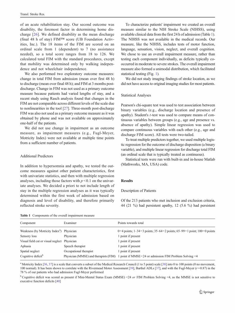
ORIGINAL ARTICLE
Post-Stroke Apathy and Hypersomnia Lead to WorseOutcomes from Acute Rehabilitation
Ari L. Harris & Jessica Elder & Nicholas D. Schiff &Jonathan D. Victor & Andrew M. Goldfine
Received: 18 June 2013 /Revised: 17 September 2013 /Accepted: 6 October 2013# Springer Science+Business Media New York 2013
Abstract Apathy and hypersomnia occur after stroke and, bydefinition, reduce participation in rehabilitation, but their effecton outcome from acute rehabilitation is not known. We per-formed a retrospective review of 213 patients admitted to astroke-specialized acute rehabilitation unit in the United States.All patients had ischemic or hemorrhagic stroke, and no demen-tia or dependence on others pre-stroke.We diagnosed apathy andhypersomnia using standardized documentation by treating ther-apists.We usedmultiple regression analysis to control for overallimpairment (combination of strength, cognitive and sensorymeasures), age, time since stroke, and stroke type (ischemic orhemorrhagic). Forty-four (21 %) of the patients had persistentapathy, and 12 (5.6 %) had persistent hypersomnia. Both groupswere more impaired in cognition, sustained attention, and morelikely to be treated for depression. Patients with apathy were 2.4times more likely to go to a nursing home, and had dischargeFIM scores 12 points below the mean. Patients withhypersomnia were ten timesmore likely to go to a nursing home,and had discharge FIM scores 16 points below the mean. Thesefindings indicate that studies to prospectively define these
clinical factors and potential confounds using standardized toolsare indicated, and if confirmed, justify studies to identify thesepatients early and develop targeted interventions.
Keywords Apathy . Hypersomnia . Stroke . Rehabilitation
Introduction
In addition to the well-recognized motor and sensory deficitsin patients with stroke, apathy and hypersomnia can reducegoal-directed behavior and therefore participation in rehabili-tation. If reduction in participation due to apathy orhypersomnia affects the rehabilitation process, it could explainsome of the variability in recovery [1, 2], and treatment ofthese conditions could improve patients' response to rehabil-itation interventions, reducing disability and improving out-comes. But, as a prerequisite to initiating clinical trials of suchtreatments, we first need to know the prevalence of theseconditions in the acute rehabilitation period, and whether theyhave an independent effect on outcome.
Apathy is a reduction of goal-directed behavior in thesetting of intact consciousness, and can be due to impairedemotional reactivity, motor planning deficits, or inability toself-initiate behaviors [3]. In stroke, it is associated withdamage or reduced blood flow to prefrontal cortex and basalganglia [4–6]. Additionally, it may occur as a consequence ofcoexisting illnesses such as depression and neurodegenerativediseases, with potentially overlapping mechanisms [7–9].
Considering all etiologies, a recent meta-analysis of 24studies found that apathy occurs in 29.5–40.2 % of patientsafter stroke, and is typically associated with worse disabilityand enduring cognitive deficits [10]. None of the studies in themeta-analysis were from the American acute rehabilitationpopulation; two were performed in Japan where patients enterrehabilitation >1 month after stroke: Hama and colleagues
A. L. Harris :A. M. Goldfine (*)Burke Medical Research Institute, Weill Cornell Medical College,785 Mamaroneck Avenue, White Plains, NY 10605, USAe-mail: [email protected]
A. L. HarrisDepartment of Neurology, Physicians Office Building Suite 324,Providence, RI, USA
J. ElderDepartment of Biostatistics and Epidemiology, Weill CornellMedical College, Burke Medical Research Institute, 785Mamaroneck Avenue, White Plains, NY 10605, USA
N. D. Schiff : J. D. VictorBrain and Mind Research Institute and Department of Neurology,Weill Cornell Medical College, 1300 York Avenue, New York,NY 10605, USA
Transl. Stroke Res.DOI 10.1007/s12975-013-0293-y

[11] found that 40 % of patients had apathy by patient reportand 19 % by structured interview of the caregiver; Santa andcolleagues [12] found 20 % of patients with apathy by patientreport [12]. Studies that followed patients over time found thatapathy generally remains present even up to 1 year [13, 14].
Hypersomnia is excessive total sleep and can be due todysfunction of the brain's arousal network or nighttime sleepdisruption [15]. It can occur after stroke from focal injury tobasal forebrain or diencephalic structures [16], or be a featureof delirium in the setting of metabolic disturbances [17]. Threestudies found that hypersomnia occurred between 4 % and18 % of patients in the first days after stroke [18–20], and onestudy found it in 14 % of 44 patients in acute rehabilitation[21]. A more recent study, only reported in a review article,found that in the chronic phase (21±18 months) after stroke27 % of patients had hypersomnia [15].
The published literature is difficult to apply to the Ameri-can acute rehabilitation because none specifically looked ateffect on outcome in this population. Furthermore, studies ofapathy in similar time periods after stroke typically excludedpatients with aphasia or severe cognitive deficits to allow foruse of a patient-report measure such as the Apathy Scale [22].To address these limitations, we conducted a preliminarystudy, using existing medical records, to study effects ofapathy and hypersomnia on outcomes from acute rehabilita-tion. Instead of using a patient-report scale, we operationallydefined the presence of these conditions using treating thera-pists' observations, ensuring that all patients could be includ-ed. We hypothesized that, independent of stroke severity andother factors, apathy and hypersomnia would have a negativeimpact on outcome, as judged by disposition location (nursinghome vs. home) and disability.
Methods
Patient Subjects
We conducted a retrospective electronic chart review of pa-tients treated in the Stroke Program at Burke RehabilitationHospital in 2011. We included 362 patients admitted from anacute care hospital with a diagnosis of ischemic or hemorrhag-ic stroke. We then excluded 149 patients who: completed lessthan 7 days of rehabilitation; had a diagnosis of a neurodegen-erative disease; required assistance of another person for dailyactivities prior to the stroke; were transferred out of rehabilita-tion to a hospital for greater than 3 days; were admitted morethan once within the year (we excluded the later admission); orhad inadequate documentation required for analysis. This re-sulted in a total of 213 patients for analysis.
The study was approved by the Institutional Review Boardof Burke Rehabilitation Center.
Available Documentation
Data for patient behaviors and other clinical characteristicswere obtained from physician and therapist electronic clinicaldocumentation, as well as billing, laboratory and other elec-tronic clinical databases.
Definition of Apathy and Hypersomnia
Patients were classified as having apathy or hypersomnia basedon behaviors documented by physical and speech therapistsapproximately three to five times per week. This observationalapproach allowed us to include patients with aphasia andcognitive deficits that would be unable to complete self-reportscales, and ensured that the behaviors were present duringtherapy sessions. To limit the effect of transient disturbancesof arousal (e.g., a night of inadequate sleep or infection), werequired that the behaviors were documented in at least half ofthe daily progress notes, with a minimum of three total notes.
To define apathy and hypersomnia, we chose from a list ofterms that therapists used to document patient behavior. Thislist included: "hypoarousal," "internally distracted," "impul-sivity," "decreased initiation," "externally distracted," "flataffect," "perseveration," "restlessness," "lability," "confabula-tion." Our operational definition of apathy was that the ther-apists selected the term "decreased initiation," implying thatpatients did not participate in therapy unless encouraged. Thiswas the closest term to the core of the definition of apathy—"a lack of motivation"— used by most authors [23, 24]. "Flataffect" is also a feature of apathy [23], though we only reporton it here as a clinical descriptor. Our operational definition ofhypersomnia was that the therapist selected the term"hypoarousal," used to describe patients who had their eyesclosed and appeared sleepy during the therapy session. Notethat while this definition of hypersomnia includes patientswho were sleepy due to disturbed nighttime sleep, our clinicalexperience is that this is an uncommon cause of persistenthypersomnia and most patients identified by the operationaldefinition had excessive total sleep. Because this study isbased on a retrospective review of routine clinical records inwhich most patients had a single therapist for the course oftheir treatment, we were unable to assess inter-rater reliability.
We also recorded speech therapist documentation of find-ings in the first 48 h after admission that have been reported toco-occur with apathy, including impaired sustained attention,and impaired executive dysfunction [5, 25]. Therapists used avariety of tasks to test for these, and then documented theirpresence or absence based on an overall impression.
Outcome Measures
Our first outcome was discharge disposition (home vs.nursing home), as home discharge is a fundamental goal
Transl. Stroke Res.
of an acute rehabilitation stay. Our second outcome wasdisability, the foremost factor in determining home dis-charge [26]. We defined disability as the mean discharge(final 48 h of stay) FIM™ score (UB Foundation Activ-ities, Inc.). The 18 items of the FIM are scored on anordinal scale from 1 (dependent) to 7 (no assistanceneeded), so a total score ranges from 18 to 126. Wecalculated total FIM with the standard procedures, exceptthat mobility was determined only by walking indepen-dence and not wheelchair independence.
We also performed two exploratory outcome measures:change in total FIM from admission (mean over first 48 h)to discharge (mean over final 48 h); and FIM at 3 months post-discharge. Change in FIM was not used as a primary outcomemeasure because patients had varied lengths of stay, and arecent study using Rasch analysis found that changes in theFIM are not comparable across different levels of the scale dueto nonlinearities in the tool [27]. Three-month post-dischargeFIMwas also not used as a primary outcomemeasure as it wasobtained by phone and was not available on approximatelyone-half of the patients.
We did not use change in impairment as an outcomemeasure, as impairment measures (e.g., Fugl-Meyer,Motricity Index) were not available at multiple time pointsfrom a sufficient number of patients.
Additional Predictors
In addition to hypersomnia and apathy, we tested the out-come measures against other patient characteristics, firstwith univariate statistics, and then with multiple regressionanalyses, including those factors with p <0.1 on the univar-iate analyses. We decided a priori to not include length ofstay in the multiple regression analyses as it was typicallydetermined within the first week of admission based ondiagnosis and level of disability, and therefore primarilyreflected stroke severity.
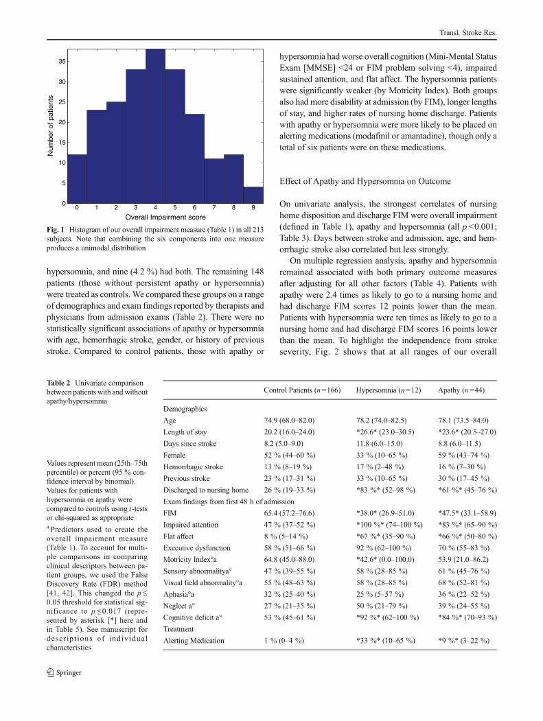
To characterize patients' impairment we created an overallmeasure similar to the NIH Stroke Scale (NIHSS), usingavailable clinical data from the first 24 h of admission (Table 1).The NIHSS was not available in the medical records. Ourmeasure, like the NIHSS, includes tests of motor function,language, sensation, vision, neglect, and overall cognition.We chose to use an overall impairment measure, rather thantesting each component individually, as deficits typically co-occurred in moderate to severe strokes. The overall impairmentmeasure also formed a unimodal distribution, which facilitatedstatistical testing (Fig. 1).
We did not study imaging findings of stroke location, as wedid not have access to original imaging studies for most patients.
Statistical Analyses
Pearson's chi-square test was used to test association betweenbinary variables (e.g., discharge location and presence ofapathy). Student's t-test was used to compare means of con-tinuous variables between groups (e.g., age and presence vs.absence of apathy). Simple linear regression was used tocompare continuous variables with each other (e.g., age anddischarge FIM score). All tests were two-tailed.
To treat multiple predictors together, we used multiple logis-tic regression for the outcome of discharge disposition (a binaryvariable), and multiple linear regression for discharge total FIM(an ordinal scale that is typically treated as continuous).
Statistical tests were run with built-in and in-house Matlab(Mathworks, MA, USA) code.
Results
Description of Patients
Of the 213 patients who met inclusion and exclusion criteria,44 (21 %) had persistent apathy, 12 (5.6 %) had persistent
Table 1 Components of the overall impairment measure
Component Examiner Points towards total
Weakness (by Motricity Indexa) Physician 0=4 points; 1–34=3 points; 35–64=2 points; 65–99=1 point; 100=0 points
Sensory loss Physician 1 point if present
Visual field cut or visual neglect Physician 1 point if present
Aphasia Speech therapist 1 point if present
Spatial neglect Occupational therapist 1 point if present
Cognitive deficitb Physician (MMSE) and therapists (FIM) 1 point if MMSE<24 or admission FIM Problem Solving <4
aMotricity Index [36, 37] is a scale that converts a subset of the Medical Research Council (1 to 5 point) scale [38] into 0 to 100 points (0 no movement,100 normal). It has been shown to correlate with the Rivermead Motor Assessment [39], Barthel ADLs [37], and with the Fugl-Meyer (r =0.87) in the78 % of our patients who had admission Fugl-Meyer performedb Cognitive deficit was scored as present if Mini-Mental Status Exam (MMSE) <24 or FIM Problem Solving <4, as the MMSE is not sensitive toexecutive function deficits [40]
Transl. Stroke Res.
hypersomnia, and nine (4.2 %) had both. The remaining 148patients (those without persistent apathy or hypersomnia)were treated as controls.We compared these groups on a rangeof demographics and exam findings reported by therapists andphysicians from admission exams (Table 2). There were nostatistically significant associations of apathy or hypersomniawith age, hemorrhagic stroke, gender, or history of previousstroke. Compared to control patients, those with apathy or
hypersomnia had worse overall cognition (Mini-Mental StatusExam [MMSE] <24 or FIM problem solving <4), impairedsustained attention, and flat affect. The hypersomnia patientswere significantly weaker (by Motricity Index). Both groupsalso had more disability at admission (by FIM), longer lengthsof stay, and higher rates of nursing home discharge. Patientswith apathy or hypersomnia were more likely to be placed onalerting medications (modafinil or amantadine), though only atotal of six patients were on these medications.
Effect of Apathy and Hypersomnia on Outcome
On univariate analysis, the strongest correlates of nursinghome disposition and discharge FIM were overall impairment(defined in Table 1), apathy and hypersomnia (all p <0.001;Table 3). Days between stroke and admission, age, and hem-orrhagic stroke also correlated but less strongly.
On multiple regression analysis, apathy and hypersomniaremained associated with both primary outcome measuresafter adjusting for all other factors (Table 4). Patients withapathy were 2.4 times as likely to go to a nursing home andhad discharge FIM scores 12 points lower than the mean.Patients with hypersomnia were ten times as likely to go to anursing home and had discharge FIM scores 16 points lowerthan the mean. To highlight the independence from strokeseverity, Fig. 2 shows that at all ranges of our overall
0 1 2 3 4 5 6 7 8 90
5
10
15
20
25
30
35
Num
ber
of p
atie
nts
Overall Impairment score
Fig. 1 Histogram of our overall impairment measure (Table 1) in all 213subjects. Note that combining the six components into one measureproduces a unimodal distribution
Table 2 Univariate comparisonbetween patients with and withoutapathy/hypersomnia
Values represent mean (25th–75thpercentile) or percent (95 % con-fidence interval by binomial).Values for patients withhypersomnia or apathy werecompared to controls using t-testsor chi-squared as appropriatea Predictors used to create theoverall impairment measure(Table 1). To account for multi-ple comparisons in comparingclinical descriptors between pa-tient groups, we used the FalseDiscovery Rate (FDR) method[41, 42]. This changed the p ≤0.05 threshold for statistical sig-nificance to p ≤0.017 (repre-sented by asterisk [*] here andin Table 5). See manuscript fordescr ip t ions of individualcharacteristics
Control Patients (n=166) Hypersomnia (n =12) Apathy (n =44)
Demographics
Age 74.9 (68.0–82.0) 78.2 (74.0–82.5) 78.1 (73.5–84.0)
Length of stay 20.2 (16.0–24.0) *26.6* (23.0–30.5) *23.6* (20.5–27.0)
Days since stroke 8.2 (5.0–9.0) 11.8 (6.0–15.0) 8.8 (6.0–11.5)
Female 52 % (44–60 %) 33 % (10–65 %) 59 % (43–74 %)
Hemorrhagic stroke 13 % (8–19 %) 17 % (2–48 %) 16 % (7–30 %)
Previous stroke 23 % (17–31 %) 33 % (10–65 %) 30 % (17–45 %)
Discharged to nursing home 26 % (19–33 %) *83 %* (52–98 %) *61 %* (45–76 %)
Exam findings from first 48 h of admission
FIM 65.4 (57.2–76.6) *38.0* (26.9–51.0) *47.5* (33.1–58.9)
Impaired attention 47 % (37–52 %) *100 %* (74–100 %) *83 %* (65–90 %)
Flat affect 8 % (5–14 %) *67 %* (35–90 %) *66 %* (50–80 %)
Executive dysfunction 58 % (51–66 %) 92 % (62–100 %) 70 % (55–83 %)
Motricity Index°a 64.8 (45.0–88.0) *42.6* (0.0–100.0) 53.9 (21.0–86.2)
Sensory abnormalitya° 47 % (39–55 %) 58 % (28–85 %) 61 % (45–76 %)
Visual field abnormality°a 55 % (48–63 %) 58 % (28–85 %) 68 % (52–81 %)
Aphasia°a 32 % (25–40 %) 25 % (5–57 %) 36 % (22–52 %)
Neglect a° 27 % (21–35 %) 50 % (21–79 %) 39 % (24–55 %)
Cognitive deficit a° 53 % (45–61 %) *92 %* (62–100 %) *84 %* (70–93 %)
Treatment
Alerting Medication 1 % (0–4 %) *33 %* (10–65 %) *9 %* (3–22 %)
Transl. Stroke Res.
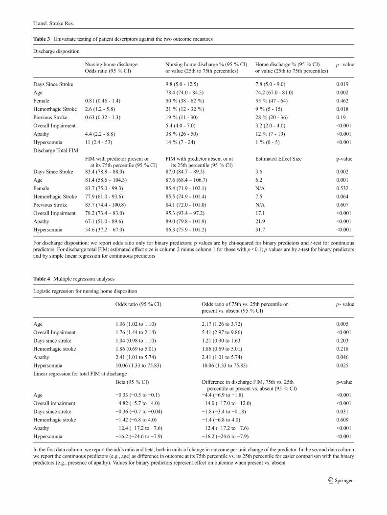
Table 3 Univariate testing of patient descriptors against the two outcome measures
Discharge disposition
Nursing home dischargeOdds ratio (95 % CI)
Nursing home discharge % (95 % CI)or value (25th to 75th percentiles)
Home discharge % (95 % CI)or value (25th to 75th percentiles)
p- value
Days Since Stroke 9.8 (5.0 - 12.5) 7.8 (5.0 - 9.0) 0.019
Age 78.4 (74.0 - 84.5) 74.2 (67.0 - 81.0) 0.002
Female 0.81 (0.46 - 1.4) 50 % (38 - 62 %) 55 % (47 - 64) 0.462
Hemorrhagic Stroke 2.6 (1.2 - 5.8) 21 % (12 - 32 %) 9 % (5 - 15) 0.018
Previous Stroke 0.63 (0.32 - 1.3) 19 % (11 - 30) 28 % (20 - 36) 0.19
Overall Impairment 5.4 (4.0 - 7.0) 3.2 (2.0 - 4.0) <0.001
Apathy 4.4 (2.2 - 8.8) 38 % (26 - 50) 12 % (7 - 19) <0.001
Hypersomnia 11 (2.4 - 53) 14 % (7 - 24) 1 % (0 - 5) <0.001
Discharge Total FIM
FIM with predictor present orat its 75th percentile (95 % CI)
FIM with predictor absent or atits 25th percentile (95 % CI)
Estimated Effect Size p-value
Days Since Stroke 83.4 (78.8 – 88.0) 87.0 (84.7 – 89.3) 3.6 0.002
Age 81.4 (58.6 – 104.3) 87.6 (68.4 – 106.7) 6.2 0.001
Female 83.7 (75.0 - 99.3) 85.4 (71.9 - 102.1) N/A 0.532
Hemorrhagic Stroke 77.9 (61.0 - 93.6) 85.5 (74.9 - 101.4) 7.5 0.064
Previous Stroke 85.7 (74.4 - 100.8) 84.1 (72.0 - 101.0) N/A 0.607
Overall Impairment 78.2 (73.4 – 83.0) 95.3 (93.4 – 97.2) 17.1 <0.001
Apathy 67.1 (51.0 - 89.6) 89.0 (79.8 - 101.9) 21.9 <0.001
Hypersomnia 54.6 (37.2 – 67.0) 86.3 (75.9 - 101.2) 31.7 <0.001
For discharge disposition: we report odds ratio only for binary predictors; p values are by chi-squared for binary predictors and t-test for continuouspredictors. For discharge total FIM: estimated effect size is column 2 minus column 1 for those with p<0.1; p values are by t-test for binary predictorsand by simple linear regression for continuous predictors
Table 4 Multiple regression analyses
Logistic regression for nursing home disposition
Odds ratio (95 % CI) Odds ratio of 75th vs. 25th percentile orpresent vs. absent (95 % CI)
p- value
Age 1.06 (1.02 to 1.10) 2.17 (1.26 to 3.72) 0.005
Overall Impairment 1.76 (1.44 to 2.14) 5.41 (2.97 to 9.86) <0.001
Days since stroke 1.04 (0.98 to 1.10) 1.21 (0.90 to 1.63 0.203
Hemorrhagic stroke 1.86 (0.69 to 5.01) 1.86 (0.69 to 5.01) 0.218
Apathy 2.41 (1.01 to 5.74) 2.41 (1.01 to 5.74) 0.046
Hypersomnia 10.06 (1.33 to 75.83) 10.06 (1.33 to 75.83) 0.025
Linear regression for total FIM at discharge
Beta (95 % CI) Difference in discharge FIM, 75th vs. 25thpercentile or present vs. absent (95 % CI)
p-value
Age −0.33 (−0.5 to −0.1) −4.4 (−6.9 to −1.8) <0.001
Overall impairment −4.82 (−5.7 to −4.0) −14.0 (−17.0 to −12.0) <0.001
Days since stroke −0.36 (−0.7 to −0.04) −1.8 (−3.4 to −0.18) 0.031
Hemorrhagic stroke −1.42 (−6.8 to 4.0) −1.4 (−6.8 to 4.0) 0.609
Apathy −12.4 (−17.2 to −7.6) −12.4 (−17.2 to −7.6) <0.001
Hypersomnia −16.2 (−24.6 to −7.9) −16.2 (−24.6 to −7.9) <0.001
In the first data column, we report the odds ratio and beta, both in units of change in outcome per unit change of the predictor. In the second data columnwe report the continuous predictors (e.g., age) as difference in outcome at its 75th percentile vs. its 25th percentile for easier comparison with the binarypredictors (e.g., presence of apathy). Values for binary predictors represent effect on outcome when present vs. absent
Transl. Stroke Res.
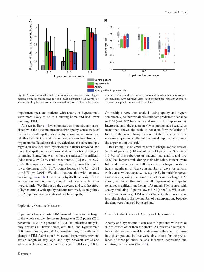
impairment measure, patients with apathy or hypersomniawere more likely to go to a nursing home and had lowerdischarge FIM.
As seen in Table 4, hypersomnia was more strongly asso-ciated with the outcome measures than apathy. Since 20 % ofthe patients with apathy also had hypersomnia, we wonderedwhether the effect of apathy was merely due to the subset withhypersomnia. To address this, we calculated the same multipleregression analyses with hypersomnia patients removed. Wefound that apathy remained correlated with fraction dischargedto nursing home, but was no longer statistically significant(odds ratio 2.19, 95 % confidence interval [CI] 0.91 to 5.29,p =0.082). Apathy remained significantly correlated withlower discharge FIM (10.73 points lower, 95 % CI −15.71to −5.75, p <0.001). We also illustrate this with separatebars in Fig. 2a and b. Thus, apathy by itself had a significantassociation with outcome, though not nearly as large ashypersomnia. We did not do the converse and test the effectof hypersomnia with apathy patients removed, as only threeof 12 hypersomnia patients did not have apathy.
Exploratory Outcome Measures
Regarding change in total FIM from admission to discharge,in the whole sample, the mean change was 23.2 points (25thpercentile 15.7; 75th percentile 30.3). On univariate analysis,only apathy (4.4 fewer points, p =0.013) and hypersomnia(7.0 fewer points, p =0.024), correlated significantly withchange in FIM. Admission FIM, overall impairment, previousstroke, length of stay, age, and days between stroke andadmission did not correlate with change in FIM (all p >0.2).
On multiple regression analysis using apathy and hyper-somnia only, neither remained significant predictors of changein FIM (p =0.062 for apathy and p =0.13 for hypersomnia).Interpretation of the change in FIM is problematic because, asmentioned above, the scale is not a uniform reflection offunction: the same change in score at the lower end of thescale may represent a different functional improvement than atthe upper end of the scale.
Regarding FIM at 3 months after discharge, we had data on52 % of patients (110 out of the 213 patients). Seventeen(15 %) of this subgroup of patients had apathy, and two(2 %) had hypersomnia during their admission. Patients werefollowed up at a mean of 120 days after discharge (no statis-tically significant difference in number of days for patientswith versus without apathy, t -test p =0.3). In multiple regres-sion analysis, using the same predictors as discharge FIMabove, we found that age, overall impairment and apathyremained significant predictors of 3-month FIM scores, withapathy predicting 13 points lower FIM (p <0.01). While con-sistent with discharge FIM scores (Table 4), these results areless reliable due to the low number of participants and becausethe data were obtained by telephone.
Other Potential Causes of Apathy and Hypersomnia
Apathy and hypersomnia can occur in patients with strokedue to causes other than the stroke. As this was a retrospec-tive study, we were unable to determine the specific causein a given patient, but we were able to test for the preva-lence of three potential causes: infection, depression andsedating medications (Table 5).
0
20
40
60
80
100
120
Impairment score range
Dis
char
ge F
IM
Control patientHypersomniaApathyApathy without Hypersomnia
BA
0−2 3−5 6−90−2 3−5 6−90
0.2
0.4
0.6
0.8
1
Impairment score range
Fra
ctio
n di
scha
rged
to n
ursi
ng h
ome
Fig. 2 Presence of apathy and hypersomnia are associated with highernursing home discharge rates (a) and lower discharge FIM scores (b),after controlling for our overall impairment measure (Table 1). Error bars
in a are 95 % confidence limits by binomial statistics. b Encircled dotsare medians; bars represent 25th–75th percentiles; whiskers extend toextreme data points not considered outliers
Transl. Stroke Res.
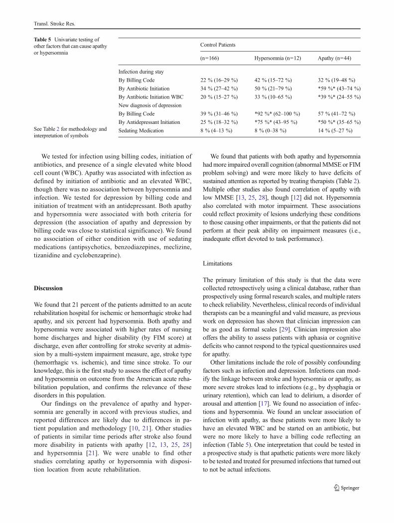
We tested for infection using billing codes, initiation ofantibiotics, and presence of a single elevated white bloodcell count (WBC). Apathy was associated with infection asdefined by initiation of antibiotic and an elevated WBC,though there was no association between hypersomnia andinfection. We tested for depression by billing code andinitiation of treatment with an antidepressant. Both apathyand hypersomnia were associated with both criteria fordepression (the association of apathy and depression bybilling code was close to statistical significance). We foundno association of either condition with use of sedatingmedications (antipsychotics, benzodiazepines, meclizine,tizanidine and cyclobenzaprine).
Discussion
We found that 21 percent of the patients admitted to an acuterehabilitation hospital for ischemic or hemorrhagic stroke hadapathy, and six percent had hypersomnia. Both apathy andhypersomnia were associated with higher rates of nursinghome discharges and higher disability (by FIM score) atdischarge, even after controlling for stroke severity at admis-sion by a multi-system impairment measure, age, stroke type(hemorrhagic vs. ischemic), and time since stroke. To ourknowledge, this is the first study to assess the effect of apathyand hypersomnia on outcome from the American acute reha-bilitation population, and confirms the relevance of thesedisorders in this population.
Our findings on the prevalence of apathy and hyper-somnia are generally in accord with previous studies, andreported differences are likely due to differences in pa-tient population and methodology [10, 21]. Other studiesof patients in similar time periods after stroke also foundmore disability in patients with apathy [12, 13, 25, 28]and hypersomnia [21]. We were unable to find otherstudies correlating apathy or hypersomnia with disposi-tion location from acute rehabilitation.
We found that patients with both apathy and hypersomniahadmore impaired overall cognition (abnormalMMSE or FIMproblem solving) and were more likely to have deficits ofsustained attention as reported by treating therapists (Table 2).Multiple other studies also found correlation of apathy withlow MMSE [13, 25, 28], though [12] did not. Hypersomniaalso correlated with motor impairment. These associationscould reflect proximity of lesions underlying these conditionsto those causing other impairments, or that the patients did notperform at their peak ability on impairment measures (i.e.,inadequate effort devoted to task performance).
Limitations
The primary limitation of this study is that the data werecollected retrospectively using a clinical database, rather thanprospectively using formal research scales, and multiple ratersto check reliability. Nevertheless, clinical records of individualtherapists can be a meaningful and valid measure, as previouswork on depression has shown that clinician impression canbe as good as formal scales [29]. Clinician impression alsooffers the ability to assess patients with aphasia or cognitivedeficits who cannot respond to the typical questionnaires usedfor apathy.
Other limitations include the role of possibly confoundingfactors such as infection and depression. Infections can mod-ify the linkage between stroke and hypersomnia or apathy, asmore severe strokes lead to infections (e.g., by dysphagia orurinary retention), which can lead to delirium, a disorder ofarousal and attention [17]. We found no association of infec-tions and hypersomnia. We found an unclear association ofinfection with apathy, as these patients were more likely tohave an elevated WBC and be started on an antibiotic, butwere no more likely to have a billing code reflecting aninfection (Table 5). One interpretation that could be tested ina prospective study is that apathetic patients were more likelyto be tested and treated for presumed infections that turned outto not be actual infections.
Table 5 Univariate testing ofother factors that can cause apathyor hypersomnia
See Table 2 for methodology andinterpretation of symbols
Control Patients
(n=166) Hypersomnia (n=12) Apathy (n=44)
Infection during stay
By Billing Code 22 % (16–29 %) 42 % (15–72 %) 32 % (19–48 %)
By Antibiotic Initiation 34 % (27–42 %) 50 % (21–79 %) *59 %* (43–74 %)
By Antibiotic Initiation WBC 20 % (15–27 %) 33 % (10–65 %) *39 %* (24–55 %)
New diagnosis of depression
By Billing Code 39 % (31–46 %) *92 %* (62–100 %) 57 % (41–72 %)
By Antidepressant Initiation 25 % (18–32 %) *75 %* (43–95 %) *50 %* (35–65 %)
Sedating Medication 8 % (4–13 %) 8 % (0–38 %) 14 % (5–27 %)
Transl. Stroke Res.
Depression is a potential confounder, as it is associatedwith worse stroke outcomes [30, 31], and can present withapathy [7]. Our database did not include formal testing ofdepression, but both groups were more likely to have billingcodes consistent with depression (trend for apathy, significantfor hypersomnia), and more than twice as likely to be treatedwith antidepressants (Table 5). Multiple previous studiesfound no association between depression and apathy afterstroke [11, 12, 28, 32], suggesting that many of these patientswere not actually depressed, but were treated as if they were.More studies are needed to disambiguate depression fromapathy and hypersomnia in stroke patients, especially as thereare reports of some antidepressants worsening apathy [33, 34].
Implications
While we have shown that apathy and hypersomnia arestrongly associated with outcome from acute rehabilitation,we do not address the mechanism of this effect. There areseveral possibilities, not mutually exclusive. One possibility isthat the behavioral abnormalities of these conditions result inmore dependence on others, and therefore need for institution-al care. This is supported by our finding of lower FIM scoreafter controlling for overall impairment (Fig. 2b and Table 4).A second possibility is that patients with these conditions haveslower rates of recovery due to decreased participation intherapy. This could be formally evaluated in a prospectivestudy with serial measurements of impairment as well asparticipation. A third possibility is that both apathy andhypersomnia are signs of under-aroused brains, which arenot performing as well as they could (as proposed in [35]).
Our findings demonstrate that apathy and hypersomnia arecommon in patients undergoing acute rehabilitation afterstroke. Both conditions contributed to explaining the rangeof outcomes of patients in acute rehabilitation, and should beadded as covariates in prospective observational and interven-tional studies. Both can be measured by purely observationalmeans, allowing inclusion of patients with language and cog-nitive disorders [21, 32], and should use validated and blindedmeasures to the extent possible. If prospective trials confirmtheir relevance, targeted treatments should be developed basedon studies of underlyingmechanism. Adequate treatment at anearly stage could potentially improve patient response to acuterehabilitation, thereby lowering costs of care by increasing thefraction of patients discharged home.
Acknowledgments We thank Cathy Dwyer, Janet Herbold and Mi-chael Reding from the Burke Rehabilitation Hospital for assistance withacquiring and interpreting the original data. We gratefully acknowledgefunding support from the Burke Medical Research Institute.
Conflict of Interest Ari Harris declares that he has no conflict of interest.Jessica Elder declares that she has no conflict of interest. Nicholas Schiff
declares that he has no conflict of interest. Jonathan Victor declares that hehas no conflict of interest. Andrew Goldfine declares that he has no conflictof interest.
References
1. Duncan PW, Goldstein LB, Matchar D, Divine GW, Feussner J.Measurement of motor recovery after stroke. Outcome assessmentand sample size requirements. Stroke. 1992;23(8):1084–9.
2. Prabhakaran S, Zarahn E, Riley C, Speizer A, Chong JY, Lazar RM,et al. Inter-individual variability in the capacity for motor recoveryafter ischemic stroke. Neurorehabil Neural Repair. 2008;22(1):64–71. doi:10.1177/1545968307305302.
3. Levy R, Dubois B. Apathy and the functional anatomy of the pre-frontal cortex–basal ganglia circuits. Cereb Cortex. 2006;16(7):916–28. doi:10.1093/cercor/bhj043.
4. Murakami T, Hama S, Yamashita H, Onoda K, Kobayashi M,Kanazawa J, et al. Neuroanatomic pathways associated withpoststroke affective and apathetic depression. The American Journalof Geriatric Psychiatry. 2013. doi:10.1016/j.jagp.2013.01.057.
5. Okada K, Kobayashi S, Yamagata S, Takahashi K, Yamaguchi S.Poststroke apathy and regional cerebral blood flow. Stroke.1997;28(12):2437–41.
6. Onoda K, Kuroda Y, Yamamoto Y, Abe S, Oguro H, Nagai A, et al.Post-stroke apathy and hypoperfusion in basal ganglia: SPECTstudy.Cerebrovasc Dis. 2011;31(1):6–11. doi:10.1159/000319771.
7. American Psychiatric A. Diagnostic and Statistical Manual ofMentalDisorders DSM-IV-TR Fourth Edition. 4th ed. Amer Psychiatric Pub;2000.
8. Ligthart SA, Richard E, Fransen NL, Eurelings LSM, Beem L,Eikelenboom P, et al. Association of vascular factors with apathy incommunity-dwelling elderly individuals. Arch Gen Psychiatry.2012;69(6):636–42. doi:10.1001/archgenpsychiatry.2011.1858.
9. Robert P, Onyike CU, Leentjens AF, Dujardin K, Aalten P, StarksteinS, et al. Proposed diagnostic criteria for apathy in Alzheimer's diseaseand other neuropsychiatric disorders. European psychiatry : the jour-nal of the Association of European Psychiatrists. 2009;24(2):98–104.doi:10.1016/j.eurpsy.2008.09.001.
10. van Dalen JW, van Charante EPM, Nederkoorn PJ, van Gool WA.Richard E. Stroke: Poststroke apathy; 2013. doi:10.1161/STROKEAHA.112.674614.
11. Hama S, Yamashita H, Shigenobu M, Watanabe A, Hiramoto K,Kurisu K, et al. Depression or apathy and functional recovery afterstroke. Int J Geriat Psychiatry. 2007;22(10):1046–51. doi:10.1002/gps.1866.
12. Santa N, Sugimori H, Kusuda K, Yamashita Y, Ibayashi S, Iida M.Apathy and functional recovery following first-ever stroke. Int JRehab i l Res . 2008 ;31(4 ) :321–6 . do i :10 .1097 /MRR.0b013e3282fc0f0e.
13. Mayo NE, Fellows LK, Scott SC, Cameron J, Wood-Dauphinee S. Alongitudinal view of apathy and its impact after stroke. Stroke.2009;40(10):3299–307. doi:10.1161/STROKEAHA.109.554410.
14. Withall A, BrodatyH, Altendorf A, Sachdev PS. A longitudinal studyexamining the independence of apathy and depression after stroke:the Sydney Stroke Study. Int Psychogeriatr. 2011;23(2):264–73. doi:10.1017/S1041610209991116.
15. Bassetti CL, Hermann DM. Sleep and stroke. Handbook of ClinicalNeurology. 2011;99:1051–72. doi:10.1016/B978-0-444-52007-4.00021-7.
16. Bassetti C, Mathis J, Gugger M, Lovblad KO, Hess CW.Hypersomnia following paramedian thalamic stroke: a reportof 12 patients. Ann Neurol. 1996;39(4):471–80. doi:10.1002/ana.410390409.
Transl. Stroke Res.
17. Oldenbeuving AW, de Kort PLM, Jansen BPW, Algra A, KappelleLJ, Roks G. Delirium in the acute phase after stroke: incidence, riskfactors, and outcome. Neurology. 2011;76(11):993–9. doi:10.1212/WNL.0b013e318210411f.
18. Marquardsen J. The natural history of acute cerebrovascular disease:a retrospective study of 769 patients. Acta Neurol Scand. 1969;45:Suppl-38:11+.
19. Terént A, Andersson B. The prognosis for patients with cerebrovas-cular stroke and transient ischemic attacks. Ups J Med Sci.1981;86(1):63–74.
20. Kotila M. Declining incidence and mortality of stroke? Stroke.1984;15(2):255–9.
21. Reding MJ, Gardner C, Hainline B, Devinsky O. Neuropsychiatricproblems interfering with inpatient stroke rehabilitation.Neurorehabil Neural Repair. 1993;7(1):1–7. doi:10.1177/136140969300700102.
22. Starkstein SE,Mayberg HS, Preziosi TJ, Andrezejewski P, LeiguardaR, Robinson RG. Reliability, validity, and clinical correlates of apa-thy in Parkinson's disease. The Journal of Neuropsychiatry andClinical Neurosciences. 1992;4(2):134–9.
23. Starkstein SE, Leentjens AFG. The nosological position of apathy inclinical practice. J Neurol Neurosurg Psychiatr. 2008;79(10):1088–92. doi:10.1136/jnnp.2007.136895.
24. Marin RS, Wilkosz PA. Disorders of diminished motivation. J HeadTrauma Rehabil. 2005;20(4):377–88.
25. Brodaty H, Sachdev PS, Withall A, Altendorf A, Valenzuela MJ,Lorentz L. Frequency and clinical, neuropsychological and neuroim-aging correlates of apathy following stroke — the Sydney StrokeStudy. Psychol Med. 2005;35(12):1707–16. doi:10.1017/S0033291705006173.
26. Ween JE, Alexander MP, D'Esposito M, Roberts M. Factors predic-tive of stroke outcome in a rehabilitation setting. Neurology.1996;47(2):388–92.
27. Glenny C, Stolee P, Thompson M, Husted J, Berg K.Underestimating physical function gains: comparing FIM motorsubscale and interrai post acute care activities of daily living scale.Arch Phys Med Rehabil. 2012;93(6):1000–8. doi:10.1016/j.apmr.2011.12.027.
28. Starkstein SE, Fedoroff JP, Price TR, Leiguarda R, RobinsonRG. Apathy following cerebrovascular lesions. Stroke.1993;24(11):1625–30.
29. Berg A, Lönnqvist J, Palomäki H, Kaste M. Assessment of depres-sion after stroke: a comparison of different screening instruments.Stroke. 2009;40(2):523–9. doi:10.1161/STROKEAHA.108.527705.
30. Kauhanen ML, Korpelainen JT, Hiltunen P, Brusin E, Mononen H,Määttä R, et al. Poststroke depression correlates with cognitiveimpairment and neurological deficits. Stroke. 1999;30(9):1875–80.doi:10.1161/01.STR.30.9.1875.
31. Pohjasvaara T, Vataja R, Leppävuori A, Kaste M, Erkinjuntti T.Depression is an independent predictor of poor long-term functionaloutcome post-stroke. Eur J Neurol. 2001;8(4):315–9. doi:10.1046/j.1468-1331.2001.00182.x.
32. Carota A, Berney A, Aybek S, Iaria G, Staub F, Ghika-Schmid F, et al.A prospective study of predictors of poststroke depression. Neurology.2005;64(3):428–33. doi:10.1212/01.WNL.0000150935.05940.2D.
33. Kodela S, Venkata PD. Antidepressant induced apathy responsive todose reduction. Psychopharmacol Bull. 2010;43(4):76–9.
34. Padala PR, Padala KP, Monga V, Ramirez DA, Sullivan DH.Reversal of SSRI-associated apathy syndrome by discontinuationof therapy. Ann Pharmacother. 2012;46(3). doi:10.1345/aph.1Q656
35. Goldfine AM, Schiff ND. What is the role of brain mechanismsunderlying arousal in recovery of motor function after structural braininjuries? Curr Opin Neurol. 2011;24(6):564–9. doi:10.1097/WCO.0b013e32834cd4f5.
36. Demeurisse G, Demol O, Robaye E. Motor evaluation in vascularhemiplegia. Eur Neurol. 1980;19(6):382–9. doi:10.1159/000115178.
37. Wade DT, Hewer RL. Motor loss and swallowing difficulty afterstroke: frequency, recovery, and prognosis. Acta Neurol Scand.1987;76(1):50–4.
38. Medical Research C. Aids to the investigation of peripheral nerveinjuries. J Neurol Psychiatry. 1943;6(1–2).
39. Collin C, Wade D. Assessing motor impairment after stroke: a pilotreliability study. Journal of Neurology, Neurosurgery & Psychiatry.1990;53(7):576–9. doi:10.1136/jnnp.53.7.576.
40. Dubois B, Slachevsky A, Litvan I, Pillon B. The FAB A frontalassessment battery at bedside. Neurology. 2000;55(11):1621–6. doi:10.1212/WNL.55.11.1621.
41. Benjamini Y, Hochberg Y. Controlling the false discovery rate: apractical and powerful approach to multiple testing. J R Statist Soc B.1995;57(1):289–300.
42. Benjamini Y, Yekutieli D. The control of the false discovery rate inmultiple testing under dependency. Ann Stat. 2001;29(4):1165–88.
Transl. Stroke Res.