Embed Size (px)
Citation preview
Post partum Uterine Involution in the Cow
Akira OKANO and Tsuneo TOMIZUKA
Department of Animal Reproduction, National Institute of Animal Industry (Tsukuba, Ibaraki, 305 Japan)
Abstract
Seven primiparous Japanese Black cows, which were nursing their calves, were slaughtered several days post partum to examine the histological pattern of uterine involution. At Days 18 and 23 post partum, the uterine endometrium contained many phagocytes and nodular aggregates of lymphocytes. The outer diameter of the uterine glands varied considerably and the lumen was enlarged. Capillaries located under the surface epithelium were not yet completely contracted. At Days 46 and 54 post partum, however, the endometrium contained only a small amount of lumphocytes and phagocytes and uterine glands evenly distributed showed a tall glandular epithelium. These glands which were located in the basal zone hardly appeared to have a lumen. Ultrasonographical observation of the progression of uterine involution was started at Day 8 post partum and continued until Day 43 post partum. The diameter and area of the uterine horn and the endometrium in cross-sectional images of the endometrium were estimated using an image analyzer. The relationship between the estimated dimensions of the uterine horn and the number of days post partum fitted into polynomial regressions. It was concluded that the uterine involution of cows with calves was completed at approximately Day 40 post partum.
Discipline: Animal industryAdditional key words: histological observation of uterus, ultrasonograph
JARQ 30, 113- 121 (1996)
Introduction
To shorten the calving interval, it is necessary
to diagnose the progression of uterine involution post par/um. For this reason, a large number of studies on postpartum uterine involution have been carried out in the cow2
•9
•11
•16
•18
•20>.
Many histological studies have so far been carried out on the bovine pregnant uterus7
•11
•2 '> with
limited emphasis placed on the post partum uterus 9•15>.
Until recently, studies on post par/um uterine involution in the cow were carried out by rectal palpation at regular intervals 16•
19•20
> or by morphological and histological procedures on uteri removed after slaughtering11•18>. Histological studies of uteri from slaughtered cows provide information about uterine involution post partum. A rapid and objective method of diagnosing the progression of involution is thus essential. In recent years, the ultrasonographical linear scanner has been used for pregnancy diagnosis and analysis of the uterine morphology in cow 19>. The authors reported real-time ultrasono-
graphs of post partum bovine uteri obtained by the ultrasonographical linear scanner for diagnosing the progression of post par/um uterine involution in the cow.
Histological observation of uterus post partum
The uteri were removed from 5 nursing Japanese Black cows which were slaughtered at Days 18, 23, 29, 46 and 54 post par/um. Serial sections were cut and stained with hematoxylin and eosin for histological observation. Outer and inner diameters of 50 uterine glands in cross-sections located in the superficial and the basal zones were measured at random using an ocular micrometer. Height of surface epithelia at 50 points in the caruncular and the intercaruncular zones was measured in the same way.
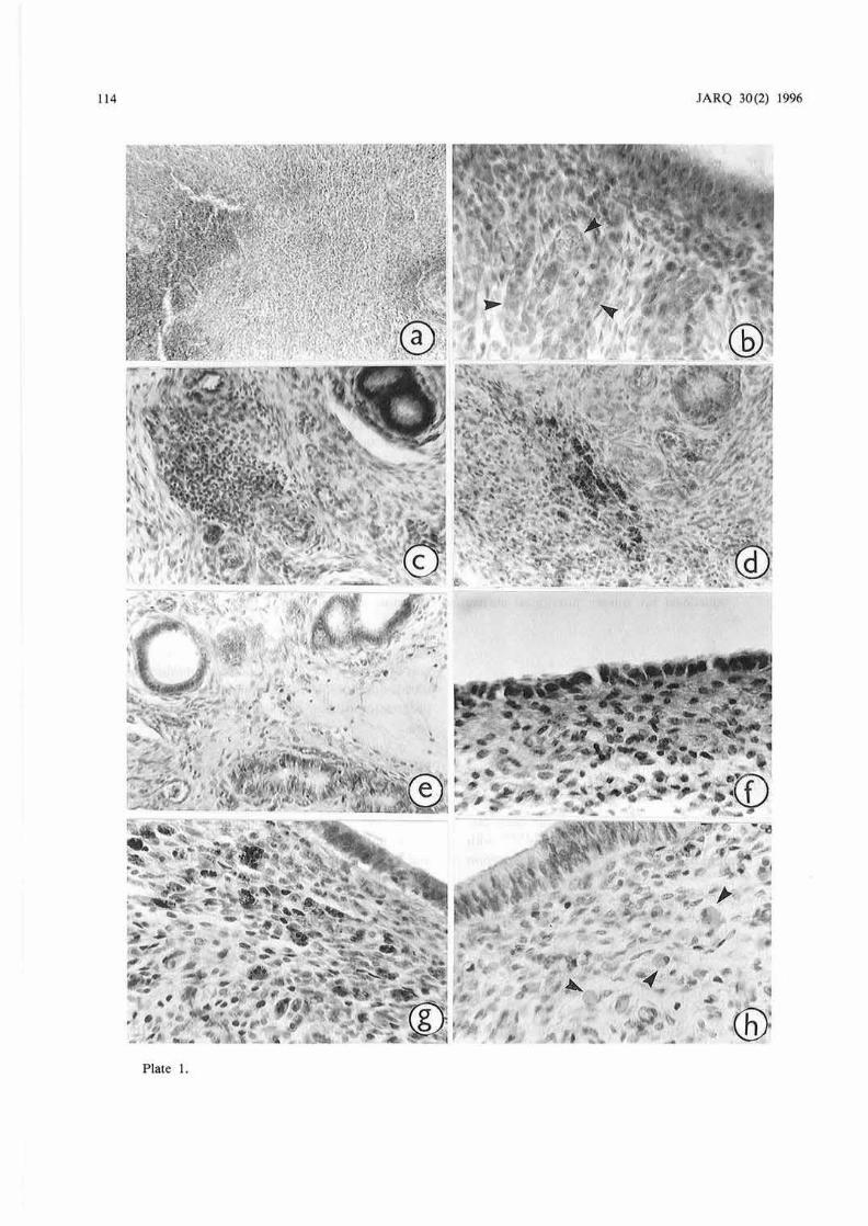
At Day 18 postpartum, in the caruncular zone, surface epithelium did not reappear. As large masses of aggregated erythrocytes were found in the endometrium (Plate la), the whole caruncles appeared edematous. Since such an edematous condition has been reported to be present already in the pregnant uterus2>, the post partum uterus at Day 18 was
114 JARQ 30(2) 1996
Plate I.

A . Okano & T. Tomiwka: Post par1u 111 U1eri11e !11volutio11 in /he Cow 115
presumably undergoing involution. The erythrocytes which infiltrated the endometrium would presumably be phagocytized by macrophages in the process of post par/um uterine involution, based on Deno's observation 6> that the function of macrophages is important for the involution of the uterus after parturition in the mouse. In the intercaruncular zone, the surface of the epithelium was thin with a height of 13.7 µm on an average and contained many lymphocytes and neutral leukocytes. Just below the surface epithelium, capillaries were not yet contracted and their inner diameters were approximately I 5 µm
(Plate lb). In the endometrium, many nodular aggregations of lymphocytes were observed (Plate le) together with masses of dark brown phagocytes (Plate Id). Sizes and shapes of the uterine glands in cross-section varied extensively. The mean outer and inner diameters of the glands, in the superficial zone, were 46.2 and 16.3 J.lm, respectively and in the basal zone, the outer diameter was 35.0 µm and the inner diameter was 10.3 J.lm (Plate le). In both superficial and basal zones, the lumen of the gland was relatively large in proportion to the outer diameter. As a result, the glandular epithelium was rather low.
At Day 23 post partum, a simple squamous epithelium reappeared from the edge of each caruncle, with a height of approximately 9.7 µm (Plate If). On the other hand, the mean height of the surface epithelium in the intercaruncular zone was 14.9 J.lm.
The histological characteristics of the endometrium at this stage were very similar to those at Day 18 stage as the endometrium contained a large amount of foreign cells such as phagocytes and lymphocytes.
The mean outer and inner diameters of uterine glands, in the superficial zone, were 46.2 and 16.5 JLID,
respectively and in the basal zone, the outer diameter was 31.3 J.lm and the inner diameter was 7.9 µm.
At Day 29 postpartum, the height of the surface epithelium in the caruncular zone and that in the intercaruncular zone were 12.6 and 15 .3 J.lm on an average, respectively, the epithelia in both zones being taller than those of the uterus at Day 23 post partum. In the endometrium, a considerable number of dark brown phagocytes were found (Plate lg) together with many lymphocytes. Variations in the size and shape of the uterine glands were negligible. The mean outer and inner diameters of the glands, in the superficial zone, were 44.1 and I 5.1 J.lm, respectively and in the basal zone, the outer diameter was 27 .6 µm and the inner diameter was 6.3 µ.m.
At Day 46 post partum, a pseudostratified surface epithelium with a height of approximately 18.6 J.lm was found in the caruncular zone, while a simple columnar epithelium with a height of approximately 19.4 µm was found in the intercaruncular zone. Although lymphocytes were hardly found in the endometrium , some phagocytes with pale hemosiderin granules which appeared to have completed their function were observed in the caruncular zone (Plate lh). The mean diameter of the phagocytes was 8 µm. The mean outer and inner diameters of the uterine glands, in the superficial zone, were 49.9 and 16.8 µ.m, respectively and in the basal zone, the outer diameter was 31.3 J.lm and the inner diameter was 7.0 µ.m.
At Day 54 postpartum, the height of the surface epithelium in the earuneular zone and that in the
P late I. a) Mass of erythrocytes in the endometrium of the caruncular zone, in a nursing cow at Day 18 post partum ( x 140)
b)
c)
d) e)
f) g)
h)
Just below the surface epithelium of the intercaruncu.lar zone, capillaries are not contracted in a nursing cow at Day 18 post part111n ( x 280)
Arrows indicate the capillaries. Nodular aggregation of lymphocytes in the endometriurn in a nursing cow at Day 13 post parlllm ( X (40) Dark phagocytes in the endometrium of a nursing cow at Day 18 pos1 par/um ( x 140) Cross-section of uterine glands in the basal zone of the cndometrium in a nursing cow at Day 18 post partum ( x 140)
The lumen of the gland is slightly enlarged. The epithelial cells of the gland are low and cuboidal. Simple squamous surface epithelium in a nursing cow at Day 23 post partum ( x 280) Just below the surface epithelium, many phagocytes with hemosiderin are present in the caruncular zone of a nursing cow at Day 29 post par/um ( x 280) Just below the surface epithelium, some phagocytes are present in the endometrium of the caruncular zone in a nursing cow at Day 46 post partum ( x 280)
These phagocytes are swollen and contain pale hemosiderin granules. Arrows indicate the phagocytes.

116
intercaruncular zone were 16.4 and 16.2 µ.m on an average, respectively. These epithelia were mostly simple columnar. In the endometrium, there were a few and scarce phagocytes. Capillaries in the endometrium at this stage were thoroughly contracted (Plate 2a). The mean outer and inner diameters of the uterine glands in the superficial zone, were 54.0 and 15.5 µ.m, respectively. The shape of most of the glands in the superficial zone was circular in crosssection (Plate 2b). Nuclei of the glandular epithelial cells contained many granules. The outer and inner diameters of the glands in the basal zone were 28.3 and 3.6 µm on an average, respectively. Most glands showed a regular shape in section, witb a small lumen (Plate 2c). Glands in both the superficial and the basal zones were distributed evenly in the endometrium. Thus, in the cases .of nursing cows, the uterine glands in the superficial zone generally became enlarged only with the increase in the number of days post partum, but the size of their lumen remained almost unchanged. The shape of the glands tended to show limited variations. On the other hand, both the outer and the inner diameters of the uterine glands in the basal zone tended to decrease. The shape of the glands in section became uniform only with the increase in the number of days postpartum.
JARQ 30(2) 1996
The first clue for the examination of uterine involution is provided by the changes in the distribution of erythrocytes and foreign cells such as lymphocytes and phagocytes in the endometrium. Incidentally in most cases of abnormal non-pregnant bovine uteri, the endometrium contains many lymphatic nodules as well 4
· 12). The presence of foreign cells such as lymphocytes and phagocytes in the endometrium probably suggests that the post partum uterus is not yet ready for the next pregnancy. At Day 46 postpartum, however, the endometrial tissues contained only a few phagocytes, which were swollen with pale hemosiderin granules. The presence of swollen phagocytes with pale hemosiderin granules in the endometrium suggests tha1 1hese cells had completed their function . At Day 54 post partum, very few phagocytes were found in the endometrium. Therefore, uterine involution may have been completed at least by Days 46 and 54 post par/um according to the foregoing histological observation. In this regard, Casida3> stated that calf suckling may accelerate the uterine involution, based on the observation of the changes in the uterine horn diameter. In studies on early weaning cows, the uterine involution was clearly retarded based on the fact that many phagocytes were still found in 1he endometrial
Plate 2.
a) Caruncular zone or the uterus with completed involution in a nursing cow at Day 54 post partum ( X 70)
b) Cross-seciion o f normal uterine glands in the superficial zone or the endometrium in a nursing cow at Day 54 post partum ( x 280)
c) Cross-section of normal uterine glands in the basal zone or the endometrium in a nursing cow at Day 54 post partum ( x 280)
These glands are evenly distributed in the endometrium.
A. Okano & T. Tomizuka: Pos1 pan um Uterine /11vo/111io11 i11 the Cow 117
tissues even at Days 44 and 62 post par/um •5>. Therefore, calf suckling is considered to be effective for the uterine involution in post partum cows.
The second clue for the examination of uterine involution is the reappearance of the surface epithelium in caruncular regions. Although the surface
epithelium in the caruncular zone did not yet reappear at Day 18 post partum, a simple squamous epithelium and a simple columnar one were observed in the same zone at Day 23 post partum. These findings are .in agreement with those of Wagner & Hanse120> in cows and Palmer 17l in sows.
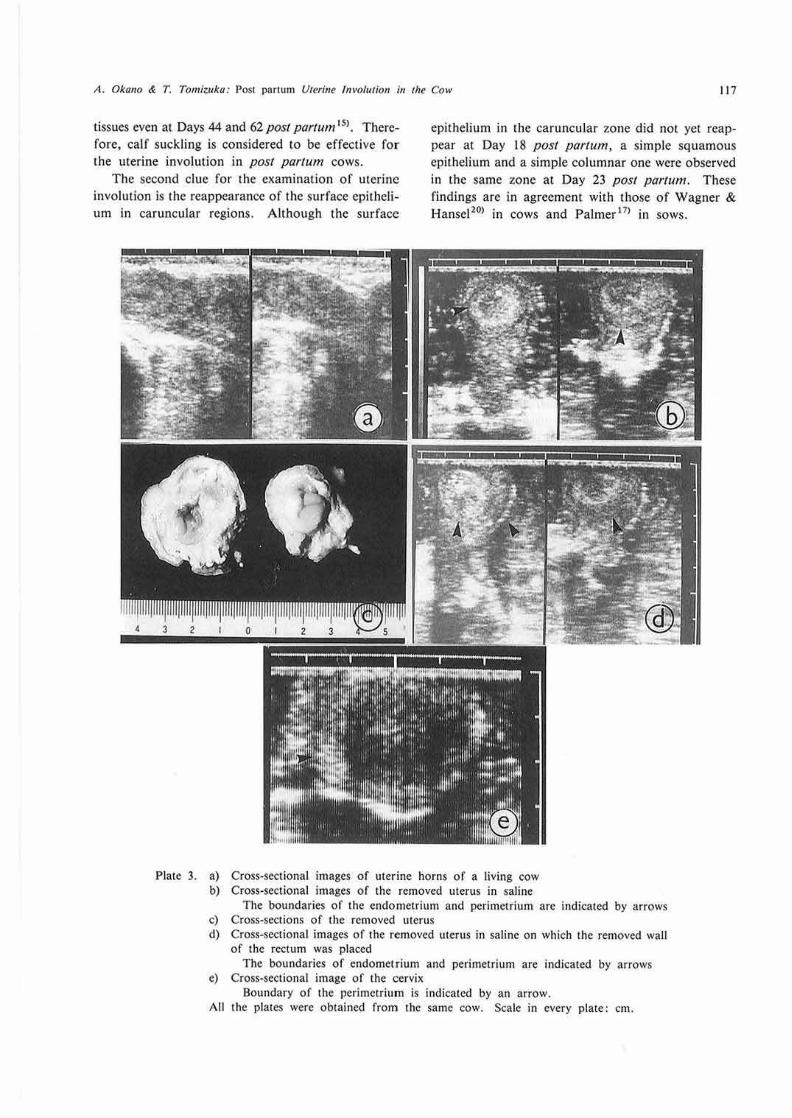
Plate 3. a) b)
c) d)
e)
All
Cross-sectional images of uterine horns of a living cow Cross-sectional images of the rnmoved uterus in saline
The boundaries of the endomctrium and perimetrium are indicated by arrows Cross-sections of the removed uterus Cross-sectional images of the removed uterus in saline on which the removed wall of the rectum was placed
The boundaries of endomel'rium and perimetrium are indicated by arrows Cross-sectional image of the cervix
Boundary of the perimetrium is indicated by an arrow. the plates were obtained from the same cow. Scale in every plate: cm.
118
The next and important clue is the pattern of changes in uterine glands. Many researchers4·s·8 •
12•13>
have stated that abnormal uterine glands generally display an enlarged lumen and low glandular epithelia. In the present study, at Day 18 post par/um, a low glandular epithelium was observed in the basal zone of the endometrium (Plate le). Therefore, at Day 18 post par/um, the uterine glands in the endometrium presumably had not yet recovered their normal condition. On the contrary, at Days 46 and 54 post partum, the uterine glands recovered their normal condition based on the outer and inner diameters of glands and the distribution of glands in the endometrial tissues (Plate 2b,c). The recovery of the normal condition of uterine glands at Days 46 and 54 postpartum is in agreement with the finding of Mochow & Olds is> . Thus, the present histological examination of uterine tissues also suggests that post par/um uterine involution is almost completed at Day 40 post partum or later though rectal
JARQ 30(2) 1996
palpation may enable to estimate that uterine involution is completed approximately between Days 28 and 47 post partum1
•11
•18>. This conclusion is also
supported by preliminary morphological observations in the current study showing that lochia were present in t he uterus isi.
Ultrasonographical observation of uterus post partum
The progression of uterine involution in Holstein dairy cows that delivered a single calf and were milked twice a day was observed at 3-day intervals from Day 8 post partum with the ultrasonographical linear scanner. The ultrasonographical linear scanner used in the present report was the Aloka-SSD-246 scanner (Aloka, Tokyo, Japan) with a 5-MHz transducer designed for intrarectal use in the cow. Real-time ultrasonographs of cross-sectional images of the uterine horn and the cervix were recorded with black
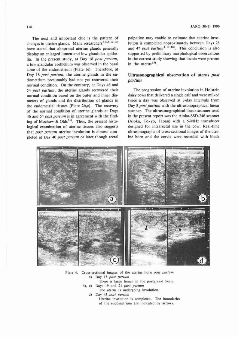
Plate 4. Cross-sectional images of the uterine horn post part11m a) Day 15 post partwr1
There is large lumen in the pos1gravid horn. b) , c) Days 19 and 21 post partum
The Ulerus is undergoing involution. d) Day 43 post parrum
Uterine invo.lution is completed. The boundaries of the endometrium are indicated by arrows.
A. Ok(l110 & T. Tomiwk(I: Pos1 parium Uterine !11vo/111io11 in the Cow 119
and white Polaroid film Type 667 (Polaroid, Cambridge, MA).
Diameters of the uterine horn and cervix and the areas of the uterine horn and endometrium were calculated from recorded cross-sectional images at various stages post partum with a Magiscan 2 image analyzer (Joyce-Loeb!, Gatesshead, England). The relationships between the number of post partum days and the calculated diameters and cross-sectional areas were fitted to square or cubic polynomial regressions.
Ultrasonographical images of the uterine horn in the living animal were almost circular (Plate 3a). The genitalia which were removed from the same animal after slaughter, and then placed in saline were observed by the ultrasonographical linear scanner. Jn this way, distinct ultrasonographs of the endometrium, myometrium and perimetrium could be obtained (Plate 3b). Diameters of the endometrium and myometrium in the cross-sectional images that were calculated by the image analyzer were in agreement with the actual diameters measured in the crosssections of the removed uterus (Plate 3c). However, when the removed uterus which was wrapped with an isolated rectal wall, was placed in saline and observed by ultrasonic scanner,' ultrasonographs of the myometrium and perimetrium became rather indistinct (Plate 3d). Therefore, to observe the myometrium and perimetrium in cross-sectional images of the uterus through image analysis, care must be exercised for determining the boundaries. The crosssectional image of the cervix was fairly clear in the ultrasonographs (Plate 3e).
Rather large lumens and lochia were observed in the ultrasonographs of uteri unti l approximately Day 15 posr partum (Plate 4a). At this stage, since the whole uterus seemed to be edematous, cross-sectional images were usually dark in ultrasonographs. Uterine involution progressed with the increase in the number of days post partum and cross-sectional images of the uterus became lighter (Plate 4b,c). In the present study, uterine involution was completed at Day 40 post partum based on the observation by ultrasonic linear scanner. It was difficult to define the boundaries of the myometrium and perimetrium in cross-sectional images of the uterus after completion of involution (Plate 4d). Areas and diameters of the uterine horn and the endometrium in the postgravid horn were larger than those of nonpostgravid horn, but both horns returned to their original condition at Day 40 post partum.
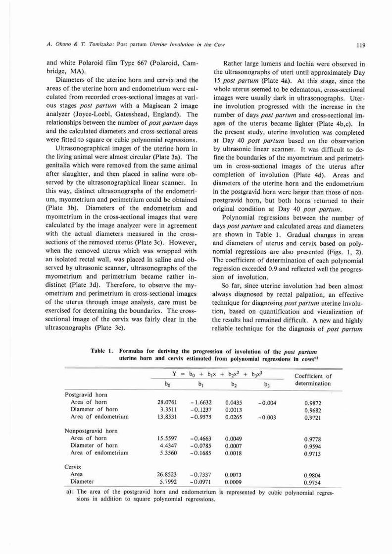
Polynomial reg·ressions between the number of days post parrum and calculated areas and diameters are shown in Table I. Gradual changes in a reas and diameters of uterus and cervix based on polynomial regressions are also presented (Figs. I, 2). The coefficient of determination of each polynomial regression exceeded 0.9 and reflected well the progression of involution.
So far, since uterine involution had been almost always diagnosed by rectal palpation, an effective technique for diagnosing post parrum uterine involution, based on quantification and visualization of the results had remained difficult. A new and highly reliable technique for the diagnosis of post partum
Table 1. Formulas for deriving the progression of involution of the post parwm uterine horn and cervix estimated from polynomial regressions in cows•>
Postgravid horn Area of horn Diameter of horn Area of endometrium
Nonpostgravid horn Area of horn Diameter of horn Area of endometrium
Cervix Area Diameter
28.0761 3.351 I
13.8531
15.5597 4.4347 5.3560
26.8523 5.7992
- 1.6632 - 0 .1237 - 0.9575
- 0 .4663 -0.0785 -0.1685
- 0.7337 -0.0971
0.0435 0.0013 0.0265
0 .0049 0 .0007 0.0018
0.0073 0.0009
-0.004
- 0.003
Coefficient of determination
0.9872 0.9682 0.9721
0.9778 0.9594 0.9713
0.9804 0.9754
a) : The area of the postgravid horn and endometrium is represented by cubic polynomial regressions in addition Lo square polynomial regressions.

120
10 20 30 40 Days posl part um
Fig. I. Curves reproducing the progression of involution in an area of the uterine horn and cervix postpartum estimated by polynomial regressions
Postgravid horn ( o ), Nonpostgravid horn ( • ), Endometrium of postgravid horn ( c.), Endometrium of nonpostgravid horn ( At. ), Cervix ( D).
uterine involution is necessary in future. Therefore, the utilization of the ultrasonic linear scanner for the diagnosis of post partum uterine involution was examined in the present report. In the case of pregnancy diagnosis, it is easy to detect a reflection of echo from the fetus and the accumulated amniotic and allantoic fluids. Until now, there had been no method to determine how a living nonpregnant bovine uterus could be observed. In the present study, it was obvious that the endometrium can be distinctly observed in any stage in ultrasonographs unlike the myomeuium and perimetrium.
In the case of rectal palpation, the uterine volume is estimated based on the finger width, and the results are sometimes inaccurate because of differences in finger width and of subjectivity among investigators. Such inaccuracies may especially occur in a relaxed and flat post par1um uterus. However, such a post partum uterus can be accurately observed crosssectionally in ultrasonographs. Accordingly, the diagnosis of uterine involution by ultrasonic linear
22
20
18
16
~ 14
l 12 ~ ~ 10
8
6
4
2
10
JARQ 30(2) 1996
20 30 40 Days post p,1rtum
Fig. 2. Curves representing the progression of involution for the diameter of uterine horn and cervix postpartum estimated by polynomial regressions
Postgravid horn ( o ), Nonpostgravid horn ( • ), Cervix ( D).
scanner is more objective than that by rectal palpation. As it is possible to detect the accumulation of lochia and retained fetal material in the uterine lumen with an ultrasonic linear scanner, post parturn uterine abnormalities can be detected in realtime ultrasonographs.
As shown in the plates, the myometrium was distinguishable from the endometrium by using the image analyzer in magnified ultrasonographs. For diagnosing post partum uterine involution, conditions of the endometrium in real-time ultrasonographs can be observed. If both ultrasonic observation and rectal palpation are carried out in a uterus undergoing involution, and the results are compared, the progression of postpartum uterine involution could be confirmed. Completion of uterine involution was found to occur approximately on Day 40 post parturn using an ultrasonic linear scanner. Similar results had previously been reported in the cow by rectal palpation2
· 1°· 14
·18>. It was reported that the uterine
volume during the process of involution estimated by rectal palpation could be fitted into a polynomial regression LIO>. Calculated figures about the post partum uterus in the present report could easily be
A. Oka110 & T. Tomiiuka: Pos1 panum Ureri11e /11volutio11 in rite Cow 121
fitted into square or cubic polynomial regressions, and they represent the progression of uterine involution. Since it was assumed that there is no basic difference in the process of post partum uterine involution between primiparous and nulliparous cows in ihe present report, cows in both conditions were analyzed.
Polynomial regressions obtained in the present study can be applied to the diagnosis of involutionary progression of post partum. Ultrasonic linear scanner, which has been introduced into many facilities for pregnancy diagnosis, can be directly applied to observe postpartum merine involution in the cow. If rectal palpation and ultrasonic observation arc combined for diagnosing uterine involution, the involutionary progress in the cow could be analyzed more precisely.
References
I) Bastidas, P. et al. (1984): Effects of rest ricted suckling on ovarian activity and uterine involution in Brahman cows. Theriogenology, 2J, 525-532.
2) Buch, N. C., Tyler, W. J. & Casida, L. E. (1955): Postpartum estrus and involution of the uterus in an experimental herd of Holstein-Friesian cows. J. Dairy Sci., 38, 73- 79.
3) Casida, L. E. (1968); Studies on the postpartum cow. Wisconsin. Res., 270, 48-52.
4) Cupps, P. T., Laben, R. C. & Mead, W. S. (1956): Histology of the pitu itaries, adrenals, ovaries and uteri of dairy cattle associated wi th different reproductive conditions. J. Dairy Sci., 39, 155-161.
5) Cupps, P . T . (1973): Uterine changes associated with impaired fertility in the dairy cow. J. Dairy Sci., 56, 878-884.
6) Deno, R. A. (1937): Uterine macrophages in the mouse and their relation 10 involution. Am. J. A111., 60, 433-471.
7) Foley, R. C ., Reece, R. P. & Leathern, J, H. (1954) : Histological observations of the bovine uterus, placenta, and corpus luteum during early pregnancy. J. Anim. Sci., 13, 131-137.
8) Fosgate, O. T., Cameron, N. W. & McLeod, R. J.
(1962): lnnuence of 17-alpha-hydroxyprogesteroneN-capronate upon post-partum reproductive activity in the bovine. J. Anim. Sci., 21, 791-793.
9) Gier, H . T. & Marion, G. B. (1968): Uterus of the cow after parturition; involution changes. Am. J. Vet. Res., 29, 83-96.
10) lzaike, Y. et al. (1984): Relationship between postpartum reproductive performance and calving number in beef cows. Jpn. J. Anim. Reprod., 30, 206-210.
11) Marion, G. B., Norwood, J. S. & Gier, H. T. (1968): Uterus of the cow after parturition: factors affecting regression. Am. J. Vet. Res., 29, 71-75.
12) Mochow, R. & Olds, D. (1966): Effect of age and number of calvings on histological characteristics of the bovine uterus. J. Dairy Sci. , 49, 642-646.
13) Moss, S., Sykes, J. F. & Wrenn, T . R. (1956): Some abnormalities of the bovine cndometrium. J, Anim. Sci., 15, 63 1-636.
14) Okano, A. & Fukuhara, R. (1980): Histological studies on postpartal involution in Japanese Black. Jpn, J. Zootech. Sci., 5 1, 284-292.
15) Okano, A . et al. (1981): Morphological involution of postpartum uterus in Japanese Black cows. Bu(!. Chugoku Natl. Exp. Stn., Ser.B, 25, 1-10.
16) Olds, D. & Coopers, C. (1970): Effects of postpartum rest period in dairy cat tle on the occurrence of breeding abnormalities and on calving intervals. J. Am. Vet. Med. Assoc., 157, 92-97.
17) Palmer, W. N., Teagne, H. S. & Venzke, W. G. ( 1965): Histo logical. changes in the reproductive tract of the sow during lactation and early postweaning. J. Anim. Sci., 24, I I 17-1125.
18) Parkins, J. L. & Kidder, H. E. (1963): Relationship of uterine involution and postpartum interval to reproductive efficiency in beef cattle. J. A11im. Sci., 22, 313-315.
19) Reeves, J. J., Rantanen, N. W. & Hauser, N. (1984): Transrectal real-time u ltrasound scanning of the cow reproductive tract. Theriogenology., 21, 485- 494.
20) Wagner. W. C. & Hansel, W. (1969): Reproductive physiology of the post partum cow. 1. Clinical and histological findings. J. Reprod. Ferr ., 18, 493-500.
21) Yamauchi, S., Kotera, K. & Kakishita, T. (1968): Histological study of the pregnant uterus. 1. General histology of the endometrium of the intercaruncular area. Jpn. J. Zootech . Sci., 39, 487-504.
(Received for publication, August 2, 1995)