Embed Size (px)
Citation preview
Post-keratoplasty infections
related to
contaminated donor tissues
Rama Paolo
San Raffaele Scientific Institute, Milano
1
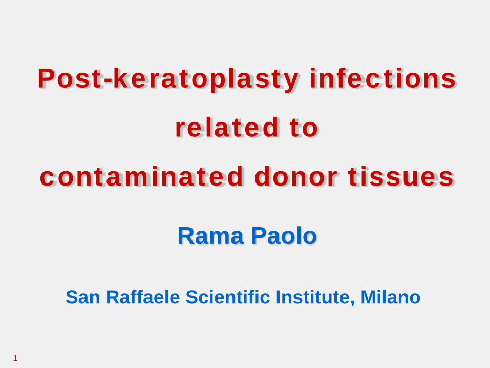
1) Keratititis
- Infiltrates (superficial, deep), all keratoplasties
- Interface infectious keratitis, DALK and EK
2) Endophthalmitis, PK and EK, rare following DALK
Infections following keratoplasties
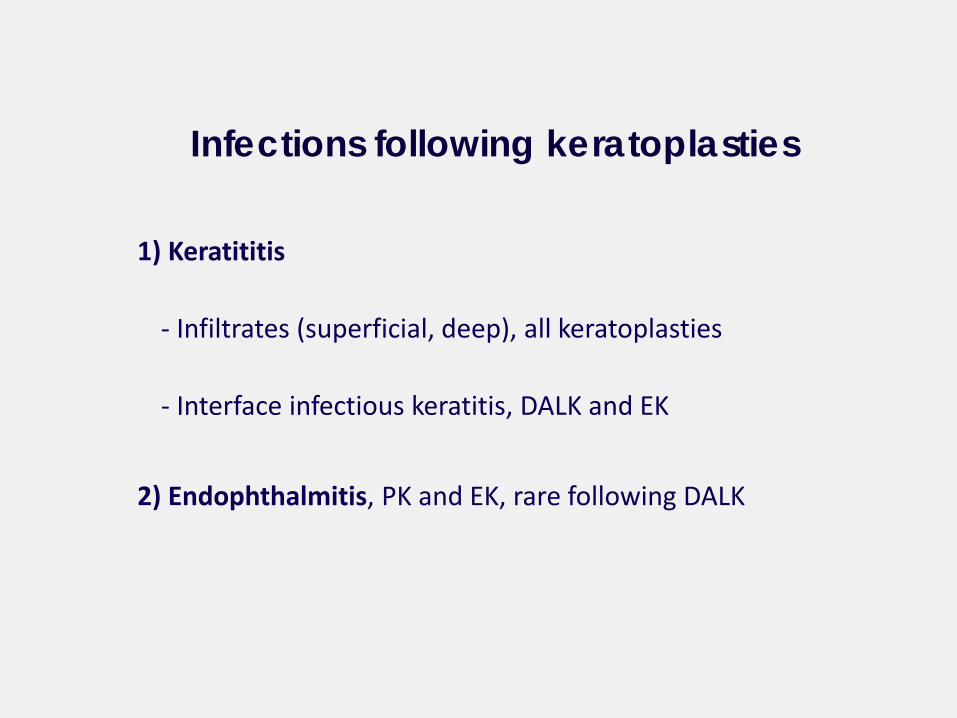
Incidence rates of infections following keratoplasty
1.7 – 7.4% in developed countries
11.9 in developing world
Host-related problems, persistent epithelial defects and loose sutures,
the major risk factor
Infections following keratoplasties
Vajpayee et al. Major review Survey Opthhalmol 2007
Endophthalmitis USA 0.42% incidence 18.083 corneal transplants
0.12% incidence 2.261.779 cataract surgery
UK 0.67% after PK Du et al. Opthhalmology 2014
Chen et al. Opthhalmology 2015
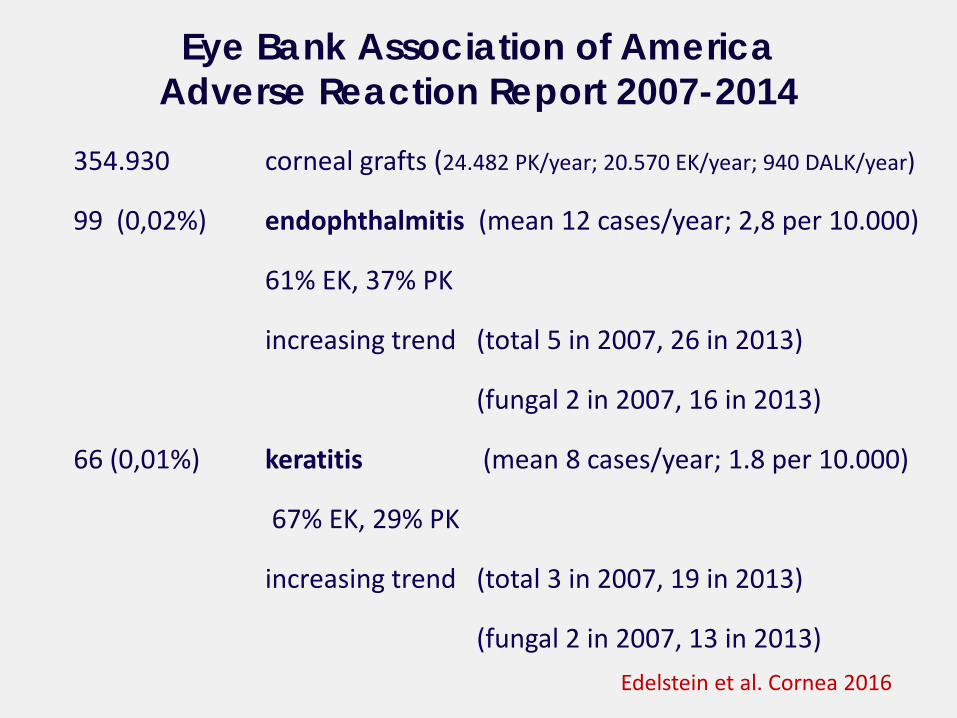
354.930 corneal grafts (24.482 PK/year; 20.570 EK/year; 940 DALK/year)
99 (0,02%) endophthalmitis (mean 12 cases/year; 2,8 per 10.000)
61% EK, 37% PK
increasing trend (total 5 in 2007, 26 in 2013)
(fungal 2 in 2007, 16 in 2013)
66 (0,01%) keratitis (mean 8 cases/year; 1.8 per 10.000)
67% EK, 29% PK
increasing trend (total 3 in 2007, 19 in 2013)
(fungal 2 in 2007, 13 in 2013)
Eye Bank Association of America Adverse Reaction Report 2007-2014
Edelstein et al. Cornea 2016
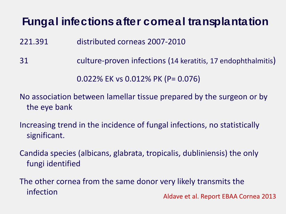
221.391 distributed corneas 2007-2010
31 culture-proven infections (14 keratitis, 17 endophthalmitis)
0.022% EK vs 0.012% PK (P= 0.076)
No association between lamellar tissue prepared by the surgeon or by the eye bank
Increasing trend in the incidence of fungal infections, no statistically significant.
Candida species (albicans, glabrata, tropicalis, dubliniensis) the only fungi identified
The other cornea from the same donor very likely transmits the infection
Fungal infections after corneal transplantation
Aldave et al. Report EBAA Cornea 2013
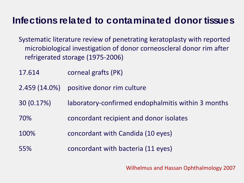
Systematic literature review of penetrating keratoplasty with reported microbiological investigation of donor corneoscleral donor rim after refrigerated storage (1975-2006)
17.614 corneal grafts (PK)
2.459 (14.0%) positive donor rim culture
30 (0.17%) laboratory-confirmed endophalmitis within 3 months
70% concordant recipient and donor isolates
100% concordant with Candida (10 eyes)
55% concordant with bacteria (11 eyes)
Infections related to contaminated donor tissues
Wilhelmus and Hassan Ophthalmology 2007
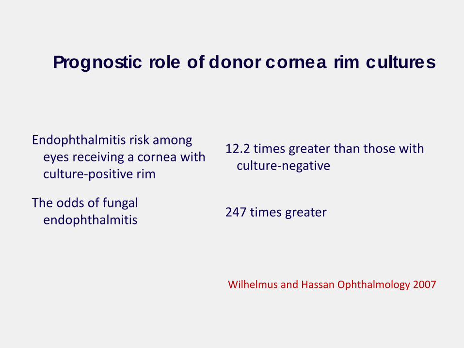
Endophthalmitis risk among eyes receiving a cornea with culture-positive rim
The odds of fungal endophthalmitis
Prognostic role of donor cornea rim cultures
Wilhelmus and Hassan Ophthalmology 2007
12.2 times greater than those with culture-negative
247 times greater
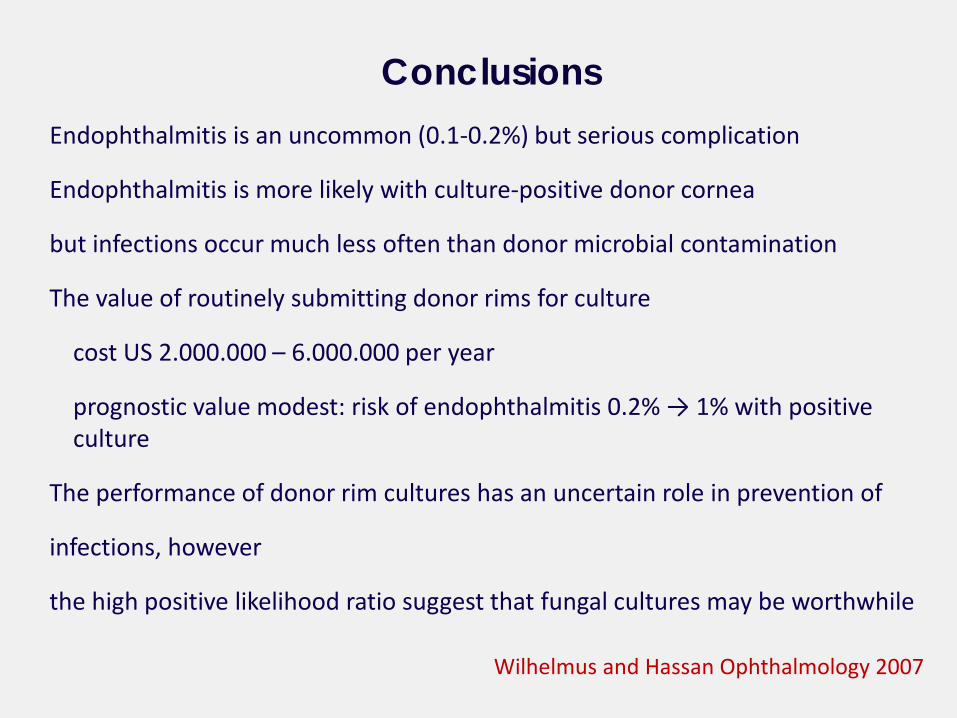
Endophthalmitis is an uncommon (0.1-0.2%) but serious complication
Endophthalmitis is more likely with culture-positive donor cornea
but infections occur much less often than donor microbial contamination
The value of routinely submitting donor rims for culture
cost US 2.000.000 – 6.000.000 per year
prognostic value modest: risk of endophthalmitis 0.2% → 1% with positive culture
The performance of donor rim cultures has an uncertain role in prevention of
infections, however
the high positive likelihood ratio suggest that fungal cultures may be worthwhile
Conclusions
Wilhelmus and Hassan Ophthalmology 2007
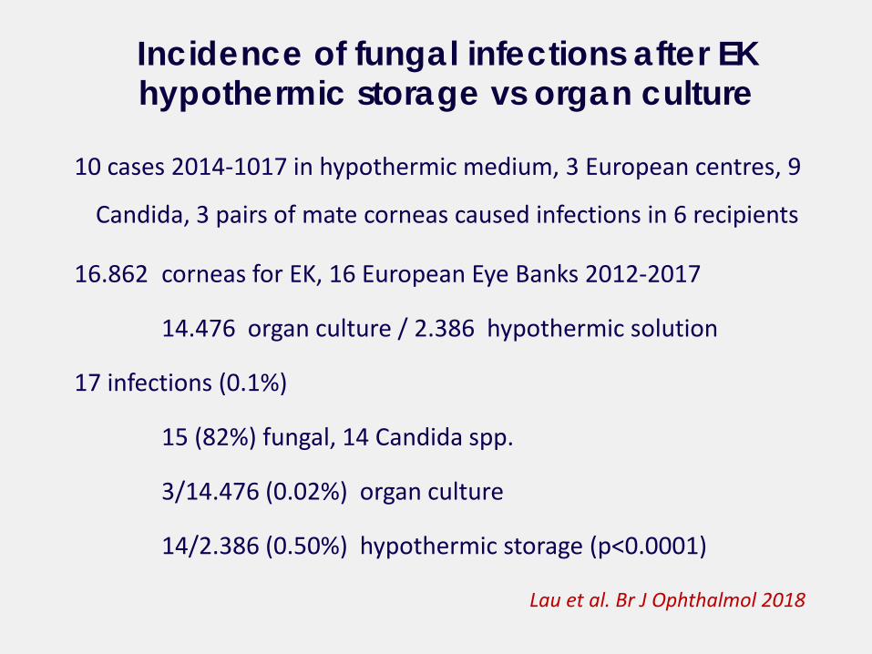
10 cases 2014-1017 in hypothermic medium, 3 European centres, 9
Candida, 3 pairs of mate corneas caused infections in 6 recipients
16.862 corneas for EK, 16 European Eye Banks 2012-2017
14.476 organ culture / 2.386 hypothermic solution
17 infections (0.1%)
15 (82%) fungal, 14 Candida spp.
3/14.476 (0.02%) organ culture
14/2.386 (0.50%) hypothermic storage (p<0.0001)
Incidence of fungal infections after EK hypothermic storage vs organ culture
Lau et al. Br J Ophthalmol 2018
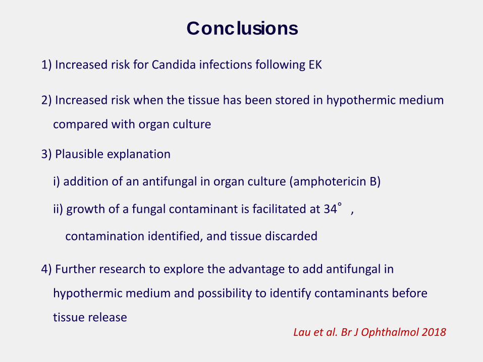
1) Increased risk for Candida infections following EK
2) Increased risk when the tissue has been stored in hypothermic medium
compared with organ culture
3) Plausible explanation
i) addition of an antifungal in organ culture (amphotericin B)
ii) growth of a fungal contaminant is facilitated at 34°,
contamination identified, and tissue discarded
4) Further research to explore the advantage to add antifungal in
hypothermic medium and possibility to identify contaminants before
tissue release
Conclusions
Lau et al. Br J Ophthalmol 2018
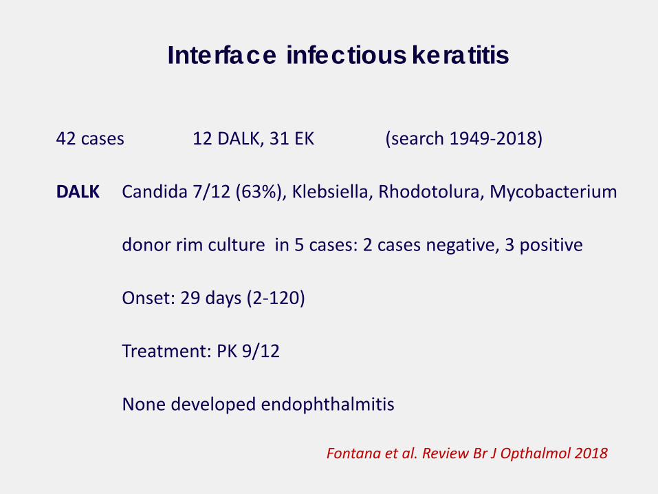
42 cases 12 DALK, 31 EK (search 1949-2018)
DALK Candida 7/12 (63%), Klebsiella, Rhodotolura, Mycobacterium
donor rim culture in 5 cases: 2 cases negative, 3 positive
Onset: 29 days (2-120)
Treatment: PK 9/12
None developed endophthalmitis
Interface infectious keratitis
Fontana et al. Review Br J Opthalmol 2018
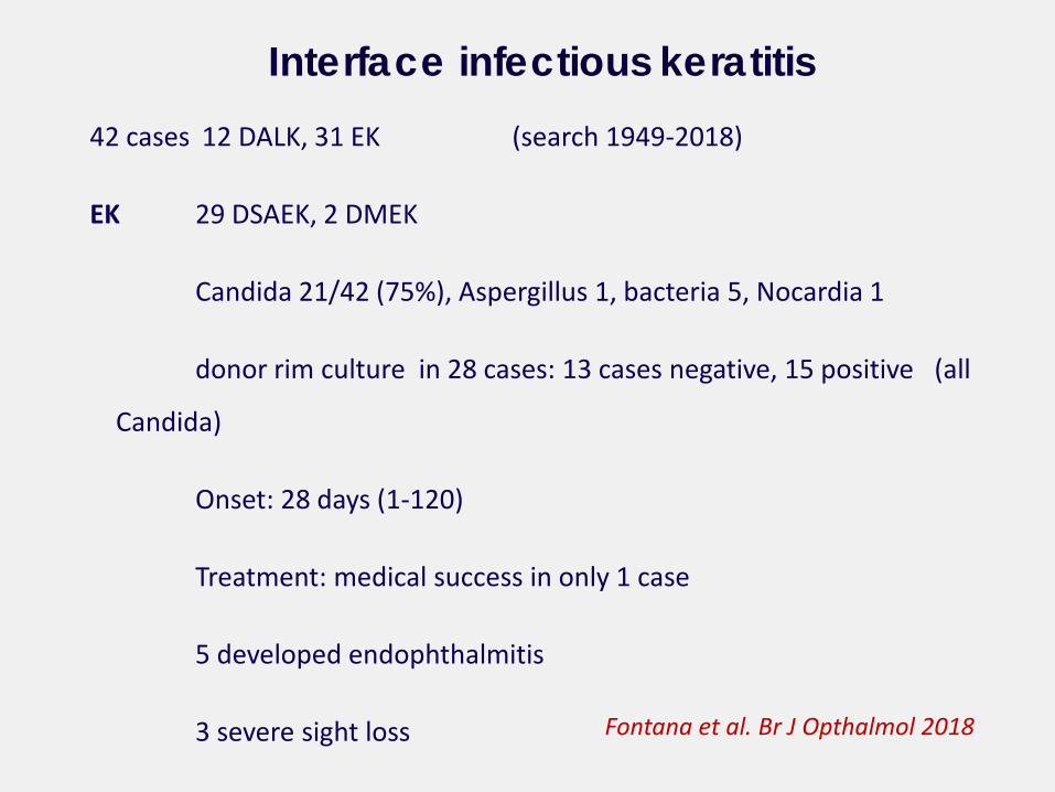
42 cases 12 DALK, 31 EK (search 1949-2018)
EK 29 DSAEK, 2 DMEK
Candida 21/42 (75%), Aspergillus 1, bacteria 5, Nocardia 1
donor rim culture in 28 cases: 13 cases negative, 15 positive (all
Candida)
Onset: 28 days (1-120)
Treatment: medical success in only 1 case
5 developed endophthalmitis
3 severe sight loss
Interface infectious keratitis
Fontana et al. Br J Opthalmol 2018
Overall perception of an increased risk of fungal infection after EK may
be the consequence of over-reporting a novel complication after a
new surgical procedure
Tissue manipulation either in the eye bank or in the operating room
does not seem to influence the risk of infections
The donor and not the processing seem to be the source of infection
Candida the most common microorganism
Conclusions
Fontana et al. Br J Ophthlmol 2018
Onset: few days – 3 months
Initial asymptomatic clinical picture and similarity to epithelial ingrowth
Conclusions
Fontana et al. Br J Ophthlmol 2018
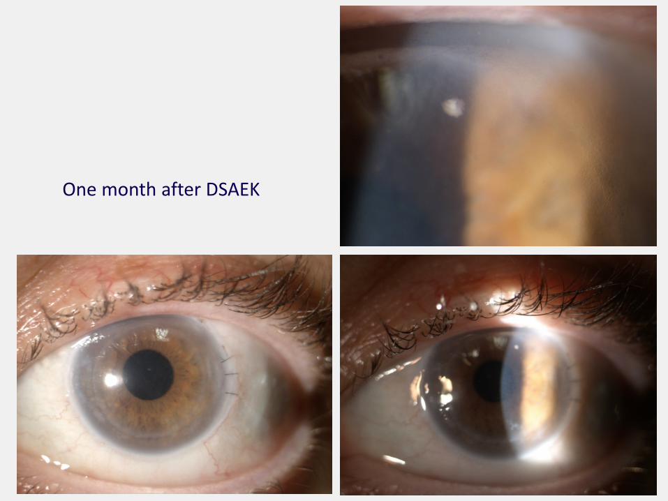
One month after DSAEK
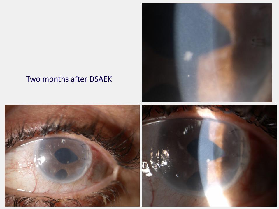
Two months after DSAEK
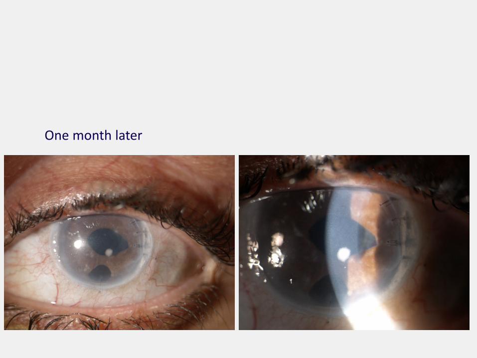
One month later
Onset: few days – 3 months
Initial asymptomatic clinical picture and similarity to epithelial ingrowth
Early warning may come from donor rim culture
In vivo confocal microscopy can be useful
In case of infection, early excisional PK is a safe and effective measure
Conclusions
Fontana et al. Br J Ophthlmol 2018
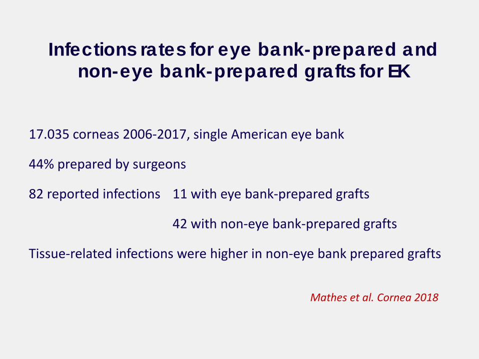
17.035 corneas 2006-2017, single American eye bank
44% prepared by surgeons
82 reported infections 11 with eye bank-prepared grafts
42 with non-eye bank-prepared grafts
Tissue-related infections were higher in non-eye bank prepared grafts
Infections rates for eye bank-prepared and non-eye bank-prepared grafts for EK
Mathes et al. Cornea 2018
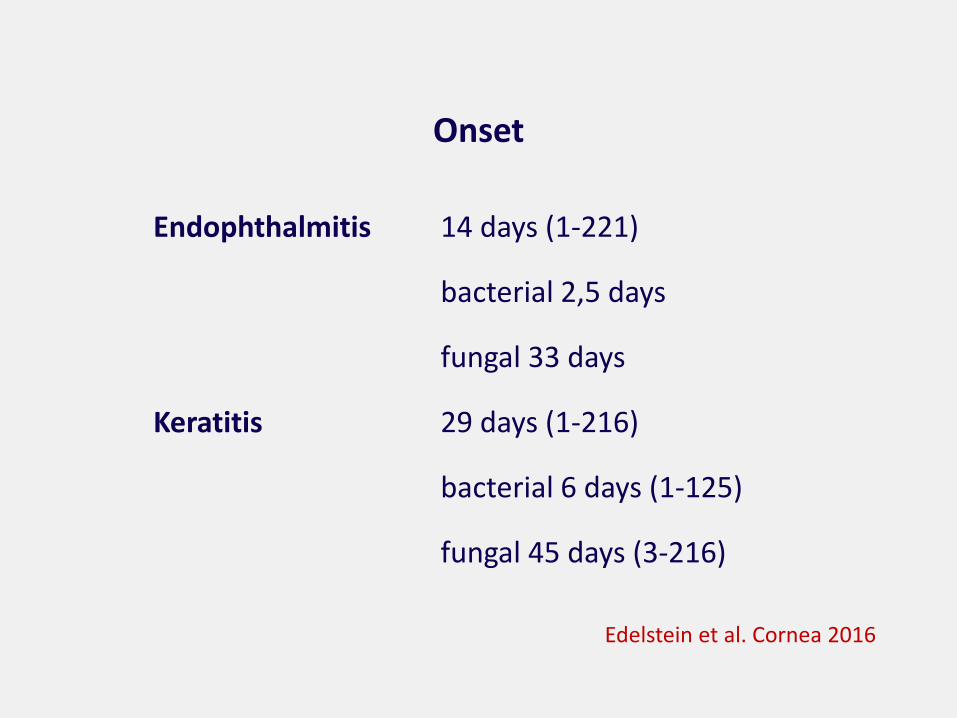
Onset
Endophthalmitis 14 days (1-221)
bacterial 2,5 days
fungal 33 days
Keratitis 29 days (1-216)
bacterial 6 days (1-125)
fungal 45 days (3-216)
Edelstein et al. Cornea 2016
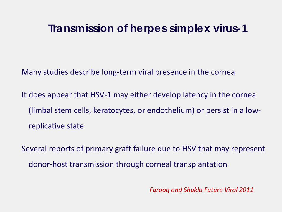
Many studies describe long-term viral presence in the cornea
It does appear that HSV-1 may either develop latency in the cornea
(limbal stem cells, keratocytes, or endothelium) or persist in a low-
replicative state
Several reports of primary graft failure due to HSV that may represent
donor-host transmission through corneal transplantation
Transmission of herpes simplex virus-1
Farooq and Shukla Future Virol 2011
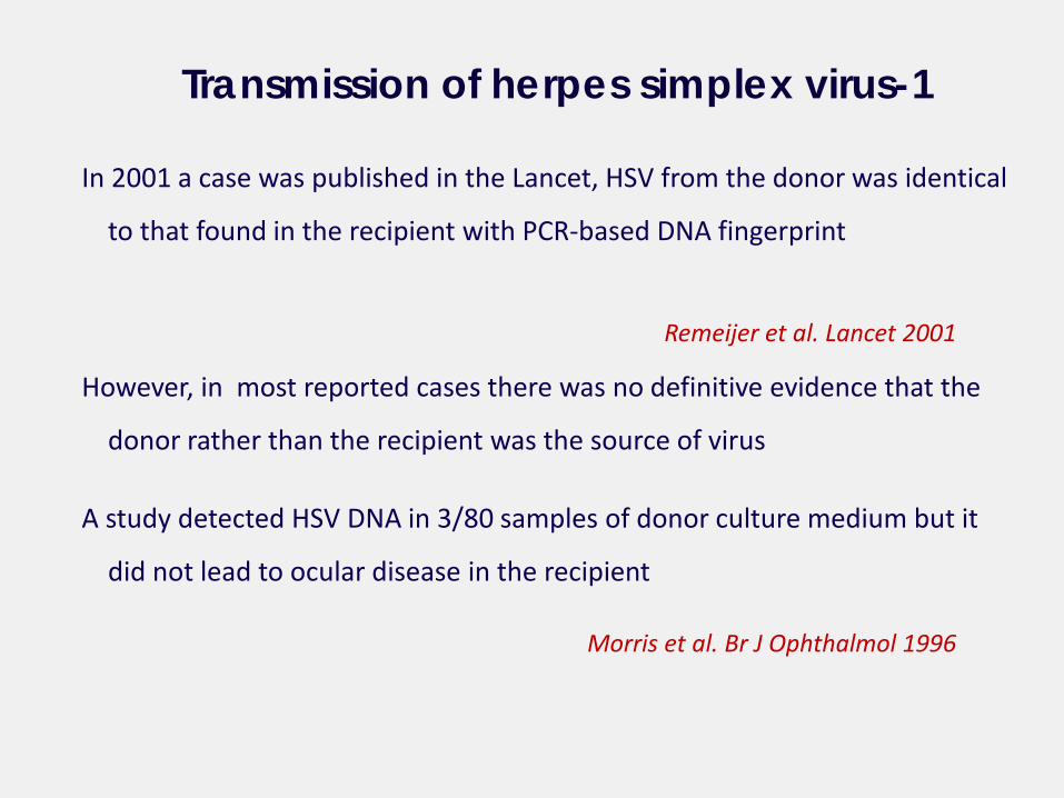
In 2001 a case was published in the Lancet, HSV from the donor was identical
to that found in the recipient with PCR-based DNA fingerprint
However, in most reported cases there was no definitive evidence that the
donor rather than the recipient was the source of virus
A study detected HSV DNA in 3/80 samples of donor culture medium but it
did not lead to ocular disease in the recipient
Transmission of herpes simplex virus-1
Remeijer et al. Lancet 2001
Morris et al. Br J Ophthalmol 1996
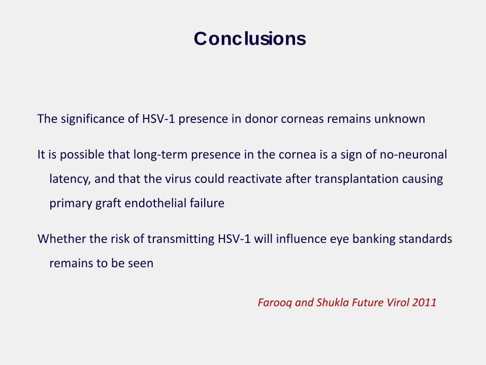
The significance of HSV-1 presence in donor corneas remains unknown
It is possible that long-term presence in the cornea is a sign of no-neuronal
latency, and that the virus could reactivate after transplantation causing
primary graft endothelial failure
Whether the risk of transmitting HSV-1 will influence eye banking standards
remains to be seen
Conclusions
Farooq and Shukla Future Virol 2011
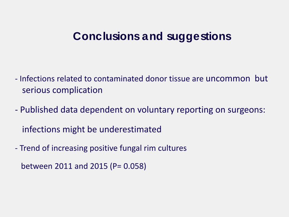
- Infections related to contaminated donor tissue are uncommon but serious complication
- Published data dependent on voluntary reporting on surgeons:
infections might be underestimated
- Trend of increasing positive fungal rim cultures
between 2011 and 2015 (P= 0.058)
Conclusions and suggestions
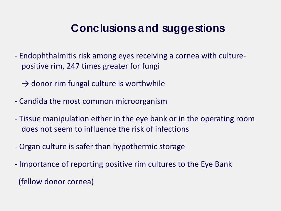
- Endophthalmitis risk among eyes receiving a cornea with culture-positive rim, 247 times greater for fungi
→ donor rim fungal culture is worthwhile
- Candida the most common microorganism
- Tissue manipulation either in the eye bank or in the operating room does not seem to influence the risk of infections
- Organ culture is safer than hypothermic storage
- Importance of reporting positive rim cultures to the Eye Bank
(fellow donor cornea)
Conclusions and suggestions
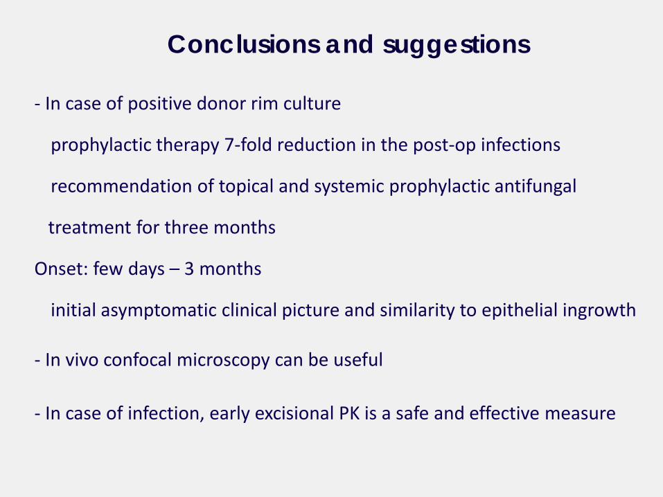
- In case of positive donor rim culture
prophylactic therapy 7-fold reduction in the post-op infections
recommendation of topical and systemic prophylactic antifungal
treatment for three months
Onset: few days – 3 months
initial asymptomatic clinical picture and similarity to epithelial ingrowth
- In vivo confocal microscopy can be useful
- In case of infection, early excisional PK is a safe and effective measure
Conclusions and suggestions

Thank you


![Ultrashort antibacterial and antifungal lipopeptides(vol vol)] of human erythrocytes. The antibiotics gentamicin and amphotericin B served as controls for bacteria and fungi, respectively,](https://img.pdfslide.us/doc/110x75/5ec1a4e4697963466236e7f0/ultrashort-antibacterial-and-antifungal-lipopeptides-vol-vol-of-human-erythrocytes.jpg)