Embed Size (px)
Citation preview

JPET #199935
1
Title Page
Post-exposure Application of Fas Receptor siRNA to Suppress Sulfur
Mustard-Induced Apoptosis in Human Airway Epithelial Cells:
Implication for a Therapeutic Approach
Brian M. Keyser, Devon K. Andres, Eric Nealley, Wesley W. Holmes, Betty Benton,
Danielle Paradiso, Ashley Appell, Chris Carpin, Dana R. Anderson, William J. Smith,
Radharaman Ray
Cellular and Molecular Biology Branch (B.M.K., D.K.A., E.N., B.B., A.A., C.C., W.J.S.,
R.R.), and Medical Toxicology Branch (W.W.H., D.P., D.R.A.),
U.S. Army Medical Research Institute of Chemical Defense,
Aberdeen Proving Ground, Maryland, 21010, USA.
JPET Fast Forward. Published on November 5, 2012 as DOI:10.1124/jpet.112.199935
Copyright 2012 by the American Society for Pharmacology and Experimental Therapeutics.
This article has not been copyedited and formatted. The final version may differ from this version.JPET Fast Forward. Published on November 5, 2012 as DOI: 10.1124/jpet.112.199935
at ASPE
T Journals on A
pril 1, 2019jpet.aspetjournals.org
Dow
nloaded from

JPET #199935
2
Running Title Page
Running Title: FasR siRNA antagonizes sulfur mustard-induced apoptosis
Corresponding author: Radharaman Ray, Research Division, US Army Medical
Research Institute of Chemical Defense, 3100 Ricketts Point Road, Aberdeen Proving
Ground, Maryland 21010-5400; e-mail: [email protected]
The number of
Text pages: 24
Tables: 0
Figures: 7
References: 40
Abstract: 242
Introduction: 678
Discussion: 1,270
Recommended section assignment: Toxicology
Nonstandard abbreviations: SM, Sulfur mustard; siRNA, small interfering RNA; FasR,
Fas receptor; sFasL, soluble Fas ligand; FasL, Fas ligand; NHBE, normal human
bronchial/tracheal epithelial; BALF, Bronchoalveolar lavage fluid; FADD, Fas-
associated death domain protein; DD, death domain; DED, death effector domain; DISC,
death-inducing signaling complex; COPD, chronic obstructive pulmonary disease;
BEBM, basal cell growth medium; IDF, idiopathic pulmonary fibrosis; ALI, acute lung
injury; i.t., intratracheal; i.n., intranasal; TNF, tumor necrosis factor; DR, death receptor;
NHEK, human epidermal keratinocytes
This article has not been copyedited and formatted. The final version may differ from this version.JPET Fast Forward. Published on November 5, 2012 as DOI: 10.1124/jpet.112.199935
at ASPE
T Journals on A
pril 1, 2019jpet.aspetjournals.org
Dow
nloaded from

JPET #199935
3
Abstract
Sulfur mustard (SM) is a vesicant chemical warfare and terrorism agent. Besides
skin and eye injury, respiratory damage has been mainly responsible for morbidity and
mortality after SM exposure. Previously, it was shown that suppressing the death receptor
(DR) response by dominant-negative FADD prior to SM exposure blocked apoptosis and
microvesication in skin. Whether antagonizing the Fas receptor (FasR) pathway by
siRNA applied after SM exposure would prevent apoptosis and thus, airway injury was
studied here. Normal human bronchial/tracheal epithelial (NHBE) cells were used as an
in vitro model and FasR siRNA, FasR agonistic antibody CH11 and FasR antagonistic
antibody ZB4 as investigative tools. In NHBE cells, both SM (300 µM) and CH11 (100
ng/ml) induced caspase-3 activation, which was inhibited by FasR siRNA and ZB4,
indicating that SM-induced apoptosis was via the Fas response. FasR siRNA inhibited
SM-induced caspase-3 activation when added to NHBE cultures up to 8 h after SM.
Results using annexin V/propidium iodide stained cells showed that both apoptosis and
necrosis were involved in cell death due to SM; FasR siRNA decreased both apoptotic
and necrotic cell populations. Bronchoalveolar lavage fluid (BALF) of rats exposed to
SM (1 mg/kg, 50 min) revealed a significant (p<0.05) increase in soluble Fas ligand and
active caspase-3 in BALF cells. These findings suggest an intervention of Fas-mediated
apoptosis as a post-exposure therapeutic strategy with a therapeutic window for SM
inhalation injury and possibly other respiratory diseases involving the Fas response.
This article has not been copyedited and formatted. The final version may differ from this version.JPET Fast Forward. Published on November 5, 2012 as DOI: 10.1124/jpet.112.199935
at ASPE
T Journals on A
pril 1, 2019jpet.aspetjournals.org
Dow
nloaded from

JPET #199935
4
Introduction
Sulfur mustard (SM; bis-(2-chloroethyl) sulfide) is a vesicating compound and
has been used as a chemical warfare agent beginning in World War I and most recently in
the Iraq/Iran conflict. Its use is now reemerging as a major threat not only to the military,
but also to civilians (Saladi et al., 2006). However, in spite of intense research on its
mechanisms of action and intervention of toxicity, there is no available therapy against
SM exposure effects. The target organs for SM injury are skin, lung and the eye. SM
exposure can result in severe skin blisters, pulmonary damage, and eye damage causing
irritation/inflammation, photophobia and neovascularization (Papirmeister et al., 1985;
Smith et al., 1990; Smith and Dunn, 1991; Petrali et al., 1993). Although SM can cause
these different tissue effects, most mortality following SM exposure has been attributed
to pulmonary damage and respiratory tract lesions. Respiratory complications of SM
poisoning in Iranian veterans have included laryngitis, tracheobronchitis, bronchiolitis,
bronchopneumonia, chronic obstructive pulmonary disease (COPD), bronchiectasis,
asthma, large airway narrowing, and pulmonary fibrosis (Bijani and Moghadamnia, 2002;
Balali-Mood and Hefazi, 2005; Emad and Emad, 2007; Ghanei and Harandi, 2007). Since
SM is a highly reactive chemical, its pathogenic mechanisms e.g., DNA damage are
initiated without much delay, but the tissue injury is not seen until after a lag period of 12
to 24 hours (Papirmeister et al., 1985; Rosenthal et al., 2003). This provides a therapeutic
window for medical management of SM injury.
It has been previously indicated that epithelial damage via apoptosis is a major
mechanism of SM injury (Rosenthal et al., 1998; Rosenthal et al., 2003). In SM injury,
both the intrinsic mitochondrial pathway and the extrinsic DR-mediated pathway are
This article has not been copyedited and formatted. The final version may differ from this version.JPET Fast Forward. Published on November 5, 2012 as DOI: 10.1124/jpet.112.199935
at ASPE
T Journals on A
pril 1, 2019jpet.aspetjournals.org
Dow
nloaded from

JPET #199935
5
involved in the apoptotic response. The intrinsic pathway utilizes the initiator caspase-9,
and the extrinsic pathway utilizes the initiator caspase-8, which interacts with caspase-9
via truncated Bid (tBid) (figure 1). It has been reported previously that the DR-mediated
pathway plays an important role in SM toxicity in normal human epidermal keratinocytes
(NHEKs) (Rosenthal et al., 1998; Rosenthal et al., 2003). However, what type(s) of
DR(s) might be involved in apoptosis due to SM was not established. The DR pathway is
initiated by the clustering and activation of the membrane receptors (e.g., Fas), leading to
the formation of a death-inducing signaling complex (DISC). For the Fas-mediated
mechanism, the DISC contains the Fas receptor, an adapter protein consisting of FADD
(Fas-associated death domain protein), which contains a DD (death domain) and a DED
(death effector domain) and procaspase-8; this complex leads to the autocatalytic
activation of procaspase-8 (Xu and Shi, 2007). Active caspase-8 then cleaves and
activates the effectors, caspases-3/-7 which in turn cleave protein substrates within the
cell, resulting in apoptosis.
In the present study, we investigated the effects of selective FasR antagonists,
ZB4 (monoclonal antagonistic antibody) and siRNA (small interfering RNA), that were
directed toward the FasR, on SM-induced apoptosis in NHBE cells as measured by
caspase-3 activation. Our goal was to elucidate the potential role and therapeutic
applications of the FasR, a member of the TNF related superfamily of death receptors, in
SM-induced apoptosis. The results from this study support previous findings that the Fas-
mediated mechanisms are responsible for apoptosis due to SM in NHBE cells (Ray et al.,
2008; Ray et al., 2010). Previous reports (Ray et al., 2008; Ray et al., 2010) have shown
that suppressing the Fas response by using a peptide inhibitor of caspase-8 prior to SM
This article has not been copyedited and formatted. The final version may differ from this version.JPET Fast Forward. Published on November 5, 2012 as DOI: 10.1124/jpet.112.199935
at ASPE
T Journals on A
pril 1, 2019jpet.aspetjournals.org
Dow
nloaded from
JPET #199935
6
exposure attenuates SM-induced apoptosis in NHBE cells. In this study, our new findings
are the following: (a) antagonizing the Fas response at an earlier stage i.e., by blocking
the FasR markedly reduces SM-induced apoptosis, (b) the FasR can be effectively
blocked by siRNA, which has been proposed as a viable novel respiratory therapeutic
approach and, moreover, (c) inhibiting the Fas response by FasR siRNA as late as 8 hours
after SM exposure attenuates SM-induced apoptosis in NHBE cells. These findings lead
us to propose that Fas suppression via blocking the FasR by siRNA is a prospective
experimental therapeutic approach for SM inhalation injury.
Methods
Cell Culture and Chemicals
SM (bis-(2-chloroethyl) sulfide; >98% pure) was obtained from the US Army
Edgewood Chemical Biological Center (Aberdeen Proving Ground, MD, USA). Frozen
stock NHBE cells and their growth media (BEBM) were obtained from Lonza
(Walkersville, MD, USA). NHBE cells were maintained in serum-free growth media
supplemented with bovine pituitary extract, hEGF (human epidermal growth factor),
hydrocortisone, epinephrine, transferrin, insulin and retinoic acid. Cells were subcultured
or used before they reached 80% confluency and up to the third passage. All other
chemicals were from Sigma (St. Louis, MO) and were of highest purity available.
SM Exposure of Cells
Cells were exposed to SM as described previously (Ray et al., 1995). Cells were grown to
about 80% confluency (visual assessment) and then exposed to SM diluted in basal cell
This article has not been copyedited and formatted. The final version may differ from this version.JPET Fast Forward. Published on November 5, 2012 as DOI: 10.1124/jpet.112.199935
at ASPE
T Journals on A
pril 1, 2019jpet.aspetjournals.org
Dow
nloaded from

JPET #199935
7
growth medium (BEGM) to a final concentration 300 µM. A concentration of 100 – 300
µM is considered to be the in vitro equivalent of a vesicating SM concentration in vivo in
an animal model. SM undergoes rapid hydrolysis in aqueous solution and was, therefore,
diluted in growth media immediately before use. Briefly, a formulation consisting of 5 µl
of neat SM oily globule frozen in 10 ml of cell growth medium was thawed by warming
to room temperature, and vortexed at top speed for 1min to solubilize the SM in the
medium and produce a 4-mM stock solution. The stock solution was then added to the
cell culture medium in flasks or multi-well plates for indicated SM concentration, and
media was not changed until cells were harvested after indicated times for further
experimental steps and analyses.
siRNA Constructs and Transfection
Small interfering RNA (siRNA) was obtained from Ambion (Austin, TX, USA).
The sense sequences were sense 5’-GCGUAUGACACAUUGAUUAtt-3’ and the
antisense 5’-UAACAAUGUGUCAUACGCtt-3’. All siRNA constructs were dissolved in
nuclease-free water that was supplied by the manufacturer. The day of the transfection,
cells were approximately at 50% confluency. Cell culture medium was aspirated and
replaced with fresh BEBM. Next, siRNA was suspended in 1.5 mL of BEBM without
growth supplements for 5 minutes; then Fugene® HD (Roche, Indianapolis, IN, USA)
was added in a 6:2 ratio (µL of Fugene® HD:µg of siRNA) for 15 minutes. The
siRNA:Fugene® HD complex was then added to NHBE cells within 15 minutes. Cells
were transfected, and 48 hours later the media was exchanged for fresh BEBM prior to
SM or the FasR CH11 exposure.
This article has not been copyedited and formatted. The final version may differ from this version.JPET Fast Forward. Published on November 5, 2012 as DOI: 10.1124/jpet.112.199935
at ASPE
T Journals on A
pril 1, 2019jpet.aspetjournals.org
Dow
nloaded from
JPET #199935
8
Caspase-3 Activation
Caspase-3 activation and sequential activity was measured by Caspase-Glo® 3/7
assay from Promega (Madison, WI, USA). Caspase-Glo® 3/7 assay is a homogeneous,
luminescent assay that measures caspase-3 and -7 activity by measuring the luminescence
generated due to the cleavage of a luminogenic substrate containing a DEVD sequence;
following this cleavage, a substrate for luciferase (amino-luciferin) is released. Caspase-
Glo® 3/7 assay was performed according to the manufacturer’s instructions. Briefly,
Caspase-Glo® reagent mixture was added in a 1:1 ratio in BEBM. Approximately one
hour following Caspase-Glo® reagent addition to the cells, caspase activity was measured
with a SpectraMax Gemini EM (MDS Analytical Technologies, Toronto, Canada).
Western Blot
The level of FasR in NHBE cells was assessed by SDS-PAGE and Western
blotting. Briefly, cells harvested in ice-cold physiological saline were lysed by boiling in
40% (w/v) urea, 1% (w/v) SDS and 3% (v/v) β-mercaptoethanol. Several protease
inhibitors present in a protease inhibitor cocktail (cat. # P8340, Sigma, St. Louis, MO,
USA) were included throughout the wash and solubilization steps to prevent FasR
degradation during the assay. The lysates were cleared by centrifugation at 16,000 x g for
10 min and immediately analyzed by SDS-PAGE. Each gel (8 – 16.5% acrylamide) lane
was loaded with approximately 50 μg protein. Semi-dry transfer to nitrocellulose
membranes was performed at 250 mA for 60 min, followed by 1 h blocking in 2% (w/v)
bovine serum albumin (this and all subsequent incubations were performed in
This article has not been copyedited and formatted. The final version may differ from this version.JPET Fast Forward. Published on November 5, 2012 as DOI: 10.1124/jpet.112.199935
at ASPE
T Journals on A
pril 1, 2019jpet.aspetjournals.org
Dow
nloaded from
JPET #199935
9
physiological saline, pH 7.5, room temperature). Rabbit monoclonal antibody (mAb) to
FasR protein was used at 1:1000 dilution and incubated overnight at 4oC. This was
followed by 1-h incubation with a 1:1000 dilution of polyclonal peroxidase-conjugated
anti-rabbit IgG. Detection was performed by fluorescent detection of ECF substrate
(Amersham, NJ, USA).
ELISA Assay
High binding ELISA Strip Plates (Santa Cruz Biotechnology, Santa Cruz, CA)
were coated with biotinylated anti-rat Fas ligand (FasL) antibody (R&D Systems,
Minneapolis, MN) in 50 mM bicarbonate buffer and blocked with BSA in tris-buffered
saline. Samples and FasL protein standard were added and incubated overnight at 4°C.
The wells were then incubated at room temperature for 1 h with FasL (N-20) antibody
(Santa Cruz Biotechnology) and probed with a horseradish peroxidase (HRP) conjugated
secondary antibody (Invitrogen, Carlsbad, CA). The peroxidase activity was detected
using 3,3’,5,5’Tetramethylbenzidine (TMB) as a substrate and stopped with a TMB stop
reagent (Sigma). The absorbance was measured at 450 nm using a ThermoMax
microplate reader (MDS Analytical Technologies, Sunnyvale, CA). The recombinant
human FasL/TNFSF6 produced in CHO cells (Sigma) was used as a standard.
Flow cytometry
Cells were stained with annexin V Alexa Fluor® 488 and propidium iodide
utilizing the dead cell apoptosis kit (Cat# V23200) from Invitrogen (Carlsbad, CA).
After the final incubation step cells were analyzed by flow cytometry. A minimum of
This article has not been copyedited and formatted. The final version may differ from this version.JPET Fast Forward. Published on November 5, 2012 as DOI: 10.1124/jpet.112.199935
at ASPE
T Journals on A
pril 1, 2019jpet.aspetjournals.org
Dow
nloaded from

JPET #199935
10
10,000 cells were analyzed using a Becton Dickinson (San Jose, CA) FACSAria II flow
cytometer. Data were analyzed using FlowJo (Tree Star, Inc., Ashland, OR) flow
cytometry analysis software.
Animals and Treatments
Male Sprague-Dawley rats (250-300 g) were purchased from Charles River
Laboratory (Wilmington, MA) and maintained in an AALAC-approved animal care
facility at the US Army Medical Research Institute of Chemical Defense (Aberdeen
Proving Ground, MD). SM (>98% pure) was diluted in absolute ethanol (EtOH). Animals
were exposed to EtOH alone or SM solutions by intratracheal inhalation to achieve a final
dose 1.0 mg/Kg as previously described(Anderson, 1996).
At 6 and 24 h after SM exposure, the rats were euthanized, and blood and
bronchoalveolar lavage fluid (BALF) were collected for biochemical analyses. BALF
was collected by lavaging the whole lung three times with 3 mL aliquots of sterile saline
at room temperature and washing three times before removal. The BALF was pooled and
centrifuged, and the supernatant and cells were collected and stored at -80°C until
analyses.
Statistical Anaylsis
Assays were performed in replicates of three. The results were averaged and
expressed as the mean + standard error of the mean (SEM). The differences between
groups at each time point were determined using a one way Analysis of Variance
This article has not been copyedited and formatted. The final version may differ from this version.JPET Fast Forward. Published on November 5, 2012 as DOI: 10.1124/jpet.112.199935
at ASPE
T Journals on A
pril 1, 2019jpet.aspetjournals.org
Dow
nloaded from

JPET #199935
11
(ANOVA) followed by post hoc comparison of multiple treatment group means using the
Tukey’s test. Statistical significance was defined as p < 0.05.
Results
ZB4 blockade of CH11-induced apoptosis in NHBE cells
First experiments conducted were concentration-response (100-500 ng/ml) and
time-course studies for CH11-induced caspase-3 activation in NHBE cells and found that
100 ng/ml CH11 added to the culture medium activated caspase-3 maximally at 24 h
(figure 2A). The concentration-response (100-600 ng/ml) for ZB4 to block CH11-
induced caspase-3 was determined: 100 ng/ml ZB4 was the minimum concentration
needed to cause 80-85% inhibition of caspase-3 activation (figure 2B). The maximum
tested concentration of 500 ng/ml ZB4 did not cause a significant caspase-3 activation,
indicating that ZB4 itself does not activate caspase-3 in NHBE cells (figure 2B). These
results clearly show that Fas-mediated caspase-3 activation is blocked by ZB4.
ZB4 antagonism of SM-induced apoptosis in NHBE cells
The effect of ZB4 (100-500 ng/ml) on caspase-3 activation at 24 h after SM (300
µM) exposure in NHBE cells was then investigated. A concentration of 100 ng/ml of
ZB4 was able to reduce the SM-induced caspase-3 activation by 23 ± 10%; this decrease
was small but significant (p<0.05) (figure 2C). A higher concentration of ZB4,
approximately 500 ng/ml, was needed to attenuate the SM-induced caspase-3 activation
by 55 ± 3% (figure 2C). Since CH11 and ZB4 act on FasRs as a specific agonist and a
This article has not been copyedited and formatted. The final version may differ from this version.JPET Fast Forward. Published on November 5, 2012 as DOI: 10.1124/jpet.112.199935
at ASPE
T Journals on A
pril 1, 2019jpet.aspetjournals.org
Dow
nloaded from

JPET #199935
12
blocker, respectively, the results presented here and above indicate that SM-induced
caspase-3 activation, i.e., apoptosis, is partially Fas-mediated.
FasR siRNA treatment attenuates SM-induced caspase-3 activation
The effect of siRNA directed towards the FasR was evaluated by measuring
caspase-3 activation due to both CH11 (100 ng/ml) (figure 3) and SM (300 µM) (figure
4). NHBE cells were grown to ~50% confluency before transfection with 1-20 nM
siRNA, scrambled siRNA (20 nM), or transfection agent only (control). Caspase-3
activation due to CH11 was measured at 48 h after siRNA transfection. A dose-dependent
reduction in CH11-induced caspase-3 activation was seen with a maximum reduction of
~80% occurring with 10 nM siRNA (figure 3A). Next, the effect of 10 nM siRNA on
FasR protein expression by Western blotting was examined. siRNA treatment abolished
almost completely the FasR band (figure 3B). These results showed that the siRNA
construct was highly effective in suppressing FasR expression as well as Fas-mediated
caspase-3 activation in NHBE cells. In other words, Fas siRNA effectively suppressed
Fas-mediated apoptosis.
Whether the observed SM-induced Fas suppression by ZB4 could be reproduced
by a molecular approach using FasR siRNA was then tested. NHBE cells were grown to
~50% before transfection with 1-20 nM siRNA, scrambled siRNA (20 nM), or control.
Forty-eight hours post-transfection a concentration of 300 µM SM was given to NHBE
cells, and approximately 24 h later caspase-3 activity was assayed using the Promega
Caspase-Glo® caspase-3 kit. As can be seen in figure 4 siRNA targeting towards the FasR
was able to decrease the caspase-3 induced activation via SM ~65%. Using each of these
This article has not been copyedited and formatted. The final version may differ from this version.JPET Fast Forward. Published on November 5, 2012 as DOI: 10.1124/jpet.112.199935
at ASPE
T Journals on A
pril 1, 2019jpet.aspetjournals.org
Dow
nloaded from
JPET #199935
13
antagonists, ZB4 and siRNA, a significant reduction of caspase-3-induced activation by
SM was seen. These results show that the major pathway in caspase-3-induced activation
of SM is the Fas pathway.
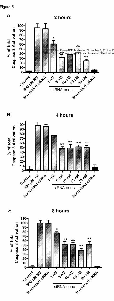
Post-exposure application of FasR siRNA
Based on the observation in NHBE cells that pre-treatment with Fas siRNA was
effective in decreasing ( ~65%) caspase-3 activation due to SM, we became interested in
seeing whether adding Fas siRNA to cell cultures at timed intervals post SM exposure
could also decrease SM-induced caspase-3 activation. First, NHBE cells (~80%
confluent) were exposed to 300 µM SM, and then Fas siRNA was applied at 2, 4 or 8 h
after SM exposure. Caspase-3 activation was measured 24 h after SM. Similar to
pretreatment, post-exposure treatment with siRNA also decreased (~40-60%) caspase-3
activation due to SM (figure 5). These results showed that Fas siRNA could prevent SM-
induced apoptosis in NHBE cells even when added to cells up to 8 h after SM; this
indicated a therapeutic window out to 8 h post-exposure.
To correlate the siRNA prevention of SM-induced caspase-3 activation with
preventing cell death, the status of surviving vs. dead cells in NHBE cultures was
determined by flow cytometry using Annexin V (apoptosis marker) and propidium iodide
(PI) (necrosis marker) staining. The results showed proportions of cells as live and dead
(via both apoptosis and necrosis) (figure 6). Fas siRNA was added to NHBE cells 8 h
after 300 µM SM exposure followed by Annexin V/PI staining and analyses
approximately 24 h after SM exposure. Cell death (apoptosis/necrosis) due to SM alone
(figure 6, block A) was 47 ± 2% (n=3). Treatment with 5-20 nM Fas siRNA decreased
This article has not been copyedited and formatted. The final version may differ from this version.JPET Fast Forward. Published on November 5, 2012 as DOI: 10.1124/jpet.112.199935
at ASPE
T Journals on A
pril 1, 2019jpet.aspetjournals.org
Dow
nloaded from
JPET #199935
14
cell death to 18-32% (n=3) (figure 6, blocks B-E). Treatment with siRNA caused a
reduction in the cells stained with either Annexin V or PI and a concomitant increase in
unstained cells. A careful examination of these results showed that in SM-exposed (300
µM) NHBE cultures, siRNA treatment was able to reduce the number of cells that were
in the late apoptosis/early necrosis phases.
Soluble FasL in the BALF of rats exposed to SM via inhalation
The findings described above indicated a Fas-mediated mechanism of cell death
due to SM in vitro in an airway epithelial (NHBE) cell culture model. To test a
therapeutic approach based on this mechanism i.e., prevention of airway injury due to SM
via Fas intervention, the next step would be to validate these observations in vivo in a
suitable animal model. To accomplish this objective, a rat SM inhalation model as
described previously (Anderson, 1996) was utilized. Anesthetized rats were exposed to
SM (in ethanol solution) via spontaneous inhalation by intubation. Subsequently,
pulmonary damage to include epithelial effects was assessed by biochemical analyses of
bronchoalveolar lavage fluid (BALF) constituents as well as cellular components in the
BALF. In these experiments, BALF samples from sham (ethanol inhalation) control and
SM-exposed (1.0 mg/kg, 50 min) rats were collected at either 6 or 24 h following SM
exposure. Soluble Fas ligand (sFasL) in BALF and caspase-3 activity in BALF cells were
then determined using ELISA and luminometric caspase-3 assay, respectively (figure 7).
Results showed significant (p<0.01) increases in both sFasL in BALF and caspase-3
activity in BALF cells at 6 and 24 h following both doses of SM. Although limited
This article has not been copyedited and formatted. The final version may differ from this version.JPET Fast Forward. Published on November 5, 2012 as DOI: 10.1124/jpet.112.199935
at ASPE
T Journals on A
pril 1, 2019jpet.aspetjournals.org
Dow
nloaded from
JPET #199935
15
information has been obtained, these results suggest a Fas-mediated mechanism of
apoptosis in vivo similar to the observations in vitro using NHBE cell cultures.
Discussion
The results in this study demonstrate that a highly selective and effective siRNA
construct was able to down-regulate the expression and functionality of the FasR in a
normal human airway epithelial cell culture model. In cultured NHBE cells, both SM
(300 µM) and the agonistic FasR antibody, CH11 activated caspase-3, which was
inhibited by the antagonistic FasR antibody ZB4. These results indicated that SM-
induced caspase-3 activation was via the Fas response (figure 2). The siRNA construct at
a low (10 nM) concentration markedly inhibited caspase-3 activation due to both CH11
and SM (figures 3, 4). However, the extent of inhibition was more when caspase-3 was
activated by CH11 than by SM, possibly because SM stimulation of caspse-3 involves
mechanisms additional to those pertaining to the Fas pathway (figure 1). siRNA inhibited
(~60%) SM-induced caspase-3 activation even when added to NHBE culture as late as 8
h after SM exposure (figure 5). Moreover, flow cytometric studies revealed that the
siRNA inhibition of caspase-3 activation due to SM was concomitant with reduced cell
death via both apoptosis and necrosis (figure 6). A more detailed analysis of these results
showed the following. In cells exposed to SM (300 µM), but not treated with siRNA, cell
death occurred via early apoptosis, late apoptosis/early necrosis, and necrosis (figure 6A).
It was noteworthy that the majority of the cells dying were in early and late apoptosis.
However, when SM-exposed (300 µM) cells were treated with siRNA the proportions of
these types of cell deaths were decreased in a siRNA concentration-dependent manner
This article has not been copyedited and formatted. The final version may differ from this version.JPET Fast Forward. Published on November 5, 2012 as DOI: 10.1124/jpet.112.199935
at ASPE
T Journals on A
pril 1, 2019jpet.aspetjournals.org
Dow
nloaded from
JPET #199935
16
(e.g., 5 nM [figure 6B] and 10 nM [figure 6C]). These observations indicated that cell
death due to SM involves early apoptosis progressing toward late apoptosis and necrosis.
Moreover, it seems possible to prevent the late phases of cell death by intervening at
early apoptosis.
Finally, the in vivo studies in the rat SM inhalation model, higher levels of both
sFasL in BALF (figure 7A) and caspase-3 activity in BALF cells (figure 7B) were
observed. These in vivo results combined with the in vitro findings suggest a mechanism
of SM inhalation injury that involves an early Fas-mediated apoptosis of airway epithelial
cells, which may eventually progress to necrosis. Depending on the degree of damage,
these cells may detach from the substratum and cause a sloughing off of the epithelial
tissue. Respiratory distress and even death following SM inhalation are believed to be
due to inflammatory responses combined with the detachment and sloughing off of the
dying airway epithelial cells. These contribute to tissue aggregate, generation of
pseudomembranes and mucus plug responsible for choking and death. We propose to
intervene in this mechanism at one of the component events, i.e., epithelial detachment,
by inhibiting the Fas-mediated apoptosis using siRNA. If successful, the siRNA
antagonism of the FasR or some other component of the DISC, e.g., the FADD, may
provide a highly prospective therapeutic approach for SM inhalation injury.
In the present study, the pathway of caspase-3 activation, i.e., apoptosis due to
SM, is via the FasR was demonstrated. Additionally,blocking the Fas pathway by FasR
siRNA reduced cell death due to SM was shown. Most intriguing was our observation
that this Fas intervention was effective even when FasR siRNA was added to cells as late
as 8 h after SM exposure. The siRNA effect is not due to its reaction with SM in the cell
This article has not been copyedited and formatted. The final version may differ from this version.JPET Fast Forward. Published on November 5, 2012 as DOI: 10.1124/jpet.112.199935
at ASPE
T Journals on A
pril 1, 2019jpet.aspetjournals.org
Dow
nloaded from
JPET #199935
17
culture medium. SM is sparingly soluble in water, but forms a saturated solution at ~5
mM which is way above the concentration used in our experiments i.e., 300 µM
(Papirmeister, 1991). SM is a strong electrophile due to the formation of a highly reactive
sulfonium intermediate in aqueous medium. This gives rise to its bifunctional alkylating
form that reacts readily with cellular functional molecules e.g., DNA, RNA, proteins, etc.
to initiate its toxic mechanisms. At body temperature (37°C), SM hydrolyzes with a half-
life (t1/2) of <10 minutes in water (Papirmeister, 1991), but ~15 minutes in cell culture
medium (pH 7.4) (unpublished results). Therefore, most of SM would have disappeared
by about an hour, which would be long before the addition of siRNA at 2 – 8 hours after
cell exposure. It is known that the actual manifestation of these toxic events e.g., skin
blister, corneum and lung epithelial separation are not observed until 12 – 24 h after SM
exposure. This indicates a substantial lag period between the onset of toxic cellular
mechanisms and their pathological manifestations, implying a therapeutic window. Thus,
the post-exposure efficacy of FasR siRNA intervention in preventing the SM effects on
airway epithelial cells in vitro strongly supports the idea of its being a prospective
molecular therapeutic for inhalation injury due to SM.
In addition to SM toxicity, the Fas pathway is known to be an important mediator
of other disease states, e.g., idiopathic pulmonary fibrosis (IPF), acute lung injury (ALI),
acute respiratory distress syndrome (ARDS), etc. (Beheshti et al., 2006; Emmler et al.,
2007; Kuwano, 2008; Perl et al., 2008). In all of these cases, Fas and Fas-ligand are
upregulated in the airway epithelial cells and are thought to contribute to cellular
apoptosis (Matute-Bello et al., 1999; Albertine et al., 2002; Hagimoto et al., 2002; Lee et
al., 2008). The severity of the infection in septic ALI was highly correlated with the Fas-
This article has not been copyedited and formatted. The final version may differ from this version.JPET Fast Forward. Published on November 5, 2012 as DOI: 10.1124/jpet.112.199935
at ASPE
T Journals on A
pril 1, 2019jpet.aspetjournals.org
Dow
nloaded from

JPET #199935
18
FasL system (Albertine et al., 2002); furthermore, ARDS non-survivors were shown to
have markedly higher levels of FasL than survivors (Matute-Bello et al., 1999). Active
antagonism of FasR has been shown to reduce the development of endotoxin-mediated
ALI, hemorrhage-induced septic ALI, and bleomycin-induced pulmonary fibrosis in
animal models (Kuwano et al., 1999; Kitamura et al., 2001; Perl et al., 2007). These
observations point to a critical role of Fas in each of these diseases. Therefore, the
antagonism of Fas via FasR, as we have proposed, may be an effective novel treatment
strategy for respiratory diseases involving the Fas-mediated mechanisms.
The idea of Fas intervention has been put forth by others as well (Perl et al., 2007;
Kuwano, 2008). There are several ways to antagonize the FasR (e.g., the Fas antagonistic
antibody ZB4 and Fas:Ig, a fusion protein that blocks FasL-FasR interaction); however,
the pitfalls of these approaches are their possible non-selective effects. Small interfering
RNA (siRNA), however, does not have these pitfalls and has been shown to be highly
selective. For these reasons, siRNA has emerged as a major thrust of the big
pharmaceutical companies. Many have shown that siRNA is easily delivered via either
intranasal (i.n.) or intratracheal (i.t.) route without any modifications to the lung (Massaro
et al., 2004; Zhang et al., 2004; Perl et al., 2005; Thomas et al., 2005; Aigner, 2007; de
Fougerolles et al., 2007; Thomas et al., 2007). The delivery of siRNA via these two
routes has been shown not to induce any significant activation of type I interferons via
activation of TLR-3, TLR-7, TLR-8, TLR-9, or protein kinase-R pathways nor via the
more classic proinflammatory responses (Alexopoulou et al., 2001; Akira, 2003; Moss
and Taylor, 2003; Sledz et al., 2003; Lomas-Neira et al., 2005; Perl et al., 2005; Robbins
and Rossi, 2005; Perl et al., 2008). This correlates with observations that intravenous
This article has not been copyedited and formatted. The final version may differ from this version.JPET Fast Forward. Published on November 5, 2012 as DOI: 10.1124/jpet.112.199935
at ASPE
T Journals on A
pril 1, 2019jpet.aspetjournals.org
Dow
nloaded from

JPET #199935
19
delivery of siRNA does not elicit an immune response (Heidel et al., 2004). Furthermore,
siRNA administrated to the lung either i.n. or i.t. has been shown to localize only to the
lung and does not become systemically available (Lomas-Neira et al., 2005; Perl et al.,
2005). The above discussions clearly indicate that siRNA may be a useful tool for in vitro
research as well as in vivo applications with a great potential for being on the forefront of
a new class of molecular medicine.
Acknowledgments
We acknowledge Dr. Alfred Sciuto of our institute for his valuable advice and support in
this research.
Authorship Contribution
Participated in research design: Keyser, Andres, Anderson, Smith, Ray.
Conducted experiments: Keyser, Andres, Nealley, Holmes, Benton, Paradiso, Appell,
Anderson, Carpin.
Contributed new reagents or analytic tools: Keyser.
Performed data analysis: Keyser, Andres, Nealley, Holmes, Ray.
Wrote or contributed to the writing of the manuscript: Keyser, Andres, Nealley, Holmes,
Anderson, Ray.
Other: Keyser, Holmes, Anderson and Ray acquired funding for the research.
This article has not been copyedited and formatted. The final version may differ from this version.JPET Fast Forward. Published on November 5, 2012 as DOI: 10.1124/jpet.112.199935
at ASPE
T Journals on A
pril 1, 2019jpet.aspetjournals.org
Dow
nloaded from

JPET #199935
20
References Aigner A (2007) Nonviral in vivo delivery of therapeutic small interfering RNAs. Curr
Opin Mol Ther 9:345-352. Akira S (2003) Toll-like receptor signaling. J Biol Chem 278:38105-38108. Albertine KH, Soulier MF, Wang Z, Ishizaka A, Hashimoto S, Zimmerman GA, Matthay
MA and Ware LB (2002) Fas and fas ligand are up-regulated in pulmonary edema fluid and lung tissue of patients with acute lung injury and the acute respiratory distress syndrome. Am J Pathol 161:1783-1796.
Alexopoulou L, Holt AC, Medzhitov R and Flavell RA (2001) Recognition of double-stranded RNA and activation of NF-kappaB by Toll-like receptor 3. Nature 413:732-738.
Anderson DR, Yourick, J.J., Moeller, R.B., Petrali, J.P., Young G. D., Byers, S.L. (1996) Pathologic changes in rat lungs following acute sulfur mustard inhalation. Inhal Toxicol 8:285-297.
Balali-Mood M and Hefazi M (2005) The pharmacology, toxicology, and medical treatment of sulphur mustard poisoning. Fundam Clin Pharmacol 19:297-315.
Beheshti J, Mark EJ, Akbaei HM, Aslani J and Ghanei M (2006) Mustard lung secrets: long term clinicopathological study following mustard gas exposure. Pathol Res Pract 202:739-744.
Bijani K and Moghadamnia AA (2002) Long-term effects of chemical weapons on respiratory tract in Iraq-Iran war victims living in Babol (North of Iran). Ecotoxicol Environ Saf 53:422-424.
de Fougerolles A, Vornlocher HP, Maraganore J and Lieberman J (2007) Interfering with disease: a progress report on siRNA-based therapeutics. Nat Rev Drug Discov 6:443-453.
Emad A and Emad Y (2007) Levels of cytokine in bronchoalveolar lavage (BAL) fluid in patients with pulmonary fibrosis due to sulfur mustard gas inhalation. J Interferon Cytokine Res 27:38-43.
Emmler J, Hermanns MI, Steinritz D, Kreppel H, Kirkpatrick CJ, Bloch W, Szinicz L and Kehe K (2007) Assessment of alterations in barrier functionality and induction of proinflammatory and cytotoxic effects after sulfur mustard exposure of an in vitro coculture model of the human alveolo-capillary barrier. Inhal Toxicol 19:657-665.
Ghanei M and Harandi AA (2007) Long term consequences from exposure to sulfur mustard: a review. Inhal Toxicol 19:451-456.
Hagimoto N, Kuwano K, Inoshima I, Yoshimi M, Nakamura N, Fujita M, Maeyama T and Hara N (2002) TGF-beta 1 as an enhancer of Fas-mediated apoptosis of lung epithelial cells. J Immunol 168:6470-6478.
Heidel JD, Hu S, Liu XF, Triche TJ and Davis ME (2004) Lack of interferon response in animals to naked siRNAs. Nat Biotechnol 22:1579-1582.
Kitamura Y, Hashimoto S, Mizuta N, Kobayashi A, Kooguchi K, Fujiwara I and Nakajima H (2001) Fas/FasL-dependent apoptosis of alveolar cells after lipopolysaccharide-induced lung injury in mice. Am J Respir Crit Care Med 163:762-769.
Kuwano K (2008) Involvement of epithelial cell apoptosis in interstitial lung diseases. Intern Med 47:345-353.
This article has not been copyedited and formatted. The final version may differ from this version.JPET Fast Forward. Published on November 5, 2012 as DOI: 10.1124/jpet.112.199935
at ASPE
T Journals on A
pril 1, 2019jpet.aspetjournals.org
Dow
nloaded from

JPET #199935
21
Kuwano K, Hagimoto N, Kawasaki M, Yatomi T, Nakamura N, Nagata S, Suda T, Kunitake R, Maeyama T, Miyazaki H and Hara N (1999) Essential roles of the Fas-Fas ligand pathway in the development of pulmonary fibrosis. J Clin Invest 104:13-19.
Lee KS, Choi YH, Kim YS, Baik SH, Oh YJ, Sheen SS, Park JH, Hwang SC and Park KJ (2008) Evaluation of bronchoalveolar lavage fluid from ARDS patients with regard to apoptosis. Respir Med 102:464-469.
Lomas-Neira JL, Chung CS, Wesche DE, Perl M and Ayala A (2005) In vivo gene silencing (with siRNA) of pulmonary expression of MIP-2 versus KC results in divergent effects on hemorrhage-induced, neutrophil-mediated septic acute lung injury. J Leukoc Biol 77:846-853.
Massaro D, Massaro GD and Clerch LB (2004) Noninvasive delivery of small inhibitory RNA and other reagents to pulmonary alveoli in mice. Am J Physiol Lung Cell Mol Physiol 287:L1066-1070.
Matute-Bello G, Liles WC, Steinberg KP, Kiener PA, Mongovin S, Chi EY, Jonas M and Martin TR (1999) Soluble Fas ligand induces epithelial cell apoptosis in humans with acute lung injury (ARDS). J Immunol 163:2217-2225.
Moss EG and Taylor JM (2003) Small-interfering RNAs in the radar of the interferon system. Nat Cell Biol 5:771-772.
Papirmeister B (1991) Medical defense against mustard gas : toxic mechanisms and pharmacological implications. CRC Press, Boca Raton.
Papirmeister B, Gross CL, Meier HL, Petrali JP and Johnson JB (1985) Molecular basis for mustard-induced vesication. Fundam Appl Toxicol 5:S134-149.
Perl M, Chung CS, Lomas-Neira J, Rachel TM, Biffl WL, Cioffi WG and Ayala A (2005) Silencing of Fas, but not caspase-8, in lung epithelial cells ameliorates pulmonary apoptosis, inflammation, and neutrophil influx after hemorrhagic shock and sepsis. Am J Pathol 167:1545-1559.
Perl M, Chung CS, Perl U, Lomas-Neira J, de Paepe M, Cioffi WG and Ayala A (2007) Fas-induced pulmonary apoptosis and inflammation during indirect acute lung injury. Am J Respir Crit Care Med 176:591-601.
Perl M, Lomas-Neira J, Chung CS and Ayala A (2008) Epithelial cell apoptosis and neutrophil recruitment in acute lung injury-a unifying hypothesis? What we have learned from small interfering RNAs. Mol Med 14:465-475.
Petrali JP, Oglesby SB, Hamilton TA and Mills KR (1993) Comparative morphology of sulfur mustard effects in the hairless guinea pig and a human skin equivalent. J Submicrosc Cytol Pathol 25:113-118.
Ray R, Legere RH, Majerus BJ and Petrali JP (1995) Sulfur mustard-induced increase in intracellular free calcium level and arachidonic acid release from cell membrane. Toxicol Appl Pharmacol 131:44-52.
Robbins MA and Rossi JJ (2005) Sensing the danger in RNA. Nat Med 11:250-251. Rosenthal DS, Simbulan-Rosenthal CM, Iyer S, Spoonde A, Smith W, Ray R and
Smulson ME (1998) Sulfur mustard induces markers of terminal differentiation and apoptosis in keratinocytes via a Ca2+-calmodulin and caspase-dependent pathway. J Invest Dermatol 111:64-71.
Rosenthal DS, Velena A, Chou FP, Schlegel R, Ray R, Benton B, Anderson D, Smith WJ and Simbulan-Rosenthal CM (2003) Expression of dominant-negative Fas-
This article has not been copyedited and formatted. The final version may differ from this version.JPET Fast Forward. Published on November 5, 2012 as DOI: 10.1124/jpet.112.199935
at ASPE
T Journals on A
pril 1, 2019jpet.aspetjournals.org
Dow
nloaded from
JPET #199935
22
associated death domain blocks human keratinocyte apoptosis and vesication induced by sulfur mustard. J Biol Chem 278:8531-8540.
Saladi RN, Smith E and Persaud AN (2006) Mustard: a potential agent of chemical warfare and terrorism. Clin Exp Dermatol 31:1-5.
Sledz CA, Holko M, de Veer MJ, Silverman RH and Williams BR (2003) Activation of the interferon system by short-interfering RNAs. Nat Cell Biol 5:834-839.
Smith WJ and Dunn MA (1991) Medical defense against blistering chemical warfare agents. Arch Dermatol 127:1207-1213.
Smith WJ, Gross CL, Chan P and Meier HL (1990) The use of human epidermal keratinocytes in culture as a model for studying the biochemical mechanisms of sulfur mustard toxicity. Cell Biol Toxicol 6:285-291.
Thomas M, Lu JJ, Chen J and Klibanov AM (2007) Non-viral siRNA delivery to the lung. Adv Drug Deliv Rev 59:124-133.
Thomas M, Lu JJ, Ge Q, Zhang C, Chen J and Klibanov AM (2005) Full deacylation of polyethylenimine dramatically boosts its gene delivery efficiency and specificity to mouse lung. Proc Natl Acad Sci U S A 102:5679-5684.
Xu G and Shi Y (2007) Apoptosis signaling pathways and lymphocyte homeostasis. Cell Res 17:759-771.
Zhang X, Shan P, Jiang D, Noble PW, Abraham NG, Kappas A and Lee PJ (2004) Small interfering RNA targeting heme oxygenase-1 enhances ischemia-reperfusion-induced lung apoptosis. J Biol Chem 279:10677-10684.
This article has not been copyedited and formatted. The final version may differ from this version.JPET Fast Forward. Published on November 5, 2012 as DOI: 10.1124/jpet.112.199935
at ASPE
T Journals on A
pril 1, 2019jpet.aspetjournals.org
Dow
nloaded from
JPET #199935
23
Footnotes
a. This work was supported by the Defense Threat Reduction Agency – Joint Science
and Technology Office, Medical S&T Division [Grants CBM.RESP.01.10.RC.015,
3.F0011-08-RC-C]. Additionally, this research was supported in part by an appointment
to the Postgraduate Research Parpticipation Program at the U.S. Army Medical Research
Institute of Chemical Defense administered by the Oak Ridge Institute for Science and
Education through an interagency agreement between the U.S. Department of Energy and
USAMRMC.
Disclaimer: The views expressed in this article are those of the author(s) and do not
reflect the official policy of the Department of Army, Department of Defense, or the U.S.
Government.
The experimental protocol was approved by the Animal Care and Use Committee at the
United States Army Medical Research Institute of Chemical Defense and all procedures
were conducted in accordance with the principles stated in the Guide for the Care and
Use of Laboratory Animals and the Animal Welfare Act of 1966 (P.L. 89-544), as
amended.
Figure legends
This article has not been copyedited and formatted. The final version may differ from this version.JPET Fast Forward. Published on November 5, 2012 as DOI: 10.1124/jpet.112.199935
at ASPE
T Journals on A
pril 1, 2019jpet.aspetjournals.org
Dow
nloaded from

JPET #199935
24
Figure 1. Extrinsic and Intrinsic Apoptosis Cascade.
Figure 2. Concentration-response of caspase-3 activation in NHBE cells due to CH11 and
ZB4. (A) NHBE cells were incubated with varying concentrations of CH11 and 24 h later
caspase-3 activity was examined. Data are shown as percent of total caspase-3 activation.
Experiments were done in triplicate with a 5 wells per experiment, ± SEM. (B) ZB4 at
various concentrations and 100 ng/ml CH11 were presented concurrently to NHBE cells;
twenty-four hours later caspase-3 activation was measured. Lined bars indicate addition
of 100 ng/ml CH11; solid black ZB4 only. Data are shown as percent of total caspase-3
activation as compared to 100 ng/ml CH11 alone.
(C) NHBE cells were exposed to ZB4 at various concentrations and 300 µM SM
concurrently; 24 h later caspase-3 activation was measured. Hatched bars indicate
addition of 300 µM SM; rightmost 500 ng/ml ZB4 is ZB4 alone. Data are shown as
percent of total caspase-3 activation as compared to 300 µM SM alone. Experiments
were done in triplicate with a 5 wells per experiment, ± SEM., * p<0.05, **p<0.01 as
compared to (B) 100 ng/ml CH11 or (C) 300 µM SM alone.
Figure 3. A. siRNA concentration-response of caspase-3 activity after CH11 addition in
NHBE cells. Various siRNA concentrations were given to NHBE cells 48 h prior to 100
ng/ml CH11 addition. Twenty-four hours after CH11 addition, caspase-3 activation was
measured. Data are shown as percent of total caspase-3 activation compared to 100 ng/ml
CH11. Experiments were done in triplicate with a 5 wells per experiment, ± SEM **
This article has not been copyedited and formatted. The final version may differ from this version.JPET Fast Forward. Published on November 5, 2012 as DOI: 10.1124/jpet.112.199935
at ASPE
T Journals on A
pril 1, 2019jpet.aspetjournals.org
Dow
nloaded from
JPET #199935
25
p<0.01 as compared to 100 ng/ml CH11 alone. Lined bars indicate addition of 100 ng/ml
CH11.
B. NHBE cells were harvested 48 h after transfection with 10 nM siRNA. Cells were
lysed and 50 µg protein was loaded per well. FasR monoclonal Ab (1:1000) and GAPDH
Ab (1:1000) were used and detected by ECF substrate.
Figure 4. FasR siRNA dose response for caspase-3 activation after 300 µM sulfur
mustard (SM) addition to cultured NHBE cells. Cells were treated with various siRNA
concentrations 48 h prior to 300 µM SM addition. Twenty-four hours after 300 µM SM
addition, caspase-3 activation was measured. Data are shown as percent of total caspase-3
activation compared to 300 µM SM. Experiments were done in triplicate with a 5 wells
per experiment, ± SEM., ** p<0.01 as compared to 100 ng/ml 300 µM SM alone.
Figure 5. FasR siRNA post-exposure concentration response to 300 µM SM. FasR
siRNA was added at 2, 4, and 8 h (A-C respectively) after 300 µM SM exposure, and
caspase 3 activation was measured 24 h after 300 µM SM exposure. Hatched bars
indicate 300 µM SM exposure, black bars vehicle control. Experiments were done in
triplicate with a 5 wells per experiment, ± SEM., ** p<0.01, *p<0.05 as compared to 300
µM SM alone.
Figure 6. Annexin V and propidium iodide staining of 300 µM SM in NHBE cells.
NHBE cells were exposed to 300 µM SM, then were given (B) 5 nM, (C) 10 nM, (D) 15
nM, or (E) 20 nM of FasR siRNA 8 h after SM exposure. Approximately 24 h following
This article has not been copyedited and formatted. The final version may differ from this version.JPET Fast Forward. Published on November 5, 2012 as DOI: 10.1124/jpet.112.199935
at ASPE
T Journals on A
pril 1, 2019jpet.aspetjournals.org
Dow
nloaded from

JPET #199935
26
SM exposure, NHBE cells were stained with annexin V/propidium iodide and analyzed
by flow cytometry. Results from a representative experiment are shown above. Averaged
data for each quadrant: (F) unstained, (G) AV only, (H) AV/PI and (I) PI only is also
shown. n=3, ± SEM, * p <0.05, **p<0.01 as compared to 300 µM SM only.
Figure 7. Measurement of sFasL and caspase-3 in BALF in a rat SM inhalation model.
(A) sFasL or (B) caspase-3 was measured in BALF or BALF cells, respectively, of rats
exposed to 1.0 mg/kg SM by inhalation by intubation; BALF and BALF cells were
collected at 6 or 24 h following SM inhalation. n=3 rats, ± SEM, * p <0.05, **p<0.01 as
compared to vehicle (EtOH) control.
This article has not been copyedited and formatted. The final version may differ from this version.JPET Fast Forward. Published on November 5, 2012 as DOI: 10.1124/jpet.112.199935
at ASPE
T Journals on A
pril 1, 2019jpet.aspetjournals.org
Dow
nloaded from

This article has not been copyedited and formatted. The final version may differ from this version.JPET Fast Forward. Published on November 5, 2012 as DOI: 10.1124/jpet.112.199935
at ASPE
T Journals on A
pril 1, 2019jpet.aspetjournals.org
Dow
nloaded from

This article has not been copyedited and formatted. The final version may differ from this version.JPET Fast Forward. Published on November 5, 2012 as DOI: 10.1124/jpet.112.199935
at ASPE
T Journals on A
pril 1, 2019jpet.aspetjournals.org
Dow
nloaded from

This article has not been copyedited and formatted. The final version may differ from this version.JPET Fast Forward. Published on November 5, 2012 as DOI: 10.1124/jpet.112.199935
at ASPE
T Journals on A
pril 1, 2019jpet.aspetjournals.org
Dow
nloaded from

This article has not been copyedited and formatted. The final version may differ from this version.JPET Fast Forward. Published on November 5, 2012 as DOI: 10.1124/jpet.112.199935
at ASPE
T Journals on A
pril 1, 2019jpet.aspetjournals.org
Dow
nloaded from
This article has not been copyedited and formatted. The final version may differ from this version.JPET Fast Forward. Published on November 5, 2012 as DOI: 10.1124/jpet.112.199935
at ASPE
T Journals on A
pril 1, 2019jpet.aspetjournals.org
Dow
nloaded from

This article has not been copyedited and formatted. The final version may differ from this version.JPET Fast Forward. Published on November 5, 2012 as DOI: 10.1124/jpet.112.199935
at ASPE
T Journals on A
pril 1, 2019jpet.aspetjournals.org
Dow
nloaded from

This article has not been copyedited and formatted. The final version may differ from this version.JPET Fast Forward. Published on November 5, 2012 as DOI: 10.1124/jpet.112.199935
at ASPE
T Journals on A
pril 1, 2019jpet.aspetjournals.org
Dow
nloaded from