Embed Size (px)
Citation preview

THE JOURNAL OF COMPARATIVE NEUROLOGY 234:523-535 (1985)
Possible Target Neurons of 5- Hydroxytryptamine Fibers in the Lamprey
Spinal Cord: Immunohistochemistry Combined With Intracellular Staining With
Lucifer Yellow
PAUL A.M. VAN DONGEN, TOMAS HOKFELT, STEN GRILLNER, ALBERT A.J. VERHOFSTAD, AND HARRY W.M. STEINBUSCH
Departments of Histology (P.A.M.V.D., T.H.) and Physiology I11 (S.G.), Karolinska Institute, Stockholm, Sweden, Department of Anatomy and Embryology, Catholic
University, Nijmegen, The Netherlands (A.A. J.V.), and Department of Pharmacology, Free University, Amsterdam, The Netherlands (H.W.M.S.)
ABSTRACT Intracellular recordings were made from 76 neurons belonging to var-
ious cell types in the lamprey spinal cord, and these neurons were subse- quently stained with Lucifer yellow. Sections were made of spinal cords containing Lucifer-yellow-filled neurons, and in the same sections 5-hydrox- ytrytamine (5-HT)-containing neurons and fibers were made visible with immunohistochemical methods. Motoneurons and lateral cells appeared to send part of their dendrites into a dense ventromedial 5-HT plexus, and these dendrites were adjacent to 5-HT varicosities. No or few 5-HT varicosi- ties have been found adjacent to cell bodies or dendrites of sensory dorsal cells, giant interneurons, and edge cells. The combined application of intra- cellular staining and immunohistochemistry appeared to be suited to screen for possible transmitter-identified contacts on morphologically identified neurons.
Key words: 5-hydroxytryptamine, Lucifer yellow, intracellular staining, lamprey, spinal cord, identified neurons
In the lamprey spinal cord numerous 5-hydroxytrypta- mine-(5-HT)-containing neurons and fibers are present (Baumgarten, '72; Van Dongen et al., '85). Many longitudi- nally oriented 5-HT fibers are seen in the various "axon columns," into which the dendrites of the spinal neurons extend, and a dense ventromedial 5-HT plexus has been found. As a basis for future electrophysiological studies aiming at understanding the action of 5-HT in the lamprey spinal cord, it is of interest to know the target neurons of the 5-HT fibers, as well as the type of contacts involved. The combined application of intracellular staining and im- munohistochemistry (Kawata et al., '83; Reaves et al., '83; Hoffert et al., '83) is a suitable approach to investigate this problem. Intracellular staining with horseradish peroxi- dase (HRP; Snow et al., '76) or with Lucifer yellow (LY; Stewart, '78) make virtually all processes of neurons visible (Takato and Goldring, '79). However, for a combination with immunofluorescence, LY is more appropriate. In this study, intracellular recordings were made from various neurons in the lamprey spinal cord, and LY was injected 0 1985 ALAN R. LISS, INC.
intracellularly by ionophoresis. Here we report data on the relationship between 5-HT fibers and sensory dorsal neu- rons, motoneurons, giant interneurons, lateral cells, and edge cells (cf. Rovainen, '79).
MATERIALS AND METHODS Animals
Eleven silver lampreys (Zchtyomyzon unicuspis, 0.15-0.32 m) were used, obtained from Iowa (USA). They were kept in aerated aquaria at a temperature of 5-10°C.
Preparation The preparation was mainly performed according to
Cohen and Wallen ('SO). Pieces (0.06 m) of the spinal cord were dissected from the region of the dorsal fins (n = 5), or the region between the last gill opening and the dorsal fins
Address reprint requests to: T. Hokfelt, Dept. of Histology, Ka- rolinska Institute, P.O. Box 60400, S-104 01 Stockholm, Sweden.

524 P.A.M. VAN DONGEN ET AL.
Fig. 1. Micrographs of various types of LY-filled neurons in whole-mount preparations of the spinal cord (horizontal cords); the edge of the spinal cords is indicated by a curved arrow, the midline by a stippled line, and the axon (if visible in the micrograph) by a straight arrow (all micrographs of
this figure at the same magnification). Only a part of the neuron could be in focus due to the thickness of the spinal cord (200 pm). A, B. Motoneurons. C. Lateral cell. D. Edge cell. E. Sensory dorsal cell. F, G. Giant interneurons.
(n = 61, and placed into the experiment chamber. Under microscopic control, the perimeningeal tissue and the men- inx primitiva dorsal to the cord were removed, the ventral roots were cut, and the spinal cord was removed from the notochord. The spinal cord was placed ventral side up, stretched to its in situ length, and fixed with miniature "staples" which were placed over the cord. The ventral perimeningeal tissue and meninx primitiva were also re- moved. Neuronal cell bodies were clearly visible with the dissection microscope in the transilluminated spinal cord, which aided the microelectrode impalement. Spinal cords dissected this way were used for 2 days.
Recordings Intracellular recordings were made with glass micropi-
pettes filled with 5% Lucifer yellow CH (LY, Sigma) in 0.1 M LiCl. Occasionally, a part of the spinal cord was damaged during the dissection; no recordings were made from dam- aged segments. When stable recordings were obtained from neurons with resting potentials below at least -40 mV, and action potential amplitudes of at least 45 mV, the cells were
filled with LY by ionophoresis with a negative current of 2.54.0 nA for 30 minutes. Within 3 hours after completion of the dye injection, a piece of spinal cord (three to ten segments) containing the LY-filled neurons was removed from the experiment chamber and fixed (see below).
Histochemistry The histochemical preparation was performed mainly ac-
cording to Van Dongen et al. ('85). A piece of spinal cord with LY-filled neurons was put on a small piece of sylgard gel, stretched with miniature needles to its in situ length, and fixed. Wholemounts of these spinal cords with LY-filled neurons were inspected with a Zeiss fluorescence micro- scope (see below). The LY-filled cells were classified (see below), and pictures were taken at different focal depths. Horizontal serial cryostat sections (14 pm) were made, pro- cessed for 5-HT immunofluorescence (Steinbusch et al. '78) with tetramethyl-rhodamine-isothiocyanate (TR1TC)conju- gated second antibodies (cf. Coons, '58; Hokfelt et al., '73, '75, '831, and inspected with the microscopic equipment described earlier (Van Dongen et al., '85). For LY, the follow-

5-HT VARICOSITIES NEAR LAMPREY SPINAL NEURONS 525
TABLE 1. Classification of Lucifer-Yellow-Filled Neurons, According to the Criteria Mentioned in the Methods-Section
Sensory dorsal cells Motoneuronlike cells Giant interneurons Lateral cells Edge cells 5-HT cells
Unclassified Small cell body Medium cell body Axon identified Axon not identified Ipsilateral dendrites Bilateral dendrites
Nos. of neurons
39 (34) 4 (4) 18 (16) 3 (2) 4 (4) 7 (7) 3 (1)
37 (29) 6 (6) 31 (23) 16 (14) 21 (15) 34 (27) 3 (2)
The nos. between parentheses indicate the nos. of LY-filled neumns with intact 5-€IT immunostaining.
ing Zeiss filters were used excitation filter KP 500, and emission filter LP 520 and sometimes also either BP 546 or KP 560 to reduce the TRITC fluorescence; and for TRITC: excitation filter BP 546 and sometimes also LP 455 to reduce LY fluorescence, and emission filter LP 590 (Hokfelt et al., ’83). Single or sequentially exposed black-and-white (Tri-X, Agfa-Gevaert) pictures were made with filter set- tings for TRlTc (5-HT) and LY. In one series, which in- cluded 13 LY-filled neurons, no 5-HT immunoreactive elements could be made visible (probably due to the fixa- tion); however, the distribution of dendrites and the general shape of these neurons were similar to those obtained in sections where 5-HT immunoreactivity was observed. Com- plete series of photonegatives or color slides were taken of some neurons. These were projected, and the LY-filled con- tours of the sections were drawn superimposed upon each other. Thereby, projections on a horizontal plane of neurons and all their processes were obtained ((‘reconstructions’’).
Classification The LY-filled neurons were classified according to the
following criteria (cf. Rovainen, ’79; Figs. 1, 10). Sensory dorsal cells. Sensory dorsal cells are large neu-
rons located in the medial columns (cf. Rovainen, ’67; Mar- tin and Wickelgren, ’71). These cells are bipolar, having a rostrally and a caudally directed ipsilateral axon;and often a few short, dendritelike processes (Boman, Christensen and Grillner, unpublished).
Motoneuivns. Motoneurons are medium-sized neurons located in the lateral gray column with an axon that can be followed into the ventral root. Such neurons have been found to innervate muscle fibers (Teravainen and Rovainen ’71; Wallen et al., ’85), but it cannot be entirely excluded that some of these neurons innervate the viscera (Johnels, ’56). Neurons whose axons were seen to turn toward the ventral roots have been included, even if the axons could not be traced out into the roots.
Giant interneuivns. Giant interneurons are very large neurons located in the caudal two-thirds of the spinal cord (Rovainen ’67, ’74). They have large dendrites that extend over the whole mediolateral width of the spinal cord ipsi- and contralateral to the cell body. They also have a promi- nent axon that ascends, gradually turning to the side of the
spinal cord contralateral to the cell body. These axons could be followed to the most rostral part of the piece of spinal cord used in this study.
Edge cells. Edge cells are medium-sized neurons located in the lateral column, situated near the lateral margin of the spinal cord (Rovainen, ’74). The cells have processes extending to the most lateral margin of the spinal cord, where these processes form nestlike ramifications (Grillner et al., ’84). Edge cells have ipsilateral and sometimes also contralateral dendrites. Most edge cells have ascending axons.
Lateral cells. Lateral cells are neurons located in the rostral part of the spinal cord (rostral to the cloaca) with a large elongated cell body that extends into the lateral axon column (Rovainen, ’74). Lateral cells have ipsilateral den- drites. Only cells having descending axons are regarded as “lateral cells” (cf. Rovainen et al., ’73).
5-HT cells. 5-HT cells are small (generally less than 15 pm) midline neurons located ventral to the central canal (Baumgarten, ’72; Van Dongen et al., ’85).
Unclassified neumns. A heterogeneous group of neurons (41%) did not fulfill the above-mentioned criteria (cf. Table 1).
RESULTS Lucifer-yellow-stained neurons
Of 76 well-filled neurons, 39 could be classified in one of the categories described above (Table 1). With the use of excitation filter KP 500 and emission filter LP 520, the red TRITC (5-HT) fluorescent cell bodies and fibers and the yellow LY-filled neurons were simultaneously visible. This is best shown on color prints, but it is possible to demon- strate this with three black-and-white prints (see below). To establish the presence of 5-HT varicosities adjacent to the LY-filled neurons, all sections were scanned using a 400 x magnification in order to find 5-HT fibers and LY-filled elements with a sharp image in the same focal depth. If the distance between a 5-HT varicosity and a LY-filled cell body or dendrite was 1 pm or less, it was called an “adjacent varicosity.”
Generally, 5-HT varicosities were found to be located at a distance from motoneuonal cell bodies (Fig. ZA), which always was larger than 2 pm. All 18 motoneurons extended several (ten to 15) small (about 1.4 pm in diameter) ventromedial dendritic branches into the very dense 5-HT plexus (Figs. 2B, 9). Several 5-HT varicos- ities were seen adjacent to motoneuronal dendrites in this region, which was not the case in other regions. In our sample, 16 motoneurons had a dendritic tree that extended over less than 200 p m in a rostrocaudal direction (Fig. lB), while the dendritic tree of two of these neurons extended over some 500 pm (Fig. 1A). These two groups might corre- spond to motoneurons that supply the ventral myotomes on the one hand, and the dorsal myotomes (WallBn et al., ’85) or the dorsal fin muscles on the other hand (Ftovainen and Birnberger ’71). Neurons of both groups had dendrites which extended into the ventromedial 5-HT plexus. The medial dendrites of four motoneurons extended into the region of the 5-HT cell bodies; the distance between these dendrites and the 5-HT cell bodies could be less than 2 pm (Fig. 3).
Lateral cells (n = 4). Usually, 5-HT varicosities re- mained remote from the somata of the lateral cells: the distance was rarely less than 2 pm and always more than 1 pm (Fig. 4A). Three out of the four lateral cells were seen to send one or two dendrites from the cell body toward the
Motoneumm (n = 18).

526 P.A.M. VAN DONGEN ET AL
Fig. 2. Motoneurons in horizontal sections. A. Cell body in the interme- diate plane. B. Ventral dendrite in the Muller-fiber plane; note the ventro- medial dendrites in the 5-HT plexus. In Figures 3-7, the following protocol applies. Left LY (exc. filter KP 500, emiss. filter LP 520 plus KP 560).
Middle: 5-HT (exc. filter BP 546 often plus LP 455, emiss. filter LP 590). Right: double exposure showing both LY and 5-HT (all micrographs of the Figures 2-8 are a t the same magnification). Bar = 50 pm.

5-HT VARICOSITIES NEAR LAMPREY SPINAL NEURONS 527
Fig. 3. Medial dendrites of a motoneuron in the region of the 5-HT cell bodies; a dendrite is found (arrow) at a distance less than 2 pm from a 5-HT cell body. A. LY. B. 5-HT. C. Double exposure showing both LY and 5-HT. Bar = 50 urn.
midline, entering the 5-HT plexus with a few fine branches (Figs. 4B, 9) adjacent to 5-HT varicosities.
Sensory dorsal cells (n = 4). 5-HT varicosities were usu- ally remote from the somata of the dorsal cells, and the distance always remained more than 1 pm (Fig. 5A). The axons of the dorsal cells followed a longitudinal course in the lateral part of the medial column in the dorsal plane, where the density of 5-HT fibers was high, and they were often accompanied by 5-HT fibers, but no adjacent (distance < 1 pm) 5-HT varicosities have been found (Fig. 5B).
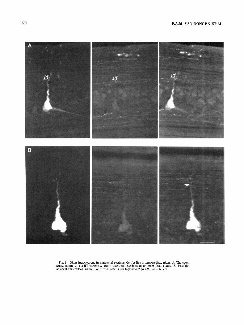
Giant interneurons (n = 3). 5-HT varicosities adjacent to the cell bodies or the dendrites of giant interneurons were rare (Fig. 6A). A single case of 5-HT varicosities at a distance of about 1 pm or less from a lateral dendrite of a giant interneuron is shown in Figure 6B. The ventral den- drites of giant interneurons did not penetrate into the 5-HT plexus, but remained dorsal and lateral to this plexus.
Edge cells (n = 5) . The cell bodies of the edge cells described in this study were all located at the very lateral margin of the spinal cord. Usually, 5-HT varicose fibers passed at a distance of 10, 20, or more pm from the edge cells' somata; 5-HT varicosities adjacent to these somata appeared to be rare or absent (cf. Fig. 7A). A varicosity possibly adjacent to a ventromedial dendrite has been found in a single case. Some of the edge cells had ventromedial dendrites, but these did not penetrate into the 5-HT plexus, but rather remained dorsal to it (Fig. 7B).
5-HT cells (n = 3). Small cell bodies in the midline column of the spinal cord could sometimes be seen in the living preparation and impaled. These small cells were only recorded from for a short period of time (< 15 minutes). 5- HT-immunoreactive material was present in these LY-filled cells (Fig. 8A,B). Although a limited proportion of their
axonal tree was filled, it could be seen that they were multipolar.
Unclassified neurons (n = 37). A large heterogeneous population of unclassified neurons was found (Table 1). This was expected since the majority of the lamprey spinal cord neurons has not yet been classified (cf. Rovainen '79; Fig. 10). Most of these unclassified cells had a medium-sized cell body and ipsilateral dendrites. In many cases (N = 21) no axon was found. Of the 37 unclassifed neurons, seven ex- tended their dendrites into the ventromedial 5-HT plexus. No further attention will be paid here to these unclassified neurons.
DISCUSSION Comments on the technique
The combined application of intracellular staining and immunohistochemistry in serial sections enabled the light microscopical detection of transmitter-identified varicosi- ties adjacent to cell bodies or dendrites of LY-filled neurons (cf. Kawata et al., '83; Reaves et al., '83; Hoffert et al., '83). With light microscopical methods, however, no definite con- clusions can be drawn with regard to synaptic connections between 5-HT-containing nerve endings and LY-filled den- drites. Moreover, although LY "stains" fine branches of neuronal dendrites (see also Takato and Goldring, '79), the dendritic tree may not be completely filled out by LY, and thus the distance between dendrite and 5-HT terminal may be overestimated. Furthermore, there is evidence from studies on the rat that 5-HT in cortical areas may exert its action at nonsynaptic release sites (Descarries et al., '75). By this method only adjacent varicosities could be identi- fied oriented toward the LY-filled element in the same plane as the sections. That is, in the present 14-pm horizon-

528 P.A.M. VAN DONGEN ET AJL
Fig. 4. Lateral cells in horizontal sections. A. Cell body in intermediate plane. B. Dendrite in the Miiller-fiber plane reaching the 5-HT plexus. For further details, see legend to Figure 2. Bar = 50 gm.

5-m VARICOSITIES NEAR LAMPREY SPINAL NEURONS
Fig. 5. Dorsal cell in horizontal section. A. Cell body in the dorsal cell plane. B. Axon in the dorsal plane. For further details, see legend to Figure 2. Bar = 50 prn.
529
530 P.A.M. VAN DONGEN ET AL
Fig. 6. Giant interneurons in horizontal sections. Cell bodies in intermediate plane. A. The open arrow points to a 5-HT varicosity and a giant cell dendrite in different focal planes. B. Possibly adjacent varicosities (arrow). For further details, see legend to Figure 2. Bar = 50 pm.

5-HT VARICOSITIES NEAR LAMPREY SPINAL NEURONS
Fig. 7. Edge cells in horizontal sections. A. Cell body in intermediate plane. B. Muller-fiber plane containing the most ventromedial extension of the dendrite (straight arrows) being still remote from 5-HT varicosities, 5-HT cell bodies (curved arrows), or 5-HT plexus. For further details, see legend to Figure 2. Bar = 50 pm.
531

532 P.A.M. VAN DONGEN ET AL
LATERAL .- MARGIN
CAUDAL
- -- . AXON
Fig. 8. 5-HT cells in horizontal section. A. LY-filled cell (arrow). B. 5-HT immunofluorescence of the same section; note 5-HT-positive material in the LY-filled neuron (arrow). Bar = 50 pm.
r--
’ {
l -
I
.. ! ’ . . I
- 1 I
. 100 Nrn
. . ,
MOTONEURON I- - MIDLINE
5 -HT
- _ _ _ _ LATERAL INTERNEURON
Fig. 9. Projection of the cell body, axon, and dendrites of a motoneuron and a lateral cell on a horizontal plane (“reconstruction”). The extension of the 5-HT plexus is indicated. In a separate section to the right of both neurons, the medial parts of the dendritic tree in sections containing the 5- HT plexus are shown; some dendritic branches were connected by branches in more dorsal or ventral planes, and in those cases, no connection is indicated in the drawings.

THE
LA
MP
RE
Y S
PIN
AL
CO
RD
ide
nti
fie
d n
eu
ron
s
ab
ou
t 1000 n
eu
ron
s p
er
se
gm
en
t
ab
ou
t 100 s
eg
me
nts
pe
r a
nim
al
ou
rs
dl
ro
ot
ga
ng
lio
n c
ell
s
7-2
0
larg
e c
eii
s p
er
/
d/
/'
gia
nt
inte
rne
uro
ns
12-35 p
er
co
rd
on
ly I
n c
au
da
l h
alf
o
f th
e c
ord
on
e p
er 3-5 s
eg
me
nts
pe
r s
eg
me
nt
in t
he
sp
ina
l c
ord
6-1
2 p
er
ga
ng
lio
n
70-100 p
er
ve
ntr
al
roo
t t
_---
- 'a late
ral
inte
r tr
al
on
ly i
n
ros
tra
l
. C
C-
inte
rne
uro
ns
(_
__
__
- ---
- --
(wit
h c
ros
sin
g,
ca
ud
al
ax
on
)
/Mu
ller
ax
on
s a
bo
ut
24
BR
AIN
'ne
uro
ns
tw
o-t
hir
ds
1-3 p
er
se
gm
en
t
01
th
e c
ord
ga
ng
llo
n
Fig.
10. S
chem
atic
sur
vey
of t
he i
dent
ifie
d ne
uron
s in
the
lam
prey
spi
nal
cord
and
the
ir c
onne
c-
tions
. The
dot
s in
the
5-H
T se
ctio
n in
dica
te t
hat m
ost o
f th
e 5-
HT
fibe
rs a
re lo
ngitu
dina
l. D
orsa
l ro
ot
gang
lia a
re in
dica
ted
as ro
und
elem
ents
bes
ide
the
spin
al c
ord.
The
num
bers
ind
icat
ed p
er s
egm
ent
are
deri
ved
from
Fre
ud (1878). R
ovai
nen
('79)
, Sel
zer (
'791,
Rov
aine
n an
d D
ill ('84), a
nd V
an D
onge
n et
al
. ('8
5). O
nly
for
grap
hic
reas
ons,
the
term
inal
s of
the
5-H
T ce
lls a
re d
raw
n in
the
ros
tral
dir
ectio
n,
alth
ough
no
pred
omin
ant r
ostr
al o
r cau
dal o
rien
tatio
n of
the
se fi
bers
has
bee
n fo
und.
en
w
w

P.A.M. VAN DONGEN ET AL 534
tal sections, adjacent varicosites could be detected, if they were lateral, medial, rostral, or caudal, but not dorsal or ventral to the LY-filled elements. In the latter cases, the distance between elements in different dorsal-ventral focal planes could not be estimated reliably.
Dendrites in the 5-HT plexus The most prominent positive result of this study is that
motoneurons and lateral cells in the lamprey spinal cord have ventromedial dendrites extending into the 5-HT plexus (cf. Fig. 10). The patterns of dendritic branching are, how- ever, different (Fig. 9): the motoneurons send several (ten to 15) and the lateral cells only one or two fine branches into the 5-HT plexus. In the lamprey 5-HT plexus, the density of 5-HT varicosities is much higher than in other regions of the lamprey spinal cord (Van Dongen et al., '85). Therefore it may be assumed that 5-HT released from vari- cosities in the lamprey spinal cord influences the motoneu- rons and lateral cells, either via synaptic or via more diffuse nonsynaptic contacts. Except from possible contacts with dendrites of motoneurons and lateral cells and some un- identified neurons in the ventromedial 5-HT plexus, we could only rarely find evidence for contacts between 5-HT varicosities and LY-filled dendrites.
An intrinsic premotor spinal 5-HT system The dendrites of identified neurons in the ventromedial
5-HT plexus belong to motoneurons or lateral cells; these neurons are, respectively, motoneurons or premotor inter- neurons (Rovainen, '79; Fig. 10). Few, if any, 5-HT contacts have been found on the primary sensory dorsal and edge cells, or on giant interneurons that can be regarded as secondary sensory neurons (Rovainen, '67, '79; Martin and Wickelgren, '71; Grillner et al., '84). 5-HT neurons thus influence motor rather than sensory neurons in the lam- prey spinal cord. It would be interesting to know whether another group of identified premotor interneurons with crossed caudal axons (CC-interneurons; Buchanan, '82) having dendritic ramifications around the Muller axons also extend their processes into the 5-HT plexus.
ComDarison with other vertebrates
tion, which can be elicited in the lamprey spinal cord in vitro, is not influenced by application of the 5-HT receptor antagonist methysergide (Grillner et al., '81). The applica- tion of 5-HT is reported to slow down the frequency of the rhythmic locomotor activity, while the amplitude of the locomotor bursts in the ventral root recordings increases (Harris-Warrick and Cohen, '83; Grillner and Wilen, un- published). These findings imply that the 5-HT neurons probably affect the motor output. Further investigations are in progress on the action of 5-HT applied close to den- drites of motoneurons and lateral cells.
ACKNOWLEDGMENTS The kind assistance of several people is greatly appreci-
ated Dr. Peter Wallen and Dr. Russel Hill for help during the electrophysiological experiments, Helene Axegren for the preparation of many dissections, Anne Peters for histo- logical help, Waldtraut Hiort for part of the photographic material, Dr. Vicky Holets for grammatical corrections, and Styrbjorn Bergelt for the reconstructions and part of the photographic material. This project was suported by the Swedish Medical Research Council (project numbers 2887 and 3026, and grant number K83-14V-6655-01).
LITERATURE CITED Baumgarten, H.G. (1972) Biogenic amines in the cyclostome and lower
vertebrate brain. Prog. Histochem. Cytochem. 4:l-90. Buchanan, J.T. (1982) Identification of interneurons with contralateral,
caudal axons in the lamprey spinal cord: Synaptic interactions and morphology. J. Neurophysiol. 47:961-975.
Carlsson, A., B. Falck, K. Fuxe and N.A. Hillarp (1964) Cellular localiza- tion of monoamines in the spinal cord. Acta Physiol. Scand. 60:112-119.
Cohen, A.H., and P. Wallen (1980) The neuronal correlate of locomotion in fish. "Fictive swimming" induced in an in uitro preparation of the lamprey spinal cord. Exp. Brain. Res. 41:11-18.
Coons, A.H. (1958) Fluorescent antibody methods. In J.F. Danielli (ed): General Cytochemical Methods, vol. 1. New York: Academic Press, pp. 399422.
Dahlstrom, A., and K. Fuxe (1965) Evidence for the existence of monoamine neurons in the central nervous system. 11. Experimentally induced changes in the intraneuronal amine levels of bulbospinal neuron sys- tems. Acta physiol. Scand. [Suppl.] 2473-36.
Descarries, L., A. Beaudet, and K.C. Watkins (1975) Serotonin nerve termi- nals in adult rat neocortex. Brain Res. lOOr563-588.
DiTirro, F.J., G.F. Martin, and R.H. Ho (1983) A developmental study of In fishes, the motor regions are substance P, somatostatin, enkephalin, and serotonin immunoreactive
elements in the spinal cord of the North American opossum. J. Comp. Neural. 216241-261.
innervated by intrinsic spinal 5-HT neurons while the sen- sory regions of the spinal cord receive 5-HT processes from suspraspinal origin (Parent and Northcutt, '82; Ritchie et al., '84). ln the various investigated (opossum, rat' cat' dog' monkey)7 both sensory and motor regions Of the spinal cord receive many 5-HT terminals (Dahlstrom and Fuxe '65; Steinbusch, '81; Martin et al., '82; Kojima et al., '82, '83; DiTirro et al., '83). The mammalian spinal 5- HT fibers arise exclusively, or almost exclusively, from brainstem 5-HT neurons (Carlsson et al., '64; DahlstrGm
Freud, S. (1878) iiber Spinalganglien and Ruckenmark des Petromyzon. Sitzungsber. Kaiserl. Akad. Wiss. (Wien), Mathemat. Naturwiss. Classe 77331-167.
Grillner, S., A. McClellan, K. Sigvardt, P. Wallen, and M. Wilen (1981) Activation of NMDA.receptors elicits "fictive locomotion" in lamprey spinal cord in vitro. Acta Physiol. Scand. 113549-551,
intraspinal mechanoreceptor. Science 223:500-503.
swimming in the lamprey spinal cord. Abstr. Soc. Neurosci. 9r753.
Grillner, S., T. Williams, and P. A. Lagerback (1984) The edge cell, a possible
Harris-Warrick, R.M., and A.H. Cohen (1983) Serotonin modulates fictive
and Fuxe' '65; 'liveras et '77; '80)' In mam- Hoffert, M.J., V. Miletic, M.A. Ruda, and R. Dubner (1983) Immunocyto.
no 5-HT bodies have been found (e.g', chemical identification of serotonin axonal contacts on characterized Dahlstrom and Fuxe, '65; Steinbusch, '81h except in nee- natal (5-75 days) opossums (DiTirro et al., '83) and in mon- keys treated with tryptophan and pargyline (LaMotte et al., '82). In any case, at least part of the propriospinal 5-HT innervation of motor regions, found in the lamprey and the Atlantic to be taken Over by supraspinal 5- HT neurons in mammals.
neurons in laminae I and 11 of the cat dorsal horn. Brain Res. 267r361- 364.
Hokfelt, T., K. Fuxe, M. Goldstein, and T.H. Joh (1973) Immunohistochemi. cal localization Of three catecholamine synthetizing enzymes: Aspects On
Hokfelt, T., K. Fuxe, and M. Goldstein (1975) Applications of immunohisto- chemistry to studies on monoamine cell systems with special reference to nervous tissue. Ann. N.Y. Acad. Sci.; 254r407-432.
Histochemistry 33r231-254.
Hokfelt, T., G. Skagerberg, L. Skirboll, and A. Bjorklund (1983) Combina- tion of retrograde tracing and neurotransmitter histochemistry. In A. Bjorklund and T. Hokfelt (eds): Handbook of Chemical Neuroanatomy, Vol. 1. Methods in Chemical Neuroanatomy. Amsterdam: Elsevier, pp.
The action of 5-HT in the lamprey spinal cord The actions Of 5-HT On Or lateral are -
not known. However, the motor pattern underlying locomo- 228-285.

5-HT VARICOSITIES NEAR LAMPREY SPINAL NEURONS 535
Johnels, A.G. (1956) On the peripheral autonomic nervous system of the trunk region of Lampetra planeri. Acta Zool. 37251-286.
Kawata, M., Y. Sano, K. Irenaga and H. Yamashita (1983) Immunohisto- chemical identification of lucifer yellow-labeled neurons in the rat su- praoptic nucleus. Histochemistry 78:21-26.
Kojima, M., Y. Takeuchi, M. Goto, and Y. Sano (1982) Immunohistochemical study on the distribution of serotonin fibers in the spinal cord of the dog. Cell Tissue Res. 226:477-491.
Kojima, M., Y. Takeuchi, M. Goto, and Y. Sano (1983) Immunohistochemical study on the localization of serotonin fibers and terminals in the spinal cord of the monkey (Macaca fuscuta). Cell Tissue Res. 229:23-36.
LaMotte, C.C., D.R. Johns, and N.C. de Lanerolle (1982) Immunohistochem- ical evidence of indoleamine neurons in monkey spinal cord. J. Comp. Neurol. 206:359-370.
Martin, A.R., and W.O. Wickelgren (1971) Sensory cells in the spinal cord of the sea lamprey. J. Physiol. (Lond.) 212:65-83.
Martin, G.F., T. Cabana, and A.O. Humbertson, Jr. (1982) The brainstem origin of monoaminergic projections to the spinal cord of the North American opossum. A study using fluorescent tracers and fluorescence histochemistry. Brain Res. Bull. 9:217-226.
Mizukawa, K. (1980) The segmental detailed topographical distribution of monoaminergic terminals and their pathways in the spinal cord of the cat. Anat. Anz. 147:125-144.
Oliveras, J.L., S. Bourgin, F. Herry, J.M. Besson and M. Hamon (1977) The topographical distribution of serotoninergic terminals in the spinal cord of the cat: Biochemical mapping by the combined use of microdissection and microassay procedures. Brain Res. 138:393-406.
Parent, A,, and R.G. Northcutt (1982) The monoamine-containing neurons in the brain of the garfish, Lepisosteus osseus. Brain Res. Bull. 9189- 204.
Reaves, T.A., Jr., A. Hou-Yu, E.A. Zimmerman and J.N. Hayward (1983) Supraoptic neurons in the rat hypothalamo-neurohypophysial explant: Double labeling with lucifer yellow injection and immunocytochemical identification of vasopressin- and neurophysin-containing neuroendo- crine cells. Neurosci. Lett. 37:137-142.
Ritchie, T.C., L.J. Roos, B.J. Williams and R.B. Leonard (1984) The descend- ing and intrinsic serotoninergic innervation of an Elsmobranch spinal cord. J. Comp. Neurol. 224:395-406.
Rovainen, C.M. (1967) Physiological and anatomical studies on large neu- rons of the central nervous system of the sea lamprey (Petromyzon marinus). 11. Dorsal cells and giant interneurons. J. Neurophysiol. 30: 1024-1042.
Rovainen, C.M., and K.L. Birnberger (1971) Identification and properties of
motoneurons to fin muscle of the sea lamprey. J. Neurophysiol. 34:974- 982.
Rovainen, C.M., P.A. Johnson, E.A. Roazch and J.A. Mankovsky (1973) Projection of individual axons in lamprey spinal cord determined by tracings through serial sections. J. Comp. Neurol. 149:193-202.
Rovainen, C.M. (1974) Synaptic interactions of identified nerve cells in the spinal cord of the sea lamprey. J. Comp. Neurol. 154:189-206.
Rovainen, C.M. (1979) Neurobiology of lampreys. Physiol. Rev. 59r1007- 1077.
Rovainen, C.M., and D.A. Dill (1984) Counts of axons in electron microscopic sections of ventral roots in lampreys. J. Comp. Neurol. 225:433-440.
Selzer, M.E. (1979) Variability in maps of identified neurons in the sea lamprey spinal cord examined by a wholemount technique. Brain Res. 163:181-193.
Snow, P.J., P.K. Rose, and A.G. Brown (1976) Tracing axons and axon collaterals of spinal neurons using intracellular injection of horseradish peroxidase. Science 191:312-313.
Steinbusch, H.W.M., A.A.J. Verhofstad and H.W.J. Joosten (1978) Localiza- tion of serotonin in the central nervous system by immunohistochemis- try: Description of a specific and sensitive technique and some applica- tions. Neuroscience 3311-819.
Steinbusch, H.W.M. (1981) Distribution of serotonin-immunoreactivity in the central nervous system of the rat. Cell bodies and terminals. Neu- roscience 6:557-618.
Stewart, W.W. (1978) Functional connections between cells as revealed by dye-coupling with a highly fluorescent naphtalimide tracer. Cell 14:741- 759.
Takato, M., and S. Goldring (1979) Intracellular marking with Lucifer yellow CH and horseradish peroxidase of cells electrophysiologically characterized as glia in the cerebral cortex of the cat. J. Comp. Neurol. 186: 173-188.
Teravainen, H., and C.M. Rovainen (1971) Fast and slow motoneurons to body muscle of the sea lamprey. J. Neurophysiol. 34:990-998.
Van Dongen, P.A.M., T. Hokfelt, S. Grillner, H.W.M. Steinbusch, A.A.J. Verhofstad, A.C. Cuello, and L. Terenius (1985) Immunohistochemical demonstration of some putative neurotransmitters in the lamprey spinal cord and spinal ganglia. 5-Hydroxytryptamine-, tachykinin- and neuro- peptide-Y-like immunoreactive neurons and fibers. J. Comp. Neurol. 234r501-522.
Wallen, P., S. Grillner, J. Feldman, and S. Bergelt (1985) Dorsal and ventral myotome motoneurons and their input during fictive locomotion in lamprey. J. Neurosci. (in press).