Embed Size (px)
Citation preview

24‐11‐10
POSITIONING AND LANDMARKS
• WHY IS THIS IMPORTANT?
1. Dose
2. Isocenter
3. Consistency with Stanford CT Scans
1
4th Stanford Computed Tomography Workshop4th Stanford Computed Tomography WorkshopRADIATION DOSE AND CONTRAST MEDIA IN NEURO & BODY CTRADIATION DOSE AND CONTRAST MEDIA IN NEURO & BODY CT
Lucas Learning Center, Department of Radiology, Stanford UniversityLucas Learning Center, Department of Radiology, Stanford UniversityThursday, NOV. 18, 2010Thursday, NOV. 18, 2010
Caryn Damits, RT (R) CT
POSITIONING AND LANDMARKS
WHY IS THIS IMPORTANT?
1. Dose
2. Isocenter
3. Consistency with Stanford CT Scans
Isocenter
• In imaging physics and radiation oncology, the `isocenter` is the point in space through which the central ray of the radiation beams passes
Why is this important? Why is this important?
Influences the MA table
XY axisCentering Importance
Useful when using SMART MA with GE Scanners
MA Table demonstrating X Y Axis
Perfect Head RXScan Range: Skull base to vertex Angle: Base of skull to Supra‐orbital Meatal Line

24‐11‐10
POSITIONING AND LANDMARKS
• WHY IS THIS IMPORTANT?
1. Dose
2. Isocenter
3. Consistency with Stanford CT Scans
2
The Perfect Head Scan
AVOID THE LENS
Worse Case Scenario
The KYPHOTICHead
Worse Case ScenarioPost‐op
Head holder
What can you do?
Do your best to CENTER and Stabilize patient
Scan an AP and LAT scout *
Large Scan FOV
Revert FOV Back to 25
Use Tools available
Options
Tools available to stabilize patient: tape, sponges, wedges
Example of wedge
24‐11‐10
POSITIONING AND LANDMARKS
• WHY IS THIS IMPORTANT?
1. Dose
2. Isocenter
3. Consistency with Stanford CT Scans
3
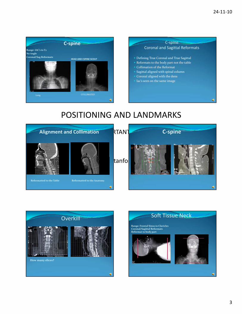
C‐spineRange: IAC’s to T2
No Angle
Coronal/Sag ReformatsHEAD AND CSPINE SCOUT
Long COLLIMATED
C‐spineCoronal and Sagittal Reformats
Defining True Coronal and True Sagittal
Reformats to the body part not the table
Collimation of the Reformat
Sagittal aligned with spinal column
Coronal aligned with the dens
Iac’s seen on the same image
Alignment and Collimation
Reformatted to the Table Reformatted to the Anatomy
C‐spine
True Sagittal True Coronal
Align sagittal with dens Align with spinal column
Overkill
How many slices?
Soft Tissue Neck
Range: Frontal Sinus to Clavicles Coronal/Sagittal ReformatsReformat to body part
24‐11‐10
POSITIONING AND LANDMARKS
• WHY IS THIS IMPORTANT?
1. Dose
2. Isocenter
3. Consistency with Stanford CT Scans
4
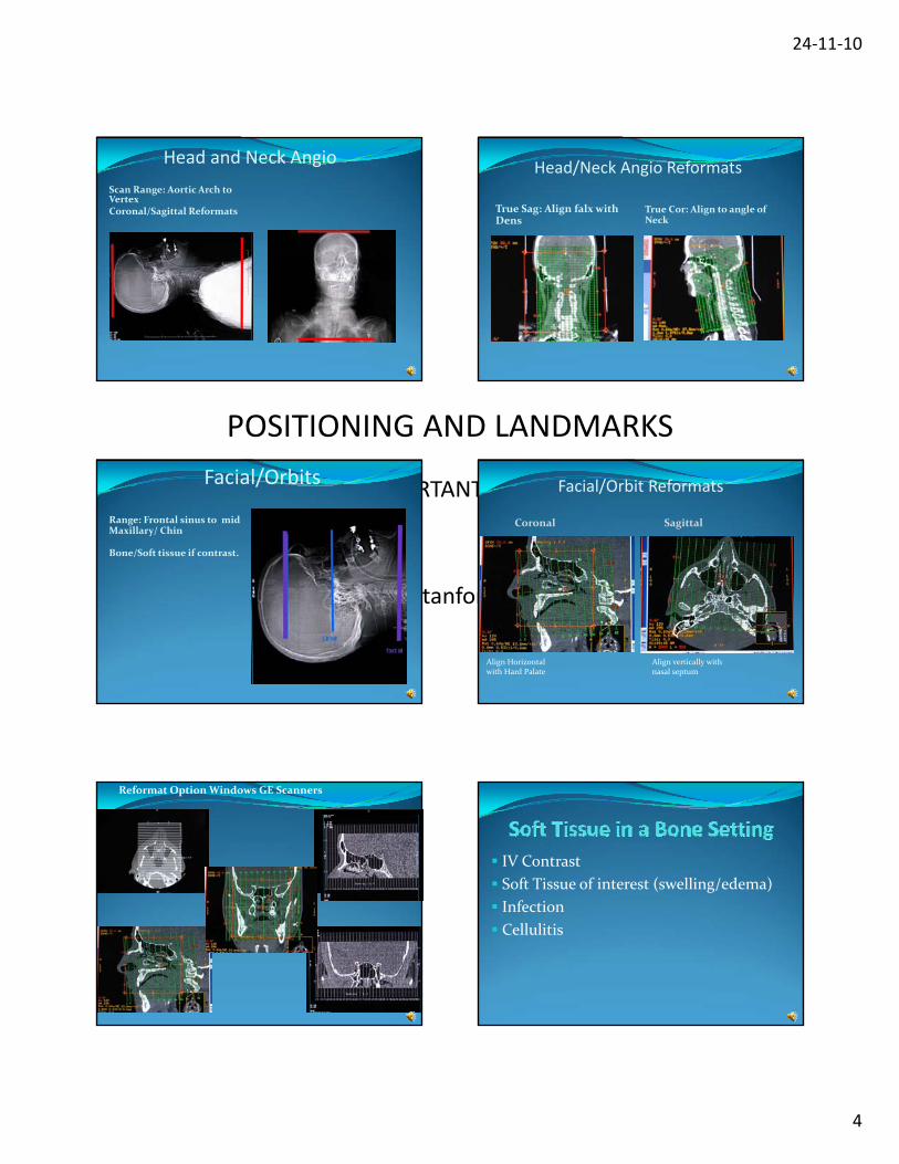
Head and Neck Angio
Scan Range: Aortic Arch to VertexCoronal/Sagittal Reformats
Head/Neck Angio Reformats
True Sag: Align falx with Dens
True Cor: Align to angle of Neck
Facial/Orbits
Range: Frontal sinus to mid Maxillary/ Chin
Bone/Soft tissue if contrast.
Facial/Orbit Reformats
Coronal Sagittal
Align Horizontal with Hard Palate
Align vertically with nasal septum
Reformat Option Windows GE Scanners
IV Contrast
Soft Tissue of interest (swelling/edema)
I f ti Infection
Cellulitis
24‐11‐10
POSITIONING AND LANDMARKS
• WHY IS THIS IMPORTANT?
1. Dose
2. Isocenter
3. Consistency with Stanford CT Scans
5
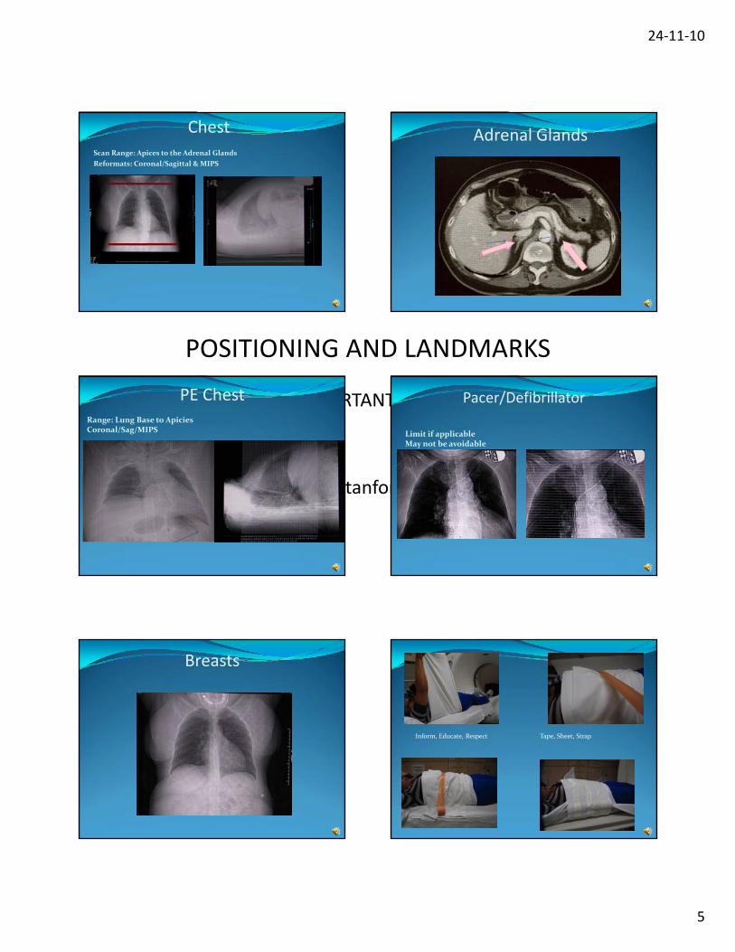
Chest Scan Range: Apices to the Adrenal Glands
Reformats: Coronal/Sagittal & MIPS
Adrenal Glands
PE ChestRange: Lung Base to ApiciesCoronal/Sag/MIPS
Pacer/Defibrillator
Limit if applicableMay not be avoidable
Breasts
Tape, Sheet, Strap Inform, Educate, Respect p pp
24‐11‐10
POSITIONING AND LANDMARKS
• WHY IS THIS IMPORTANT?
1. Dose
2. Isocenter
3. Consistency with Stanford CT Scans
6
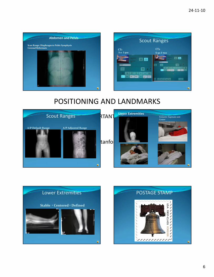
Abdomen and Pelvis
Scan Range: Diaphragm to Pubic SymphysisCoronal Reformats
Scout Ranges
CT1S 0‐ I 500
CT2
S 50‐I 700
Scout Ranges
A/P Default Range A/P Adjusted Range
Upper ExtremitiesForearm: Supinate and Center
Lower Extremities
Stable • Centered • Defined
POSTAGE STAMP

24‐11‐10
POSITIONING AND LANDMARKS
• WHY IS THIS IMPORTANT?
1. Dose
2. Isocenter
3. Consistency with Stanford CT Scans
7
Postage Stamp
Wide collimation
Good Collimation
FOV Comparison40 FOV 50 fFOV
What Can You Do
Examine the Reason
Define the Frame
C lli A i lCollimate Appropriately
Collimate to the FrameDid you know?
Initial scan from tableside?
Scan in a Decube position
Tables have Markers

24‐11‐10
POSITIONING AND LANDMARKS
• WHY IS THIS IMPORTANT?
1. Dose
2. Isocenter
3. Consistency with Stanford CT Scans
8
Decube scanning
Table Marker
Gantry Buttons What have we learned
Isocenter
Utilize tools available
True Coronal and Sagittal
Scan Ranges
Positioning of large Breasts (Smart mA)
Collimation
Initiate scan from Tableside
FOV’s
What happened?What do you see?

24‐11‐10
POSITIONING AND LANDMARKS
• WHY IS THIS IMPORTANT?
1. Dose
2. Isocenter
3. Consistency with Stanford CT Scans
9
BRA = Streak artifact
What is this?
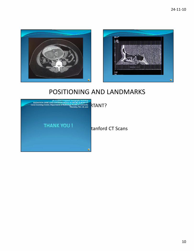
24‐11‐10
POSITIONING AND LANDMARKS
• WHY IS THIS IMPORTANT?
1. Dose
2. Isocenter
3. Consistency with Stanford CT Scans
10
4th Stanford Computed Tomography Workshop4th Stanford Computed Tomography WorkshopRADIATION DOSE AND CONTRAST MEDIA IN NEURO & BODY CTRADIATION DOSE AND CONTRAST MEDIA IN NEURO & BODY CT
Lucas Learning Center, Department of Radiology, Stanford UniversityLucas Learning Center, Department of Radiology, Stanford UniversityThursday, Nov. 18, 2010Thursday, Nov. 18, 2010