Embed Size (px)
Citation preview

BASIC SCIENCE
unication
Short CommNanomedicine: Nanotechnology, Biology, and Medicine 7 (2011) 131–136
Portable nanofiber meshes dictate cell orientation throughoutthree-dimensional hydrogels
Ying Yang, BSc, MSc, PhD⁎, Ian Wimpenny, BSc, Mark Ahearne, BEng, MSc, PhDInstitute for Science and Technology in Medicine, Medical School, Keele University, Stoke-on-Trent, United Kingdom
Received 28 August 2010; accepted 27 December 2010
www.nanomedjournal.com
Abstract
In this study, a new technique that controls individual cell orientation using nanofiber meshes within three-dimensional (3D) hydrogels isreported. Highly aligned and fragile electrospun nanofibers (average diameter 500 nm) were manufactured into portable and handleable mesheswith average line density of 45 nanofibers per 100 μm and thickness ranging between 0.5 and 3.0 μm. Through a facile and reproduciblefabrication process, the nanofiber meshes can be incorporated into 3D hydrogels via a bottom-up, layer-by-layer assembly process, resulting inmacroscopic and highly organized scaffolds. The nanofibers dictated the orientation of the cytoskeleton of individual cells in a very precisemanner, allowing altering of the orientation of a cell population throughout the thickness of the hydrogel. Addition of nanofibers affected cellphenotype and protein synthesis. This nanofiber-cell-hydrogel composite enables replication of the cellular and matrix architecture found inmany natural tissues, offering a novel protocol for electrospun nanofibers in regenerative medicine and bioengineering.
From the Clinical Editor: A novel protocol for highly organized nanofiber meshes incorporated into 3D hydrogels can be used to direct theoverlying cell population cytoskeleton direction, phenotype, and protein synthesis. Nanospun matrices offers a significant advancement forcontrolled tissue bioengineering and regenerative medicine applications.© 2011 Elsevier Inc. All rights reserved.
Key words: Portable; Nanofiber; Electrospinning; Alignment; Hydrogel; Cell orientation; Contact guidance
It has been well documented that cytoskeletal morphologyand orientation of secreted extracellular matrix (ECM) areclosely related.1 Control of the orientation and morphology ofcells can be used to manipulate ECM architecture and toorchestrate the development of artificial tissues following theconcept of “contact guidance.”2 The application of contactguidance has successfully immobilized cell populations indefined regions of a hydrogel by incorporation of chemoattrac-tants or adhesive molecules into patterned hydrogels viaphotopolymerization. These encapsulated cells had no spatialorientation at the cytoskeleton level. To generate tissues withcomplicated ECM architectures, cytoskeletal orientation in athree-dimensional construct must be controlled, which can beachieved by altering the topography of a cell culture substrate atmicro- or nanoscale since cellular attachment and morphologyhave been shown to be affected by nanotopography.3
The study is partially supported by BBSRC CASE studentship (BB/D526453/1) for I.W. and BBSRC grant (BB/F002866/1) for Y.Y. and M.A.
⁎Corresponding author: Institute for Science and Technology inMedicine (Hartshill Campus) Medical School, Keele University, Stoke-on-Trent ST4 7QB, United Kingdom.
E-mail address: [email protected] (Y. Yang).
1549-9634/$ – see front matter © 2011 Elsevier Inc. All rights reserved.doi:10.1016/j.nano.2010.12.011
Please cite this article as: Y. Yang, I. Wimpenny, M. Ahearne, Portable nanofibNanomedicine: NBM 2011;7:131-136, doi:10.1016/j.nano.2010.12.011
Several groups have demonstrated that aligned electrospunnanofibers can enhance cell adhesion, differentiation, andorientation in regenerative medicine.4,5 However, the fragilenature of nanofibers prevents them from being handled withoutfirst being deposited to a substrate or forming dense meshes.Nanofiber meshes with dense fiber packing result in subcellularpore sizes (b1 μm), which limits cell culture to the surface layerof nanofibers. Recent attempts to create layered micro- andnanofiber composites using microfibers as a substrate fornanofiber deposition in alternating layers were successful butwere limited to the use of random nanofiber layers instead ofaligned ones.6 Selective dissolution of sacrificial fibers (i.e.,water-soluble polyethylene oxide) in thick, dual-compositionnanofiber meshes improved cell infiltration, but individual cellalignment in such meshes was very limited.7,8 To achieveindividual cell orientation, zonal organization of the cellpopulation, and cell infiltration in a 3D format, nanofibers arerequired with a low packing density, a high degree of alignment,and freestanding capability. This poses the problem of how toproduce and handle these fragile nanofibers.
In this study we report a novel technique to fabricate portablealigned nanofiber meshes with a low packing density (thickness0.53.0 μm) and the integration of them into hydrogels to dictate
er meshes dictate cell orientation throughout three-dimensional hydrogels.

Figure 1. (A) The strategies used in this study to fabricate handleable aligned nanofiber meshes. (a) The electrospinning setup with the parallel electrodes andcompound collectors; (b) the electric field profile between the syringe needle (positive) and the parallel (negative) electrodes; (c) fabrication of workingnanofiber meshes using cellulose acetate frames. (B) Schematic drawing showing the assembly of the nanofiber-hydrogel composite via layer-by-layertechnique. (a) Multiple nanofiber meshes and spacers assembled onto a hydrogel base; (b) cells and hydrogel solution added to the assembled nanofibers andsolidification; (c) formation of the composites. (C) Scanning electron micrograph images showing (a) the size and arrangement of the nanofiber (scale bar =15 μm); and (b) the cross-section of an acellular nanofiber-hydrogel composite with three nanofiber mesh layers and different internal orientations. Thenanofibers on the top and bottom layers were aligned perpendicular to the cross-section, whereas the fibers on the middle layer were parallel horizontally to thesection (scale bar = 10 μm).
132 Y. Yang et al / Nanomedicine: Nanotechnology, Biology, and Medicine 7 (2011) 131–136
the orientation of cells in distinct layers throughout 3Dconstructs. We also report how the cell orientation wasassociated with a change in cell phenotype and protein synthesis.To our knowledge there has been no technique capable ofproducing portable, freestanding, highly aligned, electrospunnanofiber meshes with submicron or a few microns thickness, forlayer-by-layer applications in hydrogels.
Methods
Electrospinning of aligned and freestanding nanofiber meshes
2% Poly-L,D-lactic acid (96% L/4% D; Purac BV, Gorinchem,Netherlands) dissolved in a mixture of chloroform anddimethylformamide (7:3 volume ratio), was used in productionof the nanofibers. The polymer solution was delivered at the rateof 0.025 mL/min to an 18G needle using a syringe pump. Acomposite collector was created consisting of a combination ofpermanent copper plate and detachable (mobile) collectors toproduce highly aligned, freestanding nanofiber meshes. Electro-des were connected to the needle and the permanent collector,charged at ±6 kV (Spellman HV, Pulborough, United Kingdom).The mobile collector comprised a nonconductive textile frame(30 cm × 10 cm) with a steel wire attached across the length ofthe frame. The steel wire was connected to the copper plate
(Figure 1, A, a). After electrospinning, the mobile collector wasdisconnected from the permanent collector. Aligned nanofiberswere removed from the mobile collector using cellulose acetatesubframes (16 cm2) sprayed with an aerosol adhesive to producehandleable working nanofiber meshes (Figure 1, A, c).Deposition to the frame was homogeneous over a width of∼15 cm. Fiber density decreased beyond this range, although thefibers maintained their alignment; these fibers were not used forthis study. Random nanofibers were produced using a steel ring,electrically connected to the copper plate, in a similar fashion tothe mobile collector. No textile frame was used, becauserandomly deposited fibers can be isolated (using celluloseacetate subframes) directly from the steel ring. To ensure that therandom fiber density was comparable to that of the aligned fibersamples, the duration of electrospinning was reduced propor-tionally to the area of deposition for the random fibers; in otherwords, the area was approximately half, so the spinning durationwas approximately half.
Assembly of nanofiber scaffolds
A scaffold with multiple layers of nanofiber meshes wasconstructed by a bottom-up, layer-by-layer assembly method(Figure 1, B). An aligned nanofiber mesh was placed across thesurface of a 200-μm-thick hydrogel base. A square filter-paperframe (180 μm thick) was placed on top to act as a spacer

Figure 2. Quantitative analysis of cellular response to nanofibers from time-lapse images of corneal fibroblasts cultured on collagen hydrogels with andwithout nanofiber meshes. (A) Ratio of attached cells to total cells againstculture time (n = 8). ⁎ indicates a significant difference (P b 0.05) betweenhydrogel-only specimens and hydrogel with random nanofibers specimens.(B) The percentage of aligned cells in two separate directions with twoperpendicularly arranged nanofiber meshes (n = 3). ⁎ indicates significantdifferences (P b 0.05) between consecutive groups of orientations.
133Y. Yang et al / Nanomedicine: Nanotechnology, Biology, and Medicine 7 (2011) 131–136
between nanofiber layers and also to anchor the hydrogel aftercuring. This process was repeated to create a 3D nanofiberscaffold. The nanofiber meshes were assembled so that thenanofiber layers were arranged in parallel (0°), perpendicular(90°), or 45° to the adjacent nanofiber layer.
Formation of nanofiber-cell-hydrogel composites
Hydrogels were formed using 3.5 mg/mL collagen type Isolution from rat tail (BD Biosciences, Oxford, UnitedKingdom). Human corneal fibroblasts at fourth passage werecultivated as described,6 and bovine nucleus pulposus (NP) cellsat second passage were used. The use of human cornealfibroblasts for this research has received approval from BlackCountry Research Ethics Committee (06/Q2702/44). A total of 3× 10⁴ corneal cells in supplemented medium9 or 3 × 10⁴ NP cellsin Dulbecco's Modified Eagle medium/F12 supplemented with10% fetal calf serum and 1% antibiotic-antimycotic solutionwere seeded on the assembled nanofiber mesh scaffolds. Threehours after seeding, 200 μL of collagen solution were placedover the top of the construct and cured at 37°C. The nanofiber-cell-hydrogel composites were cultured at 37°C and 5% CO2.
Characterization of cell morphology and thehydrogel composites
Time-lapse images were recorded using a light microscope(Olympus CKX41; Olympus, Essex, United Kingdom) with acharge-coupled device camera (Sony XC-ST50; Sony, Surrey,United Kingdom) in an incubator at 37°C, 5% CO2. Cell-seededhydrogels were stained with Calcein-AM (Sigma-Aldrich,Dorset, United Kingdom) using the manufacturer's protocoland imaged using a confocal laser scanning microscope (CLSM;Olympus FV300). Immunostaining of type I and II collagenswas undertaken for NP cells (tagged with fluorescein isothio-cyanate) with counterstaining of cell skeleton for F-actin(phalloidin-TRITC). The signal was amplified using anavidin–biotin–peroxidase reagent (Vectastain Elite ABC kit,Vector Laboratories, Peterborough, United Kingdom). Thequantitative analysis of the cell alignment, cell attachment, andcollagen productions were carried out via ImageJ software(National Institutes of Health, Bethesda, Maryland). All the datawere analyzed using a two-way analysis of variance (ANOVA)test when two or more factors were being examined to determineif the factors were statistically significant, and then the factorswere individually analyzed using an ANOVA Tukey test with95% confidence interval.
Nanofiber diameters and densities were determined by theimages taken at a field emission scanning electron microscope(Hitachi S4500; Hitachi, Berkshire, United Kingdom). Theaverage line density of meshes was calculated by counting thenumber of nanofibers per 100-μm length in the mesh for fourdifferent meshes (ImageJ). The cross-sections of an acellularnanofiber hydrogel were produced using a method similar to thatstated above except for using 5% gelatin (wt/vol). Twenty-micron sections of the composite were cut using a cryotome andobserved by light microscopy.
Results and discussion
The primary breakthrough of the present technique is theestablishment of a protocol for the development of portablealigned nanofibers that are easily handled and applied to formcomposites. The unique collector system developed in this studyfacilitates portable nanofiber mesh formation. During electro-spinning, spiraling fibers attached to the conductive wire on themobile collector during electrospinning and bridged across to thenonconductive frame (Figure 1, A, b). The distance betweenthe mobile and permanent collectors prevented the nanofiberdeposition between them. Controlling the spinning timeproduced a consistent thickness of nanofiber meshes. Oncedisconnected from the permanent collector, the mobile collectorwith nanofibers could be handled without distortion of the thinlayer of oriented fibers. Adhesion of nanofibers to celluloseacetate subframes permitted easy handling of the alignednanofibers, circumventing the need for deposition to a substrate(Figure 1, A, c). The mobile collector captured 58-cm-longfibers with an average diameter of∼500 nm (Figure 1, C, a). Theaverage line density of the meshes was 45 nanofibers per100 μm, and thickness ranged between 0.5 and 3.0 μm.
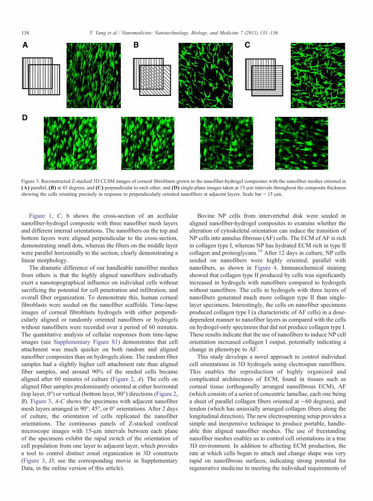
Figure 3. Reconstructed Z-stacked 3D CLSM images of corneal fibroblasts grown in the nanofiber-hydrogel composites with the nanofiber meshes oriented in(A) parallel, (B) at 45 degrees, and (C) perpendicular to each other; and (D) single-plane images taken at 15-μm intervals throughout the composite thicknessshowing the cells orienting precisely in response to perpendicularly oriented nanofibers at adjacent layers. Scale bar = 15 μm.
134 Y. Yang et al / Nanomedicine: Nanotechnology, Biology, and Medicine 7 (2011) 131–136
Figure 1, C, b shows the cross-section of an acellularnanofiber-hydrogel composite with three nanofiber mesh layersand different internal orientations. The nanofibers on the top andbottom layers were aligned perpendicular to the cross-section,demonstrating small dots, whereas the fibers on the middle layerwere parallel horizontally to the section, clearly demonstrating alinear morphology.
The dramatic difference of our handleable nanofiber meshesfrom others is that the highly aligned nanofibers individuallyexert a nanotopographical influence on individual cells withoutsacrificing the potential for cell penetration and infiltration, andoverall fiber organization. To demonstrate this, human cornealfibroblasts were seeded on the nanofiber scaffolds. Time-lapseimages of corneal fibroblasts hydrogels with either perpendi-cularly aligned or randomly oriented nanofibers or hydrogelswithout nanofibers were recorded over a period of 60 minutes.The quantitative analysis of cellular responses from time-lapseimages (see Supplementary Figure S1) demonstrates that cellattachment was much quicker on both random and alignednanofiber composites than on hydrogels alone. The random fibersamples had a slightly higher cell attachment rate than alignedfiber samples, and around 90% of the seeded cells becamealigned after 60 minutes of culture (Figure 2, A). The cells onaligned fiber samples predominantly oriented at either horizontal(top layer, 0°) or vertical (bottom layer, 90°) directions (Figure 2,B). Figure 3, A-C shows the specimens with adjacent nanofibermesh layers arranged in 90°, 45°, or 0° orientations. After 2 daysof culture, the orientation of cells replicated the nanofiberorientations. The continuous panels of Z-stacked confocalmicroscope images with 15-μm intervals between each planeof the specimens exhibit the rapid switch of the orientation ofcell population from one layer to adjacent layer, which providesa tool to control distinct zonal organization in 3D constructs(Figure 3, D; see the corresponding movie in SupplementaryData, in the online version of this article).
Bovine NP cells from intervertebal disk were seeded inaligned nanofiber-hydrogel composites to examine whether thealteration of cytoskeletal orientation can induce the transition ofNP cells into annulus fibrosus (AF) cells. The ECM of AF is richin collagen type I, whereas NP has hydrated ECM rich in type IIcollagen and proteoglycans.10 After 12 days in culture, NP cellsseeded on nanofibers were highly oriented, parallel withnanofibers, as shown in Figure 4. Immunochemical stainingshowed that collagen type II produced by cells was significantlyincreased in hydrogels with nanofibers compared to hydrogelswithout nanofibers. The cells in hydrogels with three layers ofnanofibers generated much more collagen type II than single-layer specimens. Interestingly, the cells on nanofiber specimensproduced collagen type I (a characteristic of AF cells) in a dose-dependent manner to nanofiber layers as compared with the cellson hydrogel-only specimens that did not produce collagen type I.These results indicate that the use of nanofibers to induce NP cellorientation increased collagen I output, potentially indicating achange in phenotype to AF.
This study develops a novel approach to control individualcell orientations in 3D hydrogels using electrospun nanofibers.This enables the reproduction of highly organized andcomplicated architectures of ECM, found in tissues such ascorneal tissue (orthogonally arranged nanofibrous ECM), AF(which consists of a series of concentric lamellae, each one beinga sheet of parallel collagen fibers oriented at ∼60 degrees), andtendon (which has uniaxially arranged collagen fibers along thelongitudinal direction). The new electrospinning setup provides asimple and inexpensive technique to produce portable, handle-able thin aligned nanofiber meshes. The use of freestandingnanofiber meshes enables us to control cell orientations in a true3D environment. In addition to affecting ECM production, therate at which cells began to attach and change shape was veryrapid on nanofibrous surfaces, indicating strong potential forregenerative medicine in meeting the individual requirements of

Figure 4. NP cells in nanofiber-hydrogel composites immunochemically stained for collagen I and II expression (tagged with fluorescein isothiocyanate) andcounterstained for F-actin (phalloidin-TRITC). (A) CLSM images. Top two panels, hydrogel control with cells; bottom two panels, three layers of alignednanofibers in a hydrogel composite, with cells (scale bar = 50 μm. (B) Quantitative analysis of the protein expression. * indicates a significant difference inaverage colour intensity ratio between groups, P b 0.05.
135Y. Yang et al / Nanomedicine: Nanotechnology, Biology, and Medicine 7 (2011) 131–136
specific tissues. These composites demonstrate the massivepotential for mimicking complex tissues, with layered control ofcellular orientations and appropriate synthesis of matrix proteins.
Acknowledgments
We are grateful to Professor Sally Roberts for providingdisc cells.
Appendix A. Supplementary data
Supplementary materials related to this article can be foundonline at doi:10.1016/j.nano.2010.12.011.
References
1. den Braber ET, de Ruijter JE, Ginsel LA, von Recum AF, Jansen JA.Orientation of ECM protein deposition, fibroblast cytoskeleton, and

136 Y. Yang et al / Nanomedicine: Nanotechnology, Biology, and Medicine 7 (2011) 131–136
attachment complex components on silicone microgrooved surface.J Biomed Mater Res 1998;40:291-300.
2. Mitchell D, McCartney MD, Buck RC. Comparison of the degree ofcontact guidance between tumor cells and normal cells in vitro. CancerRes 1981;41:3046-51.
3. Dalby MJ, Yarwood SJ, Riehle MO, Johnstone HJH, Affrossman S,Curtis ASG. Increasing fibroblast response to materials using nanotopo-graphy: morphological and genetic measurements of cell response to13-nm-high polymer demixed islands. Exp Cell Res 2002;276:1-9.
4. Wise JK, Yarin AL, Megaridis CM, Cho M. Chondrogenic differenti-ation of human mesenchymal stem cells on oriented nanofibrousscaffolds: engineering the superficial zone of articular cartilage. TissueEng Part A 2009;15:913-21.
5. Xie J, MacEwan MR, Li X, Sakiyama-Elbert SE, Xia Y. Neuriteoutgrowth on nanofiber scaffolds with different orders, structures, andsurface properties. ACS Nano 2009;3:1151-9.
6. Moroni L, Schotel R, Hamann D, deWijn J, van Blitterswijk C. 3D fiber-deposited electrospun integrated scaffolds enhance cartilage tissueformation. Adv Funct Mater 2008;18:53-60.
7. Guimarães A, Martins A, Pinho ED, Faria S, Reis RL, Neves NM.Solving cell infiltration limitations of electrospun nanofiber meshesfor tissue engineering applications. Nanomedicine (Lond) 2010;5:539-54.
8. Baker BM, Nathan AS, Gee AO, Mauck RL. The influence of an alignednanofibrous topography on human mesenchymal stem cell fibrochon-drogenesis. Biomaterials 2010;31:6190-200.
9. Ahearne M, Yang Y, El Haj AJ, Then KY, Liu KK. Characterizing theviscoelastic properties of thin hydrogel-based constructs for tissueengineering applications. J R Soc Interface 2005;2:455-63.
10. Hayes AJ, Benjamin M, Ralphs JR. Roles of actin stress fibres in thedevelopment of the intervertebral disc: cytoskeletal control of extracel-lular matrix assembly. Dev Dyn 1999;215:179-89.