Embed Size (px)
Citation preview
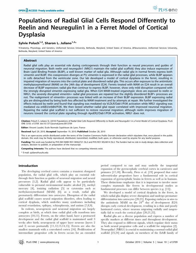
Populations of Radial Glial Cells Respond Differently toReelin and Neuregulin1 in a Ferret Model of CorticalDysplasiaSylvie Poluch1,2, Sharon L. Juliano1,2*
1 Anatomy, Physiology, and Genetics, Uniformed Services University, Bethesda, Maryland, United States of America, 2 Neuroscience, Uniformed Services University,
Bethesda, Maryland, United States of America
Abstract
Radial glial cells play an essential role during corticogenesis through their function as neural precursors and guides ofneuronal migration. Both reelin and neuregulin1 (NRG1) maintain the radial glial scaffold; they also induce expression ofBrain Lipid Binding Protein (BLBP), a well known marker of radial glia. Although radial glia in normal ferrets express bothvimentin and BLBP, this coexpression diverges at P3; vimentin is expressed in the radial glial processes, while BLBP appearsin cells detached from the ventricular zone. Our lab developed a model of cortical dysplasia in the ferret, resulting inimpaired migration of neurons into the cortical plate and disordered radial glia. This occurs after exposure to the antimitoticmethylazoxymethanol (MAM) on the 24th day of development (E24). Ferrets treated with MAM on E24 result in an overalldecrease of BLBP expression; radial glia that continue to express BLBP, however, show only mild disruption compared withthe strongly disrupted vimentin expressing radial glia. When E24 MAM-treated organotypic slices are exposed to reelin orNRG1, the severely disrupted vimentin+ radial glial processes are repaired but the slightly disordered BLBP+ processes arenot. The realignment of vimentin+ processes was linked with an increase of their BLBP expression. BLBP expressing radialglia are distinguished by being both less affected by MAM treatment and by attempts at repair. We further investigated theeffects induced by reelin and found that signaling was mediated via VLDLR/Dab1/Pi3K activation while NRG1 signaling wasmediated via erbB3/erbB4/Pi3K. We then tested whether radial glial repair correlated with improved neuronal migration.Repairing the radial glial scaffold is not sufficient to restore neuronal migration; although reelin improves migration ofneurons toward the cortical plate signaling through ApoER2/Dab1/PI3K activation, NRG1 does not.
Citation: Poluch S, Juliano SL (2010) Populations of Radial Glial Cells Respond Differently to Reelin and Neuregulin1 in a Ferret Model of Cortical Dysplasia. PLoSONE 5(10): e13709. doi:10.1371/journal.pone.0013709
Editor: Vincent Laudet, Ecole Normale Superieure de Lyon, France
Received April 29, 2010; Accepted September 19, 2010; Published October 28, 2010
This is an open-access article distributed under the terms of the Creative Commons Public Domain declaration which stipulates that, once placed in the publicdomain, this work may be freely reproduced, distributed, transmitted, modified, built upon, or otherwise used by anyone for any lawful purpose.
Funding: This work was funded by PHS NIH NIMH RO1 MH62721 (SLJ) and PHS RO1 NS24014 (SLJ). The funders had no role in study design, data collection andanalysis, decision to publish, or preparation of the manuscript.
Competing Interests: The authors have declared that no competing interests exist.
* E-mail: [email protected]
Introduction
The developing cerebral cortex contains a transient elongated
population, the radial glial cells, which play an essential role
through their function as guides of neuronal migration and neural
precursors [1,2]. Radial glial cells appear to be particularly
vulnerable to prenatal environmental insults: alcohol [3], methyl
mercury [4], ionizing radiation [5] or cytotoxins such as
methylazoxymethanol (MAM) [6]; as a result, radial glia
prematurely differentiate into astrocytes. Disruption of the radial
glial scaffold causes neural migration disorders, often leading to
cortical dysplasia, which underlies many syndromes including
mental retardation, epilepsy, schizophrenia, and autism [7,8,9].
In rodents, neurogenesis and neuronal migration are largely
complete at birth, at which time radial glial cells transform into
astrocytes [10,11]. Ferrets, on the other hand, have a protracted
development and the radial glial scaffold is maintained until 3
weeks after birth; neurogenesis of upper layer neurons (layers 2
and 3) occurs postnatally [11,12,13,14,15]. Ferrets are also the
smallest mammals with a convoluted cortex [16]. Proliferation of
intermediate progenitor cells in ferrets occurs for an extended
period compared to rats and may underlie the tangential
expansion of the gyrencephalic cerebral cortex in carnivores and
primates [17,18]. Recently, Fietz et al. [19] proposed that outer
subventricular progenitors have a fundamental role in cortical
expansion of gyrencephalic brains in ferrets as well as in humans.
These distinctions emphasize that it is important to involve more
complex mammals like ferrets in developmental studies as
fundamental processes can differ between species (e.g. [15]).
We developed a model of cortical dysplasia in the ferret, in
which radial glia display severe disruption and undergo premature
differentiation into astrocytes [20,21]. Exposing embryos in utero to
the antimitotic MAM on the 24th day of development (E24)
disrupts early cortical development, resulting in a thin and poorly
laminated cortex, where neurons migrating radially and tangen-
tially fail to reach the cortical plate [20,21,22].
Radial glia are a diverse population and express a number of
specific markers at different times and throughout development.
They also respond to different signals, which regulate their status
as both neural progenitor cells and a scaffold for migration [2].
Neuregulin1 (NRG1) is crucial to maintaining a normal radial glial
scaffold [23,24] and signals via members of the ErbB family of
PLoS ONE | www.plosone.org 1 October 2010 | Volume 5 | Issue 10 | e13709
Report Documentation Page Form ApprovedOMB No. 0704-0188
Public reporting burden for the collection of information is estimated to average 1 hour per response, including the time for reviewing instructions, searching existing data sources, gathering andmaintaining the data needed, and completing and reviewing the collection of information. Send comments regarding this burden estimate or any other aspect of this collection of information,including suggestions for reducing this burden, to Washington Headquarters Services, Directorate for Information Operations and Reports, 1215 Jefferson Davis Highway, Suite 1204, ArlingtonVA 22202-4302. Respondents should be aware that notwithstanding any other provision of law, no person shall be subject to a penalty for failing to comply with a collection of information if itdoes not display a currently valid OMB control number.
1. REPORT DATE APR 2010 2. REPORT TYPE
3. DATES COVERED 00-00-2010 to 00-00-2010
4. TITLE AND SUBTITLE Populations of Radial Glial Cells Respond Differently to Reelin andNeuregulin1 in a Ferret Model of Cortical Dysplasia
5a. CONTRACT NUMBER
5b. GRANT NUMBER
5c. PROGRAM ELEMENT NUMBER
6. AUTHOR(S) 5d. PROJECT NUMBER
5e. TASK NUMBER
5f. WORK UNIT NUMBER
7. PERFORMING ORGANIZATION NAME(S) AND ADDRESS(ES) Uniformed Services University,Anatomy, Physiology, and Genetics,Bethesda,MD,20814
8. PERFORMING ORGANIZATIONREPORT NUMBER
9. SPONSORING/MONITORING AGENCY NAME(S) AND ADDRESS(ES) 10. SPONSOR/MONITOR’S ACRONYM(S)
11. SPONSOR/MONITOR’S REPORT NUMBER(S)
12. DISTRIBUTION/AVAILABILITY STATEMENT Approved for public release; distribution unlimited
13. SUPPLEMENTARY NOTES
14. ABSTRACT Radial glial cells play an essential role during corticogenesis through their function as neural precursorsand guides of neuronal migration. Both reelin and neuregulin1 (NRG1) maintain the radial glial scaffold;they also induce expression of Brain Lipid Binding Protein (BLBP), a well known marker of radial glia.Although radial glia in normal ferrets express both vimentin and BLBP, this coexpression diverges at P3;vimentin is expressed in the radial glial processes, while BLBP appears in cells detached from theventricular zone. Our lab developed a model of cortical dysplasia in the ferret, resulting in impairedmigration of neurons into the cortical plate and disordered radial glia. This occurs after exposure to theantimitotic methylazoxymethanol (MAM) on the 24th day of development (E24). Ferrets treated withMAM on E24 result in an overall decrease of BLBP expression; radial glia that continue to express BLBP,however, show only mild disruption compared with the strongly disrupted vimentin expressing radial glia.When E24 MAM-treated organotypic slices are exposed to reelin or NRG1, the severely disruptedvimentin+ radial glial processes are repaired but the slightly disordered BLBP+ processes are not. Therealignment of vimentin+ processes was linked with an increase of their BLBP expression. BLBPexpressing radial glia are distinguished by being both less affected by MAM treatment and by attempts atrepair. We further investigated the effects induced by reelin and found that signaling was mediated viaVLDLR/Dab1/Pi3K activation while NRG1 signaling was mediated via erbB3/erbB4/Pi3K. We then testedwhether radial glial repair correlated with improved neuronal migration. Repairing the radial glialscaffold is not sufficient to restore neuronal migration; although reelin improves migration of neuronstoward the cortical plate signaling through ApoER2/Dab1/PI3K activation, NRG1 does not.
15. SUBJECT TERMS

16. SECURITY CLASSIFICATION OF: 17. LIMITATION OF ABSTRACT Same as
Report (SAR)
18. NUMBEROF PAGES
17
19a. NAME OFRESPONSIBLE PERSON
a. REPORT unclassified
b. ABSTRACT unclassified
c. THIS PAGE unclassified
Standard Form 298 (Rev. 8-98) Prescribed by ANSI Std Z39-18
receptor tyrosine kinases [25,26,27]. Radial glial disruption in E24
MAM treated cortex is likely to be caused in part by reduction of
NRG1, because exogenous replacement results in realignment in
E24 MAM treated organotypic slices [28].
Reelin is another key protein active during cortical development
as the lack of reelin results in aberrant migration of cortical
neurons and misaligned radial glial cells [29,30,31,32]. Exogenous
reelin promotes radial glial extension and rescues the radial glial
scaffold in reeler hippocampus [33]. Reelin signaling requires
binding to receptors of the lipoprotein family, very low density-
lipoprotein (VLDLR) and the apolipoprotein E receptor
(ApoER2), which triggers tyrosine phosphorylation of the
cytoplasmic adapter protein Disabled-1 (Dab1) [34,35,36]. Dab1
is expressed in cortical neurons [37] as well as in radial glial cells
[38]. In E24 MAM treated ferrets an exogenous source of reelin
secreted at the pial surface improves neuronal migration as well as
radial glial morphology [39].
In addition to radial process extension, reelin and NRG1 promote
expression of Brain Lipid Binding Protein (BLBP) in cortical radial
glia [40,41,32]. Although the function of BLBP during cortical
development is not fully understood, BLBP expression strongly
correlates with the migration of neurons along the radial glia [21].
BLBP appears to be required for radial process elongation, since the
addition of anti-BLBP antibodies inhibit this process [42].
We show here that in normal newborn ferrets, vimentin and
BLBP are strongly expressed in radial glia. In our ferret model of
cortical dysplasia, the expression of BLBP in radial glia is
decreased after MAM treatment; however the remaining BLBP+radial glial cells are relatively spared from disruption compared
with the severely disorganized vimentin+ cells. Both reelin and
NRG1 realign the disorganized vimentin+ radial glial cells.
Although the morphology of BLBP+ cells was not improved from
their mild disruption after these treatments, the expression of
BLBP was increased. This suggests that at least two distinct
populations of radial glial cells exist in ferrets that respond
differently to damage and attempts at repair. Exogenous reelin
improves not only the radial glial scaffold but also radial migration
toward the cortical plate whereas NRG1 has no effect on neuronal
migration. In addition, distinct signaling elements appear to
initiate movement out of the ventricular zone, but do not play a
role in allowing further movement toward the cortical plate.
Materials and Methods
Ethics StatementThe use of animals and the methods of this study were approved by
the Institutional Animal Care and Use Committee (IACUC) at
USUHS and under Animal Welfare Assurance number A3448-01.
The experiments were performed at an AAALAC accredited institute.
AnimalsTimed pregnant ferrets (Mustella putorius) were purchased from
Marshall Farms (New Rose, NY); ferret kits are born after 41 days
of gestation. Pregnant ferrets, anaesthetized with isofluorane using
a mask (5%), were injected intraperitoneally (IP) with methylazoxy
methanol acetate (MAM, Midwest Research Institute, Kansas
City, MO, 14 mg/kg) diluted in a sterile saline buffer. Normal and
MAM treated fetuses at E27, E33, or E38-E40 were obtained by
caesarean section under sterile conditions using isofluorane
anesthesia under the supervision of a veterinarian. We also used
normal and MAM treated newborn kits (postnatal day 0, P0), as
well as normal P3, P14, and P28 normal ferrets, which were
anesthetized with an IP injection of pentobarbital sodium (50 mg/
kg) prior to brain removal.
Organotypic cultureBrains obtained from E39-E40 embryos were cut under sterile
conditions into 400 mm thick coronal slices using a tissue chopper
(Stoelting, Wood Dale, IL). During the dissection, brains and slices
were perfused with cold, oxygenated artificial cerebrospinal fluid
(containing in mM: CaCl2 2.4, KCl 3.2, MgSO4 1.2, NaCl 124,
NaHCO3 26, NaH2PO4 1.2, glucose 10). Coronal cortical slices
containing the somatosensory cortex [13,20] were placed on
inserts (Millipore, Bedford, MA) in 6-well plates using MEM
medium (Gibco, Carlsbad, CA) containing 10% decomplemented
horse serum (Gibco) and 4% G1,2 solution (0.5 mg/mL
gentamycin, 15% glucose, 50 mM L-glutamine). A number of
organotypic slices were incubated for 1 hour in medium
supplemented with BrdU (100 mg/ml), which was removed and
then replaced with fresh medium. After 2 days in culture (DIC) in
an incubator (95% CO2; 37uC), the organotypic slices were fixed
for 2 hours by immersion in 4% phosphate buffered paraformal-
dehyde. In some cases, fixed slices were also cryoprotected and
subsequently re-sectioned at 14 mm using a cryostat.
Coculture of organotypic ferret slices with HEK cellsA number of MAM treated slices were co-cultured with: (1)
HEK 293T cells, transfected with the mouse reelin cDNA
construct pCrl [43], which produces and secretes the full length
reelin protein [44,39], (2) HEK 293T cells, transfected with the
type I NRG1 encoding plasmid (NRG1-Ig), which secretes the full
length of the isoform type I NRG1 [45] and (3) HEK 293T cells,
transfected with the type III NRG1 (NRG1-CRD), which express
a non-secreted/membrane type III NRG1 [45]. Prior to the
coculture, HEK 293T cells were cultured in Dulbecco’s modified
Eagle’s medium (MEDIATECH Inc., Herndon, VA) (control
HEK cells) supplemented with Geneticin (G418, 0.5 mg/ml)
(reelin+, NRG1-Ig, or NRG1-CRD HEK cells). The HEK cells
were placed in Matrigel (BD Biosciences, Bedford, MA) and
positioned next to the pial surface [39].
Drugs and chronic treatmentsIn some experiments, the medium was supplemented with
recombinant mouse reelin (1 nM, US Biological, Swampscott,
MA) corresponding to the central fragment. We also used
recombinant NRG1 (1 nM, R&D systems, Minneapolis, MN)
obtained from the Human DNA sequence encoding the EGF
domain of NRG1 b1. To further understand the effects mediated
by reelin or NRG1, the culture medium was complemented with
pathway inhibitors such as: LY294002 (inhibitor of PI3K; 50 mM,
Calbiochem, San Diego, CA), PP2 (a Src kinases inhibitor; 10 mM,
Calbiochem), TDZD-8 (a GSK-3b Inhibitor I, 56 mM, Calbio-
chem), or SP600125 (a JNK Inhibitor II, 10 mM, Calbiochem). To
block ApoER2 and VLDLR, we used human recombinant RAP
(300 nM, Calbiochem). NRG1 signaling was inhibited by using
blocking antibodies for NRG1 receptors, erbB-3 or erbB-4 (20 mg/ml,
LabVision, Fremont, CA) (See Figure S1).
ImmunohistochemistryFor fluorescence immunocytochemistry, slices were incubated
overnight at 4uC with mouse IgG monoclonal antibodies against:
vimentin clone V9 (1/100, Sigma), MAP2abc (1/200, Sigma)
or rabbit polyclonal antibodies against: BLBP (1/300, Che-
micon and Abcam) and GABA (1/300, Sigma). After washes in
PBS, the corresponding secondary antibodies were used (anti-
rabbit, or anti-mouse Alexa-488 or Alexa-546, 1/200, Molecular
Probes). The sections were washed in PBS and mounted in
Mowiol.
Cortical Dysplasia and Repair
PLoS ONE | www.plosone.org 2 October 2010 | Volume 5 | Issue 10 | e13709
BrdU immunoreactivityThe fixed slices were placed in 70% cold ethanol for 10 minutes
at 4uC, followed by 1 hour in 2N HCl at 37uC. Slices were then
placed in borate buffer (pH 8.5) and washed in PBS. The following
antibodies were used: anti-rat BrdU (1/100, Becton Dickinson,
Franklin Lakes, NJ) and goat anti-rat IgG conjugated with CY2
(1/200, Jackson ImmunoResearch West Grove, PA) or goat anti-
rat IgG Alexa-488 (1/200, Molecular Probes).
Quantification of BrdU immunoreactive cellsTo determine the ability of cells to migrate in organotypic
cultures of either normal, E24 MAM treated cortex alone, or after
coculture with HEK 293T cells embedded in Matrigel (as describe
above), we plotted the distribution of BrdU+ cells after 2 DIC.
Boundaries were drawn indicating the pia of a ferret slice and the
outer edge of the VZ. This region was divided into 3 equal bins for
each coculture and the number of cells per bin counted in a slab
500 mm in width in the somatosensory cortex. The bins
correspond to the intermediate zone close to the VZ (i.e., the
lowest part of the intermediate zone, IZL), a region in the IZ, but
closer to the cortical plate (i.e., the upper part of the intermediate
zone, IZU), and the region corresponding to the cortical plate (CP)
(Figure 5c–d and Figure 7c–d). Histograms were made to indicate
the position of BrdU+ cells across animals in each condition. To
compare across samples, the number of cells/bin were calculated
as a percent of the total number of cells in each slice.
Quantification of radial glial morphology and phenotypeMAM treatment leads to early radial glial differentiation, which
can be reversed by treatment with exogenous NRG1 or reelin
[28,39]. To quantify the change in morphology, treated and
untreated E24 MAM slices were double labeled for 2 specific
radial glial markers, BLBP and vimentin. All data were collected
from the somatosensory cortex. The angle of deviation for each
marker was measured as described previously [21,28] using Image
Tool (UTHSCSA, San Antonio, Texas). To determine the
phenotype of radial glial cells in normal or MAM treated cortex,
the number of processes expressing vimentin as well as BLBP, or
only vimentin, or only BLBP was computed. Since radial glial cells
can have several vertical processes, the data refer to radial glial
processes and not radial glial cells. The result is expressed as a
percentage of processes vimentin+ BLBP+ or BLBP- vimentin+ or
vimentin- BLBP+. We used a 25X objective on a microscope
equipped with an Apotome to acquire multiple z-stack images (at
least 5 z-sections, with ,5 mm interval), which were collapsed into
a single image; the degrees of deviation and the phenotype of
radial glial processes were measured in a 250 mm2 zone within the
cortical plate. On average, this zone contains 57 radial glial
processes.
Statistical AnalysisA total of 115 MAM treated embryos were obtained from 16
pregnant ferrets and 27 normal ferret embryos or kits obtained from
13 pregnant ferrets. All data are obtained from at least
two independent experiments from different litters. For all data,
a 2 way ANOVA was conducted followed by a Holm-Sidak
test for comparisons between groups. Statistical analyses were
performed using SigmaStat (Systat Software, Inc, Chicago, Illinois).
Image AcquisitionFor the acquisition of fluorescence images, we used an Axiovert
200 microscope (Zeiss) equipped with an Apotome and Axio-
vision 4.7.
Results
Radial Glial phenotype in embryonic and postnataldevelopment in normal ferret
Vimentin is an excellent marker for radial glia in ferrets, but few
others have been explored in this species [11,14]. We also know
that the radial glial phenotype differs among mammals. To further
understand the relevant proteins/intermediate filaments expressed
in ferret neocortex, we tested several other markers and observed
that BLPB was strongly expressed. To expand our assessment of
diversity among radial glia in normal ferret, we used immuno-
staining against both vimentin and BLBP during embryonic and
postnatal development. Vimentin is expressed early, since it labels
radial glial processes throughout the initial, mid, and final stages of
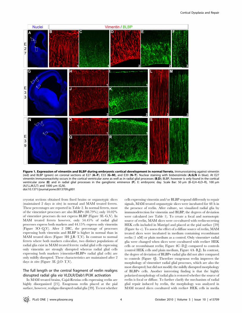
corticogenesis in ferrets (E27, E33 and E39) (Figure 1). BLBP is
also strongly expressed at E27 in the VZ, but not in radial
processes (Figure 1C–E); whereas in the ganglionic eminence,
radial processes express BLBP (Figure 1F). From E33 to E39 and
P0, vimentin and BLBP colocolize in radial glial processes
(Figure 1L–M,S–T). Elongated radial glia immunoreactive for
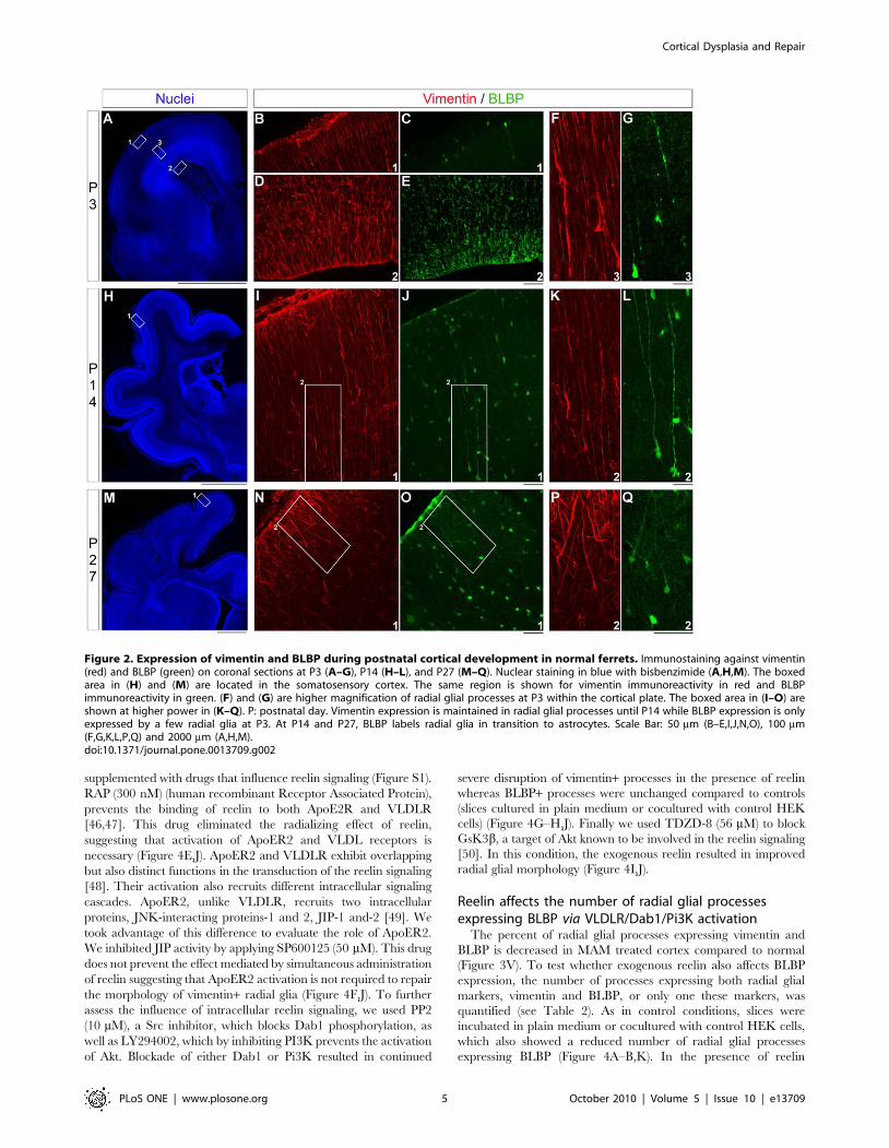
vimentin are present from P3 to P14 (Figure 2F,K). At P3, BLBP
immunoreactivity decreases in the VZ, and few BLBP+ cells are
observed close to the pia (Figure 2C–E,G). These cells, also seen at
P14, express vimentin and show an elongated process oriented
toward the pia; interestingly, their cell bodies are in the cortical
plate (Figure 2H,L). Four weeks after birth (P27), vimentin and
BLBP label only radial glia in transition to astrocytes as shown
here in the somatosensory cortex (Figure 2M–Q).
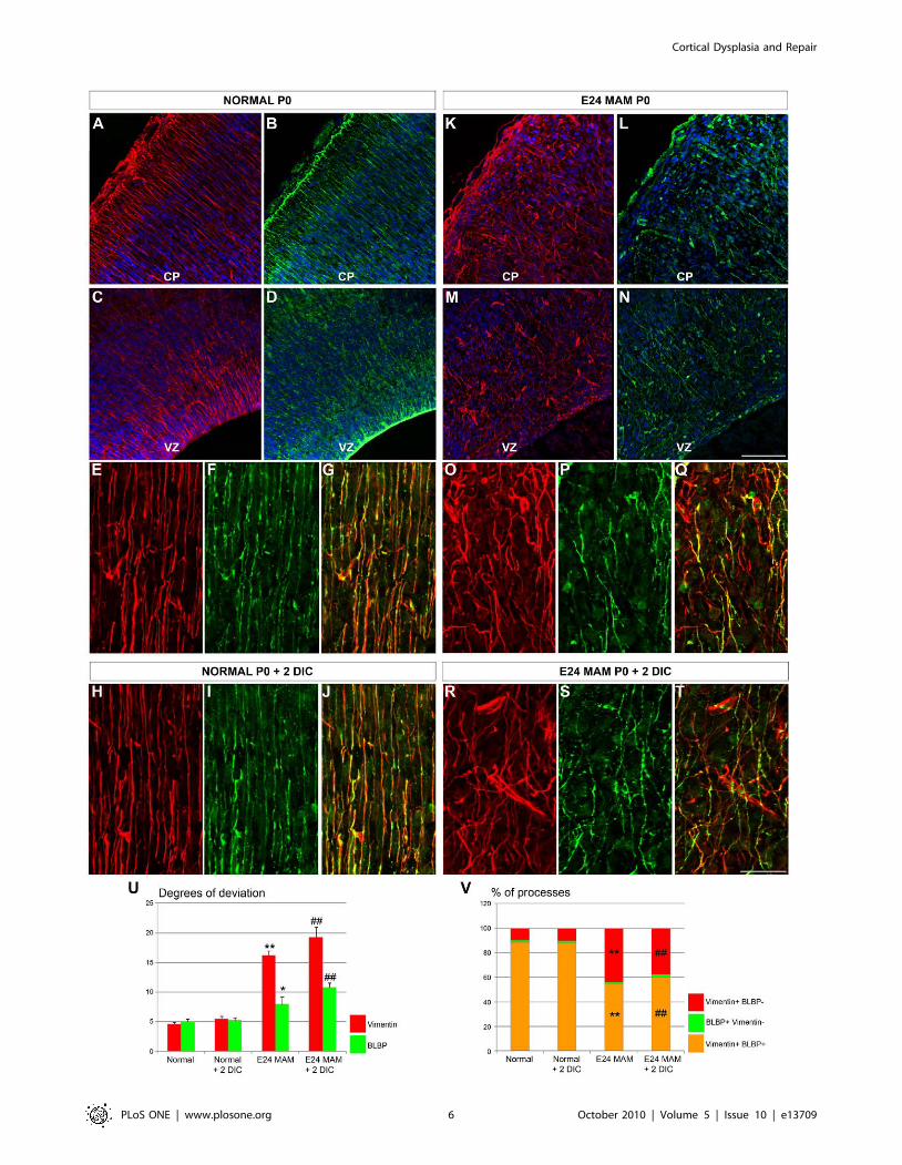
Radial glial morphology is affected in MAM treated ferretsExposure to MAM at E24 leads to a severely disrupted radial
glial scaffold [14,21]. To assess whether all radial glial cells were
disrupted, we compared the morphology of vimentin and BLBP
immunoreactive radial glia in normal and MAM treated ferrets
(E39-E40 or P0). These two markers and their colocalization were
detected immunohistochemically in 20 mm thick sections or in
organotypic slices maintained 2 DIC. As previously shown
(Figure 1) in normal ferrets, vimentin+ and BLBP+ radial glia
are elongated, parallel, and extend toward the pial surface, as
opposed to an obviously disrupted appearance in E24 MAM-
treated cortex (Figure 3A–T). To quantify radial glial morphology
in normal and MAM treated brains, we calculated the degree of
deviation of radial glial processes as described previously
[21,28,39]. A low degree of deviation indicates that radial glia
are elongated and parallel whereas a higher degree of deviation
reveals disrupted and misaligned cells. Degrees of deviation are
reported in Table 1. In normal ferrets, vimentin+ and BLBP+radial glia display a low degree of deviation (Figure 3E–G,U). In
MAM treated ferrets, a high degree of deviation occurs in
vimentin+ radial glia (Figure 3O–Q,U). BLBP+ radial glia are
substantially less disrupted after MAM treatment compared to the
vimentin+ population (Figure 3E–F,O–P). Although the degree of
deviation for BLBP+ radial glia is lower, it is significantly different
from normal ferrets (p = 0.013; Figure 3F,I,P,S). The degree of
deviation of radial glial processes (vimentin+ and BLBP+) observed
in newborn ferrets is maintained after 2 days in vitro (in plain
medium) compared with acute sections obtained from normal and
E24 MAM treated ferrets (Figure 3H–T,U).
Downregulation of BLBP in MAM treated ferret radial glialprocesses
Are vimentin and BLBP expression quantitatively changed? To
answer this question, the proportion of radial glial processes single
or double-labeled for vimentin and BLBP was determined on
Cortical Dysplasia and Repair
PLoS ONE | www.plosone.org 3 October 2010 | Volume 5 | Issue 10 | e13709
cryostat sections obtained from fixed brains or organotypic slices
(maintained 2 days in vitro) in normal and MAM treated ferrets.
These percentages are reported in Table 2. In normal ferrets, most
of the vimentin+ processes are also BLBP+ (88.79%); only 10.02%
of vimentin+ processes do not express BLBP (Figure 3E–G,V). In
MAM treated ferrets however, only 54.45% of radial glial
processes express both markers and 44.15% express only vimentin
(Figure 3O–Q,V). After 2 DIC, the percentage of processes
expressing both vimentin and BLBP is higher in normal than in
MAM treated slices (Figure 3H–J,R–T,V). In contrast to normal
ferrets where both markers colocalize, two distinct populations of
radial glia exist in MAM treated ferrets: radial glial cells expressing
only vimentin are strongly disrupted whereas radial glial cells
expressing both markers (vimentin+BLBP+ radial glial cells) are
only mildly disrupted. These characteristics are maintained after 2
days in vitro (Figure 3E–J,O–T,V).
The full length or the central fragment of reelin realignsdisrupted radial glia via VLDLR/Dab1/Pi3K activation
In MAM treated brains, Cajal-Retzius cells expressing reelin are
highly disorganized [21]. Exogenous reelin placed at the pial
surface, however, realigns disrupted radial glia [39]. To test whether
cells expressing vimentin and/or BLBP respond differently to repair
signals, MAM treated organotypic slices were incubated for 48 h in
the presence of reelin. After culture, we visualized radial glia by
immunodetection for vimentin and BLBP; the degrees of deviation
were calculated (see Table 1). To create a focal and normotopic
source of reelin, MAM slices were co-cultured with reelin-secreting
HEK cells included in Matrigel and placed at the pial surface [39]
(Figure 4a–c). To assess the effect of a diffuse source of reelin, MAM
treated slices were incubated in medium containing recombinant
reelin (1 nM) or plain medium as a control. Only vimentin+ radial
glia were changed when slices were cocultured with reelin+ HEK
cells or recombinant reelin; Figure 4C–D,J) compared to controls
(control HEK cells and plain medium, Figure 4A–B,J). In contrast,
the degree of deviation of BLBP+ radial glia did not alter compared
to controls (Figure 4J). Therefore exogenous reelin improves the
morphology of vimentin+ radial glial processes, which are also the
most disrupted; but did not modify the mildly disrupted morphology
of BLBP+ cells. Another interesting finding is that the highly
polarized morphology of radial glia is restored whether the source of
reelin is focal or diffuse. To further clarify the mechanism of radial
glial repair induced by reelin, the morphology was analyzed in
MAM treated slices cocultured with reelin+ HEK cells in media
Figure 1. Expression of vimentin and BLBP during embryonic cortical development in normal ferrets. Immunostaining against vimentin(red) and BLBP (green) on coronal sections at E27 (A–F), E33 (G–M), and E39 (N–T). Nuclear staining with bisbenzimide (A,G,N in blue). At E27vimentin immunoreactivity occurs in the cortical ventricular zone as well as in radial glial processes (B,D); BLBP, however is only found in the corticalventricular zone (E) and in radial glial processes in the ganglionic eminence (F). E: embryonic day. Scale Bar: 50 mm (B–E,H–K,O–R), 100 mm(A,F,L,M,S,T) and 1000 mm (G,N).doi:10.1371/journal.pone.0013709.g001
Cortical Dysplasia and Repair
PLoS ONE | www.plosone.org 4 October 2010 | Volume 5 | Issue 10 | e13709
supplemented with drugs that influence reelin signaling (Figure S1).
RAP (300 nM) (human recombinant Receptor Associated Protein),
prevents the binding of reelin to both ApoE2R and VLDLR
[46,47]. This drug eliminated the radializing effect of reelin,
suggesting that activation of ApoER2 and VLDL receptors is
necessary (Figure 4E,J). ApoER2 and VLDLR exhibit overlapping
but also distinct functions in the transduction of the reelin signaling
[48]. Their activation also recruits different intracellular signaling
cascades. ApoER2, unlike VLDLR, recruits two intracellular
proteins, JNK-interacting proteins-1 and 2, JIP-1 and-2 [49]. We
took advantage of this difference to evaluate the role of ApoER2.
We inhibited JIP activity by applying SP600125 (50 mM). This drug
does not prevent the effect mediated by simultaneous administration
of reelin suggesting that ApoER2 activation is not required to repair
the morphology of vimentin+ radial glia (Figure 4F,J). To further
assess the influence of intracellular reelin signaling, we used PP2
(10 mM), a Src inhibitor, which blocks Dab1 phosphorylation, as
well as LY294002, which by inhibiting PI3K prevents the activation
of Akt. Blockade of either Dab1 or Pi3K resulted in continued
severe disruption of vimentin+ processes in the presence of reelin
whereas BLBP+ processes were unchanged compared to controls
(slices cultured in plain medium or cocultured with control HEK
cells) (Figure 4G–H,J). Finally we used TDZD-8 (56 mM) to block
GsK3b, a target of Akt known to be involved in the reelin signaling
[50]. In this condition, the exogenous reelin resulted in improved
radial glial morphology (Figure 4I,J).
Reelin affects the number of radial glial processesexpressing BLBP via VLDLR/Dab1/Pi3K activation
The percent of radial glial processes expressing vimentin and
BLBP is decreased in MAM treated cortex compared to normal
(Figure 3V). To test whether exogenous reelin also affects BLBP
expression, the number of processes expressing both radial glial
markers, vimentin and BLBP, or only one these markers, was
quantified (see Table 2). As in control conditions, slices were
incubated in plain medium or cocultured with control HEK cells,
which also showed a reduced number of radial glial processes
expressing BLBP (Figure 4A–B,K). In the presence of reelin
Figure 2. Expression of vimentin and BLBP during postnatal cortical development in normal ferrets. Immunostaining against vimentin(red) and BLBP (green) on coronal sections at P3 (A–G), P14 (H–L), and P27 (M–Q). Nuclear staining in blue with bisbenzimide (A,H,M). The boxedarea in (H) and (M) are located in the somatosensory cortex. The same region is shown for vimentin immunoreactivity in red and BLBPimmunoreactivity in green. (F) and (G) are higher magnification of radial glial processes at P3 within the cortical plate. The boxed area in (I–O) areshown at higher power in (K–Q). P: postnatal day. Vimentin expression is maintained in radial glial processes until P14 while BLBP expression is onlyexpressed by a few radial glia at P3. At P14 and P27, BLBP labels radial glia in transition to astrocytes. Scale Bar: 50 mm (B–E,I,J,N,O), 100 mm(F,G,K,L,P,Q) and 2000 mm (A,H,M).doi:10.1371/journal.pone.0013709.g002
Cortical Dysplasia and Repair
PLoS ONE | www.plosone.org 5 October 2010 | Volume 5 | Issue 10 | e13709
Cortical Dysplasia and Repair
PLoS ONE | www.plosone.org 6 October 2010 | Volume 5 | Issue 10 | e13709
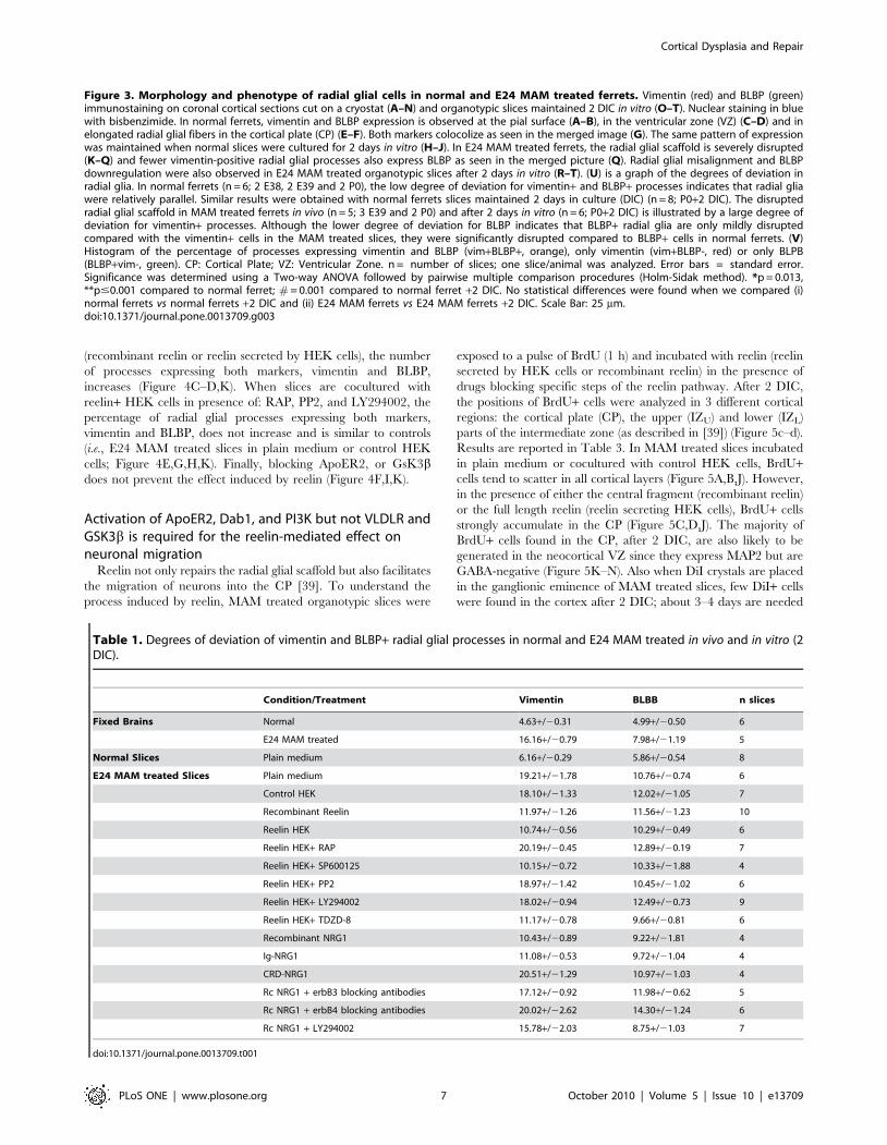
(recombinant reelin or reelin secreted by HEK cells), the number
of processes expressing both markers, vimentin and BLBP,
increases (Figure 4C–D,K). When slices are cocultured with
reelin+ HEK cells in presence of: RAP, PP2, and LY294002, the
percentage of radial glial processes expressing both markers,
vimentin and BLBP, does not increase and is similar to controls
(i.e., E24 MAM treated slices in plain medium or control HEK
cells; Figure 4E,G,H,K). Finally, blocking ApoER2, or GsK3bdoes not prevent the effect induced by reelin (Figure 4F,I,K).
Activation of ApoER2, Dab1, and PI3K but not VLDLR andGSK3b is required for the reelin-mediated effect onneuronal migration
Reelin not only repairs the radial glial scaffold but also facilitates
the migration of neurons into the CP [39]. To understand the
process induced by reelin, MAM treated organotypic slices were
exposed to a pulse of BrdU (1 h) and incubated with reelin (reelin
secreted by HEK cells or recombinant reelin) in the presence of
drugs blocking specific steps of the reelin pathway. After 2 DIC,
the positions of BrdU+ cells were analyzed in 3 different cortical
regions: the cortical plate (CP), the upper (IZU) and lower (IZL)
parts of the intermediate zone (as described in [39]) (Figure 5c–d).
Results are reported in Table 3. In MAM treated slices incubated
in plain medium or cocultured with control HEK cells, BrdU+cells tend to scatter in all cortical layers (Figure 5A,B,J). However,
in the presence of either the central fragment (recombinant reelin)
or the full length reelin (reelin secreting HEK cells), BrdU+ cells
strongly accumulate in the CP (Figure 5C,D,J). The majority of
BrdU+ cells found in the CP, after 2 DIC, are also likely to be
generated in the neocortical VZ since they express MAP2 but are
GABA-negative (Figure 5K–N). Also when DiI crystals are placed
in the ganglionic eminence of MAM treated slices, few DiI+ cells
were found in the cortex after 2 DIC; about 3–4 days are needed
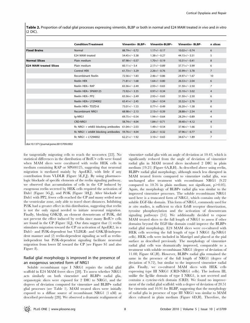
Table 1. Degrees of deviation of vimentin and BLBP+ radial glial processes in normal and E24 MAM treated in vivo and in vitro (2DIC).
Condition/Treatment Vimentin BLBB n slices
Fixed Brains Normal 4.63+/20.31 4.99+/20.50 6
E24 MAM treated 16.16+/20.79 7.98+/21.19 5
Normal Slices Plain medium 6.16+/20.29 5.86+/20.54 8
E24 MAM treated Slices Plain medium 19.21+/21.78 10.76+/20.74 6
Control HEK 18.10+/21.33 12.02+/21.05 7
Recombinant Reelin 11.97+/21.26 11.56+/21.23 10
Reelin HEK 10.74+/20.56 10.29+/20.49 6
Reelin HEK+ RAP 20.19+/20.45 12.89+/20.19 7
Reelin HEK+ SP600125 10.15+/20.72 10.33+/21.88 4
Reelin HEK+ PP2 18.97+/21.42 10.45+/21.02 6
Reelin HEK+ LY294002 18.02+/20.94 12.49+/20.73 9
Reelin HEK+ TDZD-8 11.17+/20.78 9.66+/20.81 6
Recombinant NRG1 10.43+/20.89 9.22+/21.81 4
Ig-NRG1 11.08+/20.53 9.72+/21.04 4
CRD-NRG1 20.51+/21.29 10.97+/21.03 4
Rc NRG1 + erbB3 blocking antibodies 17.12+/20.92 11.98+/20.62 5
Rc NRG1 + erbB4 blocking antibodies 20.02+/22.62 14.30+/21.24 6
Rc NRG1 + LY294002 15.78+/22.03 8.75+/21.03 7
doi:10.1371/journal.pone.0013709.t001
Figure 3. Morphology and phenotype of radial glial cells in normal and E24 MAM treated ferrets. Vimentin (red) and BLBP (green)immunostaining on coronal cortical sections cut on a cryostat (A–N) and organotypic slices maintained 2 DIC in vitro (O–T). Nuclear staining in bluewith bisbenzimide. In normal ferrets, vimentin and BLBP expression is observed at the pial surface (A–B), in the ventricular zone (VZ) (C–D) and inelongated radial glial fibers in the cortical plate (CP) (E–F). Both markers colocolize as seen in the merged image (G). The same pattern of expressionwas maintained when normal slices were cultured for 2 days in vitro (H–J). In E24 MAM treated ferrets, the radial glial scaffold is severely disrupted(K–Q) and fewer vimentin-positive radial glial processes also express BLBP as seen in the merged picture (Q). Radial glial misalignment and BLBPdownregulation were also observed in E24 MAM treated organotypic slices after 2 days in vitro (R–T). (U) is a graph of the degrees of deviation inradial glia. In normal ferrets (n = 6; 2 E38, 2 E39 and 2 P0), the low degree of deviation for vimentin+ and BLBP+ processes indicates that radial gliawere relatively parallel. Similar results were obtained with normal ferrets slices maintained 2 days in culture (DIC) (n = 8; P0+2 DIC). The disruptedradial glial scaffold in MAM treated ferrets in vivo (n = 5; 3 E39 and 2 P0) and after 2 days in vitro (n = 6; P0+2 DIC) is illustrated by a large degree ofdeviation for vimentin+ processes. Although the lower degree of deviation for BLBP indicates that BLBP+ radial glia are only mildly disruptedcompared with the vimentin+ cells in the MAM treated slices, they were significantly disrupted compared to BLBP+ cells in normal ferrets. (V)Histogram of the percentage of processes expressing vimentin and BLBP (vim+BLBP+, orange), only vimentin (vim+BLBP-, red) or only BLPB(BLBP+vim-, green). CP: Cortical Plate; VZ: Ventricular Zone. n = number of slices; one slice/animal was analyzed. Error bars = standard error.Significance was determined using a Two-way ANOVA followed by pairwise multiple comparison procedures (Holm-Sidak method). *p = 0.013,**p#0.001 compared to normal ferret; # = 0.001 compared to normal ferret +2 DIC. No statistical differences were found when we compared (i)normal ferrets vs normal ferrets +2 DIC and (ii) E24 MAM ferrets vs E24 MAM ferrets +2 DIC. Scale Bar: 25 mm.doi:10.1371/journal.pone.0013709.g003
Cortical Dysplasia and Repair
PLoS ONE | www.plosone.org 7 October 2010 | Volume 5 | Issue 10 | e13709
for tangentially migrating cells to reach the neocortex [22]. No
statistical differences in the distribution of BrdU+ cells were found
when MAM slices were cocultured with reelin HEK cells in
medium containing RAP or SP600125, suggesting that neuronal
migration is mediated mainly by ApoER2, with little if any
contribution from VLDLR (Figure 5E,F,J). By using pharmaco-
logic blockade of specific elements of the reelin signaling pathway,
we observed that accumulation of cells in the CP induced by
exogenous reelin secreted by HEK cells required the activation of
Dab1 (Figure 5G,J), and Pi3K (Figure 5H,J). After blockade of
Dab1 with PP2, fewer cells reached the CP and many settled near
the ventricular zone, only able to travel short distances. Inhibiting
Pi3K had a greater effect in this distribution, suggesting that reelin
is not the only signal needed to initiate neuronal migration.
Finally, blocking GSK3b, an element downstream of Pi3K, did
not prevent the effect induced by reelin since many BrdU+ cells
are found in the CP (Figure 5I,J). Our data suggest that (1) reelin
stimulates migration toward the CP via activation of ApoER2, in a
Dab1- and Pi3K-dependent but VLDLR- and GSK3b-indepen-
dent manner and (2) reelin-dependent signaling as well as reelin-
independent but Pi3K-dependent signaling facilitate neuronal
migration from lower IZ toward the CP (see Figure S1 and also
Figure 8).
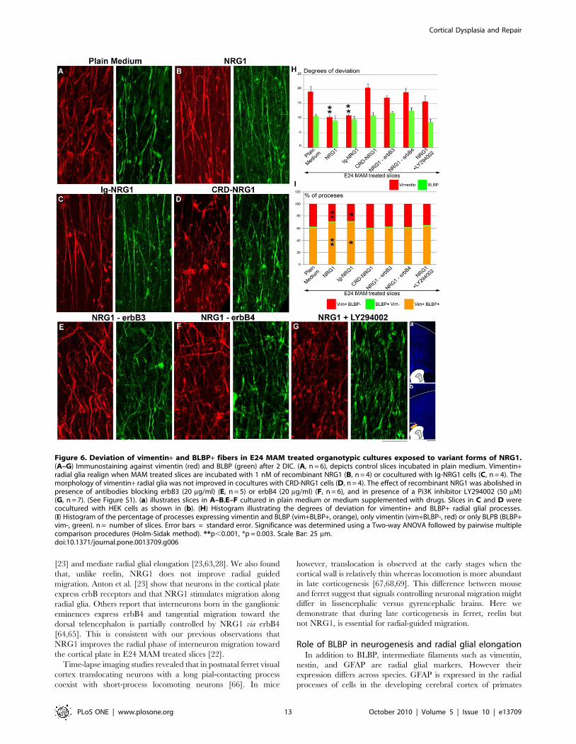
Radial glial morphology is improved in the presence ofan exogenous secreted form of NRG1
Soluble recombinant type I NRG1 repairs the radial glial
scaffold in E24 MAM ferret slices [28]. To assess whether NRG1
acts similarly on both vimentin+ and BLBP+ radial glia,
organotypic slices were exposed for 2 DIC to NRG1, and the
degrees of deviation computed for vimentin+ and BLBP+ radial
glial processes (see Table 1). MAM treated slices were initially
exposed to a diffuse source of recombinant NRG1 (1 nM) as
described previously [28]. We observed a dramatic realignment of
vimentin+ radial glia with an angle of deviation at 10.43, which is
significantly reduced from the angle of deviation of vimentin+radial glia in MAM treated slices incubated 2 DIC in plain
medium (19.21) (Figure 6A,B,H). As described above using reelin,
BLBP+ radial glial morphology, although much less disrupted in
MAM treated ferrets compared to vimentin+ radial glia, was
unchanged after treatment with recombinant NRG1 (9.22
compared to 10.76 in plain medium; not significant, p.0.05).
Again, the morphology of BLBP+ radial glia was similar to the
improved vimentin+ processes. The soluble recombinant NRG1
used here is a truncated form of NRG1, which contains only the
soluble EGF-like domain. This form of NRG1, commonly used for
in vitro studies, is sufficient to elicit ErbB receptor dimerization,
tyrosine phosphorylation and the activation of downstream
signaling pathways [51]. We additionally decided to expose
MAM treated slices to the full length of NRG1 to assess if other
domains beyond the EGF-like domain could improve the BLBP+radial glial morphology. E24 MAM slices were co-cultured with
HEK cells secreting the full length of type I NRG1 (Ig-NRG1
cells). HEK cells were included in Matrigel and placed at the pial
surface as described previously. The morphology of vimentin+radial glial cells was dramatically improved, comparable to a
treatment with soluble recombinant NRG1 (degree of deviation of
11.08; Figure 6C,H). However, BLBP+ radial glia remained the
same in the presence of the full length of NRG1 (degree of
deviation of 9.72), but similar to the improved vimentin+ radial
glia. Finally, we co-cultured MAM slices with HEK cells
expressing type III NRG1 (CRD-NRG1 cells). The isoform III,
unlike the Ig-like domain of type I NRG1, is not secreted and
contains a cysteine-rich domain (CRD). We found no improve-
ment of the radial glial scaffold: with a degree of deviation of 20.51
for vimentin and 10.91 for BLBP, suggesting that the morphology
of radial glia in presence of type III NRG1 was similar to MAM
slices cultured in plain medium (Figure 6D,H). Therefore, the
Table 2. Proportion of radial glial processes expressing vimentin, BLBP or both in normal and E24 MAM treated in vivo and in vitro(2 DIC).
Condition/Treatment Vimentin+ BLBP+ Vimentin-BLBP+ Vimentin+ BLBP- n slices
Fixed Brains Normal 88.79+/20.72 1.17+/20.17 10.02+/20.74 6
E24 MAM treated 54.45+/23.38 1.38+/20.27 44.15+/23.51 5
Normal Slices Plain medium 87.98+/20.57 1.70+/20.19 10.31+/20.41 8
E24 MAM treated Slices Plain medium 60.11+/23.4 2.17+/20.89 37.71+/23.99 6
Control HEK 61.73+/23.29 2.26+/20.76 35.99+/23.78 7
Recombinant Reelin 72.56+/21.83 2.46+/20.86 24.97+/21.67 10
Reelin HEK 71.81+/21.68 1.64+/20.80 26.53+/22.04 6
Reelin HEK+ RAP 65.56+/22.49 2.93+/20.65 31.50+/22.50 7
Reelin HEK+ SP600125 73.92+/23.35 0.97+/20.34 25.10+/23.02 4
Reelin HEK+ PP2 65.56+/22.49 2.93+/20.65 31.50+/22.50 6
Reelin HEK+ LY294002 65.41+/22.45 1.26+/20.54 33.32+/22.76 9
Reelin HEK+ TDZD-8 73.01+/21.53 0.77+/20.49 26.20+/21.58 6
Recombinant NRG1 68.98+/22.13 2.13+/20.39 28.88+/22.54 4
Ig-NRG1 69.75+/20.54 1.94+/20.64 28.29+/20.89 4
CRD-NRG1 58.74+/24.04 1.84+/20.71 39.40+/24.12 4
Rc NRG1 + erbB3 blocking antibodies 61.04+/22.03 1.49+/20.54 37.46+/21.66 5
Rc NRG1 + erbB4 blocking antibodies 59.76+/24.04 2.26+/20.32 37.96+/20.77 6
Rc NRG1 + LY294002 62.21+/21.92 3.10+/20.65 34.67+/21.89 7
doi:10.1371/journal.pone.0013709.t002
Cortical Dysplasia and Repair
PLoS ONE | www.plosone.org 8 October 2010 | Volume 5 | Issue 10 | e13709
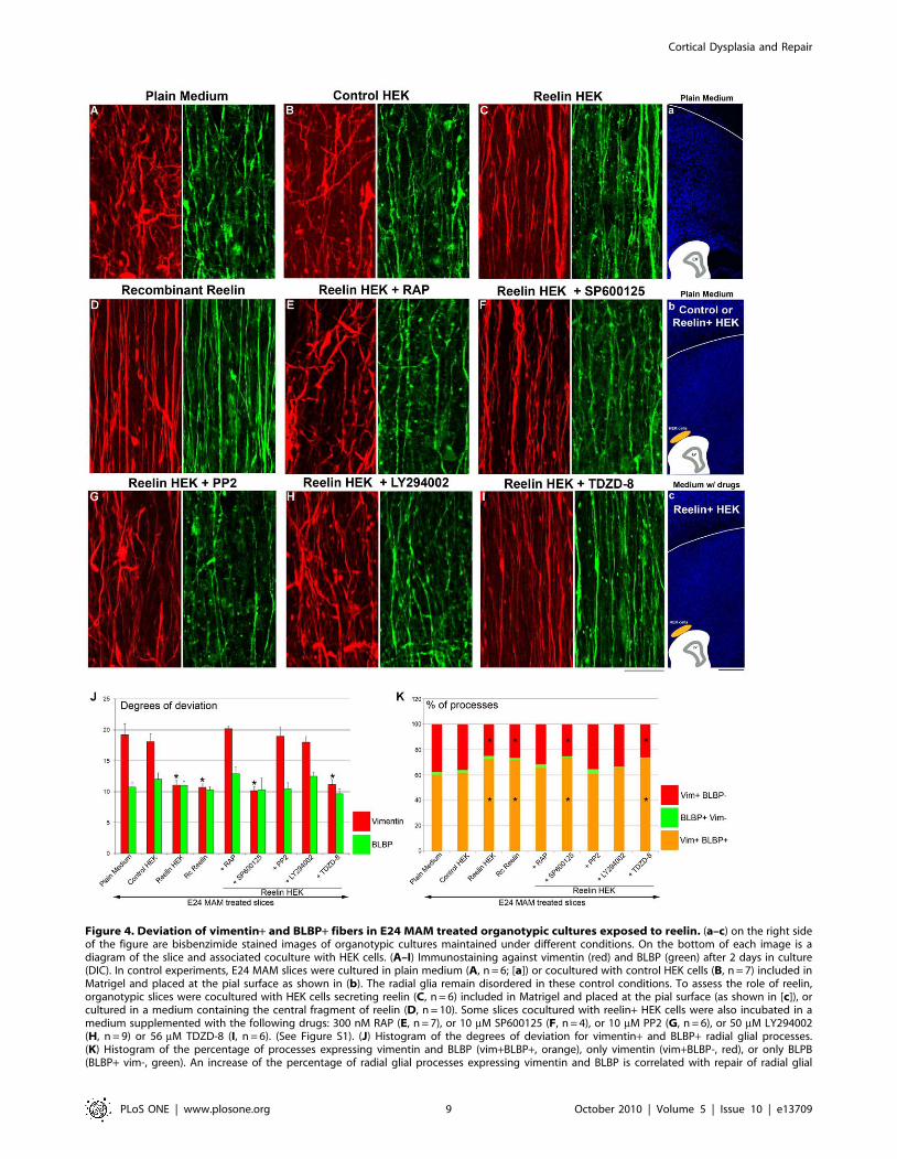
Figure 4. Deviation of vimentin+ and BLBP+ fibers in E24 MAM treated organotypic cultures exposed to reelin. (a–c) on the right sideof the figure are bisbenzimide stained images of organotypic cultures maintained under different conditions. On the bottom of each image is adiagram of the slice and associated coculture with HEK cells. (A–I) Immunostaining against vimentin (red) and BLBP (green) after 2 days in culture(DIC). In control experiments, E24 MAM slices were cultured in plain medium (A, n = 6; [a]) or cocultured with control HEK cells (B, n = 7) included inMatrigel and placed at the pial surface as shown in (b). The radial glia remain disordered in these control conditions. To assess the role of reelin,organotypic slices were cocultured with HEK cells secreting reelin (C, n = 6) included in Matrigel and placed at the pial surface (as shown in [c]), orcultured in a medium containing the central fragment of reelin (D, n = 10). Some slices cocultured with reelin+ HEK cells were also incubated in amedium supplemented with the following drugs: 300 nM RAP (E, n = 7), or 10 mM SP600125 (F, n = 4), or 10 mM PP2 (G, n = 6), or 50 mM LY294002(H, n = 9) or 56 mM TDZD-8 (I, n = 6). (See Figure S1). (J) Histogram of the degrees of deviation for vimentin+ and BLBP+ radial glial processes.(K) Histogram of the percentage of processes expressing vimentin and BLBP (vim+BLBP+, orange), only vimentin (vim+BLBP-, red), or only BLPB(BLBP+ vim-, green). An increase of the percentage of radial glial processes expressing vimentin and BLBP is correlated with repair of radial glial
Cortical Dysplasia and Repair
PLoS ONE | www.plosone.org 9 October 2010 | Volume 5 | Issue 10 | e13709
soluble EGF-like domain of type I NRG1, applied diffusely or
focally, is sufficient to realign vimentin+ radial glia, which are
highly disrupted in MAM treated animals; BLBP+ radial glia,
although less disrupted, remain unchanged.
Blockade of erbB3/erbB4 or inhibition of Pi3K signalingprevents the realignment of radial glia induced by NRG1
To further elucidate the mechanism of radial glial radialization
induced by NRG1, MAM slices were cultured in medium
containing recombinant NRG1 (1 nM) combined with HER-3
Ab-5 or HER-4 Ab-3, two antibodies blocking the binding of NRG1
to erbB3 and erbB4 respectively (Figure S1). Radialization of
vimentin+ radial glia mediated by NRG1 was prevented by
blocking erbB3 or erbB4 receptors (Figure 6E,F,H). The morphol-
ogy of BLBP+ processes however demonstrated only slight changes,
which were not significant. Since erbB receptors activate the Akt/
Pi3K signaling pathway (for review see [52]), slices were cultured
with recombinant NRG1 combined with LY294002 (50 mM), a
Pi3K inhibitor. In these conditions, the effect of NRG1 on
vimentin+ radial glia was significantly reduced when Pi3K was
inhibited. Blocking Pi3K does not alter the morphology of BLBP+radial glia (Figure 6G,H). Together, these results indicate that
NRG1 via activation of erbB3/erbB4 receptors and Pi3K induces a
realignment of vimentin+ radial glia (see also Figure 8).
Increased expression of BLBP in the presence of NRG1 iserbB3/4 and Pi3K dependent
The number of radial glial processes co-expressing vimentin and
BLBP was significantly increased compared to control when slices
were incubated in the presence of NRG1 or cocultured with Ig-
NRG1 cells (Figure 6I, Table 2). No significant difference was
found when slices were cocultured with CRD-NRG1 cells
compared to MAM treated slices incubated in plain medium
(control experiment). The percent of vimentin+ radial glial
processes expressing BLBP in presence of NRG1 while blocking
erbB3, erbB4 or Pi3K was similar to control (Figure 6I, Table 2).
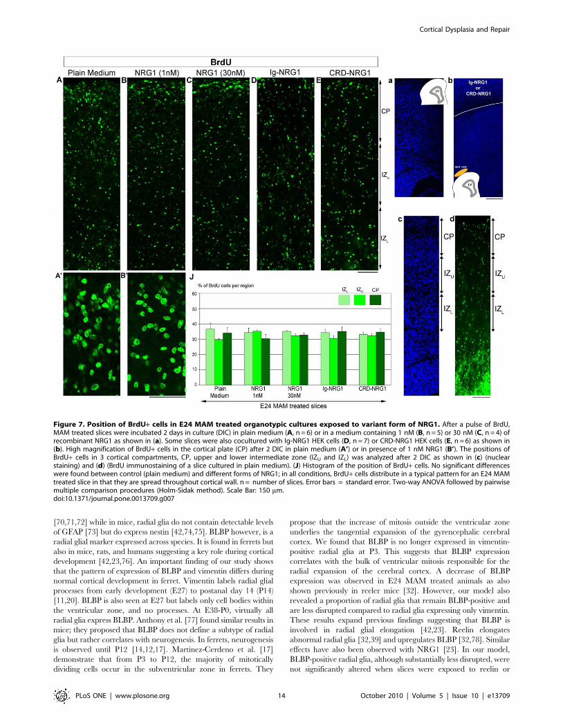
NRG1 does not improve neuronal migration toward thecortical plate
In MAM treated ferrets, disruption of radial glia is associated with
abnormal migration; neurons are scattered in all cortical layers
compared to normal ferrets where neurons accumulate in an inside-
out pattern [21]. Since NRG1 can repair the radial glial scaffold
[28], we tested whether neuronal migration was also restored. Slices
were exposed to a pulse of BrdU+ and the distribution of BrdU+cells was evaluated after 2 DIC as described in Figure 4 (see also
Figure 7c–d and Table 3 for details). Slices were incubated in plain
medium or in medium containing 1 nM of soluble recombinant
NRG1 or a 30 nM concentration, which has been shown to
promote migration along the radial glial scaffold in mice [23]. In the
presence of NRG1 (1 nM or 30 nM), BrdU+ cells scattered in the
cortical wall, similar to control conditions (slices incubated in plain
medium) (Figure 7A–C,J). We also used forskolin, a drug known to
enhance the level of NRG1 receptors available at the membrane, via
an increase of intracellular cyclic AMP [53]. Slices were treated with
forskolin alone (2 mM) or forskolin (2 mM) combined with NRG1
(1 nM). Forskolin treatment left the distribution of BrdU+ cells
unchanged suggesting that failure to migrate into the CP is not due
to decreased receptors at the membrane (Table 3). To test whether
the full length type I NRG1 or membrane bound type III NRG1
could improve radial migration, MAM slices were co-cultured with
Ig-NRG1 or CRD-NRG1 HEK cells. Figure 7D,E,J show that
BrdU+ cells failed to accumulate in the CP in both conditions.
Migration into the CP was not improved in any of the conditions
supplying exogenous NRG1: recombinant NRG1 (1 nM w/o
forskoline, or 30 nM), co-culture with cell lines expressing the
secreted form of type I NRG1 (Ig-NRG1 cells) or the membrane
form of type III NRG1 (CRD-NRG1 cells). This suggests that a
normal radial glial scaffold is not sufficient to restore neuronal
migration and other factor(s) are essential to direct migrating
neurons toward the CP (see also Figure 8).
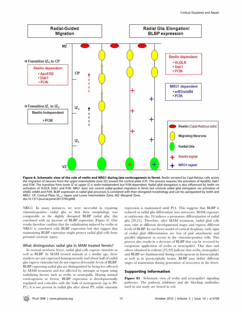
Discussion
We show here that severely disrupted dyplasic brains can be
repaired by specific application of reelin or NRG1. In our model
of cortical dysplasia, reelin restores the radial glial scaffold as well
as glia-guided migration; NRG1, however, had a more limited
effect since radial glia were realigned, but neuronal migration was
not improved (Figure 8). The differential expression of BLBP and
vimentin during normal and abnormal cortical development
suggest a key role of BLBP in radial glial elongation and possibly in
protection against environmental insults.
VLDLR and ApoER2 exert different functions duringcortical development
Corticogenesis in E24 MAM treated ferrets is severely
disrupted; misaligned radial glial cells differentiate into astrocytes
and neurons fail to reach the cortical plate [14,21,22]. Reelin is
not missing but ectopic Cajal-Retzius cells most likely provide
aberrant signaling due to their abnormal placement [14,21,39].
We previously demonstrated that an exogenous source of reelin
placed at the pial surface was sufficient to restore the radial glial
scaffold and neuronal migration toward the cortical plate [39].
Here we demonstrate that these effects are Dab1- and Pi3K-
dependent but possibly mediated through different receptors. The
role of the reelin receptors ApoER2 and VLVDR was evaluated
using RAP, which blocks both receptors [46,47], or using
SP600125 which blocks ApoER2 by inhibiting JIP activity [49].
We found that during late corticogenesis glia-guided migration is
influenced by ApoER2 as demonstrated previously in mice by
Hack et al. [48] RAP prevents the repair of radial gial cells
induced by reelin whereas SP600125 had no effect. This result
suggests that reelin signaling is mediated mainly by VLDLR
during late corticogenesis with little if any contribution from
ApoER2. This finding may be mitigated by the observation that
ApoER2 is expressed in stem cells, while JIP-1 and JIP-2 are not.
Radial glia, of course, are a form of stem cells, therefore the results
of Stockinger et al. [49] suggest that JIP-1 and JIP-2 are expressed
in neurons but not in radial glial cells. However, a population of
stem cells produced from mouse cortical cells of 15–17 old day
embryos and grown in the presence of growth factors may not
express the same set of factors/genes produced by newborn ferret
radial glia in vivo or grown in organotypic cultures. In addition,
Hack et al. [48] reported that radial glial morphology is not
affected in ApoER2-/- mice, strongly supporting the idea that
ApoER2 is not necessary for maintaining radial glial elongation.
morphology (lower degrees of deviation). This effect induced by reelin is mediated via VLDLR/Dab1/Pi3K activation. n = number of slices. Error bars= standard error. Significance was determined using a Two-way ANOVA followed by pairwise multiple comparison procedures (Holm-Sidak method).Significant pairwise comparisons are between control (i.e. Plain Medium) and tested conditions. *p#0.002. Scale Bar: 25 mm (A–I) and 500 mm (1–3).doi:10.1371/journal.pone.0013709.g004
Cortical Dysplasia and Repair
PLoS ONE | www.plosone.org 10 October 2010 | Volume 5 | Issue 10 | e13709
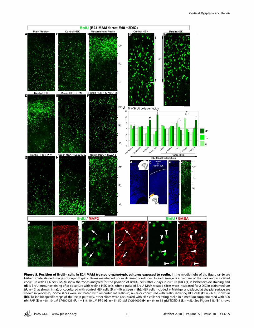
Figure 5. Position of BrdU+ cells in E24 MAM treated organotypic cultures exposed to reelin. In the middle right of the figure (a–b) arebisbenzimide stained images of organotypic cultures maintained under different conditions. In each image is a diagram of the slice and associatedcoculture with HEK cells. (c–d) show the zones analyzed for the position of BrdU+ cells after 2 days in culture (DIC) (c) is bisbenzimide staining and(d) is BrdU immunostaining after coculture with reelin+ HEK cells. After a pulse of BrdU, MAM treated slices were incubated for 2 DIC in plain medium(A, n = 6) as shown in (a), or cocultured with control HEK cells (B, n = 8) as seen in (b); HEK cells included in Matrigel and placed at the pial surface areshown in yellow (b). Some slices were incubated with recombinant reelin (C, n = 8) or cocultured with reelin secreting HEK cells (D, n = 6 as shown in[b]). To inhibit specific steps of the reelin pathway, other slices were cocultured with HEK cells secreting reelin in a medium supplemented with 300nM RAP (E, n = 8), 10 mM SP600125 (F, n = 11), 10 mM PP2 (G, n = 5), 50 mM LY294002 (H, n = 6), or 56 mM TDZD-8 (I, n = 5). (See Figure S1). (B’) shows
Cortical Dysplasia and Repair
PLoS ONE | www.plosone.org 11 October 2010 | Volume 5 | Issue 10 | e13709
Others report that the N-terminal portion of reelin binds to
cadherin-related neuronal receptors and the integrin receptors
[54,55]. Jossin et al. [56] demonstrate that the central fragment of
reelin binds to ApoER2 and VLDLR but does not bind to
cadherin-related neuronal receptors. We found no differences
when slices were treated with recombinant reelin, which consists of
the central fragment of reelin, or when slices were cocultured with
HEK cells secreting the full length of reelin. Since the central
fragment of reelin is sufficient to both realign the radial glial
scaffold and improve neuronal migration, it is likely that neither
the integrin receptors nor the cadherin-related neuronal receptors
are essential for migration in our model.
Reelin is essential to glia-guided migration ingyrencephalic cortex
Reelin not only repairs radial glia but also improves migration
of neurons that are likely to be generated in the neocortical
ventricular zone since they express MAP2 and are also GABA-
negative. In the presence of exogenous reelin, neurons move into
the cortical plate but only into the lower intermediate zone when
Dab1 and Pi3K were inhibited, suggesting that reelin signaling is
essential for neuronal migration from the intermediate zone
toward the cortical plate. This is supported by Uchida et al. [57]
who demonstrated that radially migrating neurons in the
subventricular/intermediate zone strongly express functional
VLDLR and ApoER2 receptors, which then downregulate in
the cortical plate. We found that interfering with Pi3K function
produced more cells accumulating in the lower intermediate zone
than blocking Dab1, indicating that in addition to reelin, another
unknown Pi3K-dependent signaling pathway is also involved as
suggested by Jossin and Goffinet (2007) [58]. Another possibility is
that blockade of Pi3K exhibits a more severe phenotype due to the
pleiotropic functions controlled by PI3K/Akt signaling [59]. Jossin
and Goffinet (2007) proposed that inhibition of Pi3K impairs the
polarity of neurons so they accumulate in the intermediate zone.
Morphological transition from multipolar to bipolar neurons is
essential to reach the cortical plate [60,61]. In birds, the pallium
develops in an outside-inside gradient and migrating neurons
display a multipolar morphology. Nomura et al. [62] found that
avian migrating neurons adopt a bipolar shape when reelin
signaling is experimentally increased. These studies, as well as our
observations, clearly indicate that reelin by its duel function on
radial glia and neuronal migration is fundamental to the
development of 6-layered lissencephalic as well as gyrencephalic
cortices.
NRG1 repairs the radial glial scaffold but not radialmigration
NRG1 signaling is also essential for the normal development of
radial glia [23,63,28]. In E24 MAM treated ferrets, NRG1 is
reduced and treatment with recombinant NRG1 realigns radial
glial morphology [28]. We demonstrate here that NRG1 realigns
vimentin-positive but does not significantly alter BLBP-positive
radial glia. We also show that the effect mediated by NRG1 is
erbB3/erbB4 and Pi3K-dependent. This is consistent with previous
reports showing that erbB3 and erbB4 are expressed by radial glia
a high magnification of BrdU+ cells in the cortical plate (CP) in slices cocultured with control HEK cells while (D’) shows an image of BrdUimmunoreactivity in an organotypic slice cultured with reelin+ HEK cells. (J) is a histogram indicating the distribution of BrdU+ cells after differenttreatments. (K–N) Organotypic MAM treated slice (E40) incubated 2 DIC with recombinant reelin (1nM), resectioned using a cryostat at 14 mM.Double immunostaining for BrdU (K, green) and MAP2 (L, red). (K’–L’) Higher magnification of the boxed area in L. The arrows indicate BrdU-positivecells that are also MAP2-positive. Double immunostaining for BrdU (M, green) and GABA (N, red). The arrow heads indicate BrdU+ cells that areGABA-negative. CP: Cortical Plate; IZu-L: Upper and Lower Intermediate Zone. n = number of slices. Error bars = standard error. Significance wasdetermined using a Two-way ANOVA followed by pairwise multiple comparison procedures (Holm-Sidak method). *p = 0.017, **p = 0.003,***p = 0.001 compared to CP in control medium. #p = 0.017, ##p = 0.025 compared to IZL. Scale Bar: 50 mm (A–I), 25 mm (K’,L’,M,N), 250 mm (K–L),and 500 mm (a–d).doi:10.1371/journal.pone.0013709.g005
Table 3. Distribution of BrdU+ cells in the cortical plate (CP), the upper IZ (IZu) and in the lower IZ (IZL).
Condition/Treatment CP IZ upper IZ lower n slices
Plain medium 33.98+/23.61 29.46+/21.15 36.54+/24.08 6
Control HEK 33.83+/21.78 33.18+/21.35 32.97+/21.10 8
Recombinant Reelin 44.68+/23.02 33.81+/22.81 21.50+/21.26 8
Reelin HEK 53.98+/24.08 25.46+/22.50 20.54+/23.77 6
Reelin HEK+ RAP 29.98+/21.48 34.21+/21.34 35.79+/22.17 8
Reelin HEK+ SP600125 34.06+/22.93 34.78+/23.35 30.07+/23.07 11
Reelin HEK+ PP2 27.59+/22.35 32.67+/20.98 39.72+/23.01 5
Reelin HEK+ LY294002 23.78+/22.97 29.14+/21.62 47.06+/24.39 6
Reelin HEK+ TDZD-8 45.09+/23.48 30.55+/22.31 24.35+/23.66 5
Recombinant NRG1 (1 nM) 30.31+/22.88 35.30+/20.81 34.37+/22.85 5
Recombinant NRG1 (30 nM) 32.66+/21.67 32.28+/21.19 35.05+/20.65 4
Ig-NRG1 35.15+/21.78 30.47+/21.96 34.37+/21.10 7
CRD-NRG1 34.67+/22.29 32.23+/21.33 33.09+/21.67 6
Forskolin 31.80+/21.89 36.07+/22.59 32.11+/21.47 3
Recombinant NRG1 + Forskolin 33.22+/21.63 32.87+/20.87 33.90+/20.75 3
Organotypic slices were obtained from E24 MAM treated ferrets and maintained in vitro (2 DIC).doi:10.1371/journal.pone.0013709.t003
Cortical Dysplasia and Repair
PLoS ONE | www.plosone.org 12 October 2010 | Volume 5 | Issue 10 | e13709
[23] and mediate radial glial elongation [23,63,28]. We also found
that, unlike reelin, NRG1 does not improve radial guided
migration. Anton et al. [23] show that neurons in the cortical plate
express erbB receptors and that NRG1 stimulates migration along
radial glia. Others report that interneurons born in the ganglionic
eminences express erbB4 and tangential migration toward the
dorsal telencephalon is partially controlled by NRG1 via erbB4
[64,65]. This is consistent with our previous observations that
NRG1 improves the radial phase of interneuron migration toward
the cortical plate in E24 MAM treated slices [22].
Time-lapse imaging studies revealed that in postnatal ferret visual
cortex translocating neurons with a long pial-contacting process
coexist with short-process locomoting neurons [66]. In mice
however, translocation is observed at the early stages when the
cortical wall is relatively thin whereas locomotion is more abundant
in late corticogenesis [67,68,69]. This difference between mouse
and ferret suggest that signals controlling neuronal migration might
differ in lissencephalic versus gyrencephalic brains. Here we
demonstrate that during late corticogenesis in ferret, reelin but
not NRG1, is essential for radial-guided migration.
Role of BLBP in neurogenesis and radial glial elongationIn addition to BLBP, intermediate filaments such as vimentin,
nestin, and GFAP are radial glial markers. However their
expression differs across species. GFAP is expressed in the radial
processes of cells in the developing cerebral cortex of primates
Figure 6. Deviation of vimentin+ and BLBP+ fibers in E24 MAM treated organotypic cultures exposed to variant forms of NRG1.(A–G) Immunostaining against vimentin (red) and BLBP (green) after 2 DIC. (A, n = 6), depicts control slices incubated in plain medium. Vimentin+radial glia realign when MAM treated slices are incubated with 1 nM of recombinant NRG1 (B, n = 4) or cocultured with Ig-NRG1 cells (C, n = 4). Themorphology of vimentin+ radial glia was not improved in cocultures with CRD-NRG1 cells (D, n = 4). The effect of recombinant NRG1 was abolished inpresence of antibodies blocking erbB3 (20 mg/ml) (E, n = 5) or erbB4 (20 mg/ml) (F, n = 6), and in presence of a Pi3K inhibitor LY294002 (50 mM)(G, n = 7). (See Figure S1). (a) illustrates slices in A–B,E–F cultured in plain medium or medium supplemented with drugs. Slices in C and D werecocultured with HEK cells as shown in (b). (H) Histogram illustrating the degrees of deviation for vimentin+ and BLBP+ radial glial processes.(I) Histogram of the percentage of processes expressing vimentin and BLBP (vim+BLBP+, orange), only vimentin (vim+BLBP-, red) or only BLPB (BLBP+vim-, green). n = number of slices. Error bars = standard error. Significance was determined using a Two-way ANOVA followed by pairwise multiplecomparison procedures (Holm-Sidak method). **p,0.001, *p = 0.003. Scale Bar: 25 mm.doi:10.1371/journal.pone.0013709.g006
Cortical Dysplasia and Repair
PLoS ONE | www.plosone.org 13 October 2010 | Volume 5 | Issue 10 | e13709
[70,71,72] while in mice, radial glia do not contain detectable levels
of GFAP [73] but do express nestin [42,74,75]. BLBP however, is a
radial glial marker expressed across species. It is found in ferrets but
also in mice, rats, and humans suggesting a key role during cortical
development [42,23,76]. An important finding of our study shows
that the pattern of expression of BLBP and vimentin differs during
normal cortical development in ferret. Vimentin labels radial glial
processes from early development (E27) to postanal day 14 (P14)
[11,20]. BLBP is also seen at E27 but labels only cell bodies within
the ventricular zone, and no processes. At E38-P0, virtually all
radial glia express BLBP. Anthony et al. [77] found similar results in
mice; they proposed that BLBP does not define a subtype of radial
glia but rather correlates with neurogenesis. In ferrets, neurogenesis
is observed until P12 [14,12,17]. Martinez-Cerdeno et al. [17]
demonstrate that from P3 to P12, the majority of mitotically
dividing cells occur in the subventricular zone in ferrets. They
propose that the increase of mitosis outside the ventricular zone
underlies the tangential expansion of the gyrencephalic cerebral
cortex. We found that BLBP is no longer expressed in vimentin-
positive radial glia at P3. This suggests that BLBP expression
correlates with the bulk of ventricular mitosis responsible for the
radial expansion of the cerebral cortex. A decrease of BLBP
expression was observed in E24 MAM treated animals as also
shown previously in reeler mice [32]. However, our model also
revealed a proportion of radial glia that remain BLBP-positive and
are less disrupted compared to radial glia expressing only vimentin.
These results expand previous findings suggesting that BLBP is
involved in radial glial elongation [42,23]. Reelin elongates
abnormal radial glia [32,39] and upregulates BLBP [32,78]. Similar
effects have also been observed with NRG1 [23]. In our model,
BLBP-positive radial glia, although substantially less disrupted, were
not significantly altered when slices were exposed to reelin or
Figure 7. Position of BrdU+ cells in E24 MAM treated organotypic cultures exposed to variant form of NRG1. After a pulse of BrdU,MAM treated slices were incubated 2 days in culture (DIC) in plain medium (A, n = 6) or in a medium containing 1 nM (B, n = 5) or 30 nM (C, n = 4) ofrecombinant NRG1 as shown in (a). Some slices were also cocultured with Ig-NRG1 HEK cells (D, n = 7) or CRD-NRG1 HEK cells (E, n = 6) as shown in(b). High magnification of BrdU+ cells in the cortical plate (CP) after 2 DIC in plain medium (A’) or in presence of 1 nM NRG1 (B’). The positions ofBrdU+ cells in 3 cortical compartments, CP, upper and lower intermediate zone (IZU and IZL) was analyzed after 2 DIC as shown in (c) (nuclearstaining) and (d) (BrdU immunostaining of a slice cultured in plain medium). (J) Histogram of the position of BrdU+ cells. No significant differenceswere found between control (plain medium) and different forms of NRG1; in all conditions, BrdU+ cells distribute in a typical pattern for an E24 MAMtreated slice in that they are spread throughout cortical wall. n = number of slices. Error bars = standard error. Two-way ANOVA followed by pairwisemultiple comparison procedures (Holm-Sidak method). Scale Bar: 150 mm.doi:10.1371/journal.pone.0013709.g007
Cortical Dysplasia and Repair
PLoS ONE | www.plosone.org 14 October 2010 | Volume 5 | Issue 10 | e13709
NRG1. In many instances, we were successful in repairing
vimentin-positive radial glia so that their morphology was
comparable to the slightly disrupted BLBP radial glia; this
correlated with an increase of BLBP expression (Figure 8). Our
results therefore confirm that the radialization induced by reelin or
NRG1 is correlated with BLBP expression but also suggest that
maintaining BLBP expression might protect radial glial cells from
prenatal cytotoxic injury.
What distinguishes radial glia in MAM treated ferrets?In normal newborn ferret, radial glial cells express vimentin as
well as BLBP. In MAM treated animals at a similar age, these
markers are not expressed homogeneously and about half of radial
glia express vimentin but do not express detectable levels of BLBP.
BLBP expressing radial glia are distinguished by being less affected
by MAM treatment and less affected by attempts at repair using
radializing factors such as reelin or neuregulin. During normal
corticogenesis in ferrets, BLBP expression is developmentally
regulated and coincides with the bulk of neurogenesis (up to P0-
P1); it is not present in radial glia after about P3, while vimentin
expression is maintained until P14. This suggests that BLBP is
reduced as radial glia differentiate into astrocytes. MAM exposure
at embryonic day 24 induces a premature differentiation of radial
glia [20,21]. Therefore, after MAM treatment, radial glial cells
may exist at different developmental stages and express different
levels of BLBP. In our ferret model of cortical dysplasia, early signs
of radial glial differentiation are loss of pial attachment and
parallel alignment as occurs in the vimentin-positive cells. This
process also results in a decrease of BLBP that can be reversed by
exogenous application of reelin or neuregulin1. Our data and
others obtained in rodents [23,32] indicate that reelin, neuregulin1
and BLBP are fundamental during corticogenesis in lissencephalic
as well as in gyrencephalic brains. BLBP may define different
stages of maturation during generation of neocortex in the ferret.
Supporting Information
Figure S1 Schematic view of reelin and neuregulin1 signaling
pathways. The pathway inhibitors and the blocking antibodies
used in our study are boxed in red.
Figure 8. Schematic view of the role of reelin and NRG1 during late corticogenesis in ferret. Reelin secreted by Cajal-Retzius cells assiststhe migration of neurons from the upper intermediate zone (IZ) toward the cortical plate (CP). This process requires the activation of ApoER2, Dab1and Pi3K. The transition from lower IZ to upper IZ is reelin-independent but Pi3K-dependent. Radial glial elongation is also influenced by reelin viaactivation of VLDLR, Dab1 and Pi3K. NRG1 does not control radial-guided migration in ferret but controls radial glial elongation via activation oferbB3, erbB4 and Pi3K. BLBP expression in radial glial processes is correlated with their elongated morphology and can be upregulated by reelin andNRG1. CP: Cortical Plate; IZu-L: Upper and Lower Intermediate Zone; MZ: Marginal Zone.doi:10.1371/journal.pone.0013709.g008
Cortical Dysplasia and Repair
PLoS ONE | www.plosone.org 15 October 2010 | Volume 5 | Issue 10 | e13709
Found at: doi:10.1371/journal.pone.0013709.s001 (2.25 MB
TIF)
Acknowledgments
We thank Gabriel Corfas for the Ig- and CRD-NRG1 HEK cells and
Brian Howell for the reelin-secreting HEK cells; Mireille Rossel, Michael
Davis, and Tom McFate for helpful comments; and Sarah Dhandu and
LaToya Hyson for excellent care of the ferrets.
Author Contributions
Conceived and designed the experiments: SP SLJ. Performed the
experiments: SP SLJ. Analyzed the data: SP SLJ. Wrote the paper: SP SLJ.
References
1. Rakic P (2007) The radial edifice of cortical architecture: from neuronalsilhouettes to genetic engineering. Brain research reviews 55(2): 204–219.
2. Corbin JG, Gaiano N, Juliano SL, Poluch S, Stancik E, et al. (2008) Regulationof neural progenitor cell development in the nervous system. Journal of
neurochemistry 106(6): 2272–2287.
3. Miller MW, Robertson S (1993) Prenatal exposure to ethanol alters the postnatal
development and transformation of radial glia to astrocytes in the cortex. The
Journal of comparative neurology 337(2): 253–266.
4. Choi BH, Yee S, Robles M (1996) The effects of glutathione glycoside in methyl
mercury poisoning. Toxicology and applied pharmacology 141(2): 357–364.
5. Roper SN, Abraham LA, Streit WJ (1997) Exposure to in utero irradiation
produces disruption of radial glia in rats. Developmental neuroscience 19(6):521–528.
6. Ross ME (2002) Brain malformations, epilepsy, and infantile spasms.
International review of neurobiology 49: 333–352.
7. Pang T, Atefy R, Sheen V (2008) Malformations of cortical development. The
neurologist 14(3): 181–191.
8. Spalice A, Parisi P, Nicita F, Pizzardi G, Del Balzo F, et al. (2009) Neuronal
migration disorders: clinical, neuroradiologic and genetics aspects. Acta Paediatr
98(3): 421–433.
9. Verrotti A, Spalice A, Ursitti F, Papetti L, Mariani R, et al. (2009) New trends in
neuronal migration disorders. Eur J Paediatr Neurol.
10. Schmechel DE, Rakic P (1979) A Golgi study of radial glial cells in developing
monkey telencephalon: morphogenesis and transformation into astrocytes.Anatomy and embryology 156(2): 115–152.
11. Voigt T (1989) Development of glial cells in the cerebral wall of ferrets: direct
tracing of their transformation from radial glia into astrocytes. The Journal ofcomparative neurology 289(1): 74–88.
12. Jackson CA, Peduzzi JD, Hickey TL (1989) Visual cortex development in the,ferret. I. Genesis and migration of visual cortical neurons. J Neurosci 9(4):
1242–1253.
13. Juliano SL, Palmer SL, Sonty RV, Noctor S, Hill GF, 2nd (1996) Development
of local connections in ferret somatosensory cortex. The Journal of comparative
neurology 374(2): 259–277.
14. Noctor SC, Scholnicoff NJ, Juliano SL (1997) Histogenesis of ferret
somatosensory cortex. The Journal of comparative neurology 387(2): 179–193.
15. Poluch S, Jablonska B, Juliano SL (2008) Alteration of interneuron migration in
a ferret model of cortical dysplasia. Cereb Cortex 18(1): 78–92.
16. Welker W (1990) Why does cerebral cortex fissure and fold? A review of
determinants of gyri and sulci. In: Cerebral cortex, vol. 8B. Comparative
structure and evolution of cerebral cortex, part II (Jones EG, Peters A, eds). pp3–136.
17. Martinez-Cerdeno V, Noctor SC, Kriegstein AR (2006) The role of intermediateprogenitor cells in the evolutionary expansion of the cerebral cortex. Cereb
Cortex 16 Suppl 1: i152–161.
18. Fish JL, Dehay C, Kennedy H, Huttner WB (2008) Making bigger brains-theevolution of neural-progenitor-cell division. Journal of cell science 121(Pt 17):
2783–2793.
19. Fietz SA, Kelava I, Vogt J, Wilsch-Brauninger M, Stenzel D, et al. (2010) OSVZ
progenitors of human and ferret neocortex are epithelial-like and expand byintegrin signaling. Nature neuroscience 13(6): 690–699.
20. Noctor SC, Palmer SL, Hasling T, Juliano SL (1999) Interference with the
development of early generated neocortex results in disruption of radial glia andabnormal formation of neocortical layers. Cereb Cortex 9(2): 121–136.
21. Hasling TA, Gierdalski M, Jablonska B, Juliano SL (2003) A radialization factorin normal cortical plate restores disorganized radial glia and disrupted migration
in a model of cortical dysplasia. The European journal of neuroscience 17(3):467–480.
22. Poluch S, Juliano SL (2007) A normal radial glial scaffold is necessary for
migration of interneurons during neocortical development. Glia 55(8): 822–830.
23. Anton ES, Marchionni MA, Lee KF, Rakic P (1997) Role of GGF/neuregulin
signaling in interactions between migrating neurons and radial glia in thedeveloping cerebral cortex. Development (Cambridge, England) 124(18):
3501–3510.
24. Rio C, Rieff HI, Qi P, Khurana TS, Corfas G (1997) Neuregulin and erbBreceptors play a critical role in neuronal migration. Neuron 19(1): 39–50.
25. Corfas G, Roy K, Buxbaum JD (2004) Neuregulin 1-erbB signaling and themolecular/cellular basis of schizophrenia. Nature neuroscience 7(6): 575–580.
26. Birchmeier C (2009) ErbB receptors and the development of the nervous system.Experimental cell research 315(4): 611–618.
27. Lemmon MA (2009) Ligand-induced ErbB receptor dimerization. Experimental
cell research 315(4): 638–648.
28. Gierdalski M, Sardi SP, Corfas G, Juliano SL (2005) Endogenous neuregulin
restores radial glia in a (ferret) model of cortical dysplasia. J Neurosci 25(37):
8498–8504.
29. Caviness VS, Jr., Sidman RL (1973) Time of origin or corresponding cell classes
in the cerebral cortex of normal and reeler mutant mice: an autoradiographic
analysis. The Journal of comparative neurology 148(2): 141–151.
30. Pinto-Lord MC, Evrard P, Caviness VS, Jr. (1982) Obstructed neuronal
migration along radial glial fibers in the neocortex of the reeler mouse: a Golgi-
EM analysis. Brain Res 256(4): 379–393.
31. Hunter-Schaedle KE (1997) Radial glial cell development and transformation
are disturbed in reeler forebrain. Journal of neurobiology 33(4): 459–472.
32. Hartfuss E, Forster E, Bock HH, Hack MA, Leprince P, et al. (2003) Reelin
signaling directly affects radial glia morphology and biochemical maturation.
Development (Cambridge, England) 130(19): 4597–4609.
33. Frotscher M, Haas CA, Forster E (2003) Reelin controls granule cell migration
in the dentate gyrus by acting on the radial glial scaffold. Cereb Cortex 13(6):
634–640.
34. Trommsdorff M, Gotthardt M, Hiesberger T, Shelton J, Stockinger W, et al.
(1999) Reeler/Disabled-like disruption of neuronal migration in knockout mice
lacking the VLDL receptor and ApoE receptor 2. Cell 97(6): 689–701.
35. D’Arcangelo G, Homayouni R, Keshvara L, Rice DS, Sheldon M, et al. (1999)
Reelin is a ligand for lipoprotein receptors. Neuron 24(2): 471–479.
36. Hiesberger T, Trommsdorff M, Howell BW, Goffinet A, Mumby MC, et al.
(1999) Direct binding of Reelin to VLDL receptor and ApoE receptor 2 induces
tyrosine phosphorylation of disabled-1 and modulates tau phosphorylation.
Neuron 24(2): 481–489.
37. Howell BW, Hawkes R, Soriano P, Cooper JA (1997) Neuronal position in the
developing brain is regulated by mouse disabled-1. Nature 389(6652): 733–737.
38. Luque JM, Morante-Oria J, Fairen A (2003) Localization of ApoER2, VLDLR
and Dab1 in radial glia: groundwork for a new model of reelin action during
cortical development. Brain research 140(2): 195–203.
39. Schaefer A, Poluch S, Juliano S (2008) Reelin is essential for neuronal migration
but not for radial glial elongation in neonatal ferret cortex. Developmental
neurobiology 68(5): 590–604.
40. Gaiano N, Nye JS, Fishell G (2000) Radial glial identity is promoted by Notch1
signaling in the murine forebrain. Neuron 26(2): 395–404.
41. Patten BA, Peyrin JM, Weinmaster G, Corfas G (2003) Sequential signaling
through Notch1 and erbB receptors mediates radial glia differentiation.
J Neurosci 23(14): 6132–6140.
42. Feng L, Hatten ME, Heintz N (1994) Brain lipid-binding protein (BLBP): a
novel signaling system in the developing mammalian CNS. Neuron 12(4):
895–908.
43. D’Arcangelo G, Miao GG, Chen SC, Soares HD, Morgan JI, et al. (1995) A
protein related to extracellular matrix proteins deleted in the mouse mutant
reeler. Nature 374(6524): 719–723.
44. Arnaud L, Ballif BA, Cooper JA (2003) Regulation of protein tyrosine kinase
signaling by substrate degradation during brain development. Molecular and
cellular biology 23(24): 9293–9302.
45. Chen S, Velardez MO, Warot X, Yu ZX, Miller SJ, et al. (2006) Neuregulin 1-
erbB signaling is necessary for normal myelination and sensory function.
J Neurosci 26(12): 3079–3086.
46. Andersen OM, Benhayon D, Curran T, Willnow TE (2003) Differential binding
of ligands to the apolipoprotein E receptor 2. Biochemistry 42(31): 9355–9364.
47. Strasser V, Fasching D, Hauser C, Mayer H, Bock HH, et al. (2004) Receptor
clustering is involved in Reelin signaling. Molecular and cellular biology 24(3):
1378–1386.
48. Hack I, Hellwig S, Junghans D, Brunne B, Bock HH, et al. (2007) Divergent
roles of ApoER2 and Vldlr in the migration of cortical neurons. Development
(Cambridge, England) 134(21): 3883–3891.
49. Stockinger W, Brandes C, Fasching D, Hermann M, Gotthardt M, et al. (2000)
The reelin receptor ApoER2 recruits JNK-interacting proteins-1 and -2. The
Journal of biological chemistry 275(33): 25625–25632.
50. Beffert U, Morfini G, Bock HH, Reyna H, Brady ST, et al. (2002) Reelin-
mediated signaling locally regulates protein kinase B/Akt and glycogen synthase
kinase 3beta. The Journal of biological chemistry 277(51): 49958–49964.
51. Fergusson KM, Darling PJ, Mohan MJ, Macatee TL, Lemmon MA (2000)
Extracellular domains drive homo- but not hetero-dimerization of erbB
receptors. EMBO J 19(17): 4632–4643.
52. Mei L, Xiong WC (2008) Neuregulin 1 in neural development, synaptic
plasticity and schizophrenia. Nature reviews 9(6): 437–452.
Cortical Dysplasia and Repair
PLoS ONE | www.plosone.org 16 October 2010 | Volume 5 | Issue 10 | e13709
53. Rahmatullah M, Schroering A, Rothblum K, Stahl RC, Urban B, et al. (1998)
Synergistic regulation of Schwann cell proliferation by heregulin and forskolin.
Molecular and cellular biology 18(11): 6245–6252.
54. Senzaki K, Ogawa M, Yagi T (1999) Proteins of the CNR family are multiple
receptors for Reelin. Cell 99(6): 635–647.
55. Dulabon L, Olson EC, Taglienti MG, Eisenhuth S, McGrath B, et al. (2000)
Reelin binds alpha3beta1 integrin and inhibits neuronal migration. Neuron
27(1): 33–44.
56. Jossin Y, Ignatova N, Hiesberger T, Herz J, Lambert de Rouvroit C, et al. (2004)
The central fragment of Reelin, generated by proteolytic processing in vivo, is
critical to its function during cortical plate development. J Neurosci 24(2):
514–521.
57. Uchida T, Baba A, Perez-Martinez FJ, Hibi T, Miyata T, et al. (2009)
Downregulation of functional Reelin receptors in projection neurons implies that
primary Reelin action occurs at early/premigratory stages. J Neurosci 29(34):
10653–10662.
58. Jossin Y, Goffinet AM (2007) Reelin signals through phosphatidylinositol 3-
kinase and Akt to control cortical development and through mTor to regulate
dendritic growth. Molecular and cellular biology 27(20): 7113–7124.
59. Yap TA, Garrett MD Walton MI, Raynaud F, de Bono JS, Workman P (2008)
Targeting the PI3K-AKT-mTOR pathway: progress, pitfalls, and promises.
Curr Opin Pharmacol 8(4): 393–412.
60. Nadarajah B, Alifragis P, Wong RO, Parnavelas JG (2003) Neuronal migration
in the developing cerebral cortex: observations based on real-time imaging.
Cereb Cortex 13(6): 607–611.
61. Tabata H, Nakajima K (2003) Multipolar migration: the third mode of radial
neuronal migration in the developing cerebral cortex. J Neurosci 23(31):
9996–10001.
62. Nomura T, Takahashi M, Hara Y, Osumi N (2008) Patterns of neurogenesis and
amplitude of Reelin expression are essential for making a mammalian-type
cortex. PloS one 3(1): e1454.
63. Schmid RS, McGrath B, Berechid BE, Boyles B, Marchionni M, et al. (2003)
Neuregulin 1-erbB2 signaling is required for the establishment of radial glia and
their transformation into astrocytes in cerebral cortex. Proceedings of the
National Academy of Sciences of the United States of America 100(7):
4251–4256.
64. Yau HJ, Wang HF, Lai C, Liu FC (2003) Neural development of the neuregulin
receptor ErbB4 in the cerebral cortex and the hippocampus: preferential
expression by interneurons tangentially migrating from the ganglionic
eminences. Cereb Cortex 13(3): 252–264.65. Flames N, Long JE, Garratt AN, Fischer TM, Gassmann M, et al. (2004) Short-
and long-range attraction of cortical GABAergic interneurons by neuregulin-1.
Neuron 44(2): 251–261.66. Borrell V, Kaspar BK, Gage FH, Callaway EM (2006) In vivo evidence for
radial migration of neurons by long-distance somal translocation in thedeveloping ferret visual cortex. Cereb Cortex 16(11): 1571–1583.
67. Nadarajah B, Brunstrom JE, Grutzendler J, Wong RO, Pearlman AL (2001)
Two modes of radial migration in early development of the cerebral cortex.Nature neuroscience 4(2): 143–150.
68. Weissman T, Noctor SC, Clinton BK, Honig LS, Kriegstein AR (2003)Neurogenic radial glial cells in reptile, rodent and human: from mitosis to
migration. Cereb Cotex 13(6): 550–559.69. Noctor SC, Martinez-Cerdeno V, Ivic L, Kriegstein AR (2004) Cortical neurons
arise in symmetric and asymmetric division zones and migrate through specific
phases. Nature neuroscience 7(2): 136–144.70. Levitt P, Rakic P (1980) Immunoperoxidase localization of glial fibrillary acidic
protein in radial glial cells and astrocytes of the developing rhesus monkey brain.The Journal of comparative neurology 193(3): 815–840.
71. Zecevic N (2004) Specific characteristic of radial glia in the human fetal
telencephalon. Glia 48(1): 27–35.72. Howard BM, Zhicheng M, Filipovic R, Moore AR, Antic SD, et al. (2008)
Radial glia cells in the developing human brain. Neuroscientist 14(5): 459–473.73. Cambell, Gotz (2002) Radial glia: multi-purpose cells for vertebrate brain
development. Trends in neurosciences 25(5): 235–238.74. Shibata T, Yamada K, Watanabe M, Ikenaka K, Wada K, et al. (1997)
Glutamate transporter GLAST is expressed in the radial glia-astrocyte lineage of
developing mouse spinal cord. J Neurosci 17(23): 9212–9219.75. Hartfuss E, Galli R, Heins N, Gotz M (2001) Characterization of CNS precursor
subtypes and radial glia. Developmental biology 229(1): 15–30.76. Howard B, Chen Y, Zecevic N (2006) Cortical progenitor cells in the developing
human telencephalon. Glia 53(1): 57–66.
77. Anthony TE, Mason HA, Gridley T, Fishell G, Heintz N (2005) Brain lipid-binding protein is a direct target of Notch signaling in radial glial cells. Genes &
development 19(9): 1028–1033.78. Keilani S, Sugaya K (2008) Reelin induces a radial glial phenotype in human
neural progenitor cells by activation of Notch-1. BMC developmental biology 8:69.
Cortical Dysplasia and Repair
PLoS ONE | www.plosone.org 17 October 2010 | Volume 5 | Issue 10 | e13709