Embed Size (px)
Citation preview
1
Università Degli Studi di Milano
Faculty of Agricultural and Food Sciences
Department of Agricultural and Environmental Sciences. Production, Landscape, Agroenergy
Ph.D. School “Agriculture, Environment, and Bioenergy”
XXXII Cycle 2016‐2019
The microbiota associated with the invasive Popillia japonica (Coleoptera: Scarabaeidae) and
evaluation of the entomopathogenic activity of the isolated nematodes
Nizar Fahmi Mohamed Salem GODA
Matriculation: R11610
Supervisor: Dr. Matteo MONTAGNA
Co-supervisor: Dr. Bessem CHOUAIA
Coordinator: Prof. Dr. Daniele BASSI
Academic year 2019-2020
2
Table of Contents
Abstract .................................................................................................................................................................. 4
Introductory chapter ............................................................................................................................................. 6
1. Introduction ................................................................................................................................................... 6
2. Microbiota of invasive beetles .......................................................................................................................... 7
2.1. Microbiota of beetles consider forest pests ............................................................................................... 7
2.2. Microbiota associated with beetles considered pest of crops .................................................................. 8
3.The Japanese beetle Popillia japonica (invasion and distribution, biology, and control) .......................... 11
3.1. History of invasion and distribution worldwide .................................................................................... 11
3.2. Biology ....................................................................................................................................................... 12
3.3. Control ....................................................................................................................................................... 13
4. Conclusion ........................................................................................................................................................ 14
References .................................................................................................................................................... 15
First study: Developmental stages and gut microenvironments influence gut microbiota dynamics in the
invasive beetle Popillia japonica Newman (Coleoptera: Scarabaeidae) .......................................................... 20
Summary .......................................................................................................................................................... 20
1. Introduction ............................................................................................................................................. 20
2. Materials and methods ............................................................................................................................ 23
2.1. Collection and processing of insect and soil samples .................................................................... 23
2.2. DNA extraction, amplicon library preparation, sequencing and bioinformatics ...................... 23
2.3. Diversity analyses ............................................................................................................................ 25
2.4. Changes in microbiota composition ............................................................................................... 27
2.5. Measurement of the gut physicochemical properties ................................................................... 28
3. Results ........................................................................................................................................................... 29
3.1. Alpha, beta and phylogenetic diversity of the gut microbiota ..................................................... 29
3.2. Factors affecting gut microbiota composition ............................................................................... 31
3.3. Taxonomic composition of P. japonica gut microbiota ................................................................ 37
3.4. Spatio-temporal changes in the microbiota taxonomic composition .......................................... 39
3.5. Phylogenetic relationship of Christensenellaceae associated with P. japonica ........................... 42
4. Discussion ................................................................................................................................................. 43
References ........................................................................................................................................................ 48
3
Second study: Potentially entomopathogenic nematode isolated from Popillia japonica (Coleoptera:
Scarabaeidae): bioassay, molecular characterization and the associated microbiota ................................... 59
Summary .......................................................................................................................................................... 59
1. Introduction ............................................................................................................................................. 59
2. Materials and methods ............................................................................................................................ 60
2.1. Collection of the P. japonica individuals, nematode isolation and manipulations .......................... 60
2.2. DNA extraction and molecular identification of the nematode ................................................... 61
2.3. Entomopathogenic activity on Galleria mellonella ....................................................................... 61
2.4. Characterization of the nematode microbiota using 16S rRNA metabarcoding ....................... 62
3. Results ....................................................................................................................................................... 62
3.1. Molecular characterization of the nematode ................................................................................ 62
3.2. Entomopathogenic activity on Galleria mellonella ....................................................................... 63
3.3. Taxonomic composition of the microbial community of the nematode ...................................... 64
4. Discussion ................................................................................................................................................. 66
References ........................................................................................................................................................ 67
Conclusion ............................................................................................................................................................ 72
Annexes ................................................................................................................................................................. 73
4
Abstract
Popillia japonica Newman (Coleoptera: Scarabaeidae) is a highly polyphagous invasive beetle
originating from Japan. It attacks more than 300 plant species, causing severe damage, and can adapt
rapidly to new environments. Recently, several studies have been carried out in order to study insect-
associated microorganisms and their impact on insect physiology. These microbiota-based studies have
also investigated the impact of symbionts on their host’s potential to adapt to changing conditions, thus
contributing to an insect’s invasive potential. However, to date, no study has been reported regarding the
microbial community associated with P. japonica and the factors that may influence the composition of
these communities. Therefore, our group decided to investigate the bacterial community associated with
P. japonica at different developmental stages (i.e., larvae, pupae and adults (male, female) using NGS
approaches. The aim of this thesis is to investigate the microbiota associated with the three gut regions
of different developmental stages of P. japonica (i.e., larvae, pupae and adults) in order to address the
following biological questions: 1) Do the developmental stages (i.e., larvae vs. adults) influence the
bacterial community associated with P. japonica? 2) Are the bacterial communities which are associated
with the three gut regions different? 3) Does the soil have an impact on shaping the bacterial community
associated with the different developmental stages of P. japonica (i.e., larvae, pupae and adults)? Further,
we seek to: 4) investigate the isolation and molecular characterization of the nematode associated with
the third larvae of P. japonica and the microbiota associated with the isolated nematode and 5) test the
entomopathogenic activity of the nematode associated with the third larvae of P. japonica.
Regarding the P. japonica bacterial community, our results show that soil microbes represent an
important source of gut bacteria for P. japonica larvae; but, as the insect develops, its gut microbiota
richness and diversity decrease substantially. These changes are in parallel with changes in community
composition. Regarding the nematode associated with the third larvae of P. japonica, the results of both
the BLAST search and the reconstruction of a phylogenetic tree using the maximum-likelihood method
from 18S rRNA sequences confirm the attribution of the isolated nematode to the genus Oscheius. The
isolated nematode leads to the mortality of more than 50% of the host after five days. The microbiota
study of the nematode shows that its bacterial community is dominated by bacteria belonging to the
genus Ochrobactrum, which includes entomopathogenic species.
The results achieved during the Ph.D. program and reported in the present thesis were published as
research articles:
5
1- Chouaia B, Goda N, Mazza G, Alali S, Florian F, Gionechetti F, Callegari M, Gonella E, Magoga G,
Fusi M, Crotti E, Daffonchio D, Alma A, Paoli F, Roversi PF, Marianelli L, Montagna M. 2019.
Developmental stages and gut microenvironments influence gut microbiota dynamics in the invasive
beetle Popillia japonica Newman (Coleoptera: Scarabaeidae). Environ Microbiol. 21:4343-4359.
2- Goda N, Alali S, Mirzaei M, Brunetti M. Potentially entomopathogenic nematode isolated from
Popillia japonica (Coleoptera: Scarabaeidae): bioassay, molecular characterization and the associated
microbiota. submitted to the Turkish Journal of Agriculture and Forestry.
6
Introductory chapter
1. Introduction
The insect is the largest group among the arthropod phylum, and beetles are the largest insect order
forming the order Coleoptera, with over 400,000 described species representing about 40% of all insect-
described species so far (Bouchard et al., 2011). Beetles are found in almost every habitat occupied by
insects. They feed on a wide variety of plants, fungi, decomposing dead animals, plant debris and other
invertebrates. The invasive beetles have serious economic consequences for agriculture and forestry
(Greathead et al.,1992). For instance, the Colorado potato beetle Leptinotarsa decemlineata is considered
to be a serious agricultural pest which causes complete defoliation in the case of several Solanum plants;
likewise, the invasive beetle Agrilus mali can cause significant economic losses of wild apple in the
Tianshan (West China) forests.
The gut microbiota associated with insects impact their physiology and ecology, which play an important
role in insects’ evolution, nutrition, reproduction and protection during their developmental stages (Engel
and Moran, 2013). For instance, the p-endosymbiont Buchnera provides essential amino acids to aphids
that are not present on plant phloem (Shigenobu et al., 2000), as well as the gut bacteria Enterococcus
faecalis associated with the omnivorous beetle (Lundgren and Lehman, 2010). The gut bacteria increase
weight gain and affect the expression of genes governing insulin and vitellogenin levels in the young
adult honeybees (Zheng et al., 2017). The gut symbiont is also involved in insecticide resistance in
Riptortus pedestris (Hemiptera) and Bactrocera dorsalis (Diptera) (Xia et al., 2018). Meanwhile, the
cellulolytic activity of the gut bacteria associated with the invasive subcortical beetle, Agrilus planipenni,
has resulted in extensive mortality in urban and forest ash trees (Vasanthakumar et al., 2008). Studying
and understanding symbiotic bacteria behavior have underlined their impact on economically important
insects, such as invasive beetles, and how they facilitate their invasive success. Consequently, the
importance of symbiotic bacteria associated with insects, as a promising tool in insect pest management,
has increased, opening the way for new ideas and better solutions to improve methods for the
management of invasive beetles by investigating their microbial community’s composition and
manipulating them in a way that guarantees better control and management of those pests (van den Bosch
and Welte, 2017).
7
In this introductory chapter, we have provided examples of studies on microbiota associated with forest
and agricultural invasive beetles. We have also discussed the biology, worldwide distribution and
management of P. japonica, which is the main scope of this thesis.
2. Microbiota of invasive beetles
2.1. Microbiota of beetles consider forest pests
Agrilus mali (Coleoptera: Buprestidae) is an invasive wood borer beetle which attacks wild apple forests
(Malus sieversii) in Western China, causing severe damage and tree death (Xia et al., 2018) (Table.1).
The different developmental stages of the intestinal microbial communities of A. mali (larvae, pupae and
newly emerging adults) have been investigated, with the results showing that the core taxa are assigned
to genera such as Klebsiella, Stenotrophomonas, Serratia, Enhydrobacter, Achromobacter,
Corynebacterium, Micrococcus and Acinetobacter. The persistence of such taxa in the gut microbiota
during different development stages indicates that they play an important role in the fitness of A. mali,
for example, Stenotrophomonas is reported to have cellulose degradation capabilities (Zhang et al.,
2018). Another study found that the role of both bacteria Pantoea sp. and Pseudomonas orientalis is
associated with A. mali in the breakdown of plant cell wall compounds: Pantoea sp. is able to synthesize
the four
enzymes responsible for plant cell wall degradation and P. orientalis engages in lignin peroxidase
activity (Bozorov et al., 2019).
A previous study was carried out to characterize the gut microbial communities across different life
stages of Agrilus planipennis Fairmaire, an invasive phloem-feeding and wood-boring beetle which
causes extensive mortality in urban and forest ash trees. The genera Pseudomonas, Bacillus,
Staphylococcus, Rhodococcus and Streptomyces were isolated from larvae, prepupae and adults. The
persistence of such bacteria through the developmental stages indicates that they have a specific role in
both the fitness and the adaptability of the invasive beetle A. planipennis, for example, Streptomyces spp.
is capable of digesting carboxymethylcellulose (Vasanthakumar et al., 2008).
Bark beetles (Coleoptera: Scolytidae; alt. Curculionidae: Scolytinae) form part of an economically and
ecologically important group of invasive beetles which attack subcortical trees. In particular, the genus
Dendroctonus, which is distributed in Central and North America, attack trees from the Pinaceae family
(Vasanthakumar et al., 2008). The gut bacterial communities associated with Dendroctonus rhizophagus
have been investigated using both culture-dependent and culture-independent methods, with the results
8
showing that Stenotrophomonas and Rahnella genera are the most frequently found bacteria throughout
the D. rhizophagus life cycle. In vitro, Stenotrophomonas maltophilia, Ponticoccus gilvus and Kocuria
marina have revealed the presence of cellulolytic activity, while the capability of Stenotrophomonas
maltophilia, Rahnella aquatilis, Raoultella terrigena, Ponticoccus gilvus and Kocuria marina in nitrogen
fixation and cellulose breakdown underline the important role of these bacteria in helping D. rhizophagus
development. Another study has been carried out to investigate the bacterial communities associated with
13 Dendroctonus species from infested pine trees in Mexico and the US (Morales-Jiménez et al., 2012)
(Table.1). According to the findings, Proteobacteria were the most abundant, followed by Firmicutes,
Fusobacteria, Actinobacteria, and Deinococcus-Thermus, while, at the genus level, the core bacteriome
was composed of the genera Enterobacter, Pantoea, Pseudomonas, Rahnella, Raoultella and Serratia.
A further study investigated the composition of the bacterial community in the gut of the pine engraver,
Ips pini (Say) (Coleoptera), known for colonizing red pine, which is distributed across North America,
using culture-dependent and culture-independent methods (Delalibera et al., 2007). The results revealed
that two bacterial genera, Pantoea and Stenotrophomonas, were found in all life stages of I. pini,
indicating a simple gut flora compared with wood colonizing insects, such as wood borers and termites,
and highlighting their role in helping I. pini development (Delalibera et al., 2007).
The bamboo snout beetle Cyrtotrachelus buqueti (Coleoptera: Curculionidae), regarded as a serious pest,
is found in China and other Southeast Asian countries and attacks the shoots of bamboo tree species such
as Bambusa and Dendrocalamopsis, Phyllostachys pubescens and Neosinocalamus affinis (Yang et al.,
2017). Luo and colleagues investigated the role of the gut symbiotic microbiota of C. buqueti on bamboo
lignocellulose degradation in vitro. Using 16sRNA sequencing, the gut symbiotic microbiota of adult
and larvae C. buqueti were identified. The results revealed that Lactococcus, Serratia, Dysgonomonas
and Enterococcus represent approximately 84% to 94% of the total gut symbiotic microbiota of adult
and larvae C. buqueti. Interestingly, after analyses of the resident CAZyme genes using the genomes of
the aforementioned microbes, it was possible to understand the role of the gut symbiotic microbiota of
C. buqueti in lignocellulose degradation (Luo et al., 2019).
2.2. Microbiota associated with beetles considered pest of crops
Octodonta nipae (Coleoptera: Chrysomelidae) is an invasive beetle, which was introduced into China in
2001. The beetle feeds on palm trees’ young leaves causing them to shrink and curl and their young
stems to die (Peng et al., 2018). The bacterial communities of different developmental life stages (eggs,
larvae, pupae and adults) and the reproductive organs of O. nipae have been investigated. It was
9
subsequently reported that the majority of the taxa belong to the phyla Proteobacteria, Actinobacteria
and Firmicutes and the families Dermabacteraceae, Anaplasmataceae and Enterobacteriaceae, while the
genera Serratia and Lactococcus were dominant in eggs, and Pantoea and Brachybacterium were
dominant in both larvae and pupae microbiota. Members of the Anaplasmataceae and Enterobacteriaceae
families play important roles such as nitrogen fixation, host protection against the pathogen, degradation
of uric acid and increasing host fitness (Ali et al., 2019).
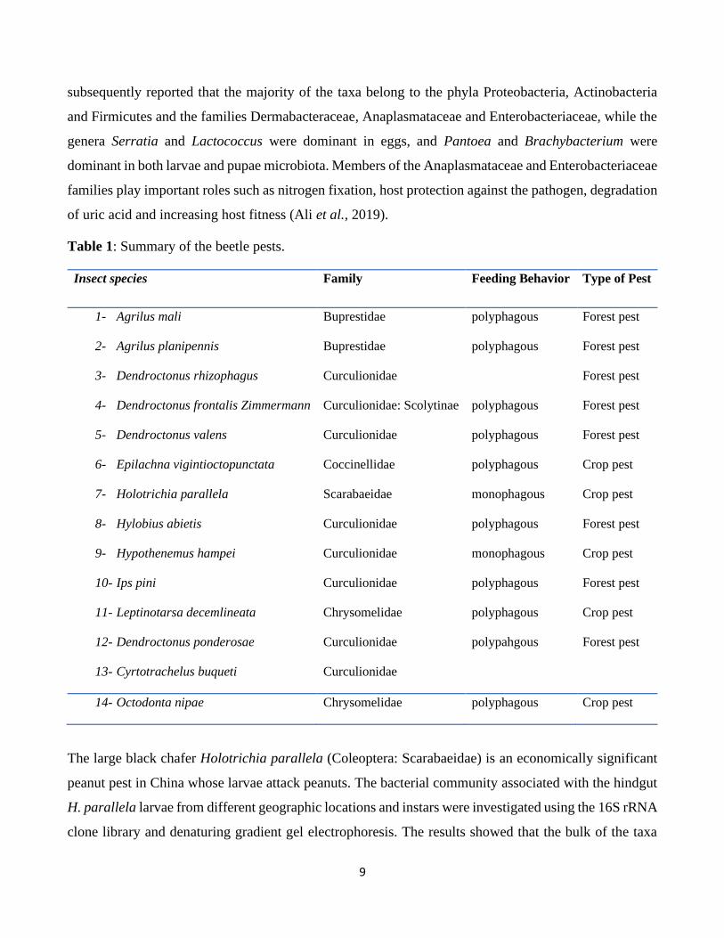
Table 1: Summary of the beetle pests.
Insect species Family Feeding Behavior Type of Pest
1- Agrilus mali Buprestidae polyphagous Forest pest
2- Agrilus planipennis Buprestidae polyphagous Forest pest
3- Dendroctonus rhizophagus Curculionidae Forest pest
4- Dendroctonus frontalis Zimmermann Curculionidae: Scolytinae polyphagous Forest pest
5- Dendroctonus valens Curculionidae polyphagous Forest pest
6- Epilachna vigintioctopunctata Coccinellidae polyphagous Crop pest
7- Holotrichia parallela Scarabaeidae monophagous Crop pest
8- Hylobius abietis Curculionidae polyphagous Forest pest
9- Hypothenemus hampei Curculionidae monophagous Crop pest
10- Ips pini Curculionidae polyphagous Forest pest
11- Leptinotarsa decemlineata Chrysomelidae polyphagous Crop pest
12- Dendroctonus ponderosae Curculionidae polypahgous Forest pest
13- Cyrtotrachelus buqueti Curculionidae
14- Octodonta nipae Chrysomelidae polyphagous Crop pest
The large black chafer Holotrichia parallela (Coleoptera: Scarabaeidae) is an economically significant
peanut pest in China whose larvae attack peanuts. The bacterial community associated with the hindgut
H. parallela larvae from different geographic locations and instars were investigated using the 16S rRNA
clone library and denaturing gradient gel electrophoresis. The results showed that the bulk of the taxa
10
belong to the Firmicutes and Proteobacteria phyla and the Ruminococcaceae, Lachnospiraceae,
Enterobacteriaceae, Desulfovibrionaceae and Rhodocyclaceae families, while the genera Bacteroidetes
and Firmicutes represented the dominant group in the first and (second, third etc.) instars, respectively
(Huang and Zhang, 2013).
The spotted leaf beetle Epilachna vigintioctopunctata Fab. (Coleoptera: Coccinellidae) is a serious pest,
with both adult and larvae forms attacking solanaceous and cucurbitaceous plants, causing serious
damage and the loss of crop production. The gut-associated bacteria in E. vigintioctopunctata has been
characterized in order to explore the relationship between the bacteria associated with the beetle and their
roles in E. vigintioctopunctata development. The results of 16S rRNA partial gene sequencing revealed
the presence of Bacillus subtilis (EVI16), B. vietnamensis (EVI09) and B. anthracis (EVI07). B. subtilis
is known as a microbial insecticide for the effective management of insect pests, while B. anthracis is
known to cause anthrax in mammals (Ramachandiran et al.,2018).
The pine weevil, Hylobius abietis (Coleoptera: Curculionidae: Molytinae), can cause damage, reaching
80% mortality, when it feeds on the stem of bark conifer seedlings. In one study, the gut bacterial
communities of different populations of H. abietis across Europe have been characterized and compared
with those of other beetles that occupy similar ecological niches. The results indicate that some OTUs
within the Enterobacteriaceae from H. abietis are closely related to those of bark beetles (Dendroctonus
ponderosae, D. frontalis, D. valens, D. rhizophagous and Ips pini), and that the microbiota of H. abietis
are different from those of closely related beetles feeding on a different diet. The study also reported that
members of the Enterobacteriaceae family are involved in terpenoid degradation and the detoxification
of plant secondary metabolites, which supports H. abietis development. This confirms that ecological
niches (Berasategui et al., 2016).
Coffea species are small trees that are economically important in countries that produce coffee beans.
The coffee berry borer Hypothenemus hampei (Coleoptera; Curculionidae: Scolytinae) is one of the most
destructive threats to coffee beans. After characterizing the bacterial composition of the H. hampei
microbial communities in specimens from major coffee-producing regions (Hawaii, India, Indonesia,
Kenya, Mexico and Puerto Rico), it was suggested that a core of organisms detected across all locations
belongs to the orders Pseudomonadales, Enterobacteriales, Turicibacteriales, Rhizobiales,
Alteromonadales and Actinomycetales, with the most abundant being Pseudomonadales (Pseudomonas
spp.). This particular study also highlighted the role of Pseudomonas fulva in the degradation of caffeine
11
in vivo, i.e., it enables H. hampei to subsist on caffeine as a source of carbon and nitrogen (Ceja-Navarro
et al., 2015).
The Colorado potato beetle Leptinotarsa decemlineata is a major pest in the case of solanaceous crops,
such as potato (Solanum tuberosum), tomato (S. lycopersicum), and eggplant (S. melongena). The
microbiota associated with this beetle have been investigated, in which 16S rDNA sequences identified
Leclercia adecarboxylata, Acinetobacter and Pseudomonas putida as the core microbiota associated with
L. decemlineata (Muratoglu et al., 2011).
3.The Japanese beetle Popillia japonica (invasion and distribution, biology, and control)
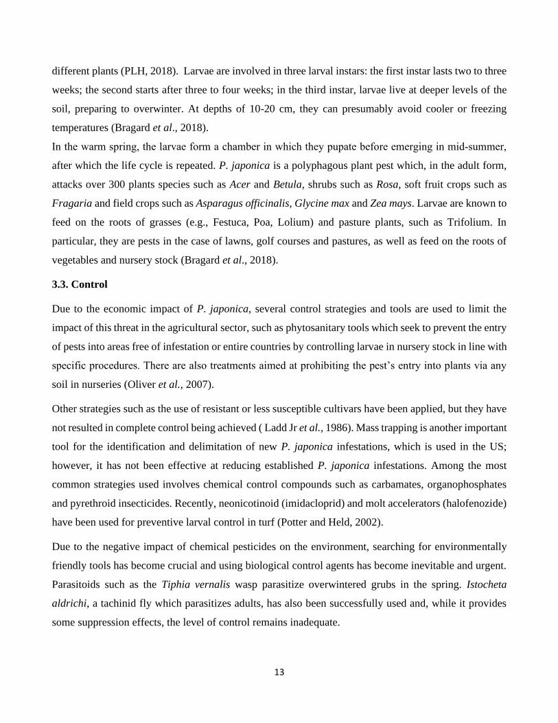
3.1. History of invasion and distribution worldwide
The Japanese beetle, Popillia japonica (Newman, 1841) (Coleoptera: Scarabaeidae), is a polyphagous
threat which is native to Japan and the far east of Russia. It was first discovered in North America in
1916, near Riverton, New Jersey, before spreading across the US. In Japan, it is common and not
considered to be a pest. On the contrary, in the US, P. japonica is a restricted and quarantined pest in all
states (Potter and Held, 2002).
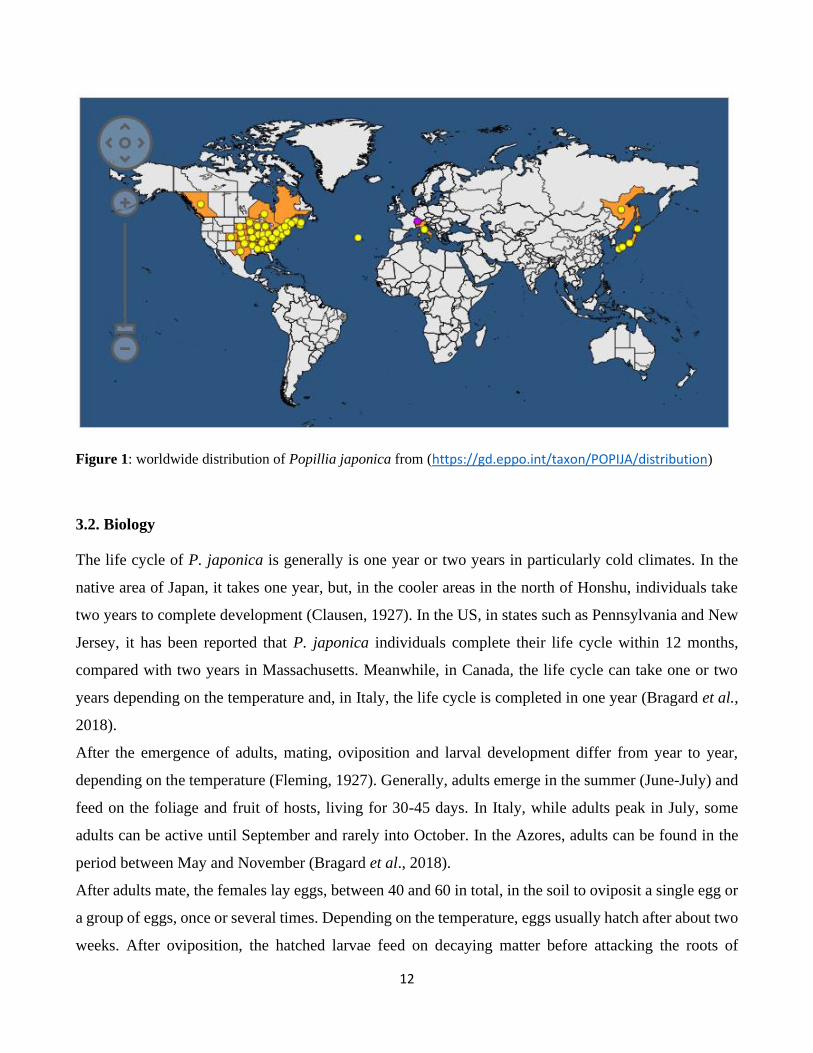
In the EPPO region, P. japonica has recently established itself in the Azores and the Canton of Ticino in
Switzerland. In Canada, the beetle has established itself in Southern Quebec, New Brunswick and Nova
Scotia (EPPO, 2018). P. japonica was recorded for the first time in Italy when an outbreak was reported
within the Ticino Valley Natural Park in Northern Italy in 2014 (Pavesi, 2014). In that year, the infested
area and the number of adults per lure traps increased to 80 km2 and about 28,000 adults using 64 double‐
lure traps, respectively. In 2016, the infested area increased to be about 500 km2 while there were 14.7
million adults by 2,100 traps. In 2017, about 800 km2 were reported to be infested and more than 48
million adults were caught by 2,100 traps(Marianelli et al., 2019). The history of the invasion of P.
japonica indicates the severe threat posed by the beetle and how rapidly the population increases,
especially the Italian population, which falls within the scope of this thesis.
12
Figure 1: worldwide distribution of Popillia japonica from (https://gd.eppo.int/taxon/POPIJA/distribution)
3.2. Biology
The life cycle of P. japonica is generally is one year or two years in particularly cold climates. In the
native area of Japan, it takes one year, but, in the cooler areas in the north of Honshu, individuals take
two years to complete development (Clausen, 1927). In the US, in states such as Pennsylvania and New
Jersey, it has been reported that P. japonica individuals complete their life cycle within 12 months,
compared with two years in Massachusetts. Meanwhile, in Canada, the life cycle can take one or two
years depending on the temperature and, in Italy, the life cycle is completed in one year (Bragard et al.,
2018).
After the emergence of adults, mating, oviposition and larval development differ from year to year,
depending on the temperature (Fleming, 1927). Generally, adults emerge in the summer (June-July) and
feed on the foliage and fruit of hosts, living for 30-45 days. In Italy, while adults peak in July, some
adults can be active until September and rarely into October. In the Azores, adults can be found in the
period between May and November (Bragard et al., 2018).
After adults mate, the females lay eggs, between 40 and 60 in total, in the soil to oviposit a single egg or
a group of eggs, once or several times. Depending on the temperature, eggs usually hatch after about two
weeks. After oviposition, the hatched larvae feed on decaying matter before attacking the roots of
13
different plants (PLH, 2018). Larvae are involved in three larval instars: the first instar lasts two to three
weeks; the second starts after three to four weeks; in the third instar, larvae live at deeper levels of the
soil, preparing to overwinter. At depths of 10-20 cm, they can presumably avoid cooler or freezing
temperatures (Bragard et al., 2018).
In the warm spring, the larvae form a chamber in which they pupate before emerging in mid‐summer,
after which the life cycle is repeated. P. japonica is a polyphagous plant pest which, in the adult form,
attacks over 300 plants species such as Acer and Betula, shrubs such as Rosa, soft fruit crops such as
Fragaria and field crops such as Asparagus officinalis, Glycine max and Zea mays. Larvae are known to
feed on the roots of grasses (e.g., Festuca, Poa, Lolium) and pasture plants, such as Trifolium. In
particular, they are pests in the case of lawns, golf courses and pastures, as well as feed on the roots of
vegetables and nursery stock (Bragard et al., 2018).
3.3. Control
Due to the economic impact of P. japonica, several control strategies and tools are used to limit the
impact of this threat in the agricultural sector, such as phytosanitary tools which seek to prevent the entry
of pests into areas free of infestation or entire countries by controlling larvae in nursery stock in line with
specific procedures. There are also treatments aimed at prohibiting the pest’s entry into plants via any
soil in nurseries (Oliver et al., 2007).
Other strategies such as the use of resistant or less susceptible cultivars have been applied, but they have
not resulted in complete control being achieved ( Ladd Jr et al., 1986). Mass trapping is another important
tool for the identification and delimitation of new P. japonica infestations, which is used in the US;
however, it has not been effective at reducing established P. japonica infestations. Among the most
common strategies used involves chemical control compounds such as carbamates, organophosphates
and pyrethroid insecticides. Recently, neonicotinoid (imidacloprid) and molt accelerators (halofenozide)
have been used for preventive larval control in turf (Potter and Held, 2002).
Due to the negative impact of chemical pesticides on the environment, searching for environmentally
friendly tools has become crucial and using biological control agents has become inevitable and urgent.
Parasitoids such as the Tiphia vernalis wasp parasitize overwintered grubs in the spring. Istocheta
aldrichi, a tachinid fly which parasitizes adults, has also been successfully used and, while it provides
some suppression effects, the level of control remains inadequate.
14
Microorganisms such as Bacillus thuringiensis have been effective in suppressing P. japonica grubs in
turf plots, while entomopathogenic nematodes such as Steinernema glaseri and Heterorhabditis
bacteriophora have been effectively applied to tackle larvae. However, the sensitivity of nematodes to
heat, soil moisture and exposure to sunlight as well as biotic factors can compromise their field efficacy.
In Italy, in order to control P. japonica and limit its spread beyond infested areas, several strategies have
been applied. For example, the use of entomopathogenic nematodes (Heterorhabditis bacteriophora) in
a field experiment resulted in a 45% reduction in P. japonica populations and fungi (Metarhizium
anisopliae) (Marianelli et al., 2019).
All previous control strategies that have been implemented has not been able to completely eradicate P.
japonica. In addition, the lack of information about the microbiota associated with P. japonica prompts
several biological questions about the composition and the role of P. japonica microbiota in helping the
invasive beetle in its development and the colonization of novel habitats.
4. Conclusion
The microbiota associated with forest and agricultural invasive beetles, especially their bacterial
symbionts, are helping their hosts in the processes of protection, colonization and adaptation in novel
habitats. As with many other insects, invasive beetles’ bacterial symbionts are mainly dominated by
bacteria from the phyla Proteobacteria, Actinobacteria, Firmicutes and Bacteroidetes. Proteobacteria
frequently harbor microbiota in various invasive beetles including both those considered to be crop pests
and those considered to be forest pests. Moreover, the Enterobacteriaceae family has been reported to be
present in both forest and agricultural invasive beetles, as well as the genus Pseudomonas. The associated
symbiotic bacteria are playing an important role by enabling their host in the protection, adaptation and
colonization of new habitats. For example, the genera Pantoea and Pseudomonas orientalis are
contributing to the suppression of plant responses against A. mail, in which Pantoea sp. can synthesize
enzymes responsible for plant cell wall degradation and P. orientalis engages in lignin peroxidase
activity. Meanwhile, the genus Streptomyces, associated with Agrilus planipennis, is capable of digesting
carboxymethylcellulose. Further, the genera Lactococcus, Serratia, Dysgonomonas and Enterococcus,
associated with C. buqueti, play an important role in lignocellulose degradation. In terms of protection,
the genus Pseudomonas, which is present in most invasive beetles, such as D. rhizophagus, I. pini, A.
planipennis, A. mali, Cryptocephalus spp. and H. hampei, is engaged in many critically important
activities such as degradation and protecting its host from the pathogens of plants. The beetle P. japonica
is a serious threat worldwide. The lack of information about the microbiota associated with P. japonica
15
undermines the importance of studying this topic and investigating the microbiota associated with P.
japonica – this is the main goal of this thesis.
References
Ali, H., Abrar, M., & Hou, Y. (2019). Pyrosequencing uncovers a shift in bacterial communities across
life stages of Octodonta nipae (Coleoptera: Chrysomelidae). Frontiers in Microbiology, 10,
466.
Berasategui, A., Axelsson, K., Nordlander, G., Schmidt, A., Borg‐Karlson, A. K., Gershenzon, J., ... &
Kaltenpoth, M. (2016). The gut microbiota of the pine weevil is similar across Europe and
resembles that of other conifer‐feeding beetles. Molecular ecology, 25(16), 4014-4031.
Bouchard, P., Bousquet, Y., Davies, A. E., Alonso-Zarazaga, M. A., Lawrence, J. F., Lyal, C. H., ... &
Smith, A. B. (2011). Family-group names in Coleoptera (Insecta). ZooKeys, (88), 1.
Bozorov, T. A., Luo, Z., Li, X., & Zhang, D. (2019). Agrilus mali Matsumara (Coleoptera:
Buprestidae), a new invasive pest of wild apple in western China: DNA barcoding and life
cycle. Ecology and evolution, 9(3), 1160-1172.
Brune, A., 2014. Symbiotic digestion of lignocellulose in termite guts. Nature Reviews Microbiology,
12(3), p.168.
Ceja-Navarro, J. A., Vega, F. E., Karaoz, U., Hao, Z., Jenkins, S., Lim, H. C., ... & Brodie, E. L.
(2015). Gut microbiota mediate caffeine detoxification in the primary insect pest of coffee.
Nature communications, 6, 7618.
Clausen, C. P., & King, J. L. (1927). The parasites of Popillia japonica in Japan and Chosen (Korea)
and their introduction into the United States (No. Folleto 2757).
Colman, D. R., Toolson, E. C., & Takacs‐Vesbach, C. D. (2012). Do diet and taxonomy influence
insect gut bacterial communities? Molecular ecology, 21(20), 5124-5137.
Davis, A. P., Govaerts, R., Bridson, D. M. & Stoffelen, P. An annotated taxonomic conspectus of the
genus Coffea (Rubiaceae). Bot. J. Linn. Soc. 152, 465–512 (2006).
EFSA Panel on Plant Health (PLH), Bragard, C., Dehnen‐Schmutz, K., Di Serio, F., Gonthier, P.,
Jacques, M. A., ... & Navas‐Cortes, J. A. (2018). Pest categorisation of Popillia japonica. Efsa
Journal, 16(11), e05438.
16
Engel, P., & Moran, N. A. (2013). The gut microbiota of insects–diversity in structure and function.
FEMS microbiology reviews, 37(5), 699-735.
European and Mediterranean Plan Protection Organization (EPPO). 2018. Global database.
(https://gd.eppo.int/taxon/POPIJA/distribution) (Accessed 30 July 2018).
Fleming, W. E. (1972). Preventing Japanese beetle dispersion by farm products and nursery stock (No.
1441). US Department of Agriculture.
Georgis, R., & Gaugler, R. (1991). Predictability in biological control using entomopathogenic
nematodes. Journal of economic entomology, 84(3), 713-720.
Greathead D.J., Greathead A.H. Biological control of insect pest by insect parasitoids and predators:
The BIOCAT database. Biocontrol. News Inf. 1992; 13:61–68. [Ref list]
Gullan, P.J.; Cranston, P.S. (2014). The Insects: An Outline of Entomology (5 ed.). John Wiley & Sons.
p. 6. ISBN 978-1-4443-3036-6.
Hernández-García, J. A., Briones-Roblero, C. I., Rivera-Orduña, F. N., & Zúñiga, G. (2017). Revealing
the gut bacteriome of Dendroctonus bark beetles (Curculionidae: Scolytinae): diversity, core
members and co-evolutionary patterns. Scientific reports, 7(1), 13864.
Huang, S., & Zhang, H. (2013). The impact of environmental heterogeneity and life stage on the
hindgut microbiota of Holotrichia parallela larvae (Coleoptera: Scarabaeidae). PLoS One, 8(2),
e57169.
Judson Linsley Gressitt. (2018) Coleopteran. Encyclopædia Britannica, inc.
https://www.britannica.com/animal/beetle.
Kistner-Thomas, E. J. (2019). The potential global distribution and voltinism of the Japanese beetle
(Coleoptera: Scarabaeidae) under current and future climates. Journal of Insect Science, 19(2),
16.
Ladd Jr, T. L., & Lawrence, K. O. (1986). Elimination of Japanese beetle larvae from plant growth
medium by using isofenphos 2• 3. J. Agric. Entomol, 3(2), 170-174.
Lu, M., Wingfield, M.J., Gillette, N.E., Mori, S.R. and Sun, J.H., 2010. Complex interactions among
host pines and fungi vectored by an invasive bark beetle. New Phytologist, 187(3), pp.859-866.
17
Lundgren, J. G., & Lehman, R. M. (2010). Bacterial gut symbionts contribute to seed digestion in an
omnivorous beetle. PLoS one, 5(5), e10831.
Luo, C., Li, Y., Chen, Y., Fu, C., Long, W., Xiao, X., ... & Yang, Y. (2019). Bamboo lignocellulose
degradation by gut symbiotic microbiota of the bamboo snout beetle Cyrtotrachelus buqueti.
Biotechnology for biofuels, 12(1), 70.
Marianelli, L., Paoli, F., Sabbatini Peverieri, G., Benvenuti, C., Barzanti, G. P., Bosio, G., ... &
Roversi, P. F. (2019). Long‐lasting insecticide‐treated nets: A new integrated pest management
approach for Popillia japonica (Coleoptera: Scarabaeidae). Integrated environmental
assessment and management, 15(2), 259-265.
Moran, N. A., & Telang, A. (1998). Bacteriocyte-associated symbionts of insects. Bioscience, 48(4),
295-304.
Morales-Jiménez, J., Zúñiga, G., Ramírez-Saad, H. C., & Hernández-Rodríguez, C. (2012). Gut-
associated bacteria throughout the life cycle of the bark beetle Dendroctonus rhizophagus
Thomas and Bright (Curculionidae: Scolytinae) and their cellulolytic activities. Microbial
Ecology, 64(1), 268-278.
Muratoglu, H., Demirbag, Z., & Sezen, K. (2011). The first investigation of the diversity of bacteria
associated with Leptinotarsa decemlineata (Coleoptera: Chrysomelidae). Biologia, 66(2), 288-
293.
Oliver, J. B., Reding, M. E., Klein, M. G., Youssef, N. N., Mannion, C. M., Bishop, B., ... & Callcott,
A. M. (2014). Chlorpyrifos immersion to eliminate third instars of Japanese beetle (Coleoptera:
Scarabaeidae) in balled and burlapped trees and subsequent treatment effects on red maple.
Journal of economic entomology, 100(2), 307-314.
Pavesi, M. (2014). Popillia japonica specie aliena invasiva segnalata in Lombardia. L’informatore
Agrario, 32, 53-55.
Peng, L. F., Li, J. L., Hou, Y. M., & Zhang, X. (2018). Descriptions of immature stages of Octodonta
nipae (Maulik) (Coleoptera, Chrysomelidae, Cassidinae, Cryptonychini). ZooKeys, (764), 91.
Potter, D. A., & Held, D. W. (2002). Biology and management of the Japanese beetle. Annual review of
entomology, 47(1), 175-205.
18
Ramachandiran, S., Sankaraiyah, K., Kavipriya, J., Vijiyalakshmi, U., & Sakunthala, C. (2018).
Identification and characterization of gut associated bacteria in Epilachna vigintioctopunctata
Fab. (Caoleoptera: Coccinellidae). Entomon, 43(1), 1-6.
Redmond, C. T., & Potter, D. A. (1995). Lack of efficacy of in vivo-and putatively in vitro-produced
Bacillus popilliae against field populations of Japanese beetle (Coleoptera: Scarabaeidae) grubs
in Kentucky. Journal of economic entomology, 88(4), 846-854.
Rosenzweig, M. L. (1995). Species Diversity in Space and Time. Cambridge: Cambridge University
Press. p. 2. ISBN 978-0-521-49952-1.
Shigenobu, S., Watanabe, H., Hattori, M., Sakaki, Y., & Ishikawa, H. (2000). Genome sequence of the
endocellular bacterial symbiont of aphids Buchnera sp. APS. Nature, 407(6800), 81.
Stork, Nigel E.; McBroom, James; Gely, Claire; Hamilton, Andrew J. (2015). "New approaches narrow
global species estimates for beetles, insects, and terrestrial arthropods". PNAS. 116 (24): 7519–
7523.
van den Bosch, T. J., & Welte, C. U. (2017). Detoxifying symbionts in agriculturally important pest
insects. Microbial biotechnology, 10(3), 531-540.
Vasanthakumar, A., Burwitz, B. J., Schloss, P. D., Klepzig, K. D., Handelsman, J., & Raffa, K. F.
(2007). Composition of the bacterial community in the gut of the pine engraver, Ips pini
(Say)(Coloptera) colonizing red pine. Symbiosos, Vol. 43: 97-104.
Vasanthakumar, A., Delalibera Jr, I., Handelsman, J., Klepzig, K. D., Schloss, P. D., & Raffa, K. F.
(2006). Characterization of gut-associated bacteria in larvae and adults of the southern pine
beetle, Dendroctonus frontalis Zimmermann. Environmental Entomology, 35(6), 1710-1717.
Vasanthakumar, A., Handelsman, J. O., Schloss, P. D., Bauer, L. S., & Raffa, K. F. (2008). Gut
microbiota of an invasive subcortical beetle, Agrilus planipennis Fairmaire, across various life
stages. Environmental Entomology, 37(5), 1344-1353.
Vasanthakumar, A., Handelsman, J. O., Schloss, P. D., Bauer, L. S., & Raffa, K. F. (2008). Gut
microbiota of an invasive subcortical beetle, Agrilus planipennis Fairmaire, across various life
stages. Environmental Entomology, 37(5), 1344-1353. (Placeholder1)
19
Xia, X., Sun, B., Gurr, G. M., Vasseur, L., Xue, M., & You, M. (2018). Gut microbiota mediate
insecticide resistance in the diamondback moth, Plutella xylostella (L.). Frontiers in
microbiology, 9, 25.
Yang, H., Su, T., Yang, W., Yang, C., Lu, L., & Chen, Z. (2017). The developmental transcriptome of
the bamboo snout beetle Cyrtotrachelus buqueti and insights into candidate pheromone-binding
proteins. PloS one, 12(6), e0179807. doi: 10.1371/journal.pone.0179807
Zhang, Z., Jiao, S., Li, X., & Li, M. (2018). Bacterial and fungal gut communities of Agrilus mali at
different developmental stages and fed different diets. Scientific reports, 8(1), 15634.
Zheng, H., Powell, J. E., Steele, M. I., Dietrich, C., & Moran, N. A. (2017). Honeybee gut microbiota
promotes host weight gain via bacterial metabolism and hormonal signaling. Proceedings of the
National Academy of Sciences, 114(18), 4775-4780.
20
First study: Developmental stages and gut microenvironments influence
gut microbiota dynamics in the invasive beetle Popillia japonica Newman
(Coleoptera: Scarabaeidae)
Summary
Popillia japonica Newman (Coleoptera: Scarabaeidae) is a highly polyphagous invasive beetle
originating from Japan. This insect is highly resilient and able to rapidly adapt to new vegetation. Insect-
associated microorganisms can play important roles in insect physiology, helping their hosts to adapt to
changing conditions and potentially contributing to an insect’s invasive potential. Such symbiotic
bacteria can be part of a core microbiota that is stably transmitted throughout the host’s life cycle or
selectively recruited from the environment at each developmental stage. The aim of this study was to
investigate the origin, stability, and turnover of the bacterial communities associated with an invasive
population of P. japonica from Italy. Our results demonstrate that soil microbes represent an important
source of gut bacteria for P. japonica larvae, but as the insect develops, it's gut microbiota richness and
diversity decreased substantially, paralleled by changes in community composition. Notably, only
16.75% of the soil bacteria present in larvae are maintained until the adult stage. We further identified
the micro-environments of different gut sections as an important factor shaping microbiota composition
in this species, likely due to differences in pH, oxygen availability and redox potential. In addition, P.
japonica also harbored a stable bacterial community across all developmental stages, consisting of taxa
well-known for the degradation of plant material, namely the families Ruminococcacae,
Christensenellaceae, and Lachnospiraceae. Interestingly, the family Christensenallaceae had so far been
observed exclusively in humans. However, the Christensenellaceae OTUs found in P. japonica belong
to different taxonomic clades within this family.
1. Introduction
Insects are the most diverse and abundant animal clades (Foottit and Adler, 2009). The diversification
and evolutionary success of insects have been partially attributed to their ability to establish associations
with different beneficial microorganisms (e.g., Douglas, 2014; Corbin et al., 2017; Sudakaran et al.,
2017; Heddi and Zaidman-Rémy, 2018). These microorganisms can play key roles for different
physiological functions such as the supply of essential nutrients missing from unbalanced diets;
contributing to the digestion of recalcitrant food components; protection from predators, parasites and
21
pathogens; and controlling mating and reproductive systems (e.g., Leftwich et al., 2017; Muhammad et
al., 2017).
As for essentially all animals, microbial communities are particularly prominent in the digestive tract
(e.g., Douglas, 2015, 2018; Clayton et al., 2018; Münger et al., 2018). The insect gut is generally
structured into foregut, midgut, and hindgut, presenting a multitude of micro-environments suitable for
microbial colonization. Differences in morphology and Physico-chemical properties between different
gut sections can greatly influence the microbial colonization patterns and community structure depending
on the host species. Gut bacteria have the potential to provide many beneficial services to their hosts and
insects display a wide range in degree of dependence on gut bacteria for basic functions. Paramount to
the evolution of intimate associations with gut microorganisms in the development of secure transmission
routes between host individuals and generations. The lack of such mechanism in most insect species may
hinder the establishment of such long-term associations. With the exception of social insects, such as
termites and ants, where social interactions provide opportunities for the transfer of gut bacteria
(Zhukova et al., 2017), insects had to develop original ways in order to transmit the important
components of their gut microbiota (Fukatsu and Hosokawa, 2002; Gonella et al., 2012; Hosokawa et
al., 2013; Mason et al., 2019). These "heritable" gut bacteria have been shown to play crucial roles in the
nutrition, protection against different pathogens and xenobiotics, modulation of immune responses, and
even extending life span (Roh et al., 2008; Kim et al., 2016; Daisley et al., 2018; Obata et al., 2018).
Several factors can influence the gut microbiota structure and composition. Among these factors, the
most important ones are diet and environment, but other factors (e.g., age) can also be at play (Wong et
al., 2011; Montagna, Chouaia, et al., 2015; Montagna, Gómez-Zurita, et al., 2015; Montagna et al., 2016;
Sanders et al., 2017; Tiede et al., 2017; Vacchini et al., 2017; Anderson et al., 2018). Although various
factors can influence the insect gut microbiota, the existence of a shared core microbial community in
some species could indicate that there are mechanisms (e.g. vertical transmission) favoring the presence
of certain members of the gut microbiota. Several studies have investigated this possibility by tracking
the changes in gut microbiota composition along the developmental stages of different insect species.
These studies showed that the transmission of the gut microbiota throughout the different developmental
stages may depend on the usefulness of certain bacteria (Zhukova et al., 2017; Malacrinò et al., 2018).
For instance, the bacterial communities of fruit flies (Tephritideae) change throughout the insect's
developmental stages to respond to the physiological needs of the host (Aharon et al., 2013; Malacrinò
et al., 2018). In holometabolous insects, the pupal stage generally represents a bottleneck where most of
22
the larval gut microbiota is lost and adult insects may have to resort to indirect ways (e.g. via
environmental transmission) to insure the transfer of beneficial bacteria from larvae to adults (Zhukova
et al., 2017). For instance, in certain bee species, certain bacterial taxa are not trans-stadially transmitted
but re-acquired from the environment (McFrederick et al., 2014). While the gut microbiota is not constant
across the developmental stages in most insects, in some cases the microbial community can be relatively
stable throughout the developmental stages. This has been observed in some Tephritid flies as well as in
the Black Soldier Fly Hermetia illucens and in the moth Plodia interpunctella (Mereghetti et al., 2017;
Yong et al., 2017; De Smet et al., 2018).
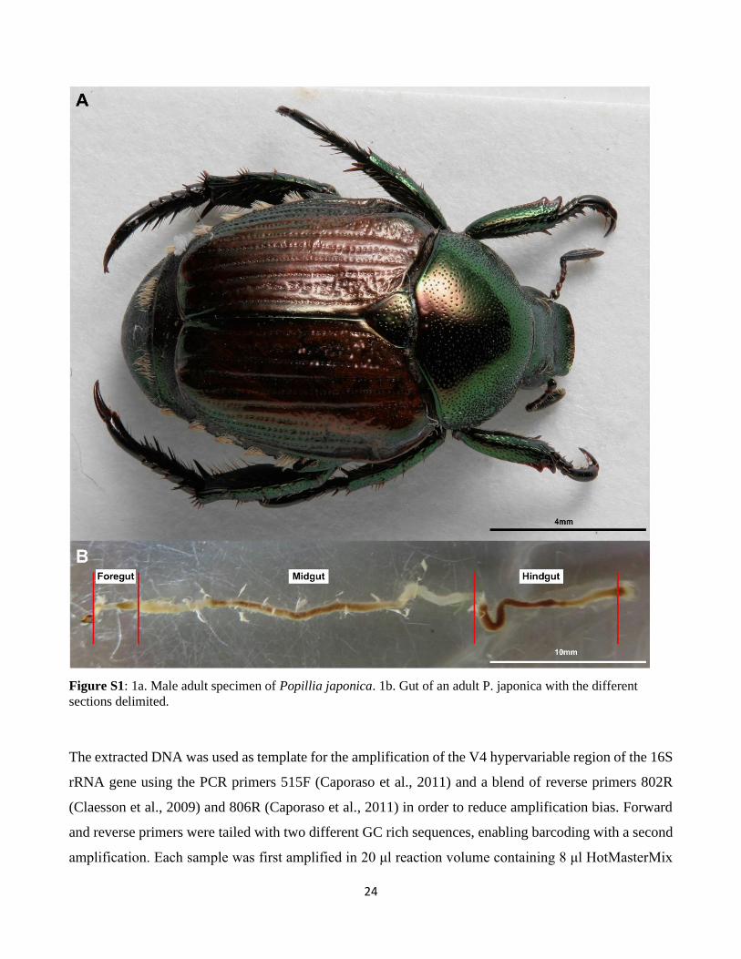
In the present study, we focused on the highly polyphagous invasive Japanese beetle Popillia japonica
Newman (Coleoptera: Scarabaeidae, Fig. S1a). This invasive insect is listed in the EPPO Annex 2 due
to the damages caused to different crops and turfs (EPPO, 2000). Native to Japan and the far east of
Russia (Fleming, 1972), this beetle became an established pest in North America in the early 1900's
(Switzer et al., 2009), in the Azores in the early 1970's (Vieira, 2008) and more recently in continental
Europe, where it was recorded for the first time in Italy in 2014 (EPPO, 2014; Pavesi, 2014) and in
Switzerland in 2017 (EPPO, 2017). Several laboratory and field trials have been carried out to limit the
spread of this pest in mainland Europe and to evaluate the environmental resilience of the infested areas
(Mazza et al., 2017; Paoli, Marianelli, Binazzi, et al., 2017; Paoli, Marianelli, Torrini, et al., 2017;
Marianelli, Paoli, Sabbatini Peverieri, et al., 2018; Marianelli, Paoli, Torrini, et al., 2018). The damages
to plants are caused by the different developmental stages of the beetle: the larvae, being underground
dwellers, feed on the plant roots and soil organic matter while adults, living in an above-ground
environment, feed on leaves and floral parts of different plant species (Fleming, 1972; Vieira, 2008).
Insect-associated bacteria can potentially contribute to an insect’s invasive potential by helping their
hosts to adapt to changing environmental conditions. Such symbiotic bacteria can be part of a core
microbiota that is stably transmitted throughout the host’s life cycle or selectively recruited from the
environment at each developmental stage. The aim of this study was to investigate microbiota dynamics
in an invasive population of P. japonica from Italy. Specifically, we addressed the following questions:
i) Does P. japonica harbor a stable core microbiota or are the bacteria mainly acquired from the
surrounding environment (i.e. rhizospheric soil exploited by larvae and pupae vs aerial environment
exploited by adults)? ii) Is the gut microbiota maintained across the post-embryonic developmental
stages (i.e. larvae, pupae, and adults) or is there a major turnover due to insect development? iii) Do
different gut micro-environments impact microbial community structure?
23
2. Materials and methods
2.1. Collection and processing of insect and soil samples
Four campaigns were organized from June to September 2017 to collect insect samples at different
developmental stages of the insect. The different stages and instars (in the case of larvae: larval instar 1
– L1; larval instar 2 – L2; larval instar 3 – L3) of the insects were collected in Oleggio (Novara, Italy;
45°36′ N, 08°38′ E, altitude ca. 230 m a.s.l.). Simultaneously, at each sampling expedition, 10 soil
samples were taken from the sampled area and combined into a single sample representative of the area,
leading to the collection of three soil samples. Insects were preserved in absolute ethanol while soil
samples in 50 ml vials, kept refrigerated on the field and then stored at -20°C before processing. All
insects were surface sterilized before dissection using the protocol described in Montagna and colleagues
(Montagna, Chouaia, et al., 2015). 90 individuals (i.e. 15 individuals of each larval instar, 15 pupae, 15
males, 15 females) were dissected under sterile conditions, and the gut (Fig. S1b) was removed in sterile
Ringer solution. The insect alimentary canal was then aseptically separated into its three compartments
(i.e. foregut, midgut, and hindgut). For each developmental stage and larval instar, five homologous gut
compartments were pooled together in a single sample, resulting in three biological replicates for each
sample category. These samples were used for DNA extraction (see table S1 for detail on the samples).
Additionally, male adults (N=9) and L3 larvae (N=6) were collected and immediately processed in order
to measure physicochemical properties (pH level, redox potential, oxygen concentration) of different gut
regions. Specimens were anesthetized at 4°C for 3’ before their dissection.
2.2. DNA extraction, amplicon library preparation, sequencing and bioinformatics
The DNA was extracted from each sample (consisting of five homologous gut compartments for a
defined insect instar and developmental stage) using the phenol-chloroform methods (Doyle and Doyle,
1990) with the modifications described in Mereghetti and colleagues (Mereghetti et al., 2017). The DNA
was then eluted in 50 μl of sterile water (Sigma-Aldrich, Saint Louis, Missouri, USA). A DNA extraction
blank was performed as control to monitor for contamination of environmental bacterial DNA. DNA
from soils was extracted using PowerSoil DNA Isolation Kit MO BIO Laboratories Inc., Carlsbad, CA)
following manufacturer's instructions. Three independent DNA extractions were performed for each of
the three representative soil samples.
24
Figure S1: 1a. Male adult specimen of Popillia japonica. 1b. Gut of an adult P. japonica with the different
sections delimited.
The extracted DNA was used as template for the amplification of the V4 hypervariable region of the 16S
rRNA gene using the PCR primers 515F (Caporaso et al., 2011) and a blend of reverse primers 802R
(Claesson et al., 2009) and 806R (Caporaso et al., 2011) in order to reduce amplification bias. Forward
and reverse primers were tailed with two different GC rich sequences, enabling barcoding with a second
amplification. Each sample was first amplified in 20 μl reaction volume containing 8 μl HotMasterMix
25
5 Prime 2.5X (Quanta Bio), 0.4 μl BSA (20 μg/μl) (Sigma-Aldrich), 1 μl EvaGreen™ 20X (Biotium),
0.8μl 515 F (10 μM) (- 5' modified with unitail 1 5'-CAGGACCAGGGTACGGTG-3'), 0.4 μl 802 R (10
μM) (- 5' modified with unitail 2 5'-CGCAGAGAGGCTCCGTG-3'), 0.4 μl 806 R (10 μM) (- 5' modified
with unitail 2 5'-CGCAGAGAGGCTCCGTG-3'), and 1 μl (50 ng) of DNA template. The PCR
amplifications were performed in a CFX 96™ PCR System (Bio-Rad) with 34 cycles of 94°C for 20 s,
52°C for 20 s, 65°C for 40 s and a final extension of 65°C for 2 min. The second PCR amplification was
performed in 25 μl reaction volume containing the same reagents as the first PCR but with 1.5 μl
barcoded/TrP1 primers (10 μM) and with 1 μl of the first PCR amplification in the following conditions:
8 cycles of 94°C for 10 s, 60°C for 10 s, 65°C for 40 s and a final extension of 72°C for 3 min. After
labeling each sample with a specific Ion Torrent (Ion Express) DNA barcode, each single library was
quality checked with agarose gel electrophoresis, quantified with Qubit Fluorometer (Thermo Fisher
Scientific) then pooled with the other libraries in equimolar amounts. The final product was then
sequenced using the Ion Torrent PGM System. Libraries preparation and sequencing were performed at
the Life Sciences Department of Trieste University, Italy. Four samples (see table S1a for details) were
excluded from the following analyses since they did not have enough reads (less than 200). The reads of
the remaining samples were analyzed using QIIME version 1.9.1 (Caporaso et al., 2010). In detail,
adapters were removed, and low-quality reads filtered (Phred < 20, read length < 250pb). Uclust (Edgar,
2010) was used to cluster the 16S rRNA sequences into Operational Taxonomic Units (OTUs) with a
similarity cut-off of 97%. Chimeras were removed using Chimeraslayer. A representative sequence for
each identified OTUs was aligned to Green-genes (http://greengenes.lbl.gov/) using Pynast (Caporaso et
al., 2010). Taxonomic assignment was performed comparing the representative OTUs to Green-genes
(release 13.8). Rare OTUs (i.e., singletons and OTUs < 10) and OTUs identified as chloroplast were
discarded. The resulting OTU table was then used for the subsequent analyses.
2.3. Diversity analyses
Bacterial OTU richness, diversity and evenness were calculated using the package Vegan (Dixon, 2003;
Oksanen et al., 2018), implemented under the R software (R Project 3.0.2; http://cran.r-project.org/)
adopting the species richness estimator Chao 1 (Chao, 1984), the Shannon H’ index (Shannon, 1948) and
the Pielou's evenness (Pielou, 1975), after sub-sampling the OTU table to obtain a total of 25,000
sequences per sample. Alpha diversity indices were compared between different groups (i.e. tissues,
developmental stages) using two-sample t-tests with 999 Monte Carlo permutations.
26
In order to evaluate if the structures of the bacterial communities associated with soil and the different
developmental stages of P. japonica were driven by species competition or by environmental factors,
thus resulting in a community dominated by closely related species (Webb et al., 2002; Mouquet et al.,
2012; O’Dwyer et al., 2012), the mean pairwise distance between all taxa in the bacterial communities
(MPD; Webb et al., 2002) was used as metric for phylogenetic structure. To allow the comparison
between the bacterial communities of the different types, null models maintaining species occurrence
frequency constant were estimated. Standard effect size and relative position of each bacterial community
with respect to the null MDP distribution, generated by 999 randomizations of the null model, were
calculated using the ses.mpd function implemented in the Rpackage picante (Kembel et al., 2010). This
standardized metric quantifies the relative excess or deficit in the phylogenetic diversity for each
community with respect to the entire species pool. Negative values reflect a relative phylogenetic
clustering of the species, while positive values indicate a relative phylogenetic evenness (or
overdispersion). SESMDP values were visualized as box-plots based on sample type (i.e., soil, larvae,
pupae, adults) and statistical differences among sample types were assessed using Welch’s one-way
ANOVA (Welch, 1951), since SESMDP values were normally distributed based on Shapiro-Wilk test
(Royston 1982) (p-value > 0.05), but the variance between groups was not homogeneous based on
Levene test (Levene, 1960) (p-value < 0.001). Hence, we used the Tamhane post-hoc test for multiple
comparisons without homoscedasticity.
The spatial (across the three gut regions) and temporal shifts (across developmental stages) of the P.
japonica bacterial community (presence/absence) were estimated using the Sørensen-based multiple-site
dissimilarity (βSOR; Baselga, 2010) implemented in the R package betapart (Baselga and Orme, 2012).
The turnover and nestedness components of this β-diversity were calculated using Simpson-based
multiple-site dissimilarity (βSIM; Baselga, 2010) and nestedness-resultant multiple-site dissimilarity
(βNES; Baselga, 2010), respectively. In addition, for each β-diversity component, the pairwise
dissimilarity values among the microbiotas of all analysed groups (i.e. soil, larvae, pupae and adults)
were calculated using the betapair function of the R package betapart (Baselga and Orme, 2012) and
visualized through heatmaps using heatmap.2 from the R package gplots.
In order to assess the difference in the microbiota structure among soil and insect samples, the sub-
sampled OTU table was subjected to a nonparametric one-way analysis of similarity ANOSIM (Clarke,
1993), implemented in the vegan library and based on the Bray-Curtis dissimilarity (999 permutations
27
permuting within gut samples of the same individuals in order to account for the non-independence of
the observations (Bray and Curtis, 1957).
The sub-sampled OTU table, after the removal of soil community samples, was used as input for a
Nonmetric Multi‐Dimensional Scaling (NMDS; Kruskal, 1964) biplot based on the Bray–Curtis
dissimilarity (Bray and Curtis, 1957), in order to graphically ordinate samples and assess the differences
among: i) the developmental stages (i.e. larvae, pupae and adults); ii) the three gut regions, and iii) to
evaluate the impact of the gut physicochemical properties on the microbiotas associated with third instar
larvae and adults. NMDS analyses were performed using the metaMDS function implemented in the R
package Vegan (Dixon, 2003; Oksanen et al., 2018). The correlation between the microbiota composition
and the tested factors (i.e. developmental stages, gut sections, gut physicochemical properties) was
investigated by fitting the NMDS ordination scores with the envfit Vegan function (Dixon, 2003;
Oksanen et al., 2018). The permutation of the community composition‐based dissimilarity matrix (taking
into account the non-independence of the different gut samples of the same individuals) allowed
assessment of the significance of the fitted factors and vectors, and a squared correlation coefficient (R2)
was calculated.
To determine the level of specificity of the microbiota composition associated with each developmental
stage or gut region, model predictions were generated using Random Forest regressors based on the
relative abundance OTU table (Knights et al., 2011). In order to classify the microbiota samples based
on the host developmental stage or gut region, the supervised_learning.py script from the QIIME pipeline
was used. cv10 was used as error correction method with 999 replicate trees.
2.4. Changes in microbiota composition
In order to identify OTUs shared between the different insect developmental stages and the soil, we only
focused on OTUs that were typical for a given sample type (i.e. larvae, pupae, adults, soil). To this end,
an OTU was considered “present” in a given sample type only when it occurred in at least 66% of the
biological replicates of that sample type (in most cases, 2 out of 3 biological replicates). These OTUs are
hereafter referred to as “core OTUs”. The “core OTUs” specific to or shared among the different
developmental stages and the soil were visualized through a Venn diagram. In addition, a bipartite
network analysis (Dormann et al., 2008) of the bacterial community associated with the P. japonica
(larvae, pupae, and adults) and the bulk soil was performed using the pairwise dissimilarity matrix
generated from the OTU table adopting the Bray-Curtis dissimilarity index (Bray and Curtis, 1957).
28
Cytoscape (Shannon et al., 2003) was used to visualize the network. Differentially abundant taxa were
determined after data normalization of the OTU table using the EdgeR package (version 3.16.5) with R
(version 3.4.4). Differentially abundant OTUs were then ranked by their log2 fold change from the most
differentially abundant to the least differentially abundant. Ranked OTUs were used to determine
enriched families between different groups using the tmod package (version 0.36) with the CERNO test
(Yamaguchi et al., 2008) and the Benjamini-Hochberg correction. The position of the OTUs belonging
to enriched families along the continuum of ranked OTUs was also assessed visually using ROC curves
(Receiver Operating Characteristic curves). The enriched families were then tested for their presence in
all samples (supplementary table S3).
The OTU sequences of enriched taxa of interest (i.e. Christensenellaceae) were retrieved from the OTU
file then aligned to complete or near-complete 16S rRNA sequences downloaded from the NCBI website
(www.ncbi.nlm.nih.gov) using Clustal W. After gap removal, the evolution model was estimated using
jModeltest according to the Akaike Information Criterion (AIC) parameter (Akaike, 1976). The
phylogenetic tree was reconstructed using maximum likelihood with the Kimura 2 parameters model and
500 bootstraps. The phylogenetic tree was reconstructed and visualized using Mega X (Kumar et al.,
2018).
In order to detect OTUs that are specific for a given gut section within the same developmental stage,
the indicator value (Dufrêne and Legendre, 1997) was calculated using the R package indicspecies (De
Cáceres and Legendre, 2009). Briefly, the indicator value of an OTU varies from 0 to 1 and attains its
maximum value when all reads of an OTU occur in all samples of only one specific gut section. We
tested the significance of the indicator value for each OTU with a Monte Carlo randomization procedure
with 999 permutations.
2.5. Measurement of the gut physicochemical properties
Physico-chemical parameters of oxygen partial pressure (pO2), pH and redox potential were measured
in the different sections of P. japonica gut (foregut, midgut, and hindgut) with microsensors and
microelectrodes (Unisense, Aarhus, Denmark). Freshly dissected guts from both L3 larvae and males
were placed on a layer of 2% (Low Melting Point) agarose prepared with Ringer’s solution (7.2 g/L
NaCl; 0.37 g/L KCl; 0.17 g/L CaCl2, pH 7.3-7.4) and immediately covered with a second layer of 0.5%
agarose prepared with Ringer’s solution (Šustr et al., 2014). Oxygen microsensors (OX-50), with a tip
diameter of 50 μm, were calibrated after an overnight polarization in water saturated with air and in 0.1
29
M sodium dithionite anoxic solution by using the CAL 300 calibration chamber (Unisense, Aarhus,
Denmark), following an overnight polarization. pH microelectrodes (PH-50), with a tip diameter of 50
μm, were calibrated with standard solutions at pH 4.0, 7.0 and 10.0. Redox potential microelectrodes
(RD-50) had a tip diameter of 50 μm and were calibrated using saturated quinhydrone solutions at pH
4.0 and 7.0. Electrode potentials for microelectrodes were measured against Ag-AgCl reference
electrodes by using a high-impedance voltmeter (Ri > 1014 Ω). Unisense microsensor multimeter
allowed to measure the current and data were recorded by using SensorTracePRO software (Unisense,
Aarhus, Denmark). Microsensors were positioned using a motorized micromanipulator (Unisense,
Aarhus, Denmark). Measurements were carried out at room temperature.
3. Results
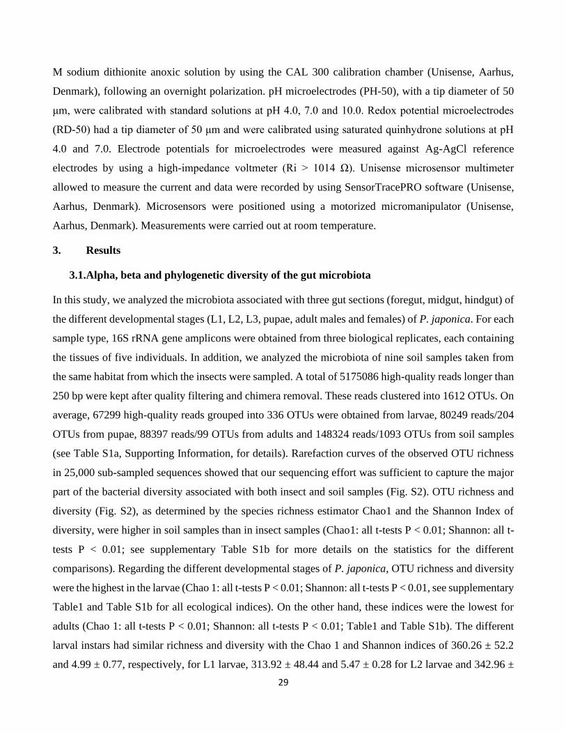
3.1.Alpha, beta and phylogenetic diversity of the gut microbiota
In this study, we analyzed the microbiota associated with three gut sections (foregut, midgut, hindgut) of
the different developmental stages (L1, L2, L3, pupae, adult males and females) of P. japonica. For each
sample type, 16S rRNA gene amplicons were obtained from three biological replicates, each containing
the tissues of five individuals. In addition, we analyzed the microbiota of nine soil samples taken from
the same habitat from which the insects were sampled. A total of 5175086 high-quality reads longer than
250 bp were kept after quality filtering and chimera removal. These reads clustered into 1612 OTUs. On
average, 67299 high-quality reads grouped into 336 OTUs were obtained from larvae, 80249 reads/204
OTUs from pupae, 88397 reads/99 OTUs from adults and 148324 reads/1093 OTUs from soil samples
(see Table S1a, Supporting Information, for details). Rarefaction curves of the observed OTU richness
in 25,000 sub-sampled sequences showed that our sequencing effort was sufficient to capture the major
part of the bacterial diversity associated with both insect and soil samples (Fig. S2). OTU richness and
diversity (Fig. S2), as determined by the species richness estimator Chao1 and the Shannon Index of
diversity, were higher in soil samples than in insect samples (Chao1: all t-tests P < 0.01; Shannon: all t-
tests P < 0.01; see supplementary Table S1b for more details on the statistics for the different
comparisons). Regarding the different developmental stages of P. japonica, OTU richness and diversity
were the highest in the larvae (Chao 1: all t-tests P < 0.01; Shannon: all t-tests P < 0.01, see supplementary
Table1 and Table S1b for all ecological indices). On the other hand, these indices were the lowest for
adults (Chao 1: all t-tests P < 0.01; Shannon: all t-tests P < 0.01; Table1 and Table S1b). The different
larval instars had similar richness and diversity with the Chao 1 and Shannon indices of 360.26 ± 52.2
and 4.99 ± 0.77, respectively, for L1 larvae, 313.92 ± 48.44 and 5.47 ± 0.28 for L2 larvae and 342.96 ±
30
43.02 and 5.74 ± 0.27 for L3 larvae (Chao 1: all t-tests p-value > 0.5; Shannon: all t-tests P > 0.5, Table
S1b). It is noteworthy that the values of Pielou’s evenness also followed a similar pattern, with the soil
having the highest value (Pielou’J = 0.84; Table1), then larvae (Pielou’J = 0.67; Table1) and with pupae
and adults having similar values (Pielou’J = 0.47 and 0.49 respectively; Table 1).
Figure S2: Alpha diversity parameters by sample or sample type. A: Chao1 index for all the samples. B: Chao1
index reported by gut section. C: Chao1 index reported by developmental stage. D: Shannon index for all the
samples. E: Shannon index reported by gut section. F: Shannon index reported by developmental stage.
Table 1: Ecological indices by developmental stage (mean ± SE)
Richness (Chao1) Diversity (Shannon) Evenness (Pielou)
Soil 1099 ± 1.35 5.88 ± 0.03 0.84 ± 0.00
Larvae 369.93 ± 28.95 3.77 ± 0.19 0.67 ± 0.03
Pupae 241.12 ± 43.51 2.49 ± 0.39 0.47 ± 0.06
Adults 129.65 ± 7.33 2.22 ± 0.18 0.49 ± 0.04
31
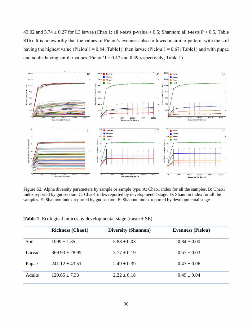
The standardized effect size of mean pairwise distance values (SES_MPD) of the bacterial communities
associated with the samples ranged from positive values for soil bacterial communities (median value of
SES_MPDSOIL = 0.78 associated with high quantiles, Table S1c) to negative values for bacterial
communities associated with the larval and pupal stages (median values SES_MPDLARVAE = -3.38
and SES_MPDPUPAE= -3.9, low quantile values, Table S1c) (Fig. 1C). SES_MDP values were
significantly different between sample types (one-way ANOVA, F = 36.75, df1 = 3, df2=21.4, p < 0.001),
namely between larvae and soil (Tamhane post-hoc test, p < 0.001) and between larvae and adults
(Tamhane post-hoc test, p = 0.001). The positive SES_MPD values for the soil communities indicate a
phylogenetic overdispersion, as expected for communities characterized by high species richness and
evenness such as those of soil. In contrast, the negative SES_MPD values for the bacterial communities
associated with larvae and pupae indicate a phylogenetic clustering of these communities, possibly due
to the selection towards certain closely related bacterial lineages by the insect gut environment or to the
adaptation of these bacteria to the gut environment. Interestingly, the bacterial communities associated
with adults were characterized by slightly negative SES_MPD values (median value of
SES_MPDADULTS= -0.53; see Supplementary Table S1c), indicating a phylogenetic evenness of these
communities (Fig. 1C). This increasing trend of SES_MPD values from larvae and pupae (negative
values) towards adults (slightly negative values) contrasted with the trend of decreasing community
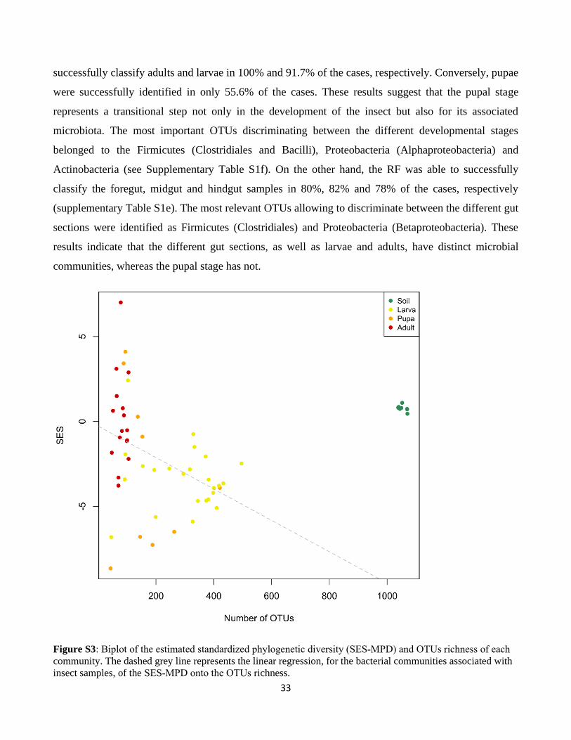
species richness from larvae to adults (Fig. S3).
3.2. Factors affecting gut microbiota composition
Soil was different from the insect samples in terms of bacterial composition (adonis: P < 0.001, R2 =
0.33; anosim: P < 0.001, R = 0.54) with few OTUs shared between soil and the different insect
developmental stages (Fig. 1A). Specifically, 891 OTUs out of the 1102 “core OTUs” of the soil were
not found in the insect samples (Fig. 1B). On the other hand, only 35 “core OTUs” present in soil were
also present in all the insect developmental stages (Fig. 1B).
Moreover, the nestedness component of the α-diversity between soil and the different insect
developmental stage was very low (0.16 on average) and the turnover was high (0.84 on average) (Fig.
S4), indicating that very few “core OTUs” were shared between soil and insect microbiotas while the
variable fraction was high. Although more bacterial OTUs were shared between the insect samples (i.e.
developmental stages and gut sections combined) than between insects and soil, these samples still
formed distinct clusters as shown by NMDS analysis (Fig 2A). Specifically, insect developmental stages
segregated along the first axis with the larvae microbiotas being clearly distinct from adult microbiotas,
32
while pupal microbiotas were intermediate. The second axis further separated the samples based on gut
sections. For larvae and adults, the microbiotas of the different gut sections formed distinct clusters with
the midgut microbiota being more different than the foregut and hindgut microbiotas. In contrast, the
pupal microbiotas showed a different pattern with a clear cluster for the hindgut, while foregut and
midgut microbiotas loosely clustered together.
Figure 1 OTU distribution among the different samples. A: Bacterial community network connecting OTUs (grey
circles) to the samples (colored circles) in which they were observed. B: Venn diagram showing the shared/specific
bacterial OTUs (at 97% similarity) between the different developmental stages and soil. C: Boxplots of the
estimated standardized phylogenetic diversity (SES-MPD) in the bacterial communities of rhizospheric soil and
Popillia japonica developmental stages.
Based on the correlations of the tested factors (i.e., developmental stages and gut sections) with the
NMDS ordinations of the insect-associated bacterial communities, the main factor driving this
segregation was the gut section (R2 = 0.18, p-value = 0.003 ) and to a lesser extent the developmental
stage. These results were further supported by the Random Forest (RF) analysis was carried out to
investigate the specificity of the microbiota of each sample category by trying to assign each sample to
its respective category based on its microbiota. The RF analysis (Supplementary Table S1d) was able to
33
successfully classify adults and larvae in 100% and 91.7% of the cases, respectively. Conversely, pupae
were successfully identified in only 55.6% of the cases. These results suggest that the pupal stage
represents a transitional step not only in the development of the insect but also for its associated
microbiota. The most important OTUs discriminating between the different developmental stages
belonged to the Firmicutes (Clostridiales and Bacilli), Proteobacteria (Alphaproteobacteria) and
Actinobacteria (see Supplementary Table S1f). On the other hand, the RF was able to successfully
classify the foregut, midgut and hindgut samples in 80%, 82% and 78% of the cases, respectively
(supplementary Table S1e). The most relevant OTUs allowing to discriminate between the different gut
sections were identified as Firmicutes (Clostridiales) and Proteobacteria (Betaproteobacteria). These
results indicate that the different gut sections, as well as larvae and adults, have distinct microbial
communities, whereas the pupal stage has not.
Figure S3: Biplot of the estimated standardized phylogenetic diversity (SES‐MPD) and OTUs richness of each
community. The dashed grey line represents the linear regression, for the bacterial communities associated with
insect samples, of the SES‐MPD onto the OTUs richness.
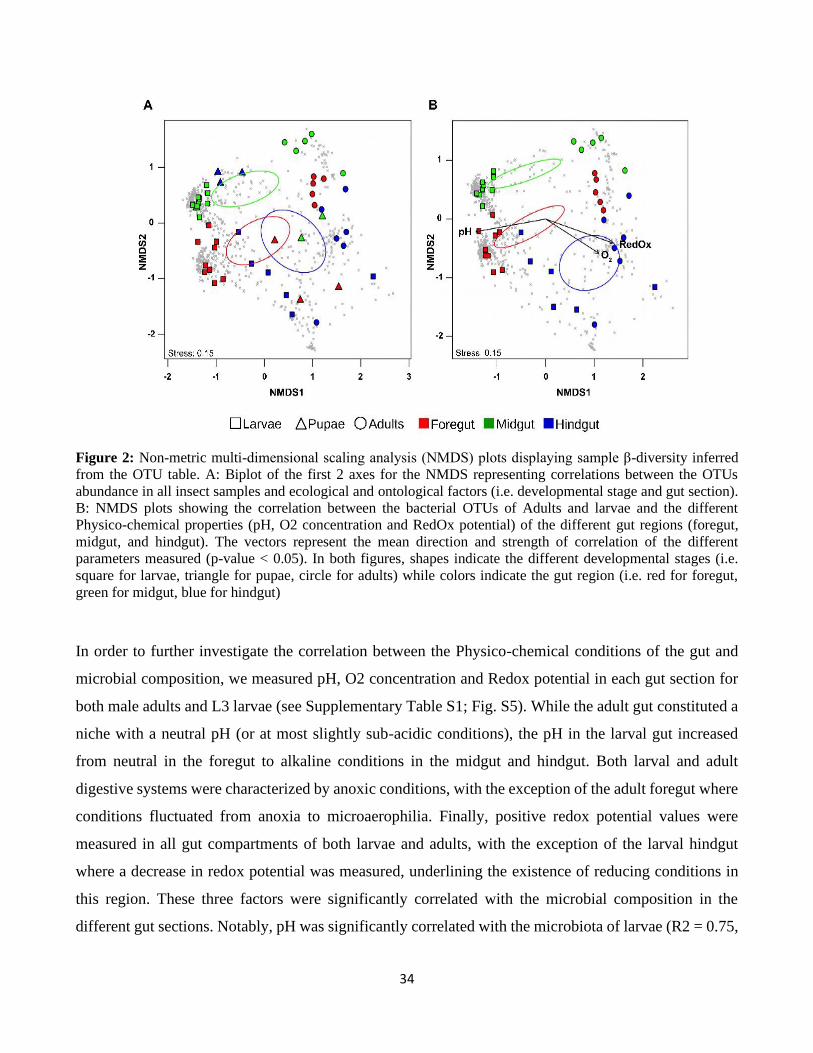
34
Figure 2: Non-metric multi-dimensional scaling analysis (NMDS) plots displaying sample β-diversity inferred
from the OTU table. A: Biplot of the first 2 axes for the NMDS representing correlations between the OTUs
abundance in all insect samples and ecological and ontological factors (i.e. developmental stage and gut section).
B: NMDS plots showing the correlation between the bacterial OTUs of Adults and larvae and the different
Physico-chemical properties (pH, O2 concentration and RedOx potential) of the different gut regions (foregut,
midgut, and hindgut). The vectors represent the mean direction and strength of correlation of the different
parameters measured (p-value < 0.05). In both figures, shapes indicate the different developmental stages (i.e.
square for larvae, triangle for pupae, circle for adults) while colors indicate the gut region (i.e. red for foregut,
green for midgut, blue for hindgut)
In order to further investigate the correlation between the Physico-chemical conditions of the gut and
microbial composition, we measured pH, O2 concentration and Redox potential in each gut section for
both male adults and L3 larvae (see Supplementary Table S1; Fig. S5). While the adult gut constituted a
niche with a neutral pH (or at most slightly sub-acidic conditions), the pH in the larval gut increased
from neutral in the foregut to alkaline conditions in the midgut and hindgut. Both larval and adult
digestive systems were characterized by anoxic conditions, with the exception of the adult foregut where
conditions fluctuated from anoxia to microaerophilia. Finally, positive redox potential values were
measured in all gut compartments of both larvae and adults, with the exception of the larval hindgut
where a decrease in redox potential was measured, underlining the existence of reducing conditions in
this region. These three factors were significantly correlated with the microbial composition in the
different gut sections. Notably, pH was significantly correlated with the microbiota of larvae (R2 = 0.75,
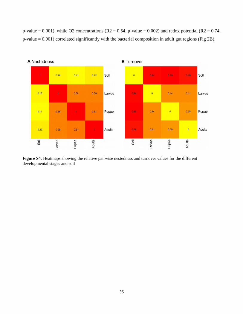
35
p-value = 0.001), while O2 concentrations (R2 = 0.54, p-value = 0.002) and redox potential (R2 = 0.74,
p-value = 0.001) correlated significantly with the bacterial composition in adult gut regions (Fig 2B).
Figure S4: Heatmaps showing the relative pairwise nestedness and turnover values for the different
developmental stages and soil
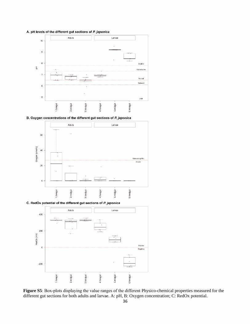
36
Figure S5: Box‐plots displaying the value ranges of the different Physico-chemical properties measured for the
different gut sections for both adults and larvae. A: pH, B: Oxygen concentration; C: RedOx potential.
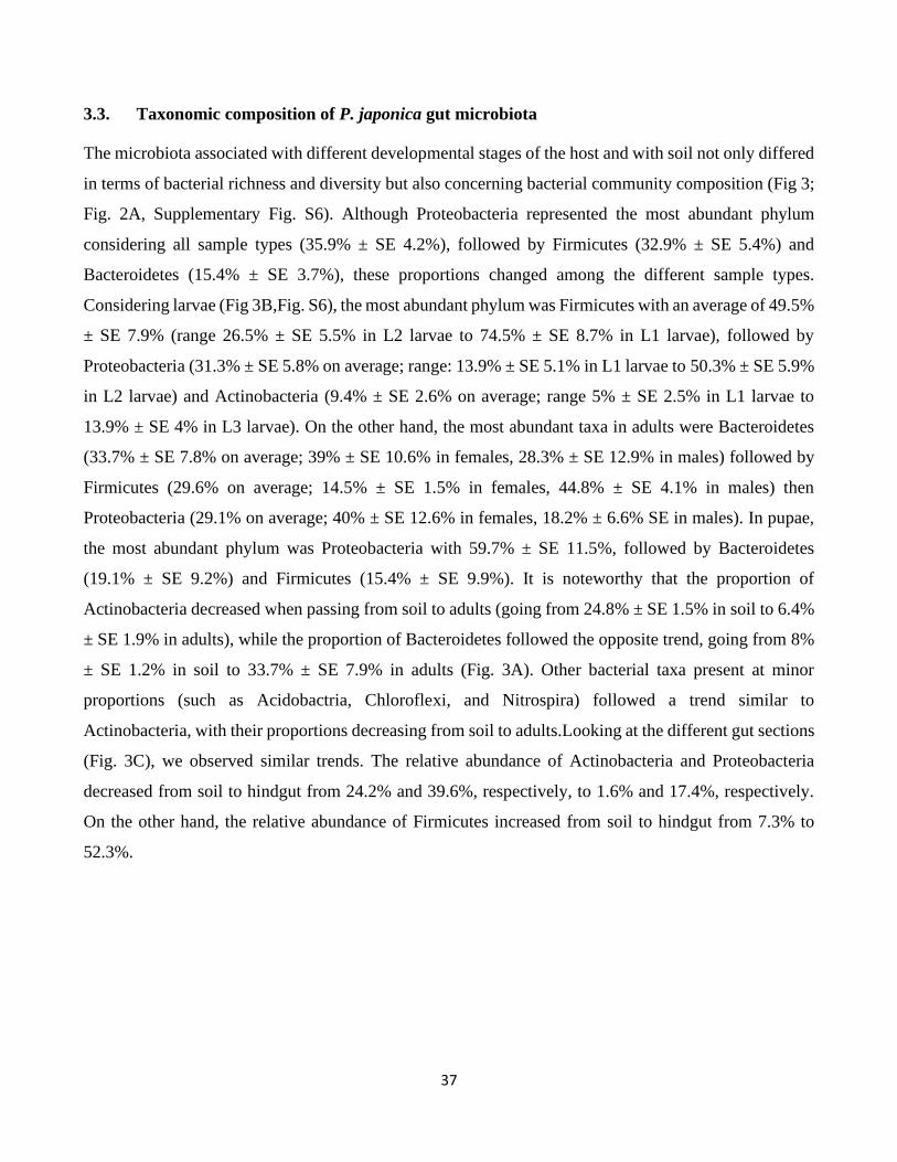
37
3.3. Taxonomic composition of P. japonica gut microbiota
The microbiota associated with different developmental stages of the host and with soil not only differed
in terms of bacterial richness and diversity but also concerning bacterial community composition (Fig 3;
Fig. 2A, Supplementary Fig. S6). Although Proteobacteria represented the most abundant phylum
considering all sample types (35.9% ± SE 4.2%), followed by Firmicutes (32.9% ± SE 5.4%) and
Bacteroidetes (15.4% ± SE 3.7%), these proportions changed among the different sample types.
Considering larvae (Fig 3B,Fig. S6), the most abundant phylum was Firmicutes with an average of 49.5%
± SE 7.9% (range 26.5% ± SE 5.5% in L2 larvae to 74.5% ± SE 8.7% in L1 larvae), followed by
Proteobacteria (31.3% ± SE 5.8% on average; range: 13.9% ± SE 5.1% in L1 larvae to 50.3% ± SE 5.9%
in L2 larvae) and Actinobacteria (9.4% ± SE 2.6% on average; range 5% ± SE 2.5% in L1 larvae to
13.9% ± SE 4% in L3 larvae). On the other hand, the most abundant taxa in adults were Bacteroidetes
(33.7% ± SE 7.8% on average; 39% ± SE 10.6% in females, 28.3% ± SE 12.9% in males) followed by
Firmicutes (29.6% on average; 14.5% ± SE 1.5% in females, 44.8% ± SE 4.1% in males) then
Proteobacteria (29.1% on average; 40% ± SE 12.6% in females, 18.2% ± 6.6% SE in males). In pupae,
the most abundant phylum was Proteobacteria with 59.7% ± SE 11.5%, followed by Bacteroidetes
(19.1% ± SE 9.2%) and Firmicutes (15.4% ± SE 9.9%). It is noteworthy that the proportion of
Actinobacteria decreased when passing from soil to adults (going from 24.8% ± SE 1.5% in soil to 6.4%
± SE 1.9% in adults), while the proportion of Bacteroidetes followed the opposite trend, going from 8%
± SE 1.2% in soil to 33.7% ± SE 7.9% in adults (Fig. 3A). Other bacterial taxa present at minor
proportions (such as Acidobactria, Chloroflexi, and Nitrospira) followed a trend similar to
Actinobacteria, with their proportions decreasing from soil to adults.Looking at the different gut sections
(Fig. 3C), we observed similar trends. The relative abundance of Actinobacteria and Proteobacteria
decreased from soil to hindgut from 24.2% and 39.6%, respectively, to 1.6% and 17.4%, respectively.
On the other hand, the relative abundance of Firmicutes increased from soil to hindgut from 7.3% to
52.3%.
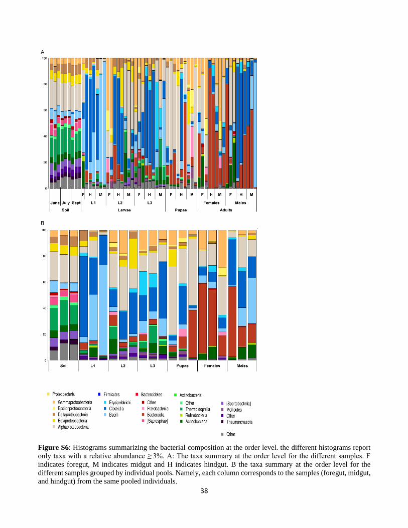
38
Figure S6: Histograms summarizing the bacterial composition at the order level. the different histograms report
only taxa with a relative abundance ≥ 3%. A: The taxa summary at the order level for the different samples. F
indicates foregut, M indicates midgut and H indicates hindgut. B the taxa summary at the order level for the
different samples grouped by individual pools. Namely, each column corresponds to the samples (foregut, midgut,
and hindgut) from the same pooled individuals.
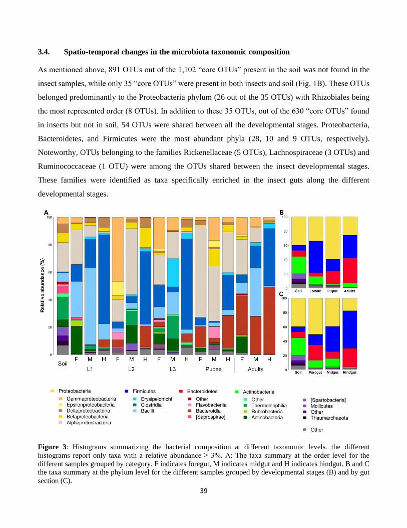
39
3.4. Spatio-temporal changes in the microbiota taxonomic composition
As mentioned above, 891 OTUs out of the 1,102 “core OTUs” present in the soil was not found in the
insect samples, while only 35 “core OTUs” were present in both insects and soil (Fig. 1B). These OTUs
belonged predominantly to the Proteobacteria phylum (26 out of the 35 OTUs) with Rhizobiales being
the most represented order (8 OTUs). In addition to these 35 OTUs, out of the 630 “core OTUs” found
in insects but not in soil, 54 OTUs were shared between all the developmental stages. Proteobacteria,
Bacteroidetes, and Firmicutes were the most abundant phyla (28, 10 and 9 OTUs, respectively).
Noteworthy, OTUs belonging to the families Rickenellaceae (5 OTUs), Lachnospiraceae (3 OTUs) and
Ruminococcaceae (1 OTU) were among the OTUs shared between the insect developmental stages.
These families were identified as taxa specifically enriched in the insect guts along the different
developmental stages.
Figure 3: Histograms summarizing the bacterial composition at different taxonomic levels. the different
histograms report only taxa with a relative abundance ≥ 3%. A: The taxa summary at the order level for the
different samples grouped by category. F indicates foregut, M indicates midgut and H indicates hindgut. B and C
the taxa summary at the phylum level for the different samples grouped by developmental stages (B) and by gut
section (C).
40
We next performed a TEA (Taxon Enrichment Analysis) to identify which bacterial families were
consistently enriched in insects compared to soil (Fig. 4). This analysis showed that among the
Firmicutes, the Ruminococcacae was significantly enriched in larvae compared to soil (P < 0.001) but
there were no differences when comparing the different developmental stages. Similarly, other bacterial
families belonging to the Firmicutes and specifically to the order Clostridiales (namely
Christensenellaceae and Lachnospiraceae) resulted to be significantly enriched in larvae and generally
in insects when compared to soil samples. These families were also enriched in the different
compartments of the gut when compared to soil (P < 0.001), independent of the insect developmental
stages. Other bacterial families, such as Rikenellaceae (Bacteroidetes) and Desulfovibrionaceae
(Proteobacteria), were also enriched in larvae compared to soil. These bacteria were also enriched in
other portions of the gut but not all of them. Desulfovibrionaceae were also enriched in the midgut and
hindgut, while Rikenellaceae were only enriched in the hindgut. Interestingly, all enriched families were
absent from the soil samples (supplementary table S3). While this family was not always present in the
foregut, Desulfovibrionaceae, Lachnospiraceae, and Ruminococcaceae were present in all midgut and
hindgut samples for all developmental stages. Rikenellaceae, on the other hand, were present in all
hindgut samples but absent from two midgut samples, namely one L1 and one pupal midgut sample
(supplementary table S3)
It is noteworthy that the TEA did not evidence any significantly enriched taxonomic group between the
different developmental stages of the insect nor did it evidence enriched taxonomic group between the
different gut sections. This is partly supported by the fact that the nestedness component of the alpha-
diversity between the different insect developmental stages was relatively high (0.59 on average),
indicating that a higher fraction of the microbiotas is shared between the different insect developmental
stages than between insects and soil.
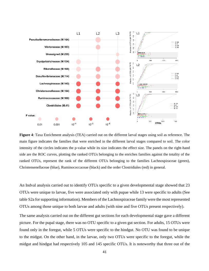
41
Figure 4: Taxa Enrichment analysis (TEA) carried out on the different larval stages using soil as reference. The
main figure indicates the families that were enriched in the different larval stages compared to soil. The color
intensity of the circles indicates the p-value while its size indicates the effect size. The panels on the right-hand
side are the ROC curves, plotting the ranked OTUs belonging to the enriches families against the totality of the
ranked OTUs, represent the rank of the different OTUs belonging to the families Lachnospiraceae (green),
Christensenellaceae (blue), Ruminococcaceae (black) and the order Clostridiales (red) in general.
An Indval analysis carried out to identify OTUs specific to a given developmental stage showed that 23
OTUs were unique to larvae, five were associated only with pupae while 13 were specific to adults (See
table S2a for supporting information). Members of the Lachnospiraceae family were the most represented
OTUs among those unique to both larvae and adults (with nine and five OTUs present respectively).
The same analysis carried out on the different gut sections for each developmental stage gave a different
picture. For the pupal stage, there was no OTU specific to a given gut section. For adults, 15 OTUs were
found only in the foregut, while 5 OTUs were specific to the hindgut. No OTU was found to be unique
to the midgut. On the other hand, in the larvae, only two OTUs were specific to the foregut, while the
midgut and hindgut had respectively 105 and 145 specific OTUs. It is noteworthy that three out of the
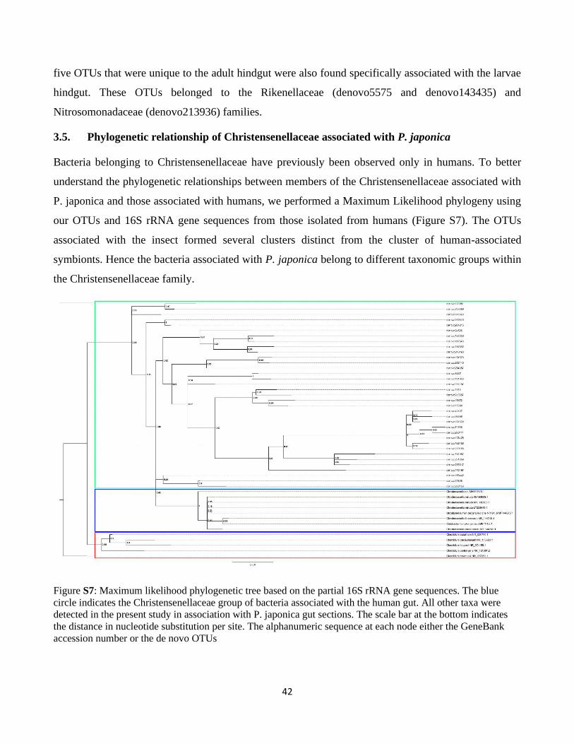
42
five OTUs that were unique to the adult hindgut were also found specifically associated with the larvae
hindgut. These OTUs belonged to the Rikenellaceae (denovo5575 and denovo143435) and
Nitrosomonadaceae (denovo213936) families.
3.5. Phylogenetic relationship of Christensenellaceae associated with P. japonica
Bacteria belonging to Christensenellaceae have previously been observed only in humans. To better
understand the phylogenetic relationships between members of the Christensenellaceae associated with
P. japonica and those associated with humans, we performed a Maximum Likelihood phylogeny using
our OTUs and 16S rRNA gene sequences from those isolated from humans (Figure S7). The OTUs
associated with the insect formed several clusters distinct from the cluster of human-associated
symbionts. Hence the bacteria associated with P. japonica belong to different taxonomic groups within
the Christensenellaceae family.
Figure S7: Maximum likelihood phylogenetic tree based on the partial 16S rRNA gene sequences. The blue
circle indicates the Christensenellaceae group of bacteria associated with the human gut. All other taxa were
detected in the present study in association with P. japonica gut sections. The scale bar at the bottom indicates
the distance in nucleotide substitution per site. The alphanumeric sequence at each node either the GeneBank
accession number or the de novo OTUs
43
4. Discussion
In this study, we demonstrate that soil bacteria represent an important source for the gut microbiota of
P. japonica larvae, but as the insect develops, the gut bacterial community experiences important changes
in richness, diversity, and composition. Specifically, 37% of the OTUs (209 OTUs) present in larvae
derived from the soil microbiota and 35 OTUs present in the soil was maintained throughout all the
developmental stages of the insect. In addition, larvae had a higher OTU richness and diversity compared
to adults. This is likely linked to the different lifestyles of the two stages: larvae are soil-dwelling and
similar in OTU numbers to other soil-dwelling arthropods such as terrestrial isopods (healthy isopods
OTUs on average 209; Dittmer et al., 2016), termites (number of OTUs consistently higher than 400; Su
et al., 2016) and ants (number of OTUs about 400; Vieira et al., 2017; Zhukova et al., 2017), while the
OTU numbers of adults are comparable to those of non-soil-dwelling insects (in 218 insect species,
average OTUs 84; Yun et al., 2014). Pupae are an intermediate state between larvae and adults in terms
of bacterial taxonomic richness and diversity, representing a bottleneck for bacterial transmission due to
metamorphosis. Nonetheless, key bacterial taxa involved in plant material degradation are still
transmitted to adults (see below for a detailed discussion). This reduction in both richness and diversity
at the pupal stage could be due to a combination of factors both random and deterministic. On the one
hand, a reduction in the number of bacterial cells during metamorphosis could have caused a random
reduction in the diversity of the microbiota. On the other hand, the observed reduction in microbiota
diversity throughout host development could be caused by one (or several) active mechanisms, such as
(i) the change of nutrition (or lifestyle) between soil-dwelling larvae and adults, (ii) specific Physico-
chemical properties (e.g., the change in gut pH between larvae and adults), and/or (iii) enzymatic
activities, among others. As a matter of fact, the observed changes (i.e. decrease in richness and diversity)
are not a constant in insect development and other studies monitoring gut microbiota changes throughout
development have shown different trends, such as an increase in species richness (Brucker and
Bordenstein 2012) or more generally the absence of a clear trend (Oliveira et al., 2018; Gao et al., 2019;
Huang et al., 2019). The trend that we observe in P. japonica could be explained by its ecology, since
soil-dwelling arthropods such as termites and woodlice consistently present higher microbiota richness
and diversity (Dittmer et al., 2016; Su et al., 2016; Vieira et al., 2017; Zhukova et al., 2017) due to their
proximity to a microbially rich and diverse environment (i.e. soil). On the other, arthropods living in
“aerial” ecosystems (i.e. plants and leaves) tend to have a less rich and diverse gut microbiota (Yun et
al., 2014; Mereghetti et al., 2019).
44
Interestingly, the decrease in microbiota richness and diversity throughout the host developmental stages
is accompanied by a shift in the phylogenetic community structure. Specifically, larvae and pupae harbor
phylogenetically clustered bacterial communities, i.e. consisting of closely related bacterial taxa. In
contrast, the adult microbiota is phylogenetically overdispersed, similarly to rhizospheric soil
communities. The observation that larvae microbiotas are phylogenetically clustered and at the same
time taxonomically rich compared to adults could be explained by a selection of certain taxonomic
groups through the gut environment. The phylogenetic overdispersion of the adult gut microbiotas
suggests that the pupal stage represents a crucial bottleneck for the gut microbiota in terms of species
richness. This might be due to the random survival of bacterial taxa present in the larvae throughout
metamorphosis (and its associated gut tissue restructuring) at the pupal stage. However, the fact that a
certain number of taxa are maintained throughout the development from larvae to adult but are absent
from soil, suggests the existence of a mechanism to specifically maintain essential bacterial partners
(e.g., Ruminococcaceae, Lachnospiraceae). In other words, the survival of certain bacterial taxa may not
be entirely random. Another possible explanation might be that the adult gut microbiota is renewed by
feeding on leaves and flowers in contrast to rhizospheric soil and/or that the Physico-chemical properties
of the adult gut are more stable than in larvae (see Fig. S5). Hence, despite the potential existence of a
mechanism to maintain and transmit a fraction of the microbiota, other bacterial taxa could still be
transient and dependent on the food source (e.g., different parts of the plant, different plant species), as
observed in D. melanogaster where acetic acid bacteria are always associated with the fly but the
presence of other bacterial taxa is dependent on the environment (Adair et al., 2018; Wong et al., 2015).
This study allowed us to identify several factors potentially shaping microbiota composition in P.
japonica. Specifically, we demonstrate that among the tested factors, microbiota composition varied
significantly between different gut sections as well as between insect developmental stages. This strong
correlation between different gut sections and microbiota diversity and composition is most likely due
to i) differences in the Physico-chemical conditions prevailing in each gut section (Fig S5) as well as ii)
biotic factors such as host enzymatic potential and immune response. It is noteworthy that the pupae
represent a transitional stage with a reshuffling of the microbiota between the larval and adult stages. In
other words, the larvae and adult microbiotas formed clearly distinct clusters, while the pupae microbiota
was more dispersed between the larvae and adult clusters. This may have had an impact on the statistical
analyses, leading to an apparently weaker effect of the developmental stages on microbiota composition.
45
Regarding the physicochemical factors, oxygen availability was the most strongly correlated with
differences in bacterial community structure between the different gut sections in adults, while intestinal
pH was the most strongly correlated factor in larvae. Although both the midgut and hindgut
compartments were largely anoxic in adults, the oxygen concentration in the midgut showed a higher
degree of variation compared to the more anoxic hindgut. This is likely due to a considerably larger
influx of oxygen via the gut epithelium in the case of the midgut, as observed in Pachnoda ephippiata
(Lemke et al., 2003). This variability in oxygen availability between the different gut compartments may
favor bacteria that are more tolerant of such fluctuations. In larvae, the pH in the midgut and hindgut was
alkaline, while the foregut had a neutral pH. It is important to note that the larvae are soil-dwellers feeding
on fresh roots and decaying soil organic matter (SOM) (Fleming, 1972). In this regard, they are similar
to other soil-dwelling macroinvertebrates, including many coleopterans, which feed on SOM and play
an important role in its degradation and stabilization (Lavelle et al., 1997; Wolters, 2000). It has been
shown that the conditions in the anterior hindgut of the humivorous termite Cubitermes spp. (i.e. high
alkalinity and oxygen influx) lead to a decrease of the molecular weight of the organic matter (Kappler
and Brune, 1999), rendering it more soluble and thus more accessible for digestion in subsequent less-
alkaline compartments (Ji et al., 2000; Kappler et al., 2000; Ji and Brune, 2001). Although the complex
microbial communities in the guts of humivorous macroinvertebrates are thought to participate in the
transformation of ingested SOM (Cazemier et al., 1997; Kane, 1997), detailed information on the
composition and activities of the gut microbiota is lacking. In view of the high midgut alkalinity in P.
japonica, it is reasonable to assume that at least some of the bacteria in the midgut are tolerant towards
high pH conditions since most bacterial taxa are also found in the more neutral gut sections of adults.
We further observed differences in microbiota composition at different taxonomic levels (from order to
OTU) between the different developmental stages of P. japonica. For instance, Actinobacteria decreased
in abundance from larvae to adults, while Bacteroidetes increased in abundance. However, no particular
taxa were found to be specifically enriched in any of the developmental stages. A similar pattern was
observed for the microbiota associated with different gut compartments (foregut, midgut, hindgut): no
particular taxon was specifically enriched in any of the compartments. Nonetheless, Proteobacteria
decreased from foregut to hindgut, while Firmicutes increased. Actinobacteria were relatively stable
between foregut and midgut but decreased in the hindgut.
In contrast, several taxa were found to be significantly enriched between soil and insect gut. Those
belonged mainly to the families Ruminococcacae, Christensenellaceae and Lachnospiraceae. Members
46
of these families are known to degrade cellulose (Flint et al., 2012; Biddle et al., 2013). The fact of
finding them enriched in the insect gut may suggest a possible symbiotic relationship where these
bacteria help their host degrade and metabolize cellulose, as in the case of the symbiotic association
between termites, protists and bacteria (Liu et al., 2013) or woodlice and certain bacterial taxa (Bredon
et al., 2018). These bacteria could be important in helping their host metabolize plant roots and leaves
and might thus contribute to its success as a polyphagous invasive insect. The bacterial taxa that were
enriched in the gut of P. japonica have been previously reported in association with various insects but
more importantly with ruminants and humans. Anaerostipes spp., Coprococcus spp. and Dorea spp.
(members of the Lachnospiraceae family) have all been previously described in association with the
human gut (Rainey, 2009) where they are hypothesized to be involved in pectin fermentation. Other
members of the Lachnospiraceae family have also been described in association with other insects
(Huang and Zhang, 2013; Bourguignon et al., 2018). The Ruminococcaceae family, represented by
Ruminococcus spp. and Oscillospira spp. in P. japonica, has also been described in association with
humans, ruminants, coleopterans, and termites (Kamagata, 2011; Huang and Zhang, 2013; Bourguignon
et al., 2018). Ruminococcus, in addition to Bacteroides spp., plays an important role in the fermentation
of hemicellulose and the degradation of different plant material through the production of Carbohydrate-
Active enzymes (CAZymes) (Jose et al., 2017). CAZymes are very important for the break-down of the
different components of lignocellulose (i.e. cellulose, lignin, hemicellulose; Bredon et al., 2018). It is
noteworthy that although some insects are able to express some of these enzymes, most of them heavily
rely on their associated microorganisms to degrade lignocellulose (Bredon et al., 2018). On the other
hand, the role of Oscillospira is still unknown and it is hypothesized that it may be involved in
lignocellulose degradation (Kamagata, 2011). Rickenellaceae, with the genus Alistipes, and
Desulfovibrionaceae have also been described in association with the guts of different animals (Koneru
et al., 2016; Ruengsomwong et al., 2016), especially termites (Reid et al., 2014; Makonde et al., 2015),
where they play an important role in the degradation of cellulose polymers (Ozbayram et al., 2018).
The taxa found to be enriched in insect samples could be preferentially present in insects due to favorable
conditions in the gut environment without an actual effect of these bacteria on the insect host. However,
the fact that these bacteria were not detected in soil suggests the presence of a more direct transmission
mechanism independent of the environmental route. In addition, the consistent presence of these bacteria
in the gut regions where plant material is degraded further argues in favor of an active role of these
bacteria and not just their presence as transient passengers.
47
In contrast to the above-mentioned bacterial families which have been observed not only in mammals
but also in insects, the family Christensenallaceae had so far been observed exclusively in humans.
Although its role in the degradation of nutrients is not yet understood, members of this family (i.e.
Christensenella minuta) have been shown to play a central role in controlling the Body Mass Index and
in helping to shape a “healthy” microbiota in humans and transfected mice (Goodrich et al., 2014).
Increased titers of C. minuta have also been correlated with longevity in humans (Biagi et al., 2016),
while decreased titers were observed during different human diseases (Petrov et al., 2017; Yu et al.,
2017). In addition, other bacteria belonging to the genus Christensenella have been isolated from
diseased humans, although no causality has been established yet (Ndongo et al., 2016). The partial 16S
rRNA gene-based phylogeny showed that the Christensenellaceae OTUs found in association with P.
japonica do not cluster with the taxa associated with humans but rather form different clusters, suggesting
that they belong to different taxonomic groups within the Christensenellaceae family (Fig. S7).
Although three biological replicates containing homologous gut regions from five individuals might be
limiting, based on the results obtained in this study we can conclude that the gut microbiota of P. japonica
is highly dynamic across the developmental stages of the insect and changes in microbiota composition
strongly correlated with the Physico-chemical properties of the gut. Despite the microbiota high
variability, 89 OTUs were maintained from larvae to adults, including 35 OTUs originating from the soil
environment. As a future perspective, it would be interesting to investigate if these OTUs represent a
stable core microbiota present in all P. japonica populations in different parts of the world or if they are
subject to change in different environments. In the first case, this might indicate a more intimate
symbiotic relationship potentially maintained via vertical transmission. In the latter case, the variable
microbiota would provide a means to investigate the origin of new invasions of this beetle, via a
comparative analysis of the local soil and insect gut microbiotas.
Declaration
BC, MM, and LM designed the experiments. BC performed the microbiota and enrichment analyses.
MM, GMG and NG performed the statistical analyses. SA performed network analyses. GMz, EG, FP,
LM, PFR, and AA performed the sampling. NG dissected the insects and extracted the DNA. FF and FF
performed the sequencing. MC, MF, EC, and DD performed the Physico-chemical analyses. BC and
MM wrote the manuscript. All authors read and commented on the manuscript.
MM: Matteo Montagna BC: Bessem ChouaiaNG: Nizar Goda LM: Leonardo Marianelli ; GMg: Giulia
Magoga ; GMz: Giuseppe Mazza ;EG: Elena Gonella ; FP: Francesco Paoli ;FF : Fiorella Florian; DD:
Daniele Daffonchio ; SA: Sumer Alali ; MC: Matteo Callegari ; MF: Marco Fusi ; EC: Elena Crotti ;
PFR: Pio Federico Roversi; AA: Alberto Alma ; FG: Fabrizia Gionechetti
48
References
Adair, K.L, Wilson, M., Bost, A., and Douglas A.E. (2018) Microbial community assembly in wild
populations of the fruit fly Drosophila melanogaster. ISME J. 12: 959-972.
Aharon, Y., Pasternak, Z., Ben Yosef, M., Behar, A., Lauzon, C., Yuval, B., and Jurkevitch, E. (2013)
Phylogenetic, Metabolic, and Taxonomic Diversities Shape Mediterranean Fruit Fly Microbiotas
during Ontogeny. Appl. Environ. Microbiol. 79: 303–313.
Akaike, H. (1976) An information criterion (AIC). Math. Sci. 14: 5–9.
Anderson, K.E., Ricigliano, V.A., Mott, B.M., Copeland, D.C., Floyd, A.S., and Maes, P. (2018) The
queen’s gut refines with age: longevity phenotypes in a social insect model. Microbiome 6: 108.
Baselga, A. (2010) Partitioning the turnover and nestedness components of beta diversity. Glob. Ecol.
Biogeogr. 19: 134–143.
Baselga, A. and Orme, C.D.L. (2012) betapart: an R package for the study of beta diversity. Methods
Ecol. Evol. 3: 808–812.
Biagi, E., Franceschi, C., Rampelli, S., Severgnini, M., Ostan, R., Turroni, S., et al. (2016) Gut
Microbiota and Extreme Longevity. Curr. Biol. 26: 1480–1485.
Biddle, A., Stewart, L., Blanchard, J., and Leschine, S. (2013) Untangling the Genetic Basis of Fibrolytic
Specialization by Lachnospiraceae and Ruminococcaceae in Diverse Gut Communities.
Diversity 5: 627–640.
Bourguignon, T., Lo, N., Dietrich, C., Šobotník, J., Sidek, S., Roisin, Y., et al. (2018) Rampant Host
Switching Shaped the Termite Gut Microbiome. Curr. Biol. 28: 649–654.e2.
Bray, J.R. and Curtis, J.T. (1957) An Ordination of the Upland Forest Communities of Southern
Wisconsin. Ecol. Monogr. 27: 325–349.
Bredon, M., Dittmer, J., Noël, C., Moumen, B., and Bouchon, D. (2018) Lignocellulose degradation at
the holobiont level: teamwork in a keystone soil invertebrate. Microbiome 6: 162.
Brucker, R.M., and Bordenstein, S.R. (2012) The roles of host evolutionary relationships (genus:
Nasonia) and development in structuring microbial communities. Evolution. 66: 349-362.
De Cáceres, M. and Legendre, P. (2009) Associations between species and groups of sites: indices and
statistical inference. Ecology 90: 3566–3574.
49
Caporaso, J.G., Kuczynski, J., Stombaugh, J., Bittinger, K., Bushman, F.D., Costello, E.K., et al. (2010)
QIIME allows analysis of high-throughput community sequencing data. Nat. Methods 7: 335–
336.
Caporaso, J.G., Lauber, C.L., Walters, W.A., Berg-Lyons, D., Lozupone, C.A., Turnbaugh, P.J., et al.
(2011) Global patterns of 16S rRNA diversity at a depth of millions of sequences per sample.
Proc. Natl. Acad. Sci. 108: 4516–4522.
Cazemier, A.E., Hackstein, J.H.P., Camp, H.J.M.O. den, Rosenberg, J., and van der Drift, C. (1997)
Bacteria in the Intestinal Tract of Different Species of Arthropods. Microb. Ecol. 33: 189–197.
Chao, A. (1984) Nonparametric Estimation of the Number of Classes in a Population. Scand. J. Stat. 11:
265–270.
Claesson, M.J., O’Sullivan, O., Wang, Q., Nikkilä, J., Marchesi, J.R., Smidt, H., et al. (2009)
Comparative Analysis of Pyrosequencing and a Phylogenetic Microarray for Exploring Microbial
Community Structures in the Human Distal Intestine. PLoS One 4: e6669.
Clarke, K.R. (1993) Non-parametric multivariate analyses of changes in community structure. Austral
Ecol. 18: 117–143.
Clayton, J.B., Gomez, A., Amato, K., Knights, D., Travis, D.A., Blekhman, R., et al. (2018) The gut
microbiome of nonhuman primates: Lessons in ecology and evolution. Am. J. Primatol. 80:
e22867.
Corbin, C., Heyworth, E.R., Ferrari, J., and Hurst, G.D.D. (2017) Heritable symbionts in a world of
varying temperature. Heredity (Edinb). 118: 10–20.
Daisley, B.A., Trinder, M., McDowell, T.W., Collins, S.L., Sumarah, M.W., and Reid, G. (2018)
Microbiota-Mediated Modulation of Organophosphate Insecticide Toxicity by Species-
Dependent Interactions with Lactobacilli in a Drosophila melanogaster Insect Model. Appl.
Environ. Microbiol. 84:.
Dittmer, J., Lesobre, J., Moumen, B., and Bouchon, D. (2016) Host origin and tissue microhabitat
shaping the microbiota of the terrestrial isopod Armadillidium vulgare. FEMS Microbiol. Ecol.
92: 92:fiw063.
Dixon, P. (2003) VEGAN, a package of R functions for community ecology. J. Veg. Sci. 14: 927–930.
50
Dormann, C.F., Gruber, B., and Fründ, J. (2008) Introducing the bipartite Package: Analysing Ecological
Networks. R news 8: 8–12.
Douglas, A.E. (2015) Multiorganismal Insects: Diversity and Function of Resident Microorganisms.
Annu. Rev. Entomol. 60: 17–34.
Douglas, A.E. (2014) Symbiosis as a General Principle in Eukaryotic Evolution. Cold Spring Harb.
Perspect. Biol. 6: a016113–a016113.
Douglas, A.E. (2018) The Drosophila model for microbiome research. Lab Anim. (NY). 47: 157–164.
Doyle, J.J. and Doyle, J.L. (1990) Isolation of Plant DNA from Fresh Tissue. Focus (Madison). 12: 13–
15.
Dufrêne, M. and Legendre, P. (1997) Species assemblages and indicator species: The need for a flexible
asymmetrical approach. Ecol. Monogr. 67: 345–366.
Edgar, R.C. (2010) Search and clustering orders of magnitude faster than BLAST. Bioinformatics 26:
2460–2461.
EPPO (2014) First report of Popillia japonica in Italy. EPPO Reporting Service no. 10 - 2014.
EPPO (2017) First report of Popillia japonica in Switzerland. EPPO Reporting Service no. 09 - 2017.
EPPO (2000) New data on quarantine pests and pests of the EPPO Alert List. EPPO Reporting Service
no. 09 - 2000.
Fleming, W.E. (1972) Biology of the Japanese beetle. USDA Technical Bulletin 1449, Washington, DC.
Flint, H.J., Scott, K.P., Duncan, S.H., Louis, P., and Forano, E. (2012) Microbial degradation of complex
carbohydrates in the gut. Gut Microbes 3: 289–306.
Foottit, R.G. and H. Adler, P. (2009) Insect Biodiversity: Science and Society Foottit,R.G. and
Adler,P.H. (eds) Wiley-Blackwell, Oxford, UK.
Fukatsu, T. and Hosokawa, T. (2002) Capsule-Transmitted Gut Symbiotic Bacterium of the Japanese
Common Plataspid Stinkbug, Megacopta punctatissima. Appl. Environ. Microbiol. 68: 389–396.
Gao, X., Li, W., Luo, J., Zhang, L., Ji, J., Zhu, X., Wang, L., Zhang, S., and Cui, J. (2019) Biodiversity
of the microbiota in Spodoptera exigua (Lepidoptera: Noctuidae). J Appl Microbiol. 126: 1199-
1208.
51
Gonella, E., Crotti, E., Rizzi, A., Mandrioli, M., Favia, G., Daffonchio, D., and Alma, A. (2012)
Horizontal transmission of the symbiotic bacterium Asaia sp. in the leafhopper Scaphoideus
titanus Ball (Hemiptera: Cicadellidae). BMC Microbiol. 12: S4.
Goodrich, J.K., Waters, J.L., Poole, A.C., Sutter, J.L., Koren, O., Blekhman, R., et al. (2014) Human
Genetics Shape the Gut Microbiome. Cell 159: 789–799.
Heddi, A. and Zaidman-Rémy, A. (2018) Endosymbiosis as a source of immune innovation. C. R. Biol.
341: 290–296.
Hosokawa, T., Hironaka, M., Inadomi, K., Mukai, H., Nikoh, N., and Fukatsu, T. (2013) Diverse
Strategies for Vertical Symbiont Transmission among Subsocial Stinkbugs. PLoS One 8: e65081.
Huang, H., Li, H., Ren, L., and Cheng, D. (2019) Microbial communities in different developmental
stages of the oriental fruit fly Bactrocera dorsalis are associated with differentially expressed
peptidoglycan recognition protein genes. Appl Environ Microbiol. pii: AEM.00803-19. doi:
10.1128/AEM.00803-19.
Huang, S. and Zhang, H. (2013) The Impact of Environmental Heterogeneity and Life Stage on the
Hindgut Microbiota of Holotrichia parallela Larvae (Coleoptera: Scarabaeidae). PLoS One 8:
e57169.
Ji, R. and Brune, A. (2001) Transformation and mineralization of 14C-labeled cellulose, peptidoglycan,
and protein by the soil-feeding termite Cubitermes orthognathus. Biol. Fertil. Soils 33: 166–174.
Ji, R., Kappler, A., and Brune, A. (2000) Transformation and mineralization of synthetic 14C-labeled
humic model compounds by soil-feeding termites. Soil Biol. Biochem. 32: 1281–1291.
Jose, V.L., Appoothy, T., More, R.P., and Arun, A.S. (2017) Metagenomic insights into the rumen
microbial fibrolytic enzymes in Indian crossbred cattle fed finger millet straw. AMB Express 7:
13.
Kamagata, Y. (2011) Genus VIII. Oscillospira Chatton and Pérard 1913, 1159. In, Vos,P., Garrity,G.,
Jones,D., Krieg,N.R., Ludwig,W., Rainey,F.A., et al. (eds), Bergey’s Manual of Systematic
Bacteriology: Volume 3: The Firmicutes. Springer New York, New York, pp. 1131–1132.
Kane, M.D. (1997) Microbial Fermentation in Insect Guts BT. In, Mackie,R.I. and White,B.A. (eds),
Gastrointestinal Microbiology: Volume 1 Gastrointestinal Ecosystems and Fermentations.
Springer US, Boston, MA, pp. 231–265.
52
Kappler, A. and Brune, A. (1999) Influence of gut alkalinity and oxygen status on mobilization and size-
class distribution of humic acids in the hindgut of soil-feeding termites. Appl. Soil Ecol. 13: 219–
229.
Kappler, A., Ji, R., and Brune, A. (2000) Synthesis and characterization of specifically 14C-labeled
humic model compounds for feeding trials with soil-feeding termites. Soil Biol. Biochem. 32:
1271–1280.
Kembel, S.W., Cowan, P.D., Helmus, M.R., Cornwell, W.K., Morlon, H., Ackerly, D.D., et al. (2010)
Picante: R tools for integrating phylogenies and ecology. Bioinformatics 26: 1463–1464.
Kim, J.K., Lee, J.B., Jang, H.A., Han, Y.S., Fukatsu, T., and Lee, B.L. (2016) Understanding regulation
of the host-mediated gut symbiont population and the symbiont-mediated host immunity in the
Riptortus-Burkholderia symbiosis system. Dev. Comp. Immunol. 64: 75–81.
Knights, D., Costello, E.K., and Knight, R. (2011) Supervised classification of human microbiota. FEMS
Microbiol. Rev. 35: 343–359.
Koneru, S.L., Salinas, H., Flores, G.E., and Hong, R.L. (2016) The bacterial community of entomophilic
nematodes and host beetles. Mol. Ecol. 25: 2312–2324.
Kruskal, J.B. (1964) Nonmetric multidimensional scaling: A numerical method. Psychometrika 29: 115–
129.
Kruskal, W.H. and Wallis, W.A. (1952) Use of Ranks in One-Criterion Variance Analysis. J. Am. Stat.
Assoc. 47: 583.
Kumar, S., Stecher, G., Li, M., Knyaz, C., and Tamura, K. (2018) MEGA X: Molecular Evolutionary
Genetics Analysis across Computing Platforms. Mol. Biol. Evol. 35: 1547–1549.
Lane, D.J. (1991) 16S/23S rRNA sequencing. In, Stackebrandt,E. and Goodfellow,M. (eds), Nucleic
Acid Techniques in Bacterial Systematics. John Wiley & Sons, Ltd, pp. 115–175.
Lavelle, P., Bignell, D., Lepage, M., Wolters, V., Roger, P.A., Ineson, P., et al. (1997) Soil function in a
changing world: the role of invertebrate ecosystem engineers. Eur. J. Soil Biol. 33: 159–193.
Leftwich, P.T., Clarke, N.V.E., Hutchings, M.I., and Chapman, T. (2017) Gut microbiomes and
reproductive isolation in Drosophila. Proc. Natl. Acad. Sci. 114: 12767 LP-12772.
53
Lemke, T., Stingl, U., Egert, M., Friedrich, M.W., and Brune, A. (2003) Physicochemical Conditions
and Microbial Activities in the Highly Alkaline Gut of the Humus-Feeding Larva of Pachnoda
ephippiata (Coleoptera: Scarabaeidae). Appl. Environ. Microbiol. 69: 6650 LP-6658.
Levene, H. (1960) Robust Tests for Equality of Variances. In, Olkin,I., Ghurye,S.G., Hoeffding,W.,
Madow,W.G., and Mann,H.B. (eds), Contributions to Probability and Statistics: Essays in Honor
of Harold Hotelling. Stanford University Press, Palo Alto, pp. 278–292.
Liu, N., Zhang, L., Zhou, H., Zhang, M., Yan, X., Wang, Q., et al. (2013) Metagenomic Insights into
Metabolic Capacities of the Gut Microbiota in a Fungus-Cultivating Termite (Odontotermes
yunnanensis). PLoS One 8: e69184.
Makonde, H.M., Mwirichia, R., Osiemo, Z., Boga, H.I., and Klenk, H.-P. (2015) 454 Pyrosequencing-
based assessment of bacterial diversity and community structure in termite guts, mounds and
surrounding soils. Springerplus 4: 471.
Malacrinò, A., Campolo, O., Medina, R.F., and Palmeri, V. (2018) Instar- and host-associated
differentiation of bacterial communities in the Mediterranean fruit fly Ceratitis capitata. PLoS
One 13: e0194131.
Marianelli, L., Paoli, F., Sabbatini Peverieri, G., Benvenuti, C., Barzanti, G.P., Bosio, G., et al. (2018)
Long-lasting insecticide-treated nets: A new integrated pest management approach for Popillia
japonica (Coleoptera: Scarabaeidae). Integr. Environ. Assess. Manag. 0:.
Marianelli, L., Paoli, F., Torrini, G., Mazza, G., Benvenuti, C., Binazzi, F., et al. (2018)
Entomopathogenic nematodes as potential biological control agents of Popillia japonica
(Coleoptera, Scarabaeidae) in Piedmont Region (Italy). J. Appl. Entomol. 142: 311–318.
Mason, C.J., Campbell, A.M., Scully, E.D., and Hoover, K. (2019) Bacterial and Fungal Midgut
Community Dynamics and Transfer Between Mother and Brood in the Asian Longhorned Beetle
(Anoplophora glabripennis), an Invasive Xylophage. Microb. Ecol. 77: 230–242.
Mazza, G., Paoli, F., Strangi, A., Torrini, G., Marianelli, L., Peverieri, G.S., et al. (2017) Hexamermis
popilliae n. sp. (Nematoda: Mermithidae) parasitizing the Japanese beetle Popillia japonica
Newman (Coleoptera: Scarabaeidae) in Italy. Syst. Parasitol. 94: 915–926.
54
McFrederick, Q.S., Mueller, U.G., Wcislo, W.T., and Hout, M.C. (2014) Host species and developmental
stage, but not host social structure, affects bacterial community structure in socially polymorphic
bees. FEMS Microbiol. Ecol. 88: 398–406.
Mereghetti, V., Chouaia, B., Limonta, L., Locatelli, D.P., and Montagna, M. (2019) Evidence for a
conserved microbiota across the different developmental stages of Plodia interpunctella. Insect
Sci.
Montagna, M., Chouaia, B., Mazza, G., Prosdocimi, E.M., Crotti, E., Mereghetti, V., et al. (2015) Effects
of the diet on the microbiota of the red palm weevil (Coleoptera: Dryophthoridae). PLoS One 10:
e0117439.
Montagna, M., Gómez-Zurita, J., Giorgi, A., Epis, S., Lozzia, G.C., and Bandi, C. (2015)
Metamicrobiomics in herbivore beetles of the genus Cryptocephalus (Chrysomelidae): toward
the understanding of ecological determinants in insect symbiosis. Insect Sci. 22: 340–352.
Montagna, M., Mereghetti, V., Gargari, G., Guglielmetti, S., Faoro, F., Lozzia, G.C., et al. (2016)
Evidence of a bacterial core in the stored products pest Plodia interpunctella: the influence of
different diets. Environ. Microbiol. 18: 4961–4973.
Mouquet, N., Devictor, V., Meynard, C.N., Munoz, F., Bersier, L.-F., Chave, J., et al. (2012)
Ecophylogenetics: advances and perspectives. Biol. Rev. 87: 769–785.
Muhammad, A., Fang, Y., Hou, Y., and Shi, Z. (2017) The Gut Entomotype of Red Palm Weevil
Rhynchophorus ferrugineus Olivier (Coleoptera: Dryophthoridae) and Their Effect on Host
Nutrition Metabolism. Front. Microbiol. 8: 2291.
Münger, E., Montiel-Castro, A.J., Langhans, W., and Pacheco-López, G. (2018) Reciprocal Interactions
Between Gut Microbiota and Host Social Behavior. Front. Integr. Neurosci. 12: 21.
Ndongo, S., Khelaifia, S., Fournier, P.-E., and Raoult, D. (2016) Christensenella massiliensis, a new
bacterial species isolated from the human gut. New Microbes New Infect. 12: 69–70.
O’Dwyer, J.P., Kembel, S.W., and Green, J.L. (2012) Phylogenetic Diversity Theory Sheds Light on the
Structure of Microbial Communities. PLOS Comput. Biol. 8: e1002832.
Obata, F., Fons, C.O., and Gould, A.P. (2018) Early-life exposure to low-dose oxidants can increase
longevity via microbiome remodelling in Drosophila. Nat. Commun. 9: 975.
55
Oksanen, J., Blanchet, F.G., Friendly, M., Kindt, R., Legendre, P., McGlinn, D., et al. (2018) vegan:
Community Ecology Package. R package version 2.5-1.
Oliveira, J.L., Cury, J.C., Gurgel-Gonçalves, R., Bahia, A.C., and Monteiro, F.A. (2018). Field-collected
Triatoma sordida from central Brazil display high microbiota diversity that varies with regard to
developmental stage and intestinal segmentation. PLoS Negl Trop Dis. 12: e0006709.
Ozbayram, E.G., Akyol, Ç., Ince, B., Karakoç, C., and Ince, O. (2018) Rumen bacteria at work:
bioaugmentation strategies to enhance biogas production from cow manure. J. Appl. Microbiol.
124: 491–502.
Paoli, F., Marianelli, L., Binazzi, F., Mazza, G., Benvenuti, C., Sabbatini Peverieri, G., et al. (2017)
Effectiveness of different doses of Heterorhabditis bacteriophora against Popillia japonica 3rd
instars: Laboratory evaluation and field application. Redia 100: 135–138.
Paoli, F., Marianelli, L., Torrini, G., Mazza, G., Benvenuti, C., Bosio, G., et al. (2017) Differential
susceptibility of Popillia japonica 3rd instars to Heterorhabditis bacteriophora (Italian strain) at
three different seasons. Biocontrol Sci. Technol. 27: 439–444.
Pavesi, M. (2014) Popillia japonica specie aliena invasiva segnalata in Lombardia. L’Informatore Agrar.
32: 53–55.
Petrov, V.A., Saltykova, I. V, Zhukova, I.A., Alifirova, V.M., Zhukova, N.G., Dorofeeva, Y.B., et al.
(2017) Analysis of Gut Microbiota in Patients with Parkinson’s Disease. Bull. Exp. Biol. Med.
162: 734–737.
Pielou, E.C. (1975) Ecological Diversity John Wiley & Sons, Ltd, New York.
Rainey, F.A. (2009) Family V. Lachnospiraceae fam. nov. In, Vos,P., Garrity,G., Jones,D., Krieg,N.R.,
Ludwig,W., Rainey,F.A., et al. (eds), Bergey’s Manual of Systematic Bacteriology: Volume 3:
The Firmicutes. Springer New York, New York, pp. 921–946.
Reid, N.M., Addison, S.L., West, M.A., and Lloyd-Jones, G. (2014) The bacterial microbiota of
Stolotermes ruficeps (Stolotermitidae), a phylogenetically basal termite endemic to New Zealand.
FEMS Microbiol. Ecol. 90: 678–688.
Roh, S.W., Nam, Y.-D., Chang, H.-W., Kim, K.-H., Kim, M.-S., Ryu, J.-H., et al. (2008) Phylogenetic
Characterization of Two Novel Commensal Bacteria Involved with Innate Immune Homeostasis
in Drosophila melanogaster. Appl. Environ. Microbiol. 74: 6171 LP-6177.
56
Royston, P. (1982). An extension of Shapiro and Wilk's W test for normality to large samples. Appl
Statist. 31: 115–124.
Ruengsomwong, S., La-ongkham, O., Jiang, J., Wannissorn, B., Nakayama, J., and Nitisinprasert, S.
(2016) Microbial Community of Healthy Thai Vegetarians and Non-Vegetarians, Their Core Gut
Microbiota, and Pathogen Risk. J. Microbiol. Biotechnol. 26: 1723–1735.
Sanders, J.G., Łukasik, P., Frederickson, M.E., Russell, J.A., Koga, R., Knight, R., and Pierce, N.E.
(2017) Dramatic Differences in Gut Bacterial Densities Correlate with Diet and Habitat in
Rainforest Ants. Integr. Comp. Biol. 57: 705–722.
Shannon, C.E. (1948) A Mathematical Theory of Communication. Bell Syst. Tech. J. 27: 379–423.
Shannon, P., Markiel, A., Ozier, O., Baliga, N.S., Wang, J.T., Ramage, D., et al. (2003) Cytoscape: A
Software Environment for Integrated Models of Biomolecular Interaction Networks. Genome
Res. 13: 2498–2504.
De Smet, J., Wynants, E., Cos, P., and Van Campenhout, L. (2018) Microbial Community Dynamics
during Rearing of Black Soldier Fly Larvae (Hermetia illucens) and Impact on Exploitation
Potential. Appl. Environ. Microbiol. 84: e02722-17.
Su, L., Yang, L., Huang, S., Su, X., Li, Y., Wang, F., et al. (2016) Comparative Gut Microbiomes of
Four Species Representing the Higher and the Lower Termites. J. Insect Sci. 16:
Sudakaran, S., Kost, C., and Kaltenpoth, M. (2017) Symbiont Acquisition and Replacement as a Source
of Ecological Innovation. Trends Microbiol. 25: 375–390.
Šustr, V., Stingl, U., and Brune, A. (2014) Microprofiles of oxygen, redox potential, and pH, and
microbial fermentation products in the highly alkaline gut of the saprophagous larva of Penthetria
holosericea (Diptera: Bibionidae). J. Insect Physiol. 67: 64–69.
Switzer, P. V, Enstrom, P.C., and Schoenick, C.A. (2009) Behavioral Explanations Underlying the Lack
of Trap Effectiveness for Small-Scale Management of Japanese Beetles (Coleoptera:
Scarabaeidae). J. Econ. Entomol. 102: 934–941.
Tiede, J., Scherber, C., Mutschler, J., McMahon, K.D., and Gratton, C. (2017) Gut microbiomes of
mobile predators vary with landscape context and species identity. Ecol. Evol. 7: 8545–8557.
57
Vacchini, V., Gonella, E., Crotti, E., Prosdocimi, E.M., Mazzetto, F., Chouaia, B., et al. (2017) Bacterial
diversity shift determined by different diets in the gut of the spotted wing fly Drosophila suzukii
is primarily reflected on acetic acid bacteria. Environ. Microbiol. Rep. 9:.
Vieira, A.S., Ramalho, M.O., Martins, C., Martins, V.G., and Bueno, O.C. (2017) Microbial
Communities in Different Tissues of Atta sexdens rubropilosa Leaf-cutting Ants. Curr.
Microbiol. 74: 1216–1225.
Vieira, V. (2008) The Japanese beetle Popillia japonica Newman, 1838 (Coleoptera: Scarabaeidae) in
the Azores islands. Boletín Soc. Entomológica Aragon. 43: 450–451.
Webb, C.O., Ackerly, D.D., McPeek, M.A., and Donoghue, M.J. (2002) Phylogenies and Community
Ecology. Annu. Rev. Ecol. Syst. 33: 475–505.
Welch, B.L. (1951) On the comparison of several mean values: an alternative approach. Biometrika. 38:
330–336.
Wolters, V. (2000) Invertebrate control of soil organic matter stability. Biol. Fertil. Soils 31: 1–19.
Wong, A.C.N., Ng, P., and Douglas, A.E. (2011) Low-diversity bacterial community in the gut of the
fruitfly Drosophila melanogaster. Environ. Microbiol. 13: 1889–1900.
Wong, A.C., Luo, Y., Jing, X., Franzenburg, S., Bost, A., and Douglas, A.E. (2015) The Host as the
Driver of the Microbiota in the Gut and External Environment of Drosophila melanogaster. Appl
EnvironMicrobiol. 81: 6232-6240.
Yamaguchi, K.D., Ruderman, D.L., Croze, E., Wagner, T.C., Velichko, S., Reder, A.T., and Salamon,
H. (2008) IFN-β-regulated genes show abnormal expression in therapy-naïve relapsing–remitting
MS mononuclear cells: Gene expression analysis employing all reported protein–protein
interactions. J. Neuroimmunol. 195: 116–120.
Yong, H.-S., Song, S.-L., Chua, K.-O., and Lim, P.-E. (2017) High Diversity of Bacterial Communities
in Developmental Stages of Bactrocera carambolae (Insecta: Tephritidae) Revealed by Illumina
MiSeq Sequencing of 16S rRNA Gene. Curr. Microbiol. 74: 1076–1082.
Yu, M., Jia, H., Zhou, C., Yang, Y., Zhao, Y., Yang, M., and Zou, Z. (2017) Variations in gut microbiota
and fecal metabolic phenotype associated with depression by 16S rRNA gene sequencing and
LC/MS-based metabolomics. J. Pharm. Biomed. Anal. 138: 231–239.
58
Yun, J.-H., Roh, S.W., Whon, T.W., Jung, M.-J., Kim, M.-S., Park, D.-S., et al. (2014) Insect Gut
Bacterial Diversity Determined by Environmental Habitat, Diet, Developmental Stage, and
Phylogeny of Host. Appl. Environ. Microbiol. 80: 5254–5264.
Zhukova, M., Sapountzis, P., Schiøtt, M., and Boomsma, J.J. (2017) Diversity and Transmission of Gut
Bacteria in Atta and Acromyrmex Leaf-Cutting Ants during Development. Front. Microbiol. 8:
1942.
59
Second study: Potentially entomopathogenic nematode isolated from
Popillia japonica (Coleoptera: Scarabaeidae): bioassay, molecular
characterization and the associated microbiota
Summary
The Japanese beetle Popillia japonica Newman is an higly invasive pest recently introducted in Europe.
In the current study a nematode is isolated from the third larvae instar of P. japonica collected in northern
Italy. Both BLAST search and the phylogenetic maximum likelihood tree inferred from 18S rRNA
sequences confirm the attribution of the isolated nematode to the genus Oscheius. The
entomopathogenicity of the isolated nematode was tested on larvae of the model organism Galleria
mellonella, the mortality of the host after five days was more than 50%. Furthermore, the microbiota
associated with the isolated nematode wes characterized using a metabarcoding approach. Our results
suggest that the bacterial community of the isolated nematode is dominated by bacteria belonging to the
genus Ochrobactrum (77%), that includes entomopathogenic species. Further studies are needed in order
to test the possibility of using this nematode as a biocontrol agent of P. japonica in Europe.
1. Introduction
The Japanese beetle (Popillia japonica Newman), native to Japan, northern China and the far eastern
Russian island of Kuril, was first reported outside its native range in the United States in 1916. Later, it
has become an established pest in North America, the Azores and more recently in Europe (Pavesi, 2014;
EPPO, 2014 https://gd.eppo.int/reporting/article-3272). In 2014, P. japonica was recorded for the first
time on the European mainland when an outbreak was reported within the Ticino Valley Natural Park,
Italy (Pavesi, 2014). The species is polyphagous and attacks different plants such as Acer spp., Glycine
max (soya bean), Malus spp. (ornamental species), Prunus spp., Rosa spp., Ulmus spp. and Vitis vinifera
(Bragard et al., 2018). The three larval instars attack roots causing plant mortality while adults feed on
leaves causing their skeletonization and on the early ripening fruit (e.g., apples, peachs, nectarines)
causing severe damages and affecting the quality of fruit (Bragard et al., 2018). In addition, P. japonica
has an indirect impact on agriculture since in the USA it is reported as a vector of Southern bean mosaic
virus and Bean pod mottle virus (Wickizer and Gergerich, 2007). Several methods have been developed
to control P. japonica. The main strategies involve the use of chemicals to target larval stages and adults
(Morris and Grewal, 2011). More environmentally friendly strategies rely on the use of organisms like
the parasitoid wasps Tiphia vernalis and Tiphia popilliavora, which attack overwintering larvae and
60
newly emerged adults, or predators such as staphylinids and carabids, which attack young larvae and
eggs (Potter and Held, 2002). Nematodes represent interesting biocontrol agents since they have a wide
host range and can quickly kill the host. They are environmentally friendly and, in association with other
biocontrol agents, are important parts of integrated pest management strategies (Grewal et al., 2005).
Entomopathogenic nematodes (EPNs) are a group of parasitic nematodes, which have evolved an
association with insect pathogenic bacteria, causing the death of the insect host (Dillman et al., 2012).
The infective juvenile is the only free-living stage able to attack and colonize the host. Once the
colonization occurred, the symbiotic bacteria (Photorhabdus spp. and Xenorhabdus spp.) harbored in the
nematode’s intestine are released into the host's hemolymph where they propagate and kill the host by
septicemia (Kaya and Gaugler, 1993). A variety of nematodes such as Steinernema spp. and
Heterorhabditis spp. have been observed within the body cavity of P. japonica larvae and used as a
biocontrol agent against this pest (Cappaert and Smitley, 2002). The commercial species Heterorhabditis
bacteriophora resulted an efficient biological control agent of P. japonica larvae in Italy (Marianelli et
al., 2018). Due to the economic impact of P. japonica, several studies searching for potential natural
enemies have been carried out and are still ongoing, especially in the recently colonized regions. In the
USA, the mermithid Psammomermis polozhentsev, (Klein et al., 1976) has been reported as the first
nematode parasitizing P. japonica, and recently a new species Hexamermis popilliae (Mazza et al., 2017)
was described from P. japonica individuals collected in Italy. During a sampling campaign aiming to
collect P. japonica individuals to study the changes of the associated microbiota throughout the host
developmental stages (Chouaia et al. 2019), an individual was found colonized by nematodes. This study
aims to identify the isolated nematodes using molecular taxonomy, evaluate its efficiency in suppressing
individuals of an insect model organism. Furthermore, we characterized the microbiota associated with
the nematode in order to look for potential bacterial symbionts with entomopathogenic activity.
2. Materials and methods
2.1. Collection of the P. japonica individuals, nematode isolation and manipulations
Nematodes were obtained from the body cavity of a third‐instar larvae specimen of P. japonica collected
in Oleggio (45°36′ N, 08°38′ E, 230 m a.s.l. - Novara, Piedmont, Italy). The nematodes were isolated
from infected P. japonica larvae and then reared using the last instar of Galleria mellonella (Lepidoptera:
Pyralidae) as host, following the method described by Kaya and Stock (1997). The parasitized G.
mellonella cadavers were rinsed in distilled water and placed in modified White’ traps (White, 1927) at
24 °C ± 0.5 °C for two weeks. During that time, the emerging infective juveniles (IJs) were collected for
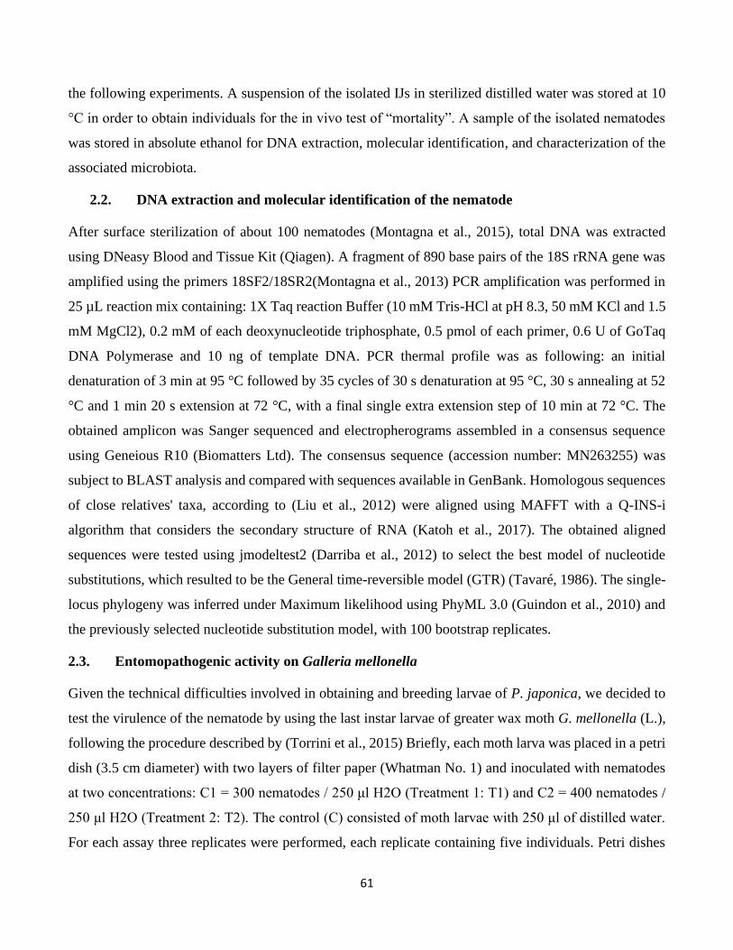
61
the following experiments. A suspension of the isolated IJs in sterilized distilled water was stored at 10
°C in order to obtain individuals for the in vivo test of “mortality”. A sample of the isolated nematodes
was stored in absolute ethanol for DNA extraction, molecular identification, and characterization of the
associated microbiota.
2.2. DNA extraction and molecular identification of the nematode
After surface sterilization of about 100 nematodes (Montagna et al., 2015), total DNA was extracted
using DNeasy Blood and Tissue Kit (Qiagen). A fragment of 890 base pairs of the 18S rRNA gene was
amplified using the primers 18SF2/18SR2(Montagna et al., 2013) PCR amplification was performed in
25 µL reaction mix containing: 1X Taq reaction Buffer (10 mM Tris-HCl at pH 8.3, 50 mM KCl and 1.5
mM MgCl2), 0.2 mM of each deoxynucleotide triphosphate, 0.5 pmol of each primer, 0.6 U of GoTaq
DNA Polymerase and 10 ng of template DNA. PCR thermal profile was as following: an initial
denaturation of 3 min at 95 °C followed by 35 cycles of 30 s denaturation at 95 °C, 30 s annealing at 52
°C and 1 min 20 s extension at 72 °C, with a final single extra extension step of 10 min at 72 °C. The
obtained amplicon was Sanger sequenced and electropherograms assembled in a consensus sequence
using Geneious R10 (Biomatters Ltd). The consensus sequence (accession number: MN263255) was
subject to BLAST analysis and compared with sequences available in GenBank. Homologous sequences
of close relatives' taxa, according to (Liu et al., 2012) were aligned using MAFFT with a Q-INS-i
algorithm that considers the secondary structure of RNA (Katoh et al., 2017). The obtained aligned
sequences were tested using jmodeltest2 (Darriba et al., 2012) to select the best model of nucleotide
substitutions, which resulted to be the General time-reversible model (GTR) (Tavaré, 1986). The single-
locus phylogeny was inferred under Maximum likelihood using PhyML 3.0 (Guindon et al., 2010) and
the previously selected nucleotide substitution model, with 100 bootstrap replicates.
2.3. Entomopathogenic activity on Galleria mellonella
Given the technical difficulties involved in obtaining and breeding larvae of P. japonica, we decided to
test the virulence of the nematode by using the last instar larvae of greater wax moth G. mellonella (L.),
following the procedure described by (Torrini et al., 2015) Briefly, each moth larva was placed in a petri
dish (3.5 cm diameter) with two layers of filter paper (Whatman No. 1) and inoculated with nematodes
at two concentrations: C1 = 300 nematodes / 250 μl H2O (Treatment 1: T1) and C2 = 400 nematodes /
250 μl H2O (Treatment 2: T2). The control (C) consisted of moth larvae with 250 μl of distilled water.
For each assay three replicates were performed, each replicate containing five individuals. Petri dishes
62
were stored at 24 °C ± 0.5 °C in darkness. The survival number of larvae for each trial was evaluated
over a period of five days by counting dead individuals at a 24 h interval. The dead larvae were placed
in modified White traps (White, 1927), after death, in order to recover the nematode infective juvenile
stages (IJs) from the host cadaver. In order to test for differences between the treatments and the control,
data collected on the number of individuals survived after five days were analyzed using the Kruskal-
Wallis test in R (version 3.5.1).
2.4. Characterization of the nematode microbiota using 16S rRNA metabarcoding
V1-V2 and V4 regions of 16S rRNA gene were sequenced using Ion Torrent platform (Life
Technologies). Firstly, the total DNA of the nematodes was used as template to amplify the V1-V2 region
using the primers 27FYM (Frank et al., 2008) and 338R (Amann et al., 1990), and to amplify the V4
region using the primers 515F (Caporaso et al., 2011) and 802R (Claesson et al., 2009) and 806R
(Caporaso et al., 2011). The PCR primers were tailed with two different GC rich sequences enabling
barcoding in a second amplification. 16S rRNA V1-V2 PCR was performed in 20 µL volume reaction
containing 8 µL of HotMasterMix 5 Prime 2.5X (Quanta Bio), 0.4 µL of BSA (20 µg/µL) (Sigma-
Aldrich), 1 µL of EvaGreen™ 20X (Biotium), 0.8µL of 27FYM (10 µM) (5' modified with unitail 1 5'-
GTGAGAGTTTGATYMTGGCTCAG -3'), 0.8 µL of 338R (10 µM) (5' modified with unitail 2 5'-
GCTGCCTCCCGTAGGAGT -3'), and 1 µL (corresponding to 50 ng) of DNA template. Library were
prepared accordingly to Chouaia et al. 2019 and sequencing was performed at Life Sciences Department
of Trieste University, Italy. The obtained reads were analyzed using QIIME 2 version 2018.11 (Bolyen
et al., 2019). After trimming, 16S rRNA sequences were clustered into Operational Taxonomic Units
(OTUs) with a similarity cut-off of 97% using the de novo clustering method implemented in the q2-
vsearch plugin (Rognes et al., 2016). Chimeras were identified and filtered using the uchime-denovo
method implemented in q2-vsearch (Edgar et al., 2011). Taxonomic assignment of OTU representative
sequences was performed using q2-feature-classifier (McDonald et al., 2012) and adopting Greengenes
13.8 as reference database. The 16S rRNA reads were deposited in the Sequence Read Archive (SRA)
of NCBI with accession number PRJNA564721
3. Results
3.1. Molecular characterization of the nematode
The 897 bp long 18S rRNA fragment amplified from the nematode DNA was assigned to the Rhabditidae
family based on a BLAST identity of 100%. The sequence showed an identity of 100% with sequences
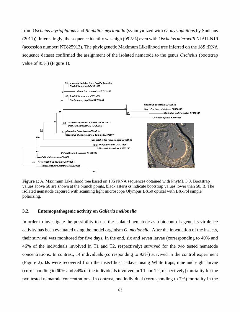
63
from Oscheius myriophilous and Rhabditis myriophila (synonymized with O. myriophilous by Sudhaus
(2011)). Interestingly, the sequence identity was high (99.5%) even with Oscheius microvilli NJAU-N19
(accession number: KT825913). The phylogenetic Maximum Likelihood tree inferred on the 18S rRNA
sequence dataset confirmed the assignment of the isolated nematode to the genus Oscheius (bootstrap
value of 95%) (Figure 1).
Figure 1: A. Maximum Likelihood tree based on 18S rRNA sequences obtained with PhyML 3.0. Bootstrap
values above 50 are shown at the branch points, black asterisks indicate bootstrap values lower than 50. B. The
isolated nematode captured with scanning light microscope Olympus BX50 optical with BX-Pol simple
polarizing.
3.2. Entomopathogenic activity on Galleria mellonella
In order to investigate the possibility to use the isolated nematode as a biocontrol agent, its virulence
activity has been evaluated using the model organism G. mellonella. After the inoculation of the insects,
their survival was monitored for five days. In the end, six and seven larvae (corresponding to 40% and
46% of the individuals involved in T1 and T2, respectively) survived for the two tested nematode
concentrations. In contrast, 14 individuals (corresponding to 93%) survived in the control experiment
(Figure 2). IJs were recovered from the insect host cadaver using White traps, nine and eight larvae
(corresponding to 60% and 54% of the individuals involved in T1 and T2, respectively) mortality for the
two tested nematode concentrations. In contrast, one individual (corresponding to 7%) mortality in the
64
control experiment. The results of the Kruskal-Wallis test indicate a significant difference in term of
survival among the groups (α2 = 6.79, df = 2, p-value = 0.0336; Table 1 Post-hoc test (Tukey’s HSD)
showed a difference between control and both treatments while no difference has been detected between
the two treatments (Table 1).
Table 1: Statistical analysis. A) Kruskal-Wallis test among T1, T2 and control groups. B) Tukey HSD tests
among the different treatments T1, T2, and control.
A) Kruskal-Wallis test
B) Post-hoc Tukey HSD
3.3. Taxonomic composition of the microbial community of the nematode
A total of 139,245 bacterial 16S rRNA sequences of the regions V1-V2 and V4 were obtained from the
DNA isolated from the nematodes. The microbiota associated with the nematode consisted of 1188
bacterial OTUs, obtained clustering the reads at 97% of sequence similarity. The nematode’s bacterial
community was characterized by values of Shannon and Pielou indices of 2 and 0.2, respectively
indicating low diversity and evenness. The most abundant taxa in the microbiota were represented by the
phylum Proteobacteria 99% (1106 OTUs), within this phylum, 50.7% of the 16S rRNA reads were
assigned to the family of Brucellaceae (259 OTUs) and 43.6% assigned to unclassified family belong to
order Rhizobiales (400 OTUs). At the genus level, the bacterial community associated with the nematode
Survival score
Chi-square
df
Assymp.sig.
6.79
2
0.0336
(I) TREATMENTS Mean Difference
(I-J)
Std. Error Sig. 95% Confidence Interval
Lower Bound Upper Bound
T1 T2 -0.333 0.385 0.420 -1.28 0.61
C -2.667* 0.385 0.000 -3.61 -1.72
T2 T1 0.333 0.385 0.420 -0.61 1.28
C -2.333* 0.385 0.001 -3.28 -1.39
C T1 2.667* 0.385 0.000 1.72 3.61
T2 2.333* 0.385 0.001 1.39 3.28
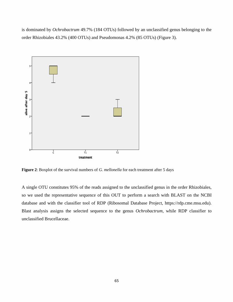
65
is dominated by Ochrobactrum 49.7% (184 OTUs) followed by an unclassified genus belonging to the
order Rhizobiales 43.2% (400 OTUs) and Pseudomonas 4.2% (85 OTUs) (Figure 3).
Figure 2: Boxplot of the survival numbers of G. mellonella for each treatment after 5 days
A single OTU constitutes 95% of the reads assigned to the unclassified genus in the order Rhizobiales,
so we used the representative sequence of this OUT to perform a search with BLAST on the NCBI
database and with the classifier tool of RDP (Ribosomal Database Project, https://rdp.cme.msu.edu).
Blast analysis assigns the selected sequence to the genus Ochrobactrum, while RDP classifier to
unclassified Brucellaceae.
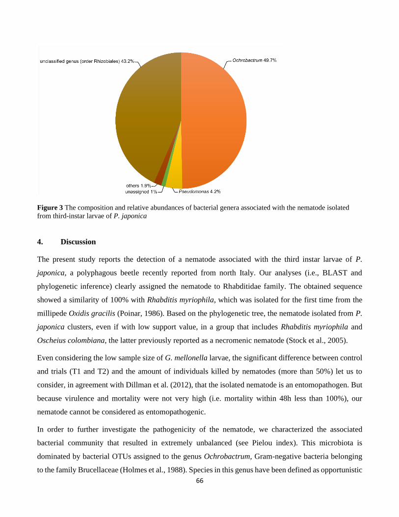
66
Figure 3 The composition and relative abundances of bacterial genera associated with the nematode isolated
from third-instar larvae of P. japonica
4. Discussion
The present study reports the detection of a nematode associated with the third instar larvae of P.
japonica, a polyphagous beetle recently reported from north Italy. Our analyses (i.e., BLAST and
phylogenetic inference) clearly assigned the nematode to Rhabditidae family. The obtained sequence
showed a similarity of 100% with Rhabditis myriophila, which was isolated for the first time from the
millipede Oxidis gracilis (Poinar, 1986). Based on the phylogenetic tree, the nematode isolated from P.
japonica clusters, even if with low support value, in a group that includes Rhabditis myriophila and
Oscheius colombiana, the latter previously reported as a necromenic nematode (Stock et al., 2005).
Even considering the low sample size of G. mellonella larvae, the significant difference between control
and trials (T1 and T2) and the amount of individuals killed by nematodes (more than 50%) let us to
consider, in agreement with Dillman et al. (2012), that the isolated nematode is an entomopathogen. But
because virulence and mortality were not very high (i.e. mortality within 48h less than 100%), our
nematode cannot be considered as entomopathogenic.
In order to further investigate the pathogenicity of the nematode, we characterized the associated
bacterial community that resulted in extremely unbalanced (see Pielou index). This microbiota is
dominated by bacterial OTUs assigned to the genus Ochrobactrum, Gram-negative bacteria belonging
to the family Brucellaceae (Holmes et al., 1988). Species in this genus have been defined as opportunistic
67
pathogens in humans (Lebuhn et al., 2006). In a previous study, Ochrobactrum anthropi has been
reported to be associated with the nematode Steinernema scapterisci (Aguillera and Smart Jr, 1993); in
addition, Ochrobactrum sp was isolated from dead larvae of G. mellonella infected by Steinernema sp.
and Heterorhabditis sp. (Razia et al., 2011). The pathogenic role of bacteria of the genus Ochrobactrum
was recently demonstrated in the case of Oscheius chongmingensis (Fu and Liu, 2019) on G. mellonella
larvae. Further studies on P. japonica larvae must be performed in order to test the possibility to use this
nematode as a biocontrol agent of this pest.
Declaration
NG designed and performed the experiments, carried out the microbiota and statistical analyses. MB
helped with the microbiota analyses. NG, MM, and MB wrote the manuscript. All authors read and
commented on the manuscript.
MM: Matteo Montagna; NG: Nizar Goda; MB: Matteo Brunetti
References
Aguillera MM, Smart Jr GC (1993). Development, reproduction, and pathogenicity of Steinernema
scapterisci in monoxenic culture with different species of bacteria. Journal of Invertebrate
Pathology 62(3): 289-294. https://doi.org/10.1006/jipa.1993.1115.
Amann RI, Binder BJ, Olson RJ, Chisholm SW, Devereux R et al. (1990). Combination of 16S rRNA-
targeted oligonucleotide probes with flow cytometry for analyzing mixed microbial populations.
Applied and Environmental Microbiology 56(6): 1919-1925.
Bolyen E, Rideout JR, Dillon MR, Bokulich NA, Abnet CC et al. (2019. Reproducible, interactive,
scalable and extensible microbiome data science using QIIME 2. Nature biotechnology, 37(8):
852-857.
Bragard C, Dehnen‐Schmutz K, Di Serio F, Gonthier P, Jacques MA et al. (2018). Pest categorisation of
Popillia japonica. Efsa Journal, 16(11): e05438. https://doi.org/10.2903/j.efsa.2018.5438.
Caporaso JG, Lauber CL, Walters WA, Berg-Lyons D, Lozupone CA et al. (2011). Global patterns of
16S rRNA diversity at a depth of millions of sequences per sample. Proceedings of the national
academy of sciences, 108(Supplement 1), 4516-4522.
https://doi.org/10.1073/pnas.1000080107.
68
Cappaert DL, Smitley DR (2002). Parasitoids and pathogens of Japanese beetle (Coleoptera:
Scarabaeidae) in southern Michigan. Environmental Entomology 31(3): 573-580.
https://doi.org/10.1603/0046-225X-31.3.573.
Claesson MJ, O'Sullivan O, Wang Q, Nikkilä J, Marchesi JR et al. (2009). Comparative analysis of
pyrosequencing and a phylogenetic microarray for exploring microbial community structures in
the human distal intestine. PloS one, 4(8): e6669. https://doi.org/10.1371/journal.pone.0006669.
Darriba D, Taboada GL, Doallo R, Posada D (2012). jModelTest 2: more models, new heuristics and
parallel computing. Nature methods 9(8): 772.
Dillman AR, Chaston JM, Adams BJ, Ciche TA, Goodrich-Blair H et al. (2012). An entomopathogenic
nematode by any other name. PLoS Pathogens 8(3), e1002527.
https://doi.org/10.1371/journal.ppat.1002527.
Edgar RC, Haas BJ, Clemente JC, Quince C, Knight R (2011). UCHIME improves sensitivity and speed
of chimera detection. Bioinformatics 27(16): 2194-2200.
https://doi.org/10.1093/bioinformatics/btr381.
Frank JA, Reich CI, Sharma S, Weisbaum JS, Wilson BA et al. (2008). Critical evaluation of two primers
commonly used for amplification of bacterial 16S rRNA genes. Applied and Environmental
Microbiology 74(8): 2461-2470. 10.1128/AEM.02272-07.
Fu JR, Liu QZ (2019). Evaluation and entomopathogenicity of gut bacteria associated with dauer
juveniles of Oscheius chongmingensis (Nematoda: Rhabditidae).MicrobiologyOpen,e00823.
https://doi.org/10.1002/mbo3.823.
Grewal PS, Ehlers RU, Shapiro-Ilan DI (2005). Nematodes as biocontrol agents. 1st ed. Cambridge,
USA: CABI.
Guindon S, Dufayard JF, Lefort V, Anisimova M, Hordijk W et al. (2010). New algorithms and methods
to estimate maximum-likelihood phylogenies: assessing the performance of PhyML 3.0.
Systematic biology 59(3): 307-321. https://doi.org/10.1093/sysbio/syq010.
Holmes B, Popoff M, Kiredjian M, Kersters K (1988). Ochrobactrum anthropi gen. nov., sp. nov. from
human clinical specimens and previously known as group Vd. International Journal of
Systematic and Evolutionary Microbiology 38(4): 406-416. https://doi.org/10.1099/00207713-
38-4-406.
69
Katoh K, Rozewicki J, Yamada KD (2017). MAFFT online service: multiple sequence alignment,
interactive sequence choice and visualization. Briefings in Bioinformatics bbx108.
https://doi.org/10.1093/bib/bbx108.
Kaya HK, Gaugler R (1993). Entomopathogenic nematodes. Annual review of entomology38: 181-206.
https://doi.org/10.1146/annurev.en.38.010193.001145.
Kaya HK, Stock SP (1997). Techniques in insect nematology. In Lawrence Lacey editor. Manual of
techniques in insect pathology. 1st ed. San Diego, CA, USA: Academic Press, pp. 281-324.
Klein M, Nickle W, Benedict P, Dunbar D (1976). Psammomermis sp. (Nematoda: Mermithidae): a new
nematode parasite of the Japanese beetle, Popillia japonica (Coleoptera: Scarabaeidae)
[Biological control]. Proceedings of the Helminthological Society of Washington (USA) 43(2):
235-236.
Lebuhn M, Bathe S, Achouak W, Hartmann A, Heulin T et al. (2006). Comparative sequence analysis
of the internal transcribed spacer 1 of Ochrobactrum species. 29(4): 265-275.
https://doi.org/10.1016/j.syapm.2005.11.003.
Liu QZ, Mráček Z, Zhang LJ, Půža V, Dong LM (2012). Re-description of Oscheius chongmingensis
(Zhang et al., 2008) (Nematoda: Rhabditidae) and its entomopathogenicity. Nematology, 14(2):
139-149. https://doi.org/10.1163/138855411X580777.
Marianelli L, Paoli F, Torrini G, Mazza G, Benvenuti C et al. (2018). Entomopathogenic nematodes as
potential biological control agents of Popillia japonica (Coleoptera, Scarabaeidae) in Piedmont
Region (Italy). Journal of applied entomology 142(3): 311-318.
https://doi.org/10.1111/jen.12470.
Mazza G, Paoli F, Strangi A, Torrini G, Marianelli L et al. (2017). Hexamermis popilliae n. sp.
(Nematoda: Mermithidae) parasitizing the Japanese beetle Popillia japonica Newman
(Coleoptera: Scarabaeidae) in Italy. Systematic parasitology 94(8): 915-926.
McDonald D, Price MN, Goodrich J, Nawrocki EP, DeSantis TZ et al. (2012). An improved Greengenes
taxonomy with explicit ranks for ecological and evolutionary analyses of bacteria and archaea.
The ISME journal 6(3): 610.
Montagna M, Sassi D, Giorgi A (2013). Pachybrachis holerorum (Coleoptera: Chrysomelidae:
Cryptocephalinae), a new species from the Apennines, Italy, identified by integration of
70
morphological and molecular data. Zootaxa, 3741(2): 243-253.
http://dx.doi.org/10.11646/zootaxa.3741.2.3.
Montagna M, Chouaia B, Mazza G, Prosdocimi EM, Crotti E et al. (2015). Effects of the diet on the
microbiota of the red palm weevil (Coleoptera: Dryophthoridae).PLoSOne,10(1), e0117439.
https://doi.org/10.1371/journal.pone.0117439.
Morris EE, Grewal PS (2011). Susceptibility of the adult Japanese beetle, Popillia japonica to
entomopathogenic nematodes. Journal of nematology 43(3-4): 196.
Pavesi M (2014). Popillia japonica specie aliena invasiva segnalata in Lombardia. L’informatore Agrario
32: 53-55 (in Italian).
Poinar GO (1986). Rhabditis myriophila sp. n. (Rhabditidae: Rhabditida), associated with the millipede
Oxidis gracilis (Polydesmida: Diplopoda). Proceedings of the Helminthological Society of
Washington, 53(2): 232-236.
Potter DA Held DW (2002). Biology and management of the Japanese beetle. Annual review of
entomology 47(1): 175-205. https://doi.org/10.1146/annurev.ento.47.091201.145153.
Razia M, Raja RK, Padmanaban K, Chellapandi P, Sivaramakrishnan SJG (2011). 16S rDNA-based
phylogeny of non-symbiotic bacteria of entomopathogenic nematodes from infected insect
cadavers. Genomics, proteomics & bioinformatics, 9(3): 104-112.
https://doi.org/10.1016/S1672-0229(11)60013-2.
Rognes T, Flouri T, Nichols B, Quince C, Mahé F (2016). VSEARCH: a versatile open source tool for
metagenomics. PeerJ 4: e2584.
Stock SP, Caicedo A, Calatayud P (2005). Rhabditis (Oscheius) colombiana n. sp.(Nematoda:
Rhabditidae), a necromenic associate of the subterranean burrower bug Cyrtomenus bergi
(Hemiptera: Cydnidae) from the Cauca Valley, Colombia. Nematology, 7(3): 363-373.
https://doi.org/10.1163/156854105774355590.
Sudhaus W (2011). Phylogenetic systematisation and catalogue of paraphyletic "Rhabditidae"
(Secernentea, Nematoda). Journal of Nematode Morphology and Systematics 14(2): 113-178.
Tavaré S, (1986). Some probabilistic and statistical problems in the analysis of DNA sequences. Lectures
on mathematics in the life sciences 17(2): 57-86.
71
Torrini G, Mazza G, Carletti B, Benvenuti C, Roversi PF et al. (2015). Oscheius onirici sp. n. (Nematoda:
Rhabditidae): a new entomopathogenic nematode from an Italian cave. Zootaxa, 3937(3): 533-
548. http://dx.doi.org/10.11646/zootaxa.3937.3.6.
White GF (1927). A method for obtaining infective nematode larvae from cultures. Science
(Washington) 66(1709): 302-303.
Wickizer SL, Gergerich RC (2007). First Report of Japanese Beetle (Popillia japonica) as a Vector of
Southern bean mosaic virus and Bean pod mottle virus. Plant Disease 91: 637-637.
https://doi.org/10.1094/PDIS-91-5-0637C.
72
Conclusion
In this PhD thesis, two research articles have been presented. The first article deals with the
developmental stages and gut microenvironments and how these influence the gut microbiota dynamics
in the invasive beetle P. japonica Newman (Coleoptera: Scarabaeidae). The results highlight the
influence of different developmental stages of P. japonica (i.e., larvae, pupae, adults), gut compartments
(i.e., foregut, midgut, hindgut) and physicochemical properties (i.e., pH, oxygen concentration, redox
potential) in shaping the bacterial community associated with P. japonica. The results also reveal that
the gut microbiota of P. japonica are highly dynamic across the developmental stages of the insect while
changes in microbiota composition are strongly correlated with the physicochemical properties of the
gut. Despite the microbiota’s high variability, in our case, 89 OTUs were maintained from larvae to
adults, including 35 OTUs originating from the soil environment. In the future, it would be worthwhile
investigating whether these OTUs represent a stable core microbiota present in all P. japonica
populations in different parts of the world or whether they are subject to change in different
environments. In the first case, this might indicate a more intimate symbiotic relationship which is
potentially maintained via vertical transmission. In the latter case, variable microbiota would provide a
means to investigate the origins of new invasions of this beetle, via a comparative analysis of local soil
and insect gut microbiotas.
In the second article, Potentially entomopathogenic nematode isolated from Popillia japonica
(Coleoptera: Scarabaeidae): bioassay, molecular characterization and the associated microbiota. Our
study of the isolated nematode suggests that it engages in entomopathogen activity. Further, it is probable
that the bacteria of the genus Ochrobactrum, which is dominant in the isolated nematode, contributes to
nematode entomopathogen activity, while the genus Ochrobactrum has been reported to be associated
with the nematode Oscheius chongmingensis and described as a pathogenic bacterium. Further studies
on P. japonica larvae must be performed in order to test the potential to use this nematode as a biocontrol
agent against P. japonica larvae.
73
Annexes
Table S1: Summary of the different ecological indices and Random Forest results for each sample. 1a: Ecological indices summary for the different
samples. 1b: summary statistics of the comparison of the different alpha diversity values between the different developmental stages. 1c: Standardized
phylogenetic evenness results for all the samples. 1d: Results of the Random Forest goodness of prediction for the developmental stages. 1e: Results
of the Random Forest goodness of prediction for the gut section. 1f: Top 10 OTU predictors of the Random Forest prediction for the developmental
stages. 1g: Top 10 OTU predictors of the Random Forest prediction for the gut sections.
Table S1a: Ecological indices summary for the different samples
Ecological indices Sample information
Sa
mp
le.I
D
Rea
ds
Ev
enn
ess.
J
Ob
serv
ed
OT
Us
chao
1
Sh
annon
mp
d.o
bs
mp
d.o
bs.
z
mp
d.o
bs.
p
Sam
ple
Sta
ge
Dev
elop
men
t
Des
crip
tion
Ox
yg
en
pH
Rx
A.1 239121 0.85 1103 1103.0 5.93 1.24 0.72 0.77 Soil Soil Soil S.S03 M.An NA NA
A.2 62966 0.66 103 125.8 3.05 1.19 -1.99 0.02 Foregut L1 Larvae FL1.2 An Sac.N P
A.4 66147 0.61 116 143.1 2.91 1.27 2.59 0.99 Foregut L2 Larvae FL2.1 An Sac.N P
A.5 63160 0.81 51 96.0 3.18 1.09 -6.56 0.00 Foregut L2 Larvae FL2.2 An Sac.N P
A.6 281025 0.85 1104 1104.6 5.97 1.23 0.54 0.71 Soil Soil Soil S.S02 M.An NA NA
A.7 73473 0.69 168 179.1 3.55 1.17 -2.62 0.01 Foregut L3 Larvae FL3.1 An Sac.N P
A.8 27298 0.81 92 137.5 3.68 1.16 -3.24 0.00 Foregut L3 Larvae FL3.2 An Sac.N P
A.9 87421 0.76 209 237.5 4.08 1.16 -2.93 0.00 Foregut L3 Larvae FL3.3 An Sac.N P
A.10 65649 0.46 308 343.5 2.62 1.09 -6.40 0.00 Foregut Pupae Pupae FP1 NA NA NA
A.11 80611 0.26 264 342.6 1.44 1.09 -7.67 0.00 Foregut Pupae Pupae FP2 NA NA NA
A.12 50309 0.42 167 181.1 2.14 1.10 -6.71 0.00 Foregut Pupae Pupae FP3 NA NA NA
A.13 208802 0.55 131 169.0 2.68 1.18 -2.25 0.02 Foregut Female Adult FF1 M.An Sac.N P
A.14 50611 0.69 73 82.0 2.94 1.15 -4.03 0.00 Foregut Female Adult FF2 M.An Sac.N P
A.15 70563 0.61 92 162.0 2.74 1.36 6.85 1.00 Foregut Female Adult FF3 M.An Sac.N P
A.16 173052 0.40 67 107.0 1.67 1.19 -1.86 0.03 Foregut Male Adult FM1 M.An Sac.N P
A.17 150020 0.66 100 127.5 3.04 1.23 0.35 0.66 Foregut Male Adult FM2 M.An Sac.N P
A.18 94199 0.63 78 96.0 2.75 1.16 -3.21 0.00 Foregut Male Adult FM3 M.An Sac.N P
A.19 92418 0.84 1087 1102.8 5.86 1.24 0.81 0.79 Soil Soil Soil S.L02 M.An NA NA
A.20 65945 0.80 536 553.2 5.01 1.17 -2.41 0.00 Midgut L1 Larvae ML1.1 An Sal.Al P.N
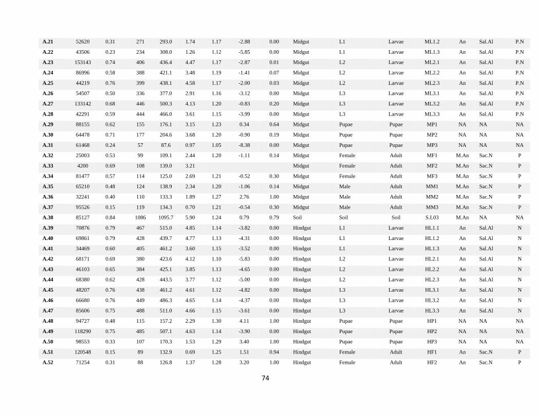
74
A.21 52620 0.31 271 293.0 1.74 1.17 -2.88 0.00 Midgut L1 Larvae ML1.2 An Sal.Al P.N
A.22 43506 0.23 234 308.0 1.26 1.12 -5.85 0.00 Midgut L1 Larvae ML1.3 An Sal.Al P.N
A.23 153143 0.74 406 436.4 4.47 1.17 -2.87 0.01 Midgut L2 Larvae ML2.1 An Sal.Al P.N
A.24 86996 0.58 388 421.1 3.48 1.19 -1.41 0.07 Midgut L2 Larvae ML2.2 An Sal.Al P.N
A.25 44219 0.76 399 438.1 4.58 1.17 -2.00 0.03 Midgut L2 Larvae ML2.3 An Sal.Al P.N
A.26 54507 0.50 336 377.0 2.91 1.16 -3.12 0.00 Midgut L3 Larvae ML3.1 An Sal.Al P.N
A.27 133142 0.68 446 500.3 4.13 1.20 -0.83 0.20 Midgut L3 Larvae ML3.2 An Sal.Al P.N
A.28 42291 0.59 444 466.0 3.61 1.15 -3.99 0.00 Midgut L3 Larvae ML3.3 An Sal.Al P.N
A.29 88155 0.62 155 176.1 3.15 1.23 0.34 0.64 Midgut Pupae Pupae MP1 NA NA NA
A.30 64478 0.71 177 204.6 3.68 1.20 -0.90 0.19 Midgut Pupae Pupae MP2 NA NA NA
A.31 61468 0.24 57 87.6 0.97 1.05 -8.38 0.00 Midgut Pupae Pupae MP3 NA NA NA
A.32 25003 0.53 99 109.1 2.44 1.20 -1.11 0.14 Midgut Female Adult MF1 M.An Sac.N P
A.33 4200 0.69 108 139.0 3.21
Midgut Female Adult MF2 M.An Sac.N P
A.34 81477 0.57 114 125.0 2.69 1.21 -0.52 0.30 Midgut Female Adult MF3 M.An Sac.N P
A.35 65210 0.48 124 138.9 2.34 1.20 -1.06 0.14 Midgut Male Adult MM1 M.An Sac.N P
A.36 32241 0.40 110 133.3 1.89 1.27 2.76 1.00 Midgut Male Adult MM2 M.An Sac.N P
A.37 95526 0.15 119 134.3 0.70 1.21 -0.54 0.30 Midgut Male Adult MM3 M.An Sac.N P
A.38 85127 0.84 1086 1095.7 5.90 1.24 0.79 0.79 Soil Soil Soil S.L03 M.An NA NA
A.39 70876 0.79 467 515.0 4.85 1.14 -3.82 0.00 Hindgut L1 Larvae HL1.1 An Sal.Al N
A.40 69861 0.79 428 439.7 4.77 1.13 -4.31 0.00 Hindgut L1 Larvae HL1.2 An Sal.Al N
A.41 34469 0.60 405 461.2 3.60 1.15 -3.52 0.00 Hindgut L1 Larvae HL1.3 An Sal.Al N
A.42 68171 0.69 380 423.6 4.12 1.10 -5.83 0.00 Hindgut L2 Larvae HL2.1 An Sal.Al N
A.43 46103 0.65 384 425.1 3.85 1.13 -4.65 0.00 Hindgut L2 Larvae HL2.2 An Sal.Al N
A.44 68380 0.62 428 443.5 3.77 1.12 -5.00 0.00 Hindgut L2 Larvae HL2.3 An Sal.Al N
A.45 48207 0.76 438 461.2 4.61 1.12 -4.82 0.00 Hindgut L3 Larvae HL3.1 An Sal.Al N
A.46 66680 0.76 449 486.3 4.65 1.14 -4.37 0.00 Hindgut L3 Larvae HL3.2 An Sal.Al N
A.47 85606 0.75 488 511.0 4.66 1.15 -3.61 0.00 Hindgut L3 Larvae HL3.3 An Sal.Al N
A.48 94727 0.48 115 157.2 2.29 1.30 4.11 1.00 Hindgut Pupae Pupae HP1 NA NA NA
A.49 118290 0.75 485 507.1 4.63 1.14 -3.90 0.00 Hindgut Pupae Pupae HP2 NA NA NA
A.50 98553 0.33 107 170.3 1.53 1.29 3.40 1.00 Hindgut Pupae Pupae HP3 NA NA NA
A.51 120548 0.15 89 132.9 0.69 1.25 1.51 0.94 Hindgut Female Adult HF1 An Sac.N P
A.52 71254 0.31 88 126.8 1.37 1.28 3.20 1.00 Hindgut Female Adult HF2 An Sac.N P
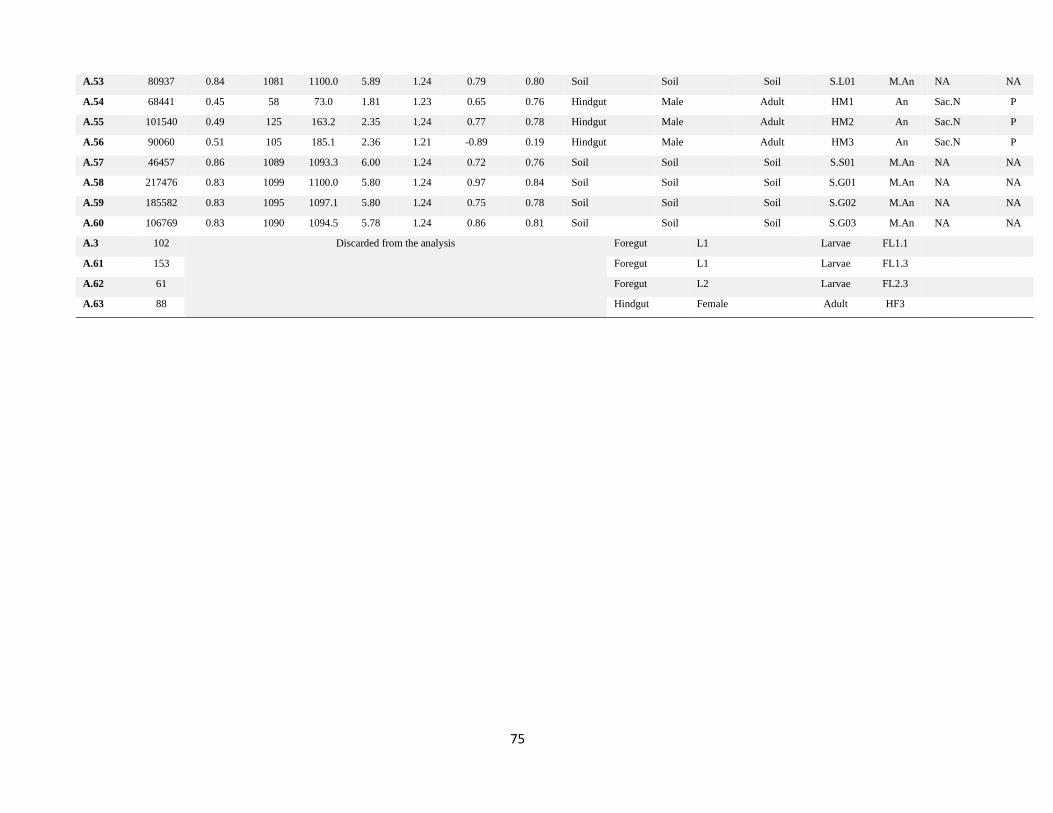
75
A.53 80937 0.84 1081 1100.0 5.89 1.24 0.79 0.80 Soil Soil Soil S.L01 M.An NA NA
A.54 68441 0.45 58 73.0 1.81 1.23 0.65 0.76 Hindgut Male Adult HM1 An Sac.N P
A.55 101540 0.49 125 163.2 2.35 1.24 0.77 0.78 Hindgut Male Adult HM2 An Sac.N P
A.56 90060 0.51 105 185.1 2.36 1.21 -0.89 0.19 Hindgut Male Adult HM3 An Sac.N P
A.57 46457 0.86 1089 1093.3 6.00 1.24 0.72 0.76 Soil Soil Soil S.S01 M.An NA NA
A.58 217476 0.83 1099 1100.0 5.80 1.24 0.97 0.84 Soil Soil Soil S.G01 M.An NA NA
A.59 185582 0.83 1095 1097.1 5.80 1.24 0.75 0.78 Soil Soil Soil S.G02 M.An NA NA
A.60 106769 0.83 1090 1094.5 5.78 1.24 0.86 0.81 Soil Soil Soil S.G03 M.An NA NA
A.3 102 Discarded from the analysis Foregut L1 Larvae FL1.1
A.61 153 Foregut L1 Larvae FL1.3
A.62 61 Foregut L2 Larvae FL2.3
A.63 88 Hindgut Female Adult HF3
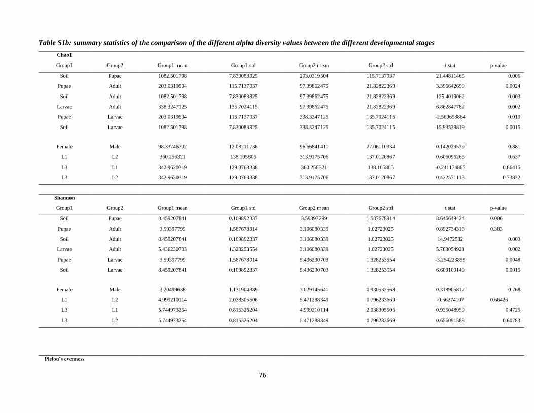
76
Table S1b: summary statistics of the comparison of the different alpha diversity values between the different developmental stages
Chao1
Group1 Group2 Group1 mean Group1 std Group2 mean Group2 std t stat p-value
Soil Pupae 1082.501798 7.830083925 203.0319504 115.7137037 21.44811465 0.006
Pupae Adult 203.0319504 115.7137037 97.39862475 21.82822369 3.396642699 0.0024
Soil Adult 1082.501798 7.830083925 97.39862475 21.82822369 125.4019062 0.003
Larvae Adult 338.3247125 135.7024115 97.39862475 21.82822369 6.862847782 0.002
Pupae Larvae 203.0319504 115.7137037 338.3247125 135.7024115 -2.569658864 0.019
Soil Larvae 1082.501798 7.830083925 338.3247125 135.7024115 15.93539819 0.0015
Female Male 98.33746702 12.08211736 96.66841411 27.06110334 0.142029539 0.881
L1 L2 360.256321 138.105805 313.9175706 137.0120867 0.606096265 0.637
L3 L1 342.9620319 129.0763338 360.256321 138.105805 -0.241174867 0.86415
L3 L2 342.9620319 129.0763338 313.9175706 137.0120867 0.422571113 0.73832
Shannon
Group1 Group2 Group1 mean Group1 std Group2 mean Group2 std t stat p-value
Soil Pupae 8.459207841 0.109892337 3.59397799 1.587678914 8.646649424 0.006
Pupae Adult 3.59397799 1.587678914 3.106080339 1.02723025 0.892734316 0.383
Soil Adult 8.459207841 0.109892337 3.106080339 1.02723025 14.9472582 0.003
Larvae Adult 5.436230703 1.328253554 3.106080339 1.02723025 5.783054921 0.002
Pupae Larvae 3.59397799 1.587678914 5.436230703 1.328253554 -3.254223855 0.0048
Soil Larvae 8.459207841 0.109892337 5.436230703 1.328253554 6.609100149 0.0015
Female Male 3.20499638 1.131904389 3.029145641 0.930532568 0.318905817 0.768
L1 L2 4.999210114 2.038305506 5.471288349 0.796233669 -0.56274107 0.66426
L3 L1 5.744973254 0.815326204 4.999210114 2.038305506 0.935048959 0.4725
L3 L2 5.744973254 0.815326204 5.471288349 0.796233669 0.656091588 0.60783
Pielou’s evenness
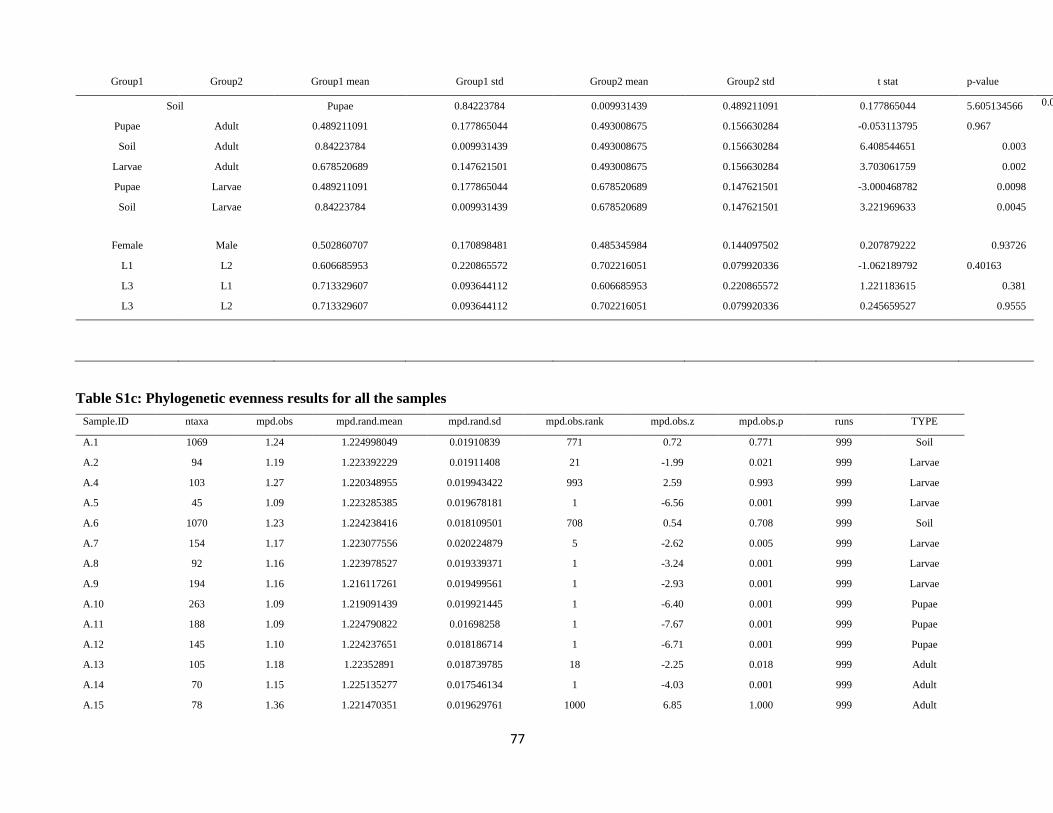
77
Group1 Group2 Group1 mean Group1 std Group2 mean Group2 std t stat p-value
Soil Pupae 0.84223784 0.009931439 0.489211091 0.177865044 5.605134566 0.006
Pupae Adult 0.489211091 0.177865044 0.493008675 0.156630284 -0.053113795 0.967
Soil Adult 0.84223784 0.009931439 0.493008675 0.156630284 6.408544651 0.003
Larvae Adult 0.678520689 0.147621501 0.493008675 0.156630284 3.703061759 0.002
Pupae Larvae 0.489211091 0.177865044 0.678520689 0.147621501 -3.000468782 0.0098
Soil Larvae 0.84223784 0.009931439 0.678520689 0.147621501 3.221969633 0.0045
Female Male 0.502860707 0.170898481 0.485345984 0.144097502 0.207879222 0.93726
L1 L2 0.606685953 0.220865572 0.702216051 0.079920336 -1.062189792 0.40163
L3 L1 0.713329607 0.093644112 0.606685953 0.220865572 1.221183615 0.381
L3 L2 0.713329607 0.093644112 0.702216051 0.079920336 0.245659527 0.9555
Table S1c: Phylogenetic evenness results for all the samples
Sample.ID ntaxa mpd.obs mpd.rand.mean mpd.rand.sd mpd.obs.rank mpd.obs.z mpd.obs.p runs TYPE
A.1 1069 1.24 1.224998049 0.01910839 771 0.72 0.771 999 Soil
A.2 94 1.19 1.223392229 0.01911408 21 -1.99 0.021 999 Larvae
A.4 103 1.27 1.220348955 0.019943422 993 2.59 0.993 999 Larvae
A.5 45 1.09 1.223285385 0.019678181 1 -6.56 0.001 999 Larvae
A.6 1070 1.23 1.224238416 0.018109501 708 0.54 0.708 999 Soil
A.7 154 1.17 1.223077556 0.020224879 5 -2.62 0.005 999 Larvae
A.8 92 1.16 1.223978527 0.019339371 1 -3.24 0.001 999 Larvae
A.9 194 1.16 1.216117261 0.019499561 1 -2.93 0.001 999 Larvae
A.10 263 1.09 1.219091439 0.019921445 1 -6.40 0.001 999 Pupae
A.11 188 1.09 1.224790822 0.01698258 1 -7.67 0.001 999 Pupae
A.12 145 1.10 1.224237651 0.018186714 1 -6.71 0.001 999 Pupae
A.13 105 1.18 1.22352891 0.018739785 18 -2.25 0.018 999 Adult
A.14 70 1.15 1.225135277 0.017546134 1 -4.03 0.001 999 Adult
A.15 78 1.36 1.221470351 0.019629761 1000 6.85 1.000 999 Adult
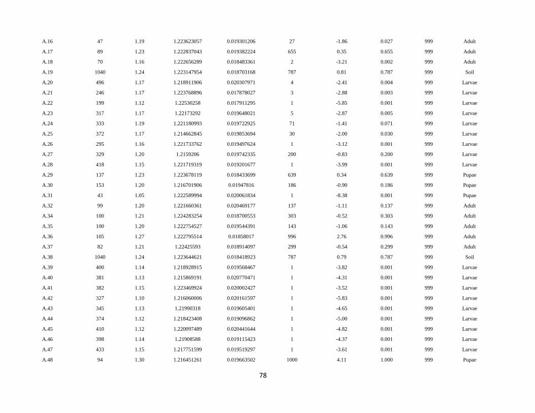
78
A.16 47 1.19 1.223623057 0.019301206 27 -1.86 0.027 999 Adult
A.17 89 1.23 1.222837043 0.019382224 655 0.35 0.655 999 Adult
A.18 70 1.16 1.222656289 0.018483361 2 -3.21 0.002 999 Adult
A.19 1040 1.24 1.223147954 0.018703168 787 0.81 0.787 999 Soil
A.20 496 1.17 1.218911906 0.020307971 4 -2.41 0.004 999 Larvae
A.21 246 1.17 1.223768896 0.017878027 3 -2.88 0.003 999 Larvae
A.22 199 1.12 1.22530258 0.017911295 1 -5.85 0.001 999 Larvae
A.23 317 1.17 1.22173292 0.019648021 5 -2.87 0.005 999 Larvae
A.24 333 1.19 1.221180993 0.019722925 71 -1.41 0.071 999 Larvae
A.25 372 1.17 1.214662845 0.019853694 30 -2.00 0.030 999 Larvae
A.26 295 1.16 1.221733762 0.019497624 1 -3.12 0.001 999 Larvae
A.27 329 1.20 1.2159206 0.019742335 200 -0.83 0.200 999 Larvae
A.28 418 1.15 1.221719319 0.019201677 1 -3.99 0.001 999 Larvae
A.29 137 1.23 1.223678119 0.018433699 639 0.34 0.639 999 Pupae
A.30 153 1.20 1.216701906 0.01947816 186 -0.90 0.186 999 Pupae
A.31 43 1.05 1.222589994 0.020061834 1 -8.38 0.001 999 Pupae
A.32 99 1.20 1.221660361 0.020469177 137 -1.11 0.137 999 Adult
A.34 100 1.21 1.224283254 0.018700553 303 -0.52 0.303 999 Adult
A.35 100 1.20 1.222754527 0.019544391 143 -1.06 0.143 999 Adult
A.36 105 1.27 1.222795514 0.01858017 996 2.76 0.996 999 Adult
A.37 82 1.21 1.22425593 0.018914097 299 -0.54 0.299 999 Adult
A.38 1040 1.24 1.223644621 0.018418923 787 0.79 0.787 999 Soil
A.39 400 1.14 1.218928915 0.019568467 1 -3.82 0.001 999 Larvae
A.40 381 1.13 1.215869191 0.020770471 1 -4.31 0.001 999 Larvae
A.41 382 1.15 1.223469924 0.020002427 1 -3.52 0.001 999 Larvae
A.42 327 1.10 1.216060006 0.020161597 1 -5.83 0.001 999 Larvae
A.43 345 1.13 1.21990318 0.019605401 1 -4.65 0.001 999 Larvae
A.44 374 1.12 1.218423408 0.019096862 1 -5.00 0.001 999 Larvae
A.45 410 1.12 1.220097489 0.020441644 1 -4.82 0.001 999 Larvae
A.46 398 1.14 1.21908588 0.019115423 1 -4.37 0.001 999 Larvae
A.47 433 1.15 1.217751599 0.019519297 1 -3.61 0.001 999 Larvae
A.48 94 1.30 1.216451261 0.019663502 1000 4.11 1.000 999 Pupae
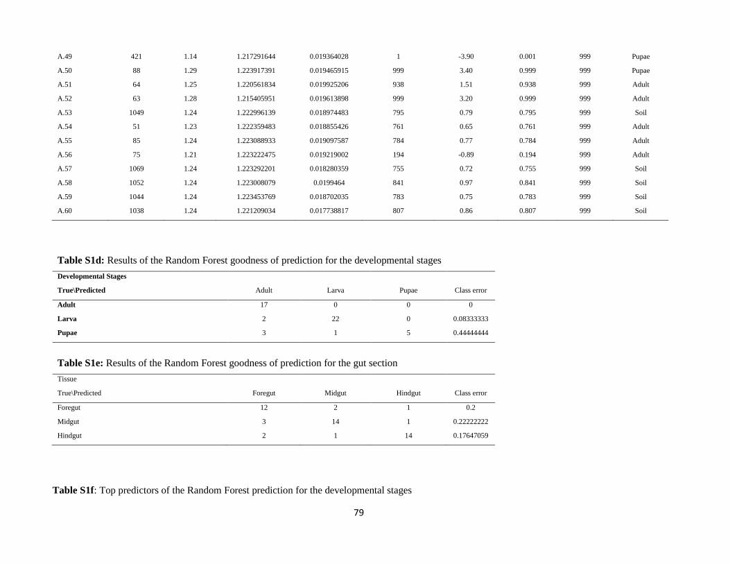
79
A.49 421 1.14 1.217291644 0.019364028 1 -3.90 0.001 999 Pupae
A.50 88 1.29 1.223917391 0.019465915 999 3.40 0.999 999 Pupae
A.51 64 1.25 1.220561834 0.019925206 938 1.51 0.938 999 Adult
A.52 63 1.28 1.215405951 0.019613898 999 3.20 0.999 999 Adult
A.53 1049 1.24 1.222996139 0.018974483 795 0.79 0.795 999 Soil
A.54 51 1.23 1.222359483 0.018855426 761 0.65 0.761 999 Adult
A.55 85 1.24 1.223088933 0.019097587 784 0.77 0.784 999 Adult
A.56 75 1.21 1.223222475 0.019219002 194 -0.89 0.194 999 Adult
A.57 1069 1.24 1.223292201 0.018280359 755 0.72 0.755 999 Soil
A.58 1052 1.24 1.223008079 0.0199464 841 0.97 0.841 999 Soil
A.59 1044 1.24 1.223453769 0.018702035 783 0.75 0.783 999 Soil
A.60 1038 1.24 1.221209034 0.017738817 807 0.86 0.807 999 Soil
Table S1d: Results of the Random Forest goodness of prediction for the developmental stages
Developmental Stages
True\Predicted Adult Larva Pupae Class error
Adult 17 0 0 0
Larva 2 22 0 0.08333333
Pupae 3 1 5 0.44444444
Table S1e: Results of the Random Forest goodness of prediction for the gut section
Tissue
True\Predicted Foregut Midgut Hindgut Class error
Foregut 12 2 1 0.2
Midgut 3 14 1 0.22222222
Hindgut 2 1 14 0.17647059
Table S1f: Top predictors of the Random Forest prediction for the developmental stages
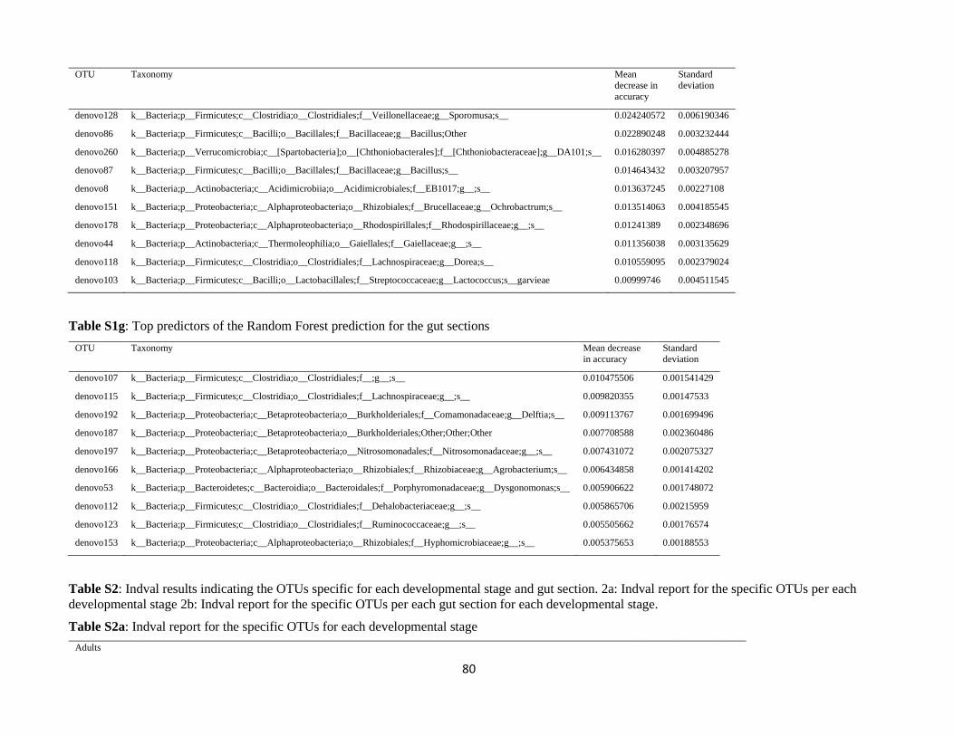
80
OTU Taxonomy Mean
decrease in
accuracy
Standard
deviation
denovo128 k__Bacteria;p__Firmicutes;c__Clostridia;o__Clostridiales;f__Veillonellaceae;g__Sporomusa;s__ 0.024240572 0.006190346
denovo86 k__Bacteria;p__Firmicutes;c__Bacilli;o__Bacillales;f__Bacillaceae;g__Bacillus;Other 0.022890248 0.003232444
denovo260 k__Bacteria;p__Verrucomicrobia;c__[Spartobacteria];o__[Chthoniobacterales];f__[Chthoniobacteraceae];g__DA101;s__ 0.016280397 0.004885278
denovo87 k__Bacteria;p__Firmicutes;c__Bacilli;o__Bacillales;f__Bacillaceae;g__Bacillus;s__ 0.014643432 0.003207957
denovo8 k__Bacteria;p__Actinobacteria;c__Acidimicrobiia;o__Acidimicrobiales;f__EB1017;g__;s__ 0.013637245 0.00227108
denovo151 k__Bacteria;p__Proteobacteria;c__Alphaproteobacteria;o__Rhizobiales;f__Brucellaceae;g__Ochrobactrum;s__ 0.013514063 0.004185545
denovo178 k__Bacteria;p__Proteobacteria;c__Alphaproteobacteria;o__Rhodospirillales;f__Rhodospirillaceae;g__;s__ 0.01241389 0.002348696
denovo44 k__Bacteria;p__Actinobacteria;c__Thermoleophilia;o__Gaiellales;f__Gaiellaceae;g__;s__ 0.011356038 0.003135629
denovo118 k__Bacteria;p__Firmicutes;c__Clostridia;o__Clostridiales;f__Lachnospiraceae;g__Dorea;s__ 0.010559095 0.002379024
denovo103 k__Bacteria;p__Firmicutes;c__Bacilli;o__Lactobacillales;f__Streptococcaceae;g__Lactococcus;s__garvieae 0.00999746 0.004511545
Table S1g: Top predictors of the Random Forest prediction for the gut sections
OTU Taxonomy Mean decrease
in accuracy
Standard
deviation
denovo107 k__Bacteria;p__Firmicutes;c__Clostridia;o__Clostridiales;f__;g__;s__ 0.010475506 0.001541429
denovo115 k__Bacteria;p__Firmicutes;c__Clostridia;o__Clostridiales;f__Lachnospiraceae;g__;s__ 0.009820355 0.00147533
denovo192 k__Bacteria;p__Proteobacteria;c__Betaproteobacteria;o__Burkholderiales;f__Comamonadaceae;g__Delftia;s__ 0.009113767 0.001699496
denovo187 k__Bacteria;p__Proteobacteria;c__Betaproteobacteria;o__Burkholderiales;Other;Other;Other 0.007708588 0.002360486
denovo197 k__Bacteria;p__Proteobacteria;c__Betaproteobacteria;o__Nitrosomonadales;f__Nitrosomonadaceae;g__;s__ 0.007431072 0.002075327
denovo166 k__Bacteria;p__Proteobacteria;c__Alphaproteobacteria;o__Rhizobiales;f__Rhizobiaceae;g__Agrobacterium;s__ 0.006434858 0.001414202
denovo53 k__Bacteria;p__Bacteroidetes;c__Bacteroidia;o__Bacteroidales;f__Porphyromonadaceae;g__Dysgonomonas;s__ 0.005906622 0.001748072
denovo112 k__Bacteria;p__Firmicutes;c__Clostridia;o__Clostridiales;f__Dehalobacteriaceae;g__;s__ 0.005865706 0.00215959
denovo123 k__Bacteria;p__Firmicutes;c__Clostridia;o__Clostridiales;f__Ruminococcaceae;g__;s__ 0.005505662 0.00176574
denovo153 k__Bacteria;p__Proteobacteria;c__Alphaproteobacteria;o__Rhizobiales;f__Hyphomicrobiaceae;g__;s__ 0.005375653 0.00188553
Table S2: Indval results indicating the OTUs specific for each developmental stage and gut section. 2a: Indval report for the specific OTUs per each
developmental stage 2b: Indval report for the specific OTUs per each gut section for each developmental stage.
Table S2a: Indval report for the specific OTUs for each developmental stage
Adults
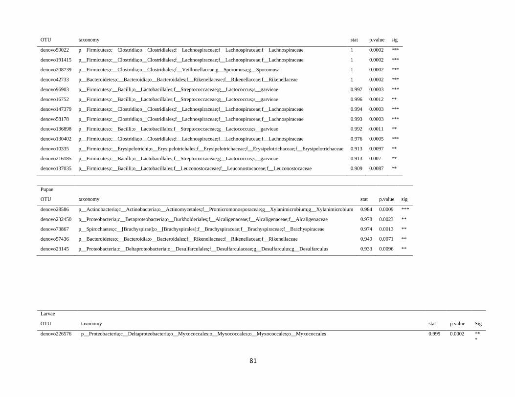
81
OTU taxonomy stat p.value sig
denovo59022 p__Firmicutes;c__Clostridia;o__Clostridiales;f__Lachnospiraceae;f__Lachnospiraceae;f__Lachnospiraceae 1 0.0002 ***
denovo191415 p__Firmicutes;c__Clostridia;o__Clostridiales;f__Lachnospiraceae;f__Lachnospiraceae;f__Lachnospiraceae 1 0.0002 ***
denovo208739 p__Firmicutes;c__Clostridia;o__Clostridiales;f__Veillonellaceae;g__Sporomusa;g__Sporomusa 1 0.0002 ***
denovo42733 p__Bacteroidetes;c__Bacteroidia;o__Bacteroidales;f__Rikenellaceae;f__Rikenellaceae;f__Rikenellaceae 1 0.0002 ***
denovo96903 p__Firmicutes;c__Bacilli;o__Lactobacillales;f__Streptococcaceae;g__Lactococcus;s__garvieae 0.997 0.0003 ***
denovo16752 p__Firmicutes;c__Bacilli;o__Lactobacillales;f__Streptococcaceae;g__Lactococcus;s__garvieae 0.996 0.0012 **
denovo147379 p__Firmicutes;c__Clostridia;o__Clostridiales;f__Lachnospiraceae;f__Lachnospiraceae;f__Lachnospiraceae 0.994 0.0003 ***
denovo58178 p__Firmicutes;c__Clostridia;o__Clostridiales;f__Lachnospiraceae;f__Lachnospiraceae;f__Lachnospiraceae 0.993 0.0003 ***
denovo136898 p__Firmicutes;c__Bacilli;o__Lactobacillales;f__Streptococcaceae;g__Lactococcus;s__garvieae 0.992 0.0011 **
denovo130402 p__Firmicutes;c__Clostridia;o__Clostridiales;f__Lachnospiraceae;f__Lachnospiraceae;f__Lachnospiraceae 0.976 0.0005 ***
denovo10335 p__Firmicutes;c__Erysipelotrichi;o__Erysipelotrichales;f__Erysipelotrichaceae;f__Erysipelotrichaceae;f__Erysipelotrichaceae 0.913 0.0097 **
denovo216185 p__Firmicutes;c__Bacilli;o__Lactobacillales;f__Streptococcaceae;g__Lactococcus;s__garvieae 0.913 0.007 **
denovo137035 p__Firmicutes;c__Bacilli;o__Lactobacillales;f__Leuconostocaceae;f__Leuconostocaceae;f__Leuconostocaceae 0.909 0.0087 **
Pupae
OTU taxonomy stat p.value sig
denovo28586 p__Actinobacteria;c__Actinobacteria;o__Actinomycetales;f__Promicromonosporaceae;g__Xylanimicrobium;g__Xylanimicrobium 0.984 0.0009 ***
denovo232450 p__Proteobacteria;c__Betaproteobacteria;o__Burkholderiales;f__Alcaligenaceae;f__Alcaligenaceae;f__Alcaligenaceae 0.978 0.0023 **
denovo73867 p__Spirochaetes;c__[Brachyspirae];o__[Brachyspirales];f__Brachyspiraceae;f__Brachyspiraceae;f__Brachyspiraceae 0.974 0.0013 **
denovo57436 p__Bacteroidetes;c__Bacteroidia;o__Bacteroidales;f__Rikenellaceae;f__Rikenellaceae;f__Rikenellaceae 0.949 0.0071 **
denovo23145 p__Proteobacteria;c__Deltaproteobacteria;o__Desulfarculales;f__Desulfarculaceae;g__Desulfarculus;g__Desulfarculus 0.933 0.0096 **
Larvae
OTU taxonomy stat p.value Sig
denovo226576 p__Proteobacteria;c__Deltaproteobacteria;o__Myxococcales;o__Myxococcales;o__Myxococcales;o__Myxococcales 0.999 0.0002 **
*
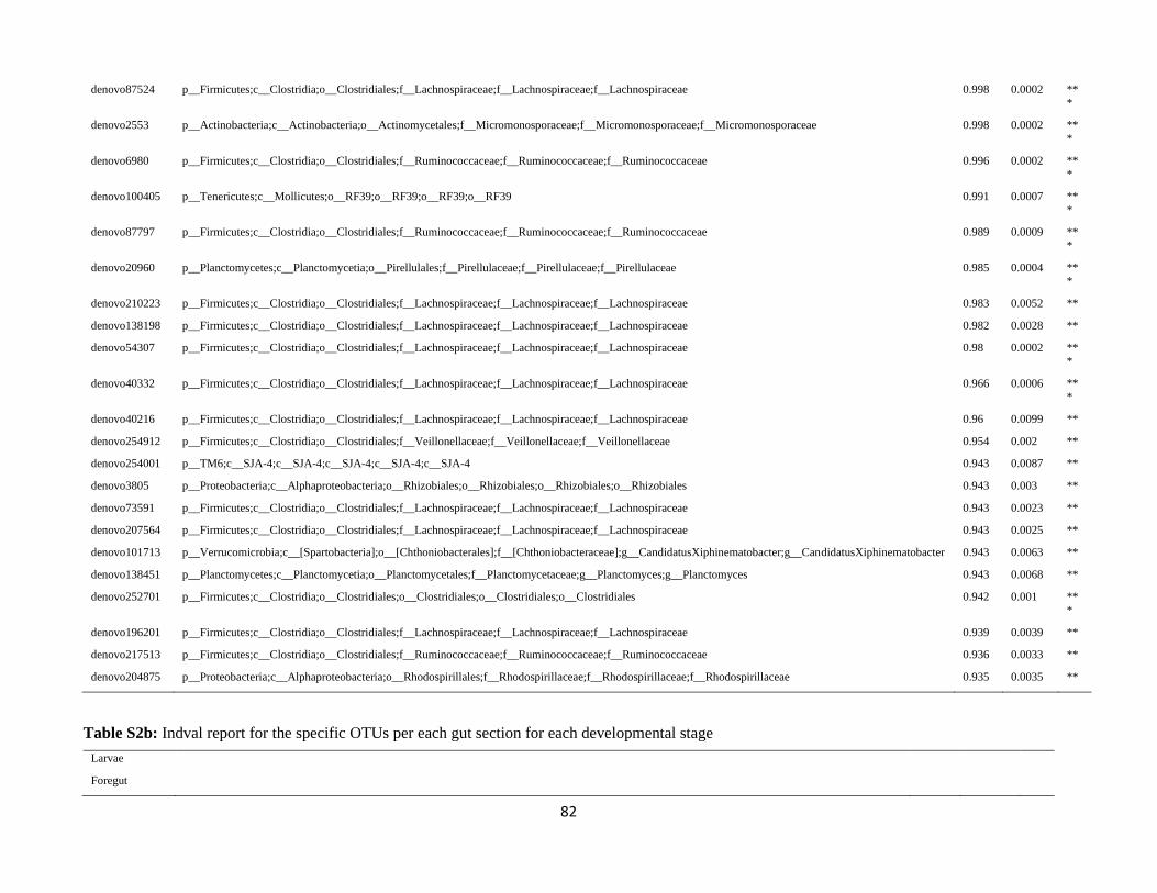
82
denovo87524 p__Firmicutes;c__Clostridia;o__Clostridiales;f__Lachnospiraceae;f__Lachnospiraceae;f__Lachnospiraceae 0.998 0.0002 **
*
denovo2553 p__Actinobacteria;c__Actinobacteria;o__Actinomycetales;f__Micromonosporaceae;f__Micromonosporaceae;f__Micromonosporaceae 0.998 0.0002 **
*
denovo6980 p__Firmicutes;c__Clostridia;o__Clostridiales;f__Ruminococcaceae;f__Ruminococcaceae;f__Ruminococcaceae 0.996 0.0002 **
*
denovo100405 p__Tenericutes;c__Mollicutes;o__RF39;o__RF39;o__RF39;o__RF39 0.991 0.0007 **
*
denovo87797 p__Firmicutes;c__Clostridia;o__Clostridiales;f__Ruminococcaceae;f__Ruminococcaceae;f__Ruminococcaceae 0.989 0.0009 **
*
denovo20960 p__Planctomycetes;c__Planctomycetia;o__Pirellulales;f__Pirellulaceae;f__Pirellulaceae;f__Pirellulaceae 0.985 0.0004 **
*
denovo210223 p__Firmicutes;c__Clostridia;o__Clostridiales;f__Lachnospiraceae;f__Lachnospiraceae;f__Lachnospiraceae 0.983 0.0052 **
denovo138198 p__Firmicutes;c__Clostridia;o__Clostridiales;f__Lachnospiraceae;f__Lachnospiraceae;f__Lachnospiraceae 0.982 0.0028 **
denovo54307 p__Firmicutes;c__Clostridia;o__Clostridiales;f__Lachnospiraceae;f__Lachnospiraceae;f__Lachnospiraceae 0.98 0.0002 **
*
denovo40332 p__Firmicutes;c__Clostridia;o__Clostridiales;f__Lachnospiraceae;f__Lachnospiraceae;f__Lachnospiraceae 0.966 0.0006 **
*
denovo40216 p__Firmicutes;c__Clostridia;o__Clostridiales;f__Lachnospiraceae;f__Lachnospiraceae;f__Lachnospiraceae 0.96 0.0099 **
denovo254912 p__Firmicutes;c__Clostridia;o__Clostridiales;f__Veillonellaceae;f__Veillonellaceae;f__Veillonellaceae 0.954 0.002 **
denovo254001 p__TM6;c__SJA-4;c__SJA-4;c__SJA-4;c__SJA-4;c__SJA-4 0.943 0.0087 **
denovo3805 p__Proteobacteria;c__Alphaproteobacteria;o__Rhizobiales;o__Rhizobiales;o__Rhizobiales;o__Rhizobiales 0.943 0.003 **
denovo73591 p__Firmicutes;c__Clostridia;o__Clostridiales;f__Lachnospiraceae;f__Lachnospiraceae;f__Lachnospiraceae 0.943 0.0023 **
denovo207564 p__Firmicutes;c__Clostridia;o__Clostridiales;f__Lachnospiraceae;f__Lachnospiraceae;f__Lachnospiraceae 0.943 0.0025 **
denovo101713 p__Verrucomicrobia;c__[Spartobacteria];o__[Chthoniobacterales];f__[Chthoniobacteraceae];g__CandidatusXiphinematobacter;g__CandidatusXiphinematobacter 0.943 0.0063 **
denovo138451 p__Planctomycetes;c__Planctomycetia;o__Planctomycetales;f__Planctomycetaceae;g__Planctomyces;g__Planctomyces 0.943 0.0068 **
denovo252701 p__Firmicutes;c__Clostridia;o__Clostridiales;o__Clostridiales;o__Clostridiales;o__Clostridiales 0.942 0.001 **
*
denovo196201 p__Firmicutes;c__Clostridia;o__Clostridiales;f__Lachnospiraceae;f__Lachnospiraceae;f__Lachnospiraceae 0.939 0.0039 **
denovo217513 p__Firmicutes;c__Clostridia;o__Clostridiales;f__Ruminococcaceae;f__Ruminococcaceae;f__Ruminococcaceae 0.936 0.0033 **
denovo204875 p__Proteobacteria;c__Alphaproteobacteria;o__Rhodospirillales;f__Rhodospirillaceae;f__Rhodospirillaceae;f__Rhodospirillaceae 0.935 0.0035 **
Table S2b: Indval report for the specific OTUs per each gut section for each developmental stage
Larvae
Foregut
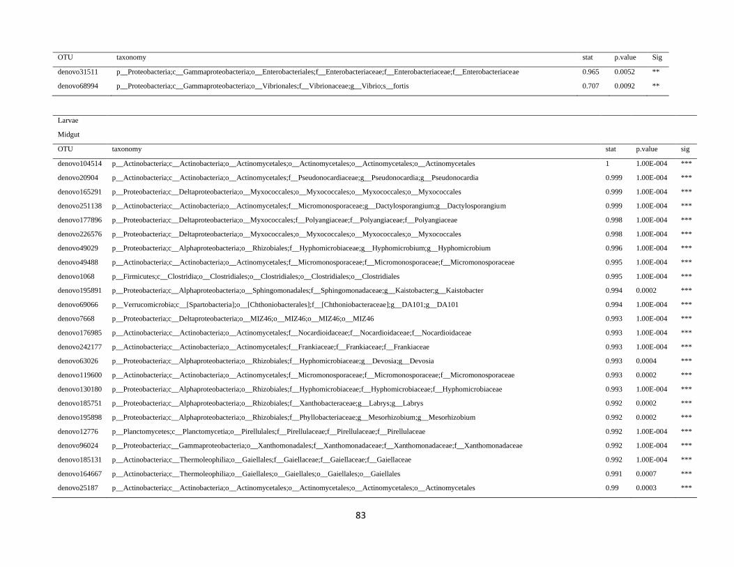
83
OTU taxonomy stat p.value Sig
denovo31511 p__Proteobacteria;c__Gammaproteobacteria;o__Enterobacteriales;f__Enterobacteriaceae;f__Enterobacteriaceae;f__Enterobacteriaceae 0.965 0.0052 **
denovo68994 p__Proteobacteria;c__Gammaproteobacteria;o__Vibrionales;f__Vibrionaceae;g__Vibrio;s__fortis 0.707 0.0092 **
Larvae
Midgut
OTU taxonomy stat p.value sig
denovo104514 p__Actinobacteria;c__Actinobacteria;o__Actinomycetales;o__Actinomycetales;o__Actinomycetales;o__Actinomycetales 1 1.00E-004 ***
denovo20904 p__Actinobacteria;c__Actinobacteria;o__Actinomycetales;f__Pseudonocardiaceae;g__Pseudonocardia;g__Pseudonocardia 0.999 1.00E-004 ***
denovo165291 p__Proteobacteria;c__Deltaproteobacteria;o__Myxococcales;o__Myxococcales;o__Myxococcales;o__Myxococcales 0.999 1.00E-004 ***
denovo251138 p__Actinobacteria;c__Actinobacteria;o__Actinomycetales;f__Micromonosporaceae;g__Dactylosporangium;g__Dactylosporangium 0.999 1.00E-004 ***
denovo177896 p__Proteobacteria;c__Deltaproteobacteria;o__Myxococcales;f__Polyangiaceae;f__Polyangiaceae;f__Polyangiaceae 0.998 1.00E-004 ***
denovo226576 p__Proteobacteria;c__Deltaproteobacteria;o__Myxococcales;o__Myxococcales;o__Myxococcales;o__Myxococcales 0.998 1.00E-004 ***
denovo49029 p__Proteobacteria;c__Alphaproteobacteria;o__Rhizobiales;f__Hyphomicrobiaceae;g__Hyphomicrobium;g__Hyphomicrobium 0.996 1.00E-004 ***
denovo49488 p__Actinobacteria;c__Actinobacteria;o__Actinomycetales;f__Micromonosporaceae;f__Micromonosporaceae;f__Micromonosporaceae 0.995 1.00E-004 ***
denovo1068 p__Firmicutes;c__Clostridia;o__Clostridiales;o__Clostridiales;o__Clostridiales;o__Clostridiales 0.995 1.00E-004 ***
denovo195891 p__Proteobacteria;c__Alphaproteobacteria;o__Sphingomonadales;f__Sphingomonadaceae;g__Kaistobacter;g__Kaistobacter 0.994 0.0002 ***
denovo69066 p__Verrucomicrobia;c__[Spartobacteria];o__[Chthoniobacterales];f__[Chthoniobacteraceae];g__DA101;g__DA101 0.994 1.00E-004 ***
denovo7668 p__Proteobacteria;c__Deltaproteobacteria;o__MIZ46;o__MIZ46;o__MIZ46;o__MIZ46 0.993 1.00E-004 ***
denovo176985 p__Actinobacteria;c__Actinobacteria;o__Actinomycetales;f__Nocardioidaceae;f__Nocardioidaceae;f__Nocardioidaceae 0.993 1.00E-004 ***
denovo242177 p__Actinobacteria;c__Actinobacteria;o__Actinomycetales;f__Frankiaceae;f__Frankiaceae;f__Frankiaceae 0.993 1.00E-004 ***
denovo63026 p__Proteobacteria;c__Alphaproteobacteria;o__Rhizobiales;f__Hyphomicrobiaceae;g__Devosia;g__Devosia 0.993 0.0004 ***
denovo119600 p__Actinobacteria;c__Actinobacteria;o__Actinomycetales;f__Micromonosporaceae;f__Micromonosporaceae;f__Micromonosporaceae 0.993 0.0002 ***
denovo130180 p__Proteobacteria;c__Alphaproteobacteria;o__Rhizobiales;f__Hyphomicrobiaceae;f__Hyphomicrobiaceae;f__Hyphomicrobiaceae 0.993 1.00E-004 ***
denovo185751 p__Proteobacteria;c__Alphaproteobacteria;o__Rhizobiales;f__Xanthobacteraceae;g__Labrys;g__Labrys 0.992 0.0002 ***
denovo195898 p__Proteobacteria;c__Alphaproteobacteria;o__Rhizobiales;f__Phyllobacteriaceae;g__Mesorhizobium;g__Mesorhizobium 0.992 0.0002 ***
denovo12776 p__Planctomycetes;c__Planctomycetia;o__Pirellulales;f__Pirellulaceae;f__Pirellulaceae;f__Pirellulaceae 0.992 1.00E-004 ***
denovo96024 p__Proteobacteria;c__Gammaproteobacteria;o__Xanthomonadales;f__Xanthomonadaceae;f__Xanthomonadaceae;f__Xanthomonadaceae 0.992 1.00E-004 ***
denovo185131 p__Actinobacteria;c__Thermoleophilia;o__Gaiellales;f__Gaiellaceae;f__Gaiellaceae;f__Gaiellaceae 0.992 1.00E-004 ***
denovo164667 p__Actinobacteria;c__Thermoleophilia;o__Gaiellales;o__Gaiellales;o__Gaiellales;o__Gaiellales 0.991 0.0007 ***
denovo25187 p__Actinobacteria;c__Actinobacteria;o__Actinomycetales;o__Actinomycetales;o__Actinomycetales;o__Actinomycetales 0.99 0.0003 ***
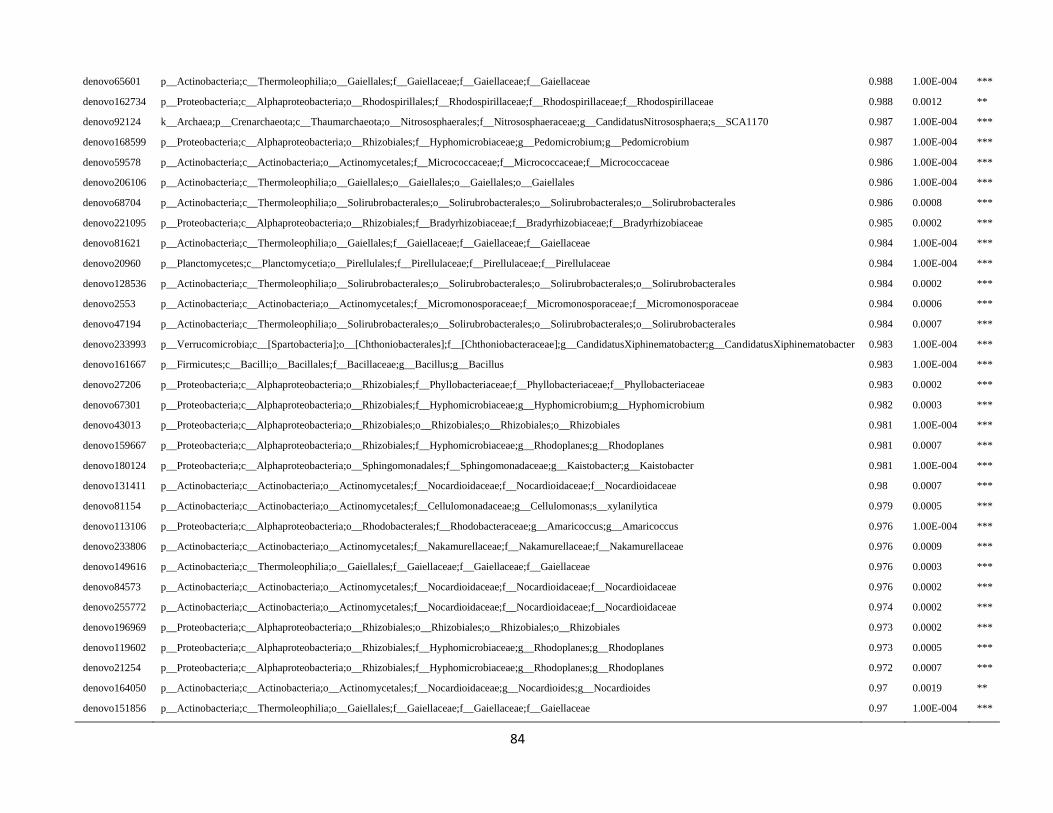
84
denovo65601 p__Actinobacteria;c__Thermoleophilia;o__Gaiellales;f__Gaiellaceae;f__Gaiellaceae;f__Gaiellaceae 0.988 1.00E-004 ***
denovo162734 p__Proteobacteria;c__Alphaproteobacteria;o__Rhodospirillales;f__Rhodospirillaceae;f__Rhodospirillaceae;f__Rhodospirillaceae 0.988 0.0012 **
denovo92124 k__Archaea;p__Crenarchaeota;c__Thaumarchaeota;o__Nitrososphaerales;f__Nitrososphaeraceae;g__CandidatusNitrososphaera;s__SCA1170 0.987 1.00E-004 ***
denovo168599 p__Proteobacteria;c__Alphaproteobacteria;o__Rhizobiales;f__Hyphomicrobiaceae;g__Pedomicrobium;g__Pedomicrobium 0.987 1.00E-004 ***
denovo59578 p__Actinobacteria;c__Actinobacteria;o__Actinomycetales;f__Micrococcaceae;f__Micrococcaceae;f__Micrococcaceae 0.986 1.00E-004 ***
denovo206106 p__Actinobacteria;c__Thermoleophilia;o__Gaiellales;o__Gaiellales;o__Gaiellales;o__Gaiellales 0.986 1.00E-004 ***
denovo68704 p__Actinobacteria;c__Thermoleophilia;o__Solirubrobacterales;o__Solirubrobacterales;o__Solirubrobacterales;o__Solirubrobacterales 0.986 0.0008 ***
denovo221095 p__Proteobacteria;c__Alphaproteobacteria;o__Rhizobiales;f__Bradyrhizobiaceae;f__Bradyrhizobiaceae;f__Bradyrhizobiaceae 0.985 0.0002 ***
denovo81621 p__Actinobacteria;c__Thermoleophilia;o__Gaiellales;f__Gaiellaceae;f__Gaiellaceae;f__Gaiellaceae 0.984 1.00E-004 ***
denovo20960 p__Planctomycetes;c__Planctomycetia;o__Pirellulales;f__Pirellulaceae;f__Pirellulaceae;f__Pirellulaceae 0.984 1.00E-004 ***
denovo128536 p__Actinobacteria;c__Thermoleophilia;o__Solirubrobacterales;o__Solirubrobacterales;o__Solirubrobacterales;o__Solirubrobacterales 0.984 0.0002 ***
denovo2553 p__Actinobacteria;c__Actinobacteria;o__Actinomycetales;f__Micromonosporaceae;f__Micromonosporaceae;f__Micromonosporaceae 0.984 0.0006 ***
denovo47194 p__Actinobacteria;c__Thermoleophilia;o__Solirubrobacterales;o__Solirubrobacterales;o__Solirubrobacterales;o__Solirubrobacterales 0.984 0.0007 ***
denovo233993 p__Verrucomicrobia;c__[Spartobacteria];o__[Chthoniobacterales];f__[Chthoniobacteraceae];g__CandidatusXiphinematobacter;g__CandidatusXiphinematobacter 0.983 1.00E-004 ***
denovo161667 p__Firmicutes;c__Bacilli;o__Bacillales;f__Bacillaceae;g__Bacillus;g__Bacillus 0.983 1.00E-004 ***
denovo27206 p__Proteobacteria;c__Alphaproteobacteria;o__Rhizobiales;f__Phyllobacteriaceae;f__Phyllobacteriaceae;f__Phyllobacteriaceae 0.983 0.0002 ***
denovo67301 p__Proteobacteria;c__Alphaproteobacteria;o__Rhizobiales;f__Hyphomicrobiaceae;g__Hyphomicrobium;g__Hyphomicrobium 0.982 0.0003 ***
denovo43013 p__Proteobacteria;c__Alphaproteobacteria;o__Rhizobiales;o__Rhizobiales;o__Rhizobiales;o__Rhizobiales 0.981 1.00E-004 ***
denovo159667 p__Proteobacteria;c__Alphaproteobacteria;o__Rhizobiales;f__Hyphomicrobiaceae;g__Rhodoplanes;g__Rhodoplanes 0.981 0.0007 ***
denovo180124 p__Proteobacteria;c__Alphaproteobacteria;o__Sphingomonadales;f__Sphingomonadaceae;g__Kaistobacter;g__Kaistobacter 0.981 1.00E-004 ***
denovo131411 p__Actinobacteria;c__Actinobacteria;o__Actinomycetales;f__Nocardioidaceae;f__Nocardioidaceae;f__Nocardioidaceae 0.98 0.0007 ***
denovo81154 p__Actinobacteria;c__Actinobacteria;o__Actinomycetales;f__Cellulomonadaceae;g__Cellulomonas;s__xylanilytica 0.979 0.0005 ***
denovo113106 p__Proteobacteria;c__Alphaproteobacteria;o__Rhodobacterales;f__Rhodobacteraceae;g__Amaricoccus;g__Amaricoccus 0.976 1.00E-004 ***
denovo233806 p__Actinobacteria;c__Actinobacteria;o__Actinomycetales;f__Nakamurellaceae;f__Nakamurellaceae;f__Nakamurellaceae 0.976 0.0009 ***
denovo149616 p__Actinobacteria;c__Thermoleophilia;o__Gaiellales;f__Gaiellaceae;f__Gaiellaceae;f__Gaiellaceae 0.976 0.0003 ***
denovo84573 p__Actinobacteria;c__Actinobacteria;o__Actinomycetales;f__Nocardioidaceae;f__Nocardioidaceae;f__Nocardioidaceae 0.976 0.0002 ***
denovo255772 p__Actinobacteria;c__Actinobacteria;o__Actinomycetales;f__Nocardioidaceae;f__Nocardioidaceae;f__Nocardioidaceae 0.974 0.0002 ***
denovo196969 p__Proteobacteria;c__Alphaproteobacteria;o__Rhizobiales;o__Rhizobiales;o__Rhizobiales;o__Rhizobiales 0.973 0.0002 ***
denovo119602 p__Proteobacteria;c__Alphaproteobacteria;o__Rhizobiales;f__Hyphomicrobiaceae;g__Rhodoplanes;g__Rhodoplanes 0.973 0.0005 ***
denovo21254 p__Proteobacteria;c__Alphaproteobacteria;o__Rhizobiales;f__Hyphomicrobiaceae;g__Rhodoplanes;g__Rhodoplanes 0.972 0.0007 ***
denovo164050 p__Actinobacteria;c__Actinobacteria;o__Actinomycetales;f__Nocardioidaceae;g__Nocardioides;g__Nocardioides 0.97 0.0019 **
denovo151856 p__Actinobacteria;c__Thermoleophilia;o__Gaiellales;f__Gaiellaceae;f__Gaiellaceae;f__Gaiellaceae 0.97 1.00E-004 ***
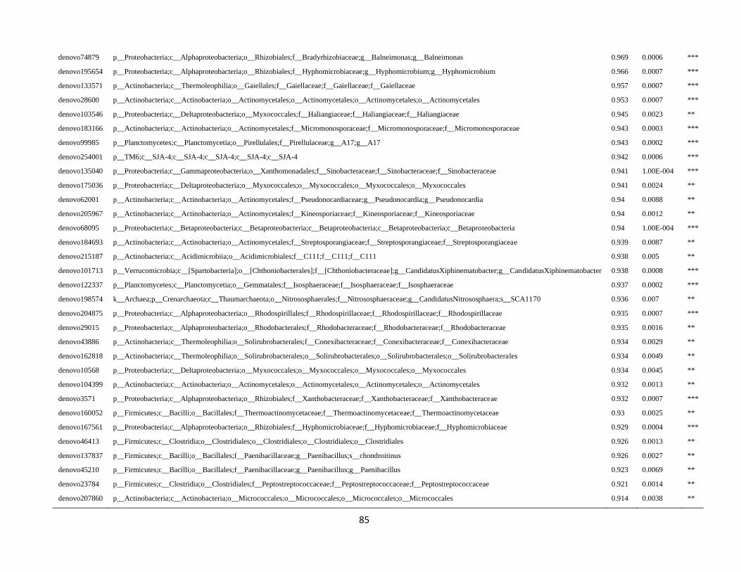
85
denovo74879 p__Proteobacteria;c__Alphaproteobacteria;o__Rhizobiales;f__Bradyrhizobiaceae;g__Balneimonas;g__Balneimonas 0.969 0.0006 ***
denovo195654 p__Proteobacteria;c__Alphaproteobacteria;o__Rhizobiales;f__Hyphomicrobiaceae;g__Hyphomicrobium;g__Hyphomicrobium 0.966 0.0007 ***
denovo133571 p__Actinobacteria;c__Thermoleophilia;o__Gaiellales;f__Gaiellaceae;f__Gaiellaceae;f__Gaiellaceae 0.957 0.0007 ***
denovo28600 p__Actinobacteria;c__Actinobacteria;o__Actinomycetales;o__Actinomycetales;o__Actinomycetales;o__Actinomycetales 0.953 0.0007 ***
denovo103546 p__Proteobacteria;c__Deltaproteobacteria;o__Myxococcales;f__Haliangiaceae;f__Haliangiaceae;f__Haliangiaceae 0.945 0.0023 **
denovo183166 p__Actinobacteria;c__Actinobacteria;o__Actinomycetales;f__Micromonosporaceae;f__Micromonosporaceae;f__Micromonosporaceae 0.943 0.0003 ***
denovo99985 p__Planctomycetes;c__Planctomycetia;o__Pirellulales;f__Pirellulaceae;g__A17;g__A17 0.943 0.0002 ***
denovo254001 p__TM6;c__SJA-4;c__SJA-4;c__SJA-4;c__SJA-4;c__SJA-4 0.942 0.0006 ***
denovo135040 p__Proteobacteria;c__Gammaproteobacteria;o__Xanthomonadales;f__Sinobacteraceae;f__Sinobacteraceae;f__Sinobacteraceae 0.941 1.00E-004 ***
denovo175036 p__Proteobacteria;c__Deltaproteobacteria;o__Myxococcales;o__Myxococcales;o__Myxococcales;o__Myxococcales 0.941 0.0024 **
denovo62001 p__Actinobacteria;c__Actinobacteria;o__Actinomycetales;f__Pseudonocardiaceae;g__Pseudonocardia;g__Pseudonocardia 0.94 0.0088 **
denovo205967 p__Actinobacteria;c__Actinobacteria;o__Actinomycetales;f__Kineosporiaceae;f__Kineosporiaceae;f__Kineosporiaceae 0.94 0.0012 **
denovo68095 p__Proteobacteria;c__Betaproteobacteria;c__Betaproteobacteria;c__Betaproteobacteria;c__Betaproteobacteria;c__Betaproteobacteria 0.94 1.00E-004 ***
denovo184693 p__Actinobacteria;c__Actinobacteria;o__Actinomycetales;f__Streptosporangiaceae;f__Streptosporangiaceae;f__Streptosporangiaceae 0.939 0.0087 **
denovo215187 p__Actinobacteria;c__Acidimicrobiia;o__Acidimicrobiales;f__C111;f__C111;f__C111 0.938 0.005 **
denovo101713 p__Verrucomicrobia;c__[Spartobacteria];o__[Chthoniobacterales];f__[Chthoniobacteraceae];g__CandidatusXiphinematobacter;g__CandidatusXiphinematobacter 0.938 0.0008 ***
denovo122337 p__Planctomycetes;c__Planctomycetia;o__Gemmatales;f__Isosphaeraceae;f__Isosphaeraceae;f__Isosphaeraceae 0.937 0.0002 ***
denovo198574 k__Archaea;p__Crenarchaeota;c__Thaumarchaeota;o__Nitrososphaerales;f__Nitrososphaeraceae;g__CandidatusNitrososphaera;s__SCA1170 0.936 0.007 **
denovo204875 p__Proteobacteria;c__Alphaproteobacteria;o__Rhodospirillales;f__Rhodospirillaceae;f__Rhodospirillaceae;f__Rhodospirillaceae 0.935 0.0007 ***
denovo29015 p__Proteobacteria;c__Alphaproteobacteria;o__Rhodobacterales;f__Rhodobacteraceae;f__Rhodobacteraceae;f__Rhodobacteraceae 0.935 0.0016 **
denovo43886 p__Actinobacteria;c__Thermoleophilia;o__Solirubrobacterales;f__Conexibacteraceae;f__Conexibacteraceae;f__Conexibacteraceae 0.934 0.0029 **
denovo162818 p__Actinobacteria;c__Thermoleophilia;o__Solirubrobacterales;o__Solirubrobacterales;o__Solirubrobacterales;o__Solirubrobacterales 0.934 0.0049 **
denovo10568 p__Proteobacteria;c__Deltaproteobacteria;o__Myxococcales;o__Myxococcales;o__Myxococcales;o__Myxococcales 0.934 0.0045 **
denovo104399 p__Actinobacteria;c__Actinobacteria;o__Actinomycetales;o__Actinomycetales;o__Actinomycetales;o__Actinomycetales 0.932 0.0013 **
denovo3571 p__Proteobacteria;c__Alphaproteobacteria;o__Rhizobiales;f__Xanthobacteraceae;f__Xanthobacteraceae;f__Xanthobacteraceae 0.932 0.0007 ***
denovo160052 p__Firmicutes;c__Bacilli;o__Bacillales;f__Thermoactinomycetaceae;f__Thermoactinomycetaceae;f__Thermoactinomycetaceae 0.93 0.0025 **
denovo167561 p__Proteobacteria;c__Alphaproteobacteria;o__Rhizobiales;f__Hyphomicrobiaceae;f__Hyphomicrobiaceae;f__Hyphomicrobiaceae 0.929 0.0004 ***
denovo46413 p__Firmicutes;c__Clostridia;o__Clostridiales;o__Clostridiales;o__Clostridiales;o__Clostridiales 0.926 0.0013 **
denovo137837 p__Firmicutes;c__Bacilli;o__Bacillales;f__Paenibacillaceae;g__Paenibacillus;s__chondroitinus 0.926 0.0027 **
denovo45210 p__Firmicutes;c__Bacilli;o__Bacillales;f__Paenibacillaceae;g__Paenibacillus;g__Paenibacillus 0.923 0.0069 **
denovo23784 p__Firmicutes;c__Clostridia;o__Clostridiales;f__Peptostreptococcaceae;f__Peptostreptococcaceae;f__Peptostreptococcaceae 0.921 0.0014 **
denovo207860 p__Actinobacteria;c__Actinobacteria;o__Micrococcales;o__Micrococcales;o__Micrococcales;o__Micrococcales 0.914 0.0038 **
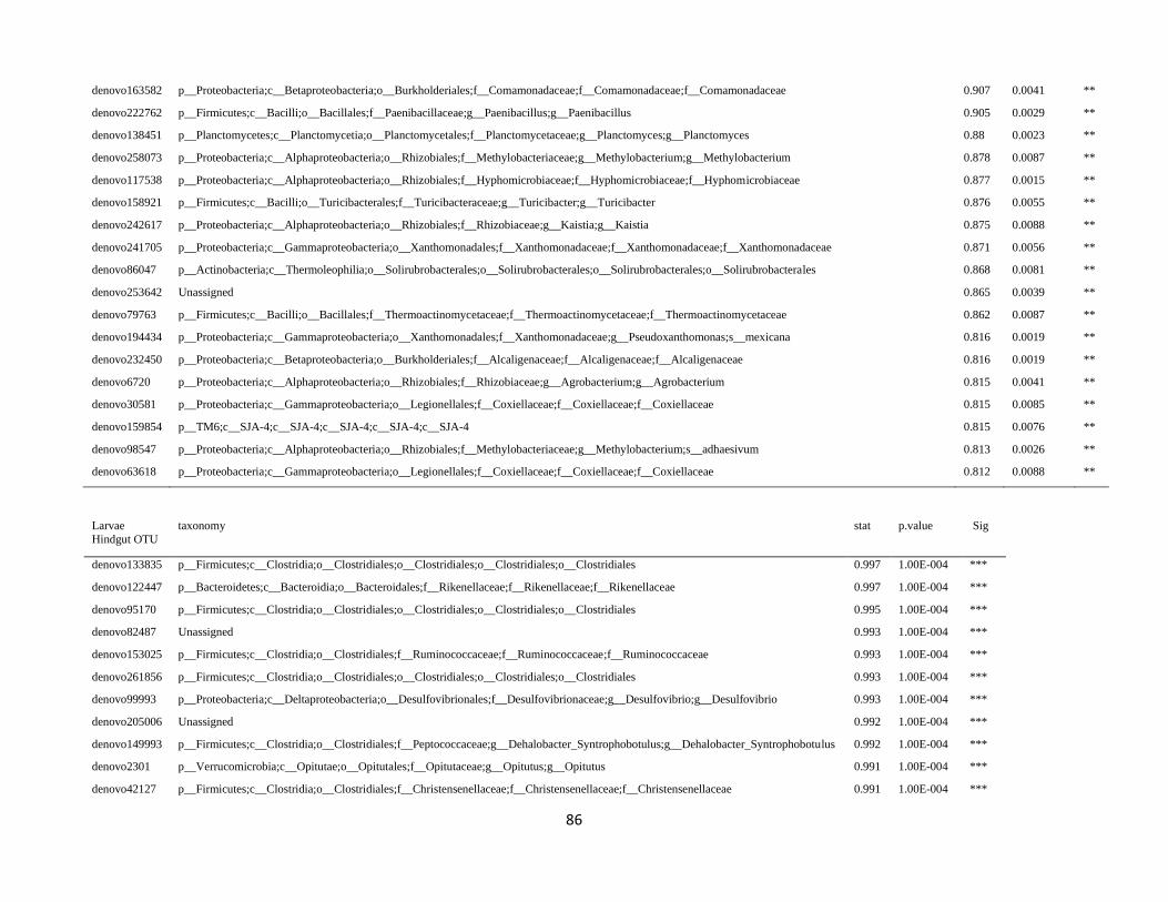
86
denovo163582 p__Proteobacteria;c__Betaproteobacteria;o__Burkholderiales;f__Comamonadaceae;f__Comamonadaceae;f__Comamonadaceae 0.907 0.0041 **
denovo222762 p__Firmicutes;c__Bacilli;o__Bacillales;f__Paenibacillaceae;g__Paenibacillus;g__Paenibacillus 0.905 0.0029 **
denovo138451 p__Planctomycetes;c__Planctomycetia;o__Planctomycetales;f__Planctomycetaceae;g__Planctomyces;g__Planctomyces 0.88 0.0023 **
denovo258073 p__Proteobacteria;c__Alphaproteobacteria;o__Rhizobiales;f__Methylobacteriaceae;g__Methylobacterium;g__Methylobacterium 0.878 0.0087 **
denovo117538 p__Proteobacteria;c__Alphaproteobacteria;o__Rhizobiales;f__Hyphomicrobiaceae;f__Hyphomicrobiaceae;f__Hyphomicrobiaceae 0.877 0.0015 **
denovo158921 p__Firmicutes;c__Bacilli;o__Turicibacterales;f__Turicibacteraceae;g__Turicibacter;g__Turicibacter 0.876 0.0055 **
denovo242617 p__Proteobacteria;c__Alphaproteobacteria;o__Rhizobiales;f__Rhizobiaceae;g__Kaistia;g__Kaistia 0.875 0.0088 **
denovo241705 p__Proteobacteria;c__Gammaproteobacteria;o__Xanthomonadales;f__Xanthomonadaceae;f__Xanthomonadaceae;f__Xanthomonadaceae 0.871 0.0056 **
denovo86047 p__Actinobacteria;c__Thermoleophilia;o__Solirubrobacterales;o__Solirubrobacterales;o__Solirubrobacterales;o__Solirubrobacterales 0.868 0.0081 **
denovo253642 Unassigned 0.865 0.0039 **
denovo79763 p__Firmicutes;c__Bacilli;o__Bacillales;f__Thermoactinomycetaceae;f__Thermoactinomycetaceae;f__Thermoactinomycetaceae 0.862 0.0087 **
denovo194434 p__Proteobacteria;c__Gammaproteobacteria;o__Xanthomonadales;f__Xanthomonadaceae;g__Pseudoxanthomonas;s__mexicana 0.816 0.0019 **
denovo232450 p__Proteobacteria;c__Betaproteobacteria;o__Burkholderiales;f__Alcaligenaceae;f__Alcaligenaceae;f__Alcaligenaceae 0.816 0.0019 **
denovo6720 p__Proteobacteria;c__Alphaproteobacteria;o__Rhizobiales;f__Rhizobiaceae;g__Agrobacterium;g__Agrobacterium 0.815 0.0041 **
denovo30581 p__Proteobacteria;c__Gammaproteobacteria;o__Legionellales;f__Coxiellaceae;f__Coxiellaceae;f__Coxiellaceae 0.815 0.0085 **
denovo159854 p__TM6;c__SJA-4;c__SJA-4;c__SJA-4;c__SJA-4;c__SJA-4 0.815 0.0076 **
denovo98547 p__Proteobacteria;c__Alphaproteobacteria;o__Rhizobiales;f__Methylobacteriaceae;g__Methylobacterium;s__adhaesivum 0.813 0.0026 **
denovo63618 p__Proteobacteria;c__Gammaproteobacteria;o__Legionellales;f__Coxiellaceae;f__Coxiellaceae;f__Coxiellaceae 0.812 0.0088 **
Larvae
Hindgut OTU
taxonomy stat p.value Sig
denovo133835 p__Firmicutes;c__Clostridia;o__Clostridiales;o__Clostridiales;o__Clostridiales;o__Clostridiales 0.997 1.00E-004 ***
denovo122447 p__Bacteroidetes;c__Bacteroidia;o__Bacteroidales;f__Rikenellaceae;f__Rikenellaceae;f__Rikenellaceae 0.997 1.00E-004 ***
denovo95170 p__Firmicutes;c__Clostridia;o__Clostridiales;o__Clostridiales;o__Clostridiales;o__Clostridiales 0.995 1.00E-004 ***
denovo82487 Unassigned 0.993 1.00E-004 ***
denovo153025 p__Firmicutes;c__Clostridia;o__Clostridiales;f__Ruminococcaceae;f__Ruminococcaceae;f__Ruminococcaceae 0.993 1.00E-004 ***
denovo261856 p__Firmicutes;c__Clostridia;o__Clostridiales;o__Clostridiales;o__Clostridiales;o__Clostridiales 0.993 1.00E-004 ***
denovo99993 p__Proteobacteria;c__Deltaproteobacteria;o__Desulfovibrionales;f__Desulfovibrionaceae;g__Desulfovibrio;g__Desulfovibrio 0.993 1.00E-004 ***
denovo205006 Unassigned 0.992 1.00E-004 ***
denovo149993 p__Firmicutes;c__Clostridia;o__Clostridiales;f__Peptococcaceae;g__Dehalobacter_Syntrophobotulus;g__Dehalobacter_Syntrophobotulus 0.992 1.00E-004 ***
denovo2301 p__Verrucomicrobia;c__Opitutae;o__Opitutales;f__Opitutaceae;g__Opitutus;g__Opitutus 0.991 1.00E-004 ***
denovo42127 p__Firmicutes;c__Clostridia;o__Clostridiales;f__Christensenellaceae;f__Christensenellaceae;f__Christensenellaceae 0.991 1.00E-004 ***
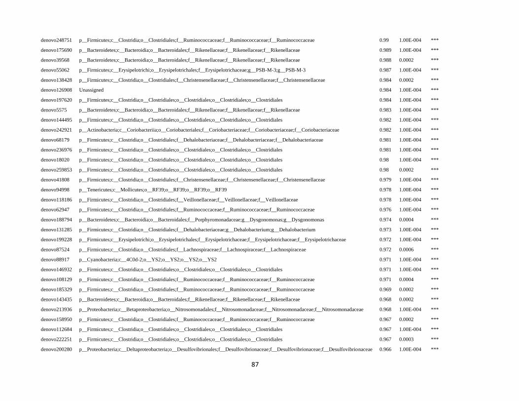
87
denovo248751 p__Firmicutes;c__Clostridia;o__Clostridiales;f__Ruminococcaceae;f__Ruminococcaceae;f__Ruminococcaceae 0.99 1.00E-004 ***
denovo175690 p__Bacteroidetes;c__Bacteroidia;o__Bacteroidales;f__Rikenellaceae;f__Rikenellaceae;f__Rikenellaceae 0.989 1.00E-004 ***
denovo39568 p__Bacteroidetes;c__Bacteroidia;o__Bacteroidales;f__Rikenellaceae;f__Rikenellaceae;f__Rikenellaceae 0.988 0.0002 ***
denovo55062 p__Firmicutes;c__Erysipelotrichi;o__Erysipelotrichales;f__Erysipelotrichaceae;g__PSB-M-3;g__PSB-M-3 0.987 1.00E-004 ***
denovo138428 p__Firmicutes;c__Clostridia;o__Clostridiales;f__Christensenellaceae;f__Christensenellaceae;f__Christensenellaceae 0.984 0.0002 ***
denovo126908 Unassigned 0.984 1.00E-004 ***
denovo197620 p__Firmicutes;c__Clostridia;o__Clostridiales;o__Clostridiales;o__Clostridiales;o__Clostridiales 0.984 1.00E-004 ***
denovo5575 p__Bacteroidetes;c__Bacteroidia;o__Bacteroidales;f__Rikenellaceae;f__Rikenellaceae;f__Rikenellaceae 0.983 1.00E-004 ***
denovo144495 p__Firmicutes;c__Clostridia;o__Clostridiales;o__Clostridiales;o__Clostridiales;o__Clostridiales 0.982 1.00E-004 ***
denovo242921 p__Actinobacteria;c__Coriobacteriia;o__Coriobacteriales;f__Coriobacteriaceae;f__Coriobacteriaceae;f__Coriobacteriaceae 0.982 1.00E-004 ***
denovo68179 p__Firmicutes;c__Clostridia;o__Clostridiales;f__Dehalobacteriaceae;f__Dehalobacteriaceae;f__Dehalobacteriaceae 0.981 1.00E-004 ***
denovo236976 p__Firmicutes;c__Clostridia;o__Clostridiales;o__Clostridiales;o__Clostridiales;o__Clostridiales 0.981 1.00E-004 ***
denovo18020 p__Firmicutes;c__Clostridia;o__Clostridiales;o__Clostridiales;o__Clostridiales;o__Clostridiales 0.98 1.00E-004 ***
denovo259853 p__Firmicutes;c__Clostridia;o__Clostridiales;o__Clostridiales;o__Clostridiales;o__Clostridiales 0.98 0.0002 ***
denovo41808 p__Firmicutes;c__Clostridia;o__Clostridiales;f__Christensenellaceae;f__Christensenellaceae;f__Christensenellaceae 0.979 1.00E-004 ***
denovo94998 p__Tenericutes;c__Mollicutes;o__RF39;o__RF39;o__RF39;o__RF39 0.978 1.00E-004 ***
denovo118186 p__Firmicutes;c__Clostridia;o__Clostridiales;f__Veillonellaceae;f__Veillonellaceae;f__Veillonellaceae 0.978 1.00E-004 ***
denovo62947 p__Firmicutes;c__Clostridia;o__Clostridiales;f__Ruminococcaceae;f__Ruminococcaceae;f__Ruminococcaceae 0.976 1.00E-004 ***
denovo188794 p__Bacteroidetes;c__Bacteroidia;o__Bacteroidales;f__Porphyromonadaceae;g__Dysgonomonas;g__Dysgonomonas 0.974 0.0004 ***
denovo131285 p__Firmicutes;c__Clostridia;o__Clostridiales;f__Dehalobacteriaceae;g__Dehalobacterium;g__Dehalobacterium 0.973 1.00E-004 ***
denovo199228 p__Firmicutes;c__Erysipelotrichi;o__Erysipelotrichales;f__Erysipelotrichaceae;f__Erysipelotrichaceae;f__Erysipelotrichaceae 0.972 1.00E-004 ***
denovo87524 p__Firmicutes;c__Clostridia;o__Clostridiales;f__Lachnospiraceae;f__Lachnospiraceae;f__Lachnospiraceae 0.972 0.0006 ***
denovo88917 p__Cyanobacteria;c__4C0d-2;o__YS2;o__YS2;o__YS2;o__YS2 0.971 1.00E-004 ***
denovo146932 p__Firmicutes;c__Clostridia;o__Clostridiales;o__Clostridiales;o__Clostridiales;o__Clostridiales 0.971 1.00E-004 ***
denovo108129 p__Firmicutes;c__Clostridia;o__Clostridiales;f__Ruminococcaceae;f__Ruminococcaceae;f__Ruminococcaceae 0.971 0.0004 ***
denovo185329 p__Firmicutes;c__Clostridia;o__Clostridiales;f__Ruminococcaceae;f__Ruminococcaceae;f__Ruminococcaceae 0.969 0.0002 ***
denovo143435 p__Bacteroidetes;c__Bacteroidia;o__Bacteroidales;f__Rikenellaceae;f__Rikenellaceae;f__Rikenellaceae 0.968 0.0002 ***
denovo213936 p__Proteobacteria;c__Betaproteobacteria;o__Nitrosomonadales;f__Nitrosomonadaceae;f__Nitrosomonadaceae;f__Nitrosomonadaceae 0.968 1.00E-004 ***
denovo158950 p__Firmicutes;c__Clostridia;o__Clostridiales;f__Ruminococcaceae;f__Ruminococcaceae;f__Ruminococcaceae 0.967 0.0002 ***
denovo112684 p__Firmicutes;c__Clostridia;o__Clostridiales;o__Clostridiales;o__Clostridiales;o__Clostridiales 0.967 1.00E-004 ***
denovo222251 p__Firmicutes;c__Clostridia;o__Clostridiales;o__Clostridiales;o__Clostridiales;o__Clostridiales 0.967 0.0003 ***
denovo200280 p__Proteobacteria;c__Deltaproteobacteria;o__Desulfovibrionales;f__Desulfovibrionaceae;f__Desulfovibrionaceae;f__Desulfovibrionaceae 0.966 1.00E-004 ***
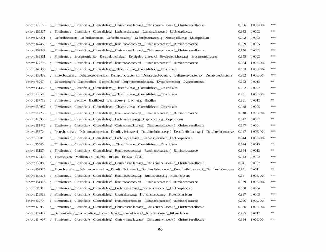
88
denovo229153 p__Firmicutes;c__Clostridia;o__Clostridiales;f__Christensenellaceae;f__Christensenellaceae;f__Christensenellaceae 0.966 1.00E-004 ***
denovo160517 p__Firmicutes;c__Clostridia;o__Clostridiales;f__Lachnospiraceae;f__Lachnospiraceae;f__Lachnospiraceae 0.963 0.0002 ***
denovo124201 p__Deferribacteres;c__Deferribacteres;o__Deferribacterales;f__Deferribacteraceae;g__Mucispirillum;g__Mucispirillum 0.962 0.0002 ***
denovo147469 p__Firmicutes;c__Clostridia;o__Clostridiales;f__Ruminococcaceae;f__Ruminococcaceae;f__Ruminococcaceae 0.959 0.0005 ***
denovo169849 p__Firmicutes;c__Clostridia;o__Clostridiales;f__Christensenellaceae;f__Christensenellaceae;f__Christensenellaceae 0.956 0.0002 ***
denovo136551 p__Firmicutes;c__Erysipelotrichi;o__Erysipelotrichales;f__Erysipelotrichaceae;f__Erysipelotrichaceae;f__Erysipelotrichaceae 0.955 0.0002 ***
denovo127793 p__Firmicutes;c__Clostridia;o__Clostridiales;f__Ruminococcaceae;f__Ruminococcaceae;f__Ruminococcaceae 0.954 1.00E-004 ***
denovo148356 p__Firmicutes;c__Clostridia;o__Clostridiales;o__Clostridiales;o__Clostridiales;o__Clostridiales 0.953 1.00E-004 ***
denovo153882 p__Proteobacteria;c__Deltaproteobacteria;c__Deltaproteobacteria;c__Deltaproteobacteria;c__Deltaproteobacteria;c__Deltaproteobacteria 0.952 1.00E-004 ***
denovo78067 p__Bacteroidetes;c__Bacteroidia;o__Bacteroidales;f__Porphyromonadaceae;g__Dysgonomonas;g__Dysgonomonas 0.952 0.0013 **
denovo151480 p__Firmicutes;c__Clostridia;o__Clostridiales;o__Clostridiales;o__Clostridiales;o__Clostridiales 0.952 0.0002 ***
denovo73359 p__Firmicutes;c__Clostridia;o__Clostridiales;o__Clostridiales;o__Clostridiales;o__Clostridiales 0.951 1.00E-004 ***
denovo117712 p__Firmicutes;c__Bacilli;o__Bacillales;f__Bacillaceae;g__Bacillus;g__Bacillus 0.951 0.0012 **
denovo259057 p__Firmicutes;c__Clostridia;o__Clostridiales;o__Clostridiales;o__Clostridiales;o__Clostridiales 0.948 0.0005 ***
denovo217233 p__Firmicutes;c__Clostridia;o__Clostridiales;f__Ruminococcaceae;f__Ruminococcaceae;f__Ruminococcaceae 0.948 1.00E-004 ***
denovo126955 p__Firmicutes;c__Clostridia;o__Clostridiales;f__Lachnospiraceae;g__Coprococcus;g__Coprococcus 0.947 0.0037 **
denovo153442 p__Firmicutes;c__Clostridia;o__Clostridiales;f__Christensenellaceae;f__Christensenellaceae;f__Christensenellaceae 0.947 0.0004 ***
denovo25672 p__Proteobacteria;c__Deltaproteobacteria;o__Desulfovibrionales;f__Desulfovibrionaceae;f__Desulfovibrionaceae;f__Desulfovibrionaceae 0.947 1.00E-004 ***
denovo59301 p__Firmicutes;c__Clostridia;o__Clostridiales;f__Lachnospiraceae;f__Lachnospiraceae;f__Lachnospiraceae 0.944 1.00E-004 ***
denovo25640 p__Firmicutes;c__Clostridia;o__Clostridiales;o__Clostridiales;o__Clostridiales;o__Clostridiales 0.944 0.0013 **
denovo13127 p__Firmicutes;c__Clostridia;o__Clostridiales;f__Ruminococcaceae;f__Ruminococcaceae;f__Ruminococcaceae 0.944 0.0012 **
denovo173388 p__Tenericutes;c__Mollicutes;o__RF39;o__RF39;o__RF39;o__RF39 0.943 0.0002 ***
denovo230089 p__Firmicutes;c__Clostridia;o__Clostridiales;f__Christensenellaceae;f__Christensenellaceae;f__Christensenellaceae 0.941 0.0002 ***
denovo163925 p__Proteobacteria;c__Deltaproteobacteria;o__Desulfovibrionales;f__Desulfovibrionaceae;f__Desulfovibrionaceae;f__Desulfovibrionaceae 0.941 0.0011 **
denovo137378 p__Firmicutes;c__Clostridia;o__Clostridiales;f__Ruminococcaceae;g__Ruminococcus;g__Ruminococcus 0.94 1.00E-004 ***
denovo184318 p__Firmicutes;c__Clostridia;o__Clostridiales;f__Ruminococcaceae;f__Ruminococcaceae;f__Ruminococcaceae 0.939 1.00E-004 ***
denovo67331 p__Firmicutes;c__Clostridia;o__Clostridiales;f__Lachnospiraceae;f__Lachnospiraceae;f__Lachnospiraceae 0.938 0.0004 ***
denovo216333 p__Firmicutes;c__Clostridia;o__Clostridiales;f__Clostridiaceae;g__Proteiniclasticum;g__Proteiniclasticum 0.937 0.0003 ***
denovo46870 p__Firmicutes;c__Clostridia;o__Clostridiales;f__Ruminococcaceae;f__Ruminococcaceae;f__Ruminococcaceae 0.936 1.00E-004 ***
denovo17998 p__Firmicutes;c__Clostridia;o__Clostridiales;f__Christensenellaceae;f__Christensenellaceae;f__Christensenellaceae 0.936 1.00E-004 ***
denovo142822 p__Bacteroidetes;c__Bacteroidia;o__Bacteroidales;f__Rikenellaceae;f__Rikenellaceae;f__Rikenellaceae 0.935 0.0012 **
denovo184067 p__Firmicutes;c__Clostridia;o__Clostridiales;f__Christensenellaceae;f__Christensenellaceae;f__Christensenellaceae 0.934 1.00E-004 ***
89
denovo123216 p__Firmicutes;c__Clostridia;o__Clostridiales;o__Clostridiales;o__Clostridiales;o__Clostridiales 0.934 0.0002 ***
denovo187502 p__Firmicutes;c__Clostridia;o__Clostridiales;f__Christensenellaceae;f__Christensenellaceae;f__Christensenellaceae 0.932 0.0019 **
denovo120961 p__Firmicutes;c__Clostridia;o__Clostridiales;f__Christensenellaceae;f__Christensenellaceae;f__Christensenellaceae 0.932 0.0004 ***
denovo135849 p__Tenericutes;c__Mollicutes;o__RF39;o__RF39;o__RF39;o__RF39 0.929 0.0003 ***
denovo24626 p__Firmicutes;c__Clostridia;o__Clostridiales;f__Christensenellaceae;f__Christensenellaceae;f__Christensenellaceae 0.928 0.0003 ***
denovo129712 p__Firmicutes;c__Clostridia;o__Clostridiales;f__Ruminococcaceae;f__Ruminococcaceae;f__Ruminococcaceae 0.928 0.0007 ***
denovo130706 p__Firmicutes;c__Clostridia;o__Clostridiales;o__Clostridiales;o__Clostridiales;o__Clostridiales 0.927 0.0035 **
denovo127239 p__Firmicutes;c__Clostridia;o__Clostridiales;f__Christensenellaceae;f__Christensenellaceae;f__Christensenellaceae 0.925 1.00E-004 ***
denovo55168 p__Firmicutes;c__Clostridia;o__Clostridiales;f__Ruminococcaceae;f__Ruminococcaceae;f__Ruminococcaceae 0.924 0.0008 ***
denovo9734 p__Proteobacteria;c__Gammaproteobacteria;o__Xanthomonadales;f__Xanthomonadaceae;f__Xanthomonadaceae;f__Xanthomonadaceae 0.921 1.00E-004 ***
denovo88585 p__Firmicutes;c__Clostridia;o__Clostridiales;f__Ruminococcaceae;f__Ruminococcaceae;f__Ruminococcaceae 0.92 0.0003 ***
denovo173993 p__Proteobacteria;c__Deltaproteobacteria;o__Desulfovibrionales;f__Desulfovibrionaceae;f__Desulfovibrionaceae;f__Desulfovibrionaceae 0.92 0.0026 **
denovo145783 p__Firmicutes;c__Clostridia;o__Clostridiales;o__Clostridiales;o__Clostridiales;o__Clostridiales 0.92 0.0005 ***
denovo198782 p__Firmicutes;c__Clostridia;o__Clostridiales;f__Lachnospiraceae;f__Lachnospiraceae;f__Lachnospiraceae 0.92 0.0036 **
denovo109754 p__Firmicutes;c__Clostridia;o__Clostridiales;f__[Mogibacteriaceae];f__[Mogibacteriaceae];f__[Mogibacteriaceae] 0.919 0.0002 ***
denovo247950 p__Firmicutes;c__Clostridia;o__Clostridiales;f__Peptococcaceae;g__Pelotomaculum;g__Pelotomaculum 0.918 0.0014 **
denovo165044 p__Firmicutes;c__Clostridia;o__Clostridiales;o__Clostridiales;o__Clostridiales;o__Clostridiales 0.918 0.0005 ***
denovo200812 p__Bacteroidetes;c__Bacteroidia;o__Bacteroidales;f__Rikenellaceae;f__Rikenellaceae;f__Rikenellaceae 0.918 0.0007 ***
denovo177004 p__Firmicutes;c__Clostridia;o__Clostridiales;f__Lachnospiraceae;f__Lachnospiraceae;f__Lachnospiraceae 0.917 0.0008 ***
denovo96838 p__Bacteroidetes;c__Bacteroidia;o__Bacteroidales;f__Rikenellaceae;g__PW3;g__PW3 0.916 0.0008 ***
denovo207564 p__Firmicutes;c__Clostridia;o__Clostridiales;f__Lachnospiraceae;f__Lachnospiraceae;f__Lachnospiraceae 0.912 0.0044 **
denovo107253 p__Firmicutes;c__Clostridia;o__Clostridiales;o__Clostridiales;o__Clostridiales;o__Clostridiales 0.912 0.0046 **
denovo258817 p__Firmicutes;c__Clostridia;o__Clostridiales;f__Christensenellaceae;f__Christensenellaceae;f__Christensenellaceae 0.911 0.0021 **
denovo196201 p__Firmicutes;c__Clostridia;o__Clostridiales;f__Lachnospiraceae;f__Lachnospiraceae;f__Lachnospiraceae 0.91 0.0051 **
denovo244579 p__Firmicutes;c__Clostridia;o__Clostridiales;o__Clostridiales;o__Clostridiales;o__Clostridiales 0.91 0.0005 ***
denovo3347 p__Firmicutes;c__Clostridia;o__Clostridiales;f__Christensenellaceae;f__Christensenellaceae;f__Christensenellaceae 0.91 0.0014 **
denovo234535 p__Firmicutes;c__Clostridia;o__Clostridiales;f__Lachnospiraceae;g__Anaerostipes;g__Anaerostipes 0.91 0.0007 ***
denovo149112 p__Firmicutes;c__Clostridia;o__Clostridiales;o__Clostridiales;o__Clostridiales;o__Clostridiales 0.909 0.0035 **
denovo43084 p__Firmicutes;c__Clostridia;o__Clostridiales;f__Ruminococcaceae;f__Ruminococcaceae;f__Ruminococcaceae 0.909 0.0009 ***
denovo181638 p__Firmicutes;c__Clostridia;o__Clostridiales;f__Christensenellaceae;f__Christensenellaceae;f__Christensenellaceae 0.908 0.0035 **
denovo6850 p__Firmicutes;c__Clostridia;o__Clostridiales;o__Clostridiales;o__Clostridiales;o__Clostridiales 0.908 0.0007 ***
denovo225907 p__Firmicutes;c__Clostridia;o__Clostridiales;f__Ruminococcaceae;f__Ruminococcaceae;f__Ruminococcaceae 0.907 0.0021 **
90
denovo128743 p__Firmicutes;c__Clostridia;o__Clostridiales;f__Ruminococcaceae;f__Ruminococcaceae;f__Ruminococcaceae 0.906 0.0026 **
denovo150305 p__Firmicutes;c__Clostridia;o__Clostridiales;o__Clostridiales;o__Clostridiales;o__Clostridiales 0.905 0.0004 ***
denovo113691 p__Bacteroidetes;c__Bacteroidia;o__Bacteroidales;f__Porphyromonadaceae;g__Dysgonomonas;g__Dysgonomonas 0.904 0.0023 **
denovo220410 p__Tenericutes;c__Mollicutes;o__RF39;o__RF39;o__RF39;o__RF39 0.902 1.00E-004 ***
denovo261215 p__Firmicutes;c__Clostridia;o__Clostridiales;f__Christensenellaceae;f__Christensenellaceae;f__Christensenellaceae 0.899 0.0029 **
denovo257270 p__Tenericutes;c__Mollicutes;o__RF39;o__RF39;o__RF39;o__RF39 0.897 0.0039 **
denovo40332 p__Firmicutes;c__Clostridia;o__Clostridiales;f__Lachnospiraceae;f__Lachnospiraceae;f__Lachnospiraceae 0.896 0.0008 ***
denovo87563 p__Firmicutes;c__Clostridia;o__Clostridiales;f__Lachnospiraceae;f__Lachnospiraceae;f__Lachnospiraceae 0.896 0.0045 **
denovo182100 p__Firmicutes;c__Clostridia;o__Clostridiales;f__Ruminococcaceae;f__Ruminococcaceae;f__Ruminococcaceae 0.895 0.0056 **
denovo54307 p__Firmicutes;c__Clostridia;o__Clostridiales;f__Lachnospiraceae;f__Lachnospiraceae;f__Lachnospiraceae 0.892 0.0005 ***
denovo254912 p__Firmicutes;c__Clostridia;o__Clostridiales;f__Veillonellaceae;f__Veillonellaceae;f__Veillonellaceae 0.889 0.0034 **
denovo244668 p__Proteobacteria;c__Betaproteobacteria;o__Burkholderiales;f__Oxalobacteraceae;f__Oxalobacteraceae;f__Oxalobacteraceae 0.887 0.0021 **
denovo93372 p__Firmicutes;c__Clostridia;o__Clostridiales;f__Lachnospiraceae;f__Lachnospiraceae;f__Lachnospiraceae 0.886 0.0044 **
denovo197872 Unassigned 0.886 0.0019 **
denovo231777 p__Firmicutes;c__Clostridia;o__Clostridiales;f__Ruminococcaceae;g__Oscillospira;g__Oscillospira 0.885 0.0038 **
denovo40216 p__Firmicutes;c__Clostridia;o__Clostridiales;f__Lachnospiraceae;f__Lachnospiraceae;f__Lachnospiraceae 0.882 0.0028 **
denovo114424 p__Tenericutes;c__Mollicutes;o__RF39;o__RF39;o__RF39;o__RF39 0.881 0.0036 **
denovo56209 p__Firmicutes;c__Clostridia;o__Clostridiales;f__Lachnospiraceae;f__Lachnospiraceae;f__Lachnospiraceae 0.881 0.003 **
denovo231269 p__Firmicutes;c__Clostridia;o__Clostridiales;f__Christensenellaceae;f__Christensenellaceae;f__Christensenellaceae 0.881 0.0091 **
denovo240210 p__Firmicutes;c__Clostridia;o__Clostridiales;o__Clostridiales;o__Clostridiales;o__Clostridiales 0.881 0.0013 **
denovo140365 p__Firmicutes;c__Clostridia;o__Clostridiales;o__Clostridiales;o__Clostridiales;o__Clostridiales 0.88 0.0015 **
denovo236364 p__Firmicutes;c__Clostridia;o__Clostridiales;f__Peptococcaceae;g__Desulfosporosinus;s__meridiei 0.879 0.0023 **
denovo212762 p__Cyanobacteria;c__4C0d-2;o__YS2;o__YS2;o__YS2;o__YS2 0.877 0.001 ***
denovo4543 p__Firmicutes;c__Clostridia;o__Clostridiales;f__Christensenellaceae;f__Christensenellaceae;f__Christensenellaceae 0.875 0.0017 **
denovo108343 p__Firmicutes;c__Clostridia;o__Clostridiales;f__Ruminococcaceae;f__Ruminococcaceae;f__Ruminococcaceae 0.874 0.0012 **
denovo57436 p__Bacteroidetes;c__Bacteroidia;o__Bacteroidales;f__Rikenellaceae;f__Rikenellaceae;f__Rikenellaceae 0.873 0.0017 **
denovo168776 p__Firmicutes;c__Clostridia;o__Clostridiales;o__Clostridiales;o__Clostridiales;o__Clostridiales 0.872 0.0008 ***
denovo69720 p__Cyanobacteria;c__4C0d-2;o__YS2;o__YS2;o__YS2;o__YS2 0.868 0.0026 **
denovo192196 p__Firmicutes;c__Clostridia;o__Clostridiales;o__Clostridiales;o__Clostridiales;o__Clostridiales 0.867 0.0031 **
denovo156767 p__Firmicutes;c__Erysipelotrichi;o__Erysipelotrichales;f__Erysipelotrichaceae;f__Erysipelotrichaceae;f__Erysipelotrichaceae 0.867 0.0038 **
denovo175497 p__Firmicutes;c__Clostridia;o__Clostridiales;f__Christensenellaceae;f__Christensenellaceae;f__Christensenellaceae 0.864 0.0065 **
denovo253981 p__Firmicutes;c__Clostridia;o__Clostridiales;f__Lachnospiraceae;f__Lachnospiraceae;f__Lachnospiraceae 0.859 0.0093 **
91
denovo18391 Unassigned 0.858 0.0029 **
denovo115642 p__Firmicutes;c__Clostridia;o__Clostridiales;f__Lachnospiraceae;f__Lachnospiraceae;f__Lachnospiraceae 0.855 0.0053 **
denovo252701 p__Firmicutes;c__Clostridia;o__Clostridiales;o__Clostridiales;o__Clostridiales;o__Clostridiales 0.85 0.005 **
denovo229168 p__Firmicutes;c__Clostridia;o__Clostridiales;f__Christensenellaceae;f__Christensenellaceae;f__Christensenellaceae 0.837 0.0064 **
denovo164524 p__Firmicutes;c__Clostridia;o__Clostridiales;o__Clostridiales;o__Clostridiales;o__Clostridiales 0.832 0.0053 **
denovo159133 p__Firmicutes;c__Clostridia;o__Clostridiales;f__Lachnospiraceae;f__Lachnospiraceae;f__Lachnospiraceae 0.821 0.0052 **
Adult
Foregut
OTU taxonomy stat p.value Sig
denovo27158 p__Firmicutes;c__Bacilli;o__Bacillales;f__Paenibacillaceae;g__Brevibacillus;g__Brevibacillus 0.998 0.0009 ***
denovo52639 p__Proteobacteria;c__Alphaproteobacteria;o__Caulobacterales;f__Caulobacteraceae;f__Caulobacteraceae;f__Caulobacteraceae 0.997 0.0008 ***
denovo72506 p__Proteobacteria;c__Alphaproteobacteria;o__Sphingomonadales;f__Sphingomonadaceae;g__Sphingomonas;g__Sphingomonas 0.996 0.0004 ***
denovo80767 p__Proteobacteria;c__Betaproteobacteria;o__Burkholderiales;f__Comamonadaceae;g__Delftia;g__Delftia 0.982 0.0023 **
denovo238230 p__Proteobacteria;c__Alphaproteobacteria;o__Sphingomonadales;f__Sphingomonadaceae;g__Sphingomonas;g__Sphingomonas 0.922 0.0037 **
denovo112746 p__Actinobacteria;c__Rubrobacteria;o__Rubrobacterales;f__Rubrobacteraceae;g__Rubrobacter;g__Rubrobacter 0.913 0.002 **
denovo126712 p__Actinobacteria;c__Rubrobacteria;o__Rubrobacterales;f__Rubrobacteraceae;g__Rubrobacter;g__Rubrobacter 0.913 0.002 **
denovo175295 p__Actinobacteria;c__Actinobacteria;o__Actinomycetales;f__Intrasporangiaceae;g__Janibacter;g__Janibacter 0.913 0.002 **
denovo197632 p__Proteobacteria;c__Betaproteobacteria;o__Burkholderiales;f__Alcaligenaceae;g__Achromobacter;g__Achromobacter 0.913 0.002 **
denovo28466 p__Actinobacteria;c__Actinobacteria;o__Actinomycetales;f__Intrasporangiaceae;f__Intrasporangiaceae;f__Intrasporangiaceae 0.913 0.0037 **
denovo248414 p__Cyanobacteria;c__4C0d-2;o__MLE1-12;o__MLE1-12;o__MLE1-12;o__MLE1-12 0.912 0.0018 **
denovo250095 p__Proteobacteria;c__Alphaproteobacteria;o__Ellin329;o__Ellin329;o__Ellin329;o__Ellin329 0.912 0.0019 **
denovo237078 p__Proteobacteria;c__Alphaproteobacteria;o__Caulobacterales;f__Caulobacteraceae;f__Caulobacteraceae;f__Caulobacteraceae 0.911 0.01 **
denovo206270 p__Proteobacteria;c__Betaproteobacteria;o__Burkholderiales;f__Alcaligenaceae;g__Achromobacter;g__Achromobacter 0.91 0.0051 **
denovo175703 p__Proteobacteria;c__Alphaproteobacteria;o__Caulobacterales;f__Caulobacteraceae;g__Caulobacter;g__Caulobacter 0.909 0.0037 **
Adult
Hindgut
OTU taxonomy stat p.value sig
denovo117074 p__Proteobacteria;c__Deltaproteobacteria;o__Desulfovibrionales;f__Desulfovibrionaceae;f__Desulfovibrionaceae;f__Desulfovibrionaceae 0.894 0.0018 **
denovo143435 p__Bacteroidetes;c__Bacteroidia;o__Bacteroidales;f__Rikenellaceae;f__Rikenellaceae;f__Rikenellaceae 0.894 0.0018 **
92
denovo213936 p__Proteobacteria;c__Betaproteobacteria;o__Nitrosomonadales;f__Nitrosomonadaceae;f__Nitrosomonadaceae;f__Nitrosomonadaceae 0.894 0.0018 **
denovo5575 p__Bacteroidetes;c__Bacteroidia;o__Bacteroidales;f__Rikenellaceae;f__Rikenellaceae;f__Rikenellaceae 0.894 0.0018 **
denovo13451 p__Euryarchaeota;c__Methanobacteria;o__Methanobacteriales;f__Methanobacteriaceae;g__Methanobrevibacter;g__Methanobrevibacter 0.865 0.0075 **
Table S3: presence‐absence matrix of the enriched families for each sample.
Type Soil Soil Soil Soil Soil Soil Soil Soil Soil
Stage Soil Soil Soil Soil Soil Soil Soil Soil Soil
Development Soil Soil Soil Soil Soil Soil Soil Soil Soil
Description S.G01 S.G02 S.G03 S.L01 S.L02 S.L03 S.S01 S.S02 S.S03
Sample A.58 A.59 A.60 A.53 A.19 A.38 A.57 A.6 A.1
k__Bacteria;p__Bacteroidetes;c__Bacteroidia;o__Bacteroidales;f__Rikenellaceae 0 0 0 0 0 0 0 0 0
k__Bacteria;p__Proteobacteria;c__Deltaproteobacteria;o__Desulfovibrionales;f__Desulfovibrionaceae 0 0 0 0 0 0 0 0 0
k__Bacteria;p__Firmicutes;c__Clostridia;o__Clostridiales;f__Lachnospiraceae 0 0 0 0 0 0 0 0 0
k__Bacteria;p__Firmicutes;c__Clostridia;o__Clostridiales;f__Ruminococcaceae 0 0 0 0 0 0 0 0 0
k__Bacteria;p__Firmicutes;c__Clostridia;o__Clostridiales;f__Christensenellaceae 0 0 0 0 0 0 0 0 0
Type Foregut Hindgut Hindgut Hindgut Midgut Midgut Midgut
Stage L1 L1 L1 L1 L1 L1 L1
Development Larvae Larvae Larvae Larvae Larvae Larvae Larvae
Description FL1.2 HL1.1 HL1.2 HL1.3 ML1.1 ML1.2 ML1.3
Sample A.2 A.39 A.40 A.41 A.20 A.21 A.22
k__Bacteria;p__Bacteroidetes;c__Bacteroidia;o__Bacteroidales;f__Rikenellaceae 0 1 1 1 1 1 0
k__Bacteria;p__Proteobacteria;c__Deltaproteobacteria;o__Desulfovibrionales;f__Desulfovibrionaceae 1 1 1 1 1 1 1
k__Bacteria;p__Firmicutes;c__Clostridia;o__Clostridiales;f__Lachnospiraceae 1 1 1 1 1 1 1
k__Bacteria;p__Firmicutes;c__Clostridia;o__Clostridiales;f__Ruminococcaceae 1 1 1 1 1 1 1
k__Bacteria;p__Firmicutes;c__Clostridia;o__Clostridiales;f__Christensenellaceae 1 1 1 1 1 1 1
Type Foregut Foregut Hindgut Hindgut Hindgut Midgut Midgut Midgut
Stage L2 L2 L2 L2 L2 L2 L2 L2
Development Larvae Larvae Larvae Larvae Larvae Larvae Larvae Larvae
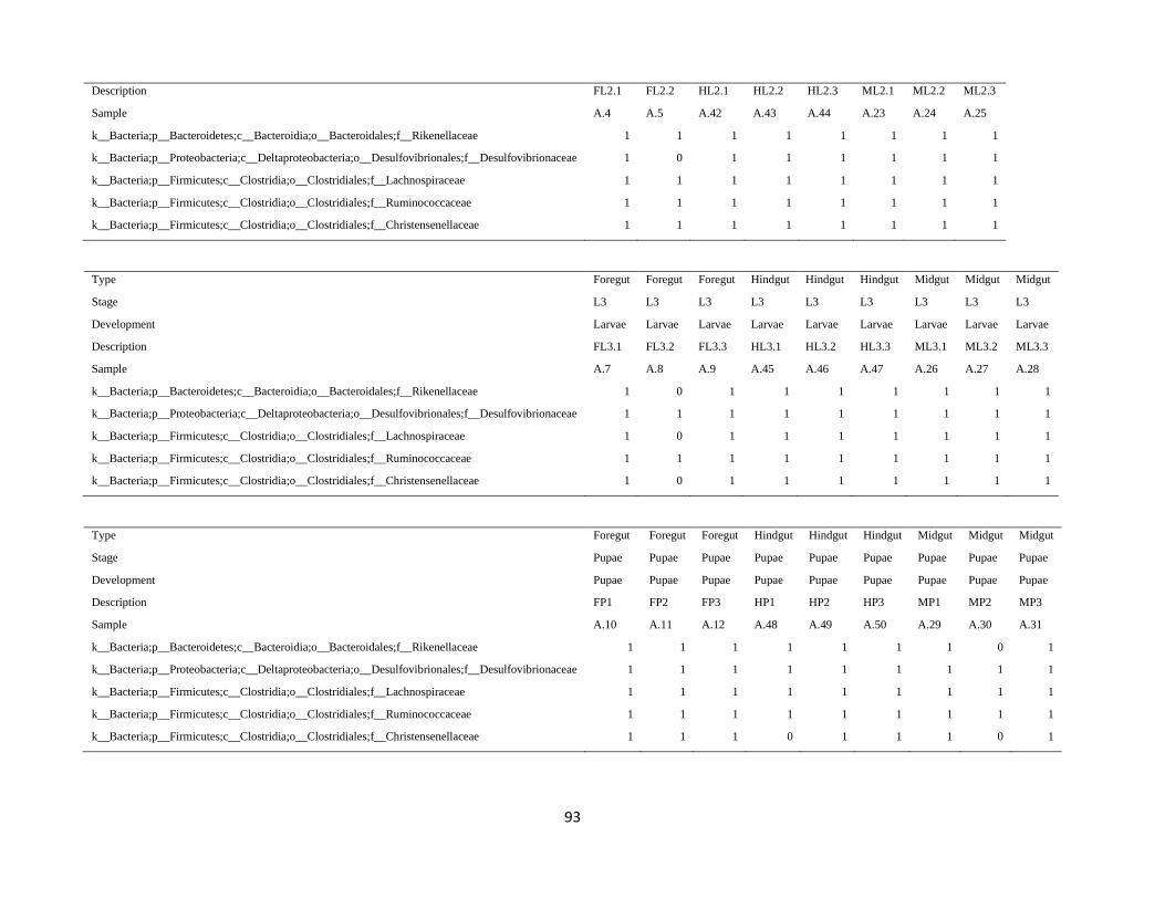
93
Description FL2.1 FL2.2 HL2.1 HL2.2 HL2.3 ML2.1 ML2.2 ML2.3
Sample A.4 A.5 A.42 A.43 A.44 A.23 A.24 A.25
k__Bacteria;p__Bacteroidetes;c__Bacteroidia;o__Bacteroidales;f__Rikenellaceae 1 1 1 1 1 1 1 1
k__Bacteria;p__Proteobacteria;c__Deltaproteobacteria;o__Desulfovibrionales;f__Desulfovibrionaceae 1 0 1 1 1 1 1 1
k__Bacteria;p__Firmicutes;c__Clostridia;o__Clostridiales;f__Lachnospiraceae 1 1 1 1 1 1 1 1
k__Bacteria;p__Firmicutes;c__Clostridia;o__Clostridiales;f__Ruminococcaceae 1 1 1 1 1 1 1 1
k__Bacteria;p__Firmicutes;c__Clostridia;o__Clostridiales;f__Christensenellaceae 1 1 1 1 1 1 1 1
Type Foregut Foregut Foregut Hindgut Hindgut Hindgut Midgut Midgut Midgut
Stage L3 L3 L3 L3 L3 L3 L3 L3 L3
Development Larvae Larvae Larvae Larvae Larvae Larvae Larvae Larvae Larvae
Description FL3.1 FL3.2 FL3.3 HL3.1 HL3.2 HL3.3 ML3.1 ML3.2 ML3.3
Sample A.7 A.8 A.9 A.45 A.46 A.47 A.26 A.27 A.28
k__Bacteria;p__Bacteroidetes;c__Bacteroidia;o__Bacteroidales;f__Rikenellaceae 1 0 1 1 1 1 1 1 1
k__Bacteria;p__Proteobacteria;c__Deltaproteobacteria;o__Desulfovibrionales;f__Desulfovibrionaceae 1 1 1 1 1 1 1 1 1
k__Bacteria;p__Firmicutes;c__Clostridia;o__Clostridiales;f__Lachnospiraceae 1 0 1 1 1 1 1 1 1
k__Bacteria;p__Firmicutes;c__Clostridia;o__Clostridiales;f__Ruminococcaceae 1 1 1 1 1 1 1 1 1
k__Bacteria;p__Firmicutes;c__Clostridia;o__Clostridiales;f__Christensenellaceae 1 0 1 1 1 1 1 1 1
Type Foregut Foregut Foregut Hindgut Hindgut Hindgut Midgut Midgut Midgut
Stage Pupae Pupae Pupae Pupae Pupae Pupae Pupae Pupae Pupae
Development Pupae Pupae Pupae Pupae Pupae Pupae Pupae Pupae Pupae
Description FP1 FP2 FP3 HP1 HP2 HP3 MP1 MP2 MP3
Sample A.10 A.11 A.12 A.48 A.49 A.50 A.29 A.30 A.31
k__Bacteria;p__Bacteroidetes;c__Bacteroidia;o__Bacteroidales;f__Rikenellaceae 1 1 1 1 1 1 1 0 1
k__Bacteria;p__Proteobacteria;c__Deltaproteobacteria;o__Desulfovibrionales;f__Desulfovibrionaceae 1 1 1 1 1 1 1 1 1
k__Bacteria;p__Firmicutes;c__Clostridia;o__Clostridiales;f__Lachnospiraceae 1 1 1 1 1 1 1 1 1
k__Bacteria;p__Firmicutes;c__Clostridia;o__Clostridiales;f__Ruminococcaceae 1 1 1 1 1 1 1 1 1
k__Bacteria;p__Firmicutes;c__Clostridia;o__Clostridiales;f__Christensenellaceae 1 1 1 0 1 1 1 0 1
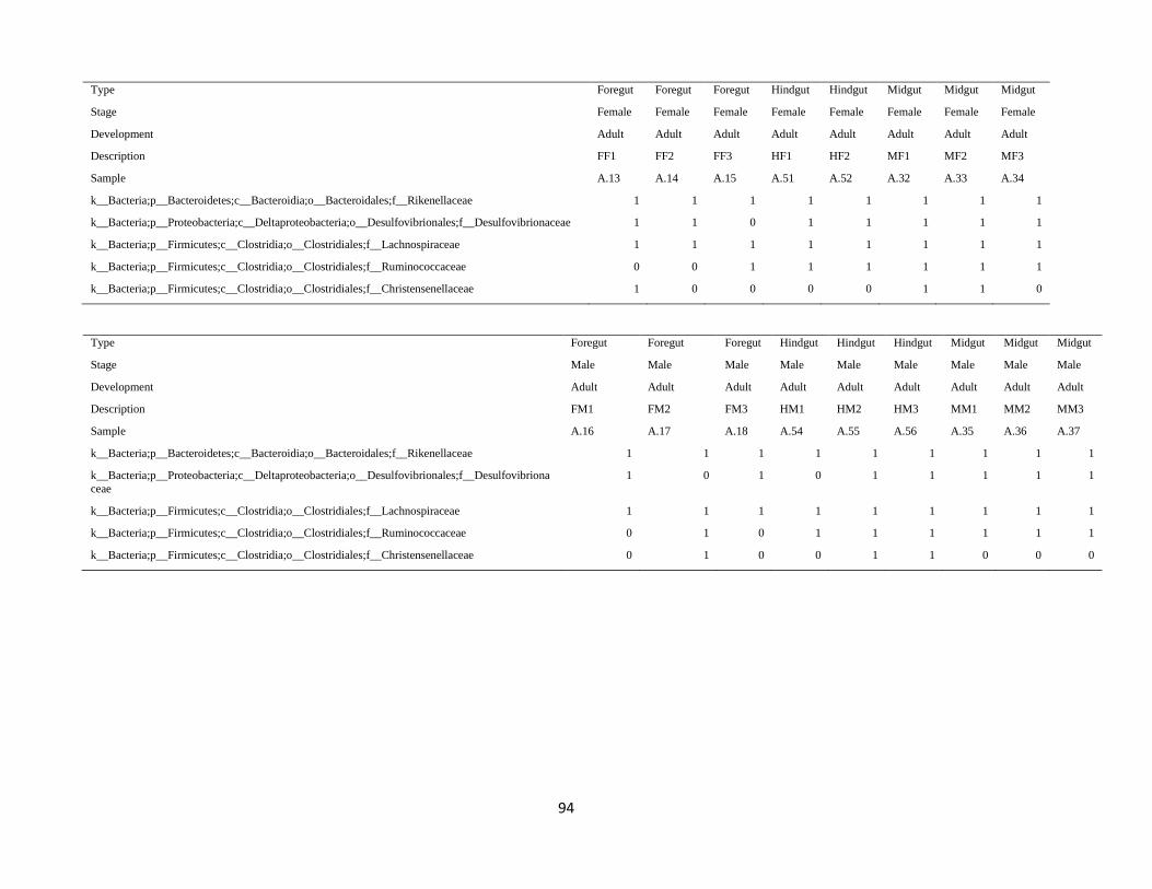
94
Type Foregut Foregut Foregut Hindgut Hindgut Midgut Midgut Midgut
Stage Female Female Female Female Female Female Female Female
Development Adult Adult Adult Adult Adult Adult Adult Adult
Description FF1 FF2 FF3 HF1 HF2 MF1 MF2 MF3
Sample A.13 A.14 A.15 A.51 A.52 A.32 A.33 A.34
k__Bacteria;p__Bacteroidetes;c__Bacteroidia;o__Bacteroidales;f__Rikenellaceae 1 1 1 1 1 1 1 1
k__Bacteria;p__Proteobacteria;c__Deltaproteobacteria;o__Desulfovibrionales;f__Desulfovibrionaceae 1 1 0 1 1 1 1 1
k__Bacteria;p__Firmicutes;c__Clostridia;o__Clostridiales;f__Lachnospiraceae 1 1 1 1 1 1 1 1
k__Bacteria;p__Firmicutes;c__Clostridia;o__Clostridiales;f__Ruminococcaceae 0 0 1 1 1 1 1 1
k__Bacteria;p__Firmicutes;c__Clostridia;o__Clostridiales;f__Christensenellaceae 1 0 0 0 0 1 1 0
Type Foregut Foregut Foregut Hindgut Hindgut Hindgut Midgut Midgut Midgut
Stage Male Male Male Male Male Male Male Male Male
Development Adult Adult Adult Adult Adult Adult Adult Adult Adult
Description FM1 FM2 FM3 HM1 HM2 HM3 MM1 MM2 MM3
Sample A.16 A.17 A.18 A.54 A.55 A.56 A.35 A.36 A.37
k__Bacteria;p__Bacteroidetes;c__Bacteroidia;o__Bacteroidales;f__Rikenellaceae 1 1 1 1 1 1 1 1 1
k__Bacteria;p__Proteobacteria;c__Deltaproteobacteria;o__Desulfovibrionales;f__Desulfovibriona
ceae
1 0 1 0 1 1 1 1 1
k__Bacteria;p__Firmicutes;c__Clostridia;o__Clostridiales;f__Lachnospiraceae 1 1 1 1 1 1 1 1 1
k__Bacteria;p__Firmicutes;c__Clostridia;o__Clostridiales;f__Ruminococcaceae 0 1 0 1 1 1 1 1 1
k__Bacteria;p__Firmicutes;c__Clostridia;o__Clostridiales;f__Christensenellaceae 0 1 0 0 1 1 0 0 0