Embed Size (px)
Citation preview
Polysaccharide drug delivery systems based on pectin and chitosan 257
Biotechnology and Genetic Engineering Reviews - Vol. 27, 257-284 (2010)
*To whom correspondence may be addressed ([email protected])
Abbreviations: GlcN: D-glucosamine; GlcNAc: N-acetyl-D-glucosamine; DA/ DAc: degree of acetyla-tion; TPP: tripolyphosphate; HG: homogalacturonan; RG-I: type I rhamnogalacturonan; DM: degree of methylation; [h]: intrinsic viscosity; s0
20,w: sedimentation coefficient; M
w: weight average molecular weight;
rg: radius of gyration; f/f
0: translational frictional ratio; L
p: persistence length
Polysaccharide drug delivery systems based on pectin and chitosan
GoRDoN A. MoRRIs1,*, M. sAMIl KöK2, sTEPHEN E. HARDING1 AND GARy G. ADAMs1,3
1NCMH Laboratory, School of Biosciences, University of Nottingham, Sutton
Bonington LE12 5RD, UK; 2Department of Food Engineering, Abant Izzet Baysal
University, 14280 Bolu, Turkey; 3Insulin and Diabetes Experimental Research
(IDER) Group, Faculty of Medicine and Health Science, University of Nottingham,
Clifton Boulevard, Nottingham NG7 2RD, UK
Abstract
Chitosans and pectins are natural polysaccharides which show great potential in drug delivery systems.
Chitosans are a family of strongly polycationic derivatives of poly-N-acetyl-D-glucosamine. This positive charge is very important in chitosan drug delivery systems as it plays a very important role in mucoadhesion (adhesion to the mucosal surface). other chitosan based drug delivery systems involve complexation with ligands to form chitosan nanoparticles with can be used to encapsulate active compounds.
Pectins are made of several structural elements the most important of which are the homogalacturonan (HG) and type I rhamnogalacturonan (RG-I) regions often described in simplified terms as the “smooth” and “hairy” regions respectively. Pectin HG regions consist of poly-glacturonic acid residues which can be partially methyl esterified. Pectins with a degree of methyl esterification (DM) > 50% are known as high methoxyl (HM) pectins and consequently low methoxyl (lM) pectins have a DM < 50%. low methoxyl pectins are of particular interest in drug delivery as they can form gels with calcium ion (Ca2+) which has potential applications especially in nasal formulations.
258 G.A. Morris et al.
In this chapter we will discuss the physicochemical properties of both chitosans and pectins and how these translate to current and potential drug delivery systems.
Introduction
The route for the delivery of drugs that is still the most popular with medical staff and patients alike is through the mouth and down the alimentary tract: the oral route. The major site for drug absorption by this route is the small intestine which offers ≈ 100 m2 of surface epithelia across which transfer can at least in principle take place. If the drug is poorly soluble, or is in the form of a controlled release dosage form, significant absorption of the drug may also occur in the large intestine (Davis, 1989). However, the clearance time through the whole alimentary tract is generally too short (4–12 h), rendering oral drug administration a very inefficient process, with much of the drug unabsorbed. More recently interest has focused on drug absorption through nasal epithelia, where again clearance problems are an issue. Consideration is also given to other delivery routes (e.g. vaginal and ocular). other important issues are the degradation of peptide-based drugs in the gastrointestinal tract and low trans-mucosal permeability. Macromolecular based carrier and mucoadhesive systems have been considered for several years and two polysaccharide based systems have emerged as particularly promising: chitosans (cationic) and low methoxy pectins (anionic).
Chitosan
CHEMICAl sTRuCTuRE
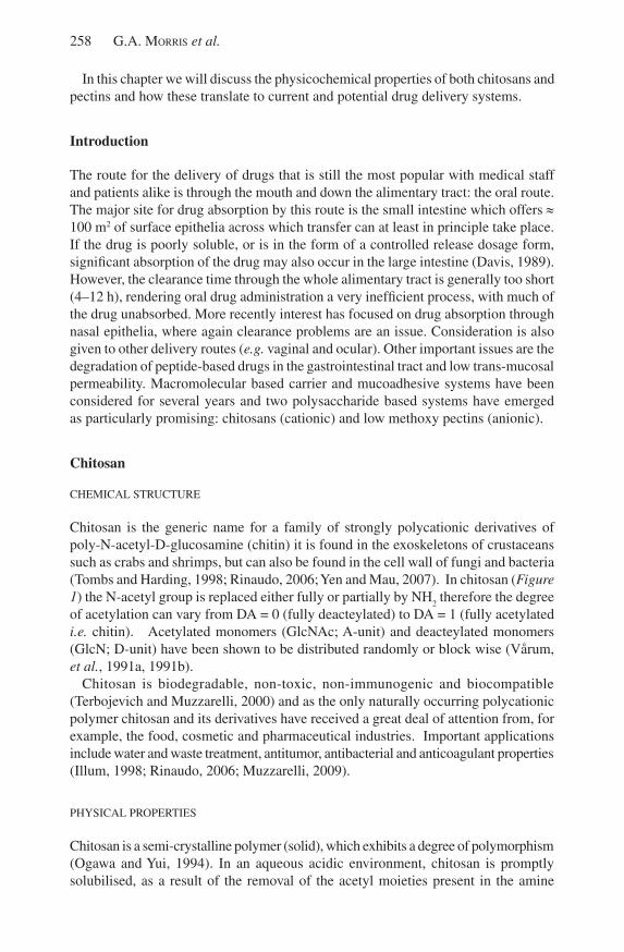
Chitosan is the generic name for a family of strongly polycationic derivatives of poly-N-acetyl-D-glucosamine (chitin) it is found in the exoskeletons of crustaceans such as crabs and shrimps, but can also be found in the cell wall of fungi and bacteria (Tombs and Harding, 1998; Rinaudo, 2006; yen and Mau, 2007). In chitosan (Figure 1) the N-acetyl group is replaced either fully or partially by NH
2 therefore the degree
of acetylation can vary from DA = 0 (fully deacteylated) to DA = 1 (fully acetylated i.e. chitin). Acetylated monomers (GlcNAc; A-unit) and deacteylated monomers (GlcN; D-unit) have been shown to be distributed randomly or block wise (Vårum, et al., 1991a, 1991b).
Chitosan is biodegradable, non-toxic, non-immunogenic and biocompatible (Terbojevich and Muzzarelli, 2000) and as the only naturally occurring polycationic polymer chitosan and its derivatives have received a great deal of attention from, for example, the food, cosmetic and pharmaceutical industries. Important applications include water and waste treatment, antitumor, antibacterial and anticoagulant properties (Illum, 1998; Rinaudo, 2006; Muzzarelli, 2009).
PHysICAl PRoPERTIEs
Chitosan is a semi-crystalline polymer (solid), which exhibits a degree of polymorphism (ogawa and yui, 1994). In an aqueous acidic environment, chitosan is promptly solubilised, as a result of the removal of the acetyl moieties present in the amine
Polysaccharide drug delivery systems based on pectin and chitosan 259
Figure 1. schematic representation of the structure repeat units of chitosan, where R = Ac or H depending on the degree of acetylation.
functional groups. This solubility is limited, however, in inorganic acids compared to its solubility in organic acids. solubilisation occurs as a consequence of the protonation of -NH
2 functional groups on the C-2 position of D-glucosamine residues. Chitosan
is a weak base with pKa values ranging from 6.2 to 7 and at physiological pH 7.4 or higher, low solubility is shown (Park, et al., 1983). However, chitosan’s solution properties are dependent on the distribution of its acetyl groups and the molecular weight of the polymer (Kubota and Eguchi, 1997). The solubility of chitosan in water increases with increasing DA (Vårum et al., 1994). With the addition of electrolytes to the solution, the aqueous solubility of chitosan is affected and salting out of chitosan can be seen as in the case of excessive hydrochloric acid use, and the resulting formation of chitosan chlorhydrate (Rinaudo, 2006). salting out can also be used to recover chitosan from solution and salting out efficiency of anions follows the Hofmeister series so
42- > H
2Po
4 ≈ HPo
42- > No
3- (leHoux and Depuis,
2007). With an extended chitosan conformation, due to the repelling effect of each positively charged deacetylated unit, the addition of electrolytes reduces the inter-chain repulsion and induces a more random coil-like conformation in the molecule (Terbojevich and Muzzarelli, 2000).
Molecular weight, pH, ionic strength, and temperature are all factors which affect the viscosity of chitosan. Hydrodynamic studies based on intrinsic viscosity ([η]), sedimentation coefficient (s0
20,w), radius of gyration (r
g) and weight average molecular
weight (Mw) have focussed on qualitative/ semi-quantitative methods of estimating
the conformation based around “power law” Mark-Houwink-Kuhn-sakurada relations (Tombs and Harding, 1998) which link intrinsic viscosity, sedimentation coefficient and radius of gyration with molar mass [η] ~ Ma, s0
20,w ~ Mb and r
g ~ Mc, where a, b
and c have defined values for specific conformation types (Table 1). The translational frictional ratio, f/f
o (Tanford, 1961), sedimentation conformation zoning (Pavlov, et al.,
1997; 1999), the “Wales-van Holde” ratio, ks/[ η] (Wales and van Holde, 1954) and
the persistence length, Lp (Kratky and Porod, 1949) have also been used to estimate
dilute solution conformation (Table 2).This has resulted in chitosan being reported to have either a rigid rod-type structure
(Terbojevich, et al., 1991; Errington, et al., 1993; Cölfen, et al., 2001; Fee, et al., 2003; Kasaai, 2006; Morris, et al., 2009a) or a semi-flexible-coil (Rinaudo, et al., 1993; Berth, et al., 1998; Brugnerotto, et al., 2001; schatz, et al., 2003; Mazeau and Rinaudo, 2004; Vold, 2004; lamarque, et al., 2005; Velásquez, et al., 2008). It has also been shown that flexibility (in terms of persistence length) is moderately influenced by DA (Terbojevich, et al., 1991; Mazeau and Rinaudo, 2004).
260 G.A. Morris et al.
Table 1. The Mark-Houwink-Kuhn-sakurada (MHKs) power law exponents (a, b and c), and the Wales – van Holde (k
s/[η]) for the conformations described by
sedimentation conformation zoning.
Zone AExtra-rigid rod
Zone BRigid rod
Zone Csemi-flexible coil
Zone DRandom coil
Zone Espherical
a > 1.4 0.8 – 1.4 0.5 – 0.8 0.2 – 0.5 0.0
b < 0.2 0.2 – 0.4 0.4 – 0.5 0.5 – 0.6 0.67
c > 0.8 0.6 – 0.8 0.5 – 0.6 0.4 – 0.5 0.33
ks/[η] < 0.2 0.2 – 0.4 0.4 – 1.0 1.0 - 1.4 1.6
Table 2. Estimations of the dilute solution conformation for pectin and chitosan.
Pectin Chitosan
a 0.62 – 0.94 0.77 – 1.1
b 0.17 0.24 – 0.25
c 0.57 0.55 – 0.56
ks/[η] 0.10 – 0.85 0.16 – 0.73
f/f0
7 – 10 11 - 16
Lp (nm) 10 - 15 4 - 35
Zone A/B/C B/C
References Anger and Berth, 1985; Axelos, et al., 1987; Axelos and Thibault, 1991, Berth, et al., 1977; Harding, et al., 1991; Garnier, et al., 1993; Malovikova, et al., 1993; Tombs and Harding, 1998; Morris, et al., 2000, 2002, 2008; Fishman, et al., 2001, 2006
Terbojevich, et al., 1991; Errington, et al., 1993; ottøy, et al., 1996; Berth, et al., 1998; Cölfen, et al, 2001; Brugnerotto, et al., 2001; Fee, et al., 2003; schatz, et al., 2003; Mazeau and Rinaudo, 2004; Vold, 2004; lamarque, et al., 2005; Rinaudo, 2006; Kasaai, 2006; Velásquez, et al., 2008; Morris, et al., 2009
CHITosAN CoMPlExATIoN
The ability of chitosan to complex with other ligands, metals for example, is well known (Rhazi, et al., 2002a,b). The proclivity for chelation is dependent on physical state, -NH
2 content and distribution of chitosan, degree of polymerization, pH and
cation content. It has been shown that following a higher degree of deacetylation, there is a characteristic increase in the degree of chelation. The chelation takes place in chitosan with a degree of polymerization greater than 6 monomeric residues (Rhazi, et al., 2002b). In addition, the intensity of chitosan chelation is governed by nature of cation in solution. studies have shown that the affinity of chitosan for divalent and trivalent cations of chloride salts shows selectivity in the following order: Cu2+ >> Hg2+ > Zn2+ > Cd2+ > Ni2+ > Co2+> Ca2+, Eur3+ > Nd3+ > Cr3+> Pr3+ (Rhazi, et al., 2002b).
Polysaccharide drug delivery systems based on pectin and chitosan 261
USAGE IN DRUG DELIvERy
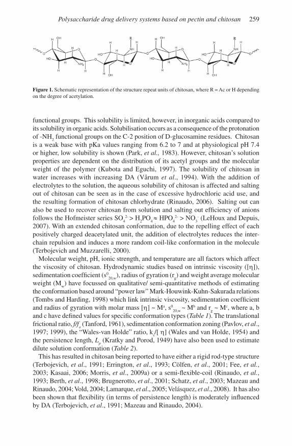
Chitosan is of great interest to the pharmaceutical industry in drug delivery and the number of publications on this subject has increased by almost an order of magnitude in the last decade (Figure 2). Many aspects including biodegradation, biodistribution and toxicity (Kean and Thanou, 2010); formulations for delivery of DNA and siRNA (Mao, et al., 2010); delivery systems for protein therapeutics (Amidi, et al., 2010); hydrogels for controlled, localized drug delivery (Bhattarai, et al., 2010); nanostructures for delivery of ocular therapeutics (de la Fuente, et al., 2010) and the targeted delivery of low molecular drugs (Park, et. al., 2010) have been reviewed in the most recent volume of Advanced Drug Delivery Reviews (Volume 62).
0
100
200
300
400
500
600
700
2000 2001 2002 2003 2004 2005 2006 2007 2008 2009
Years
Figure 2. Number of publications on chitosan in drug delivery over the last 10 years (adapted from Figure 1 in Amidi and Hennink (2010)). Reproduced with the permission of Elsevier.
The mucoadhesive properties of chitosan play an important role in its usage in oral, nasal and ocular drug delivery (Harding, et al., 1999; Illum, 2002; Harding, 2006).
MUCoADHESIoN
Mucoadhesion is the specific term for adhesion when one of the surfaces is mucus (Harding, et al., 1999). Mucus consists largely of water (> 95 %) and the high molecular weight glycoprotein mucin (Harding, et al., 1999; Harding, 2003; Harding, 2006). The key sugar residues for mucoadhesive interaction are the acidic ones (N-acetyl neuraminic acid or “sialic acid”, and some sulphated galactose) and the hydrophobic methyl containing fucose. Despite the polydispersity of these molecules compared to unglycosylated proteins, their structural hierarchy is also well understood. They consist of M ~ 500 000 g/mol basic units linked linearly into “subunits” of M ~ 2 500 000 g/mol. These subunits are further linearly arrayed into macroscopic structures (M between 5 and 50 000 000 g/mol) seen under the electron microscope (Harding, et al., 1983) or using atomic force microscopy (Deacon, et al., 2000). Chitosan interacts
262 G.A. Morris et al.
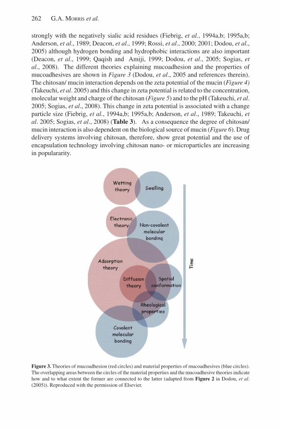
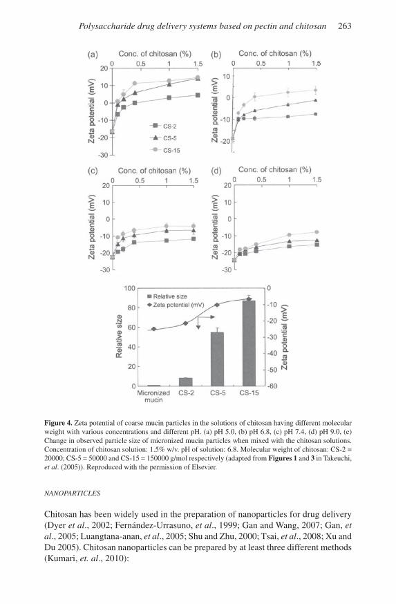
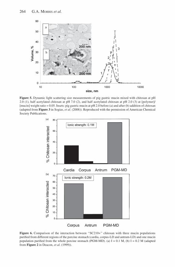
strongly with the negatively sialic acid residues (Fiebrig, et al., 1994a,b; 1995a,b; Anderson, et al., 1989; Deacon, et al., 1999; Rossi, et al., 2000; 2001; Dodou, et al., 2005) although hydrogen bonding and hydrophobic interactions are also important (Deacon, et al., 1999; Qaqish and Amiji, 1999; Dodou, et al., 2005; sogias, et al., 2008). The different theories explaining mucoadhesion and the properties of mucoadhesives are shown in Figure 3 (Dodou, et al., 2005 and references therein). The chitosan/ mucin interaction depends on the zeta potential of the mucin (Figure 4) (Takeuchi, et al. 2005) and this change in zeta potential is related to the concentration, molecular weight and charge of the chitosan (Figure 5) and to the pH (Takeuchi, et al. 2005; sogias, et al., 2008). This change in zeta potential is associated with a change particle size (Fiebrig, et al., 1994a,b; 1995a,b; Anderson, et al., 1989; Takeuchi, et al. 2005; sogias, et al., 2008) (Table 3). As a consequence the degree of chitosan/ mucin interaction is also dependent on the biological source of mucin (Figure 6). Drug delivery systems involving chitosan, therefore, show great potential and the use of encapsulation technology involving chitosan nano- or microparticles are increasing in populararity.
Figure 3. Theories of mucoadhesion (red circles) and material properties of mucoadhesives (blue circles). The overlapping areas between the circles of the material properties and the mucoadhesive theories indicate how and to what extent the former are connected to the latter (adapted from Figure 2 in Dodou, et al. (2005)). Reproduced with the permission of Elsevier.
Polysaccharide drug delivery systems based on pectin and chitosan 263
Figure 4. Zeta potential of coarse mucin particles in the solutions of chitosan having different molecular weight with various concentrations and different pH. (a) pH 5.0, (b) pH 6.8, (c) pH 7.4, (d) pH 9.0, (e) Change in observed particle size of micronized mucin particles when mixed with the chitosan solutions. Concentration of chitosan solution: 1.5% w/v. pH of solution: 6.8. Molecular weight of chitosan: Cs-2 = 20000; Cs-5 = 50000 and Cs-15 = 150000 g/mol respectively (adapted from Figures 1 and 3 in Takeuchi, et al. (2005)). Reproduced with the permission of Elsevier.
NANoPARTICLES
Chitosan has been widely used in the preparation of nanoparticles for drug delivery (Dyer et al., 2002; Fernández-urrasuno, et al., 1999; Gan and Wang, 2007; Gan, et al., 2005; luangtana-anan, et al., 2005; shu and Zhu, 2000; Tsai, et al., 2008; xu and Du 2005). Chitosan nanoparticles can be prepared by at least three different methods (Kumari, et. al., 2010):
264 G.A. Morris et al.
Figure 5. Dynamic light scattering size measurements of pig gastric mucin mixed with chitosan at pH 2.0 (1), half acetylated chitosan at pH 7.0 (2), and half acetylated chitosan at pH 2.0 (3) at [polymer]/[mucin] weight ratio = 0.05. Insets: pig gastric mucin at pH 2.0 before (a) and after (b) addition of chitosan (adapted from Figure 3 in sogias, et al. (2008)). Reproduced with the permission of American Chemical society Publications.
Figure 6. Comparison of the interaction between ‘‘sC210+’’ chitosan with three mucin populations purified from different regions of the porcine stomach (cardia, corpus-lD and antrum-lD) and one mucin population purified from the whole porcine stomach (PGM-MD). (a) I = 0.1 M, (b) I = 0.2 M (adapted from Figure 2 in Deacon, et al. (1999)).
Polysaccharide drug delivery systems based on pectin and chitosan 265
Table 3. Mucoadhesive analysis. The sedimentation coefficient ratio (scomplex
/s
mucin) as an index of (muco)adhesiveness (from Fiebrig, et al., 1994a,b; 1995a,b;
Anderson, et al., 1989) (adapted from Table 1 in Harding (2003)).
Mucoadhesive scomplex
/smucin
Conditions
DEAE-dextran 1.1–1.9a pH 6.8, 20 ºC
1.2–1.4a pH 6.8, 37 ºC
Chitosan (FA ≈ 0.11) 48 pH 6.5, 20 ºC
15 pH 4.5, 20 ºC
22 pH 2.0, 20 ºC
12 pH 2.0, 37 ºC
26 pH 4.5, 20 ºC + 3 mM bile salt
35 pH 4.5, 37 ºC + 3 mM bile salt
18 pH 4.5, 20 ºC + 6 mM bile salt
14 pH 4.5, 37 ºC + 6 mM bile salt
Chitosan (FA ≈ 0.42) 31 pH 4.5, 20 ºC
44 pH 4.5, 37 ºC
aDepends on the mixing ratio
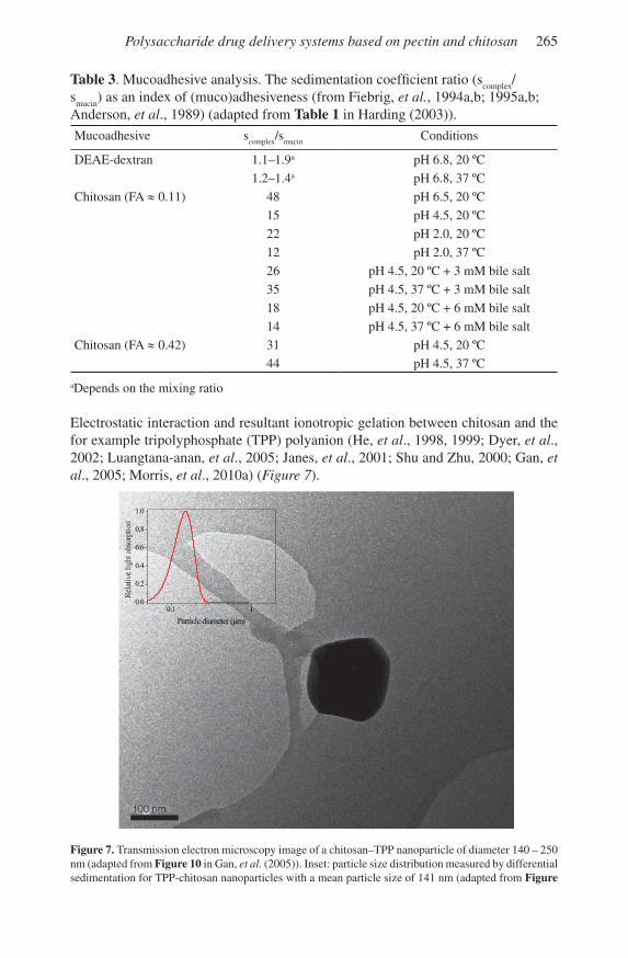
Electrostatic interaction and resultant ionotropic gelation between chitosan and the for example tripolyphosphate (TPP) polyanion (He, et al., 1998, 1999; Dyer, et al., 2002; luangtana-anan, et al., 2005; Janes, et al., 2001; shu and Zhu, 2000; Gan, et al., 2005; Morris, et al., 2010a) (Figure 7).
0.1 10.0
0.2
0.4
0.6
0.8
1.0
Particle diameter (µm)
Figure 7. Transmission electron microscopy image of a chitosan–TPP nanoparticle of diameter 140 – 250 nm (adapted from Figure 10 in Gan, et al. (2005)). Inset: particle size distribution measured by differential sedimentation for TPP-chitosan nanoparticles with a mean particle size of 141 nm (adapted from Figure
266 G.A. Morris et al.
3 in Morris, et al. (2010a)). Reproduced with the permission of Elsevier.
Micro-emulsion for preparation of chitosan – glutaraldehyde complexes for example (Genta, et. al., 1998; Dhawan, et al., 2004).
Polyelectrolyte complex (PEC) formation with for example pectin (Macleod, et al., 1999; ofori-Kwakye and Fell, 2001) or hyaluronic acid (lim, et al., 2000; Kim, et al., 2004; Kujawa, et al., 2007). This is of particular importance when a constant drug release profile is not desired (Macleod, et al., 1999; ofori-Kwakye and Fell, 2001).
The size of the nanoparticles depends on the molecular weight of the chitosan polymer and higher molecular weight chitosans produce larger nanoparticles (luangtana-anan et al., 2005; Morris, et al., 2010a). The method of cross-linking affects the mucoadhesive strength and stability of the nanoparticles (Genta, et. al., 1998; Dhawan, et al., 2004).
STABILITy
The stability (shelf-life) of chitosan in terms of molar mass, viscosity and conformation is very important to pharmaceutical industry as these properties play an important role in the function of chitosan in formulations (skaugrud, et al., 1999; Terbojevich and Muzzarelli, 2000). Chitosan storage conditions and particularly temperature may be important but whether or not chitosan depolymerisation will be detrimental to its intended application will depend on the functional significance of the changes that occur. Depolymerisation of chitosan in both the polymeric and nanoparticle form is temperature dependent (Nguyen, et al., 2007; Morris, et al., 2009b; Morris, et al., 2010a). For example it has been reported that low molar mass chitosans can cause more cell damage (Aspden, et al., 1996), although they may also prevent diabetes mellitus progression in mice to a greater extent than high molar chitosans
(Kondo, et al., 2000), show greater antibacterial activity compared with high molar mass chitosans (lui, et al., 2001) and whilst the high viscosities of high molar mass chitosans limit its biological usefulness, low molar mass chitosan is more soluble at neutral pH and therefore potentially more available in vivo (Harish Prashanth and Tharanathan, 2007). However, it has also been reported that high molar mass chitosans show greater antibacterial activity compared with low molar mass chitosans (No, et al., 2006), that nasal insulin delivery (Aspden, et al., 1997; Davis and Illum, 2000) is more effective with chitosan of molar mass greater than 100000 g/mol and the reversibility of transepithelial chemical resistance (TEER) values decrease with decreased chitosan molar mass (Holme, et al., 2000).
Pectin
CHEMICAl sTRuCTuRE
Pectins are a complex family of heteropolysaccharides that constitute a large proportion of the primary cell walls of dicotyledons and play important roles in growth, development and senescence (van Buren, 1991; Tombs and Harding, 1998; Ridley, et. al., 2001; Willats, et. al., 2001). Pectic polysaccharides are made of
Polysaccharide drug delivery systems based on pectin and chitosan 267
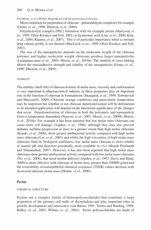
several structural elements the most important of which are the homogalacturonan (HG) and type I rhamnogalacturonan (RG-I) regions often described in simplified terms as the “smooth” and “hairy” regions respectively (Figure 8). The HG region is composed of (1→4) linked α-D-GalpA residues that can be partially methylated at C-6 (Pilnik and Voragen, 1970) and possibly partially acetyl-esterified at o-2 and/or o-3 (Rombouts and Thibault, 1986). The degree of methylation (DM) and the degree of acetylation (DAc) are defined as the number of moles of methanol or acetic acid per 100 moles of GalA. The degree of methylation in native pectins is generally in the order of DM ≈ 70-80; whereas degree of acetylation is generally much lower e.g. DAc ≈ 35 for sugar beet pectins (Rombouts and Thibault, 1986). Theoretically the degree of methoxyl esterification (DM) can range from 0-100 %. Pectins with a degree of esterification (DM) > 50% are known as high methoxyl (HM) pectins and consequently low methoxyl (lM) pectins have a DM < 50% (Walter, 1991). The RG-I region consists of disaccharide repeating unit [→4)-α-D-GalpA-(1→2)-α-l-Rhap-(1→]
n with a variety side chains consisting of l-arbinosyl and D-galactosyl
residues (Voragen, et. al., 1995). It has been reported that GalA residues in the RG-I region are partially acetylated (Ishii, 1997; Perrone, et. al., 2002) but not methylated (Komalavilas and Mort, 1989; Perrone, et. al., 2002). In the case of sugar beet pectin the neutral side chain sugars are substituted with ferulic acid (Fry, 1982; Rombouts and Thibault, 1986) and there is evidence indicating that pectin chains can be dimerised via diferulic bridges (levigne, et. al., 2004a,b). There are a number of different ways in which ferulic acid can dimerise the most common being: 5-5’; 8-o-4’; 8-5’ cyclic and 8-5’ non-cyclic dimers (Micard, et. al., 1997).
Figure 8. schematic structure for pectin: galacturonic acid ( ); galactose ( ); arabinose ( ); rhamnose ( ) and methyl groups ( ).
PHysICAl PRoPERTIEs
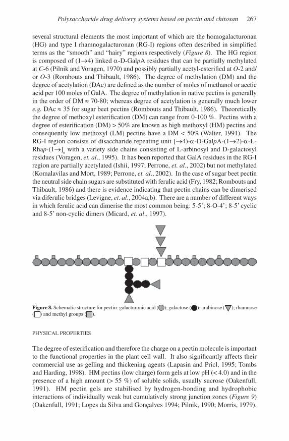
The degree of esterification and therefore the charge on a pectin molecule is important to the functional properties in the plant cell wall. It also significantly affects their commercial use as gelling and thickening agents (lapasin and Pricl, 1995; Tombs and Harding, 1998). HM pectins (low charge) form gels at low pH (< 4.0) and in the presence of a high amount (> 55 %) of soluble solids, usually sucrose (oakenfull, 1991). HM pectin gels are stabilised by hydrogen-bonding and hydrophobic interactions of individually weak but cumulatively strong junction zones (Figure 9) (oakenfull, 1991; lopes da silva and Gonçalves 1994; Pilnik, 1990; Morris, 1979).
268 G.A. Morris et al.
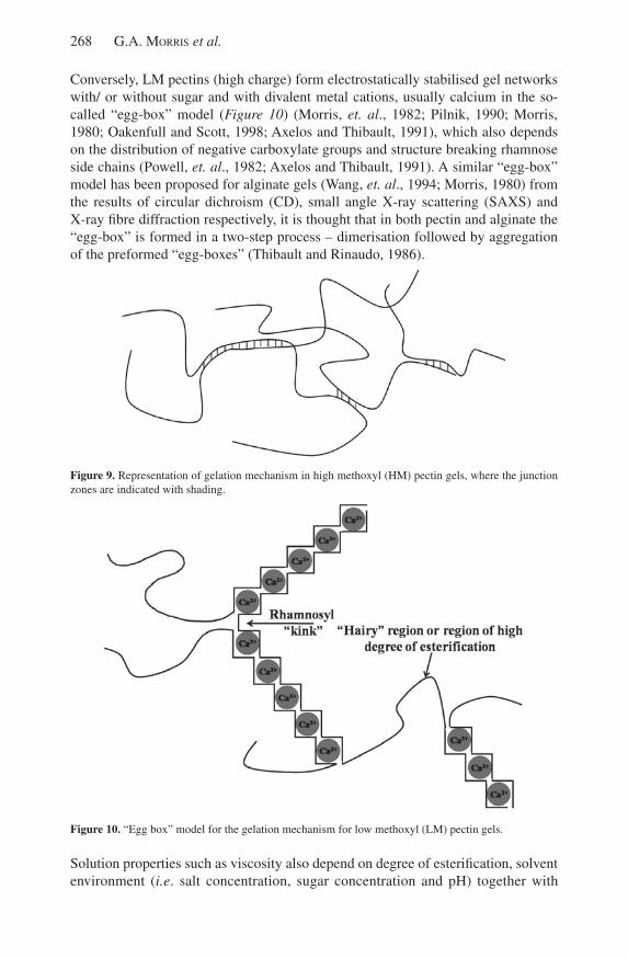
Conversely, lM pectins (high charge) form electrostatically stabilised gel networks with/ or without sugar and with divalent metal cations, usually calcium in the so-called “egg-box” model (Figure 10) (Morris, et. al., 1982; Pilnik, 1990; Morris, 1980; oakenfull and scott, 1998; Axelos and Thibault, 1991), which also depends on the distribution of negative carboxylate groups and structure breaking rhamnose side chains (Powell, et. al., 1982; Axelos and Thibault, 1991). A similar “egg-box” model has been proposed for alginate gels (Wang, et. al., 1994; Morris, 1980) from the results of circular dichroism (CD), small angle x-ray scattering (sAxs) and x-ray fibre diffraction respectively, it is thought that in both pectin and alginate the “egg-box” is formed in a two-step process – dimerisation followed by aggregation of the preformed “egg-boxes” (Thibault and Rinaudo, 1986).
Figure 9. Representation of gelation mechanism in high methoxyl (HM) pectin gels, where the junction zones are indicated with shading.
Figure 10. “Egg box” model for the gelation mechanism for low methoxyl (lM) pectin gels.
solution properties such as viscosity also depend on degree of esterification, solvent environment (i.e. salt concentration, sugar concentration and pH) together with
Polysaccharide drug delivery systems based on pectin and chitosan 269
temperature (oakenfull, 1991). Hydrodynamic studies based on intrinsic viscosity ([η]), sedimentation coefficient (s0
20,w), radius of gyration (r
g) and weight average
molecular weight (Mw) have focussed on qualitative/ semi-quantitative methods
of estimating the conformation based around “power law” Mark-Houwink-Kuhn-sakurada relations (Tombs and Harding, 1998) which link intrinsic viscosity, sedimentation coefficient and radius of gyration with molar mass [η] ∝ Ma, s0
20,w
∝ Mb and rg ∝ Mc, where a, b and c have defined values for specific conformation
types (Table 1). The translational frictional ratio, f/fo (Tanford, 1961), sedimentation
conformation zoning (Pavlov, et al., 1997; 1999), the “Wales-van Holde” ratio, ks/[η]
(Wales and van Holde, 1954) and the persistence length, Lp (Kratky and Porod, 1949)
have also been used to estimate dilute solution conformation (Table 2). A picture of a semi-flexible conformation for pectins irrespective of degree of esterification (and charge) has emerged from these studies (Anger and Berth, 1985; Axelos, et al., 1987; Axelos and Thibault, 1991, Berth, et al., 1977; Harding, et al., 1991; Garnier, et al., 1993; Malovikova, et al., 1993; Cros, et al., 1996; Tombs and Harding, 1998; Braccini, et al., 1999; Morris, et al., 2000, 2002, 2008; Fishman, et al., 2001, 2006; Noto, et al, 2005). Pectin molecular weight and chain flexibility is important in mucoadhesive interactions (Nafee, et al., 2007).
usAGE IN DRuG DElIVERy
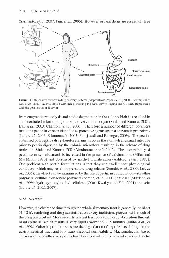
Pectins have been used is a gelling agent for a large number of years, however there has been recent interest in the use of pectin gels in controlled drug delivery (sungthongjeen, et al., 2004; lui, et al., 2003; lui, et al., 2006). This is in part due their long standing reputation of being non-toxic (GRAs – generally regarded as safe) (lui, et al., 2003; lui, et al., 2007; Watts and smith, 2009), their relatively low production costs (sungthongjeen, et al., 2004) and high availability (Beneke et al., 2009). It is proposed that pectin could be used to deliver drugs orally, nasally and vaginally (Figure 11) (Peppas, et al., 2000; sinha and Kumria, 2001; lui, et al., 2003; Nafee, et al., 2004; Valenta, 2005; lui, et al., 2007; Chelladurai, et al., 2008; Thirawong, et al., 2008), which are generally well accepted by patients (lui, et al., 2003; lui, et al., 2007; yadav, et al., 2009).
oRAL DELIvERy
The oral route is of particular interest as in general oral drug administration results in less pain, greater convenience, higher compliance and reduced infection risk as compared to subcutaneous injections (Chen and langer, 1998, yadav, et al., 2009). However, there are disadvantages associated with this route of administration such as low bioavailability due to relatively low passage of active agents across the mucosal epithelium, rapid polypeptide degradation due to action of digestive enzymes in the GI tract, enzymatic proteolysis and acidic degradation of orally administered drugs in the stomach (lui, et al., 2003; lin, et al., 2007). Various approaches have been made to increase the buccal penetration using permeation enhancers (Mesiha, et al., 1994; Carino, et al., 2000), protease inhibitors (yamamoto, et al., 1994), enteric coatings (Morishita, et. al., 1993) and (bio)polymer micro-/ nano-sphere formulations
270 G.A. Morris et al.
(sarmento, et al., 2007; Jain, et al., 2005). However, protein drugs are essentially free
Figure 11. Major sites for pectin drug delivery systems (adapted from Peppas, et al., 2000; Harding, 2003; lui, et al., 2003; Valenta, 2005) with insets showing the nasal cavity, vagina and GI tract. Reproduced with the permission of Elsevier.
from enzymatic proteolysis and acidic degradation in the colon which has resulted in a concentrated effort to target their delivery to this organ (sinha and Kumria, 2001; lui, et al., 2003; Chambin, et al., 2006). Therefore a number of different polymers including pectin have been identified as protective agents against enzymatic proteolysis (lui, et al., 2003; sriamornsak, 2003; Pourjavadi and Barzegar, 2009). The pectin-stabilised polypeptide drug therefore mains intact in the stomach and small intestine prior to pectin digestion by the colonic microflora resulting in the release of drug molecule (sinha and Kumria, 2001; Vandamme, et al., 2002). The susceptibility of pectin to enzymatic attack is increased in the presence of calcium ions (Miler and MacMilan, 1970) and decreased by methyl esterification (Ashford, et al., 1993). one problem with pectin formulations is that they can swell under physiological conditions which may result in premature drug release (semdé, et al., 2000; lui, et al., 2006), the effect can be minimised by the use of pectin in combination with other polymers: cellulosic or acrylic polymers (semdé, et al., 2000); chitosan (Macleod, et al., 1999); hydroxypropylmethyl cellulose (ofori-Kwakye and Fell, 2001) and zein (lui, et al., 2005; 2007).
NASAL DELIvERy
However, the clearance time through the whole alimentary tract is generally too short (4–12 h), rendering oral drug administration a very inefficient process, with much of the drug unabsorbed. More recently interest has focused on drug absorption through nasal epithelia, which results in very rapid absorption ~ 15 minutes (Jabbal-Gill, et al., 1998). other important issues are the degradation of peptide-based drugs in the gastrointestinal tract and low trans-mucosal permeability. Macromolecular based carrier and mucoadhesive systems have been considered for several years and pectin
Polysaccharide drug delivery systems based on pectin and chitosan 271
based systems have emerged as particularly promising. low methoxyl pectins are strongly polyanionic polyuronides from fruit used traditionally in jams and jellies (Rolin, 1993) and can also form weak gels in the presence of Ca2+ ions (8 meq/l), which occur naturally in nasal secretions (Chang and su, 1989; Illum, 2000), and their texture makes them patient friendly in nasal delivery formulations (Dale, et al., 2002; yadav, et al., 2009). These gels are pseudoplastic (sriamornsak, 2004; Thirawong, et al., 2008; Chelladurai, et al., 2008) and drug release is diffusion controlled at low pectin concentrations (lui, et al., 2007; Chelladurai, et al., 2008) and determined by gel dissolution at higher pectin concentration (lui, et al., 2007). In addition, this may hold an incorporated drug substance in the nasal cavity for a prolonged period and thereby modulate its rate of systemic absorption. Pectins do not act as an absorption enhancer, however they cause tight junctions to open and therefore alter drug release characteristics due to the chelation of calcium (Charlton, et al., 2007; McConaughy, et al., 2009). They are also highly mucoadhesive (Nafee, et al., 2004; lui, et al., 2007; Thirawong, et al., 2008), although less mucoadhesive than chitosan (Nafee, et al., 2004). Their mucoadhesive power depends on molecular weight, viscosity, the local pH and pectin functional groups (lui, et al., 2007; Thirawong, et al., 2008).
Nasal drug delivery is limited by the small sample volume that can be delivered ~ 150 µl, which is important in drug formulations especially if the drug is sparingly soluble or if a drug has to be delivered over prolonged period (lui, et al., 2007).
vAGINAL DELIvERy
like the nose the vagina is another potential site for drug delivery due to its rich blood supply, large surface area (Vermani and Garg, 2000) and well understood microflora (Valenta, 2005). Drug delivery release rates may vary during the menstrual cycle and this is especially important at the menopause (Valenta, 2005). Drug delivery systems are based on mucoadhesion (Harding, et al., 1999; Harding, 2003; 2006). The vaginal route has been demonstrated to be favourable in the delivery of many drugs e.g. propranolol, human growth hormone, etc. (see Valenta, 2005 and references therein). Furthermore it might be expected the vaginal delivery of hormonal contraception may be more efficient than the oral route (Valenta, 2005). The vaginal route offers many of the advantages of the nasal route with main disadvantage being it is only available to females. Pectin-based formulations have demonstrated highest mucoadhesive strength, highest swelling volume and lowest pH reduction in a trial (Baloĝlu, et al., 2003; 2006).
STABILITy
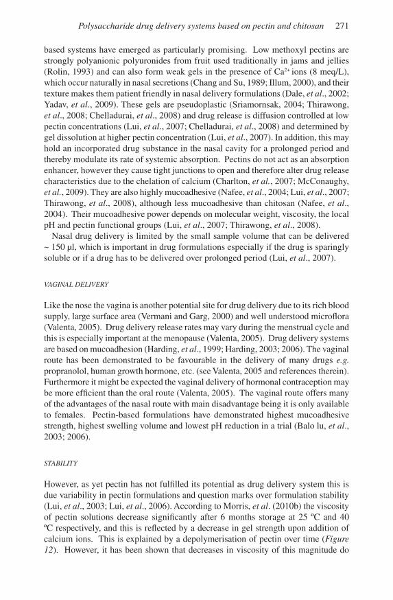
However, as yet pectin has not fulfilled its potential as drug delivery system this is due variability in pectin formulations and question marks over formulation stability (lui, et al., 2003; lui, et al., 2006). According to Morris, et al. (2010b) the viscosity of pectin solutions decrease significantly after 6 months storage at 25 ºC and 40 ºC respectively, and this is reflected by a decrease in gel strength upon addition of calcium ions. This is explained by a depolymerisation of pectin over time (Figure 12). However, it has been shown that decreases in viscosity of this magnitude do
272 G.A. Morris et al.
not significantly change the drug release rates from pectin gels in vitro (Nessa, 2003; Chelladurai, et al., 2008). In calcium pectate based tablet formulations drug release time is increased with lower degree of methyl esterification, but higher levels of calcium ions can lead to disintegration of the tablet and increased drug release (sungthongjeen, et al., 2004).
0 20 40 60 80 100 120 140 160 180
0.0
1.0x10⁻⁶
2.0x10⁻⁶
3.0x10⁻⁶
4.0x10⁻⁶
5.0x10⁻⁶
6.0x10⁻⁶
7.0x10⁻⁶
8.0x10⁻⁶
1/M
w,t -
1/M
w,t=
0 (mol
/g)
time (days)
Figure 12. 1st order kinetic plots of (mol/g) vs. time (days) for pectin of DM ~ 19 %, where closed symbols represent molar masses estimated from viscometry at 4 °C ( ), 25 °C ( ) and 40 °C ( ) (adapted from Figure 3 in Morris, et al. (2010b)). The kinetic rate constants (day-1) are (-0.8 ± 1.1) x 10-7, (5.7 ± 1.1) x 10-7 and (6.7 ± 0.2) x 10-6 at 4 ºC, 25 ºC and 40 ºC, respectively.
Conclusions
In the last decade there has been a great deal of interest in the use of polysaccharides and particularly chitosan and pectin in drug delivery systems. It is clear that both the polysaccharides either individually or together show great potential, however, many important issues still remain to be resolved fully. With chitosans, these include (i) their stability, with the important constraint that they are soluble only at pH < 6. (ii) their construction into microparticles capable of surviving the large environmental variation between mouth and intestine for oral drug delivery. In addressing (i) and (ii) issues concerning the optimal degree of acetylation and molecular weight of the chitosan need to be addressed. With low methoxy-pectin systems issues include: (i) optimal molecular weight and degree of esterification (ii) drug diffusivity (iii) interactions with mucosal tissues, (iv) stability (molecular weight/viscosity/gelation).
References
AMidi, M. And Hennink, W. e. (2010). Preface: Chitosan-based formulations of drugs, imaging agents and biotherapeutics. Advanced Drug Delivery Reviews, 62, 1–2.
Polysaccharide drug delivery systems based on pectin and chitosan 273
AMidi, M., MAstrobAttistA, e., Jiskoot, W. And Hennink, W. e. (2010). ChitosaN based delivery systems for protein therapeutics and antigens. Advanced Drug Delivery Reviews, 62, 59-82.
Anderson, M. t., HArdinG, s. e. And dAvis, s. s. (1989). on the interaction in solution of a candidate mucoadhesive polymer, diethylaminoethyl-dextran, with pig gastric mucus glycoprotein. Biochemical Society Transactions, 17, 1101–1102.
AnGer, H. And bertH, G. (1985). Gel-permeation chromatography of sunflower pectin. Carbohydrate Polymers, 5, 241-250.
AsHford, M., fell, J. J., AttWood, d., sHArMA, H. l. And WoodHeAd, P. J. (1993). An evaluation of pectin as a carrier for drug targeting to the colon. Journal of Controlled Release, 26, 213–220.
AsPden, t. J., illuM, l. And skAuGrud, Ø. (1996). Chitosan as a nasal delivery system: evaluation of insulin absorption enhancement and effect on nasal membrane integrity using rat models. European Journal of Pharmaceutical Sciences, 4, 23-31.
AsPden, t. J., MAson, J. d. t., Jones, n. s., loWe, J., skAuGrud, Ø. And illuM, i. (1997). Chitosan as a nasal delivery system: the effect of chitosan solutions on in vitro and in vivo mucociliary transport rates in human turbinates and volunteers. Journal of Pharmaceutical Sciences, 86, 509-513.
Axelos, M. A. v. lefebvre, J. And tHibAult, J-f. (1987). Conformation of a low methoxyl citrus pectin in aqueous solution, Food Hydrocolloids, 1, 569-570.
Axelos, M. A. v. And tHibAult, J-f. (1991). The chemistry of low-methoxyl pectin gelation. In: Walter R. H. (Ed.) The Chemistry and Technology of Pectin (pp 109-118). san Diego: Academic Press.
bAlolu, e., ÖzyAzici, M., HizArciolu, s. y. And kArAvAnA. H. A. (2003). An in vitro investigation for vaginal bioadhesive formulations: bioadhesive properties and swelling states of polymer mixtures. Il Farmaco, 58, 391-396.
bAlolu, e., ÖzyAzici, M., HizArciolu, s. y., senyiit, t., Özyurt, d. And Pekçetin, c. (2006). Bioadhesive controlled release systems of ornidazole for vaginal delivery. Pharmaceutical Development and Technology, 11, 477-484.
beneke, c. M., vilJoen, A. M. And HAMMAn, J. H. (2009). Polymeric plant-derived excipients in drug delivery. Molecules, 14, 2602-2620.
bertH, G., AnGer, H. And linoW, f. (1977). scattered-light photometric and viscosimetric studies of molecular mass determination of pectins in aqueous-solutions. Nahrung-Food, 31, 939-950.
bertH, G., dAutzenberG, H. And Peter, M. G. (1998). Physico-chemical characterization of chitosans varying in degree of acetylation. Carbohydrate Polymers, 36, 205-216.
bHAttArAi, n., Gunn, J. And zHAnG, M. (2010). Chitosan-based hydrogels for controlled, localized drug delivery. Advanced Drug Delivery Reviews, 62, 83-99.
brAccini, i., GrAsso, r. P. And Perez, s. (1999). Conformational and configurational features of acidic polysaccharides and their interactions with calcium ions: a molecular modeling investigation Carbohydrate Research, 317, 199-130.
bruGnerotto J., desbrières J., roberts G. And rinAudo M. (2001). Characterization of chitosan by steric exclusion chromatography. Polymer, 42, 9921–9927.
cHAMbin, o., duPuis, G, cHAMPion, d., voilley, A. And Pourcelot, y. (2006). Colon-specific drug delivery: Influence of solution reticulation properties upon pectin beads performance. International Journal of Pharmaceutics, 321, 86-93.
274 G.A. Morris et al.
cArino, G. P., JAcob, J. s. And MAtHioWitz, e. (2000). Nanosphere based oral insulin delivery. Journal of Controlled Release, 65, 261–269.
cHArlton, s. t., dAvis, s. s. And illuM, l. (2007). Evaluation of bioadhesive polymers as delivery systems for nose to brain delivery: In vitro characterisation studies. Journal of Controlled Release, 118, 225–234.
cHellAdurAi, s., MisHrA, M. And MisHrA, b. (2008). Design and Evaluation of Bioadhesive in-situ Nasal Gel of Ketorolac Tromethamine. Chemical and Pharmaceutical Bulletin, 56, 1596-1599.
cHen, H. And lAnGer, l. (1998). oral particulate delivery: status and future trends. Advanced Drug Delivery Reviews, 34, 339–350.
cHien, y. W., su, k. s. e. And cHAnG, s. f. (1989). Nasal Systemic Drug Delivery (pp 1-26). New york: Marcel Dekker Inc.
cros, s. c., GArnier, c., Axelos, M. A. v., iMbery, A. And Perez, s. (1996). solution conformations of pectin polysaccharides: determination of chain characteristics by small angle neutron scattering, viscometry and molecular modeling. Biopolymers, 39, 339-352.
cÖlfen H., bertH, G. And dAutzenberG, H. (2001). Hydrodynamic studies on chitosans in aqueous solution. Carbohydrate Polymers, 45, 373-383.
dAle, o., HJortkJAer, r. And kHArAscH, e. d. (2002). Nasal administration of opioids for pain management in adults. Acta Anaesthesiologica Scandinavica, 46, 759 –770.
dAvis, s. s. (1989). small intestine transit. In: Hardy, J. G., Davis, s. s. and Wilson, C. G. (Eds.) Drug Delivery to the Gastrointestinal Tract (pp. 49–61). Chichester: Ellis Horwood.
dAvis, s. s. And illuM, l. (2000). Chitosan for oral delivery of drugs. In: Muzzarelli, R. A. A. (Ed.). Chitosan per os: from Dietary Supplement to Drug Carrier (pp 137-164). Grottammare: Atec.
deAcon, M. P., dAvis, s. s., WHite, r. J., nordMAn, H., cArlstedt, i., n. errinGton, n., roWe, A. J. And HArdinG, s. e. (1999). Are chitosan–mucin interactions specific to different regions of the stomach? Velocity ultracentrifugation offers a clue. Carbohydrate Polymers, 38, 235–238.
deAcon, M. P., McGurk, s., roberts, c. J., WilliAMs, P. M., tendler, s. J. b., dAvies, M. c., dAvis, s. s. And HArdinG, s. e. (2000). Atomic force microscopy of gastric mucin and chitosan mucoadhesive systems, Biochemical Journal, 348, 557–563.
de lA fuente, M., rAviñA, M., PAolicelli, P., sAncHez, A., seiJo, b. And Alonso, M. J. (2010). Chitosan-based nanostructures: A delivery platform for ocular therapeutics. Advanced Drug Delivery Reviews, 62, 100-117.
dHAWAn, s., sinGlA, A. k. And sinHA, v. r. (2004). Evaluation of mucoadhesive properties of chitosan microspheres prepared by different methods. AAPS PharmSciTech, 5, 1-7.
dodou, d., breedveld, P. And WierinGA, P. A. (2005). Mucoadhesives in the gastrointestinal tract: revisiting the literature for novel applications. European Journal of Pharmaceutics and Biopharmaceutics, 60, 1–16.
dyer, A. M., HincHcliffe, M., WAtts, P. cAstile, J., JAbbAl-Gill, nAnkervis, i. r., sMitH, A. And illuM, l. (2002). Nasal delivery of insulin using novel chitosan based formulations: a comparative study in two animal models between simple chitosan formulations and chitosan nanoparticles. Pharmaceutical Research, 19, 998-1008.

Polysaccharide drug delivery systems based on pectin and chitosan 275
errinGton, n., HArdinG, s. e., våruM, k. M. And illuM, l. (1993). Hydrodynamic characterisation of chitosans varying in degree of acetylation. International Journal of Biological Macromolecules, 15, 113-117.
fee, M., errinGton, n., JuMel, k., illuM, l, sMitH, A. And HArdinG, s. e. (2003). Correlation of sEC/MAlls with ultracentrifuge and viscometric data for chitosans. European Biophysical Journal, 32, 457-464.
fernández-urrAsuno, r., cAlvo, P., ruMuñán-loPez, c., vilA-JAto, J. l. And Alonso, M. J. (1999). Enhancement of nasal absorption of insulin using chitosan nanoparticles. Pharmaceutical Research, 16, 1576-1581.
fisHMAn, M. l., cHAu, H. k., HoAGlAnd, P. d., And HotcHkiss, A. t. (2006). Microwave-assisted extraction of lime pectin. Food Hydrocolloids, 20, 1170-1177.
fisHMAn, M. l., cHAu, H. k., kolPAk, f. And brAdy, J. (2001). solvent effects on the molecular properties of pectins. Journal of Agricultural and Food Chemistry, 49, 4494-4501.
fiebriG, i., dAvis, s. s. And HArdinG, s. e. (1995a). Methods used to develop mucoadhesive drug delivery systems: bioadhesion in the gastrointestinal tract. In: Harding, s. E., Hill, s. E. and Mitchell, J. R. (Eds.) Biopolymer Mixtures (pp 373–419). Nottingham: Nottingham university Press.
fiebriG, i., HArdinG, s. e., & dAvis, s. s. (1994a). sedimentation analysis of the potential interactions between mucins and putative bioadhesive polymer. Progress in Colloid and Polymer Science, 93, 66–73.
fiebriG, i., HArdinG, s. e., roWe, A. J., HyMAn, s. c. And dAvis, s. s. (1995b). Transmission electron microscopy studies on pig gastric mucin and its interactions with chitosan. Carbohydrate Polymers, 28, 239–244.
fiebriG, i., HArdinG, s. e., stokke, b. t., våruM, k. M., JordAn, d. And dAvis, s. s. (1994b). The potential for chitosan as mucoadhesive drug carrier: studies on its interaction with pig gastric mucin on a molecular level. European Journal of Pharmaceutical Sciences, 2, 185.
fry, s. c. (1982). Phenolic compounds of the primary cell wall: feruloylated disaccharides of D-galactose and l-arabinose from spinach polysaccharide. Biochemical Journal, 203, 493-502.
GAn, Q, And WAnG, t. (2007). Chitosan nanoparticle as protein delivery carrier systematic examination of fabrication conditions for efficient loading and release. Colloids and Surfaces B: Biointerfaces, 59, 24–34.
GAn, Q, WAnG, t., cocHrAne, c. And MccArron, P. (2005). Modulation of surface charge, particle size and morphological properties of chitosan–TPP nanoparticles intended for gene delivery. Colloids and Surfaces B: Biointerfaces, 44, 65–73.
GArnier, c., Axelos M. A. v. And tHibAult, J-f. (1993). Phase-diagrams of pectin-calcium systems - influence of pH, ionic-strength, and temperature on the gelation of pectins with different degrees of methylation. Carbohydrate Research, 240, 219-232.
GentA, i., costAntini, M., Asti, A., conti, b. And MontAnAri, l. (1998). Influence of glutaraldehyde on drug release and mucoadhesive properties of chitosan microspheres. Carbohydrate Polymers, 36, 81-88.
HArdinG, s. e. (2003). Mucoadhesive interactions. Biochemical Society Transactions, 31, 1036-1041.
HArdinG, s. e. (2006). Trends in mucoadhesive analysis. Trends in Food Science and
276 G.A. Morris et al.
Technology, 17, 255-262.HArdinG, s. e., bertH, G., bAll, A., MitcHell, J. r. And GArcìA de lA torre, J. (1991).
The molecular weight distribution and conformation of citrus pectins in solution studied by hydrodynamics. Carbohydrate Polymers, 16, 1-15.
HArdinG, s. e., dAvis, s. s., deAcon, M. P. And fiebriG, i. (1999). Biopolymer mucoadhensives. In: Harding, s. E. (Ed.) Biotechnology and Genetic Engineering Reviews Vol. 16 (pp. 41-86). Andover: Intercept.
HArdinG, s. e., roWe, A. J. And creetH, J. M. (1983). Further evidence for a flexible and highly expanded spheroidal model for mucus glycoproteins in solution, Biochemical Journal, 209, 893-896.
HArisH PrAsHAntH, k. v. And tHArAnAtHAn r. n. (2007). Chitin/ chitosan: modifications and their unlimited potential – an overview. Trends in Food Science and Technology, 18, 117-131.
He, P., dAvis, s. s. And illuM, l. (1998). In-vitro evaluation of the properties of chitosan microspheres International Journal of Pharmaceutics, 166, 75–68.
He, P., dAvis, s.s. And illuM, l. (1999). Chitosan microspheres prepared by novel modified spray drying methods. Journal of Microencapsulation, 16, 343-355.
HolMe, H. k., dAvidsen, l., kristiAnsen, A. And sMidsrØd, o. (2008). Kinetics and mechanisms of the depolymerization of alginate and chitosan in aqueous solution. Carbohydrate Polymers, 73, 656-664.
illuM, l. (1998). Chitosan and its use as a pharmaceutical excipient. Pharmaceutical Research, 15, 1326-1331.
illuM, l. (2000). Improved delivery of drugs to mucosal services. European patent EP0975367B1.
illuM, l. (2002) Nasal drug delivery: new. developments and strategies. Drug Discovery Today, 7, 1184–1189.
isHii, t. (1997). o-acetylated oligosaccharides from pectins of potato tuber cell walls. Plant Physiology, 113, 1265-1272.
JAbbAl-Gill, i., fiscHer, A. n., rAPPuoli, r., dAvis, s. s. And illuM, l. (1998). stimulation of mucosal and systemic responses against Bordetella pertussis filamentous haemagglutinin and recombinant pertussis toxin after nasal administration with chitosan in mice. vaccine, 16, 2039 –2046.
JAin, d., PAndA, A. k. And MAJuMdAr, d. k. (2005). Eudragit s100 Entrapped Insulin Microspheres for oral Delivery. AAPS PharmSciTech, 6, E100-E107.
JAnes, k. A., cAlvo, P. And Alonso, M. J. (2001). Polysaccharide colloidal particles as delivery systems for macromolecules. Advanced Drug Delivery Reviews, 47, 83–97.
kAsAAi, M. r. (2006). Calculation of Mark–Houwink–sakurada (MHs) equation viscometric constants for chitosan in any solvent–temperature system using experimental reported viscometric constants data. Carbohydrate Polymers, 68, 477-488.
keAn, t. And tHAnou, M. (2010). Biodegradation, biodistribution and toxicity of chitosan. Advanced Drug Delivery Reviews, 62, 3-11.
kiM, s. J., sHin, s. r., lee, k. b., PArk, y. d. And kiM, s. i. (2004). synthesis and characteristics of polyelectrolyte complexes composed of chitosan and hyaluronic acid. Journal of Applied Polymer Science, 91, 2908–2913.
koMAlAvilAs, P. And Mort, A. J. (1989). The acetylation of o-3 of galacturonic acid in
Polysaccharide drug delivery systems based on pectin and chitosan 277
the rhamnose-rich portion of pectin. Carbohydrate Research, 189, 261-272.kondo, y., nAkAtAni, A., HAyAsHi, k. And ito, M. (2000). low molecular weight
chitosan prevents the progression of low dose streptozotocin induced slowly progressive diabetes mellitus in mice. Biological and Pharmacological Bulletin, 23, 1458-1464.
krAtky, o. And Porod, G. (1949). Röntgenungtersuchung gelöster fadenmoleküle. Recueil Des Travaux Chimiques Des Pays-Bas, 68, 1106-1109.
kubotA, n. And eGucHi, y. (1997). Facile preparation of water-soluble n-acetylated chitosan and molecular weight dependence of its water-solubility. Polymer Journal, 29, 123-127.
kuMAri, A., yAdAv, s. k. And yAdAv, s. c. (2010). Biodegradable polymeric nanoparticles based drug delivery systems. Colloids and Surfaces B: Biointerfaces, 75, 1-18.
kuJAWA, P., scHMAucH, G., viitAlA, t., bAdiA, A. And Winnik, f. M. (2007). Construction of viscoelastic biocompatible films via the layer-by-layer assembly of hyaluronan and phosphorylcholine-modified chitosan. Biomacromolecules, 8, 3169-3176.
lAMArQue, G., lucAs, J-M., viton, c. And doMArd, A. (2005). Physicochemical behavior of homogeneous series of acetylated chitosans in aqueous solution: role of various structural parameters. Biomacromolecules, 6, 131-142.
lAPAsin, r. And Pricl. s. (1995). Rheology of Industrial Polysaccharides, Theory and Applications. Blackie, london, uK.
leHoux, J-G. And dePuis, G. (2007). Recovery of chitosan from aqueous acidic solutions by salting-out: Part 1. use of inorganic salts. Carbohydrate Polymers, 68, 295-304.
leviGne, s., rAlet, M-c., QuéMéner, b. And tHibAult, J-f. (2004a). Isolation of diferulic bridges ester-linked to arabinan in sugar beet cell walls. Carbohydrate Research, 339, 2315-2319.
leviGne, s., rAlet, M-c., QuéMéner, b. c., Pollet, b. n-l., lAPierre, c. And tHibAult, J-F. (2004b). Isolation from sugar beet cell walls of arabinan oligosaccharides esterified by two ferulic acid monomers. Plant Physiology, 134, 1173-1180.
liM, s. t., MArtin, G. P., berry, d. J. And broWn, M. b. (2007). Preparation and evaluation of the in vitro drug release properties and mucoadhesion of novel microspheres of hyaluronic acid and chitosan. Journal of Controlled Release, 66, 281–292.
lin, y. H., cHen, c. H., liAnG, H. f., kulkArni, A. r., lee, P. W., cHen, c. H. And sunG, H. W. (2007). Novel nanoparticles for oral insulin delivery via the paracellular pathway. Nanotechnology, 18, 1-11.
loPes dA silvA, J. A. And GonçAlves, M. P. (1994). Rheological study into the ageing process of high methoxyl pectin/ sucrose gels. Carbohydrate Polymers, 24, 235-245.
luAnGtAnA-AnAn, M., oPAnAsoPit, P., nGAWHirunPAt, t., nuntHAnid, J., sriAMornsAk, P., liMMAtvAPirAt, s. And liM, l. y. (2005). Effect of chitosan salts and molecular weight on a nanoparticulate carrier for therapeutic protein. Pharmaceutical Development and Technology, 10, 189–196.
lui, l., fisHMAn, M. l. And Hicks, k. b. (2007). Pectin in controlled drug delivery – a review. Cellulose, 14, 15-24.
lui, l., fisHMAn, M. l., Hicks, k. b. And kende, M. (2005). Interaction of various pectin formulations with porcine colonic tissues. Biomaterials, 26, 5907–5916.
278 G.A. Morris et al.
lui, l., fisHMAn, M. l., kost, J. And Hicks, k. b. (2003). Pectin-based systems for colon-specific drug delivery via oral route. Biomaterials, 24, 3333-3343.
liu, x. f., GuAn, y. l., yAnG, d. z., li, z. And de yAo, k. (2001). Antibacterial action of chitosan and carboxymethylated chitosan. Journal of Applied Polymer Science, 79, 1324-1335.
MAcleod, G. s., collett, J. H. And fell, J. t. (1999). The potential use of mixed films of pectin, chitosan and HPMC for bimodal drug release. Journal of Controlled Release, 58, 303–310.
MAlovikovA, A., rinAudo, M. And MilAs, M. (1993). on the characterization of polygalacturonate salts in dilute-solution. Carbohydrate Polymers, 22, 87-92.
MAo, s., sun, W. And kissel, t. (2010). Chitosan-based formulations for delivery of DNA and siRNA. Advanced Drug Delivery Reviews, 62, 12-27.
MAzeAu k. And rinAudo M. (2004). The prediction of the characteristics of some polysaccharides from molecular modelling. Comparison with effective behaviour. Food Hydrocolloids, 18, 885–898.
McconAuGHy, s. d., kirklAnd, s. e., treAt, n. J. stroud, P. A. And MccorMick, c. l. (2009). Tailoring the network properties of ca2+ crosslinked aloe vera polysaccharide hydrogels for in situ release of therapeutic agents. Biomacromolecules, 9, 3277-3287.
MesiHA, M., PlAkoGiAnnis, f. And veJosotH, s. (1994). Enhanced oral absorption of insulin from desolvated fatty acid-sodium glycocholate emulsions. International Journal of Pharmaceutics, 111, 213–216.
MicArd, v., GrAbber, J. H., rAlPH, J., renArd, c. M. c. G. And tHibAult, J-f. (1997). Dehydrodiferulic acids from sugar beet pulp. Phytochemistry, 44, 1365-1368.
Miller, l. And MAcMilAn, J. d. (1970). Mode of action of pectic enzymes. 2. Further purification of exopolygalacturonate lyase and pectinesterases from Clostridium multifermerans. Journal of Bacteriology, 102, 72–78.
MorisHitA, i., MorisHitA, M., tAkAyAMA, k., MAcHidA, y. And nAGAi, t. (1993). Enteral insulin delivery by microspheres in 3 different formulations using Eudragit l100 and s100. International Journal of Pharmaceutics, 91, 29-37.
Morris, e. r. (1980). Physical probes of polysaccharide conformation and interactions. Food Chemistry, 6, 15-39.
Morris, e. r., PoWell, d. A., Gidley, M. J. And rees, d. A. (1982.) Conformation and interactions of pectins I. Polymorphism between gel and solid states of calcium polygalacturonate. Journal of Molecular Biology, 155, 507-516.
Morris, G. A., foster, t. J. And HArdinG, s. e. (2000). The effect of degree of esterification on the hydrodynamic properties of citrus pectin. Food Hydrocolloids, 14, 227-235.
Morris, G. A., foster, t. J. And HArdinG, s. e. (2002). A hydrodynamic study of the depolymerisation of a high methoxy pectin at elevated temperatures. Carbohydrate Polymers, 48, 361-367.
Morris, G. A., GArcíA de lA torre, J., orteGA, A., cAstile, J., sMitH, A., And HArdinG, s. e. (2008). Molecular flexibility of citrus pectins by combined sedimentation and viscosity analysis. Food Hydrocolloids, 22, 1435-1442.
Morris, G. A., cAstile, J., sMitH, A., AdAMs, G. G., And HArdinG, s. e. (2009a). Macromolecular conformation of chitosan in dilute solution: a new global hydrodynamic approach. Carbohydrate Polymers, 76, 616-621.
Polysaccharide drug delivery systems based on pectin and chitosan 279
Morris, G. A., cAstile, J., sMitH, A., AdAMs, G. G., And HArdinG, s. e. (2009b). The kinetics of chitosan depolymerisation at different temperatures. Polymer Degradation and Stability, 94, 1344-1348, 2009.
Morris, G. A., cAstile, J., sMitH, A., AdAMs, G. G., And HArdinG, s. e. (2010a). The effect of different storage temperatures on the stability of tripolyphosphate (TPP) – chitosan nanoparticles (submitted).
Morris, G. A., cAstile, J., sMitH, A., AdAMs, G. G. And HArdinG, s. e. (2010b). The effect of different storage temperatures on the physical properties of pectin solutions and gels (submitted).
MuzzArelli, r. A. A. (2009). Genipin-crosslinked chitosan hydrogels as biomedical and pharmaceutical aids. Carbohydrate Polymers, 77, 1-9.
nAfee, n. A., isMAil, f. A., borAie, n. A. And MortAdA, l. M. (2004). Mucoadhesive delivery systems. I. Evaluation of mucoadhesive polymers for buccal tablet formulation. Drug Development and Industrial Pharmacy, 30, 985-993.
nessA, M. u. (2003). Physiochemical characterisation of pectin solution as a vehicle for nasal drug delivery. Msc Dissertation, university of Nottingham, u.K.
nGuyen, t. t. b., Hein, s., nG, c-H. And stevens, W. f. (2007). Molecular stability of chitosan in acid solutions stored at various conditions. Journal of Applied Polymer Science, 107, 2588–2593.
no, H. k., kiM, s. H., lee, s. H., PArk, n. y. And PrinyAWiWAtkul, W. (2006). stability and antibacterial activity of chitosan solutions affected by storage temperature and time. Carbohydrate Polymers, 65, 174-178.
noto, r., MArtorAnA, v., bulone, d. And sAn biAGio, b. l. (2005). Role of charges and solvent on the conformational properties of poly(galacturonic acid) chains: a molecular dynamics study. Biomacromolecules, 6, 2555-2562.
oAkenfull, d. G. (1991). The Chemistry of High-Methoxyl Pectins. In: Walter RH (Ed.) The Chemistry and Technology of Pectin, pp 87-108. Academic Press, san Diego.
oAkenfull, d. G. And scott, A. (1998). Milk gels with low methoxy pectins. In: Phillips, G. o., Williams, P. A. and Wedlock, D. J. (Eds.) Gums and Stabilisers for the Food Industry 9 (pp 212-221). oxford: oxford university Press.
ofori-kWAkye, k. And fell, J. t. (2001). Biphasic drug release: the permeability of films containing pectin, chitosan and HPMC. International Journal of Pharmaceutics, 226, 139–145.
oGAWA, k. And yui, t. (1994). Effect of explosion on the crystalline polymorphism of chitin and chitosan. Bioscience Biotechnology and Biochemistry, 58. 968-969.
PArk, J. H., sArAvAnAkuMAr, G., kiM, k. And kWon, i. c. (2010). Targeted delivery of low molecular drugs using chitosan and its derivatives. Advanced Drug Delivery Reviews, 62, 28-41.
PArk, J. W., cHoi, k-H. And PArk, k. k. (1983). Acid-base equilibria and related properties of chitosan. Bulletin of the Korean Chemical Society, 4, 68-72.
PAvlov, G. M., roWe, A. J. And HArdinG, s. e. (1997). Conformation zoning of large molecules using the analytical ultracentrifuge. Trends in Analytical Chemistry, 16, 401-405.
PAvlov, G. M. HArdinG, s. e. And roWe, A. J. (1999). Normalized scaling relations as a natural classification of linear macromolecules according to size. Progress in Colloid and Polymer Science, 113, 76-80.
PePPAs, n. A., bures, P., leobAndunG, W. And icHikAWA, H. (2000). Hydrogels
280 G.A. Morris et al.
in pharmaceutical formulations. European Journal of Pharmaceutics and Biopharmaceutics, 50, 27-46.
Perez, s., rodríGuez-cArvAJAl, M. A. And doco, t. (2003). A complex plant cell wall polysaccharide: rhamnogalacturonan II. A structure in quest of a function. Biochimie, 85, 109-121.
Perrone, P., HeWAGe, H. c., tHoMson, H. r., bAiley, k., sAdler, i. H. And fry, s. c. (2002). Patterns of methyl and o-acetyl esterification in spinach pectins: a new complexity. Phytochemistry, 60, 67-77.
Pilnik, W. (1990). Pectin - a many splendored thing. In: Phillips, G. o., Williams, P. A. and Wedlock, D. J. (Eds.) Gums and Stabilisers for the Food Industry 5 (pp. 209-222). oxford: IRl Press.
Pilnik, W. And vorAGen A. G. J. (1970). Pectin substances and their uronides. In: Hulme, A. C. (Ed.) The Biochemistry of Fruits and their Products (pp 53-87). New york: Academic Press.
PourJAvAdi A. And bArzeGAr, s. (2009). smart pectin-based superabsorbent hydrogel as a matrix for ibuprofen as an oral non-steroidal anti-inflammatory drug delivery. Starch/ Stärke, 61, 173-187.
PoWell, d. A., Morris, e. r., Gidley, M. J. And rees, d. A. (1982). Conformation and interactions of pectins II. Influence of residue sequence on chain association in calcium pectate gels. Journal of Molecular Biology, 155, 317-331.
QAQisH, r. b. And AMiJi, M. M. (1999). synthesis of a fluorescent chitosan derivative and its application for the study of chitosan-mucin interactions. Carbohydrate Polymers, 38, 99-107.
rHAzi, M., desbriéres J., tolAiMAte, A., rinAudo, M., vottero, P. And AlAGui, A. (2002a). Contribution to the study of the complexation of copper by chitosan and oligomers. Polymer, 43, 1267–76.
rHAzi, M., desbriéres J., tolAiMAte, A., rinAudo, M., vottero, P., AlAGui, A. And el MerAy, M. (2002b). Influence of the nature of the metal ions on the complexation with chitosan.: Application to the treatment of liquid waste. European Polymer Journal, 38, 1523-1530.
ridley, b. l., o’neil, M. A. And MoHnen, d. (2001). Pectins: structure, biosynthesis and oligogalacturonide-related signalling. Phytochemistry, 57, 929-967.
rinAudo, M. (2006). Chitin and chitosan: properties and applications. Progress in Polymer Science, 31, 603-632.
rolin, r. (1993). Pectin. In: Whistler, R. l. and BeMiller, J. N. (Eds.) Industrial Gums (pp 257-293). New york: Academic Press.
roMbouts, f. M. And tHibAult, J-f. (1986). sugar beet pectins: chemical structure and gelation through oxidative coupling. In: Fishman, M. l. and Jen, J. J. (Eds.) The Chemistry and Function of Pectins (pp 49-60). Washington DC: American Chemical society.
rossi, s., ferrAri, f., bonferoni, M. c. And cArAMellA, c. (2000). Characterization of chitosan hydrochloride–mucin interaction by means of viscosimetric and turbidimetric measurements. European Journal of Pharmaceutical Sciences, 10, 251–257.
rossi, s., ferrAri, f., bonferoni, M. c. And cArAMellA, c. (2001). Characterization of chitosan hydrochloride–mucin rheological interaction: influence of polymer concentration and polymer: mucin weight ratio. European Journal of Pharmaceutical
Polysaccharide drug delivery systems based on pectin and chitosan 281
Sciences, 12, 479–485.sArMento, b., ribeiro, A., veiGA, f., ferreirA, d. And neufeld, r. (2007).
oral bioavailability of insulin contained in polysaccharide nanoparticles. Biomacromolecules, 8, 3054-3060
scHAtz, s. viton, c., delAir, t. PicHot, c. And doMArd, A. (2003). Typical physicochemical behaviors of chitosan in aqueous solution. Biomacromolecules, 4, 641-648.
seMdé, r., AMiGHi, k., devleescHouWer, M. J. And Moës, A. J. (2000). studies of pectin HM/Eudragit® Rl/Eudragit® NE film-coating formulations intended for colonic drug delivery. International Journal of Pharmaceutics, 197, 181–192.
sHu, x. z., And zHu, k. J. (2000). A novel approach to prepare tripolyphosphate: chitosan complex beads for controlled release drug delivery. International Journal of Pharmaceutics, 201, 51–58.
sinHA, v. r. And kuMriA, r. (2001). Polysaccharides in colon-specific drug delivery. International Journal of Pharmaceutics, 224, 19–38.
skAuGrud, Ø., HAGen, A., borGersen, b. And dornisH, M. (1999). Biomedical and pharmaceutical applications of alginate and chitosan. In: Harding, s. E. (Ed.) Biotechnology and Genetic Engineering Reviews vol. 16 (pp 23-40). Andover: Intercept.
soGiAs, i. A., WilliAMs, A. c. And kHutoryAnskiy, v. v. (2008). Why is chitosan mucoadhesive? Biomacromolecules, 9, 1837–1842.
sriAMornsAk, P. (2003). Chemistry of pectin and its pharmaceutical uses: a review. Silpakorn University International Journal, 3, 206–228.
sunGtHonGJeen, s., sriAMornsAk, P., PitAksuteePonG, t., soMsiri, A. And PuttiPiPAtkHAcHorn, s. (2004). Effect of degree of esterification of pectin and calcium amount on drug release from pectin-based matrix tablets. AAPS PharmSciTech, 5, 1-9.
tAkeucHi, H., tHonGborisute, J., MAtsui, y., suGiHArA, H., yAMAMoto, H. And kAWAsHiMA, y. (2005). Novel mucoadhesion tests for polymers and polymer-coated particles to design optimal mucoadhesive drug delivery systems. Advanced Drug Delivery Reviews, 57, 1583– 1594.
tAnford, c. (1961). Physical Chemistry of Macromolecules. New york: John Wiley & sons.
terboJevicH, M. cosAni, A., conio, G., MArsAno, e. And biAncHi, e. (1991). Chitosan: chain rigidity and mesophase formation. Carbohydrate Research, 209, 251-260.
terboJevicH, M. And MuzzArelli, r. A. A. (2000). Chitosan. In: Phillips, G. o. and Williams, P. A. (Eds.). Handbook of Hydrocolloids (pp 367-378). Cambridge: Woodhead Publishing ltd.
tHibAult, J-f. And rinAudo, M. (1986). Chain association of pectic molecules during calcium-induced gelation. Biopolymers, 25, 455-468.
tHirAWonG, n., kennedy, r. A. And sriAMornsAk, P. (2008). Viscometric study of pectin–mucin interaction and its mucoadhesive bond strength. Carbohydrate Polymers, 71, 170-179.
toMbs M. P., And HArdinG, s. e. (1998). An Introduction to Polysaccharide Biotechnology. london: Taylor and Francis.
tsAi, M. l., bAi, s. W., And cHen, r. H. (2008). Cavitation effects versus stretch effects resulted in different size and polydispersity of ionotropic gelation chitosan–sodium
282 G.A. Morris et al.
tripolyphosphate nanoparticle. Carbohydrate Polymers, 71, 448-457.vAlentA, c. (2005). The use of mucoadhesive polymers in vaginal delivery. Advanced
Drug Delivery Reviews, 57, 1692– 1712.vAn buren, J. P. (1991). Function of pectin in plant tissue structure and firmness. In:
Walter, R. H. (Ed). The Chemistry and Technology of Pectin (pp 1-22). san Diego: Academic Press.
vAndAMMe, t. f., lenourry, A., cHArrueAu, c. And cHAuMeil, J-c. (2002). The use of polysaccharides to target drugs to the colon. Carbohydrate Polymers, 48, 219-231.
våruM, k. M., AntHonsen, M. W., GrAsdAlen, H. And sMidsrØd, o. (1991a). Determination of the degree of N-acetylation and distribution of N-acetyl groups in partially N-deacteylated chitins (chitosans) by high-field NMR spectroscopy. Carbohydrate Research, 211, 17-23.
våruM, k. M., AntHonsen, M. W., GrAsdAlen, H. And sMidsrØd, o. (1991b). 13C NMR studies of the acetylation sequences in partially N-deacteylated chitins (chitosans). Carbohydrate Research, 217, 19-27.
våruM, k. M., ottØy, M. H. And sMidsrØd, o. (1994). Water-solubility of partially N-acetylated chitosans as a function of pH: effect of chemical composition and depolymerisation. Carbohydrate Polymers, 25, 65-70.
velásQuez, c. l., Albornoz, J. s. And bArrios, e. M. (2008). Viscosimetric studies of chitosan nitrate and chitosan chlorhydrate in acid free NaCl aqueous solution. E-Polymers, 014.
verMAni, k. And GArG, s. (2000). The scope and potential of vaginal drug delivery, Pharmaceutical Science & Technology Today, 3, 359– 364.
vold, i. M. n. (2004). Periodate oxidised Chitosans: Structure and Solution Properties. PhD Dissertation, Norwegian university of science and Technology, Trondheim, Norway.
vorAGen, A. G. J., Pilnik, W., tHibAult, J-f., Axelos, M. A. v. And renArd, c. M. G. c. (1995). Pectins. In: stephen, A. M. (Ed.) Food polysaccharides and their interactions (pp 287-340). New york: Marcel Dekker.
WAles, M. And vAn Holde, k. e. (1954). The concentration dependence of the sedimentation constants of flexible macromolecules. Journal of Polymer Science, 14, 81-86.
WAnG, z-y., WHite, J. W., konno, M., sAito, s. And nozAWA, t. (1994). A small-angle x-ray scattering study of alginate solution and its sol-gel transition by addition of divalent cations. Biopolymers, 35, 227-238.
WAtts, P. And sMitH, A. (2009). Pecsys: in situ gelling system for optimised nasal drug delivery. Expert opinion on Drug Delivery, 6, 543-552.
WillAts, W. G. t., MccArtney, l., MAckie, W. And knox J. P. (2001). Pectin: cell biology and prospects for functional analysis. Plant Molecular Biology, 47, 9-27.
xu, y. And du, y. (2005). Effect of molecular structure of chitosan on protein delivery properties of chitosan nanoparticles. International Journal of Pharmaceutics, 250, 215-226.
yAdAv, n., Morris, G. A., HArdinG, s. e., AnG, s. And AdAMs, G. G. (2009). Various non-injectable delivery systems for the treatment of diabetes mellitus. Endocrine, Metabolic & Immune Disorders - Drug Targets, 9, 1-13.
yAMAMoto, A., tAniGucHi, t., rikyuu, k., tsuJi, t., fuJitA, t., MurAkAMi, M. And
Polysaccharide drug delivery systems based on pectin and chitosan 283
MurAnisHi, s. (1994). Effects of various protease inhibitors on the intestinal absorption and degradation of insulin in rats. Pharmaceutical Research, 11, 1496–1500.
yen, M-t. And MAu, J-l. (2007). Physico-chemical characterization of fungal chitosan from shiitake stipes. LWT - Food Science and Technology, 40, 472-479.


![Bimodal Gastroretentive Drug Delivery Systems of ......a gastroretentive floating drug delivery system[12]. The drug concentrations can be controlled by formulating bimodal drug delivery](https://img.pdfslide.us/doc/110x75/5e6f0293269d113bd9170da6/bimodal-gastroretentive-drug-delivery-systems-of-a-gastroretentive-floating.jpg)