Embed Size (px)
Citation preview
Polypoidal choroidal vasculopathy in Caucasianpatients with presumed neovascular age-relatedmacular degeneration and poor ranibizumabresponseKatja Hatz,1,2 Christian Prünte1,2
1Vista Klinik, Binningen, Basel,Switzerland2Department ofOphthalmology, KantonsspitalLiestal, Liestal, Switzerland
Correspondence toDr Katja Hatz, Vista Klinik,Hauptstrasse 55,Binningen CH-4102,Switzerland;[email protected]
Received 22 March 2013Revised 25 September 2013Accepted 21 October 2013
To cite: Hatz K, Prünte C.Br J Ophthalmol PublishedOnline First: [please includeDay Month Year]doi:10.1136/bjophthalmol-2013-303444
ABSTRACTAims To determine the prevalence of polypoidalchoroidal vasculopathy (PCV) in patients with presumedneovascular age-related macular degeneration (AMD)who were considered poor responders to ranibizumab.Methods Caucasian patients with suspectedneovascular AMD, presumed to be choroidalneovascularisation, previously treated with ≥8 intravitrealinjections of ranibizumab 0.5 mg (Lucentis; Novartis AG,Basel, Switzerland) administered as required duringoptical coherence tomography-guided dosing wereretrospectively included. Eyes were categorised accordingto the time from injection 1 to injection 6 (group 1:<12 months; group 2: ≥12 months). Indocyanine greenangiography (ICGA) was used to re-evaluate eyes forPCV. Suitable candidates received reduced-fluencephotodynamic therapy/ranibizumab combination therapysupplemented by ranibizumab monotherapy, as required.Results 202 eyes were included (group 1: 73.8%;group 2: 26.2%). The prevalence of PCV in group 1(21.5%) was significantly higher than in group 2 (3.8%;p=0.003). After initiation of combination therapy, 16eyes with PCV received 3.1±2.5 ranibizumab injections/year vs 8.4±2.4 injections/year before initiation ofcombination therapy (p<0.001).Conclusions In Caucasian patients with presumedneovascular AMD, PCV prevalence is increased in eyesthat respond poorly to ranibizumab monotherapy. ICGAimproved PCV diagnosis in poor responders; combinationtherapy may be beneficial for eyes with PCV.
INTRODUCTIONAntivascular endothelial growth factor (VEGF)agents, including ranibizumab, aflibercept and bevaci-zumab, are currently the standard option for thetreatment of neovascular age-related macular degen-eration (AMD).1 The widespread use of ranibizumabintravitreal injection for the treatment of this condi-tion was based on results from the pivotal Phase IIIMinimally Classic/Occult Trial of the Anti-VEGFAntibody Ranibizumab in the Treatment ofNeovascular AMD (MARINA) and Anti-VEGFAntibody for the Treatment of Predominantly ClassicChoroidal Neovascularisation in AMD (ANCHOR)studies.2–4 Data from open-label studies using opticalcoherence tomography (OCT)-guided re-treatmentstrategies have shown that improvements in best-corrected visual acuity (BCVA) and OCT-assessedcentral retinal thickness can be achieved with fewerranibizumab injections than the fixed monthly dosingregimens used in studies such as MARINA and
ANCHOR. However, BCVA improvements withOCT-guided re-treatment indications are generallysmaller than those achieved with fixed monthlydosing.3–7 OCT images have shown that, in mostpatients, resolution of intraretinal and subretinal fluid(the presence of which corresponds with a decreasein BCVA) can be achieved 1–3 months after the startof monthly ranibizumab injections, often followed byan injection-free interval of several months beforefluid begins to reaccumulate.5 7 8 However, somepatients continue to require frequent injections.5 7 8
Why some patients have a poor response to ranibizu-mab is not well understood, but use of indocyaninegreen angiography (ICGA) has revealed the presenceof polypoidal choroidal vasculopathy (PCV) inseveral patients with neovascular AMD who did notrespond to repeated injections of anti-VEGF agents.910 Additionally, patients with retinal angiomatousproliferation (RAP) have been reported to requiremore anti-VEGF treatments than patients withnon-RAP lesions.5
Neovascular AMD may be caused by differenttypes of neovascularisation, the most common ofwhich is choroidal neovascularisation (CNV).11 PCV,characterised by a branching network of inner chor-oidal vessels with terminal aneurysmal dilations, ismost common in Asian individuals and other pig-mented races (reported in 22–55% of Asian patientswith neovascular AMD), but has also been observedin 4–14% of Caucasian patients.12 13 Despite somedebate as to whether PCV is a separate clinical entity,it is increasingly considered to be a subtype of occultCNV, particularly in Caucasian individuals.12 14–17
RAP, which begins as proliferation of new vesselswithin the retina and may merge with the choroidalcirculation as CNV in late-stage disease, is anotherrelatively recently recognised subtype of neovascularAMD that may represent up to 15% of newly diag-nosed cases.11 18 19
The primary purpose of this study was to use ICGAto determine the prevalence of PCV in Caucasianpatients with presumed neovascular AMD who wereconsidered to have poor response to ranibizumab, asindicated by the need for frequent intravitreal injec-tions during OCT-guided dosing. The prevalence ofRAP was also investigated, as was the frequency ofranibizumab injections during the 12 months beforeand after the start of combination therapy.
PATIENTS AND METHODSWe retrospectively studied the medical records ofpatients with presumed neovascular AMD, initially
Hatz K, et al. Br J Ophthalmol 2013;0:1–7. doi:10.1136/bjophthalmol-2013-303444 1
Clinical science BJO Online First, published on November 18, 2013 as 10.1136/bjophthalmol-2013-303444
Copyright Article author (or their employer) 2013. Produced by BMJ Publishing Group Ltd under licence.
on March 13, 2020 by guest. P
rotected by copyright.http://bjo.bm
j.com/
Br J O
phthalmol: first published as 10.1136/bjophthalm
ol-2013-303444 on 18 Novem
ber 2013. Dow
nloaded from
visualised by fluorescein angiography and presumed to be occultor classic CNV, diagnosed between May 2006 and September2010. ICGA had not been performed at baseline because it isnot standard practice in Europe. Patients were treated withintravitreal injections of ranibizumab 0.5 mg (Lucentis; NovartisAG, Basel, Switzerland) for ≥1 year in a routine clinical settingat Vista Klinik, Binningen, Switzerland. Patients initially receivedthree consecutive monthly ranibizumab injections and were thenre-treated pro re nata if there was OCT evidence of CNV andintraretinal or subretinal fluid ≥1 month after the last injectionor a new macular haemorrhage. To be eligible for this analysis,patients must have received ≥8 ranibizumab injections and wererequired to be re-evaluated with ICGA. All patients whoreceived ≥8 ranibizumab injections underwent ICGA, exceptthose with a known indocyanine green allergy.
In previous clinical studies with pro re nata ranibizumab treat-ment, on average 5–6 ranibizumab injections/year were required toachieve significant visual acuity improvements and OCT centralretinal thickness reductions compared with baseline.5 7 8 Therefore,we chose to categorise eyes according to the time from injection 1to injection 6 (group 1: <12 months; group 2: ≥12 months).Group 1 eyes were considered to be relatively poor responders toranibizumab, with injection frequency also reflecting CNV activityon OCT. Reinjection criteria were based on signs of CNVactivity onOCTor new intraretinal or subretinal haemorrhages.
Ophthalmological examinations performed in this studyincluded BCVA assessment, dilated fundus examination with a 90D lens, colour fundus photography, spectral-domain OCT(Spectralis OCT; Heidelberg Engineering, Heidelberg, Germany),as well as fluorescein angiography and ICGA by scanning laserophthalmoscopy (Heidelberg Retina Angiography-2; HeidelbergEngineering). An ICG movie was used in each lesion for ≥30 safter filling to show pulsating PCV. OCT was used to evaluatechanges in exudation, including intraretinal or subretinal fluid,serous or haemorrhagic retinal pigment epithelial detachmentsand intraretinal or subretinal haemorrhage. ICGA was not per-formed at initial diagnosis of AMD because it is currently not astandard primary diagnostic examination in Europe, where thepopulation is largely Caucasian and data previously have indicateda low prevalence of PCV in this group. Conversely, PCV is knownto be common in Asian populations; therefore, ICGA is generallyincluded in initial clinical examination in Asia.20
PCV was defined as evidence of at least one focal polypoidallesion in the inner choroid during early-phase ICGA. Only clearlyidentified PCV lesions were counted as such; differentiation waspossible from the pulsation of polyps that could be observed usingan ICGA movie. Furthermore, diagnosis of RAP lesions and identi-fication of CNV feeder vessels were facilitated by ICGA. Areas of
geographic atrophy observed on fluorescein angiography were alsorecorded during ophthalmological examination.
Once PCV was diagnosed, patients were offered reduced-fluence photodynamic therapy (PDT) with verteporfin (Visudyne;Novartis AG, Basel, Switzerland), followed 1 h later by intravitrealranibizumab 0.5 mg injection, if they had no lesions near the foveain combination with well-preserved BCVA (16/20–20/20) and noevidence of high-pigment epithelial detachment. Patients withlesions located very close to the optic nerve without any margin tothe papilla were not offered combination treatment, nor werepatients with good response to monotherapy—defined as>2 months with complete resolution of intraretinal or subretinalfluid following the last ranibizumab injection. Generally, patientsconsidered eligible for PDT also had ≥6 ranibizumab injections/year after initial diagnosis and before ICGA and diagnosis of PCV.PDTwas administered by a Zeiss VISULAS 690s laser (Carl ZeissMeditec AG; Jena, Germany) with a reduced fluence of 25 J/cm2
for 83 s (300 mW/cm2) 15 min after initiation of verteporfin infu-sion according to standard protocol.21 However, laser spot sizewas determined by the greatest linear dimension of the lesion,which was measured with ICGA, as described by Chan et al22 andin the efficacy and safety of verteporfin photodynamic therapy incombination with ranibizumab or alone versus ranibizumab mono-therapy in patients with symptomatic macular polypoidal chor-oidal vasculopathy (EVEREST) study by Koh et al.23 The laserspot size was chosen to cover the polyps and the surroundingabnormally dilated choroidal vessels seen on ICGA by adding500–1000 mm to the greatest linear diameter, if required. Lesionstoo close to the optic nerve were excluded. When recurrent orresidual PCV lesions were associated with exudative fluid duringfollow-up, patients could be re-treated with combined PDT andranibizumab every 3 months, whereas intravitreal ranibizumabinjections could be administered on a monthly basis.
In this paper, prevalence data are presented as percentagesand all other variables as mean±SD. Statistical analyses wereperformed with SPSS V.17.0 for Windows (SPSS; Chicago,Illinois, USA). We used a two-sided t test to compare differencesbetween groups. A p value of <0.05 was considered to be statis-tically significant.
RESULTSOf 753 eyes (675 patients) diagnosed with neovascular AMDand treated with ranibizumab for ≥1 year, 213 eyes received ≥8injections. Inability to perform ICGA because of known allergiesto iodine or severely reduced general condition in 11 patientsmeant that 11 eyes were excluded from the analysis. The totalpopulation for analysis comprised 202 eyes from 180 Caucasianpatients (table 1).
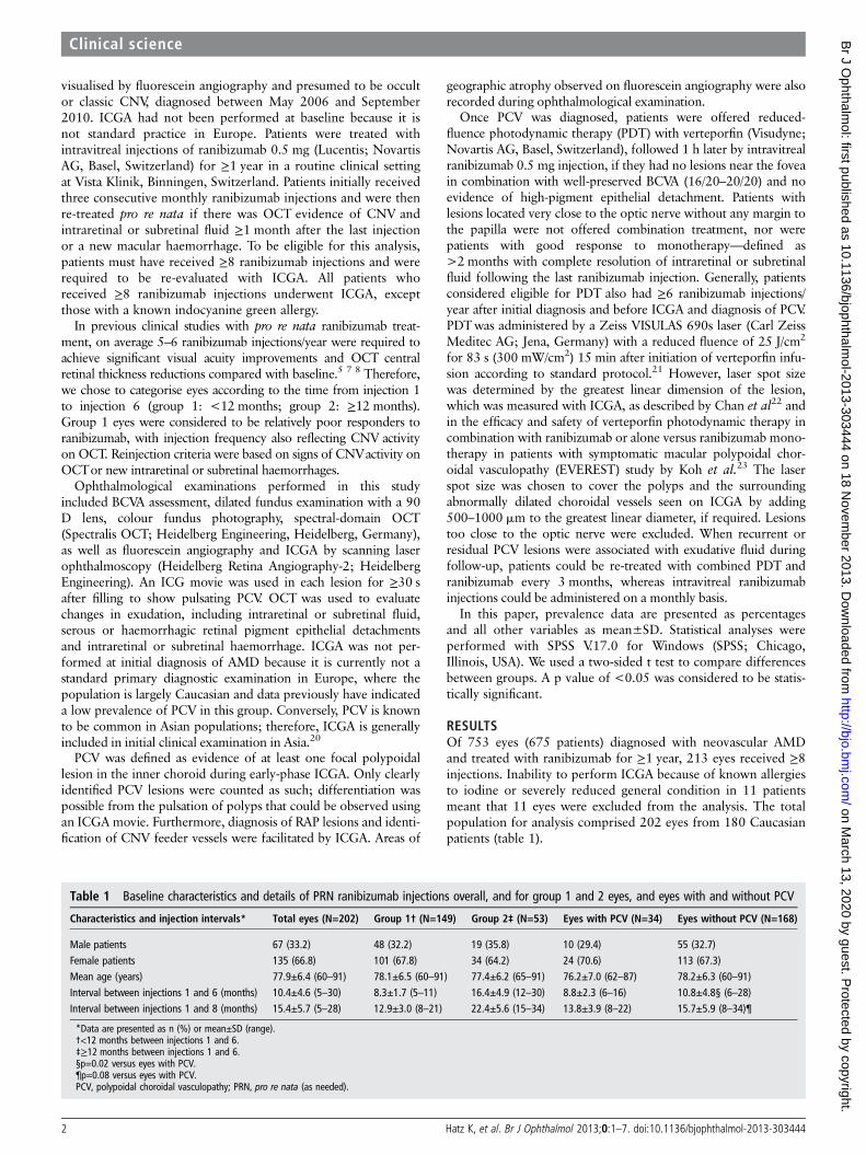
Table 1 Baseline characteristics and details of PRN ranibizumab injections overall, and for group 1 and 2 eyes, and eyes with and without PCV
Characteristics and injection intervals* Total eyes (N=202) Group 1† (N=149) Group 2‡ (N=53) Eyes with PCV (N=34) Eyes without PCV (N=168)
Male patients 67 (33.2) 48 (32.2) 19 (35.8) 10 (29.4) 55 (32.7)Female patients 135 (66.8) 101 (67.8) 34 (64.2) 24 (70.6) 113 (67.3)Mean age (years) 77.9±6.4 (60–91) 78.1±6.5 (60–91) 77.4±6.2 (65–91) 76.2±7.0 (62–87) 78.2±6.3 (60–91)Interval between injections 1 and 6 (months) 10.4±4.6 (5–30) 8.3±1.7 (5–11) 16.4±4.9 (12–30) 8.8±2.3 (6–16) 10.8±4.8§ (6–28)Interval between injections 1 and 8 (months) 15.4±5.7 (5–28) 12.9±3.0 (8–21) 22.4±5.6 (15–34) 13.8±3.9 (8–22) 15.7±5.9 (8–34)¶
*Data are presented as n (%) or mean±SD (range).†<12 months between injections 1 and 6.‡≥12 months between injections 1 and 6.§p=0.02 versus eyes with PCV.¶p=0.08 versus eyes with PCV.PCV, polypoidal choroidal vasculopathy; PRN, pro re nata (as needed).
2 Hatz K, et al. Br J Ophthalmol 2013;0:1–7. doi:10.1136/bjophthalmol-2013-303444
Clinical science
on March 13, 2020 by guest. P
rotected by copyright.http://bjo.bm
j.com/
Br J O
phthalmol: first published as 10.1136/bjophthalm
ol-2013-303444 on 18 Novem
ber 2013. Dow
nloaded from
The total population received a mean of 12.8±3.9 injectionsover 27.8±10.8 months, with ICGA performed at 15.1±10.2 months (group 1:13.1±9.1 months; group 2:20.7±1.2 months). The interval between injections 1 and 6 was<12 months in 149 eyes (73.8%; group 1) and ≥12 months in53 eyes (26.2%; group 2).
Prevalence of PCV, RAP and CNV as evaluated by ICGAOverall, 16.8% of eyes had evidence of PCVon ICGA (figure 1).The prevalence of PCV in group 1 (21.5%) was significantlyhigher than in group 2 (3.8%; p=0.003) (table 2). ICGA alsoshowed RAP lesions in 11.4% of eyes (group 1: 10.7%; group 2:13.2%) and AMD-type CNV in 71.8% of eyes (group 1: 67.8%;group 2: 83.0%). There were no significant differences betweenthe two groups in the prevalence of RAP or CNV, including CNVfeeder vessels and large areas of geographic atrophy in associ-ation with small occult CNV (table 2).
Eyes with PCV before and after PDTBaseline characteristics, such as age at diagnosis and sex, werenot significantly different between eyes with or without PCV(table 1). The mean interval between injections 1 and 6 was 8.8±2.3 months for eyes with PCV, which was significantly shorterthan for eyes without PCV (10.8±4.8 months; p=0.02).Between injections 1 and 8, the mean interval was 13.8±3.9 months for eyes with PCV and 15.7±5.9 months for eyes
without PCV, but the between-group difference did not quitereach statistical significance (p=0.08).
Before eyes were evaluated with ICGA, patients with PCVhad 6.7±3.1 ranibizumab injections over 12.1±7.2 months.After diagnosis of PCV, 16 of 34 (47.1%) eyes were treated withreduced-fluence PDT plus ranibizumab combination therapy(figures 2 and 3). Combination therapy was not performed
Figure 1 Examples of evidence of polypoidal choroidal vasculopathy observed on indocyanine green angiography not diagnosed with fluoresceinangiography. (A) Typical peripapillar location; (B) subfoveal location with extended subfoveal haemorrhage.
Table 2 Prevalence of PCV, RAP and CNV in eyes with presumedneovascular AMD requiring ≥8 intravitreal ranibizumab injectionsduring PRN treatment
Prevalence, n (%)Overall(n=202)
Group 1*(n=149)
Group 2†(n=53)
PCV 34 (16.8) 32 (21.5) 2 (3.8)‡RAP 23 (11.4) 16 (10.7) 7 (13.2)AMD-type CNV 145 (71.8) 101 (67.8) 44 (83.0)CNV feeder vessels 60 (29.7) 46 (30.9) 14 (26.4)Geographic atrophy withsmall occult CNV
9 (4.5) 6 (4.0) 3 (5.7)
*<12 months between injections 1 and 6.†≥12 months between injections 1 and 6.‡p=0.003 versus group 1.AMD, age-related macular degeneration; CNV, choroidal neovascularisation; PCV,polypoidal choroidal vasculopathy; PRN, pro re nata (as needed); RAP, retinalangiomatous proliferation.
Hatz K, et al. Br J Ophthalmol 2013;0:1–7. doi:10.1136/bjophthalmol-2013-303444 3
Clinical science
on March 13, 2020 by guest. P
rotected by copyright.http://bjo.bm
j.com/
Br J O
phthalmol: first published as 10.1136/bjophthalm
ol-2013-303444 on 18 Novem
ber 2013. Dow
nloaded from
in 18 (52.9%) eyes with PCV because of good response tomonotherapy (9 eyes), lesion location near the fovea and goodBCVA (3 eyes), pigment epithelial rupture (2 eyes), largepigment epithelial detachment (2 eyes), and refusal to receivecombination therapy (1 eye). Combination therapy was plannedin one additional eye, but no follow-up was available. No
differences in characteristics were observed between PCVlesions that required monotherapy versus those that requiredcombination therapy.
The 16 eyes with PCV that were treated with combinationtherapy received 8.4±2.4 ranibizumab injections/year before thestart of combination therapy compared with 3.1±2.5 injections/
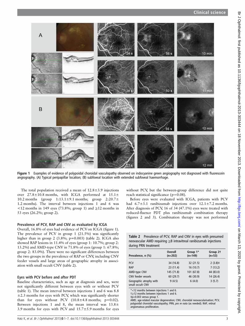
Figure 2 Images from a patient with no response to antivascular endothelial growth factor monotherapy who was treated with combinedreduced-fluence photodynamic therapy and intravitreal ranibizumab until month 15 to maintain stable visual acuity (20/40 to 20/32).
4 Hatz K, et al. Br J Ophthalmol 2013;0:1–7. doi:10.1136/bjophthalmol-2013-303444
Clinical science
on March 13, 2020 by guest. P
rotected by copyright.http://bjo.bm
j.com/
Br J O
phthalmol: first published as 10.1136/bjophthalm
ol-2013-303444 on 18 Novem
ber 2013. Dow
nloaded from
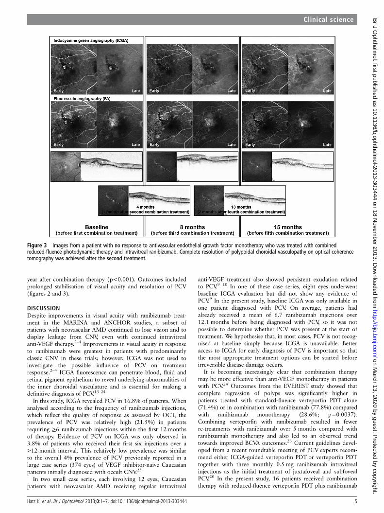
year after combination therapy (p<0.001). Outcomes includedprolonged stabilisation of visual acuity and resolution of PCV(figures 2 and 3).
DISCUSSIONDespite improvements in visual acuity with ranibizumab treat-ment in the MARINA and ANCHOR studies, a subset ofpatients with neovascular AMD continued to lose vision and todisplay leakage from CNV, even with continued intravitrealanti-VEGF therapy.2–4 Improvements in visual acuity in responseto ranibizumab were greatest in patients with predominantlyclassic CNV in these trials; however, ICGA was not used toinvestigate the possible influence of PCV on treatmentresponse.2–4 ICGA fluorescence can penetrate blood, fluid andretinal pigment epithelium to reveal underlying abnormalities ofthe inner choroidal vasculature and is essential for making adefinitive diagnosis of PCV.13 24
In this study, ICGA revealed PCV in 16.8% of patients. Whenanalysed according to the frequency of ranibizumab injections,which reflect the quality of response as assessed by OCT, theprevalence of PCV was relatively high (21.5%) in patientsrequiring ≥6 ranibizumab injections within the first 12 monthsof therapy. Evidence of PCV on ICGA was only observed in3.8% of patients who received their first six injections over a≥12-month interval. This relatively low prevalence was similarto the overall 4% prevalence of PCV previously reported in alarge case series (374 eyes) of VEGF inhibitor-naive Caucasianpatients initially diagnosed with occult CNV.25
In two small case series, each involving 12 eyes, Caucasianpatients with neovascular AMD receiving regular intravitreal
anti-VEGF treatment also showed persistent exudation relatedto PCV.9 10 In one of these case series, eight eyes underwentbaseline ICGA evaluation but did not show any evidence ofPCV.9 In the present study, baseline ICGA was only available inone patient diagnosed with PCV. On average, patients hadalready received a mean of 6.7 ranibizumab injections over12.1 months before being diagnosed with PCV, so it was notpossible to determine whether PCV was present at the start oftreatment. We hypothesise that, in most cases, PCV is not recog-nised at baseline simply because ICGA is unavailable. Betteraccess to ICGA for early diagnosis of PCV is important so thatthe most appropriate treatment options can be started beforeirreversible disease damage occurs.
It is becoming increasingly clear that combination therapymay be more effective than anti-VEGF monotherapy in patientswith PCV.24 Outcomes from the EVEREST study showed thatcomplete regression of polyps was significantly higher inpatients treated with standard-fluence verteporfin PDT alone(71.4%) or in combination with ranibizumab (77.8%) comparedwith ranibizumab monotherapy (28.6%; p=0.0037).Combining verteporfin with ranibizumab resulted in fewerre-treatments with ranibizumab over 5 months compared withranibizumab monotherapy and also led to an observed trendtowards improved BCVA outcomes.23 Current guidelines devel-oped from a recent roundtable meeting of PCV experts recom-mend either ICGA-guided verteporfin PDT or verteporfin PDTtogether with three monthly 0.5 mg ranibizumab intravitrealinjections as the initial treatment of juxtafoveal and subfovealPCV.20 In the present study, 16 patients received combinationtherapy with reduced-fluence verteporfin PDT plus ranibizumab
Figure 3 Images from a patient with no response to antivascular endothelial growth factor monotherapy who was treated with combinedreduced-fluence photodynamic therapy and intravitreal ranibizumab. Complete resolution of polypoidal choroidal vasculopathy on optical coherencetomography was achieved after the second treatment.
Hatz K, et al. Br J Ophthalmol 2013;0:1–7. doi:10.1136/bjophthalmol-2013-303444 5
Clinical science
on March 13, 2020 by guest. P
rotected by copyright.http://bjo.bm
j.com/
Br J O
phthalmol: first published as 10.1136/bjophthalm
ol-2013-303444 on 18 Novem
ber 2013. Dow
nloaded from
after they were diagnosed with PCV. Compared with the12-month period before the start of combination therapy, theneed for ranibizumab injections during the 12-month periodafter combination therapy (8.4 vs 3.1 injections) was signifi-cantly reduced. We did not systematically evaluate the impact ofcombination therapy on lesion characteristics. However, in acase series of 12 eyes with PCV lesions treated with multipleinjections of anti-VEGF agents, Cho et al9 reported that eighteyes went on to be treated with combination verteporfin PDTand an anti-VEGF agent, with complete resolution of the PCVlesion and associated macular exudation over 1.5–3 months in75% of cases. Despite stabilisation of disease in all patients, finalvisual acuity generally did not improve.9 The authors stated thatthis finding probably was a result of permanent photoreceptordamage from chronic oedema and recurrent haemorrhage.
In the present study, dynamic fluorescein angiography andICGA movies during the filling phase and images taken duringthe mid and late phases allowed RAP lesions to be clearly differ-entiated. They were identified in 11.4% of eyes. However,unlike PCV, which was most prevalent in the group of patientsrequiring more frequent ranibizumab injections during the firstyear of therapy, RAP was evenly distributed between the twogroups (group 1: 10.7% of eyes; group 2: 13.2% of eyes). MostRAP cases were found to be stage I/II in this study, which maybe the result of the patient selection criteria, that is, early detec-tion in an urban population combined with exclusion of patientswith advanced disease before they had received eight injections.The mean number of required ranibizumab injections has previ-ously been shown to increase significantly from 4.1/year in stageIIA RAP to 6.3/year in stage III RAP.26 These results support theobservation that, relative to classic and occult subtypes of CNV,stage I–II RAP is not associated with reduced responsiveness toranibizumab.7 27 Better response at earlier stages may providean explanation for the even distribution of RAP lesions in thisstudy. The selection criteria used may also explain the lowprevalence of RAP observed in this study compared with otherstudies.28–30 The RAP prevalence among occult/minimallyclassic lesions was mostly determined at the same time as preva-lence among all lesion types. A slightly lower prevalence of RAPlesions was previously reported in a large German population ofpatients with AMD, although this may be due to the investiga-tors using only fluorescein angiography for diagnosis.31
The extent of vascular maturity can affect therapeuticresponse, with immature vessels having a better response toVEGF inhibitors than mature vessels, so CNV may be relativelymature and resistant to treatment in patients with chronic AMDor a history of previous anti-VEGF therapy.9 10 In this study,ICGA revealed that 29.7% of patients had CNV feeder vesselsas signs of mature CNV (group 1: 30.9%; group 2: 26.4%).Large areas of geographic atrophy were observed in a relativelysmall proportion of patients with occult CNV in both patientgroups (4.0–5.7%).
In conclusion, the relatively high prevalence of PCV inCaucasian patients with presumed neovascular AMD requiringmultiple intravitreal injections of ranibizumab during the first12 months of treatment may help to explain some of the widevariations in the need for re-treatment among patients with pre-sumed neovascular AMD receiving OCT-guided therapy withanti-VEGF agents. Because PCV may prove to be difficult totreat with anti-VEGF therapy alone, combination therapyinvolving PDT may be needed. Therefore, differentiationbetween CNV and PCV early in the treatment sequence isimportant so that an alternative treatment strategy can beinitiated before irreversible disease progression occurs.
Acknowledgements The authors would like to acknowledge ChameleonCommunications International, which provided medical writing services and editorialsupport with funding from Heidelberg Engineering.
Funding Funding for medical writing support was provided by HeidelbergEngineering (Tiergartenstrasse 15, 69121 Heidelberg, Germany). The sponsor had norole in the design or conduct of this research.
Competing interests CP is a consultant to Novartis Pharma AG, Bayer andAllergan. KH has no competing interests.
Ethics approval Any necessary ethics committee approval was secured for thereported study (Ethikkommission beider Basel, ref. no. 362/12).
Provenance and peer review Not commissioned; externally peer reviewed.
Open Access This is an Open Access article distributed in accordance with theCreative Commons Attribution Non Commercial (CC BY-NC 3.0) license, whichpermits others to distribute, remix, adapt, build upon this work non-commercially,and license their derivative works on different terms, provided the original work isproperly cited and the use is non-commercial. See: http://creativecommons.org/licenses/by-nc/3.0/
REFERENCES1 Nguyen DH, Luo J, Zhang K, et al. Current therapeutic approaches in neovascular
age-related macular degeneration. Discov Med 2013;15:343–8.2 Brown DM, Michels M, Kaiser PK, et al. Ranibizumab versus verteporfin
photodynamic therapy for neovascular age-related macular degeneration: two-yearresults of the ANCHOR study. Ophthalmology 2009;116:57–65.
3 Brown DM, Kaiser PK, Michels M, et al. Ranibizumab versus verteporfin forneovascular age-related macular degeneration. N Engl J Med 2006;355:1432–44.
4 Rosenfeld PJ, Brown DM, Heier JS, et al. Ranibizumab for neovascular age-relatedmacular degeneration. N Engl J Med 2006;355:1419–31.
5 Fung AE, Lalwani GA, Rosenfeld PJ, et al. An optical coherencetomography-guided, variable dosing regimen with intravitreal ranibizumab (Lucentis)for neovascular age-related macular degeneration. Am J Ophthalmol2007;143:566–83.
6 Holz FG, Amoaku W, Donate J, et al. Safety and efficacy of a flexible dosingregimen of ranibizumab in neovascular age-related macular degeneration: theSUSTAIN study. Ophthalmology 2011;118:663–71.
7 Lalwani GA, Rosenfeld PJ, Fung AE, et al. A variable-dosing regimen withintravitreal ranibizumab for neovascular age-related macular degeneration: year 2 ofthe PrONTO Study. Am J Ophthalmol 2009;148:43–58.
8 Rothenbuehler SP, Waeber D, Brinkmann CK, et al. Effects of ranibizumab inpatients with subfoveal choroidal neovascularization attributable to age-relatedmacular degeneration. Am J Ophthalmol 2009;147:831–7.
9 Cho M, Barbazetto IA, Freund KB. Refractory neovascular age-related maculardegeneration secondary to polypoidal choroidal vasculopathy. Am J Ophthalmol2009;148:70–8.
10 Stangos AN, Gandhi JS, Nair-Sahni J, et al. Polypoidal choroidal vasculopathymasquerading as neovascular age-related macular degeneration refractory toranibizumab. Am J Ophthalmol 2010;150:666–73.
11 Yannuzzi LA, Negrao S, Iida T, et al. Retinal angiomatous proliferation inage-related macular degeneration. Retina 2001;21:416–34.
12 Ciardella AP, Donsoff IM, Huang SJ, et al. Polypoidal choroidal vasculopathy. SurvOphthalmol 2004;49:25–37.
13 Laude A, Cackett PD, Vithana EN, et al. Polypoidal choroidal vasculopathy andneovascular age-related macular degeneration: same or different disease? ProgRetin Eye Res 2010;29:19–29.
14 Costa RA, Navajas EV, Farah ME, et al. Polypoidal choroidal vasculopathy:angiographic characterization of the network vascular elements and a newtreatment paradigm. Prog Retin Eye Res 2005;24:560–86.
15 Imamura Y, Engelbert M, Iida T, et al. Polypoidal choroidal vasculopathy: a review.Surv Ophthalmol 2010;55:501–15.
16 Khan S, Engelbert M, Imamura Y, et al. Polypoidal choroidal vasculopathy:simultaneous indocyanine green angiography and eye-tracked spectral domainoptical coherence tomography findings. Retina 2012;32:1057–68.
17 Lima LH, Schubert C, Ferrara DC, et al. Three major loci involved in age-relatedmacular degeneration are also associated with polypoidal choroidal vasculopathy.Ophthalmology 2010;117:1567–70.
18 Freund KB, Ho IV, Barbazetto IA, et al. Type 3 neovascularization: the expandedspectrum of retinal angiomatous proliferation. Retina 2008;28:201–11.
19 Yannuzzi LA, Freund KB, Takahashi BS. Review of retinal angiomatous proliferationor type 3 neovascularization. Retina 2008;28:375–84.
20 Koh AH, Chen LJ, Chen SJ, et al. Polypoidal choroidal vasculopathy: evidence-basedguidelines for clinical diagnosis and treatment. Retina 2013;33:686–716.
21 Visudyne: EPAR – Product Information – European Medicines Agency. Annex1.Summary of product characteristics. http://www.ema.europa.eu/ema/ (last updated27 Nov 2012).
6 Hatz K, et al. Br J Ophthalmol 2013;0:1–7. doi:10.1136/bjophthalmol-2013-303444
Clinical science
on March 13, 2020 by guest. P
rotected by copyright.http://bjo.bm
j.com/
Br J O
phthalmol: first published as 10.1136/bjophthalm
ol-2013-303444 on 18 Novem
ber 2013. Dow
nloaded from
22 Chan WM, Lam DS, Lai TY, et al. Photodynamic therapy with verteporfin forsymptomatic polypoidal choroidal vasculopathy: one-year results of a prospectivecase series. Ophthalmology 2004;111:1576–84.
23 Koh A, Lee WK, Chen LJ, et al. EVEREST study: efficacy and safety of verteporfinphotodynamic therapy in combination with ranibizumab or alone versusranibizumab monotherapy in patients with symptomatic macular polypoidalchoroidal vasculopathy. Retina 2012;32:1453–64.
24 de Crecchio G, Chan RV, Manzi G, et al. Polypoidal choroidal vasculopathy: recentadvances in therapy. Curr Drug Targets 2011;12:206–11.
25 Lafaut BA, Leys AM, Snyers B, et al. Polypoidal choroidal vasculopathy inCaucasians. Graefes Arch Clin Exp Ophthalmol 2000;238:752–9.
26 Reche-Frutos J, Calvo-Gonzalez C, Perez-Trigo S, et al. Ranibizumab in retinalangiomatous proliferation (RAP): influence of RAP stage on visual outcome. Eur JOphthalmol 2011;21:783–8.
27 Konstantinidis L, Mameletzi E, Mantel I, et al. Intravitreal ranibizumab (Lucentis) inthe treatment of retinal angiomatous proliferation (RAP). Graefes Arch Clin ExpOphthalmol 2009;247:1165–71.
28 Parravano M, Pilotto E, Musicco I, et al. Reproducibility of fluorescein andindocyanine green angiographic assessment for RAP diagnosis: a multicenter study.Eur J Ophthalmol 2012;22:598–606.
29 Massacesi AL, Sacchi L, Bergamini F, et al. The prevalence of retinal angiomatousproliferation in age-related macular degeneration with occult choroidalneovascularization. Graefes Arch Clin Exp Ophthalmol 2008;246:89–92.
30 Slakter JS, Yannuzzi LA, Schneider U, et al. Retinal choroidal anastomoses andoccult choroidal neovascularization in age-related macular degeneration.Ophthalmology 2000;107:742–53.
31 Kuerzinger GR, Lang GK, Lang GE. Retinal angiomatous proliferation in age-relatedmacular degeneration. Klin Monbl Augenheilkd 2006;223:691–5.
Hatz K, et al. Br J Ophthalmol 2013;0:1–7. doi:10.1136/bjophthalmol-2013-303444 7
Clinical science
on March 13, 2020 by guest. P
rotected by copyright.http://bjo.bm
j.com/
Br J O
phthalmol: first published as 10.1136/bjophthalm
ol-2013-303444 on 18 Novem
ber 2013. Dow
nloaded from


![Comparison of Intravitreal Ranibizumab and Bevacizumab ... · chroidal nevus, melanoma, choroidal rupture, polypoidal choroidal vasculopathy (PCV) and idiopathic causes [2,4]. Among](https://img.pdfslide.us/doc/110x75/602950428aaed502c576bd94/comparison-of-intravitreal-ranibizumab-and-bevacizumab-chroidal-nevus-melanoma.jpg)


![Diabetes is a vasculopathy [autosaved]](https://img.pdfslide.us/doc/110x75/58ed40fc1a28ab99298b45f1/diabetes-is-a-vasculopathy-autosaved.jpg)


