Embed Size (px)
Citation preview

POLYETHYLENE GLYCOL (PEG) AS A KEY COMPONENT OF
LONG-CIRCULATING DELIVERY SYSTEMS FOR
THERAPY AND IMAGING
Doctoral Thesis Presented
By
Rishikesh Manohar Sawant
To,
The Graduate School of Bouvé College of Health Sciences
In Partial Fulfillment of the Requirements for the Degree of
Doctor of Philosophy in Pharmaceutical Sciences with Specialization in
Pharmaceutics & Drug Delivery Systems
NORTHEASTERN UNIVERSITY
BOSTON, MASSACHUSETTS
November, 2008

iii
ABSTRACT
The undesired side-effects of many therapies and diagnostics result from
their accumulation in the non-target tissues. There is a clear need to
design pharmaceutical delivery systems capable of delivering drugs, DNA
and diagnostics to the target tissue with minimal accumulation in the non-
target tissues. Targeted delivery of pharmaceuticals will help in reducing
accumulation and undesired side-effects in the non-target organs, and
increase the amount of drug delivered and drug bioavailability at intended
target organs. Targeting can be achieved passively by long circulation
time of the pharmaceuticals in the blood, actively by using target-specific
ligands or by combination of both. The major long term goal of this
project is to develop optimal delivery systems capable of delivering drugs,
DNA or imaging agents to the intended target site using polymeric
carriers like dextran, or nanoparticulate carriers like liposomes and
micelles.
In this study, polyethylene glycol (PEG), a biocompatible hydrophilic
polymer was used as the key component of various delivery systems to
make them long-circulating for passive targeting and/or actively targeted
via the attachment of various ligands onto their surfaces. PEG conjugates
have been used to modify existing delivery platforms like dextran, to
increase its circulation time in blood. PEG-phosphatidylethanolamine
(PEG-PE) conjugates spontaneously form micelles with a hydrophobic
lipid core to entrap super-paramagnetic iron oxide nanoparticles (SPION)
and form stable nano-sized suspensions of SPION-micelles. There was a
significant improvement in MRI signal from the SPION-micelles
compared to “plain” SPION. To prepare targeted contrast agents, para-
nitrophenyl PEG-PE (pNP-PEG-PE) conjugates were used to surface-
modify SPION-micelles with the anti-cancer nucleosome-specific

iv
monoclonal antibody 2C5 (mAb2C5). mAb2C5-SPION-micelles
demonstrated increased association with cancer cells and were able to
bring more MRI contrast signal to cancer cells in vitro. Moreover, physical
targeting of SPION-micelles into subcutaneous tumor models in mice in
vivo was also possible by using an external magnet.
Stimuli (pH)- sensitive PEG-PE conjugates [with a pH-cleavable
hydrazone bond between PEG and PE (PEG-Hz-PE) making PEG
detachable from the conjugate at lowered pH] can be used to prepare
multifunctional nanocarriers with “hidden” functions that will be
developed only upon external stimuli (lowered-pH values in tumor
interstitium). Cell-penetrating peptides (CPPs) have been shown to
effectively deliver the cargoes into the cell. Using pH-sensitive PEG-Hz-
PE, we have constructed multifunctional nanocarriers which, in addition
to prolonged circulation (via the attached PEG) and target recognition
(via the attached antibody), carry the temporarily hidden CPP function.
The CPPs attached to the nanocarriers are “shielded” with PEG chains
under normal pH values (as in blood), however upon the incubation at
low-pH values, hydrazone bond hydrolyzes, PEG detaches, and the CPPs
become exposed and help internalize the nanocarriers into the cells. This is
a significant step on the way toward “smart” multifunctional
pharmaceutical nanocarriers capable of both target accumulation and
intracellular penetration in a controlled fashion.

v
ACKNOWLEDGEMENTS
I would like to acknowledge and express by sincere thanks to all who
made it possible to complete my doctoral thesis dissertation:
First and foremost, I am extremely grateful to my Ph.D. advisor,
Prof. Vladimir Torchilin, for providing me the opportunity to work in his
lab, and the financial support throughout my Ph.D. research years. His
understanding and patience helped instill confidence and courage to
develop myself as a research scientist. His constant guidance and
mentoring helped me overcome many hurdles throughout my Ph.D.
research.
My sincere thanks to all my thesis committee members,
Dr. Robert Campbell, Dr. Kim Lewis, Dr. Robert Schatz, and
Dr. Volkmar Weissig, for their time, invaluable advice on my thesis
project and moral support.
I would also like to acknowledge the efforts of Dr. James Hurley and
Dr. Anatoly Lukyanov for help with chemical synthesis, Dr. Tatiana
Levchenko for help with the radioactivity labeling, Dmitry Mongayt for in
vitro cell-interaction studies, Dr. William Hartner for in vivo
biodistribution studies and Dr. Srinivas Sridhar’s lab for synthesis of
SPION. My heartfelt thanks to all the lab members and graduate
students from the Department of Pharmaceutical Sciences who helped
me either directly or indirectly during my dissertation work.
I, humbly, thank my parents, who have sacrificed much to ensure that I
have the best in life. I, certainly, am very fortunate to have such a
wonderful family. And finally, I would like to thank my wonderful wife
Rupa, who has been there for me through the toughest of times and
completely supporting my dreams..

vi
TABLE OF CONTENTS
ABSTRACT ..................................................................................................... iii
ACKNOWLEDGEMENTS............................................................................. v
LIST OF TABLES............................................................................................ x
LIST OF FIGURES......................................................................................... xi
1 INTRODUCTION..................................................................................... 1
1.1 STATEMENT OF THE PROBLEM ................................................... 1
1.1.1 Need for site- specific drug delivery. ............................................... 1
1.1.2 Need for long circulation of drug delivery systems. ........................ 1
1.1.3 Systems for targeted drug delivery. ................................................. 3
1.1.4 Active targeting of drug carriers. .................................................... 4
1.1.5 Polyethylene glycol (PEG) in drug delivery carriers ...................... 4
1.2 REVIEW OF THE LITERATURE...................................................... 5
1.2.1 Water-soluble polymers and prolonged circulation. ....................... 5
1.2.2 Rationale for using PEG in drug delivery systems. ......................... 6
1.2.3 PEGylation chemistry. ..................................................................... 8
2 CONCEPTS AND DESIGN OF PHARMACEUTICAL DELIVERY
SYSTEMS. ...................................................................................................... 15
2.1 PEGYLATION OF DEXTRAN TO MAKE LONG-CIRCULATING
PHARMACEUTICAL CARRIER.......................................................................... 17
2.2 PEG-LIPID CONJUGATES AS THE BASIS FOR MICELLAR
PHARMACEUTICAL NANOCARRIERS AND PEG-LIPID MICELLES WITH
MAGNETIC SENSITIVITY................................................................................. 19
2.2.1 PEG-lipid conjugates as micellar systems..................................... 19
2.2.2 Superparamagnetic iron oxide nanoparticles (SPION)................. 21

vii
2.2.3 MRI and MRI Contrast agents....................................................... 22
2.2.4 Antinuclear Autoantibodies: Monoclonal antibody 2C5 . ........... 23
2.3 DETACHABLE PEG TO MAKE STIMULI-SENSITIVE MULTIFUNCTIONAL
LIPOSOMES AND MICELLES. ........................................................................... 25
2.3.1 Cell-penetrating peptides............................................................... 25
2.3.2 Developing multifunctional nanocarrier systems. ......................... 26
2.3.3 The new concept of multifunctionality. .......................................... 28
3 OBJECTIVES AND SPECIFIC AIMS ................................................ 30
4 EXPERIMENTAL DESIGN AND METHODS .................................. 31
4.1 PEGYLATION OF DEXTRAN TO MAKE LONG-CIRCULATING
PHARMACEUTICAL CARRIERS. ....................................................................... 31
4.1.1 Materials ........................................................................................ 31
4.1.2 Synthesis of aminodextran ............................................................. 31
4.1.3 Introduction of DTPA residues into aminodextran........................ 32
4.1.4 PEGylation of aminodextran. ........................................................ 32
4.1.5 Amino group concentration. .......................................................... 33
4.1.6 Radiolabeling of PEGylated dextrans by transchelation. ............. 33
4.1.7 Biodistribution studies. .................................................................. 34
4.1.8 Statistics. ........................................................................................ 34
4.2 PEG-LIPID MICELLES WITH MAGNETIC SENSITIVITY.......................... 35
4.2.1 Materials. ....................................................................................... 35
4.2.2 Synthesis of SPION ........................................................................ 35
4.2.3 Formulation of SPION-micelles. ................................................... 36
4.2.4 Size distribution and zeta potential measurements of SPION-
micelles...................................................................................................... 36
4.2.5 Freeze-fracture electron microscopy............................................. 37
4.2.6 Relaxation rate of SPION-micelles................................................ 37
4.2.7 Preparation of SPION-immunomicelles. ....................................... 38

viii
4.2.8 ELISA of immunomicelles. ............................................................. 39
4.2.9 Cell culture..................................................................................... 39
4.2.10 Interaction of SPION-loaded mAb 2C5-immunomicelles with
cancer cells in vitro................................................................................... 40
4.2.11 Fluorescence microscopy........................................................... 40
4.2.12 Subcutaneous tumor models in mice for studying magnetic
targeting of SPION-micelles. .................................................................... 41
4.2.13 Radiolabeling of SPION-micelles with 111- Indium.................. 41
4.2.14 Magnetic targeting of SPION-micelles in tumor of tumor-bearing
mice in vivo under influence of external magnet. ..................................... 42
4.3 DETACHABLE PEG TO MAKE STIMULI-SENSITIVE MULTIFUNCTIONAL
LIPOSOMES AND MICELLES. ........................................................................... 43
4.3.1 Materials ........................................................................................ 43
4.3.2 Synthesis of pH-cleavable mPEG2000-hydrazone-phospatidyl
ethanolamine (mPEG2000-Hz-PE). ......................................................... 44
4.3.3 Acidic pH cleavability of mPEG2000-Hz-PE................................ 46
4.3.4 Kinetics of the pH-dependent degradation of mPEG2000-Hz-PE. 46
4.3.5 Preparation of pH-sensitive drug delivery systems. ...................... 47
4.3.6 Preparation of pH-sensitive immunocarriers. ............................... 48
4.3.7 ELISA. ............................................................................................ 48
4.3.8 Biotin-avidin binding. .................................................................... 49
4.3.9 Interaction of TATp-containing pH-sensitive drug delivery systems
with cells. .................................................................................................. 49

ix
5 RESULTS AND DISCUSSION ............................................................. 51
5.1 PEGYLATION OF DEXTRAN TO MAKE LONG-CIRCULATING
PHARMACEUTICAL CARRIERS. ....................................................................... 51
5.1.1 Synthesis of aminodextran and PEGylation of aminodextran. ...... 51
5.1.2 PEGylation profile of aminodextran.............................................. 52
5.1.3 Biodistribution profile of PEGylated aminodextran...................... 53
5.2 PEG-LIPID MICELLES WITH MAGNETIC SENSITIVITY.......................... 56
5.2.1 Particle size and surface potential of SPION micelles. ................. 56
5.2.2 Freeze-fracture electron microscopy............................................. 56
5.2.3 Relaxation rate of SPION-micelles................................................ 57
5.2.4 SPION-immunomicelles and their characterization...................... 58
5.2.5 In vitro cell interaction of mAb 2C5 SPION-immunomicelles. ..... 60
5.2.6 T2 relaxation rates of mAb2C5 SPION-immunomicelles on cells in
vitro. ........................................................................................................ 62
5.2.7 Magnetic targeting of SPION-micelles in tumor of tumor-bearing
mice in vivo under the influence of external magnet. .............................. 63
5.3 DETACHABLE PEG TO MAKE STIMULI-SENSITIVE MULTIFUNCTIONAL
LIPOSOMES AND MICELLES. ........................................................................... 65
5.3.1 Synthesis of pH-cleavable PEG-PE conjugate. ............................. 66
5.3.2 Immunoreactivity of multifunctional drug delivery systems. ......... 70
5.3.3 Avidin binding of multifunctional biotin-containing drug delivery
systems....................................................................................................... 71
5.3.4 TATp-mediated interaction of multifunctional drug delivery systems
with cells in vitro....................................................................................... 73
6 CONCLUSIONS ..................................................................................... 77
7 REFERENCES........................................................................................ 79
APPENDIX 1: LIST OF PUBLICATIONS................................................. 92

x
LIST OF TABLES
Table I. Some of the currently marketed PEGylated products ……………… 7
Table II. Aminodextran PEGylation efficiency and blood circulation half-
life in female C57BL/6J mice ……………………………………………………
53
Table III. Relaxation rate (1/T2) of the MCF-7 cells after the incubation
with different concentrations of micellar preparations.………………………
62
Table IV. PEG2000-Hz-PE micelle stability at different pH values (as per
cent of remaining micelles)………………………………………………………
70

xi
LIST OF FIGURES
Figure 1. Schematic representation of the enhanced permeability and
retention (EPR) effect…………………………………………… ………………
2
Figure 2. Chemistry for amino-reactive PEG conjugation ………………… 11
Figure 3. Chemistry for thiol-reactive PEG conjugation …………………… 11
Figure 4. Example of detachable “double ester” PEG……………………… 12
Figure 5. Attachment and detachment of PEG maleic anhydride with
proteins ……………………………………………………………………………
13
Figure 6. Example of detachable PEG-lipid conjugates using disulphide
linkages ……………………………………………………………………………
13
Figure 7. Chemical structure of PEG-PE……………………………………… 20
Figure 8. Micelle formation from amphiphilic unimers and drug
incorporation into micelle………………………………………………………
21
Figure 9. Schematic of interaction of the multifunctional pH-responsive
pharmaceutical nanocarrier with the target cell. pH-dependent removal of
protecting PEG chains or mAb-PEG moieties allows for the direct
interaction of the CPP moiety with the cell membrane………………………
27
Figure 10. Synthesis of PEGylated dextran… ………………………………… 52
Figure 11. Biodistribution of Dextran(PEG)21 in comparison with
Dextran: A) Liver; B) Spleen; C) Kidney; D) Lung …………………………
54

xii
Figure 12. Particle size for SPION-micelles measured using dynamic light
scattering in ZetaPlus particle size analyzer…………………………………
56
Figure 13. Freeze-fracture micrographs of (A) ‘plain’ PEG-PE micelles
and (B) SPION-loaded PEG-PE micelles ……………………………………
57
Figure 14. T2 relaxation rate (1/T2) of SPION-loaded micelles compared
to the relaxation rate of “plain” SPION in HBS pH 7.4 at room
temperature…………………………………………………………………………
58
Figure 15. Binding of mAb 2C5 SPION- immunomicelles and control
‘plain’ SPION-micelles or bovine IgG SPION-micelles to a monolayer of
nucleosomes…………………………………………………………………………
59
Figure 16. Epifluorescence microscopy of human breast cancer MCF-7
cells after incubation with SPION-micelles for 2 h……………………………
60
Figure 17. Cell-associated rhodamine fluorescence (by fluorescence
spectrometer) using micelles at concentration of A) 0.35 mM PEG-PE or
B) 0.70 mM PEG-PE . ……………………………………………………………
61
Figure 18. Accumulation of SPION-micelles in tumor tissue of tumor-
bearing Balb/C mice under influence of external magnets…………………
64
Figure 19. Schematic for the design of the multifunctional DDS used in
this study that includes pH-cleavable PEG-Hz-PE (a), and TATp (b), and
monoclonal antibody (c) attached to the surface of DDS via pH-non-
cleavable spacer……………………………………………………………………
65
Figure 20. Schematic description of the conjugation reaction for
preparing pH-sensitive PEG-Hz-PE ……………………………………………
68

xiii
Figure 21. HPLC analysis of the pH-sensitive mPEG2000-Hz-PE micelles
after incubation in pH 8.0 (A) and after incubation in pH 5 (B) at room
temperature…………………………………………………………………………
69
Figure 22. Binding of antimyosin mAb 2G4-PEG2000-Hz-PE-
immunomicelles to a monolayer of dog cardiac myosin in comparison to
the native mAb 2G4 at corresponding pH values ……………………………
71
Figure 23. Binding of pH-sensitive biotin-micelles to NeutrAvidin columns
after 15 min incubation at room temperature at pH 8.0 and at pH 5.0……
72
Figure 24. Fluorescence microscopy showing internalization of Rh-PE-
labeled-TATp-containing micelles by NIH 3T3 fibroblast cells after
incubating micelles at pH 8.0 and pH 5.0 for 30 min………………………
73
Figure 25. Fluorescence microscopy showing internalization of Rh-PE-
labeled-TATp containing pH-sensitive liposomes by U-87 MG
astrocytoma cells …………………………………………………………………
75

1
1 INTRODUCTION
1.1 STATEMENT OF THE PROBLEM
1.1.1 Need for site- specific drug delivery.
The most important requirement during treatment or diagnosis of a disease
condition is to get the maximum effect with minimum dose of pharmaceutical
agents. In most of the cases, the administered pharmaceuticals more or less
evenly distribute throughout the body with the blood, before providing the
desired therapeutic or diagnostic effect at the disease site, for example tumors,
or infarcts. This type of distribution leads to unnecessary exposure of non-
target tissue to the drugs which is the main reason for drug-related toxicities
and side-effects (1). Also because of this, the amount of the pharmaceuticals
administered has to be in “excess” to obtain an effective or therapeutically
desirable concentration at the disease site.
An obvious way to address this problem would be to formulate or design drug
delivery systems (drug carriers) that are capable of accumulating in desired
pathological sites and have minimal accumulation in non-target tissues (1-3).
Such targeted drug carriers are the solution to reduce effective drug dose, and
also drug- related toxicities.
1.1.2 Need for long circulation of drug delivery systems.
In order to design targeted delivery systems, it is important to understand and
exploit the characteristics / conditions unique to the pathological tissue to
enhance / facilitate therapy. For example, in the case of solid tumors,
uncontrolled cell proliferation demands angiogenesis (i.e. new blood vessel
development for supporting this growth). New vessel formation is often
accompanied by discontinuity or “leakiness” within the vascular endothelial
layer (4). Studies indicate the pore size of most peripheral human tumors to

2
range from 200 nm to 600 nm in diameter (5) . This “leakiness” makes the
tumor vasculature hyperpermeable for high molecular weight (≤ 40 kDa) long-
circulating macromolecules and nanoparticles like liposomes (50 -to- 400 nm)
or polymeric micelles (10 -to- 50 nm). In addition to this, defective or
underdeveloped lymphatic drainage system causes the permeabilized
macromolecules to be retained. Thus, macromolecules entering tumor tissues
are retained with higher efficiency and for a prolonged time compared to
normal tissues. Maeda and coworkers observed this passive targeting
phenomenon while studying styrene maleic anhydride–neocarzinostatin
(SMANCS) and described it as the Enhanced Permeability and Retention
Effect (EPR effect) (1, 6-8) (Figure 1).
Figure 1. Schematic representation of the enhanced permeability and retention
(EPR) effect (9).

3
Thus, pharmaceuticals or pharmaceutical carriers with increased residence time
in blood, (i.e. long-circulating) will result in repeated passages of
pharmaceuticals over the discontinuous tumor vascular bed. Long-circulating
pharmaceuticals will thus be able to extravasate more efficiently through the
vasculature and accumulate in the diseased tissue areas. Thus, long circulation
of pharmaceuticals could be viewed as the mantra to make them accumulate
more in the compromised vasculature areas in diseased tissue.
1.1.3 Systems for targeted drug delivery.
For parenteral administration, drug delivery systems to be safe and effective
they require the following properties: they should be small in particle size (nm
range), biocompatible, bio-degradable, devoid of immunogenicity, have high
drug-loading capacity, exhibit long blood circulation , extravasate into the
required pathological sites and be able to release the drug contents at the target
site for therapeutic effect (1, 10).
Particulate delivery systems like liposomes, micelles and polymeric
nanoparticles have been extensively studied with a great deal of success and
provide most of the properties desired in delivery systems for parenteral
administration. Because of their small size and ability to functionalize their
surfaces with various polymers or ligands, these particles can be used for target
specific delivery. Various other constructs or assemblies exist which can serve
the same function of targeted delivery include drug- polymer conjugates,
cyclodextrins, niosomes, solid lipid particles, lipoproteins, microemulsions,
dendrimers, metal nanoparticles, protein cages, polyplexes, and cochleates
(11). The choice of delivery system used is mostly influenced by the physico-
chemical characteristics of the drug to be delivered.
4
1.1.4 Active targeting of drug carriers.
In addition to long circulation and passive targeting, target-specific ligands
attached to the surface of drug delivery systems can provide even better
accumulation or even allow for delivery at the cellular level. Various ligands
like monoclonal antibodies, their fragments, peptides, sugar moieties, etc
having specific recognition for their target receptors have been identified and
well characterized, and can be used to achieve targeted active delivery of
pharmaceuticals and pharmaceutical carriers.
1.1.5 Polyethylene glycol (PEG) in drug delivery carriers
Among many materials used to make or modify pharmaceutical carriers (lipids,
natural and synthetic polymers, emulsions, or dendrimers) special attention
was paid to PEG, which was used both for chemical modification of various
drugs (peptide and protein, first of all) to make them more stable and long-
circulating and for the decoration of pharmaceutical carriers to improve their
pharmacokinetic properties (12-15). FDA approved PEG is a highly
hydrophilic, flexible polymer which has an inherent long circulating property.
The array of already available versatile PEG chemistries make it an attractive
polymer to be used in modifying pharmaceuticals or surfaces of
pharmaceutical carriers to achieve the desired long-circulating property or add
convenient functional groups to conjugate ligands for active targeting.
The goal of this study was to design various platforms for the efficient delivery
of therapeutic and diagnostic agents using PEG to screen its application in
engineering various delivery systems for both passive and/or active targeted
delivery of pharmaceuticals for the potential treatment and diagnosis of various
disease states.

5
1.2 REVIEW OF THE LITERATURE.
1.2.1 Water-soluble polymers and prolonged circulation.
Water-soluble polymers are popular as carriers for various therapeutic and
diagnostic agents (14-16). Conjugation with polymers increases the circulation
lifetime of many low-molecular weight drugs, by preventing their renal
filtration when the total molecular weight of the polymer-drug conjugate
exceeds approximately 40 kDa and by preventing drug degradation by the
action of various body enzymes (17). In addition, polymer conjugation
prevents the passive diffusion into cells typical for low-molecular-weight
therapeutics, thereby increasing circulation time and decreasing the
accumulation of the drug in non-targeted tissues (12, 18). This latter effect
allows for a substantial decrease in side-effects for certain drugs (19).
Conjugation with polymers also allows for an increase in therapeutic potential
of biologically active proteins and peptides (18). The use of potential
therapeutic agents of this nature is frequently hampered by their rapid
proteolytic degradation in the blood and uptake by the cells of the
reticuloendothelial system (RES). The attachment of polymer molecules to
polypeptides creates steric hindrances for their interaction with degrading
enzymes of the host and receptors of RES cells (14). The hydrophilic polymer
chains exposed to the aqueous surroundings interfere with the inter-particle
attraction caused by van der Waals forces. This prevents adsorption of blood
proteins (including opsonins) in vivo onto the surface of biologically active
proteins, peptides or any pharmaceutical carrier surface in general. Preventing
protein adsorption onto carrier surfaces not only prevents their RES mediated
clearance, but can also influence important characteristics such as
biodistribution, stability, or drug release profile. The efficacy of protection
depends both on the surface density of hydrophilic polymer blocks and the

6
thickness of the protective layer, i.e. the molecular size of the hydrophilic
polymer block (16, 20, 21).
1.2.2 Rationale for using PEG in drug delivery systems.
A number of water-soluble polymers like PEG (15, 21), M-(2-
hydroxypropylmethacrylamide) (HPMA) copolymers (22), poly(acryl amide)
and poly(vinyl pyrrolidone), poly(acryloyl morpholine), poly(2-methyl-2-
oxazoline), poly(2-ethyl-2-oxazoline), phosphatidyl polyglycerols, polyvinyl
alcohol and poly(glutamic acid) (PGA) (23-28) have been tested as steric
protectors in various nanoparticulate systems with various degrees of success.
However, PEG has emerged as one of the most popular polymers for drug
delivery, in particular for the development of protein- or peptide-based long-
circulating therapeutics (16, 18, 29). PEGylated agents have been approved by
the Food and Drug Administration (FDA) for parenteral or topical
administration and as a component of various foods, cosmetics, and drug-
delivery systems, such as liposomes, and nasal sprays. Table I shows some of
the marketed PEGylated products.

7
Table I. Some of the currently marketed PEGylated products (16).
PEG offers dramatic protection to the molecules to which it is conjugated.
Chapman and colleagues demonstrated that by attaching a single 60 kDa
branched molecule of PEG onto antibody fragments, sufficient steric protection
was achieved (30). In this case, PEGylation resulted in the modification of only
one protein reactive group, which increased the chances to obtain a modified
protein with preservation of its specific activity.
In the area of nanoparticulate delivery systems, “conventional” first-generation
liposomes (phospholipid bilayer vesicles) were beneficial for the transport of
small drug molecules by reducing the rate of drug clearance and increasing the
accumulation of drug molecules into the target organ/tissue. In spite of the
advantages offered, liposomal delivery had a number of limitations including
sequestration into organs like liver, spleen, kidney and recognition and removal
by the RES. When the liposomes were additionally surface-modified by either

8
glycolipids like monsialoganglioside or hydrophilic-polymers, such as PEG,
the liposomes were able to evade recognition by the immune system resulting
in long-circulation in the blood pool along with improved distribution to the
tumors (20, 21, 23, 31-34). The success of clinically approved Doxil® is an
superb example of such a PEGylated liposome system (33). Overall PEG
provides an excellent combination of properties with a highly flexible
hydrophilic polymer chain to act as a protecting polymer; it has very low
toxicity, devoid of any antigenicity, does not accumulate in the RES organs
and also has minimum change in biological properties of the modified
pharmaceuticals (14, 29, 35).
1.2.3 PEGylation chemistry.
PEGylation can be defined as conjugation of one or more PEG chains to
proteins, peptide, non-peptide molecules or any particle surface (29).
PEGylation technology was first developed by Davis et al. in the 1970s (36).
PEG polymers are repeating units of ethylene oxide with molecular weights
ranging from 500 Da – 30 kDa in linear chains or branched chains linked
together by chemical linkers (16) having the general structure:
HO-(CH2CH2O)n –CH2CH2-OH
PEG is synthesized by ring opening polymerization of ethylene oxide initiated
by nucleophilic attack of hydroxide ions on the epoxide ring. However PEGs
having hydroxy groups on both sides generally have higher polydispersity and
high molecular weights since polymerization can occur at both ends of the
polymer. To address this, monomethoxy PEG or mPEG are useful having the
general structure:
CH3O-(CH2CH2O)n –CH2CH2-OH

9
In order to couple the PEG chains onto different molecules such as proteins,
peptides or particle surfaces it is necessary to have PEG activated with a
functional group at one or both of the termini. The choice of the functional
group is influenced by the functional groups available on the molecule of
interest. In proteins or peptides the side chain amino groups (lysine, arginine),
sulfhydryl (cysteine), hydroxyl (serine, threonine), carboxy (aspartic acid,
glutamic acid) or N-terminal amino and C-terminal carboxy can be considered;
whereas in the case of glycoproteins, the hydroxyl groups can be utilized.
The majority of the cases for PEGylation of proteins or peptides make use of
available primary amine groups from lysine, arginine or the N-terminal amino
group. PEG chemistries for amine conjugating involve acylation and include
derivatives like PEG-dichlorotriazine, PEG-tresylate, PEG-succinimidyl
carbonate, PEG-benzotriazole carbonate, PEG-p-nitrophenyl carbonate, PEG-
trichlorophenyl carbonate, PEG-carbonylimidazole, PEG-succinimidyl
succinate, PEG tresylate. In general, active esters of PEG carboxylic acids are
commonly used as acylating agents for proteins that react with primary amines
to form stable amides (37). Some of the amino group reactions are as shown in
Figure 2.

10
Figure 2. Chemistry for amino-reactive PEG conjugation (37).

11
Conjugating to thiol groups (Figure 3) usually involves the formation of
disulfide, thioether or thioester bonds using PEG derivatives such as PEG-
maleimide, vinylsulfone, iodoacetamide and orthopyridyl disulfide (37).
Figure 3. Chemistry for thiol-reactive PEG conjugation (37).
In most cases, PEGylation chemistry involves formation of a stable irreversible
conjugate linkage because of the obvious advantage of long-term storage, as
well as effective steric protection from opsonins. However, the steric
protection property of PEG can also block the active site of the PEGylated
protein and decrease its biological activity. The best solution to prevent such a
problem requires a chemistry that allows for the removal of PEG chains from
the conjugate to expose the active or binding site to generate the native protein

12
or peptide. For example, in the case of PEG-intron, PEG-succinimidyl
carbonate was conjugated to interferon alpha-2b histidine residue (His34) to
form a carbamate bond that was released over time (38). PEG “double esters”
have also been developed for this purpose wherein hydroxy acids are attached
to carboxylic acids of PEG to create a PEG acid that has an ester linkage
between hydroxy acid and PEG acid. The terminal acid is then activated and
attached to α- and ε-amino groups. The problem however with PEG “double
esters” chemistry is that they release the protein with residual chemical groups
still attached to the protein which could lead to protein immunogenicity
(Figure 4).
Figure 4. Example of detachable “double ester” PEG (37).
13
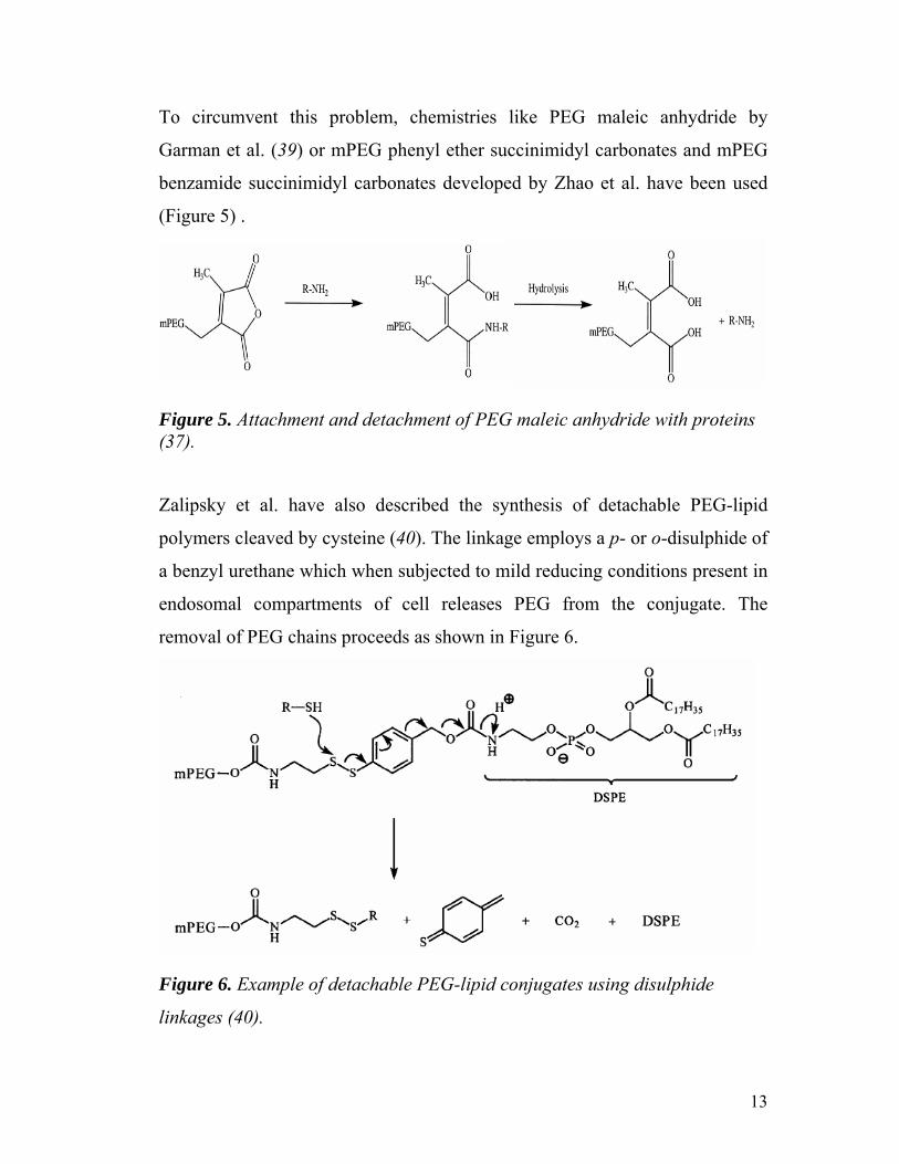
To circumvent this problem, chemistries like PEG maleic anhydride by
Garman et al. (39) or mPEG phenyl ether succinimidyl carbonates and mPEG
benzamide succinimidyl carbonates developed by Zhao et al. have been used
(Figure 5) .
Figure 5. Attachment and detachment of PEG maleic anhydride with proteins (37).
Zalipsky et al. have also described the synthesis of detachable PEG-lipid
polymers cleaved by cysteine (40). The linkage employs a p- or o-disulphide of
a benzyl urethane which when subjected to mild reducing conditions present in
endosomal compartments of cell releases PEG from the conjugate. The
removal of PEG chains proceeds as shown in Figure 6.
Figure 6. Example of detachable PEG-lipid conjugates using disulphide
linkages (40).

14
Thus there is an array of available PEGylation chemistry to tailor the
requirements for drug molecules, proteins, peptides or any particulate drug
delivery systems where long-circulation is desired.
FDA-approved PEGylated products clearly indicate the improved therapeutic
efficacy of the drugs using this technology. Even though many studies have
been conducted demonstrating the theoretical and commercial usefulness of
PEGylation technology there are many more untapped applications that are still
to be explored.

15
2 CONCEPTS AND DESIGN OF PHARMACEUTICAL
DELIVERY SYSTEMS.
Thus it is clear that PEG offers the versatile properties of flexibility,
hydrophilicity, and biocompatibility. PEG-conjugation modifies the physico-
chemical properties of the conjugated molecule thereby changing its
pharmacokinetic and biodistribution profile.
A detailed systematic investigation was carried out to explore the applications
of PEG in developing various drug carrier systems.
As a first example, PEG with a molecular mass of 5 kDa was used to modify
dextran (molecular mass 73 kDa). Dextran was initially modified to introduce
amino groups. Partial modification of these amino groups with PEG residues
resulted in a construct with a nearly doubled circulation half-life in mice of up
to 7 h (41). Residual dextran amino groups can be used for attachment of
drugs, diagnostic agents or even stimuli-responsive polymer blocks, thus
providing a convenient platform for long-circulating multifunctional polymer-
drug pharmaceutical preparations. We have prepared PEG-modified dextran to
make it long-circulating. Dextran, a polysaccharide, has hydroxyl groups
which can be used to conjugate small drug molecules proteins/peptides or
diagnostic moieties and be developed to be used as a drug carrier system.
Another application of PEG is in combination with lipids. When PEG is
conjugated to lipid molecules as in the case of PEG-phosphatidylethanolamine
(PEG-PE), the conjugates can form micelles with a central hydrophobic lipid
core and outer PEG corona. These micellar structures can be loaded with
poorly water-soluble drugs like, paclitaxel and camptothecin (10, 42, 43). PEG

16
forms the external hydrophilic corona that can offer long-circulation as well as
the convenience of attaching various ligands at the distal PEG-terminus to
make the micelles actively target the specific diseased tissue.
To develop magnetically sensitive micelles, superparamagnetic iron oxide
nanoparticles (SPION) were incorporated into PEG-PE based micelles to form
stable SPION-micelles. SPION have excellent MRI contrast properties,
however, they are not stable in physiological systems and show aggregation
(44, 45). PEG-lipid based micellar formulation not only prevented the SPION
from aggregation but also improved its MRI signal. Because of the small size
and long-circulating property, SPION-micelles can be targeted passively by
EPR effect. SPION- micelles can also be targeted to the disease site under
influence of external magnets. Moreover to prepare actively targeted MRI
contrast agents, SPION-micelles can be easily surface-modified by active
targeting ligands.
An interesting application of PEG-lipid conjugates can be achieved when the
bond between PEG and lipid is stimuli-sensitive and degradable. This stimulus
can either be the locally decreased pH, locally increased temperature, or
reducing conditions as the found in inflammation areas or tumor interstitium
(46-49). Because of its steric protection function PEG is used in targeted long-
circulating PEGylated liposomes and PEG-PE-based micelles (10, 43, 50, 51).
Cell-penetrating peptides (CPPs) like TAT can be incorporated onto the
surface of liposomes or micelles and these ligands could be easily “shielded”
or “exposed” by using stimuli (pH)-sensitive PEG-PE. PEG-PE used in this
work for liposome surface modification or for micelle preparation was made
degradable by inserting the pH-sensitive hydrazone bond between PEG and PE
(PEG-Hz-PE). Under normal pH values, TATp functions on the surface of
nanocarriers were “shielded” by long protective PEG chains. At pH 7.4-8.0,

17
TATp-containing both liposomes and micelles demonstrated very limited
internalization by NIH/3T3 or U-87 cells. However, upon brief incubation at
lowered pH values (pH 5.0-6.0), the nanocarriers lost their protective PEG
shell because of acidic hydrolysis of PEG-Hz-PE and acquired the ability to
become effectively internalized by cells via TATp moieties. This result could
be considered as the first step in the development of multifunctional stimuli-
sensitive pharmaceutical nanocarriers.
2.1 PEGylation of dextran to make long-circulating pharmaceutical
carrier.
Dextran, a natural polymer of glucose residues linked mainly with 1,6-bonds
has also been used as a drug carrier for a long time. This polymer is widely
available from natural sources and can be prepared in a variety of molecular
weights (52). For example, as early as 1978, it was shown that conjugation of
daunomycin with dextran increased the therapeutic efficacy and decreased the
toxicity of the drug (53). However, there are certain problems associated with
dextran. The degree of dextran branching (the property associated with dextran
immunogenicity) depends on the source of dextran and may vary over a broad
range (54). The high-molecular-weight dextrans used as drug carriers may
have a rather broad molecular size distribution and can cause anaphylactic
reactions (53). Although dextran-bound drugs demonstrated somewhat
increased circulation times, dextrans still undergo biodegradation via various
dextranases found in many tissues (55, 56).
On the other hand, synthesis of monofunctional (containing an activating group
at only one terminus) forms of high molecular weight PEG with narrow
polydispersity is relatively difficult to achieve (57). The disadvantage of PEGs
with molecular weights exceeding 30 kDa is their reduced excretion and

18
increased kidney accumulation. Although PEG has a very low toxicity and has
been approved for a variety of products for human use (14), this polymer is not
metabolized. While PEG molecules with the molecular weight of 30 kDa or
lower are easily cleared by renal filtration, PEGs with higher molecular
weights are cleared much more slowly (57). Such biologically uncleavable
polymers with high molecular weight can accumulate in the kidneys, decrease
the efficiency of renal filtration and cause various undesirable side effects (17).
Thus, the idea comes to mind of designing long-circulating but still
biodegradable delivery systems built from a biodegradable core polymer,
dextran and grafted with relatively short PEG residues as a good alternative to
using either high molecular PEGs or pure dextrans. Such co-polymers
synthesized using monofunctional PEG derivatives with a low-to-moderate
molecular weight will slowly degrade to fragments small enough to be
eliminated by the renal filtration. The dextran core, after the attachment of
multiple PEG residues, might still bear free reactive groups suitable for the
attachment or single-point modification of various drugs including proteins and
peptides to obtain their long-circulating forms (58).

19
2.2 PEG-lipid conjugates as the basis for micellar pharmaceutical
nanocarriers and PEG-lipid micelles with magnetic sensitivity.
2.2.1 PEG-lipid conjugates as micellar systems
PEG-PE-based lipid-core polymeric micelles have been the delivery system of
choice for various hydrophobic drugs/diagnostic agents (42, 50, 59-63). They
have prolonged blood circulation and can be easily functionalized with various
target-specific ligands like nucleosome-specific monoclonal antibody 2C5
(mAb2C5) on the water-exposed PEG termini (10, 43). Polymeric micelles, in
general, have been studied extensively for encapsulating various cancer chemo
therapeutics. In addition to long blood-circulating time and EPR effect in
tumors, they have also been shown to reduce toxicity and side-effects of the
drugs (64).
Diacyllipid-PEG or PEG-PE conjugates have been successfully used to
surface- modify liposomes to make them long circulating (65). The structure of
PEG-PE polymer is similar to that of amphiphilic co-polymers of A-B type
with acyl chains of lipids forming the hydrophobic core of the micelles that can
be used as a cargo space for encapsulating a variety of sparingly water-soluble
therapeutic and diagnostic agents. PEG-PE conjugates are commercially
available or can be easily synthesized (65).

20
Figure 7. Chemical structure of PEG-PE (50).
The chemical structure of PEG-PE is shown in Figure 7. A single molecule of
PEG-PE consists of flexible, hydrophilic PEG conjugated with a relatively
short but very hydrophobic diacyl phospholipid. The lipid allows the anchoring
of the molecule in the lipid bilayer in case of liposomes, leading to its initial
use of modifying the surface of liposomes with PEG chains.
The presence of PEG protects liposomes from the interaction with opsonins in
the blood plasma and prevents rapid uptake of liposomes by the RES. This
results in a dramatic increase in the liposome circulation time. One peculiar
observation is that the PEG-PE molecule, if used in liposomes above certain
critical limit forms micelles (66). Thus, the proportion of PEG-PE used in a
liposomal formulation is important. Later it was realized that PEG–PE micelles
have a substantial potential as particulate carriers for the delivery of
therapeutic and diagnostic agents (Figure 8). The use of lipid moieties as
hydrophobic blocks provides a very high stability of the micelles because of
very strong hydrophobic interactions between the double acyl chains of the
phospholipids residues. As with long-circulating liposomes, the exposure of
PEG residues on the surface of micelles prevents rapid uptake of the micelles
by the RES, making them long circulating. PEG-PE has very low toxicity and

21
it is currently used in the clinic as a component of Doxil® — doxorubicin-
loaded long-circulating liposomes.
Figure 8. Micelle formation from amphiphilic unimers and drug incorporation
into micelle (50).
In this work PEG-PE was used to prepare magnetically sensitive micelles.
Hydrophobic SPION particles were loaded into the PEG-PE based micellar
system to form SPION-micelles.
2.2.2 Superparamagnetic iron oxide nanoparticles (SPION).
SPION have received increased attention during the last decade due to their
characteristic inducible magnetic moments in the presence of an external
magnetic field, their small size (ca. 4 - 10 nm) and most importantly T2
magnetic resonance imaging (MRI) contrast properties. The most important
property of the SPION is their paramagnetic nature. SPION behave like
magnets only in presence of an external magnetic field and do not retain any

22
residual magnetism upon removal of the external magnetic field. This property
makes it possible for their application as MRI contrast agent or their
targetability under the influence of external magnets. However, “plain” SPION
are not stable at normal physiological conditions and show a tendency to
aggregate because of the hydrophobic nature of these particles. These magnetic
nanoparticles can be dispersed to form homogenous suspensions into suitable
solvents by using proper surface coating. Attempts have been made to stabilize
SPION by their incorporation into polymeric micelles (44).
2.2.3 MRI and MRI Contrast agents.
Currently, MRI is the most routinely used imaging modality in both clinical
and research settings. It offers outstanding resolution down to 1-2 mm in plane
at magnet field strengths as low as 1.5 Tesla. The signal of MRI depends on
the longitudinal and transverse (T2) proton relaxation times of mainly water.
The difference in proton relaxation times (because of differences in water
content of tissues) provides MRI contrast in the tissues. The tissue water
content, however, is dependent on the physiological microenvironment and
may be slightly altered depending on the pathology state. However, this slight
change in water environment is not evident until the late phases of the disease.
In order to make the differences visible at early stages and reveal the
pathologic condition at early stages, certain MRI contrast agents can be used.
MRI contrast agents, because of their intrinsic paramagnetic nature, produce
local micro-magnetic fields, and thus decrease the T1 or T2 relaxation times of
tissue water protons that results in enhanced contrast in imaging of the
pathological tissue.
The capability of a contrast agent to change T1 and T2 is defined as the
relaxivity, r1 or r2, expressed as mM-1 s-1. Depending on the r2/r1 ratios, the
MRI contrast agents are classified as positive contrast agents or negative

23
contrast agents. Positive contrast agents have low r2/r1 ratios and thus generate
hyper-intense (bright) T1-weighted images (e.g. gadolinium-based
paramagnetic chelates), while the negative contrast agents have high r2/r1 ratio
and thus generate hypo-intense (dark) T2-weighted images (e.g. iron-oxide
based superparamagnetic chelates). MRI applications are becoming more and
more dependent on contrast agents. MRI in combination with the contrast
agent has been an effective tool to get a perspective of inflammation, infarct,
tumor, atherosclerotic plaques, live stem-cell tracking, brain perfusion and
many other applications (45, 67).
2.2.4 Antinuclear Autoantibodies: Monoclonal antibody 2C5 (mAb2C5).
mAb2C5 belongs to a subset of anti-nuclear antibodies of the IgG2a isotype
(68). mAb2C5 is capable of inhibiting tumor growth of various murine and
human tumors both prophylactically and therapeutically (69). However at sub-
therapeutic levels, mAb2C5 is capable of specifically recognizing a wide
variety of tumors with no affinity for normal cells through the surface-bound
nucleosomes and thus serves as a targeting moiety to deliver drug carriers such
as liposomes and micelles into different murine and human tumors (70, 71).
Nucleosomes are an assembly of DNA and four pairs of histones, which are
released from dying cells in the center of the tumor. As these nucleosomes
migrate from the center to the periphery of the tumor, they get attached and
expressed on the surface of the neighboring live tumor cells. The uniqueness of
using mAb2C5 as the targeting ligand is that its target antigen is not tumor-
type specific, but rather a universal target that is typical of almost all tumor
types (71, 72). Thus it can be expected that the nanocarriers decorated with
mAb2C5 will recognize a broad variety of murine and human cancer cell
types. Previously we have demonstrated efficient tumor targeting capability
of mAb2C5 with several drug carriers like paclitaxel-loaded mAb2C5 surface-

24
modified immunomicelles (73) or doxorubicin-loaded mAb2C5 surface-
modified liposomes (36, 74-77).
Taking into account the benefits of using lipid-core based PEG-PE polymeric
micelles and the tumor-targetability of mAb2C5, we formulated SPION-loaded
PEG-PE micelles (SPION-micelles) and additionally surface modified SPION-
micelles with mAb2C5 to form the SPION-immunomicelles. We hypothesized
that these immunomicelles would bring an increased quantity of SPION to
cancer cells thus increasing the T2 MRI signal from these cancer cells
compared to either “plain” SPION-micelles or “non-specific” SPION-bovine-
IgG immunomicelles.
Because of the magnetic properties of SPION, we also hypothesized that we
would be able to target SPION-micelles to the tumor tissue in subcutaneous
tumor models in mice using external magnets.

25
2.3 Detachable PEG to make stimuli-sensitive multifunctional liposomes
and micelles.
Another important application of PEG is to engineer multifunctional
pharmaceutical nanoparticulates by using PEG conjugates with special
properties such as pH-sensitivity. The concept of synthesizing cleavable PEG-
lipid polymers was described earlier by Zalipsky et al. The cleavable bond was
based on p- or o-disulphide of a benzyl urethane which, when subjected to mild
reducing conditions such as in endosomal compartments of cells releases PEG
from the conjugate (40).
Ideally, a nanoparticular drug delivery system (DDS) should be able: (a) to
specifically accumulate in the required organ or tissue, and then (b) penetrate
inside target cells delivering its load (drug or DNA) intracellularly. Organ or
tissue (tumor, infarct) accumulation could be achieved by passive targeting via
the EPR effect (8, 78); or by antibody-mediated active targeting (10, 79), while
the intracellular delivery could be mediated by certain internalizable ligands
(folate, transferrin) (80, 81) or by cell-penetrating peptides (82, 83).
2.3.1 Cell-penetrating peptides
CPPs such as HIV trans-activating transcriptional activator peptide (TATp),
polyarginine (polyArg), homeodomain of Antennapedia (Antp), or Herpes
simplex virus type 1 (HSV-1) protein VP22 all contain short sequences of less
than 30 amino acids that are able to penetrate cell membranes and translocate
different cargoes into cells. For example, the minimal protein transduction
domains or the cell penetrating function domains of TAT-peptide have the
sequence Arg-Lys-Lys-Arg-Arg-Gln-Arg-Arg-Arg while the domains in Antp
have the sequence Arg-Gln-Ile-Lys-Ile-Trp-Phe-Gln-Asn-Arg-Arg-Met-Lys-

26
Trp-Lys-Lys. As is evident, the common feature of these peptides is that they
are amphipathic and net positively charged.
The exact mechanism of cell translocation is not yet clearly understood but in
general it is accepted that energy-dependent macropinocytosis is responsible
for TAT-mediated intracellular delivery of large molecules and nanoparticles
with subsequent enhanced escape from endosomes into the cell cytoplasm (84,
85). Energy-independent electrostatic interactions and hydrogen bonding seem
to be responsible for cell penetration of individual CPPs or CPP-conjugated
small molecules (86, 87).
2.3.2 Developing multifunctional nanocarrier systems.
The “ideal” drug delivery system (DDS) should simultaneously carry on its
surface various active moieties, i.e. be multifunctional and possess the ability
to “switch on” and “switch off” certain functions when necessary, for example
under the action of local stimuli typical of the target pathological zone
(increased temperature or lowered pH values characteristic for inflamed,
ischemic, and neoplastic tissues). Another important requirement is that
different properties of the multifunctional DDS should be coordinated in a
certain orchestrated manner. For example, if a system is to be constructed that
can provide the combination of the longevity allowing for target accumulation
via the EPR effect and specific cell surface binding allowing for its
internalization by target cells, two requirements have to be met. First, the half-
life of the carrier in the circulation should be long enough (hours) to fit EPR
effect requirements, and second, the internalization of the DDS by the target
cells should proceed fast enough (minutes) to prevent carrier degradation and
drug loss in the interstitial space. However, systems like this still represent a
challenge (88).

27
Intracellular transport of different biologically active molecules is one of the
key problems in drug delivery in general. Nanoparticular DDS, such as
liposomes and micelles, are frequently used to increase the efficacy of drug
and DNA delivery and targeting (50, 51). So far, multiple and not always
successful attempts have been made to deliver various drug carriers directly
into the cell cytoplasm, bypassing the endocytic pathway, to protect drugs and
DNA from the lysosomal degradation, thus enhancing drug efficiency and
DNA incorporation into the cell genome (89-92).
.
cell cell
binding site
short PEG spacer
long PEG spacer
cell-penetrating function
drug-loaded nanocarriers
bonds that could be made stimuli-sensitive
pathological zone with abnormal pH or temperature
specific ligand
cell penetration via “de-shielded” function
long protective PEG
Figure 9. Schematic of interaction of the multifunctional pH-responsive
pharmaceutical nanocarrier with the target cell. pH-dependent removal of
protecting PEG chains or mAb-PEG moieties allows for the direct interaction
of the CPP moiety with the cell membrane.

28
2.3.3 The new concept of multifunctionality.
Keeping this in mind we came up with an interesting idea to combine both the
longevity of the PEG and the cell-penetrating capability of TAT-peptide by
designing liposomes and micelles such that during the first phase of delivery, a
non-specific cell-penetrating function is shielded providing organ/tissue-
specific delivery (sterically-protecting polymer or antibody). Upon the
accumulation in the target, protecting polymer or antibody attached to the
surface of the DDS via the stimuli-sensitive bond should detach under the
action of local pathological conditions (abnormal pH or temperature) and
expose the previously hidden second function allowing for the subsequent
delivery of the carrier and its cargo inside cells. Such a DDS should be stable
in the blood for a long time (hours) to allow for an efficient target
accumulation. It has to lose the protective coat inside the target almost
instantly to allow for fast internalization (minutes). Such a delivery system can
prove beneficial as it will carry the drug directly into the target cell (88). The
schematic pattern of such system is shown in Figure 9. Intracellular trafficking,
distribution, and fate of the carrier and its cargo can be additionally controlled
by its charge and composition, which can drive it to the nuclear compartment
or toward other cell organelles.
In this study, we have prepared and tested in vitro stable targeted PEGylated
DDS (liposomes and micelles) containing the second specific function
(responsible for the intracellular internalization of the DDS), which, under
normal conditions, is shielded by the protecting polymer or polymer-antibody
conjugate, however can be “exposed” upon brief incubation at lowered pH
values characteristic of inflamed or neoplastic areas. As the first step on the
way to multifunctional drug delivery systems, we have prepared liposomes and
micelles sterically protected by the low pH-cleavable PEG chains and

29
additionally containing a monoclonal antibody attached to the surface of the
DDS via a noncleavable longer PEG spacer and an additional function (biotin
or TATp) attached to the surface of the DDS via the noncleavable anchor
shorter than the cleavable PEG chain.

30
3 OBJECTIVES AND SPECIFIC AIMS
The overall objective of this thesis project is to test applications of PEG in
constructing platforms for delivery of therapeutics and diagnostics.
Polymers like dextran and drug delivery nanocarriers like liposomes and
micelles have been used and improved by either modifying them by using PEG
to prepare more effective drug carrier formulations and thus to improve the
delivery of pharmaceuticals therapeutic outcome..
I. To develop and characterize a biodegradable drug carrier platform using
dextran and test if PEGylation of dextran improves their circulation time
in vivo.
II. To develop and characterize “targeted” SPION as an imaging agent
loaded in PEG-lipid polymeric micelles, and to use either external
magnets or nucleosome-specific monoclonal antibody 2C5 on the micelle
surface for target-specific delivery, and to test the targetability of such
micelles to cancer cells in vitro and in vivo.
III. To develop and characterize multifunctional liposomes and micelles
simultaneously carrying a number of ligands, which are expressed only
upon the action of external stimuli like local pH, using pH-degradable
PEG-lipid conjugates and test their functionality in vitro.

31
4 EXPERIMENTAL DESIGN AND METHODS
4.1 PEGylation of dextran to make long-circulating pharmaceutical
carriers.
4.1.1 Materials
Dextran with average molecular weight of 73 kDa, carbonyldiimidazole (CDI),
ethylenediamine (EDA), diethylene triamine pentaacetic acid anhydride
(DTPA) and fluorescamine were purchased from Sigma Chemical, Inc. (St.
Louis, MO). PEG-succinimidyl propionate (PEG-SPA) with a molecular
weight of 5 kDa was from Nektar Therapeutics (San Carlos, CA). All other
chemicals and organic solvents were of analytical grade. Distilled, deionized
water was used.
4.1.2 Synthesis of aminodextran
Aminodextran was synthesized from CDI-activated dextran by conjugating
EDA using a previously described procedure for attaching diamines to cross-
linked agarose (93). In order to determine the rate of CDI activation, 17 µmol
of dextran dissolved in 30 ml of formamide was incubated with 360 µmol of
CDI (21 molar excess over dextran) for 15 min, 30 min, 1 h and 3 h at room
temperature. After overnight incubation at room temperature the reaction was
stopped by the addition of at least 100-fold molar excess (over CDI) of EDA.
The resultant aminodextran was precipitated by adding 2-fold volume excess
of acetone. The white precipitate was washed with acetone and collected by
centrifuging at 1500xg for 1 h, dissolved in water, dialyzed against at least a
3000-fold excess of water for 2-4 days with several buffer changes, and freeze
32
dried. The number of EDA residues attached per dextran molecules was
estimated by determining the concentration of amino groups in the samples
using the fluorescamine method as described below.
As a typical preparative procedure, 68 µmol of dextran was dissolved in 125
ml of formamide and 1.44 mmol of CDI (21 molar excess over dextran)
dissolved in 10 ml of formamide was added. After an hour of incubation, 25 ml
of EDA was added to the mixture, and stirred at room temperature overnight.
Purification was carried out as described above.
4.1.3 Introduction of DTPA residues into aminodextran.
Aminodextran (28.5 µmol) was dissolved in 25 ml of formamide containing
0.8 ml of pyridine and mixed with of DTPA anhydride dissolved in 0.5 ml of
formamide (2-fold molar excess over aminodextran). The mixture was
incubated for 1-5 h at room temperature, dialyzed against water and freeze
dried.
4.1.4 PEGylation of aminodextran.
Aminodextran (20.5 µmol) was dissolved in 50 ml of formamide containing 2
ml of pyridine. To this solution, 103 µmol of PEG-SPA dissolved in 5 ml of
formamide was added. To study the kinetics of PEG attachment, the mixture
was incubated for 30 min, 2 h, 5 h and overnight at room temperature with
stirring. The reaction was stopped by adding 5 ml of water per 10 ml of the
reaction mixture. The sample was purified by dialysis and freeze-dried as
described above. The number of PEG residues attached per each aminodextran
molecule was estimated by amino group loss determined by the fluorescamine
method (see below).

33
For preparative purposes, 20 µmol of aminodextran was dissolved in 50 ml of
formamide containing 1.2 ml of pyridine. To this solution, 300 µmol or 600
µmol of PEG-SPA dissolved in formamide was added. The mixture was
incubated at room temperature with stirring overnight. Organic solvents and
unbound PEG were removed by dialysis against water using dialysis bags with
a cutoff size of 50 kDa (Spectrum Medical Industries, Rancho Dominguez,
CA) and freeze-dried. For biodistribution studies, PEGylated aminodextran
samples were acetylated in formamide containing pyridine by addition of a 10-
fold molar excess of acetic anhydride and purified as described above.
4.1.5 Amino group concentration.
The number of primary amino groups was estimated using fluorescamine
according to a procedure adapted from a previous publication (94). The
material of interest was dissolved at about 0.2 mg/ml in 10 mM phosphate
buffered saline, pH 7.4. A 1.5 ml aliquot of the sample was mixed with 0.5 ml
of 3 mg/ml fluorescamine solution in acetone and incubated in the dark at
room temperature for 30 min. The number of amino groups was determined by
fluorescence at an excitation wavelength of 400 nm and an emission
wavelength of 460 nm on a F-2000 spectrofluorimeter (Hitachi, Japan) using a
sample of commercially available aminodextran with a known number of
amino groups per dextran molecule as a standard (Molecular Probes, OR).
4.1.6 Radiolabeling of PEGylated dextrans by transchelation.
DTPA-modified aminodextran or PEGylated aminodextran was dissolved in 10
mM HEPES buffered saline, pH 7.4 (HBS) at a concentration of 10 mg/ml. To
1 ml of this solution, 10-20 µCi of 111In in 0.1 ml of 0.1 M sodium citrate, pH
3.7 (a weak chelator for 111In) was added and incubated for 1 h at room

34
temperature. Any unbound 111In was removed by dialysis using 25 kDa
MWCO dialysis tubing against HBS at 4oC overnight.
4.1.7 Biodistribution studies.
In vivo biodistribution studies were carried out using 111In-labeled dextran or
PEG-dextran in female C57BL/6J mice (Charles River Laboratories,
Wilmington, MA). The mice were injected with 100 µl of 10 mg/ml of 111In-
labeled dextran or PEG-dextran via the tail vein. The animals were allowed
free access to food and water. Mice were sacrificed by cervical dislocation at
time points between 0.5 and 17 h post injection with 4 animals in a group for
each time point. Blood, liver, spleen, renal and lung samples were collected
and analyzed by γ-counting for the presence of the dextran-associated 111In
radioactivity as CPM using a Beckman 5500B gamma-counter. Dextran
circulation half-lives were determined assuming a first order elimination
process from the Log [% of injected dose in blood] vs. Time plots using a
computer program, Origin (Microcal Software, Inc, Northampton, MA).
4.1.8 Statistics.
Data is represented as mean ± SE, unless specified otherwise. Student’s t-test
was applied for test of significance. P ≤ 0.05 was considered statistically
significant.

35
4.2 PEG-lipid micelles with magnetic sensitivity.
4.2.1 Materials.
1,2-distearoyl-sn-glycero-3-phosphoethanolamine-N-[poly(ethyleneglycol)
2000] (PEG2000-PE) and 1,2-dipalmitoyl-sn-glycero-3-phosphoethanolamine-
N-[lissamine rhodamine B sulfonyl] [ammonium salt] (Rhodamine-PE or Rh-
PE) and 1,2-Dimyristoyl-sn-Glycero-3-Phosphoethanolamine-N-DTPA
(DTPA-PE) were purchased from Avanti Polar Lipids (Alabaster, AL, USA).
Trimethylamine oxide [(CH3)3NO], Iron(0) pentacarbonyl [Fe(CO)5], hexane,
ethanol, octyl ether and all other chemicals were purchased from Sigma Chem.,
Inc. (St. Louis, MO, USA). HOECHST 33342 was purchased from Invitrogen
Corp. (Carlsbad, CA, USA). Dulbecco’s Modified Eagle’s Medium, Hanks’
Balanced Salt Solution (HBSS), and penicillin/streptomycin stock solutions
were purchased from CellGro (Kansas City, MO, USA). Tissue culture grade
fetal bovine serum and trypsin was obtained from ICN Biomedicals (Costa
Nesa, CA, USA). The production and purification of the anti-nucleosomal
cancer-specific mAb 2C5 were carried out by Harlan Bioproducts
(Indianapolis, IL, USA) using the hybridoma cell line from our laboratory.
Control bovine IgG antibody was purchased from Millipore Corp. (Bedford,
MA, USA). p-Nitrophenylcarbonyl-polyethyleneglycol-3400-phosphatidyl-
ethanolamine (pNP-PEG3400-PE) was synthesized in our lab following the
procedure described in (95). Cancer cell lines were purchased from the
American Type Culture Collection (Rockville, MD, USA). Distilled and
deionized water was used in all experiments.
4.2.2 Synthesis of superparamagnetic iron oxide nanoparticles (SPION).
The nanoparticles were synthesized using the thermal decomposition method
developed by Hyeon et. al. with some slight modifications (96). Briefly, 1.28 g

36
oleic acid was dissolved in 10 mL octyl ether, heated to about 100°C. To this
solution 200 µl of Fe(CO)5 was added. The mixture was refluxed at 280°C for
1 hour and then cooled to room temperature. The solution changed color from
pale-yellow green to dark black during the heating process. To this mixture
0.34 g of (CH3)3NO was added along with 500µl of octyl ether to facilitate
complete transfer of the salt. The mixture was than heated to 130°C for 2 hours
under an argon atmosphere. The temperature was slowly increased to reflux
temperature of 280°C and maintained for an additional 1 hour. The iron oxide
nanoparticles were precipitated from this solution using ethanol, separated
from the mother liquor by centrifugation, dried under argon, weighed and re-
dispersed in hexane to get a 1.5 mg/mL SPION solution.
4.2.3 Formulation of SPION-micelles.
SPION-micelles were prepared by the rehydration method of dry lipid film and
SPION. Briefly, 10 mg of mPEG2000-PE in chloroform was mixed with 0.25
mg of SPIONs in hexane. The organic liquids were removed using a rotary
evaporator to dryness. To remove any residual organic liquid the film was
freeze-dried overnight. Next day, the film was rehydrated using 1 mL HBS, pH
7.4. The film was vortexed vigorously for 5 min followed by bath sonication
for 5 min to get SPION-loaded PEG-PE micelles. Unincorporated SPION were
removed by applying an external magnet and the supernatant was removed to a
fresh vial.
4.2.4 Size distribution and zeta potential measurements of SPION-micelles.
The size distribution and surface potential of pure micelles and the SPION-
micelles were determined by dynamic light scattering (DLS) measurement
with a Brookhaven Instruments 90Plus particle size analyzer. The

37
measurements were performed five times. For the zeta potential measurement,
a phase analysis light scattering (PALS) method was used. For each sample 5
runs with 10 cycles each were performed and an average value was obtained
from the five measurements. For DLS size measurements the samples were
prepared in HBS pH 7.4 whereas for the zeta potential measurements the
samples were prepared in 1 mM KCl.
4.2.5 Freeze-fracture electron microscopy.
For determining the structure of the micelles after the incorporation of SPION,
the freeze fracture electron microscopy was carried out for ‘plain’ PEG-PE
micelles and SPION-micelles (10 mg/mL in HBS pH 7.4). The sample was
quenched by using the sandwich technique and liquid nitrogen-cooled propane.
A cooling rate of 10,000 Kelvin per sec avoids ice crystal formation and
artifacts possibly caused by the cryofixation process. The cryo-fixed samples
were stored in liquid nitrogen for less than 2 hours before processing. The
fracturing process was carried out in JEOL JED-9000 freeze-etching
equipment, and the exposed fracture planes were shadowed with platinum for
30 sec at an angle of 25–35° and with carbon for 35 sec [2 kV, 60–70 mA, 1 x
10-5 torr]. The replicas were cleaned with fuming HNO3 for 24 h followed by
repeated agitation with fresh chloroform/methanol [1:1 by vol] at least five
times and examined with a JEOL 100 CX electron microscope.
4.2.6 Relaxation rate of SPION-micelles.
When used as a contrast agent for MRI, SPION increases the relaxation rate of
water and has the greatest effect on the T2 (spin-spin) relaxation rate.
However, this change in the relaxation rate depends on the size of the SPION,
degree of aggregation, surface coating thickness, etc. In vitro relaxation
parameters of both, ‘plain’ SPION and SPION-micelle samples were measured

38
using a benchtop 5 mHz RADX NMR Proton Spin Analyzer at room
temperature in HBS, pH 7.4, at different SPION concentrations ranging from
0.002 -to- 0.125 mg/mL.
4.2.7 Preparation of SPION-immunomicelles.
First, mAb 2C5 or nonspecific control bovine IgG was conjugated to pNP-
PEG3400-PE as previously with some modifications (36, 75-77, 97). Briefly, 14
mg PEG2000-PE (97 mol %), 0.45 mg pNP-PEG3400-PE (2 mol %), 0.064 mg
Rh-PE (1 mol %) in chloroform and 0.35 mg SPION in hexane was dried in a
rotary evaporator and freeze-dryer to form a thin film. The film was hydrated
with 1 mL of 5mM citrate buffered saline, pH 5.0, by vortexing for 5 min.
followed by bath sonication for 5 min to form the micelle solution. Separately,
1 mL solution (1.56 mg/mL) of the mAb 2C5 or non-specific bovine gamma
immunoglobulin antibody (IgG) was prepared in 50 mM phosphate-buffered
saline, pH 8.7, and incubated with the above micelle solution with stirring
overnight at 4°C to allow the attachment of the antibody to the activated PEG
terminus with the simultaneous hydrolysis of non-reacted pNP groups, thus
forming the SPION-2C5-immunomicelles. The formulations were then purified
using dialysis bags MWCO 250 kDa against HBS, pH 7.4, with 4 changes
within 6 hours. After purification, the antibody concentration in the micelle
preparation was estimated by the BCA protein assay kit (Pierce Biotechnology,
Inc. Rockford, IL, USA) according to manufacturer’s recommendations. The
micelle size was confirmed by the dynamic light scattering in ZetaPlus particle
size analyzer. (Brookhaven Instruments Corporation Holtsville, NY, USA).

39
4.2.8 ELISA of immunomicelles.
The specific activity of the immunomicelles was determined by ELISA as
described below. An ELISA assay (indirect, using an enzyme-tagged
secondary Ab) was performed to show the ability of the SPION-
immunomicelles to recognize the target antigen, nucleosomes (NS). The 96-
well plates were coated with 50 µL of 40 µg/mL poly-L-lysine (MW 30-70
kDa). After discarding the poly-L-lysine solution, the wells were blocked with
200 µL of TBS containing 0.05% w/v Tween 20 and 2 mg/mL casein (TBST-
casein) for 1h at RT, and then incubated with 50 µL of 40 µg/mL NS in TBST-
casein for 1 h at RT and washed three times with TBST. The wells were
incubated with different concentrations of the native mAb 2C5 and mAb2C5-
and IgG-modified SPION immunomicelles for 1 h at RT. After incubation, the
wells were washed as before and incubated with 50 µL/well of 1:5000 dilution
of goat anti-mouse IgG peroxidase conjugate (ICN Biomedicals, Inc., Aurora,
OH) in TBST-casein for 1 h at RT. The wells were again washed as before,
and each well was incubated with 100 µL of enhanced Kblue TMB peroxidase
substrate (Neogen Corporation, Lexington, KY) for15 min. The microplate was
read at a dual wavelength of 620 nm with the reference filter at 492 nm using a
Labsystems Multiskan MCC/340 microplate reader installed with
GENESISLITE windows based microplate software.
4.2.9 Cell culture.
Human breast carcinoma MCF-7 and murine breast carcinoma 4T1 cells
(American Type Culture Collection, Manassas, VA, USA) were propagated
using DMEM cell culture medium at 37 °C, 5% CO2. DMEM media were
supplemented with 10% FBS, 1 mM Na-pyruvate, 50 U/mL penicillin, and
50 µg/mL streptomycin.

40
4.2.10 Interaction of SPION-loaded mAb 2C5-immunomicelles with cancer
cells in vitro.
After the initial passage in tissue culture flasks, MCF-7 cells were grown on
coverslips in 6-well tissue culture plates (100,000 cells per well) in DMEM
with 10% BSA. After the cells reached a confluence of 70–80%, the plates
were washed and blocked with 1% BSA in DMEM. Rhodamine-PE (Rh-PE)-
labeled micelle samples in a serum-free medium were added to the cells at a
final PEG–PE concentration of 0.35-0.70 mM and incubated for 2 h at 37°C,
5% CO2. After the incubation, the cells were washed with HBSS. The cells
were then digested using trypsin and collected in HBSS. T2 relaxation times
for the cell samples were measured using a Varian INOVA 500MHz NMR
Spectrometer at room temperature in HBSS. The rhodamine fluorescence
associated with the cells was also measured using the Hitachi L2000
fluorospectrometer at the excitation wavelength of 550 nm and emission
wavelength of 590 nm.
4.2.11 Fluorescence microscopy.
After the initial passage in tissue culture flasks, MCF-7 cells were grown on
coverslips in 6-well tissue culture plates (50,000 cells per well) in DMEM with
10% BSA. After the cells reached a confluence of 40-50 % the plates were
washed and blocked with 1% BSA in DMEM. Rh-PE-labeled micelle samples
in serum-free medium were added to the cells at a final PEG–PE concentration
of 0.35-0.70 mM and incubated for 2 h at 37°C, 5% CO2. After the incubation,
the cells were washed with HBSS. The cells were stained with HOECHST
33342 at 5 µg/mL for 3 min followed by washing with HBSS. Individual
coverslips were mounted cell-side down onto fresh glass slides with PBS. Cells
were viewed and pictures were taken with a Nikon Eclipse E400 microscope
using the epifluorescence microscopy.

41
4.2.12 Subcutaneous tumor models in mice for studying magnetic targeting
of SPION-micelles.
All the animal work was performed at either in our lab facility or the animal
care facility at Northeastern University. The studies were in compliance with
the institutional guidelines. Balb/C female mice (Charles River Laboratories,
Wilmington, MA) 6-8 weeks of age were injected subcutaneously with murine
mammary epithelial tumor 4T1 cell suspension at 1 x 10⁵ cells in 100 µL
media into the right flank of the mice. Mice were allowed free access to food
and water and were monitored regularly for tumor volume and body weight
changes. The tumor dimensions were measured with vernier caliper and the
tumor volume was calculated using the formula:
Tumor volume = [Length x (Width)2] / 2 ; with length being the longer axis
(76).
4.2.13 Radiolabeling of SPION-micelles with 111- Indium.
To radiolabel with 111-Indium (111In), SPION-micelles formulations were
prepared as described above in section 4.2.4 along with addition of 0.5 mol%
of DTPA-PE to the lipid film. DTPA-PE-containing SPION-micelles were
incubated for 1 hour with 111In at room temperature, to allow for the trans-
chelation of 111In from a weak citrate complex into a strong DTPA complex.
The unbound 111In was removed by dialysis overnight against 4 L HBS pH 7.4
at 4°C using MWCO 2000 dialysis tubing.

42
4.2.14 Magnetic targeting of SPION-micelles in tumor of tumor-bearing
mice in vivo under influence of external magnet.
Subcutaneous 4T1 tumors were grown in BALB/C mice as described above.
After the tumor volume reached approx. 0.5 – 0.6 mm3, the mice were injected
with 100 µL of Indium- (111In)-labeled SPION-micelles at PEG-PE
concentration of 33.6 mg/mL via tail vein. Test group consist of animals with
magnets attached externally onto the tumors using cyanoacrylate glue and the
other control group will be non-magnet bearing mice. Mice under anesthesia
were sacrificed by cervical dislocation at time points 10 min, 30 min, 1 hour,
and 4 hours post injection with 6 animals in a group for each time point. Tumor
tissue was collected, weighed and analyzed by γ-counting for the presence of
the SPION-micelle-associated 111In radioactivity as CPM using a Beckman
5500B gamma-counter. From this data, the amount of the accumulated
radioactivity per gram of tissue was calculated as percent of injected dose of
radioactivity.

43
4.3 Detachable PEG to make stimuli-sensitive multifunctional liposomes
and micelles.
4.3.1 Materials
Cell lines, mouse fibroblast NIH 3T3 and astrocytoma U-87 MG, were
purchased from the American Type Culture Collection (Manassas, VA). All
cell culture media, DMEM, and RPMI 1640, heat-inactivated fetal bovine
serum (FBS), and concentrated solutions of sodium pyruvate and
penicillin/streptomycin stock solutions were purchased from Cellgro®
(Herndon, VA). TAT-peptide (11-mer:
TyrGlyArgLysLysArgArgGlnArgArgArg; molecular mass, 1,560 Da; three
reactive amino groups) was prepared by Research Genetics (Huntsville, AL).
Monoclonal antibody (mAb) 2G4 was produced and purified in our laboratory.
Also, pNP-PEG3400-PE was synthesized and purified according to an
established method in our laboratory (95). Egg phosphatidylcholine (PC),
cholesterol, 1,2-distearoyl-sn-glycero-3-phosphoethanolamine-N-[methoxy
(polyethyleneglycol)-750] (ammonium salt) (mPEG750-PE), 1,2-distearoyl-sn-
glycero-3-phosphoethanolamine-N-[methoxy (polyethylene glycol)-2000]
(ammonium Salt) (mPEG2000-PE), 1,2-dipalmitoyl-sn-glycero-3-phospho-
ethanolamine-N-(cap biotinyl) (sodium salt) (biotin-PE) 1,2-dipalmitoyl-sn-
glycero-3-phosphothioethanol (sodium salt) (PE-SH) and rhodamine-PE (Rh-
PE) were obtained from Avanti Polar Lipids, Inc. (Alabaster, AL). mPEG2000-
butyraldehyde (mPEG2000-CHO) and mPEG5000-butyraldehyde (mPEG5000-
CHO) was obtained from Nektar™ (Huntsville, AL). Control bovine antibody
IgG was obtained from Serologicals Proteins, Inc. (Kankakee, IL). 3-(2-
pyridyldithio) propionyl hydrazide (PDPH) and Immobilized NeutrAvidin™
Protein was purchased from Pierce Biotechnology, Inc. (Rockford, IL). Bovine

44
serum albumin and all other chemicals and buffer solution components were
from Sigma (St. Louis, MO) and were of analytical grade.
4.3.2 Synthesis of pH-cleavable mPEG2000-hydrazone-phospatidyl
ethanolamine (mPEG2000-Hz-PE).
Developing the method of coupling oxidized antibody to the PEG terminus
through a hydrazone bond as suggested by Hansen et al (98), we devised our
own scheme of conjugate reaction for synthesis of pH-cleavable PEG-PE. The
reaction was performed in two steps: first, activation of mPEG2000-CHO with
PDPH, and second, conjugation of 1,2-dipalmitoyl-sn-glycero-3-
phosphothioethanol (Na salt) (PE-SH) to activated mPEG-CHO.
For step I of the synthesis, 150 mg (64 µmole) of mPEG2000-CHO was
dissolved in dry chloroform containing 3.5 molar excess of 3-(2-pyridyldithio)
propionyl hydrazide (PDPH) to obtain 50 mg/ml solution of mPEG2000-CHO.
The mixture was incubated for 48 h at room temperature with stirring under
argon. TLC (CHCl3 : CH3OH : H2O - 80:20:2) revealed that the reaction was
complete. The starting material mPEG2000-CHO did not absorb UV and was
positive to Dragendorff spray, while PDPH absorbed UV and was negative for
Dragendorff spray. The product, mPEG2000-Hz-PDP, absorbed UV and was
positive to Dragendorff spray. Organic solvents were then removed using a
rotary evaporator. mPEG2000-Hz-PDP was then dissolved in deionized water
(adjusted to pH 10-11 using 1 M NaOH) and purified from unreacted PDPH
using a Sepharose G25 column and deionized water (adjusted to pH 10.5 using
1 M NaOH). Pooled fractions containing mPEG2000-Hz-PDP (Dragendorff and
UV positive) were freeze-dried.

45
For step II of the reaction, 20 mg (26 µmole) of 1,2-dipalmitoyl-sn-glycero-3-
phosphothioethanol (sodium salt) (PE-SH) was dissolved in dry chloroform
containing 1.5 molar excess of mPEG2000-Hz-PDP, to get a 10 mg/mL solution
of PE-SH. The solution was supplemented with 15 µL (approx. 3-fold molar
excess over PEG) of triethylamine (TEA). The sample was incubated overnight
at room temperature with stirring under argon. TLC (CHCl3:CH3OH:H2O -
80:20:2) revealed that the reaction was complete. The starting material
mPEG2000-Hz-PDP was positive to Dragendorff spray and negative for
molybdenum blue, while PE-SH was positive to molybdenum blue and
negative for Dragendorff spray. The product, mPEG2000-Hz-PE, was positive to
both Dragendorff spray and molybdenum blue. The organic solvents were then
removed using a rotary evaporator. The mPEG2000-Hz-PE micelles were
formed in deionized water (adjusted to pH 10.5 using 1 M NaOH) by
vortexing. The micelles were separated from the unbound PEG and released
pyridine-2-thione on CL-4B column using deionized water (adjusted to pH
10.5 using 1 M NaOH) as an eluent.
Pooled fractions containing mPEG2000-Hz-PE were freeze-dried, and extracted
with chloroform. mPEG2000-Hz-PE was stored as 10 mg/mL in chloroform at -
80º C under argon until further use. mPEG5000-Hz-PE used in some
experiments was synthesized in the same way starting with mPEG5000-CHO. 1H
NMR (500 MHz, CDCl3) δ (ppm) for mPEG2000-Hz-PE: 0.87 (t, CH3 of lipid,
6H), 1.27 (b,s, CH2, ≈ 56H), 2.29 (t, OCOCH2, 4H), 2.40 (t, COCH2CH2S,
2H), 2.45 (t, SCH2CH2O, 2H), 2.5 (t, COCH2CH2S, 2H), 2.59 (m, CH2CH=N,
2H), 3.1 (t, CH2CH, 2H) , 3.39 (s, OCH3 of PEG, 3H) and 3.5 (bm, PEG, ≈
184H). Thus, there was a clear indication for the presence of the expected
conjugate.

46
4.3.3 Acidic pH cleavability of mPEG2000-Hz-PE.
TLC analysis. TLC-verified degradation of the polymer conjugates after pH
treatment and spots corresponding to plain PEG and plain PE were observed
after incubation of the polymers at pH 5.0 for 15 minutes at 37° C.
HPLC analysis. Micelles of mPEG2000-Hz-PE were prepared containing 1 mol
% of rhodamine-PE as fluorescent marker as follows. Lipid film was prepared
by mixing chloroform solutions of both the lipids in a round bottom flask and
then removing chloroform using a rotary evaporator. To ensure complete
removal of any traces of chloroform further drying was done using a
lyophilizer. The appropriate volume of pH 8.5 phosphate buffer (100 mM
phosphate, 150 mM sodium chloride) was added and vortexed for 2 min to
form a 0.5 mM solution of mPEG2000-Hz-PE micelles. The sample was then
divided into equal volumes and incubated at different pH values. For pH 7.4
treatment, a 50 µL aliquot of the first half of the micelle formulation was
applied, as is, to Shodex KW-804 size exclusion column at regular intervals
using pH 7.4 phosphate buffer (100 mM phosphate, 150 mM sodium sulfate)
as eluent and run at 1.0 ml/min. Both UV (from 200 to 400 nm) and
fluorescence (excitation 550 nm and emission 590 nm) was used to monitor the
micelles. To the second half of the micelle formulation an appropriate volume
of 1N HCl was added to a final pH of ~ 5.0, aliquots of which were then
analyzed as above at different intervals. As a control, micelles of mPEG2000-PE
(non-pH-sensitive micelles) were prepared and analyzed after treatment at both
the pH values as above.
4.3.4 Kinetics of the pH-dependent degradation of mPEG2000-Hz-PE.
The degradation of the micelles spontaneously formed by mPEG2000-Hz-PE
under the action of the acidic pH was studied by following the presence or
absence of micelles over the period of time in buffer solutions at different pHs

47
(i.e. pH values 6.0, 7.0, 8.0 and 10). Rh-PE-labeled micelles of mPEG2000-Hz-
PE conjugate were prepared in phosphate buffer (10mM phosphate, 150mM
NaCl) solutions of different pH values. The pH of the solution was adjusted
using appropriate amounts of either 1N HCl or NaOH. Aliquots (50 µl) were
sampled at different time intervals for size exclusion chromatographic analysis
in 100mM phosphate buffer (pH 7.0) containing 150mM sodium sulphate
using a fluorescence detector (excitation 550 nm and emission 590 nm). The
area under the micelle peak (mean retention time: 9.35 min) was determined
for each chromatogram.
4.3.5 Preparation of pH-sensitive drug delivery systems.
Micelles. For micelle preparations, a mixture of mPEG750-PE, pH-sensitive
mPEG2000-Hz-PE, biotin-PE (or TATp-PE), and Rh-PE at a molar ratio of
40:54:5:1 was prepared in chloroform. Chloroform was removed on a rotary
evaporator followed by freeze-drying on a Freezone 4.5 (Labconco, Kansas
City, MO). The film was hydrated with PBS, pH 8.0 (10 mM phosphate, 150
mM sodium chloride) at room temperature and vortexed for 5 min. Micelle
size was determined by using a Coulter N4 Plus submicron particle analyzer.
Liposomes. For liposome preparations, a mixture of phosphatidylcholine and
cholesterol in 7:3 molar ratio and with the addition of various quantities (up to
18% mol) of PEG5000-Hz-PE or mPEG5000-PE was prepared in chloroform.
When required, the composition for liposome preparation was supplemented
with 0.5 to 1 % mol of TATp- PEG2000-PE [prepared as described earlier in
(92)] and with 0.5 % mol of Rh-PE (for the fluorescent labeling). Chloroform
was removed on a rotary evaporator followed by freeze-drying on a Freezone
4.5 (Labconco, Kansas City, MO). The film obtained was hydrated with HBS
buffer (pH 8.0) at room temperature for 5 min. The lipid dispersion was

48
extruded 20 times through polycarbonate filters (pore size 200 nm) by using a
Micro extruder (Avanti). Vesicle size was determined by using a Coulter N4
Plus submicron particle analyzer.
4.3.6 Preparation of pH-sensitive immunocarriers.
First, mAb 2G4 or nonspecific control bovine IgG was conjugated to pNP-
PEG3400-PE as in (95) with some modifications. Briefly, pNP-PEG3400-PE and
mPEG750-PE was dried in a rotary evaporator and freeze-dryer to form a thin
film. The film was hydrated with 5mM citrate buffered saline, pH 5.0, and
vortexed. Antibody solution was prepared in 50mM tris-buffered saline, pH 8.7
and incubated with a 10-fold molar excess of pNP-PEG3400-PE for 24 h at 4°C
to allow the attachment of the antibody to the activated PEG terminus with the
simultaneous hydrolysis of non-reacted pNP groups, thus forming the
antibody-micelle solution. Then, the required aliquot of this solution was added
to liposomes or micelles prepared as described above and incubated for an hour
to allow for the quantitative incorporation of the modified antibody into the
appropriate drug delivery systems (DDS) (73).
4.3.7 ELISA.
An ELISA assay (indirect, using an enzyme-tagged secondary Ab) was
performed to show the ability of the pH-sensitive immunocarriers to recognize
the target antigen at different pH values (pH 8.0 and 5.0).
First, ELISA plates were coated with 50 µl of 10 µg/ml cardiac myosin and
incubated overnight at 4°C. Then, each well was washed three times with 200
µl of TBST (TBS containing 0.05% w/v Tween-20), and incubated with 50 µl
of serial dilutions of 2G4 antibody (or non-specific IgG) in TBST-Casein
(TBST with 2 mg/mL casein) for 1 h at RT. After incubation, the wells were
washed as before and incubated with 50 µl/well of 1:5000 dilution of goat anti-

49
mouse IgG peroxidase conjugate (ICN Biomedicals, Inc., Aurora, OH) in
TBST-Casein for 1 h at RT. The wells were again washed as before, and each
well was incubated with 100 µl of enhanced Kblue® TMB peroxidase substrate
(Neogen Corporation, Lexington, KY) for 15 min. The microplate was read at
a dual wavelength of 620nm with the reference filter at 492nm using a
Labsystems Multiskan MCC/340 microplate reader installed with GENESIS-
LITE windows based microplate software.
4.3.8 Biotin-avidin binding.
To test in a simple test-system, the binding of biotin-bearing Rh-PE-labeled
DDS before and after incubation at lowered pH values, the corresponding
samples were kept for 15 min at pH 8.0 or pH 5.0 and then applied onto the
Immobilized NeutrAvidin™ protein column. The retention of the
corresponding preparation on the column was estimated following the decrease
in the sample rhodamine fluorescence at 550/590 nm after passing through the
NeutrAvidin™ column.
4.3.9 Interaction of TATp-containing pH-sensitive drug delivery systems
with cells.
For experiments with micelles, NIH 3T3 cells (fibroblasts) have been chosen.
After the initial passage in tissue culture flasks, NIH 3T3 cells were grown on
coverslips in 6-well tissue culture plates (100,000 cells per well) in DMEM
with 10% BSA. After 48 h the plates were washed twice with PBS, pH 7.4, and
then treated with various Rh-PE-labeled micelle samples (without and with
pre-incubation for 15 min at pH 5.0) in serum-free medium (2 ml/well, 30µg
total PEG-PE/ml). After a 1 h incubation period, the media were removed and
the plates washed with serum-free medium three times. Individual coverslips
were mounted cell-side down onto fresh glass slides with PBS. Cells were

50
viewed with a Nikon Eclipse E400 microscope under bright light, or under
epifluorescence with rhodamine/TRITC.
For experiments with the liposomes, U-87 MG cells (astrocytoma) have been
chosen. After the initial passage in tissue culture flasks, U-87 MG cells were
grown on coverslips in 6-well tissue culture plates (20,000 cells per well) in
DMEM with 10% BSA. After 48 h the plates were washed twice with PBS, pH
7.4, and then treated with various Rh-PE-labeled liposome samples (with and
without pre-incubation for 20 min at pH 5.0) in serum-free medium (2 ml/well,
30 µg total lipid/ml). After 1 hour incubation, the media were removed and the
plates washed with serum-free medium three times. Individual coverslips were
mounted cell-side down onto fresh glass slides with PBS. Cells were viewed
with a Nikon Eclipse E400 microscope under bright light, or under
epifluorescence with rhodamine/TRITC.

51
5 RESULTS AND DISCUSSION
5.1 PEGylation of dextran to make long-circulating pharmaceutical
carriers.
5.1.1 Synthesis of aminodextran and PEGylation of aminodextran.
The synthesis is outlined in Figure 10. First, hydroxyl groups of dextran were
activated with carbonyldiimidazole (CDI). CDI forms imidazole carbamate
complexes with dextran hydroxyl groups. These complexes react with amino
groups forming urethane bonds. In our design, ethylenediamine (EDA) was
added to the activated dextran without purification of the latter. To avoid the
formation of cross-linked dextrans, EDA was used in at least 100-fold molar
excess over CDI. The quantification of amino groups in the sample obtained by
the fluorescamine method has shown that, on average, 21 ± 5 amino groups
were introduced into each dextran molecule with a molecular weight of 73
kDa. This corresponds to a quantitative yield of CDI hydroxy group activation.
The incubation of dextran with CDI for longer than 30 min before the addition
of EDA did not result in a reaction yield increase. Incubation for 15 min,
however, resulted in the introduction of substantially fewer amino groups.
These data suggested an incubation of 30 min to 5 h for the CDI activation of
dextran. The aminodextran preparation was further modified with PEG-SPA
(Figure 10). To characterize the kinetics of this step, the number of PEG
residues attached per each aminodextran molecule was quantified by amino
group loss after incubation of aminodextran in the presence of 5 molar (over
aminodextran) excess of PEG-SPA for 0.5 to 17 h (data not shown). The
results indicated that the reaction's completion requires at least overnight
incubation.

52
DEXTRAN-NH2
PEG-SPA
N
O
O
O C
O
O CH2 CH2 O CH3n
AMINODEXTRAN-PEG
SPA
AMINODEXTRAN
C
O
O CH2 CH2 O CH3DEXTRAN-NHn
NH
CH2
CH2
OH
OH
HOH H HOH
H CH2O
OO
H
OH
H
O
H
HOH
H
O
C
O
NH2
m
AMINODEXTRAN-PEG
NH
CH2
CH2
OH
OH
HOH H HOH
H CH2O
OO
H
OH
H
O
H
HOH
H
O
C
O
NH
m
PEG-SPA+
+
DEXTRAN-NH2
PEG-SPA
N
O
O
O C
O
O CH2 CH2 O CH3nN
O
O
O C
O
O CH2 CH2 O CH3n
AMINODEXTRAN-PEG
SPASPA
AMINODEXTRAN
C
O
O CH2 CH2 O CH3DEXTRAN-NHn
C
O
O CH2 CH2 O CH3DEXTRAN-NH C
O
O CH2 CH2 O CH3DEXTRAN-NHn
NH
CH2
CH2
OH
OH
HOH H HOH
H CH2O
OO
H
OH
H
O
H
HOH
H
O
C
O
NH2
m
NH
CH2
CH2
OH
OH
HOH H HOH
H CH2O
OO
H
OH
H
O
H
HOH
H
O
C
O
NH2
NH
CH2
CH2
OH
OH
HOH H HOH
H CH2O
OO
H
OH
H
O
H
HOH
H
O
C
O
NH2
m
AMINODEXTRAN-PEG
NH
CH2
CH2
OH
OH
HOH H HOH
H CH2O
OO
H
OH
H
O
H
HOH
H
O
C
O
NH
mAMINODEXTRAN-PEG
NH
CH2
CH2
OH
OH
HOH H HOH
H CH2O
OO
H
OH
H
O
H
HOH
H
O
C
O
NH
m
NH
CH2
CH2
OH
OH
HOH H HOH
H CH2O
OO
H
OH
H
O
H
HOH
H
O
C
O
NH
NH
CH2
CH2
OH
OH
HOH H HOH
H CH2O
OO
H
OH
H
O
H
HOH
H
O
C
O
NH
mm
PEG-SPA+
+
a bDEXTRAN-NH2
PEG-SPA
N
O
O
O C
O
O CH2 CH2 O CH3n
AMINODEXTRAN-PEG
SPA
AMINODEXTRAN
C
O
O CH2 CH2 O CH3DEXTRAN-NHn
NH
CH2
CH2
OH
OH
HOH H HOH
H CH2O
OO
H
OH
H
O
H
HOH
H
O
C
O
NH2
m
AMINODEXTRAN-PEG
NH
CH2
CH2
OH
OH
HOH H HOH
H CH2O
OO
H
OH
H
O
H
HOH
H
O
C
O
NH
m
PEG-SPA+
+
DEXTRAN-NH2
PEG-SPA
N
O
O
O C
O
O CH2 CH2 O CH3nN
O
O
O C
O
O CH2 CH2 O CH3n
AMINODEXTRAN-PEG
SPASPA
AMINODEXTRAN
C
O
O CH2 CH2 O CH3DEXTRAN-NHn
C
O
O CH2 CH2 O CH3DEXTRAN-NH C
O
O CH2 CH2 O CH3DEXTRAN-NHn
NH
CH2
CH2
OH
OH
HOH H HOH
H CH2O
OO
H
OH
H
O
H
HOH
H
O
C
O
NH2
m
NH
CH2
CH2
OH
OH
HOH H HOH
H CH2O
OO
H
OH
H
O
H
HOH
H
O
C
O
NH2
NH
CH2
CH2
OH
OH
HOH H HOH
H CH2O
OO
H
OH
H
O
H
HOH
H
O
C
O
NH2
m
AMINODEXTRAN-PEG
NH
CH2
CH2
OH
OH
HOH H HOH
H CH2O
OO
H
OH
H
O
H
HOH
H
O
C
O
NH
mAMINODEXTRAN-PEG
NH
CH2
CH2
OH
OH
HOH H HOH
H CH2O
OO
H
OH
H
O
H
HOH
H
O
C
O
NH
m
NH
CH2
CH2
OH
OH
HOH H HOH
H CH2O
OO
H
OH
H
O
H
HOH
H
O
C
O
NH
NH
CH2
CH2
OH
OH
HOH H HOH
H CH2O
OO
H
OH
H
O
H
HOH
H
O
C
O
NH
mm
PEG-SPA+
+
a b
Figure 10. Synthesis of PEGylated dextran. a. General scheme of
aminodextran conversion into Dextran-PEG. b. Chemical specifics of (a).
5.1.2 PEGylation profile of aminodextran.
For the biodistribution study, aminodextran was modified with PEG-SPA at
15/1 mol/mol and 30/1 mol/mol ratios. The degree of PEGylation of the
samples obtained is shown in Table II. Modification at initial PEG-
SPA/aminodextran ratio of 15/1 mol/mol resulted in introduction of 12±2 PEG
residues per aminodextran molecule indicating that the reaction yield of PEG
attachment was close to 80% (12 of possible 15 PEG residues were attached).
The addition of 30 molar excess of PEG-SPA over aminodextran resulted in
the PEGylation of almost all of the available amino groups.

53
Initial PEG-SPA -to-
Aminodextran ratio
(mol/mol)
PEG/Aminodextran
attached
(mol/mol,
mean±S.D.)
Circulation half-Life
(hours)
0/1 (Non modified
Aminodextran)
15/1
30/1
0
12±2
21±2
4.0
5.3
7.0
Table II. Aminodextran PEGylation efficiency and blood circulation half-life
in female C57BL/6J mice (n=3).
5.1.3 Biodistribution profile of PEGylated aminodextran.
Blood clearance and biodistribution of dextran with 12 or 21 PEG molecules
attached [Dextran(PEG)12 or Dextran(PEG)21] in comparison with an
appropriate aminodextran with its amino groups acylated (non-PEGylated
aminodextran) was studied in female C57BL/6J mice. The blood concentration
profiles for both samples showed a good linear fit in Log [% of dose] vs. time
coordinates (R2≥97) indicating that the samples followed a first order
elimination process. The PEGylation of dextran caused a substantial increase
in circulation time. The circulation half-life of Dextran(PEG)21 was 7.0 h as
opposed to 4.0 h for Dextran. The circulation half-life of Dextran(PEG)12 was
intermediate at 5.3 h (Table II). The results indicate that the modification of
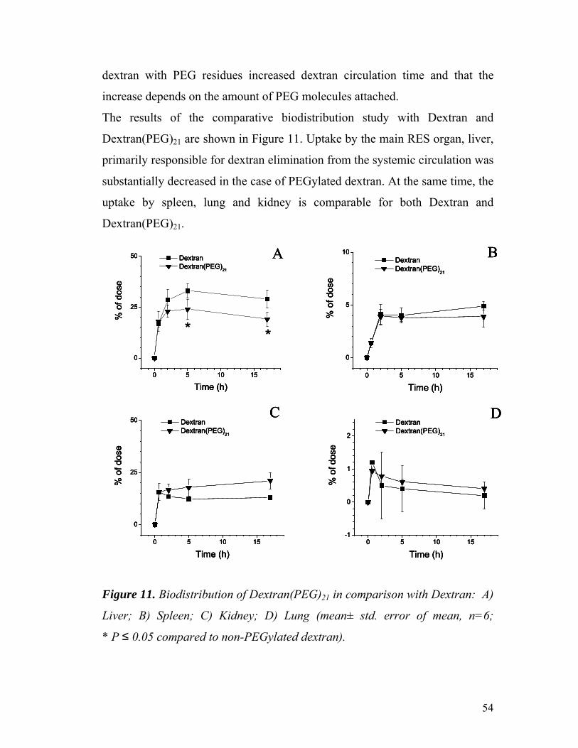
54
dextran with PEG residues increased dextran circulation time and that the
increase depends on the amount of PEG molecules attached.
The results of the comparative biodistribution study with Dextran and
Dextran(PEG)21 are shown in Figure 11. Uptake by the main RES organ, liver,
primarily responsible for dextran elimination from the systemic circulation was
substantially decreased in the case of PEGylated dextran. At the same time, the
uptake by spleen, lung and kidney is comparable for both Dextran and
Dextran(PEG)21.
0 5 10 15
0
5
10
% o
f dos
e
Time (h)
Dextran Dextran(PEG)21
0 5 10 15
0
25
50
% o
f dos
e
Time (h)
Dextran Dextran(PEG)21
0 5 10 15-1
0
1
2
% o
f dos
e
Time (h)
Dextran Dextran(PEG)21
0 5 10 15
0
25
50
% o
f dos
e
Time (h)
Dextran Dextran(PEG)21
A B
C D
0 5 10 15
0
5
10
% o
f dos
e
Time (h)
Dextran Dextran(PEG)21
0 5 10 15
0
25
50
% o
f dos
e
Time (h)
Dextran Dextran(PEG)21
0 5 10 15-1
0
1
2
% o
f dos
e
Time (h)
Dextran Dextran(PEG)21
0 5 10 15
0
25
50
% o
f dos
e
Time (h)
Dextran Dextran(PEG)21
A B
C D
**
0 5 10 15
0
5
10
% o
f dos
e
Time (h)
Dextran Dextran(PEG)21
0 5 10 15
0
25
50
% o
f dos
e
Time (h)
Dextran Dextran(PEG)21
0 5 10 15-1
0
1
2
% o
f dos
e
Time (h)
Dextran Dextran(PEG)21
0 5 10 15
0
25
50
% o
f dos
e
Time (h)
Dextran Dextran(PEG)21
A B
C D
0 5 10 15
0
5
10
% o
f dos
e
Time (h)
Dextran Dextran(PEG)21
0 5 10 15
0
25
50
% o
f dos
e
Time (h)
Dextran Dextran(PEG)21
0 5 10 15-1
0
1
2
% o
f dos
e
Time (h)
Dextran Dextran(PEG)21
0 5 10 15
0
25
50
% o
f dos
e
Time (h)
Dextran Dextran(PEG)21
A B
C D
**
Figure 11. Biodistribution of Dextran(PEG)21 in comparison with Dextran: A)
Liver; B) Spleen; C) Kidney; D) Lung (mean± std. error of mean, n=6;
* P ≤ 0.05 compared to non-PEGylated dextran).

55
In conclusion, the activation of dextran with CDI provides a convenient
method for the preparation of aminodextran by the attachment of EDA
residues. Introduced amino groups can be used for the further attachment of
PEG residues using PEG-SPA with a nearly quantitative reaction yield. The
modification with PEG decreases the uptake of dextran by RES organs and
substantially increases the dextran circulation time. The increase in circulation
time depends on the number of PEG residues introduced. Thus, PEGylated
dextrans (with residual reactive groups, such as amino groups) can be used as
convenient long-circulating carriers for therapeutic and diagnostic agents.

56
5.2 PEG-lipid micelles with magnetic sensitivity.
5.2.1 Particle size and surface potential of SPION micelles.
The SPION-micelle formulation was stable and did not show signs of
aggregation of SPION for approx 2 weeks at 4°C. The size of the micelles was
generally in the range of 30 – 50 nm (Figure 12). The surface potential for the
SPION-micelles was – 26.7 ± 3.1 mV and was not different than pure
mPEG2000-PE micelles of – 29.7 ± 2.4 mV.
Figure 12. Particle size for SPION-micelles measured using dynamic light
scattering in ZetaPlus particle size analyzer.
5.2.2 Freeze-fracture electron microscopy.
Freeze fracture electron microscopy was used to study the morphology of the
SPION-micelles and confirm whether there is a difference from the
morphology of ‘plain’ PEG-PE micelles as well as for determining whether the
SPION were loaded inside the lipid core of the PEG-PE micelles or were
present as aggregates externally. As can be clearly seen from the electron
micrographs taken from several freeze-fracture preparations of either ‘plain’
PEG-PE micelles or SPION-loaded PEG-PE micelles, both samples contain

57
medium concentration of small (ca. 30-to-50 nm), overall spherical particles,
which indicated that the normal morphology of the micelles was maintained.
They display their shadows behind the structure, which is typical for ‘tight-
core’ particles such as micelles. The only clear difference in electron
micrographs of the two samples was that in case of SPION-loaded PEG-PE
micelles, the structures show an additional darker inner core, a clear indication
of encapsulated SPION (typical micrographs in Figure 13).
A BA BAA BB
Figure 13. Freeze-fracture micrographs of (A) ‘plain’ PEG-PE micelles and
(B) SPION-loaded PEG-PE micelles (red arrows pointing at darker inner
core).
5.2.3 Relaxation rate of SPION-micelles.
For any MRI contrast the relaxation rate depends on the size of the contrast
agent, degree of aggregation and concentration. PEG-PE based polymeric
micelles were able to prevent the SPION form aggregation and hence the T2
relaxation rate was much greater in case of SPION-micelles when compared to
‘plain’ SPION at the same concentration (Figure 14). For example, at
0.0625 mg/mL of SPION, the relaxation rate of SPION-micelles was more
than 30 times the relaxation rate of ‘plain’ SPION. This is a very important and

58
desirable property for developing MRI contrast agents as more intense MRI
signals will be obtained at low concentrations of SPION, thus preventing
unnecessary high doses of the contrast agents to obtain clinically relevant MRI
images of the pathological organs.
0
0.05
0.1
0.15
0.2
0.25
0 0.025 0.05 0.075 0.1 0.125 0.15Concentration of SPION (mg/mL)
T2 re
laxa
tion
rate
(1/T
2) [s
ec-1
]
SPION-micelles
SPIONs0
0.05
0.1
0.15
0.2
0.25
0 0.025 0.05 0.075 0.1 0.125 0.15Concentration of SPION (mg/mL)
T2 re
laxa
tion
rate
(1/T
2) [s
ec-1
]
SPION-micelles
SPIONs
Figure 14. T2 relaxation rate (1/T2) of SPION-loaded micelles compared to
the relaxation rate of “plain” SPION in HBS pH 7.4 at room temperature
(mean± S.D., n=6).
Thus, the incorporation of SPIONs into mPEG-PE based polymeric micelles
prevented the SPION aggregation and also improved the MRI signal at low
concentration.
5.2.4 SPION-immunomicelles and their characterization.
In order to further improve the SPION-micelles we modified their outer PEG
corona with tumor cell-targeting nucleosome-specific mAb 2C5, using a

59
simple and reproducible chemistry using pNP-PEG3400-PE. (95). As was seen
previously, the attachment of mAb 2C5 did not change the size of the SPION-
immunomicelles (10, 36, 44, 74-77, 99). These SPION-immunomicelles were
also fluorescently-labeled using Rh-PE to follow their interaction with cells in
vitro. The conjugation of the antibody to the Rh-PE-labeled SPION-micelles
was gentle enough, since the ELISA assay indicated that the activity of the
mAb 2C5-SPION-immunomicelles towards the specific antigen, nucleosomes,
was maintained almost at the level of native unmodified mAb 2C5, while the
‘plain’ SPION- micelles or non-specific bovine IgG-modified SPION-micelles
did not show any binding to the monolayer of the nucleosomal antigen (Figure
15).
00.20.40.60.8
11.21.41.61.8
2
0 1 2 3 4Concentration of protein (µg/mL)
Abs
orba
nce
at 6
20-4
92 n
m
Native mAb 2C5"Plain" SPION-micellesmAb2C5 SPION-micellesBovine IgG SPION- micelles
Figure 15. Binding of mAb 2C5 SPION- immunomicelles and control ‘plain’
SPION-micelles or bovine IgG SPION-micelles to a monolayer of nucleosomes
(mean ± S.D., n=3).
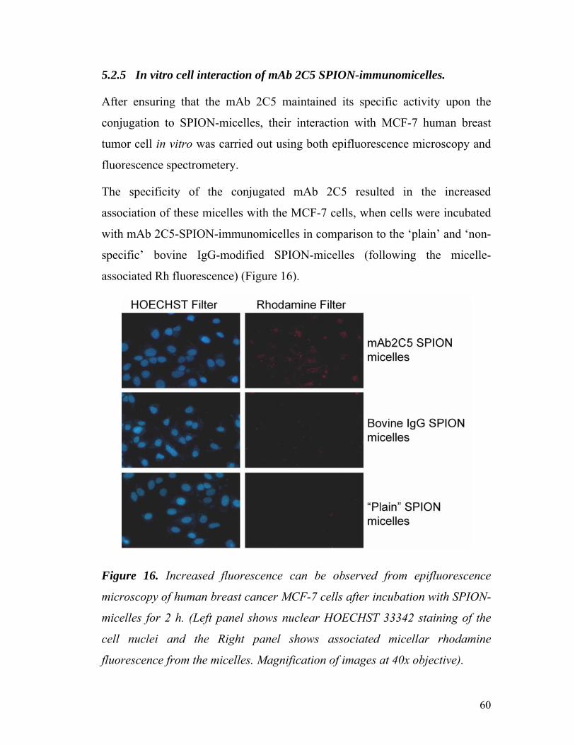
60
5.2.5 In vitro cell interaction of mAb 2C5 SPION-immunomicelles.
After ensuring that the mAb 2C5 maintained its specific activity upon the
conjugation to SPION-micelles, their interaction with MCF-7 human breast
tumor cell in vitro was carried out using both epifluorescence microscopy and
fluorescence spectrometery.
The specificity of the conjugated mAb 2C5 resulted in the increased
association of these micelles with the MCF-7 cells, when cells were incubated
with mAb 2C5-SPION-immunomicelles in comparison to the ‘plain’ and ‘non-
specific’ bovine IgG-modified SPION-micelles (following the micelle-
associated Rh fluorescence) (Figure 16).
Figure 16. Increased fluorescence can be observed from epifluorescence
microscopy of human breast cancer MCF-7 cells after incubation with SPION-
micelles for 2 h. (Left panel shows nuclear HOECHST 33342 staining of the
cell nuclei and the Right panel shows associated micellar rhodamine
fluorescence from the micelles. Magnification of images at 40x objective).
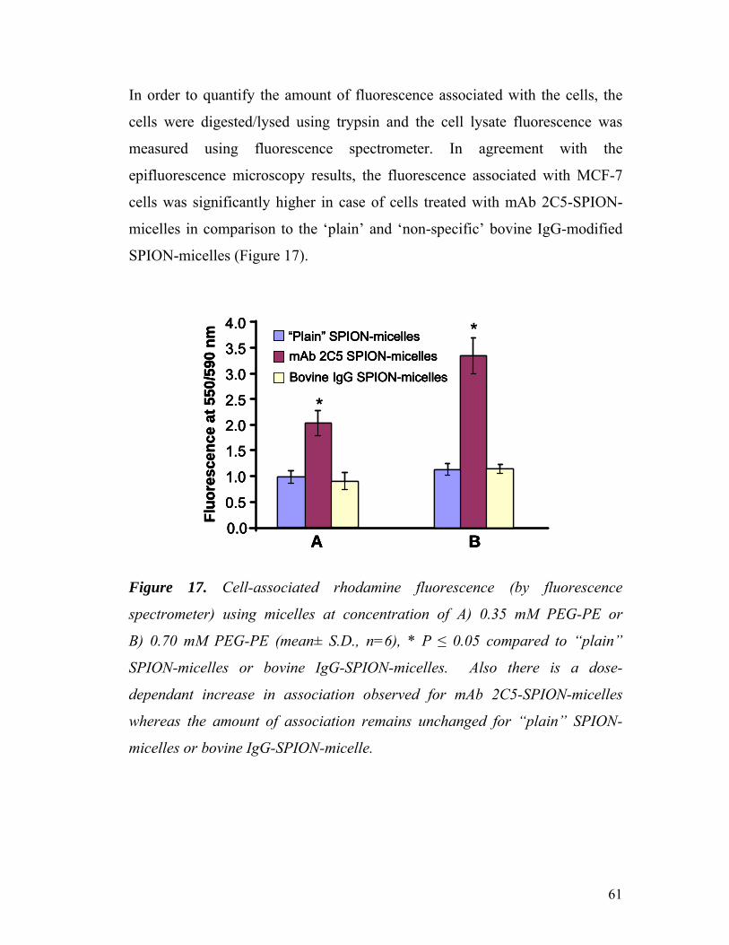
61
In order to quantify the amount of fluorescence associated with the cells, the
cells were digested/lysed using trypsin and the cell lysate fluorescence was
measured using fluorescence spectrometer. In agreement with the
epifluorescence microscopy results, the fluorescence associated with MCF-7
cells was significantly higher in case of cells treated with mAb 2C5-SPION-
micelles in comparison to the ‘plain’ and ‘non-specific’ bovine IgG-modified
SPION-micelles (Figure 17).
0.0
Fluo
resc
ence
at 5
50/5
90 n
m “Plain” SPION-micelles
A B
0.5
1.0
1.5
2.0
2.5
3.0
3.5
4.0
mAb 2C5 SPION-micellesBovine IgG SPION-micelles
0.0
Fluo
resc
ence
at 5
50/5
90 n
m “Plain” SPION-micelles
A B
0.5
1.0
1.5
2.0
2.5
3.0
3.5
4.0
mAb 2C5 SPION-micellesBovine IgG SPION-micelles
*
*
0.0
Fluo
resc
ence
at 5
50/5
90 n
m “Plain” SPION-micelles
A B
0.5
1.0
1.5
2.0
2.5
3.0
3.5
4.0
mAb 2C5 SPION-micellesBovine IgG SPION-micelles
0.0
Fluo
resc
ence
at 5
50/5
90 n
m “Plain” SPION-micelles
A B
0.5
1.0
1.5
2.0
2.5
3.0
3.5
4.0
mAb 2C5 SPION-micellesBovine IgG SPION-micelles
*
*
Figure 17. Cell-associated rhodamine fluorescence (by fluorescence
spectrometer) using micelles at concentration of A) 0.35 mM PEG-PE or
B) 0.70 mM PEG-PE (mean± S.D., n=6), * P ≤ 0.05 compared to “plain”
SPION-micelles or bovine IgG-SPION-micelles. Also there is a dose-
dependant increase in association observed for mAb 2C5-SPION-micelles
whereas the amount of association remains unchanged for “plain” SPION-
micelles or bovine IgG-SPION-micelle.

62
5.2.6 T2 relaxation rates of mAb2C5 SPION-immunomicelles on cells in
vitro.
The goal of developing cancer cell-targeted SPION-micelles was to ensure that
the higher amount of SPION is carried to the cancer cells for their more
efficient MR imaging. We determined the T2 relaxation time of the lysed cells
after their co-incubation with plain, non-specific IgG-modified or mAb 2C5-
modified SPION-micelles. The signal associated with the cells was determined
using 500 MHz NMR. We observed a significant increase in cell-associated T2
relaxation rate (1/T2) in case of cells treated with mAb 2C5 SPION-
immunomicelles (compared to non-targeted SPION micelles) (Table III). This
increased signal is the direct result of target cell recognition by mAb 2C5-
SPION-immunomicelles.
Concentration of PEG-PE applied to MCF-7 cells. Formulations
0.35 mM 0.70 mM
‘Plain’ SPION-micelles 4.3 ± 0.02 s-1 5.2 ± 0.05 s-1
mAb 2C5 SPION-micelles 5.6 ± 0.01 s-1 * 7.1 ± 0.02 s-1 *
Bovine IgG SPION- micelles 4.3 ± 0.00 s-1 6.0 ± 0.02 s-1
Table III. Relaxation rate (1/T2) of the MCF-7 cells after the incubation with
different concentrations of micellar preparations. (The relaxation rate of the
sample increases when larger quantity of SPION is associated with the cells).
(n=3, mean±std. error of mean,* p<0.05 when compared to ‘Plain’ SPION-
micelles and Bovine IgG SPION-micelles)

63
PEG-PE-based polymeric micelles offer a convenient carrier system for
suspending SPION in aqueous medium. The formulations were stable with no
apparent aggregation compared to ‘plain’ SPION. SPION-loaded PEG-PE
micelles provided dramatically improved T2 relaxation rates. These SPION-
micelles can be easily modified with anti-cancer nucleosome-specific mAb
2C5 with the preservation of the antibody specific activity to make them cancer
cell-specific. mAb 2C5-SPION-immunomicelles specifically recognize and
bind cancer cells in vitro, bring more SPION to these cells, and have the
potential to serve as an MRI contrast agent with improved T2 for better tumor
imaging. In addition, the specific accumulation of mAb 2C5-SPION-
immunomicelles into the cancer cells make these micelles good candidates for
use in conjunction with AC-field-assisted hyperthermic cancer therapy.
5.2.7 Magnetic targeting of SPION-micelles in tumor of tumor-bearing
mice in vivo under the influence of external magnet.
Since SPION have magnetic properties in presence of external magnetic fields,
we hypothesized that SPION-micelles could be targeted to the intended target
area using external magnets.
There is significantly more tumor accumulation of SPION-micelles in presence
of external magnet than compared to control non-magnet bearing BALB/c
mice at all time points (Figure 18). For example, at time 4 hr (240 min) the
amount of SPION-micelles accumulated in external magnet-bearing mice is
twice that of control non-magnet bearing mice. Thus targeting can be achieved
using SPION-micelles and external magnets. The SPION-micelles could
moreover be co-loaded with poorly soluble anti-cancer drugs to direct them
specifically to the tumor area. Magnetically targeted SPION-micelles may
effectively reduce the dose of anti-cancer drug required to get therapeutic
effects.

64
The only limitation to magnetic-targeting remaining, is that the tumor must be
easily accessible for focusing magnetic fields. Solid magnets as used in this
study or electro-magnets could be used as the source for focusing magnetic
fields in the intended target area. Also, depending on the field strength used
there is scope for improvement of the accumulation of the SPION-micelles.
0.00%
0.50%
1.00%
1.50%
2.00%
2.50%
3.00%
3.50%
10 30 60 240Time (min)
% in
ject
ed S
PIO
N-m
icel
les
per g
ram
of t
umor
With magnetWithout magnet
p < 0.05
p < 0.05
p < 0.05
*p = 0.0504
Figure 18. Accumulation of SPION-micelles in tumor tissue of tumor-bearing
Balb/C mice under influence of external magnets (n=6, mean±std. error of
mean).

65
5.3 Detachable PEG to make stimuli-sensitive multifunctional liposomes
and micelles.
The particular design of the multifunctional DDS we have used in this study is
presented in Figure 19. The suggested DDS (liposome or micelle) bears on its
surface a “hidden” function (we have used biotin and TATp moieties for this
purpose) inserted into the liposome membrane or micelle core via its
modification with PE moiety; protecting PEG chains (PEG2000) attached to the
surface via the pH-cleavable bond; and specific antibody attached to the
surface via non-cleavable long PEG spacers (PEG3400). In some experiments
with liposomes, cleavable PEG5000-Hz-PE and non-cleavable TATp-PEG2000-
PE conjugates were used (88).
Incubation at lowered pH
Degradation of PEG chains
Targeting via target specific antibody and long circulation
a
b
c
aa a
b
Internalization via TAT peptide
Incubation at lowered pH
Degradation of PEG chains
Incubation at lowered pH
Degradation of PEG chains
Targeting via target specific antibody and long circulation
a
b
c
aa a
b
Targeting via target specific antibody and long circulation
a
b
c
aa a
b
Internalization via TAT peptideInternalization via TAT peptide
Figure 19. Schematic for the design of the multifunctional DDS used in this
study that includes pH-cleavable PEG-Hz-PE (a), and TATp (b), and
monoclonal antibody (c) attached to the surface of DDS via pH-non-cleavable
spacer.
66
Such DDS (Figure 19) are expected to demonstrate specific targeted properties
(via antibody-mediated recognition) at both normal (7.5-8.0) and acidic (5.0-
6.0) pH values, however, the incubation of the DDS at lowered pH should
eliminate (detach) protecting PEG chains and un-shield the second function.
After exposure to the lowered pH, in addition to immune recognition, DDS
should be able to bind with the avidin column if the second, “hidden” function
is biotin, or demonstrate internalization by cells if the second, “hidden”
function is TATp. As an antibody, we have used the cardiac myosin-specific
monoclonal 2G4 antibody (mAb 2G4) (100). The coupling of mAb 2G4 and
biotin or TATp to the DDS surface was performed using the reactive derivative
of PEG-PE conjugate activated at the free PEG terminus with a
p-nitrophenylcarbonyl (pNP) group (pNP-PEG-PE) (95).
5.3.1 Synthesis of pH-cleavable PEG-PE conjugate.
The scheme of the synthesis of PEG-PE conjugated via the pH-cleavable
hydrazone group (PEG-Hz-PE) is shown in the Figure 20. The synthesis was
carried out in 2 steps. The first step involves the conjugation of 3-(2-
pyridyldithio) propionyl hydrazide (PDPH) to mPEG2000-CHO. The hydrazide
group in PDPH reacts with the aldehyde group of mPEG2000-CHO to form the
acidic-pH-labile hydrazone bond. Since this bond is vulnerable to hydrolysis,
efforts were taken to carry out the reaction in anhydrous conditions. Use of
PDPH as the cross linker not only offers the advantage of forming the
hydrazone bond but also introducing pyridyldisulfanyl groups for subsequent
conjugation of PEG to the thiol component of 1,2-dipalmitoyl-sn-glycero-3-
phosphothioethanol (PE-SH) in the second step of the synthesis. The PE-SH in
the conjugate serves as a hydrophobic anchor to assure its association with the
lipid bilayer of liposomes or the hydrophobic core of micelles. All steps of the

67
reaction were followed on TLC to confirm the progress of the reaction. The
structure of the final conjugate, mPEG2000-phospatidyl ethanolamine hydrazone
(mPEG2000-Hz-PE) was confirmed by proton NMR characterization.

68
Step 1.
SSN
HH2N
ONO
HCH3O(CH2CH2O)n +
SSN
H
ONN
CH3O(CH2CH2O)n
mPEG-butyraldhehyde PDPH
dry CHCl3stirring, room temperature48h
mPEG-Hz-PDP
Step 2.
OP
O O C15H31O
O
C15H31
O
HS
NaOO
SSN
H
ONN
CH3O(CH2CH2O)n +
OP
O O C15H31O
O
C15H31
O
NaOO
SSN
H
ON
CH3O(CH2CH2O)n
stirring, room temperatureovernight
PE-SHmPEG-Hz-PDP
dry CHCl3,TEA
mPEG-Hz-PE
Figure 20. Schematic description of the conjugation reaction for preparing
pH-sensitive PEG-Hz-PE.

69
The stability of the conjugate and the kinetics of its degradation were analyzed
by size-exclusion HPLC following the area under the micelle peak on the
chromatogram after conjugate incubation for different time intervals at
different pH values. The conjugate spontaneously forms micelles in aqueous
solutions similar to “normal” PEG-PE conjugate. Rhodamine-PE was
incorporated into the micelles as a fluorescent tag, and the sample was
monitored using a fluorescence detector with excitation at 550 nm and
emission at 590 nm. As a typical example, HPLC for mPEG2000-Hz-PE is
shown in Figure 21. After appropriate pH treatment the peak at retention time
9.7 min (peak for micelle) observed in case of intact micelles (pH 8.0)
disappears after incubation at pH 5.0. This disappearance is indicative of the
destruction of the micelle structure due to the loss of the PEG corona, while
non-pH-sensitive micelles produce the peak at both pH values (data not
shown).
Retention Time (min)
400350300250200150100
500
0 2 4 6 8
Inte
nsity
(mV
)
Micelle peak
Absence of the micelle peak
12 14 1610
A
B
Retention Time (min)
400350300250200150100
500
400350300250200150100
500
0 2 4 6 8
Inte
nsity
(mV
)
Micelle peak
Absence of the micelle peak
12 14 1610
A
B
Figure 21. HPLC analysis of the pH-sensitive mPEG2000-Hz-PE micelles
after incubation in pH 8.0 (A) and after incubation in pH 5 (B) at room
temperature.

70
Table IV shows the representative data for the degradation kinetics of PEG2000-
Hz-DPPE micelles at different pH values. It is clearly seen that such micelles
are quite stable at high pH value (8 and above) but they disintegrate within a
few minutes at pH 5.0. This result was also confirmed by TLC, where two
separate spots for PEG and PE were observed after a few minutes of incubation
at pH 5.0 with no TLC spot corresponding to intact PEG-PE (data not shown).
pH value
Incubation Time 5.0 7.0 8.0 10.0
20 min 3 56 94 99
40 min 2.5 28 62 99
60 min 2 10 53 99
Table IV. PEG2000-Hz-PE micelle stability at different pH values (as per cent
of remaining micelles)
5.3.2 Immunoreactivity of multifunctional drug delivery systems.
Fully assembled 2G4 antibody-bearing DDS demonstrated clear
immunoreactivity towards the antigen, the monolayer of dog cardiac myosin in
the standard ELISA test at both pH 8.0 and 5.0 in PEG-PE-based 2G4-
immunomicelles (Figure 22). There is a decreased affinity for the antibodies
modified with the pNP-PEG-PE anchor and incorporated into the micelle
structure. The same pattern is observed for immunoliposomes. This decrease
is more apparent than real, since not all DDS-attached antibodies, even
remaining active, can interact with the substrate because of their steric

71
orientation to the DDS surface, and as was shown earlier, is compensated by
the multipoint interaction of antibody-modified drug delivery systems with the
target (74, 101). Thus, the systems prepared are immunologically active at both
pH values. Control preparations bearing a non-specific IgG did not show any
binding with myosin at any pH.
0.0
0.2
0.4
0.6
0.8
1.0
1.2
1.4
1.6
1.8
2.0
0.0 2.0 4.0 6.0 8.0 10.0
Concentration of mAb 2G4 (µg/mL)
Abs
orba
nce
at 6
20 -4
92 n
m
Native mAb 2G4, pH 8.0Native mAb 2G4, pH 5.0mAb 2G4 Immunomicelles, pH 8.0mAb 2G4 Immunomicelles, pH 5.0
Figure 22. Binding of antimyosin mAb 2G4-PEG2000-Hz-PE-immunomicelles
to a monolayer of dog cardiac myosin in comparison to the native mAb 2G4 at
corresponding pH values (mean ± S.D.; n =3).
5.3.3 Avidin binding of multifunctional biotin-containing drug delivery
systems.
Avidin-biotin complexation was used initially as an easy-to-use test system to
follow the shielding and un-shielding of the second hidden function in pH-
sensitive drug delivery systems. Liposomes and micelles containing 5 % mol
of the biotin-PE in addition to 2G4-PEG3400-PE and pH-sensitive PEG2000-Hz-
PE, were prepared and labeled with rhodamine-PE, and their ability to interact
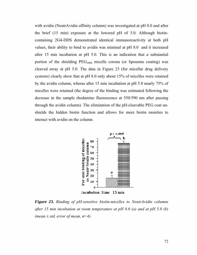
72
with avidin (NeutrAvidin affinity column) was investigated at pH 8.0 and after
the brief (15 min) exposure at the lowered pH of 5.0. Although biotin-
containing 2G4-DDS demonstrated identical immunoreactivity at both pH
values, their ability to bind to avidin was minimal at pH 8.0 and it increased
after 15 min incubation at pH 5.0. This is an indication that a substantial
portion of the shielding PEG2000 micelle corona (or liposome coating) was
cleaved away at pH 5.0. The data in Figure 23 (for micellar drug delivery
systems) clearly show that at pH 8.0 only about 15% of micelles were retained
by the avidin column, wheras after 15 min incubation at pH 5.0 nearly 75% of
micelles were retained (the degree of the binding was estimated following the
decrease in the sample rhodamine fluorescence at 550/590 nm after passing
through the avidin column). The elimination of the pH-cleavable PEG coat un-
shields the hidden biotin function and allows for more biotin moieties to
interact with avidin on the column.
Figure 23. Binding of pH-sensitive biotin-micelles to NeutrAvidin columns
after 15 min incubation at room temperature at pH 8.0 (a) and at pH 5.0 (b)
(mean ± std. error of mean, n=4).

73
5.3.4 TATp-mediated interaction of multifunctional drug delivery systems
with cells in vitro.
For in vitro cell culture experiments, we have used rhodamine-labeled DDS
similar to those described above, but containing TATp moieties attached to
their surface instead of biotin groups. In this case, we have investigated DDS
internalization by various cells (non-targets for the 2G4 antibody) at pH 8.0
and after a brief (20-30 min) exposure at pH 5.0. TATp-containing DDS
interact differently with cells at pH 8.0 than after incubation at pH 5.0, which
indicated that at pH 5.0 a substantial portion of the shielding PEG is lost from
the micelle corona (or liposome coating). Figure 24 clearly shows that while
cleavable PEG-PE-based TATp-containing micelles kept at pH 8.0 show only
marginal association with NIH-3T3 murine fibroblasts, the same micelles pre-
incubated for 30 min at pH 5.0 demonstrated dramatically enhanced
association with the cells (higher fluorescence), i.e. better accessibility of
TATp moieties for cell interaction.
A BA B
Figure 24. Fluorescence microscopy showing internalization of Rh-PE-
labeled-TATp-containing micelles by NIH 3T3 fibroblast cells after incubating
micelles at pH 8.0 (A) and pH 5.0 (B) for 30 min.

74
In the case of TAT-bearing liposomes (Figure 25), we have shown that the
incorporation of 9 % mol of PEG-PE strongly diminished TATp-uptake of
liposomes (Figure 25B), and the incorporation of 18 % mol of PEG-PE
completely eliminated it (Figure 25C). However when pH-degradable PEG-PE
was used, 20 min preincubation of both preparations at pH 5.0 significantly
increased the association of both preparations with cells, bringing the cell
binding of the liposomes with 9 % mol PEG (Figure 25D) almost back to the
level of PEG-free TATp-liposomes (Figure 25A), and significantly improving
the cell binding of the TATp-liposomes with 18 % mol of the initial PEG
(Figure 25E). These results clearly confirm that the elimination of the pH-
cleavable PEG coat de-shield the hidden TATp function and allows for better
association of DDS with cells.

75
B C
D E
A
Figure 25. Fluorescence microscopy showing internalization of Rh-PE-
labeled-TATp containing liposomes by U-87 MG astrocytoma cells using:
PEG-free TATp-liposomes (A); 9 % mol pH-non-cleavable PEG-PE at pH 7.4
(B); 18 % mol pH-non-cleavable PEG-PE at pH 7.4 (C); 9 % mol pH-
cleavable PEG-Hz-PE after incubation at pH 5.0 for 20 min (D)*, 18 % mol
pH-cleavable PEG-Hz-PE after incubation at pH 5.0 for 20 min (E)*.
* The pH of these formulations was raised back to pH 7.4 after their
incubation at pH 5.0 and prior to incubation with cells.

76
Using pH-sensitive PEG-PE conjugates double-targeted drug carriers have
been developed, which, in addition to prolonged circulation (via the attached
PEG) and target recognition (via the attached antibody) can carry a temporarily
hidden function which can be developed under the action of certain local
stimuli (such as lowered pH). This represents a significant step on the way
towards “smart” multifunctional pharmaceutical nanocarriers capable of both
target accumulation and intra-cellular penetration in a controlled fashion.

77
6 CONCLUSIONS
PEG is an important biomedical polymer and has proved to be backbone for
successful design and application of many therapeutic or diagnostic delivery
systems.
A. PEG modification of biocompatible and biodegradable polymer dextran
results in dextran-based degradable polymeric drug carrier which has
improved and longer blood-circulating properties.
B. Polymeric micelles based on PEG-PE conjugates can be successfully used
for various purposes including the preparation of actively and passively
targeted tumor-targeted magnetically sensitive micelles loaded with
SPION.
C. PEG-based conjugate with lowered pH-sensitive bond between PEG and
PE allow for the preparation of multifunctional pharmaceutical nanocarriers
where the cell-penetrating function is sterically shielded by PEG chains
under normal conditions in the circulation, but becomes exposed and
functional after PEG detaches in the acidic environment in the tumor.

78
P.S. Some of the data in this thesis work has already been published in (58, 88)
and other parts of the data have been submitted for publication.

79
7 REFERENCES
(1) Torchilin, V. P. (2000) Drug targeting. European Journal of
Pharmaceutical Sciences 11 Suppl 2, S81-91.
(2) Presant, C. A., Wolf, W., Waluch, V., Wiseman, C., Kennedy, P.,
Blayney, D., and Brechner, R. R. (1994) Association of intratumoral
pharmacokinetics of fluorouracil with clinical response. Lancet 343,
1184-7.
(3) Jain, R. K. (1994) Barriers to drug delivery in solid tumors. Scientific
American 271, 58-65.
(4) Jain, R. K. (1990) Vascular and interstitial barriers to delivery of
therapeutic agents in tumors. Cancer Metastasis Reviews 9, 253-66.
(5) Yuan, F., Dellian, M., Fukumura, D., Leunig, M., Berk, D. A.,
Torchilin, V. P., and Jain, R. K. (1995) Vascular permeability in a
human tumor xenograft: molecular size dependence and cutoff size.
Cancer Research 55, 3752-6.
(6) Iwai, K., Maeda, H., and Konno, T. (1984) Use of oily contrast medium
for selective drug targeting to tumor: enhanced therapeutic effect and X-
ray image. Cancer Research 44, 2115-21.
(7) Matsumura, Y., and Maeda, H. (1986) A new concept for
macromolecular therapeutics in cancer chemotherapy: mechanism of
tumoritropic accumulation of proteins and the antitumor agent smancs.
Cancer Research 46, 6387-92.
(8) Maeda, H., Wu, J., Sawa, T., Matsumura, Y., and Hori, K. (2000)
Tumor vascular permeability and the EPR effect in macromolecular
therapeutics: a review. Journal of Controlled Release 65, 271-84.
(9) Muzykantov, V., and Torchilin, V. (2002) Biomedical aspects of drug
targeting, Kluwer Academic Pub., Boston.

80
(10) Torchilin, V. P. (2004) Targeted polymeric micelles for delivery of
poorly soluble drugs. Cellular and Molecular Life Sciences 61, 2549-59.
(11) Torchilin, V. P. (2006) Nanoparticulates as Drug Carriers, Imperial
College Press, London.
(12) Duncan, R., and Spreafico, F. (1994) Polymer conjugates.
Pharmacokinetic considerations for design and development. Clinical
Pharmacokinetics 27, 290-306.
(13) Roberts, M. J., and Harris, J. M. (1998) Attachment of degradable
poly(ethylene glycol) to proteins has the potential to increase
therapeutic efficacy. Journal of Pharmaceutical Sciences 87, 1440-5.
(14) Harris, J. M., Martin, N. E., and Modi, M. (2001) Pegylation: a novel
process for modifying pharmacokinetics. Clinical Pharmacokinetics 40,
539-51.
(15) Duncan, R. (2003) The dawning era of polymer therapeutics. Nature
Reviews Drug Discovery 2, 347-60.
(16) Molineux, G. (2003) Pegylation: Engineering Improved
Biopharmaceuticals for Oncology. Pharmacotherapy 23, 3S-8S.
(17) Torchilin, V. P. (1991) Immobilized enzymes in medicine, Springer-
Verlag, Berlin ; New York.
(18) Vicent, M. J., and Duncan, R. (2006) Polymer conjugates: nanosized
medicines for treating cancer. Trends in Biotechnology 24, 39-47.
(19) Minko, T., Kopeckova, P., and Kopecek, J. (2000) Efficacy of the
chemotherapeutic action of HPMA copolymer-bound doxorubicin in a
solid tumor model of ovarian carcinoma. International Journal of
Cancer 86, 108-17.
(20) Torchilin, V. P., Trubetskoy, V. S., Whiteman, K. R., Caliceti, P.,
Ferruti, P., and Veronese, F. M. (1995) New synthetic amphiphilic
polymers for steric protection of liposomes in vivo. Journal of
Pharmaceutical Sciences 84, 1049-53.

81
(21) Torchilin, V. P., and Trubetskoy, V. S. (1995) Which polymers can
make nanoparticulate drug carriers long-circulating? Advanced Drug
Delivery Reviews 16, 141-155.
(22) Kwon, G. S. (2005) Polymeric drug delivery systems, Taylor & Francis,
Boca Raton.
(23) Papahadjopoulos, D., Allen, T. M., Gabizon, A., Mayhew, E., Matthay,
K., Huang, S. K., Lee, K. D., Woodle, M. C., Lasic, D. D., Redemann,
C., and et al. (1991) Sterically stabilized liposomes: improvements in
pharmacokinetics and antitumor therapeutic efficacy. Proceedings of the
National Academy of Sciences of the United States of America 88,
11460-4.
(24) Ranucci, E., Spagnoli, G., Sartore, L., Ferruti, P., Caliceti, P., Schiavon,
O., and Veronese, F. M. (1994) Synthesis and molecular weight
characterization of low molecular weight end-functionalized poly(4-
acryloylmorpholine). Macromolecular Chemistry and Physics 195,
3469-3479.
(25) Woodle, M. C. (1998) Controlling liposome blood clearance by surface-
grafted polymers. Advanced Drug Delivery Reviews 32, 139-152.
(26) Lasic, D. D., Vallner, J. J., and Working, P. K. (1999) Sterically
stabilized liposomes in cancer therapy and gene delivery. Current
Opinion in Molecular Therapeutics 1, 177-85.
(27) Torchilin, V. P., Levchenko, T. S., Whiteman, K. R., Yaroslavov, A. A.,
Tsatsakis, A. M., Rizos, A. K., Michailova, E. V., and Shtilman, M. I.
(2001) Amphiphilic poly-N-vinylpyrrolidones: synthesis, properties and
liposome surface modification. Biomaterials 22, 3035-44.
(28) Homsi, J., Simon, G. R., Garrett, C. R., Springett, G., De Conti, R.,
Chiappori, A. A., Munster, P. N., Burton, M. K., Stromatt, S., Allievi,
C., Angiuli, P., Eisenfeld, A., Sullivan, D. M., and Daud, A. I. (2007)
Phase I Trial of Poly-L-Glutamate Camptothecin (CT-2106)

82
Administered Weekly in Patients with Advanced Solid Malignancies.
Clinical Cancer Research 13, 5855-5861.
(29) Veronese, F. M., and Pasut, G. (2005) PEGylation, successful approach
to drug delivery. Drug Discovery Today 10, 1451-8.
(30) Chapman, A. P., Antoniw, P., Spitali, M., West, S., Stephens, S., and
King, D. J. (1999) Therapeutic antibody fragments with prolonged in
vivo half-lives. Nature Biotechnology 17, 780-3.
(31) Lasic, D. D., Martin, F. J., Gabizon, A., Huang, S. K., and
Papahadjopoulos, D. (1991) Sterically stabilized liposomes: a
hypothesis on the molecular origin of the extended circulation times.
Biochimica et Biophysica Acta 1070, 187-92.
(32) Gabizon, A., and Papahadjopoulos, D. (1988) Liposome formulations
with prolonged circulation time in blood and enhanced uptake by
tumors. Proceedings of the National Academy of Sciences of the United
States of America 85, 6949-53.
(33) Gabizon, A., and Martin, F. (1997) Polyethylene glycol-coated
(pegylated) liposomal doxorubicin. Rationale for use in solid tumours.
Drugs 54 Suppl 4, 15-21.
(34) Report Final Report on the Safety Assessment of Polyethylene (2007)
Informa Healthcare 115 - 127
(35) Torchilin, V. P., Omelyanenko, V. G., Papisov, M. I., Bogdanov, A. A.,
Jr., Trubetskoy, V. S., Herron, J. N., and Gentry, C. A. (1994)
Poly(ethylene glycol) on the liposome surface: on the mechanism of
polymer-coated liposome longevity. Biochimica et Biophysica Acta
1195, 11-20.
(36) Elbayoumi, T. A., and Torchilin, V. P. (2008) Tumor-specific antibody-
mediated targeted delivery of Doxil® reduces the manifestation of
auricular erythema side effect in mice. International Journal of
Pharmaceutics 357, 272-279

83
(37) Roberts, M. J., Bentley, M. D., and Harris, J. M. (2002) Chemistry for
peptide and protein PEGylation. Advanced Drug Delivery Reviews 54,
459-76.
(38) Kozlowski, A., and Harris, J. M. (2001) Improvements in protein
PEGylation: pegylated interferons for treatment of hepatitis C. Journal
of Controlled Release 72, 217-24.
(39) Garman, A. J., and Kalindjian, S. B. (1987) The preparation and
properties of novel reversible polymer-protein conjugates. 2-omega-
Methoxypolyethylene (5000) glycoxymethylene-3-methylmaleyl
conjugates of plasminogen activators. FEBS Letters 223, 361-5.
(40) Zalipsky, S., Qazen, M., Walker, J. A., 2nd, Mullah, N., Quinn, Y. P.,
and Huang, S. K. (1999) New detachable poly(ethylene glycol)
conjugates: cysteine-cleavable lipopolymers regenerating natural
phospholipid, diacyl phosphatidylethanolamine. Bioconjugate
Chemistry 10, 703-7.
(41) Lukyanov, A. N., Sawant, R. M., Hartner, W. C., and Torchilin, V. P.
(2004) PEGylated dextran as long-circulating pharmaceutical carrier.
Journal of Biomaterials Science Polymer Edition 15, 621-30.
(42) Dabholkar, R. D., Sawant, R. M., Mongayt, D. A., Devarajan, P. V., and
Torchilin, V. P. (2006) Polyethylene glycol-phosphatidylethanolamine
conjugate (PEG-PE)-based mixed micelles: some properties, loading
with paclitaxel, and modulation of P-glycoprotein-mediated efflux.
International Journal of Pharmaceutics 315, 148-57.
(43) Torchilin, V. P. (2007) Micellar nanocarriers: pharmaceutical
perspectives. Pharmaceutical Research 24, 1-16.
(44) Ai, H., Flask, C., Weinberg, B., Shuai, X. T., Pagel, M. D., Farrell, D.,
Duerk, J., and Gao, J. (2005) Magnetite-Loaded Polymeric Micelles as
Ultrasensitive Magnetic-Resonance Probes. Advanced Materials 17,
1949-1952.

84
(45) Corot, C., Robert, P., Idee, J. M., and Port, M. (2006) Recent advances
in iron oxide nanocrystal technology for medical imaging. Advanced
Drug Delivery Reviews 58, 1471-504.
(46) Engin, K., Leeper, D. B., Thistlethwaite, A. J., Tupchong, L., and
McFarlane, J. D. (1994) Tumor extracellular pH as a prognostic factor
in thermoradiotherapy. International Journal of Radiation
Oncology•Biology•Physics 29, 125-32.
(47) Engin, K., Leeper, D. B., Cater, J. R., Thistlethwaite, A. J., Tupchong,
L., and McFarlane, J. D. (1995) Extracellular pH distribution in human
tumours. International Journal of Hyperthermia 11, 211-6.
(48) Stubbs, M., McSheehy, P. M., Griffiths, J. R., and Bashford, C. L.
(2000) Causes and consequences of tumour acidity and implications for
treatment. Molecular Medicine Today 6, 15-9.
(49) Jayasundar, R., and Singh, V. P. (2002) In vivo temperature
measurements in brain tumors using proton MR spectroscopy.
Neurology India 50, 436-9.
(50) Torchilin, V. P. (2005) Lipid-core micelles for targeted drug delivery.
Current Drug Delivery 2, 319-27.
(51) Torchilin, V. P. (2005) Recent advances with liposomes as
pharmaceutical carriers. Nature Reviews Drug Discovery 4, 145-60.
(52) Mehvar, R. (2000) Dextrans for targeted and sustained delivery of
therapeutic and imaging agents. Journal of Controlled Release 69, 1-25.
(53) Bernstein, A., Hurwitz, E., Maron, R., Arnon, R., Sela, M., and
Wilchek, M. (1978) Higher antitumor efficacy of daunomycin when
linked to dextran: in vivo and in vitro studies. Journal of the National
Cancer Institute 60, 379-84.
(54) Walker, G. J. (1978) Biochemistry of carbohydrates II, University Park
Press, Baltimore.

85
(55) Preobrazhenskaya, M. E., Minakova, A. L., and Rosenfeld, E. L. (1974)
Studies of the dextranase activity of pig-spleen acid alpha-D-
glucosidase. Carbohydrate Research 38, 267-77.
(56) Larsen, C. (1989) Dextran prodrugs - structure and stability in relation
to therapeutic activity. Advanced Drug Delivery Reviews 3, 103-154.
(57) Yamaoka, T., Tabata, Y., and Ikada, Y. (1994) Distribution and tissue
uptake of poly(ethylene glycol) with different molecular weights after
intravenous administration to mice. Journal of Pharmaceutical Sciences
83, 601-6.
(58) Lukyanov, A. N., Sawant, R. M., Hartner, W. C., and Torchilin, V. P.
(2004) PEGylated dextran as long-circulating pharmaceutical carrier.
Journal of Biomaterials Science Polymer Edition 15, 621-30.
(59) Jones, M., and Leroux, J. (1999) Polymeric micelles - a new generation
of colloidal drug carriers. European Journal of Pharmaceutics and
Biopharmaceutics 48, 101-11.
(60) Jones, M. C., Ranger, M., and Leroux, J. C. (2003) pH-sensitive
unimolecular polymeric micelles: synthesis of a novel drug carrier.
Bioconjugate Chemistry 14, 774-81.
(61) Roby, A., Erdogan, S., and Torchilin, V. P. (2006) Solubilization of
poorly soluble PDT agent, meso-tetraphenylporphin, in plain or
immunotargeted PEG-PE micelles results in dramatically improved
cancer cell killing in vitro. European Journal of Pharmaceutics and
Biopharmaceutics 62, 235-40.
(62) Skidan, I., Dholakia, P., and Torchilin, V. (2008) Photodynamic therapy
of experimental B-16 melanoma in mice with tumor-targeted
5,10,15,20-tetraphenylporphin-loaded PEG-PE micelles. Journal of
Drug Targeting 16, 486-93.
(63) Jones, M. C., Gao, H., and Leroux, J. C. (2008) Reverse polymeric
micelles for pharmaceutical applications. Journal of Controlled Release.

86
(64) Yokoyama, M., Okano, T., Sakurai, Y., Fukushima, S., Okamoto, K.,
and Kataoka, K. (1999) Selective delivery of adriamycin to a solid
tumor using a polymeric micelle carrier system. Journal of Drug
Targeting 7, 171-86.
(65) Klibanov, A. L., Maruyama, K., Torchilin, V. P., and Huang, L. (1990)
Amphipathic polyethyleneglycols effectively prolong the circulation
time of liposomes. FEBS Letters 268, 235-7.
(66) Bedu-Addo, F. K., Tang, P., Xu, Y., and Huang, L. (1996) Interaction of
polyethyleneglycol-phospholipid conjugates with cholesterol-
phosphatidylcholine mixtures: sterically stabilized liposome
formulations. Pharmaceutical Research 13, 718-24.
(67) Mulder, W. J., Strijkers, G. J., van Tilborg, G. A., Griffioen, A. W., and
Nicolay, K. (2006) Lipid-based nanoparticles for contrast-enhanced
MRI and molecular imaging. NMR in Biomedicine 19, 142-64.
(68) Iakoubov, L. Z., and Torchilin, V. P. (1997) A novel class of antitumor
antibodies: nucleosome-restricted antinuclear autoantibodies (ANA)
from healthy aged nonautoimmune mice. Oncoogy Reearch 9, 439-46.
(69) Chakilam, A. R., Pabba, S., Mongayt, D., Iakoubov, L.Z., Torchilin,
V.P. (2004) A single monoclonal antinuclear autoantibody with
nucleosome-restricted specificity inhibits growth of diverse human
tumors in nude mice. Cancer Therapy 2, 353-64.
(70) Iakoubov, L. Z., and Torchilin, V. P. (1997) A novel class of antitumor
antibodies: nucleosome-restricted antinuclear autoantibodies (ANA)
from healthy aged nonautoimmune mice. Oncology Research 9, 439-46.
(71) Iakoubov, L. Z., and Torchilin, V. P. (1998) Nucleosome-releasing
treatment makes surviving tumor cells better targets for nucleosome-
specific anticancer antibodies. Cancer Detection and Prevention 22,
470-5.

87
(72) Koutouzov, S., Cabrespines, A., Amoura, Z., Chabre, H., Lotton, C.,
and Bach, J. F. (1996) Binding of nucleosomes to a cell surface
receptor: redistribution and endocytosis in the presence of lupus
antibodies. European Journal of Immunology 26, 472-86.
(73) Torchilin, V. P., Lukyanov, A. N., Gao, Z., and Papahadjopoulos-
Sternberg, B. (2003) Immunomicelles: targeted pharmaceutical carriers
for poorly soluble drugs. Proceedings of the National Academy of
Sciences of the United States of America 100, 6039-44.
(74) Lukyanov, A. N., Elbayoumi, T. A., Chakilam, A. R., and Torchilin, V.
P. (2004) Tumor-targeted liposomes: doxorubicin-loaded long-
circulating liposomes modified with anti-cancer antibody. Journal of
Controlled Release 100, 135-44.
(75) Gupta, B., Levchenko, T. S., Mongayt, D. A., and Torchilin, V. P.
(2005) Monoclonal antibody 2C5-mediated binding of liposomes to
brain tumor cells in vitro and in subcutaneous tumor model in vivo.
Journal of Drug Targeting 13, 337-43.
(76) Elbayoumi, T. A., and Torchilin, V. P. (2006) Enhanced accumulation
of long-circulating liposomes modified with the nucleosome-specific
monoclonal antibody 2C5 in various tumours in mice: gamma-imaging
studies. European Journal of Nuclear Medicine and Molecular Imaging
33, 1196-205.
(77) Elbayoumi, T. A., and Torchilin, V. P. (2007) Enhanced cytotoxicity of
monoclonal anticancer antibody 2C5-modified doxorubicin-loaded
PEGylated liposomes against various tumor cell lines. European
Journal of Pharmaceutical Sciences 32, 159-68.
(78) Palmer, T. N., Caride, V. J., Caldecourt, M. A., Twickler, J., and
Abdullah, V. (1984) The mechanism of liposome accumulation in
infarction. Biochimica et Biophysica Acta 797, 363-8.

88
(79) Jaracz, S., Chen, J., Kuznetsova, L. V., and Ojima, I. (2005) Recent
advances in tumor-targeting anticancer drug conjugates. Bioorganic &
Medicinal Chemistry 13, 5043-54.
(80) Widera, A., Norouziyan, F., and Shen, W. C. (2003) Mechanisms of
TfR-mediated transcytosis and sorting in epithelial cells and
applications toward drug delivery. Advanced Drug Delivery Reviews 55,
1439-66.
(81) Gabizon, A., Shmeeda, H., Horowitz, A. T., and Zalipsky, S. (2004)
Tumor cell targeting of liposome-entrapped drugs with phospholipid-
anchored folic acid-PEG conjugates. Advanced Drug Delivery Reviews
56, 1177-92.
(82) Lochmann, D., Jauk, E., and Zimmer, A. (2004) Drug delivery of
oligonucleotides by peptides. European Journal of Pharmaceutics and
Biopharmaceutics 58, 237-51.
(83) Gupta, B., Levchenko, T. S., and Torchilin, V. P. (2005) Intracellular
delivery of large molecules and small particles by cell-penetrating
proteins and peptides. Advanced Drug Delivery Reviews 57, 637-51.
(84) Snyder, E. L., and Dowdy, S. F. (2004) Cell penetrating peptides in
drug delivery. Pharmaceutical Research 21, 389-93.
(85) Wadia, J. S., Stan, R. V., and Dowdy, S. F. (2004) Transducible TAT-
HA fusogenic peptide enhances escape of TAT-fusion proteins after
lipid raft macropinocytosis. Nature Medicine 10, 310-5.
(86) Rothbard, J. B., Jessop, T. C., Lewis, R. S., Murray, B. A., and Wender,
P. A. (2004) Role of membrane potential and hydrogen bonding in the
mechanism of translocation of guanidinium-rich peptides into cells.
Journal of the American Chemical Society 126, 9506-7.
(87) Shen, H., Mai, J. C., Qiu, L., Cao, S., Robbins, P. D., and Cheng, T.
(2004) Evaluation of peptide-mediated transduction in human CD34+
cells. Human Gene Therapy 15, 415-9.
89
(88) Sawant, R. M., Hurley, J. P., Salmaso, S., Kale, A., Tolcheva, E.,
Levchenko, T. S., and Torchilin, V. P. (2006) "SMART" drug delivery
systems: double-targeted pH-responsive pharmaceutical nanocarriers.
Bioconjugate Chemistry 17, 943-9.
(89) Tachibana, R., Harashima, H., Shono, M., Azumano, M., Niwa, M.,
Futaki, S., and Kiwada, H. (1998) Intracellular regulation of
macromolecules using pH-sensitive liposomes and nuclear localization
signal: qualitative and quantitative evaluation of intracellular
trafficking. Biochemical and Biophysical Research Communications
251, 538-44.
(90) Wattiaux, R., Laurent, N., Wattiaux-De Coninck, S., and Jadot, M.
(2000) Endosomes, lysosomes: their implication in gene transfer.
Advanced Drug Delivery Reviews 41, 201-8.
(91) Maheshwari, A., Mahato, R. I., McGregor, J., Han, S., Samlowski, W.
E., Park, J. S., and Kim, S. W. (2000) Soluble biodegradable polymer-
based cytokine gene delivery for cancer treatment. Molecular Therapy
2, 121-30.
(92) Torchilin, V. P., Rammohan, R., Weissig, V., and Levchenko, T. S.
(2001) TAT peptide on the surface of liposomes affords their efficient
intracellular delivery even at low temperature and in the presence of
metabolic inhibitors. Proceedings of the National Academy of Sciences
of the United States of America 98, 8786-91.
(93) Bethell, G. S., Ayers, J. S., Hancock, W. S., and Hearn, M. T. (1979) A
novel method of activation of cross-linked agaroses with 1,1'-
carbonyldiimidazole which gives a matrix for affinity chromatography
devoid of additional charged groups. Journal of Biological Chemistry
254, 2572-4.

90
(94) Lorenzen, A., and Kennedy, S. W. (1993) A fluorescence-based protein
assay for use with a microplate reader. Analytical Biochemistry 214,
346-8.
(95) Torchilin, V. P., Levchenko, T. S., Lukyanov, A. N., Khaw, B. A.,
Klibanov, A. L., Rammohan, R., Samokhin, G. P., and Whiteman, K. R.
(2001) p-Nitrophenylcarbonyl-PEG-PE-liposomes: fast and simple
attachment of specific ligands, including monoclonal antibodies, to
distal ends of PEG chains via p-nitrophenylcarbonyl groups. Biochimica
et Biophysica Acta 1511, 397-411.
(96) Hyeon, T., Lee, S. S., Park, J., Chung, Y., and Na, H. B. (2001)
Synthesis of highly crystalline and monodisperse maghemite
nanocrystallites without a size-selection process. Journal of the
American Chemical Society 123, 12798-801.
(97) Lukyanov, A. N., Hartner, W. C., and Torchilin, V. P. (2004) Increased
accumulation of PEG-PE micelles in the area of experimental
myocardial infarction in rabbits. Journal of Controlled Release 94, 187-
93.
(98) Hansen, C. B., Kao, G. Y., Moase, E. H., Zalipsky, S., and Allen, T. M.
(1995) Attachment of antibodies to sterically stabilized liposomes:
evaluation, comparison and optimization of coupling procedures.
Biochimica et Biophysica Acta 1239, 133-44.
(99) Elbayoumi, T. A., Pabba, S., Roby, A., and Torchilin, V. P. (2007)
Antinucleosome antibody-modified liposomes and lipid-core micelles
for tumor-targeted delivery of therapeutic and diagnostic agents.
Journal of Liposome Research 17, 1-14.
(100) Liang, W., Levchenko, T., Khaw, B. A., and Torchilin, V. (2004) ATP-
containing immunoliposomes specific for cardiac myosin. Current Drug
Delivery 1, 1-7.

91
(101) Klibanov, A. L., Muzykantov, V. R., Ivanov, N. N., and Torchilin, V. P.
(1985) Evaluation of quantitative parameters of the interaction of
antibody-bearing liposomes with target antigens. Analytical
Biochemistry 150, 251-7.

92
APPENDIX 1: List of Publications
Below is the list of articles that were published during the course of my
Ph.D. program at Northeastern University.
(1) Sawant, R. R., Sawant, R. M., and Torchilin, V. P. (2008) Mixed PEG-
PE/vitamin E tumor-targeted immunomicelles as carriers for poorly
soluble anti-cancer drugs: improved drug solubilization and enhanced in
vitro cytotoxicity. European Journal of Pharmaceutics and
Biopharmaceutics 70, 51-7.
(2) Sawant, R. R., Sawant, R. M., Kale, A. A., and Torchilin, V. P. (2008)
The architecture of ligand attachment to nanocarriers controls their
specific interaction with target cells. Journal of Drug Targeting 16, 596-
600.
(3) Sawant, R. M., Cohen, M. B., Torchilin, V. P., and Rokhlin, O. W.
(2008) Prostate cancer-specific monoclonal antibody 5D4 significantly
enhances the cytotoxicity of doxorubicin-loaded liposomes against
target cells in vitro. Journal of Drug Targeting 16, 601-4.
(4) Agarwal, A., Lvov, Y., Sawant, R. M., and Torchilin, V. (2008) Stable
nanocolloids of poorly soluble drugs with high drug content prepared
using the combination of sonication and layer-by-layer technology.
Journal of Controlled Release 128, 255-60.
93
(5) Sawant, R. M., Hurley, J. P., Salmaso, S., Kale, A., Tolcheva, E.,
Levchenko, T. S., and Torchilin, V. P. (2006) "SMART" drug delivery
systems: double-targeted pH-responsive pharmaceutical nanocarriers.
Bioconjugate Chemistry 17, 943-9.
(6) Erdogan, S., Roby, A., Sawant, R. M., Hurley, J., and Torchilin, V. P.
(2006) Gadolinium-loaded polychelating polymer-containing cancer
cell-specific immunoliposomes. Journal of Liposome Research 16, 45-
55.
(7) Dabholkar, R. D., Sawant, R. M., Mongayt, D. A., Devarajan, P. V.,
and Torchilin, V. P. (2006) Polyethylene glycol-
phosphatidylethanolamine conjugate (PEG-PE)-based mixed micelles:
some properties, loading with paclitaxel, and modulation of P-
glycoprotein-mediated efflux. International Journal of Pharmaceutics
315, 148-57.
(8) Lukyanov, A. N., Sawant, R. M., Hartner, W. C., and Torchilin, V. P.
(2004) PEGylated dextran as long-circulating pharmaceutical carrier.
Journal of Biomaterials Science Polymer Edition 15, 621-30.
(9) Sawant, R. M., Sawant, R. R., Gultepe, E., Nagesha, D.,
Papahadjopoulos-Sternberg, B., Sridhar, S., and Torchilin, V.P.
Nanosized Cancer Cell-Targeted Polymeric Immunomicelles Loaded
with Superparamagnetic Iron Oxide Nanoparticles. (Submitted)