Embed Size (px)
Citation preview
Polycythemia Vera(lots of red cells - for real)
• An uncommon disorder - distinguish from other causes of erythrocytosis
• Diagnosis depends on knowledge of erythropoeisis
• Complications most commonly from thrombosis and vascular incidents
• Long natural history with treatment
Definition of Erythrocytosis
• Normal hematocrit at FMLH:– Male 47 5 percent– Female 42 5 percent
• Normal hemoglobin at FMLH:– Male 15 2 gm/dl– Female 13.5 1.5 gm/dl
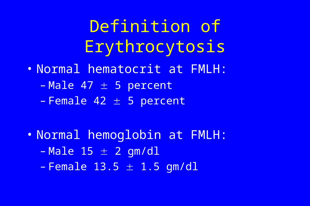
Absolute vs. Relative Erythrocytosis
Normal Spurious Polycythemia
Plasma Vol
RBC
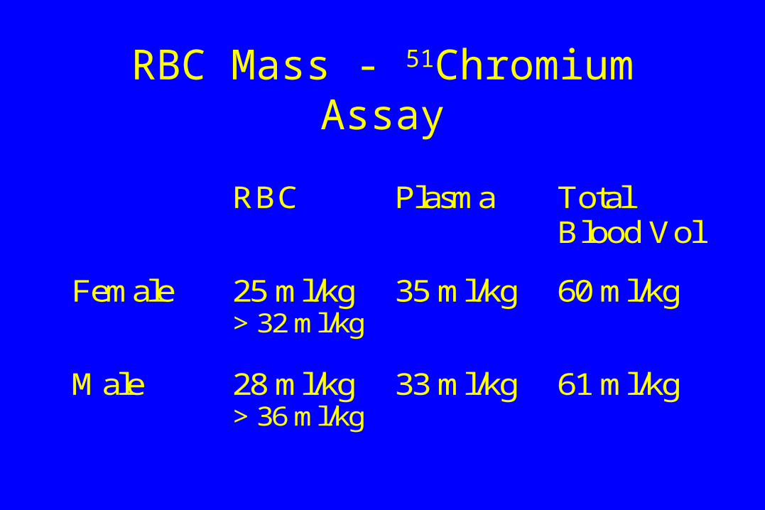
RBC Mass - 51Chromium Assay
RBC Plasma TotalBlood Vol
Female 25 ml/kg> 32 ml/kg
35 ml/kg 60 ml/kg
Male 28 ml/kg> 36 ml/kg
33 ml/kg 61 ml/kg
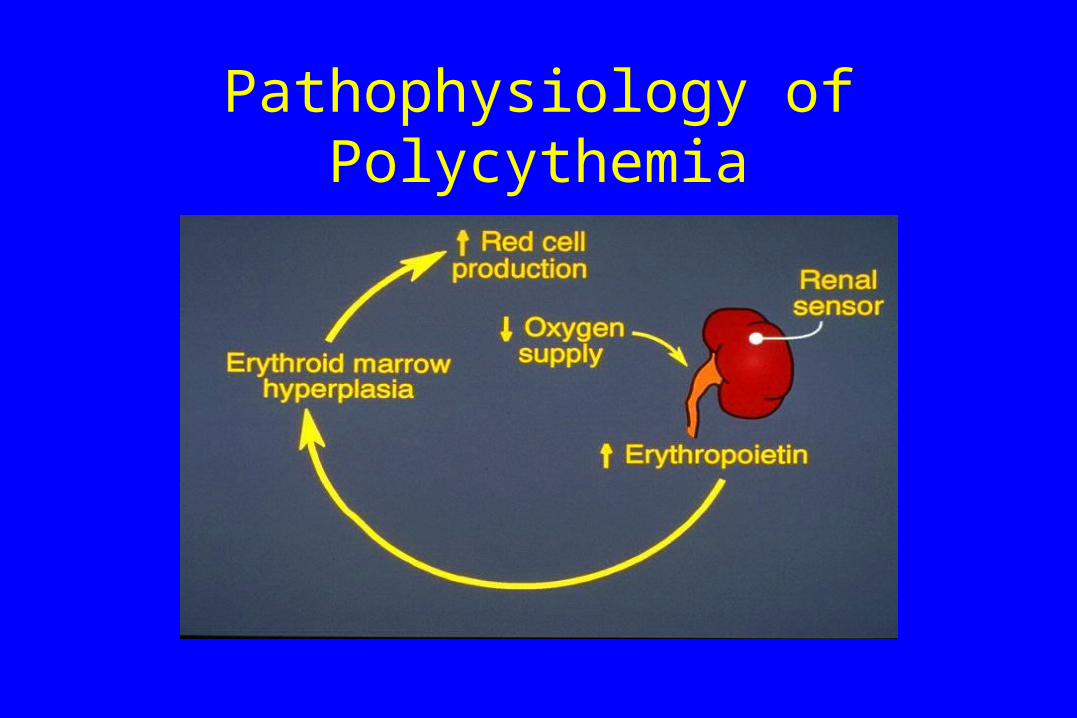
Pathophysiology of Polycythemia
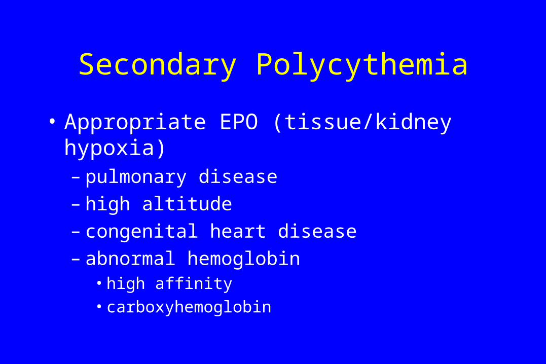
Secondary Polycythemia
• Appropriate EPO (tissue/kidney hypoxia)– pulmonary disease– high altitude– congenital heart disease– abnormal hemoglobin
• high affinity
• carboxyhemoglobin
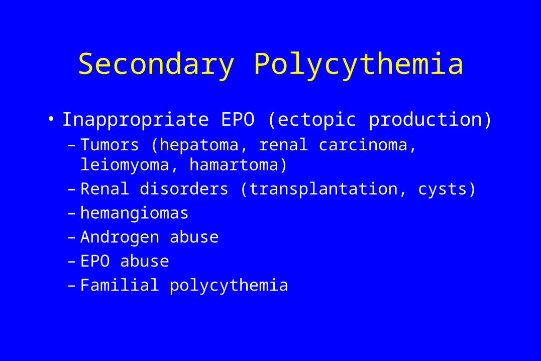
Secondary Polycythemia
• Inappropriate EPO (ectopic production)– Tumors (hepatoma, renal carcinoma, leiomyoma,
hamartoma)– Renal disorders (transplantation, cysts)– hemangiomas– Androgen abuse– EPO abuse– Familial polycythemia
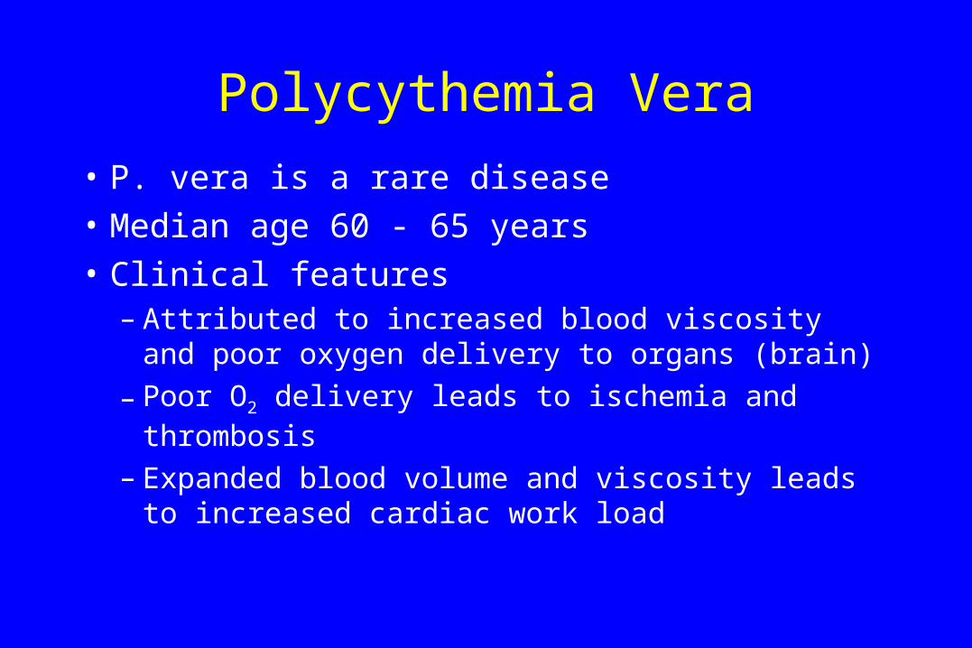
Polycythemia Vera
• P. vera is a rare disease
• Median age 60 - 65 years
• Clinical features– Attributed to increased blood viscosity and poor
oxygen delivery to organs (brain)
– Poor O2 delivery leads to ischemia and thrombosis
– Expanded blood volume and viscosity leads to increased cardiac work load
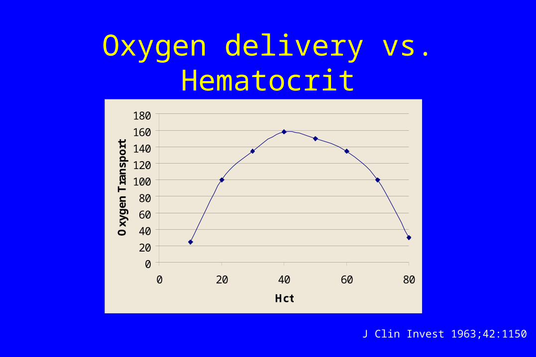
Oxygen delivery vs. Hematocrit
0
20
40
60
80
100
120
140
160
180
0 20 40 60 80
Hct
Oxy
gen
Tra
nsp
ort
J Clin Invest 1963;42:1150
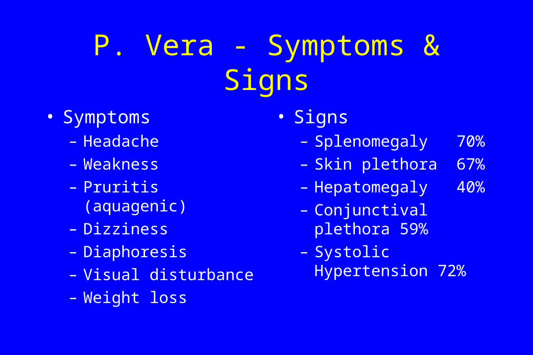
P. Vera - Symptoms & Signs
• Symptoms– Headache
– Weakness
– Pruritis (aquagenic)
– Dizziness
– Diaphoresis
– Visual disturbance
– Weight loss
• Signs– Splenomegaly 70%
– Skin plethora 67%
– Hepatomegaly 40%
– Conjunctival plethora 59%
– Systolic Hypertension 72%
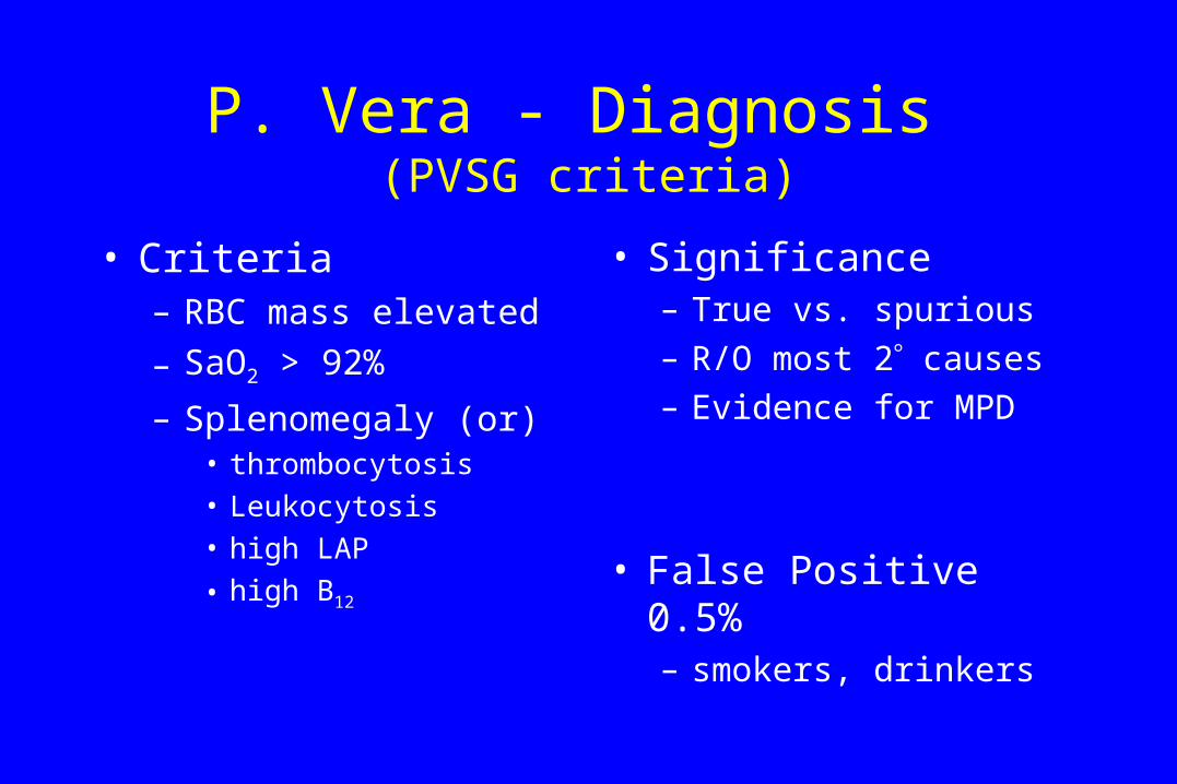
P. Vera - Diagnosis (PVSG criteria)
• Criteria– RBC mass elevated
– SaO2 > 92%
– Splenomegaly (or)• thrombocytosis
• Leukocytosis
• high LAP
• high B12
• Significance– True vs. spurious
– R/O most 2 causes
– Evidence for MPD
• False Positive 0.5%– smokers, drinkers
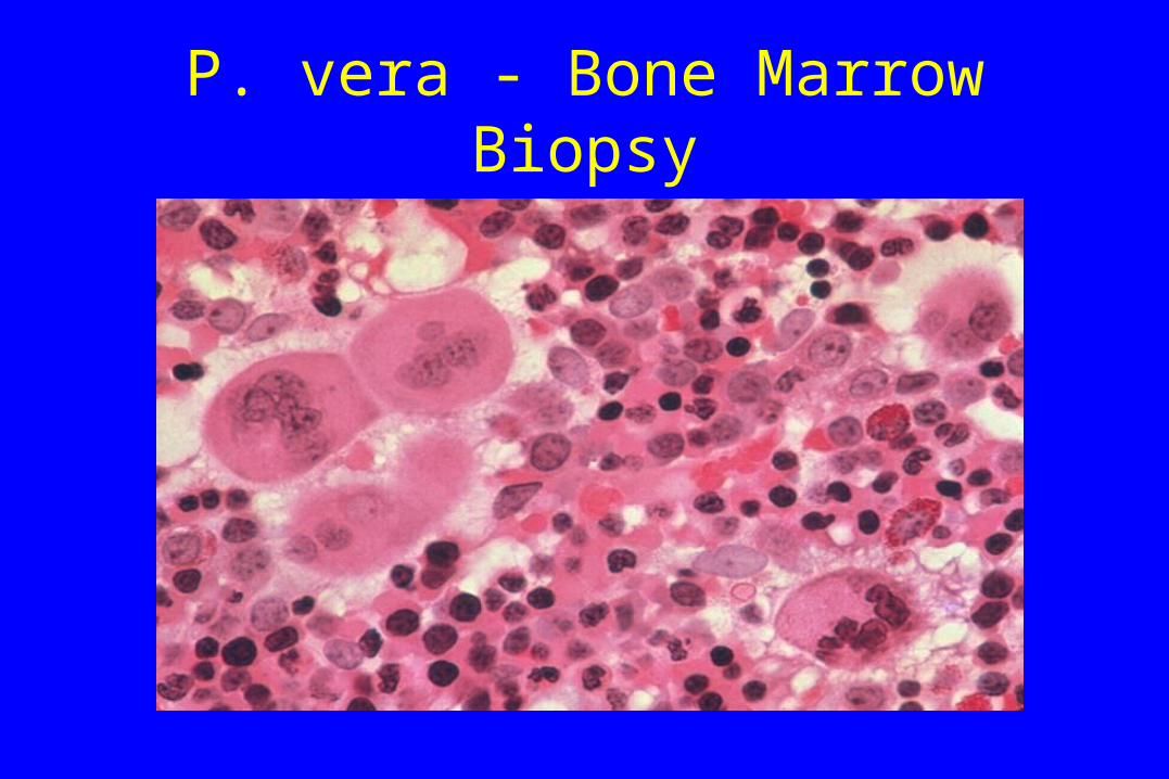
P. vera - Bone Marrow Biopsy

P. Vera - Natural History
PVSG GISPThrombosis/embolism 31% 30%AML 19% 15%Other cancer 15% 16%Hemorrhage 6% 3%Myelofibrosis 4% 3%Other 25% 35%
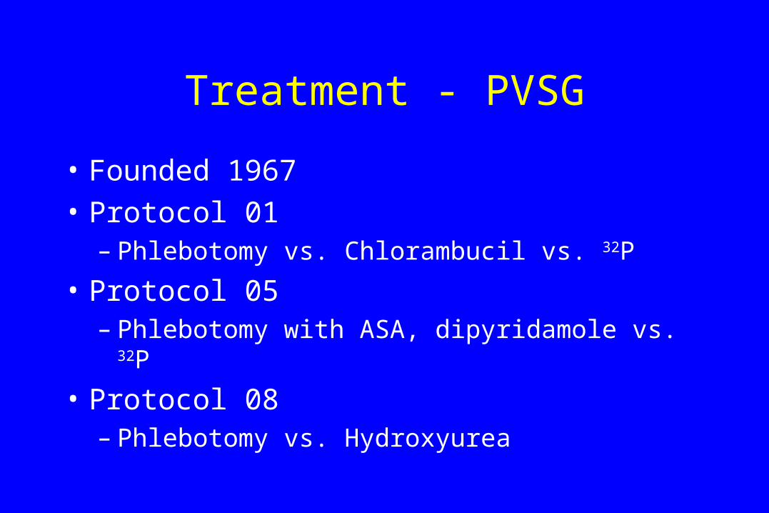
Treatment - PVSG
• Founded 1967
• Protocol 01– Phlebotomy vs. Chlorambucil vs. 32P
• Protocol 05– Phlebotomy with ASA, dipyridamole vs. 32P
• Protocol 08– Phlebotomy vs. Hydroxyurea
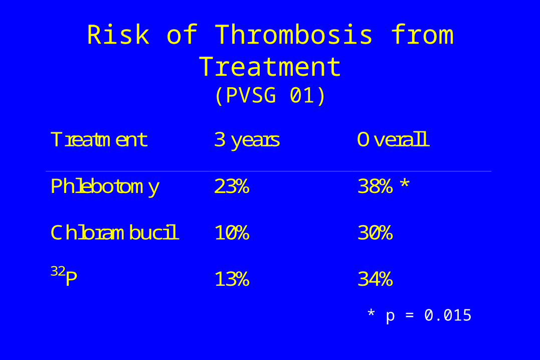
Risk of Thrombosis from Treatment(PVSG 01)
Treatment 3 years Overall
Phlebotomy 23% 38%*
Chlorambucil 10% 30%
32P 13% 34%
* p = 0.015
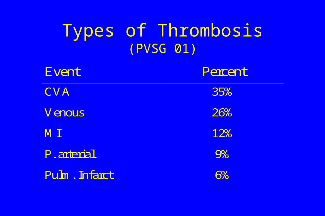
Types of Thrombosis(PVSG 01)
Event Percent
CVA 35%
Venous 26%
MI 12%
P. arterial 9%
Pulm. Infarct 6%
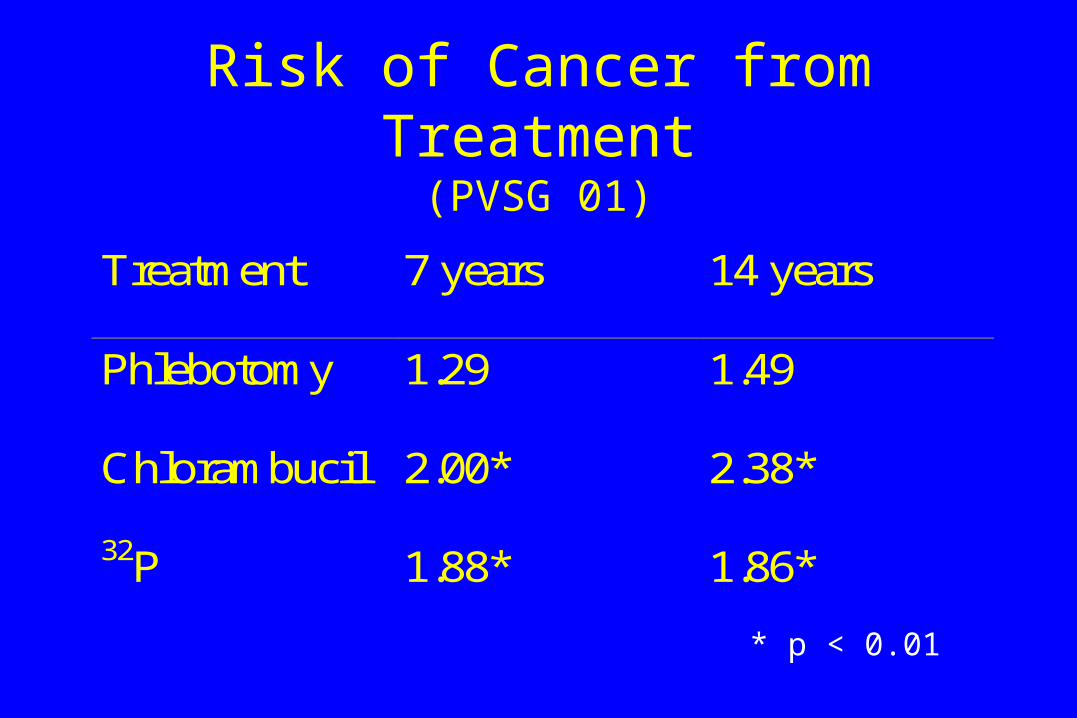
Risk of Cancer from Treatment(PVSG 01)
Treatment 7 years 14 years
Phlebotomy 1.29 1.49
Chlorambucil 2.00* 2.38*
32P 1.88* 1.86*
* p < 0.01
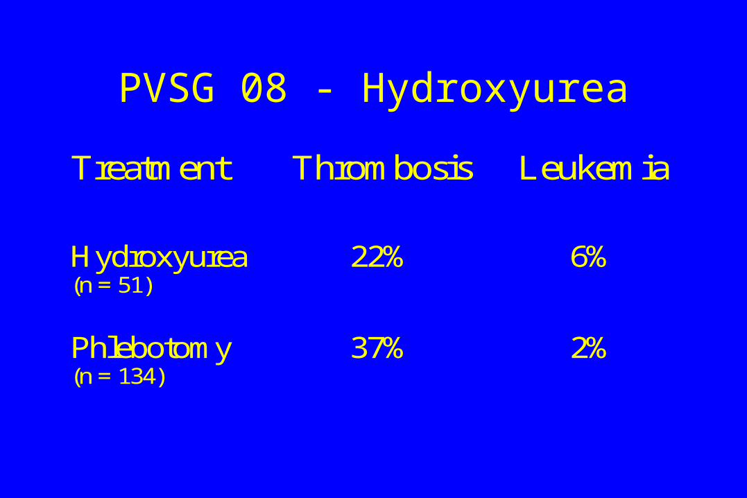
PVSG 08 - Hydroxyurea
Treatment Thrombosis Leukemia
Hydroxyurea(n = 51)
22% 6%
Phlebotomy(n = 134)
37% 2%
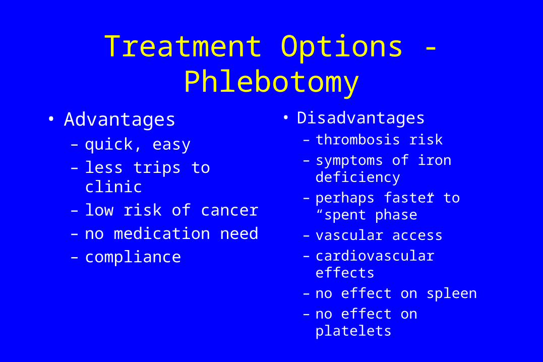
Treatment Options - Phlebotomy
• Advantages– quick, easy
– less trips to clinic
– low risk of cancer
– no medication need
– compliance
• Disadvantages– thrombosis risk
– symptoms of iron deficiency
– perhaps faster to “spent phase”
– vascular access
– cardiovascular effects
– no effect on spleen
– no effect on platelets

Treatment Options - 32P
• Advantages– quick and effective
– thrombosis risk low
– no medication
– follow-up need minimal
– compliance easier
– reduces spleen size
– lowers all counts
– few side-effects
• Disadvantages– risk of leukemia
– uncontrolled effects
– childbearing risk
– radiation issues
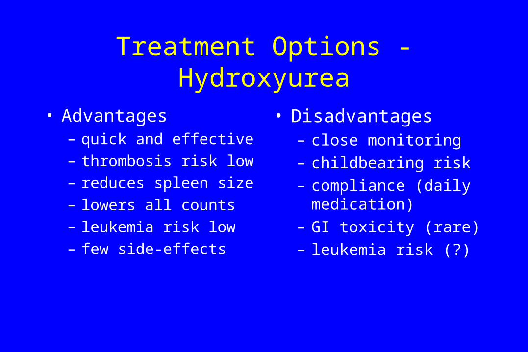
Treatment Options - Hydroxyurea
• Advantages– quick and effective
– thrombosis risk low
– reduces spleen size
– lowers all counts
– leukemia risk low
– few side-effects
• Disadvantages– close monitoring
– childbearing risk
– compliance (daily medication)
– GI toxicity (rare)
– leukemia risk (?)
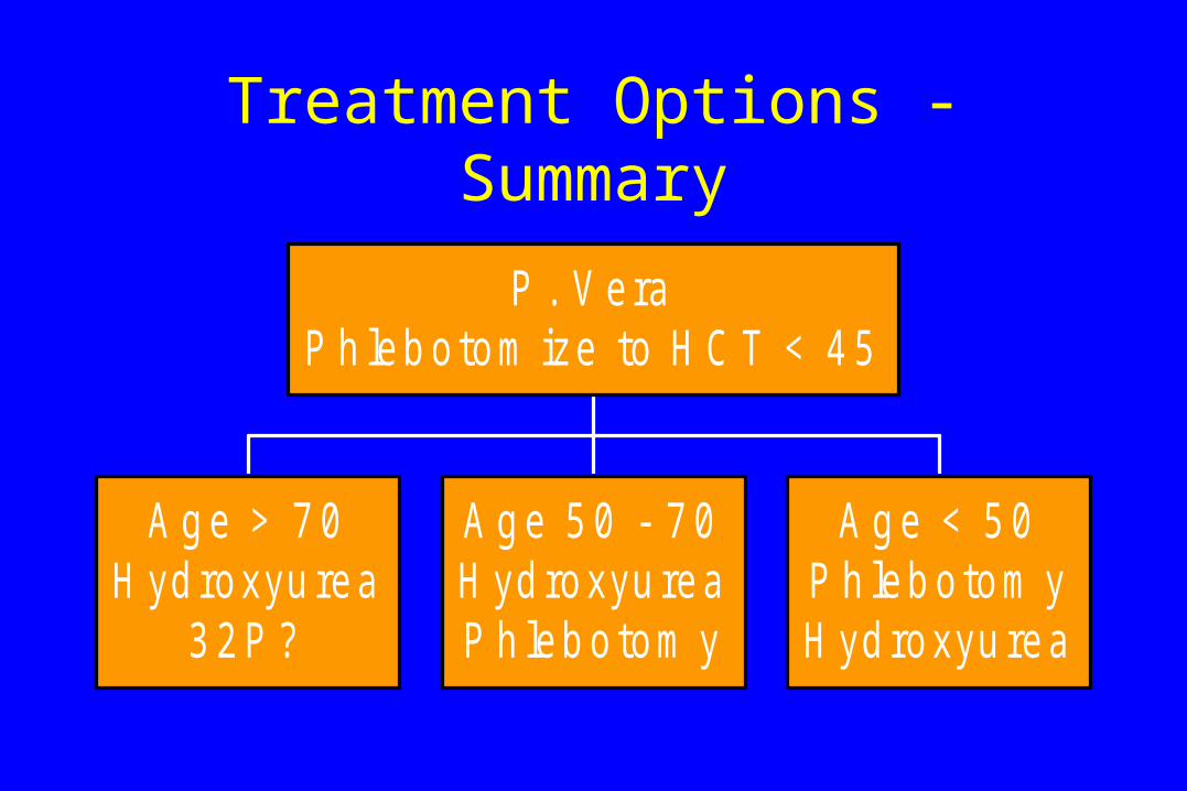
Treatment Options - Summary
A g e > 7 0H yd roxyu rea
3 2 P ?
A g e 5 0 - 7 0H yd roxyu reaP h leb o tom y
A g e < 5 0P h leb o tom yH yd roxyu rea
P . V eraP h leb o tom ize to H C T < 4 5