Embed Size (px)
Citation preview
Polyaniline nanofibres as templates for the covalent immobilisation of
biomolecules
Emer Lahiffa,1
, Carol Lynamb,1,*
, Niamh Gilmartinb, Dermot Diamond
a and Richard O’Kennedy
b
aCLARITY: The Centre for Sensor Web Technologies,
National Centre for Sensor Research,
Dublin City University,
Dublin 9, Ireland.
bSchool of Biotechnology and
Biomedical Diagnostics Institute,
National Centre for Sensor Research,
Dublin City University,
Dublin 9, Ireland.
* Corresponding author at: School of Biotechnology and
Biomedical Diagnostics Institute,
National Centre for Sensor Research, Dublin City University, Dublin 9, Ireland
Telephone: +353-1-7007717
Fax: +353-1-7006558
E-mail address: [email protected]
1 EL and CL contributed equally

2
Abstract
The attachment of antibodies onto polyaniline nanofibres using covalent chemistry was
investigated for the first time. Polyaniline nanofibres were functionalised post-polymerisation to
attach either amide or carboxylic acid side-groups. These templates could then be further
modified to attach antibodies, specifically in this instance mouse immunoglobulin G (IgG). The
resultant conjugates were characterised using a variety of techniques including infrared, UV-
visible and Raman spectroscopy. Conjugates were then used to detect secondary antibodies (anti-
IgG). Results from enzyme-linked immunoassay studies indicate successful binding of the
antibody to the polyaniline nanofibres. Carboxyl functionalised polyaniline nanofibres are shown
in particular to decrease non-specific binding in the immunoassay. Direct electrical
communication between polyaniline nanofibres covalently linked to peroxidase-labelled
antibodies was observed during cyclic voltammetry, which demonstrates their potential for
further development as nano-dimensional immunosensors.
Keywords: polyaniline nanofibers, conducting polymer functionalization, antibody conjugation,
immunoassay
3
1. Introduction
Conducting polymers combine the advantages of polymers (being lightweight, inexpensive and
easily processed) with the ability to transport charge [1]. Their unique combination of physical,
optical and electrical properties, has led to considerable interest in their use as platforms for a
wide variety of applications. Examples of conducting polymers used in applications
encompassing electrochromic devices, printable electronics and photochromics include
polythiophene, polypyrrole and polyaniline [2,3,4]. Polyaniline (PAni) has been previously used
as a mediator in biosensor applications, due to its inherent electro-activity, favourable charge
storage stability and ease of preparation [5,6,7]. Interfacing biomolecules with conducting
polymers is a logical step in order to accomplish bio-composite materials which can be used for
controlling stimuli-responsive actions involved in drug release, bio-feedback devices, neural
implants and artificial muscles [8].
Nanodimensional PAni can be readily prepared, exhibiting markedly improved properties from
those of the bulk materials, including increased surface area and sharp switching [9]. The
properties unique to one dimensional nanostructures such as nanowires, nanotubes and
nanofibres have been the focus of much research, partly due to the promise associated with
nanoscale devices for sensors and electronics. For example Virji et al. [9] howed how PAni
nanofibre films have far superior performance in both sensor sensitivity and response time in
comparison to their bulk counterparts. This is due to faster gas diffusion, enabled by the porous
high-surface area nanofibrillar film structure.
4
Recently, monolithic microstructures from nanofibers of PAni have been reported [10]. Since the
porous properties of monolithic polymers can be easily tuned, monolithic capillary columns have
encountered wide interest for applications in capillary electrochromatography, for example
functionalised monoliths have been used in protein analysis [11]. Using a capillary coated with
polyaniline achieved the separation of the end products of glycosylation [12]. Relative to packed
columns, monoliths have the advantages of preparation simplicity and availability for surface
modification [13,14]. The large surface area of monolithic beds enables a relatively high sample
loading capacity. The attachment of proteins to affinity columns have seen use in
immunopurification, [15] and the detection of selected analytes by chromatographic
immunoassays [16].
The attachment of biomolecules to conducting polymer nanostructures means that bio-active
functionalities are anchored to a redox active nano-dimensional system. The nanostructured form
of PAni offers a very large surface-to-volume ratio thus facilitating the immobilisation of a
greater number of biomolecules, and an enhancement of sensor sensitivity and response times
[9,17]. One of the most common methods for immobilising biomolecules onto surfaces achieved
either physically or by electrochemical techniques, is adsorption [18]. The latter is achieved by
applying a potential to the electrode surface, which enhances electrostatic interactions and
therefore immobilisation [10]. Using adsorption as the method of immobilisation however has
the disadvantage that the biomolecule can easily desorb from the surface during use. Conversely,
covalent attachment of biomolecules to nanomaterial surfaces ensures sufficient immobilisation
without leaching of the biomolecule from the substrate surface during use [19].
5
The authors have recently reported the successful post-synthesis functionalisation of PAni
nanofibres with mercaptoundecanoic acid and cysteamine; thereby incorporating functional
handles (COOH and NH2) which allow for further derivatisation of the polymer [20,21]. Post
functionalisation, PAni maintains its nano-morphology and ability to switch optical, electrical
and electrochemical properties in response to its immediate chemical environment. In this paper,
we describe for the first time how PAni nanofibres modified with either carboxyl or amino
terminated chains allow for a one step covalent derivatisation of the polymer with biomolecules
(in particular we demonstrate the technique for antibodies). Biomolecule immobilisation onto
electrode surfaces is of importance for the development of enzyme–catalysed biofuel cells,
amperometric biosensors and other biotechnological applications. Here we report a ‘generic’
approach for immobilisation. PAni-antibody conjugates are of interest as they are low cost and
easily scalable. The polyaniline serves as an immobilisation matrix and also plays an active role
in transduction. Alternatively these materials could find application as solid phases in affinity
monolithic micro-columns for protein separation.
6
2. Experimental Procedures
2.1 Chemicals: Aniline (BDH), HCl (Fisher Scientific), ammonium peroxydisulfate (Aldrich),
mercaptoundecanoic acid (Aldrich), mercaptopropionic acid (Aldrich), cysteamine (Aldrich) and
pH 4 buffer tablets (Fluka) were used. The aniline monomer was purified by vacuum distillation
before use. Mouse immunoglobulin G (IgG) (Sigma), anti-mouse IgG labelled with horse radish
peroxidase (derived from goat, Sigma), 3,3’,5,5’-tetramethylbenzidine dihydrochloride (TMB,
Sigma) were used as supplied.
2.2 Nanofibre Synthesis: Polyaniline nanofibres were synthesised, under ambient conditions, by
interfacial polymerisation between an aqueous and an organic layer, as reported previously [20].
The product was purified by centrifugation (4000 rpm/5 min/3 cycles) and suspended as a colloid
in deionised water. Fibres were modified with mercaptoundecanoic acid (HS(CH2)10COOH),
mercaptopropionic acid (HS(CH2)2COOH) or cysteamine (HS(CH2)2NH2), by refluxing at 100
°C for 2 hours in an aqueous pH 4 buffer. Four sets of fibres were functionalised; two with
cysteamine (0.1 mmol and 0.3 mmol) and two with 0.1 mmol carboxyl groups with different
spacer lengths (C10 and C2). All experiments were carried out under identical conditions using
~45 mg PAni. The product was purified by centrifugation (4000 rpm/5 min/3 cycles) and re-
suspended as a colloid in deionised water.
2.3 Covalent Antibody Immobilisation onto PAni: Briefly, 100 µl of mouse IgG, at a
concentration of 0.1 mg/ml was added to 1 ml of the PAni nanofibre dispersions (0.2 mg/ml) for
1 hour with shaking (200 rpm) at 37 °C. Antibodies were covalently immobilised by the addition
of 4 mg ethyl diaminocarbodiimide (EDC), the zero length crosslinker, to the dispersion. The
7
PAni-IgG mixture was centrifuged and washed in distilled water 3 times. Any remaining
unreacted sites on the nanofibres were blocked with 5 wt. % milk marvel and incubated
overnight at 4 °C. The PAni-IgG mixture was then centrifuged (10 min at 4000 rpm) and washed
3 times with distilled water. Anti-mouse IgG labelled with horse radish peroxidise (HRP) was
diluted 1 in 1000 in 1 % (w/v) milk marvel/PBS to give a final concentration of 0.6 µg/ml and 1
ml of this solution was added to the PAni-IgG pellet, re-dispersed and incubated at 37 °C for one
hour. The PAni mixture was then centrifuged (10 min at 4000 rpm) and washed a further 3 times
with water and the PAni pellet re-suspended in 0.5 ml water.
2.4 Antibody Detection: HRP-labelled antibodies were detected using the 3,3′,5,5′-
tetramethylbenzidine dihydrochloride (TMB) assay prepared as per the manufacturer’s
instructions. 200 µl of TMB solution was added to 250 µl of the antibody-labelled PAni solution
and incubated in the dark at room temperature for 10 min. This PAni-TMB dispersion was then
centrifuged (10 min at 4000 rpm) and 50 µl of the supernatant was added to a microtitre well.
The reaction was stopped by the addition of 50 µl of 10% (v/v) HCl and the absorbance was read
at 450 nm. 10 µl of each PAni and PAni/Ab-HRP suspension was cast as a film onto clean
glassy carbon electrodes and used as the working electrode in cyclic voltammetry.
2.5 Characterisation: Covalent bonding of side-chains to PAni nanofibres was investigated using
Fourier Transform infrared spectra (FTIR) recorded in transmission mode from 400 to 4000 cm-
1, for 100 repeat scans, with a resolution of 8 cm
-1 and at 1 cm
-1 intervals on a Perkin Elmer
Spectrum GX FTIR. Samples were dried under vacuum at 50 °C for 4 hours prior to FTIR and
then mixed with dried KBr. Dedoped samples were also analysed using solid state nuclear
8
magnetic resonance (SSNMR). 13
C spectra were obtained using a Varian VNMRS spectrometer
operating at 100.56 MHz for carbon, and spectra are referenced to tetramethylsilane. Thermal
Gravimetric Analysis (TGA) decomposition curves were obtained using a Mettler-Toledo
TGA/SDTA851. Samples were heated from 25-700 °C at 10 °C/minute under nitrogen.
Nanofibres were coated with 10 nm Au/Pd and imaged using field emission scanning electron
microscopy (FESEM) at an accelerating voltage of 20 kV on a S-4300 Hitachi system. Raman
spectroscopy was carried out on an Avalon 785 nm Raman spectrometer at 2 cm-1
resolution, 3
seconds per scan and 10-20 collections. A 785 nm laser line was used as it can detect both doped
and de-doped PAni features. UV-vis spectroscopy was carried out on a Perkin Elmer UV-vis
NIR Lambda 900 Spectrometer at 1 nm resolution. Characterisation of the samples by enzyme
assay was obtained using a Tecan Sunrise microplate reader. Absorbance readings were taken at
a wavelength of 450 nm. Transmission electron microscopy (TEM) analysis was performed
using a Jeol 2100 microscope (200 kV). Cyclic voltammetry was performed using an eDAQ e-
corder (401) and potentiostat/galvanostat (EA 160) with Chart v5.1.2/EChem v 2.0.2 software
(ADInstruments), and a PC computer, in phosphate buffered saline solution (PBS - 0.2 M, pH
7.4) with Ag/AgCl reference electrode and platinum mesh counter electrode.
9
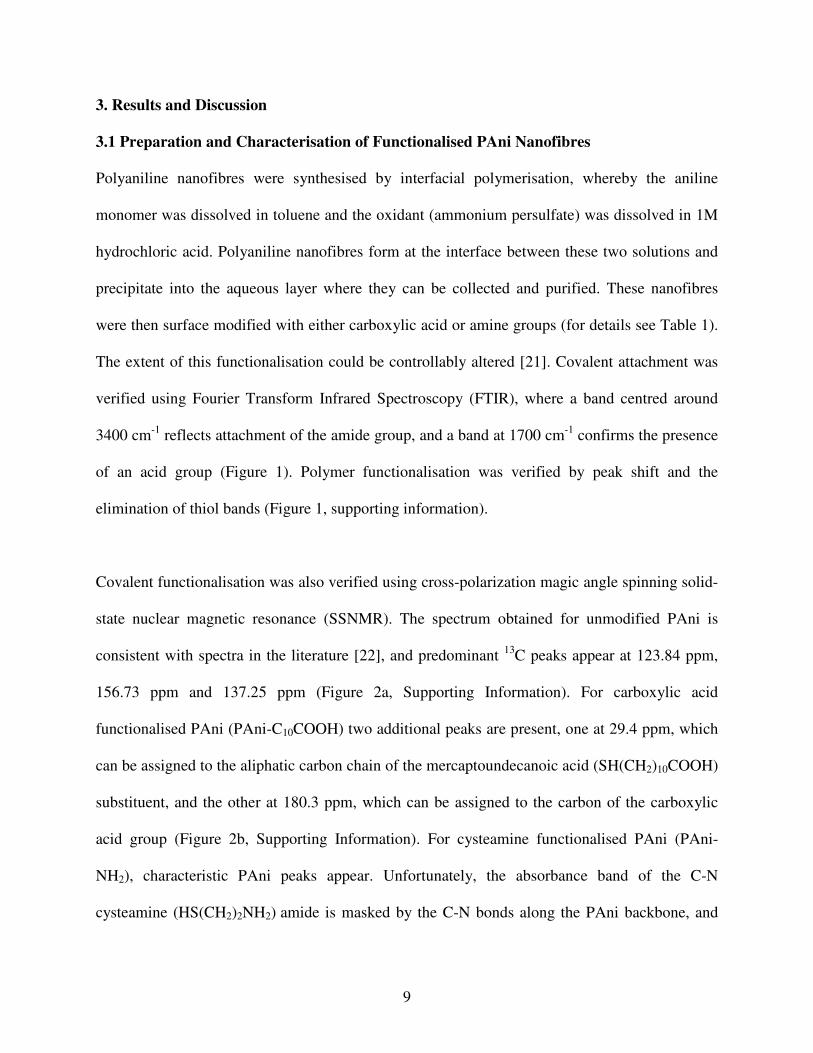
3. Results and Discussion
3.1 Preparation and Characterisation of Functionalised PAni Nanofibres
Polyaniline nanofibres were synthesised by interfacial polymerisation, whereby the aniline
monomer was dissolved in toluene and the oxidant (ammonium persulfate) was dissolved in 1M
hydrochloric acid. Polyaniline nanofibres form at the interface between these two solutions and
precipitate into the aqueous layer where they can be collected and purified. These nanofibres
were then surface modified with either carboxylic acid or amine groups (for details see Table 1).
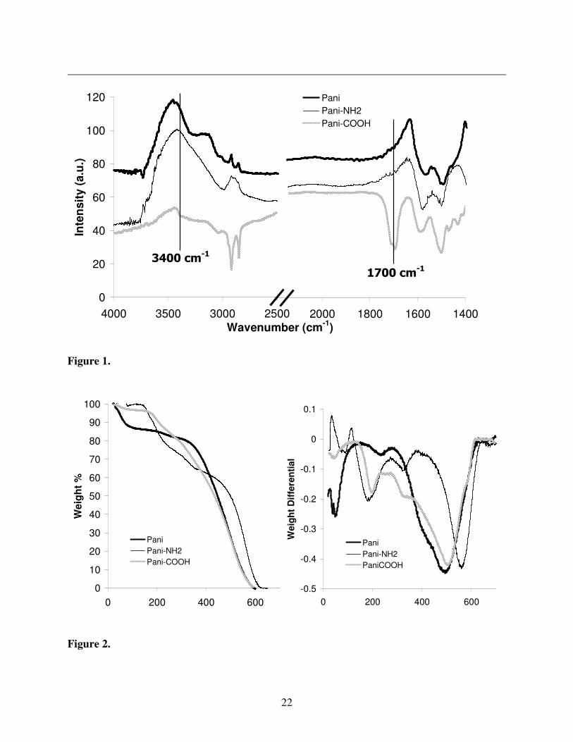
The extent of this functionalisation could be controllably altered [21]. Covalent attachment was
verified using Fourier Transform Infrared Spectroscopy (FTIR), where a band centred around
3400 cm-1
reflects attachment of the amide group, and a band at 1700 cm-1
confirms the presence
of an acid group (Figure 1). Polymer functionalisation was verified by peak shift and the
elimination of thiol bands (Figure 1, supporting information).
Covalent functionalisation was also verified using cross-polarization magic angle spinning solid-
state nuclear magnetic resonance (SSNMR). The spectrum obtained for unmodified PAni is
consistent with spectra in the literature [22], and predominant 13
C peaks appear at 123.84 ppm,
156.73 ppm and 137.25 ppm (Figure 2a, Supporting Information). For carboxylic acid
functionalised PAni (PAni-C10COOH) two additional peaks are present, one at 29.4 ppm, which
can be assigned to the aliphatic carbon chain of the mercaptoundecanoic acid (SH(CH2)10COOH)
substituent, and the other at 180.3 ppm, which can be assigned to the carbon of the carboxylic
acid group (Figure 2b, Supporting Information). For cysteamine functionalised PAni (PAni-
NH2), characteristic PAni peaks appear. Unfortunately, the absorbance band of the C-N
cysteamine (HS(CH2)2NH2) amide is masked by the C-N bonds along the PAni backbone, and
10
therefore cannot be isolated (Figure 2c, Supporting Information). The SSNMR spectra confirm
the formation of PAni-COOH nanofibres, and are consistent with the formation of PAni-NH2
nanofibres.
Results obtained by thermal gravimetric analysis (TGA) are also consistent with nanofibre
functionalisation. These reveal that PAni, PAni-C10COOH and PAni-NH2 have different
decomposition profiles (Figure 2). Unmodified polyaniline decomposes over a temperature
range between 300 °C and 620 °C, while other decomposition peaks outside this temperature
range can be attributed to trapped solvent/dopant in the material [21]. In contrast to this,
functionalised nanofibres show two significant areas of decomposition. For PAni-C10COOH
the peak centred at 504 °C is consistent with a PAni component, while the second peak at
196 °C is due to a modified PAni-C10COOH surface component (ca. 10 wt%). In the case of
PAni-NH2 B, decomposition peaks appear at 558 °C due to the PAni component, while the
amide-modified surface component manifests at 184 °C (ca. 19wt%). These TGA results are
in agreement with previously published data which shows that the degree of nanofibre
functionalisation can be controlled for both PAni-COOH and PAni-NH2.
3.2 Antibody immobilisation
Covalent immobilisation of antibodies onto functionalised polyaniline (PAni) nanofibres was
investigated and involved attachment of antibodies to both carboxyl and amino functionalised
PAni nanofibres (Pani-COOH and Pani-NH2 respectively). To determine the effect of chain
length on antibody immobilisation, carboxyl functionalised PAni nanofibres with two carbon
(PAni-(CH2)2-COOH), and ten carbon (PAni-(CH2)10-COOH) spacer chains were examined. In
11
the case of amine functionalised PAni nanofibres, two different concentrations of cysteamine
were used to compare the effect of amino-group density with respect to antibody immobilisation
(products from 0.1 mmol and 0.3 mmol cysteamine reactions are denoted Pani-C2NH2 A and
Pani-C2NH2 B, respectively, see Table 1).
For the immobilisation, both types of PAni-COOH and PAni-NH2 dispersions were mixed with
the primary antibody, mouse immunoglobulin G (IgG), and the cross linker ethyl
diaminocarbodiimide (EDC). A range of blocking agents were tested (ovalbumin, bovine serum
albumin (BSA), Tween 20®, BSA/milk marvel (1:1), milk marvel, gelatin, Super Block®, KLH
protein and soya milk) and the results indicate that milk marvel was the most effective blocking
agent against non-specific binding (See figure 3, supporting Information). Milk marvel therefore
was used for all PAni samples tested. As future work will involve adaptation of this method for a
sandwich immunoassay, an unlabelled antibody was immobilised onto the PAni nanofibres.
3.3 Characterisation of antibody conjugates
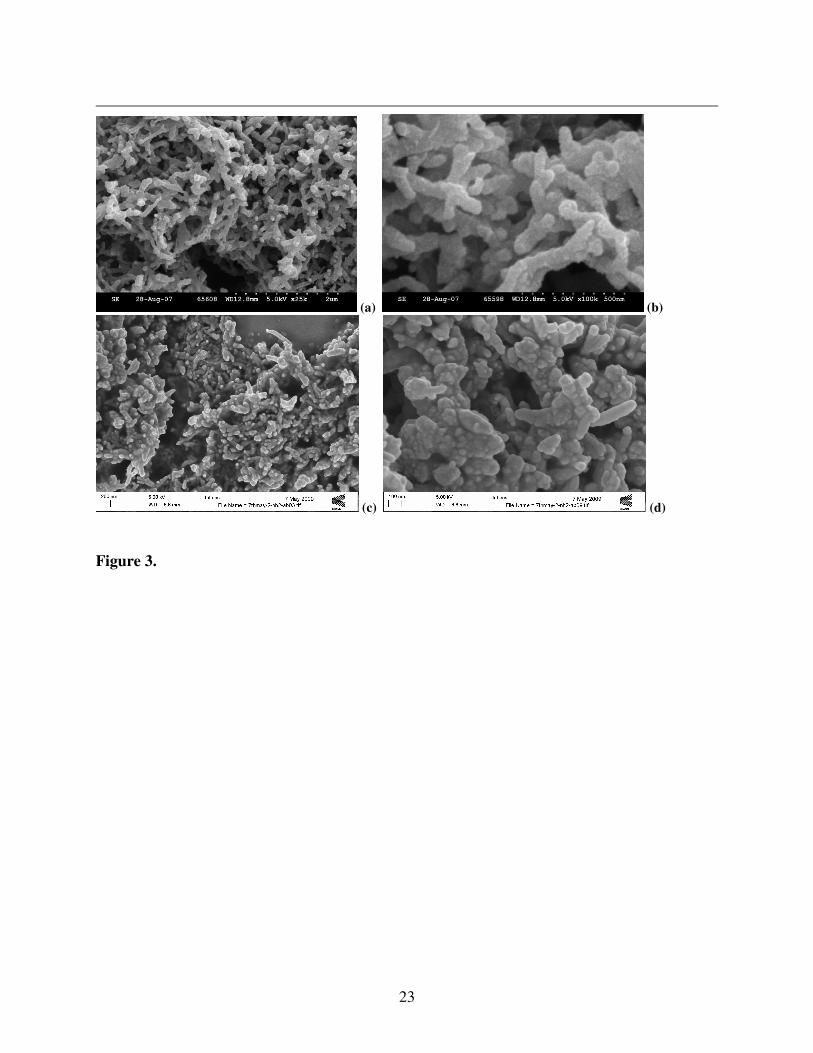
The nanofibre morphology of PAni can be verified using scanning electron microscopy (SEM).
Nanofibres synthesised by interfacial polymerisation have a uniform diameter distribution and a
high surface area, thus making them ideal for use as a sensing platform. Post-functionalisation,
the nanofibre morphology appears essentially unaffected. However, greater aggregation of the
nanofibres is observed following antibody immobilisation, which results in an apparently less
porous film structure (Figure 3). This is analogous to functionalisation of PAni with carboxyl
and amino groups, in that the degree of aggregation scales with the level of functionalisation
[20]. Films do however maintain a high surface-to-volume ratio in comparison with bulk PAni.
12
The surface topography of the nanofibres appears altered suggesting that the attachment of
biomolecules was successful.
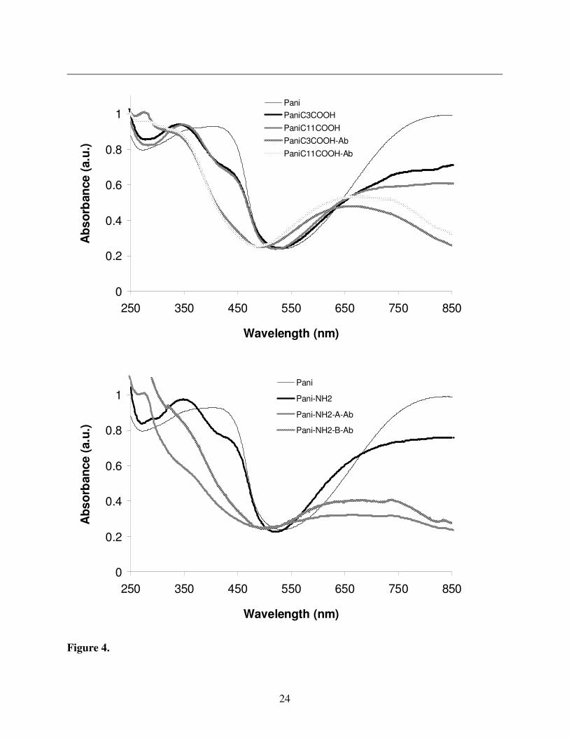
Functionalisation affects the delocalised bonding along the PAni polymer backbone, and this can
be monitored by analysing spectroscopic changes in the materials (in particular UV-vis and
Raman spectra were examined). For UV-vis spectra, a polaron band at 450 nm with a free carrier
tail above 600 nm indicates that PAni is in the doped (emeraldine salt, therefore conductive) state
[23]. For a dedoped (emeraldine base, therefore non-conductive) state these bands disappear and
a π-π* transition band at 341 nm dominates, with a band at 660cm
-1 due to the quinoid fraction of
the PAni backbone [24]. For unmodified PAni in an aqueous environment the material remains
in the doped form (Figure 4). However, with functionalised PAni-NH2 and PAni-COOH in an
aqueous environment, features of the dedoped state begin to emerge. This is due to the fact that
functionalisation disrupts the delocalised π-system of chemical bonds along the PAni polymer
backbone. For PAni-antibody conjugates, further dedoping of the material is observed due to the
higher pH environment that the nanofibres are exposed to during conjugation. Aromatic amino
acids associated with antibodies absorb strongly near 280 nm and a peak corresponding to this is
clearly seen for all spectra of the PAni-antibody conjugates. Therefore UV-vis results suggest
that antibody functionalisation was successful.
The dedoped state of PAni-antibody conjugates was also reflected in Raman spectra of the
material (Figure 4, Supporting Information). Spectra of PAni in the doped conductive state have
signature bands between 1300-1400 cm-1
, and these are present for PAni-NH2. Strong bands
13
between 1400-1500 cm-1
reflect a transition to the dedoped state, and these emerge for the PAni-
NH2-antibody conjugate.
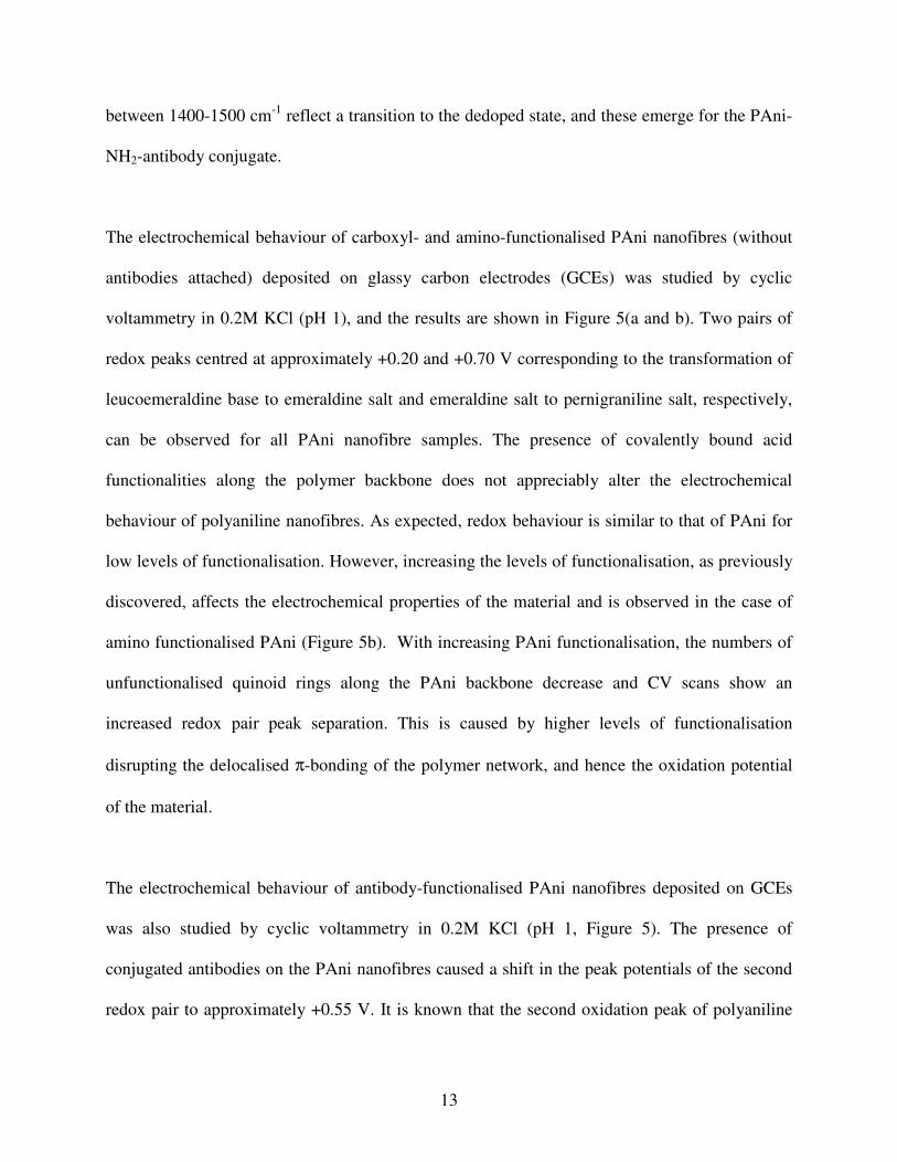
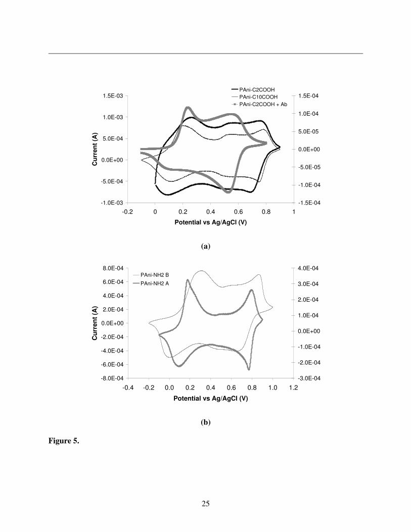
The electrochemical behaviour of carboxyl- and amino-functionalised PAni nanofibres (without
antibodies attached) deposited on glassy carbon electrodes (GCEs) was studied by cyclic
voltammetry in 0.2M KCl (pH 1), and the results are shown in Figure 5(a and b). Two pairs of
redox peaks centred at approximately +0.20 and +0.70 V corresponding to the transformation of
leucoemeraldine base to emeraldine salt and emeraldine salt to pernigraniline salt, respectively,
can be observed for all PAni nanofibre samples. The presence of covalently bound acid
functionalities along the polymer backbone does not appreciably alter the electrochemical
behaviour of polyaniline nanofibres. As expected, redox behaviour is similar to that of PAni for
low levels of functionalisation. However, increasing the levels of functionalisation, as previously
discovered, affects the electrochemical properties of the material and is observed in the case of
amino functionalised PAni (Figure 5b). With increasing PAni functionalisation, the numbers of
unfunctionalised quinoid rings along the PAni backbone decrease and CV scans show an
increased redox pair peak separation. This is caused by higher levels of functionalisation
disrupting the delocalised π-bonding of the polymer network, and hence the oxidation potential
of the material.
The electrochemical behaviour of antibody-functionalised PAni nanofibres deposited on GCEs
was also studied by cyclic voltammetry in 0.2M KCl (pH 1, Figure 5). The presence of
conjugated antibodies on the PAni nanofibres caused a shift in the peak potentials of the second
redox pair to approximately +0.55 V. It is known that the second oxidation peak of polyaniline
14
moves to a less positive potential with increasing pH. The shift to +0.55 V may be due to the
presence of conjugated antibodies adjacent to polymer chains, changing the local chemical and
hence electrochemical environment of the polymer.
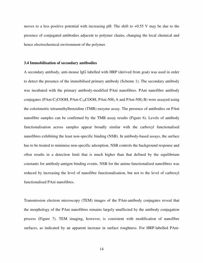
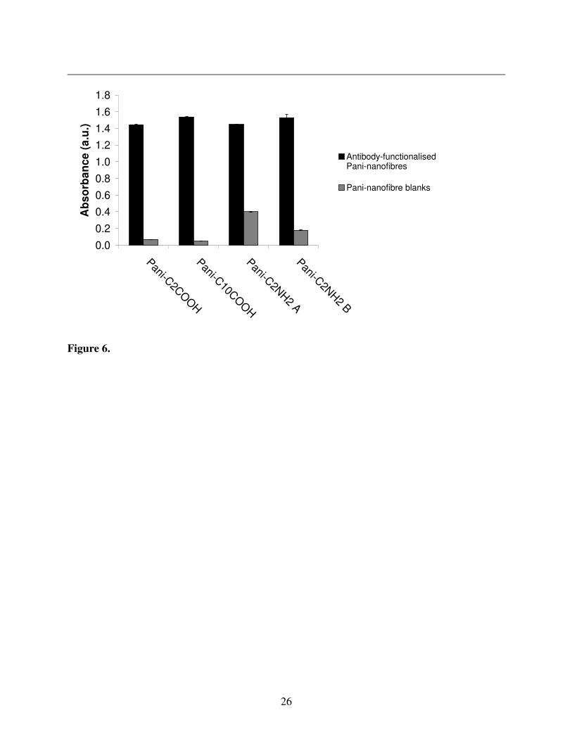
3.4 Immobilisation of secondary antibodies
A secondary antibody, anti-mouse IgG labelled with HRP (derived from goat) was used in order
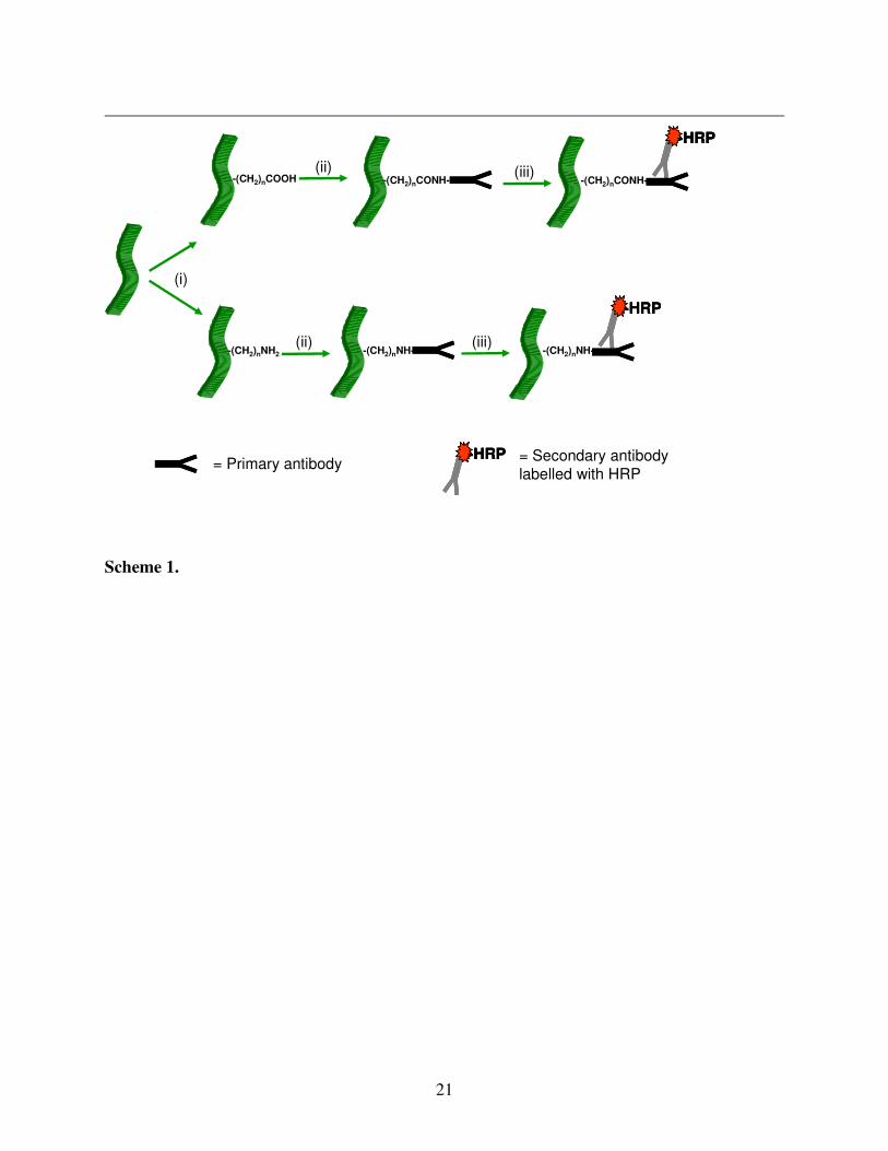
to detect the presence of the immobilised primary antibody (Scheme 1). The secondary antibody
was incubated with the primary antibody-modified PAni nanofibres. PAni nanofibre antibody
conjugates (PAni-C2COOH, PAni-C10COOH, PAni-NH2 A and PAni-NH2 B) were assayed using
the colorimetric tetramethylbenzidine (TMB) enzyme assay. The presence of antibodies on PAni
nanofibre samples can be confirmed by the TMB assay results (Figure 6). Levels of antibody
functionalisation across samples appear broadly similar with the carboxyl functionalised
nanofibres exhibiting the least non-specific binding (NSB). In antibody-based assays, the surface
has to be treated to minimise non-specific adsorption. NSB controls the background response and
often results in a detection limit that is much higher than that defined by the equilibrium
constants for antibody-antigen binding events. NSB for the amine-functionalised nanofibres was
reduced by increasing the level of nanofibre functionalisation, but not to the level of carboxyl
functionalised PAni nanofibres.
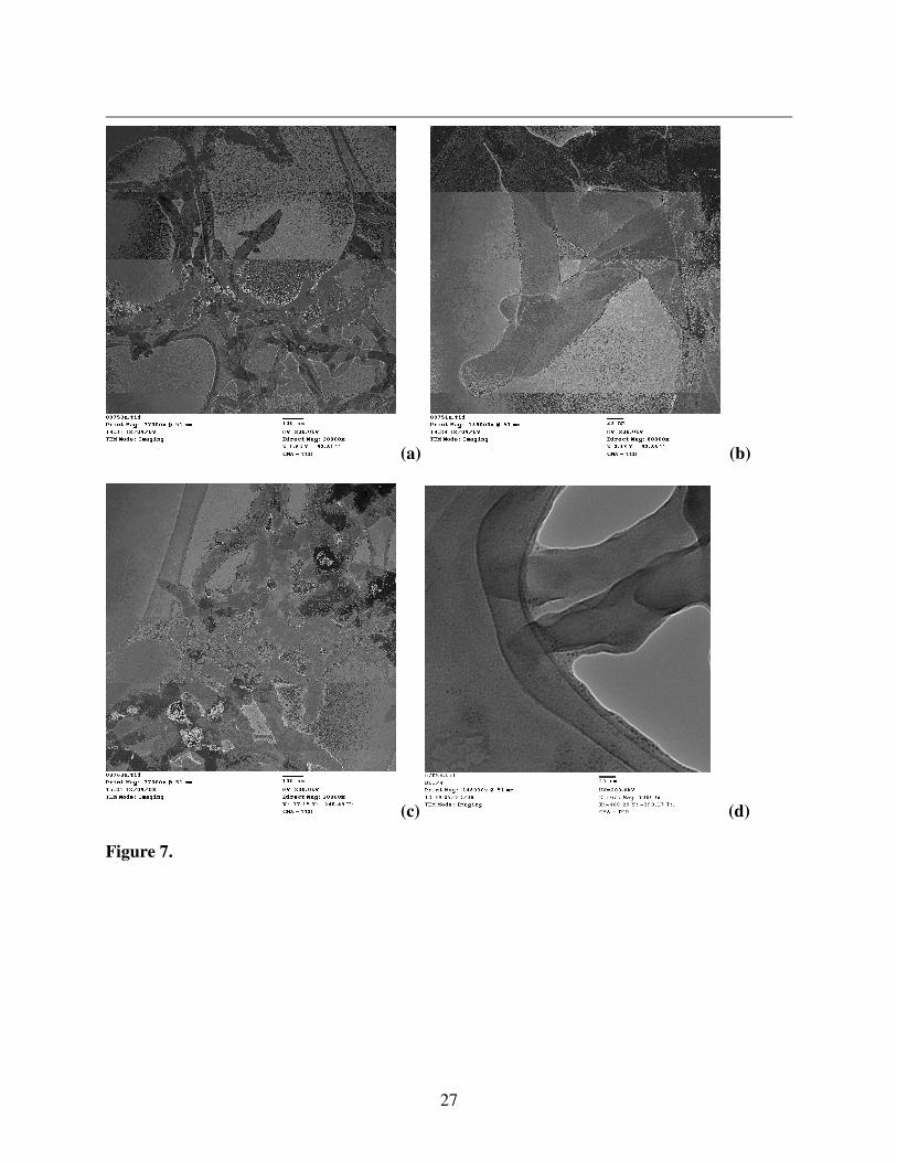
Transmission electron microscopy (TEM) images of the PAni-antibody conjugates reveal that
the morphology of the PAni nanofibres remains largely unaffected by the antibody conjugation
process (Figure 7). TEM imaging, however, is consistent with modification of nanofibre
surfaces, as indicated by an apparent increase in surface roughness. For HRP-labelled PAni-
15
antibody conjugates, dark spheres on the surface of the nanofibres can be observed, consistent
with the size of HRP labels (Figure 7d). EDXS analysis on films of these PAni-antibody
conjugates confirms the presence of iron from the metal centre of the HRP enzyme label on the
secondary antibody (Figure 5, Supporting Information).
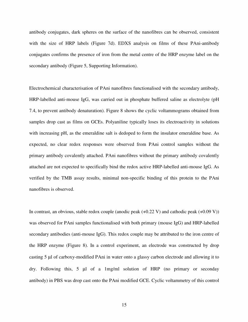
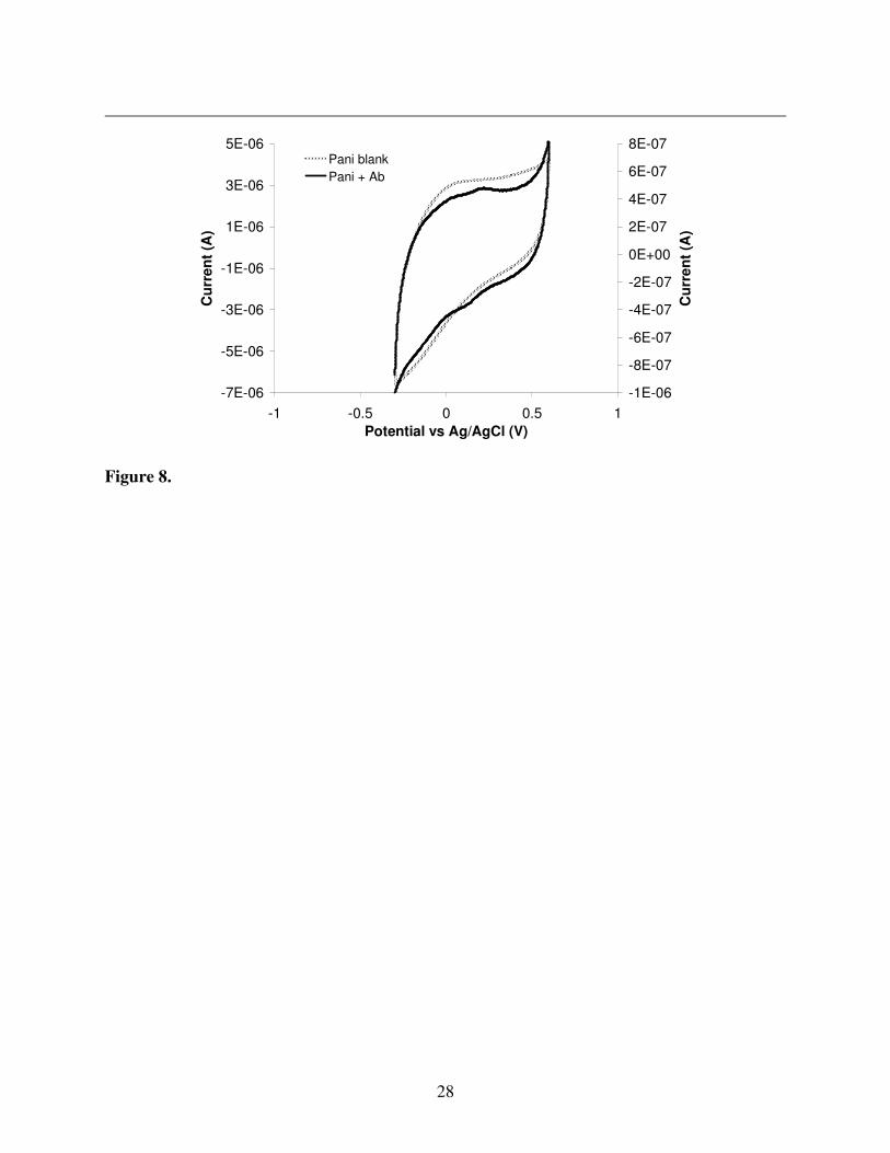
Electrochemical characterisation of PAni nanofibres functionalised with the secondary antibody,
HRP-labelled anti-mouse IgG, was carried out in phosphate buffered saline as electrolyte (pH
7.4, to prevent antibody denaturation). Figure 8 shows the cyclic voltammograms obtained from
samples drop cast as films on GCEs. Polyaniline typically loses its electroactivity in solutions
with increasing pH, as the emeraldine salt is dedoped to form the insulator emeraldine base. As
expected, no clear redox responses were observed from PAni control samples without the
primary antibody covalently attached. PAni nanofibres without the primary antibody covalently
attached are not expected to specifically bind the redox active HRP-labelled anti-mouse IgG. As
verified by the TMB assay results, minimal non-specific binding of this protein to the PAni
nanofibres is observed.
In contrast, an obvious, stable redox couple (anodic peak (+0.22 V) and cathodic peak (+0.09 V))
was observed for PAni samples functionalised with both primary (mouse IgG) and HRP-labelled
secondary antibodies (anti-mouse IgG). This redox couple may be attributed to the iron centre of
the HRP enzyme (Figure 8). In a control experiment, an electrode was constructed by drop
casting 5 µl of carboxy-modified PAni in water onto a glassy carbon electrode and allowing it to
dry. Following this, 5 µl of a 1mg/ml solution of HRP (no primary or seconday
antibody) in PBS was drop cast onto the PAni modified GCE. Cyclic voltammetry of this control
16
sample in PBS (not shown) exhibited a stable redox couple at approximately +0.20 V. This
confirms that the redox couple obtained with the PAni nanofibres functionalised with HRP-
labelled anti-mouse IgG was definitely due to the HRP moiety. These results strongly suggest
that direct electron transfer was achieved between the PAni nanofibres and the covalently
bioconjugated peroxidase enzymes.
4. Conclusion
The covalent attachment of antibodies to conducting polymer nanofibres was successfully
demonstrated. Attachment can be achieved using a controllable and relatively simple process,
which can easily be scaled up for bulk production. Antibody conjugates can then be used to
detect the presence of secondary antibodies. HRP-labelled antibodies can also be attached, thus
suggesting potential application in nanostructured immunosensors for point of care detection. For
clinically relevant point of care immunoassays, non-specific adsorption must be minimised, and
in particular carboxyl functionalised polyaniline nanofibres were shown to decrease non-specific
binding in the immunoassay. This platform could be further developed to attach other enzymes,
antibodies and oligonucleotides. Thus providing a material which is if interest for a range of
applications including affinity columns for protein separation, biosensors, biofuel cells and
bioelectronic systems.
Acknowledgment
This material is based upon research supported by the Science Foundation Ireland under Grant
No. 05/CE3/B754. CL acknowledges the EU Seventh Framework Programme for support in the
17
form of a Marie Curie Re-Integration Grant. EL and DD acknowledge SFI 07/CE/I1147 -
"CLARITY: Centre for Sensor Web Technologies", and Enterprise Ireland PC/2008/0149.
Figure captions:
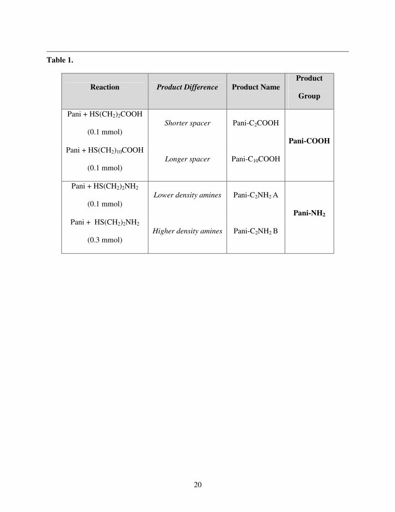
Table 1: A summary of modified nanofibres investigated.
Scheme 1. PAni nanofibre modification, where conditions involve (i) thiol reflux, (ii) EDC
coupling of primary antibody and (iii) incubation with HRP-labelled secondary antibody.
Figure 1: FTIR spectra for functionalised nanofibres show either a carboxylic acid band at 1700
cm-1
or an amine band at 3400 cm-1
.
Figure 2: TGA curves of PAni, PAni-COOH and PAni-NH2 have clearly different profiles (left).
Plotting of the differential of TGA curves allows for a clearer interpretation of results (right).
Figure 3. SEM images show high surface area functionalised PAni-NH2 nanofibres (a-b), and
PAni-NH2 antibody conjugates (c-d).
Figure 4. UV-vis spectra of PAni-COOH and PAni-NH2, along with their antibody conjugates
(all in aqueous solution). (Spectra have been normalised for clarity).
Figure 5. Cyclic voltammetry carried out in 0.2 M KCl (pH 1) (a, b) as electrolyte at 100 mV/s
scan rate.
Figure 6. Indirect ELISA using Pani-nanofibres immobilised with mouse IgG, an anti-mouse
HRP-labelled antibody as a secondary antibody and using the HRP substrate, 3,3´,5,5´-
tetramethylbenzidine (TMB) for detection. Pani-nanofibres not immobilised with antibodies
were used as a negative control.
18
Figure 7. TEM images show high surface area PAni nanofibres (a and b) and PAni-COOH
antibody conjugates (c and d).
Figure 8. Cyclic voltammetry carried out in phosphate buffered saline (pH 7.4) as electrolyte at
100 mV/s scan rate.
References
[1] A.G. MacDiarmid, J.C. Chiang, A.F. Richter, A.J. Epstein, Synthetic Met. 18 (1987) 285-
290.
[2] D.F.Acevedo, J. Balach, C.R. Rivarola, M.C. Miras, C.A. Barbero, Faraday Discussions 131
(2006) 235-252.
[3] C. Barbero, M.C. Miras, B. Schnyder, O. Haas, R. Kotz, J. Mater. Chem. 4 (1994) 1775-
1783.
[4] W.R. Small, F. Masdarolomoor, G.G. Wallace, M. Panhuis, J. Mater. Chem. 17 (2007) 4359-
4361.
[5] H. Peng, L. Zhang, C. Soeller, J. Travas-Sejdic, Biomaterials 30 (2009) 2132-2148.
[6] H. Peng, C. Soeller, N.A. Vigar, V. Caprio, J. Travas-Sejdic, Biosens. Bioelectron. 22 (2007)
1868-1873.
[7] Z. Wang, S. Liu, P. Wu, C. Cai, Anal. Chem. 81 (2009) 1638-1645.
[8] G. Wallace, G. Spinks, Soft Matter 3 (2007) 665–671.
[9] S.Virji, J.X. Huang, R.B. Kaner, B.H. Weiller, Nano Letters 4 (2004) 491-496.
[10] B. Shedd, C.O. Baker, M. J.Heller, R.B. Kaner, H.T. Hahn, Materials Science and
Engineering B 162 (2009) 111-115.
[11] X. Sun, W. Yang, T. Pan, A.T. Woolley, Anal. Chem. 80 (2008) 5126-5130.
[12] P.F.G. de Sa, C. Robb, E. Resende, P. McCarthy, S.C. Yang, P.R. Brown, J.A. Dain,
Journal of Capillary Electrophoresis and Microchip Technology 7 (2002) 61-65.
[13] M.J. Benes, D. Horak, F.J. Svec, Sep. Sci. 28 (2005) 1855-1875.
[14] S. Eeltink, F. Svec, Electrophoresis 28 (2007) 137-147.
19
[15] S. Anastase-Ravion, Z. Ding, A. Pelle, A.S. Hoffman, D. Letourneur, J. Chromatogr. B 761
(2001) 247-254.
[16] M. Lonnberg, J. Carlsson, J. Chromatogr, A 1127 (2006) 175-182.
[17] S. Virji, J.D. Fowler, C.O. Baker, J.X. Huang, R.B. Kaner, B.H. Weiller, Small 1 (2005)
624-627.
[18] X. Jiang, Q. Xu, S.K.W. Dertinger, A.D. Stroock, T. Fu, G.M. Whitesides, Anal. Chem. 77
(2005) 2338-2347.
[19] A.C. Henry, T.J. Tutt, M. Galloway, Y.Y. Davidson, C.S. McWhorter, S.A. Soper, R.L.
McCarley, Anal. Chem. 72 (2000) 5331-5337.
[20] E. Lahiff, T. Woods, W. Blau, G.G. Wallace, D. Diamond, Synthetic Met. 159 (2009) 741-
748.
[21] E. Lahiff, S. Scarmagnani, B. Schazmann, A. Cafolla, D. Diamond, International Journal of
Nanomanufacturing 5 (2010) Nos. ½.
[22] S. Kaplan, E.M. Conwell, A.F. Richter, A.G. Macdiarmid, J. Am. Chem. Soc. 110 (1988)
7647-7651.
[23] M. Inoue, F. Medrano, M. Nakamura, M.B. Inoue, Q. Fernando, J. Mater. Chem. 4 (1994)
1811-1814.
[24] T. Yasuda, I. Yamaguchi, T. Yamamoto, J. Mater. Chem. 13 (2003) 2138-2144.
20
Table 1.
Reaction Product Difference Product Name
Product
Group
Pani + HS(CH2)2COOH
(0.1 mmol)
Shorter spacer Pani-C2COOH
Pani + HS(CH2)10COOH
(0.1 mmol)
Longer spacer Pani-C10COOH
Pani-COOH
Pani + HS(CH2)2NH2
(0.1 mmol)
Lower density amines Pani-C2NH2 A
Pani + HS(CH2)2NH2
(0.3 mmol)
Higher density amines Pani-C2NH2 B
Pani-NH2
21
-(CH2)nCONH-
-HRP-HRP-HRP
-(CH2)nCONH--(CH2)nCOOH
-(CH2)nNH2 -(CH2)nNH-
-HRP-HRP-HRP
-(CH2)nNH-
-HRP-HRP-HRP= Primary antibody
= Secondary antibody labelled with HRP
(i)
(ii)
(ii)
(iii)
(iii)
Scheme 1.
22
0
20
40
60
80
100
120
2500300035004000
Wavenumber (cm-1
)
Inte
nsit
y (
a.u
.)
3400 cm-1
1400160018002000
Pani
Pani-NH2
Pani-COOH
1700 cm-1
Figure 1.
0
10
20
30
40
50
60
70
80
90
100
0 200 400 600
We
igh
t %
Pani
Pani-NH2
Pani-COOH
-0.5
-0.4
-0.3
-0.2
-0.1
0
0.1
0 200 400 600
Weig
ht
Dif
fere
nti
al
Pani
Pani-NH2
PaniCOOH
Figure 2.
23
(a) (b)
(c) (d)
Figure 3.
24
0
0.2
0.4
0.6
0.8
1
250 350 450 550 650 750 850
Wavelength (nm)
Ab
so
rban
ce
(a.u
.)
Pani
PaniC3COOH
PaniC11COOH
PaniC3COOH-Ab
PaniC11COOH-Ab
0
0.2
0.4
0.6
0.8
1
250 350 450 550 650 750 850
Wavelength (nm)
Ab
so
rban
ce
(a.u
.)
Pani
Pani-NH2
Pani-NH2-A-Ab
Pani-NH2-B-Ab
Figure 4.
25
-1.0E-03
-5.0E-04
0.0E+00
5.0E-04
1.0E-03
1.5E-03
-0.2 0 0.2 0.4 0.6 0.8 1
Potential vs Ag/AgCl (V)
Cu
rren
t (A
)
-1.5E-04
-1.0E-04
-5.0E-05
0.0E+00
5.0E-05
1.0E-04
1.5E-04PAni-C2COOH
PAni-C10COOH
PAni-C2COOH + Ab
(a)
-8.0E-04
-6.0E-04
-4.0E-04
-2.0E-04
0.0E+00
2.0E-04
4.0E-04
6.0E-04
8.0E-04
-0.4 -0.2 0.0 0.2 0.4 0.6 0.8 1.0 1.2
Potential vs Ag/AgCl (V)
Cu
rre
nt
(A)
-3.0E-04
-2.0E-04
-1.0E-04
0.0E+00
1.0E-04
2.0E-04
3.0E-04
4.0E-04PAni-NH2 B
PAni-NH2 A
(b)
Figure 5.
26
0.0
0.2
0.4
0.6
0.8
1.0
1.2
1.4
1.6
1.8
Pani-C2C
OO
H
Pani-C10C
OO
H
Pani-C2N
H2 A
Pani-C2N
H2 B
Ab
so
rba
nc
e (
a.u
.)
Antibody-functionalisedPani-nanofibres
Pani-nanofibre blanks
Figure 6.
27
(a) (b)
(c) (d)
Figure 7.
28
-7E-06
-5E-06
-3E-06
-1E-06
1E-06
3E-06
5E-06
-1 -0.5 0 0.5 1
Potential vs Ag/AgCl (V)
Cu
rre
nt
(A)
-1E-06
-8E-07
-6E-07
-4E-07
-2E-07
0E+00
2E-07
4E-07
6E-07
8E-07
Cu
rre
nt
(A)
Pani blank
Pani + Ab
Figure 8.