Embed Size (px)
Citation preview
Poly(ADP-Ribose) Polymerases in Host-Pathogen Interactions,Inflammation, and Immunity
Pamlea N. Brady,a Anupam Goel,b Margaret A. Johnsona
aDepartment of Chemistry, University of Alabama at Birmingham, Birmingham, Alabama, USAbCatalent Pharma Solutions, Philadelphia, Pennsylvania, USA
SUMMARY . . . . . . . . . . . . . . . . . . . . . . . . . . . . . . . . . . . . . . . . . . . . . . . . . . . . . . . . . . . . . . . . . . . . . . . . . . . . . . . . . . . . . . . . 1INTRODUCTION . . . . . . . . . . . . . . . . . . . . . . . . . . . . . . . . . . . . . . . . . . . . . . . . . . . . . . . . . . . . . . . . . . . . . . . . . . . . . . . . . . 2FUNCTION AND CLASSIFICATION OF PARP ENZYMES . . . . . . . . . . . . . . . . . . . . . . . . . . . . . . . . . . . . . 3
PARPs in DNA Repair . . . . . . . . . . . . . . . . . . . . . . . . . . . . . . . . . . . . . . . . . . . . . . . . . . . . . . . . . . . . . . . . . . . . . . . . . . 5PARP as a Chromatin-Remodeling Protein . . . . . . . . . . . . . . . . . . . . . . . . . . . . . . . . . . . . . . . . . . . . . . . . . . . 5PARPs as Transcription Factor Coactivators/Corepressors . . . . . . . . . . . . . . . . . . . . . . . . . . . . . . . . . . 6
PARPs IN HOST-PATHOGEN INTERACTIONS . . . . . . . . . . . . . . . . . . . . . . . . . . . . . . . . . . . . . . . . . . . . . . . . . . 7PARPs in Antiviral Responses . . . . . . . . . . . . . . . . . . . . . . . . . . . . . . . . . . . . . . . . . . . . . . . . . . . . . . . . . . . . . . . . . 7PARPs in Proviral Responses . . . . . . . . . . . . . . . . . . . . . . . . . . . . . . . . . . . . . . . . . . . . . . . . . . . . . . . . . . . . . . . . . 10PARP and Bacterial Pathogens . . . . . . . . . . . . . . . . . . . . . . . . . . . . . . . . . . . . . . . . . . . . . . . . . . . . . . . . . . . . . . 11Plant PARPs . . . . . . . . . . . . . . . . . . . . . . . . . . . . . . . . . . . . . . . . . . . . . . . . . . . . . . . . . . . . . . . . . . . . . . . . . . . . . . . . . . . 11Remaining Questions . . . . . . . . . . . . . . . . . . . . . . . . . . . . . . . . . . . . . . . . . . . . . . . . . . . . . . . . . . . . . . . . . . . . . . . . 12
PARPs IN INFLAMMATION AND IMMUNITY . . . . . . . . . . . . . . . . . . . . . . . . . . . . . . . . . . . . . . . . . . . . . . . . 13PARP as a Transcriptional Coactivator of Inflammatory Pathways . . . . . . . . . . . . . . . . . . . . . . . 14PARP, NAD� Metabolism, and Inflammation . . . . . . . . . . . . . . . . . . . . . . . . . . . . . . . . . . . . . . . . . . . . . . . 15NAD� Metabolism and Infection . . . . . . . . . . . . . . . . . . . . . . . . . . . . . . . . . . . . . . . . . . . . . . . . . . . . . . . . . . . . 19Role of PARG . . . . . . . . . . . . . . . . . . . . . . . . . . . . . . . . . . . . . . . . . . . . . . . . . . . . . . . . . . . . . . . . . . . . . . . . . . . . . . . . . 20MARylation and Its Reversal . . . . . . . . . . . . . . . . . . . . . . . . . . . . . . . . . . . . . . . . . . . . . . . . . . . . . . . . . . . . . . . . . 21Role of PARP in Inflammatory Autoimmune Disorders . . . . . . . . . . . . . . . . . . . . . . . . . . . . . . . . . . . . 23Arthritis . . . . . . . . . . . . . . . . . . . . . . . . . . . . . . . . . . . . . . . . . . . . . . . . . . . . . . . . . . . . . . . . . . . . . . . . . . . . . . . . . . . . . . . . 24Lupus . . . . . . . . . . . . . . . . . . . . . . . . . . . . . . . . . . . . . . . . . . . . . . . . . . . . . . . . . . . . . . . . . . . . . . . . . . . . . . . . . . . . . . . . . . 24Hyperthyroidism (Graves’ Disease) . . . . . . . . . . . . . . . . . . . . . . . . . . . . . . . . . . . . . . . . . . . . . . . . . . . . . . . . . . 25
PARP STRUCTURE AND INHIBITION . . . . . . . . . . . . . . . . . . . . . . . . . . . . . . . . . . . . . . . . . . . . . . . . . . . . . . . . . 25The PARP Signature Sequence and Its Significance . . . . . . . . . . . . . . . . . . . . . . . . . . . . . . . . . . . . . . . 25Coupling DNA Damage Detection to Catalytic Activity . . . . . . . . . . . . . . . . . . . . . . . . . . . . . . . . . . . 25Inhibitor Classes . . . . . . . . . . . . . . . . . . . . . . . . . . . . . . . . . . . . . . . . . . . . . . . . . . . . . . . . . . . . . . . . . . . . . . . . . . . . . . 27
CONCLUSION . . . . . . . . . . . . . . . . . . . . . . . . . . . . . . . . . . . . . . . . . . . . . . . . . . . . . . . . . . . . . . . . . . . . . . . . . . . . . . . . . . . 29ACKNOWLEDGMENTS . . . . . . . . . . . . . . . . . . . . . . . . . . . . . . . . . . . . . . . . . . . . . . . . . . . . . . . . . . . . . . . . . . . . . . . . . 29REFERENCES . . . . . . . . . . . . . . . . . . . . . . . . . . . . . . . . . . . . . . . . . . . . . . . . . . . . . . . . . . . . . . . . . . . . . . . . . . . . . . . . . . . . . 29AUTHOR BIOS . . . . . . . . . . . . . . . . . . . . . . . . . . . . . . . . . . . . . . . . . . . . . . . . . . . . . . . . . . . . . . . . . . . . . . . . . . . . . . . . . . . 47
SUMMARY The literature review presented here details recent research involvingmembers of the poly(ADP-ribose) polymerase (PARP) family of proteins. Among the17 recognized members of the family, the human enzyme PARP1 is the most exten-sively studied, resulting in a number of known biological and metabolic roles. Thisreview is focused on the roles played by PARP enzymes in host-pathogen interac-tions and in diseases with an associated inflammatory response. In mammalian cells,several PARPs have specific roles in the antiviral response; this is perhaps best illus-trated by PARP13, also termed the zinc finger antiviral protein (ZAP). Plant stress re-sponses and immunity are also regulated by poly(ADP-ribosyl)ation. PARPs promoteinflammatory responses by stimulating proinflammatory signal transduction path-ways that lead to the expression of cytokines and cell adhesion molecules. Hence,PARP inhibitors show promise in the treatment of inflammatory disorders and condi-tions with an inflammatory component, such as diabetes, arthritis, and stroke. Thesefunctions are correlated with the biophysical characteristics of PARP family enzymes.This work is important in providing a comprehensive understanding of the molecu-
Citation Brady PN, Goel A, Johnson MA. 2019.Poly(ADP-ribose) polymerases in host-pathogen interactions, inflammation, andimmunity. Microbiol Mol Biol Rev 83:e00038-18.https://doi.org/10.1128/MMBR.00038-18.
Copyright © 2018 American Society forMicrobiology. All Rights Reserved.
Address correspondence to Margaret A.Johnson, [email protected].
Published 19 December 2018
REVIEW
crossm
March 2019 Volume 83 Issue 1 e00038-18 mmbr.asm.org 1Microbiology and Molecular Biology Reviews
on March 9, 2020 by guest
http://mm
br.asm.org/
Dow
nloaded from
on March 9, 2020 by guest
http://mm
br.asm.org/
Dow
nloaded from
on March 9, 2020 by guest
http://mm
br.asm.org/
Dow
nloaded from
lar basis of pathogenesis and host responses, as well as in the identification of inhib-itors. This is important because the identification of inhibitors has been shown to beeffective in arresting the progression of disease.
KEYWORDS ADP-ribosylation, poly(ADP-ribose) polymerases, poly(ADP-ribose),antiviral responses, autoimmunity, enzyme inhibition, host-pathogen interactions,inflammation, stress granules, transcriptional regulation
INTRODUCTION
Poly(ADP-ribose) (PAR) is an unusual nucleic acid that is derived from NAD� by theaction of poly(ADP-ribose) polymerase (PARP) enzymes. This polymer is usually
found as a posttranslational modification of proteins and performs a wide variety ofsignaling, regulatory, and metabolic functions in cells. Essential functions in DNAdamage repair, RNA biology, stress responses, the cell cycle and cell death pathways,gene expression regulation, chromatin remodeling, and others have been identified (1).PAR was originally identified in 1963 by the laboratories of P. Chambon and coworkersas the insoluble portion of hen liver nuclear extracts (2). Subsequently its primarystructure was determined by the Mandel, Hayaishi, and Kawamura laboratories inde-pendently (3–5). Enzymes responsible for poly(ADP-ribose) polymerase and mono(ADP-ribosyl)transferase (MART) activities were subsequently discovered in the 1980s and1990s, respectively (6–9). A distinction is now made between PARPs, which catalyzeADP-ribose (ADPR) polymerization or poly(ADP-ribosyl)ation, and enzymes that transfer asingle ADP-ribose monomer. The latter is termed mono(ADP-ribosyl)ation (MARylation). Theentire enzyme family is now also referred to as diphtheria toxin-like ADP-ribosyltransferases(ARTDs), referring to their mechanistic similarity to the ADP-ribosylating diphtheria toxinproteins (10).
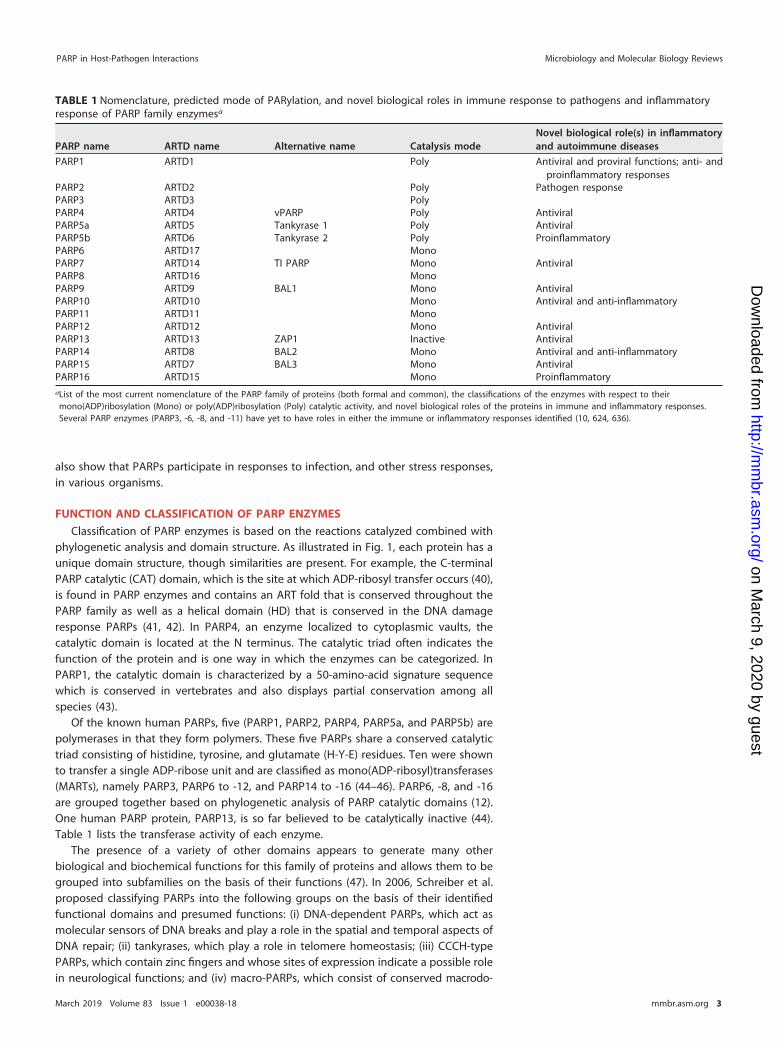
Seventeen human PARP enzymes have been identified (10). In addition, approxi-mately 1,900 other PARP family enzymes have been identified in other organisms,primarily eukaryotes (11–13). Table 1 lists the most current nomenclature of PARPs/ARTDs along with other names associated with PARP protein family members.
PARP family members have essential roles in the cell. Well-characterized roles exist,for example, in DNA damage repair. PARP1 and PARP2 are important components ofthe single-strand break (SSB) repair and base excision repair pathways. PARP1 andPARP3 participate in double-strand break (DSB) repair (reviewed in reference 14). Otherroles include regulating the cell cycle, regulating transcription, participating in chro-matin remodeling, and interacting with epigenetic mechanisms (15–18). When actingas transcriptional cofactors, PARPs participate in regulating circadian rhythms, guidingembryonic development, reprogramming somatic cells, and cellular differentiation(19–27). Additional roles in RNA biochemistry have recently been discovered. PARPsparticipate in the regulation of ribosome biogenesis and nucleolar structure and assistin controlling mRNA stability and translation (22, 28–34). PARP activity also impacts theregulation of alternative splicing and RNA silencing (35–37).
PARPs play significant roles in the development of cancer and have been shown tocontribute to six of the eight “hallmarks of cancer”: metastasis, replicative immortality,angiogenesis, cell death resistance, avoidance of growth suppression, proliferativesignaling, deregulation of cellular energetics, and avoidance of immune destruction(38). Through these functions, PARP inhibition has become important in treating severaltypes of cancer, and several PARP inhibitors have been successful in clinical trials.Examples include the compounds olaparib and veliparib, which are applied to thetreatment of ovarian, breast, and lung cancers (39).
This review discusses emerging roles of PARPs and poly(ADP-ribosyl)ation (PARyla-tion) in inflammation, immunity, and host-pathogen interactions. Increasing evidenceshows that PARPs have importance in viral infection, often being coopted by both DNAand RNA viruses. PARPs also contribute to inflammatory responses, and PARP inhibitionshows promise for the treatment of chronic inflammatory conditions. Recent studies
Brady et al. Microbiology and Molecular Biology Reviews
March 2019 Volume 83 Issue 1 e00038-18 mmbr.asm.org 2
on March 9, 2020 by guest
http://mm
br.asm.org/
Dow
nloaded from
also show that PARPs participate in responses to infection, and other stress responses,in various organisms.
FUNCTION AND CLASSIFICATION OF PARP ENZYMES
Classification of PARP enzymes is based on the reactions catalyzed combined withphylogenetic analysis and domain structure. As illustrated in Fig. 1, each protein has aunique domain structure, though similarities are present. For example, the C-terminalPARP catalytic (CAT) domain, which is the site at which ADP-ribosyl transfer occurs (40),is found in PARP enzymes and contains an ART fold that is conserved throughout thePARP family as well as a helical domain (HD) that is conserved in the DNA damageresponse PARPs (41, 42). In PARP4, an enzyme localized to cytoplasmic vaults, thecatalytic domain is located at the N terminus. The catalytic triad often indicates thefunction of the protein and is one way in which the enzymes can be categorized. InPARP1, the catalytic domain is characterized by a 50-amino-acid signature sequencewhich is conserved in vertebrates and also displays partial conservation among allspecies (43).
Of the known human PARPs, five (PARP1, PARP2, PARP4, PARP5a, and PARP5b) arepolymerases in that they form polymers. These five PARPs share a conserved catalytictriad consisting of histidine, tyrosine, and glutamate (H-Y-E) residues. Ten were shownto transfer a single ADP-ribose unit and are classified as mono(ADP-ribosyl)transferases(MARTs), namely PARP3, PARP6 to -12, and PARP14 to -16 (44–46). PARP6, -8, and -16are grouped together based on phylogenetic analysis of PARP catalytic domains (12).One human PARP protein, PARP13, is so far believed to be catalytically inactive (44).Table 1 lists the transferase activity of each enzyme.
The presence of a variety of other domains appears to generate many otherbiological and biochemical functions for this family of proteins and allows them to begrouped into subfamilies on the basis of their functions (47). In 2006, Schreiber et al.proposed classifying PARPs into the following groups on the basis of their identifiedfunctional domains and presumed functions: (i) DNA-dependent PARPs, which act asmolecular sensors of DNA breaks and play a role in the spatial and temporal aspects ofDNA repair; (ii) tankyrases, which play a role in telomere homeostasis; (iii) CCCH-typePARPs, which contain zinc fingers and whose sites of expression indicate a possible rolein neurological functions; and (iv) macro-PARPs, which consist of conserved macrodo-
TABLE 1 Nomenclature, predicted mode of PARylation, and novel biological roles in immune response to pathogens and inflammatoryresponse of PARP family enzymesa
PARP name ARTD name Alternative name Catalysis modeNovel biological role(s) in inflammatoryand autoimmune diseases
PARP1 ARTD1 Poly Antiviral and proviral functions; anti- andproinflammatory responses
PARP2 ARTD2 Poly Pathogen responsePARP3 ARTD3 PolyPARP4 ARTD4 vPARP Poly AntiviralPARP5a ARTD5 Tankyrase 1 Poly AntiviralPARP5b ARTD6 Tankyrase 2 Poly ProinflammatoryPARP6 ARTD17 MonoPARP7 ARTD14 TI PARP Mono AntiviralPARP8 ARTD16 MonoPARP9 ARTD9 BAL1 Mono AntiviralPARP10 ARTD10 Mono Antiviral and anti-inflammatoryPARP11 ARTD11 MonoPARP12 ARTD12 Mono AntiviralPARP13 ARTD13 ZAP1 Inactive AntiviralPARP14 ARTD8 BAL2 Mono Antiviral and anti-inflammatoryPARP15 ARTD7 BAL3 Mono AntiviralPARP16 ARTD15 Mono ProinflammatoryaList of the most current nomenclature of the PARP family of proteins (both formal and common), the classifications of the enzymes with respect to theirmono(ADP)ribosylation (Mono) or poly(ADP)ribosylation (Poly) catalytic activity, and novel biological roles of the proteins in immune and inflammatory responses.Several PARP enzymes (PARP3, -6, -8, and -11) have yet to have roles in either the immune or inflammatory responses identified (10, 624, 636).
PARP in Host-Pathogen Interactions Microbiology and Molecular Biology Reviews
March 2019 Volume 83 Issue 1 e00038-18 mmbr.asm.org 3
on March 9, 2020 by guest
http://mm
br.asm.org/
Dow
nloaded from
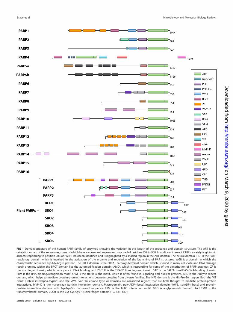
FIG 1 Domain structure of the human PARP family of enzymes, showing the variation in the length of the sequence and domain structure. The ART is thecatalytic domain of the sequences, some of which have a conserved sequence comprised of residues 859 to 908. In addition, in select PARPs, a catalytic glutamicacid corresponding to position 988 of PARP1 has been identified and is highlighted by a shaded region in the ART domain. The helical domain (HD) is the PARPregulatory domain which is involved in the activation of the enzyme and regulation of the branching of PAR structures. WGR is a domain in which thecharacteristic sequence Trp-Gly-Arg is present. The BRCT domain is the BRCA1 carboxyl-terminal domain which is found in many cell cycle and DNA damagerepair proteins. Within the BRCT domain lies the automodification domain (AMD), which is responsible for some of the dimerization of PARP enzymes. ZF isthe zinc finger domain, which participate in DNA binding, and ZF/THP is the TiPARP homologous domain. SAP is the SAF/Acinus/PIAS-DNA-binding domain.RRM is the RNA-binding/recognition motif. SAM is the sterile alpha motif, which is often found in signaling and nuclear proteins. ARD is the Ankyrin repeatdomain, which helps to mediate protein-protein interactions between proteins from diverse families. The HPS domain is the His-Pro-Ser region. Both the VIT(vault protein interalpha-trypsin) and the vWA (von Willebrand type A) domains are conserved regions that are both thought to mediate protein-proteininteractions. MVP-ID is the major-vault particle interaction domain. Macrodomain, poly(ADP-ribose) interaction domain; WWE, iso(ADP-ribose) and protein-protein interaction domain with Trp-Trp-Glu conserved sequence. UIM is the MAO interaction motif, GRD is a glycine-rich domain. And TMD is thetransmembrane domain. CCCH is the Cys-Cys-Cys-His zinc finger domain (10, 181, 637).
Brady et al. Microbiology and Molecular Biology Reviews
March 2019 Volume 83 Issue 1 e00038-18 mmbr.asm.org 4
on March 9, 2020 by guest
http://mm
br.asm.org/
Dow
nloaded from
mains that are linked to a PARP domain. There are several PARP family members that arenot classed among these groups, i.e., PARP4, PARP6, PARP8, PARP10, and PARP11 (47).
PARPs in DNA Repair
Though PARPs perform numerous functions, there are several broad categories thatare well established. The first is as a DNA damage response protein. PARP1, PARP2, andPARP3 are DNA-dependent enzymes that are activated upon binding to DNA damageand play important roles in the repair of DNA strand breaks (48). To date, three PARPenzymes (PARP1, -2, and -3) have been identified to play a role in DNA repair, with therole of PARP1 having been studied the most extensively (49–51). PARP1 has beenproposed as a general DNA damage sensor and is activated by several types ofdamaged DNA, including hairpins, cruciform DNA, loops, nicked DNA, blunt ends, andoverhangs (52, 53). PARP1 may also interact with other non-B DNA structures, such asG quadruplexes (54). In contrast, PARP2 and -3 play more specific roles in DNA repairpathways. PARP2 participates in the base excision repair (BER) pathway, interactingwith the X-ray repair cross-complementing protein 1 (XRCC1) and DNA ligase III (55, 56).The depletion of PARP2 results in an increased sensitivity to ionizing radiation andalkylating agents, which is consistent with a role in single-strand break (SSB) repair (55,57). PARP2 is the closest paralogue of PARP1 and in the presence of DNA damage isresponsible for most of the residual PARP activity in PARP1�/� cells (50, 58). Whileeither PARP1 or PARP2 can be knocked out in mice without causing serious defects, adouble knockout is lethal in the embryo, suggesting that the functions of these twoenzymes are partly redundant (57).
In contrast, PARP3 has been shown to play a role in double-strand break (DSB) repairby interacting with the aprataxin-like factor (APLF) at damaged sites (49, 59). PARP3 alsohelps to promote accurate ligation of DSB by XRCC4 and DNA ligase IV during thenonhomologous end-joining (NHEJ) process (49, 60). PARP3 was shown to ADP-ribosylate histone H2B, and this was required for the SSB response (61). PARP2 andPARP3 enzymes are activated by a smaller subset of damaged DNA types, which isconsistent with their specific roles. Thus, PARP2 is activated by DNA containing “flapsand gaps” (62), while PARP3 is activated by blunt ends (49). Both enzymes are activatedby 5=-phosphorylated nicks (42).
PARP1 to -3 also have the capacity to ADP-ribosylate DNA, with the modificationoccurring at double-strand break termini. This activity may help to mark the locationsof strand breaks and recruit repair proteins (63–65).
PARP as a Chromatin-Remodeling Protein
A second category of biological function for PARPs is their role in chromatinremodeling. MARylation and PARylation of histones were observed as early as 1980 (66,67). PARP was soon observed to participate in the relaxation or decondensation ofchromatin structure (68–71). The addition of a high-molecular-weight, negativelycharged polymer to histone proteins loosens the tightly packed structure of chromatinby electrostatic repulsion, promoting the access of chromatin-remodeling factors (68).PARP was shown to participate in puffing in Drosophila, a process which requires theloosening of chromatin structure, promoting remodeling and facilitating transcription (72).Not only PARylation but also MARylation has unique roles; for example, MARylation ofhistone H3 promotes the access of p300, leading to cellular proliferation through the�-catenin pathway (73). Changes in nucleosome structure due to PARP binding andPARylation were shown to lead to high gene transcription levels and could produce, forexample, a strong inflammatory response due to recruitment of nuclear factor kappa-light-chain-enhancer of activated B cells (NF-�B) at the interleukin-1� (IL-1�) and tumornecrosis factor (TNF) promoters where PARP1 is constitutively associated (74). Thenucleosome-binding capability of PARP1 was shown to be key to its transcription factorcoactivator activity for certain loci (75) (see also “PARPs as Transcription Factor Coacti-vators/Corepressors” below).
PARP in Host-Pathogen Interactions Microbiology and Molecular Biology Reviews
March 2019 Volume 83 Issue 1 e00038-18 mmbr.asm.org 5
on March 9, 2020 by guest
http://mm
br.asm.org/
Dow
nloaded from
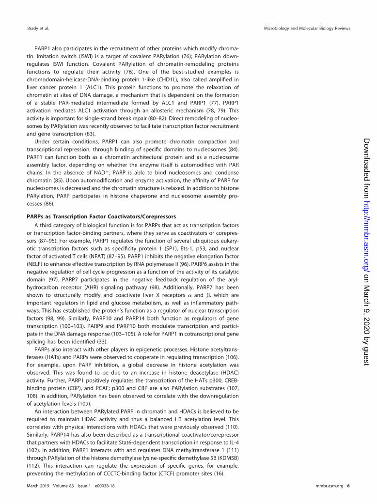
PARP1 also participates in the recruitment of other proteins which modify chroma-tin. Imitation switch (ISWI) is a target of covalent PARylation (76); PARylation down-regulates ISWI function. Covalent PARylation of chromatin-remodeling proteinsfunctions to regulate their activity (76). One of the best-studied examples ischromodomain-helicase-DNA-binding protein 1-like (CHD1L), also called amplified inliver cancer protein 1 (ALC1). This protein functions to promote the relaxation ofchromatin at sites of DNA damage, a mechanism that is dependent on the formationof a stable PAR-mediated intermediate formed by ALC1 and PARP1 (77). PARP1activation mediates ALC1 activation through an allosteric mechanism (78, 79). Thisactivity is important for single-strand break repair (80–82). Direct remodeling of nucleo-somes by PARylation was recently observed to facilitate transcription factor recruitmentand gene transcription (83).
Under certain conditions, PARP1 can also promote chromatin compaction andtranscriptional repression, through binding of specific domains to nucleosomes (84).PARP1 can function both as a chromatin architectural protein and as a nucleosomeassembly factor, depending on whether the enzyme itself is automodified with PARchains. In the absence of NAD�, PARP is able to bind nucleosomes and condensechromatin (85). Upon automodification and enzyme activation, the affinity of PARP fornucleosomes is decreased and the chromatin structure is relaxed. In addition to histonePARylation, PARP participates in histone chaperone and nucleosome assembly pro-cesses (86).
PARPs as Transcription Factor Coactivators/Corepressors
A third category of biological function is for PARPs that act as transcription factorsor transcription factor-binding partners, where they serve as coactivators or corepres-sors (87–95). For example, PARP1 regulates the function of several ubiquitous eukary-otic transcription factors such as specificity protein 1 (SP1), Ets-1, p53, and nuclearfactor of activated T cells (NFAT) (87–95). PARP1 inhibits the negative elongation factor(NELF) to enhance effective transcription by RNA polymerase II (96). PARP6 assists in thenegative regulation of cell cycle progression as a function of the activity of its catalyticdomain (97). PARP7 participates in the negative feedback regulation of the aryl-hydrocarbon receptor (AHR) signaling pathway (98). Additionally, PARP7 has beenshown to structurally modify and coactivate liver X receptors � and �, which areimportant regulators in lipid and glucose metabolism, as well as inflammatory path-ways. This has established the protein’s function as a regulator of nuclear transcriptionfactors (98, 99). Similarly, PARP10 and PARP14 both function as regulators of genetranscription (100–103). PARP9 and PARP10 both modulate transcription and partici-pate in the DNA damage response (103–105). A role for PARP1 in cotranscriptional genesplicing has been identified (33).
PARPs also interact with other players in epigenetic processes. Histone acetyltrans-ferases (HATs) and PARPs were observed to cooperate in regulating transcription (106).For example, upon PARP inhibition, a global decrease in histone acetylation wasobserved. This was found to be due to an increase in histone deacetylase (HDAC)activity. Further, PARP1 positively regulates the transcription of the HATs p300, CREB-binding protein (CBP), and PCAF; p300 and CBP are also PARylation substrates (107,108). In addition, PARylation has been observed to correlate with the downregulationof acetylation levels (109).
An interaction between PARylated PARP in chromatin and HDACs is believed to berequired to maintain HDAC activity and thus a balanced H3 acetylation level. Thiscorrelates with physical interactions with HDACs that were previously observed (110).Similarly, PARP14 has also been described as a transcriptional coactivator/corepressorthat partners with HDACs to facilitate Stat6-dependent transcription in response to IL-4(102). In addition, PARP1 interacts with and regulates DNA methyltransferase 1 (111)through PARylation of the histone demethylase lysine-specific demethylase 5B (KDM5B)(112). This interaction can regulate the expression of specific genes, for example,preventing the methylation of CCCTC-binding factor (CTCF) promoter sites (16).
Brady et al. Microbiology and Molecular Biology Reviews
March 2019 Volume 83 Issue 1 e00038-18 mmbr.asm.org 6
on March 9, 2020 by guest
http://mm
br.asm.org/
Dow
nloaded from
New functions within and outside these categories continue to be discovered.Recent proteomic work has identified ADP-ribosylation substrates, which is useful forunderstanding the metabolic pathways involving PARPs (113–117). The results of thesestudies suggest that PARPs regulate many more biological functions than have beenidentified to date (113). Among the novel functions identified for this family of enzymesare their roles in host-pathogen interactions. Some of the most recent findings arediscussed below.
PARPs IN HOST-PATHOGEN INTERACTIONSPARPs in Antiviral Responses
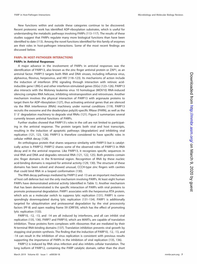
A major advance in the involvement of PARPs in antiviral responses was theidentification of PARP13, also known as the zinc finger antiviral protein or ZAP1, as anantiviral factor. PARP13 targets both RNA and DNA viruses, including influenza virus,alphavirus, filovirus, herpesvirus, and HIV (118–123). Its mechanisms of action includethe induction of interferon (IFN) signaling through interaction with retinoic acid-inducible gene I (RIG-I) and other interferon-stimulated genes (ISGs) (123–126). PARP13also interacts with the Moloney leukemia virus 10 homologue (MOV10) RNA-inducedsilencing complex RNA helicase, inhibiting retrotransposition and retroviruses. Anothermechanism involves the physical interaction of PARP13 with argonaute proteins totarget them for ADP-ribosylation (127), thus activating antiviral genes that are silencedvia the RNA interference (RNAi) machinery under normal conditions (119). PARP13recruits the exosome and the deadenylase poly(A)-specific RNase (PARN), as well as the5=-3= degradation machinery to degrade viral RNAs (121). Figure 2 summarizes severalcurrently known antiviral functions of PARPs.
Further studies showed that PARP13’s roles in the cell are not limited to participat-ing in the antiviral response. The protein targets both viral and host transcripts,resulting in the induction of apoptotic pathways (degradation) and inhibiting viralreplication (121, 123, 126). PARP13 is therefore considered to have specific roles incellular mRNA decay (128).
An orthologous protein that shares sequence similarity with PARP13 but is catalyt-ically active is PARP12. PARP12 shares some of the observed roles of PARP13 in RNAdecay and in the antiviral response. Like PARP13, it recognizes specific sequences inviral RNA and DNA and degrades retroviral RNA (121, 122, 125). Both proteins containzinc finger domains in the N-terminal region. Recognition of RNA by these nucleicacid-binding domains is required for antiviral activity (129, 130). The structure of thesedomains has been solved and showed unusual, CCCH-type zinc fingers with cavitiesthat could bind RNA in a looped conformation (130).
The RNA decay pathways mediated by PARP12 and -13 are an important mechanismof host cell defense but not the only mechanism involving PARPs. At least eight humanPARPs have demonstrated antiviral activity (identified in Table 1). Another mechanismthat has been demonstrated is the specific interaction of PARPs with viral proteins topromote proteasomal degradation. PARP1 associates with the herpesvirus RTA protein,which acts as a molecular switch to suppress lytic replication (131). PARP1 is corre-spondingly downregulated during lytic replication (131–134). PARP1 is additionallytargeted for ubiquitination and proteasomal degradation by the viral processivityfactors (PF-8) and open reading frame 59 (ORF59), which has the effect of promotinglytic replication (133).
PARP10, -12, -13, and -14 are all induced by interferons, and all can inhibit viralreplication (135, 136). PARP7 and PARP10, which are MARTs, are capable of translationinhibition. These proteins form complexes with ribosomes that are mediated by theirN-terminal RNA-binding domains (137). Translation inhibition prevents viral growth bystopping viral protein synthesis. The finding that the induction of PARP10, -12, -13, and-14 can result in the inhibition of virus replication is consistent with previous resultssupporting the importance of PARPs in the inhibition of viral replication (135, 136).
PARP12 is induced by RNA virus infection and also inhibits cellular translation. Thelong isoform of PARP12, containing the PARP catalytic domain, rather than the short
PARP in Host-Pathogen Interactions Microbiology and Molecular Biology Reviews
March 2019 Volume 83 Issue 1 e00038-18 mmbr.asm.org 7
on March 9, 2020 by guest
http://mm
br.asm.org/
Dow
nloaded from
isoform was required for this antiviral activity, which was active against positive,negative, and ambisense RNA viruses (135). Since this mechanism appears to involvecatalytic activity and/or the catalytic domain, it may be separate from the RNA decayfunctions of the protein.
Another important antiviral mechanism involves the formation of cellular stressgranules (SGs). The introduction of viral RNA into the cytoplasm results in the activation
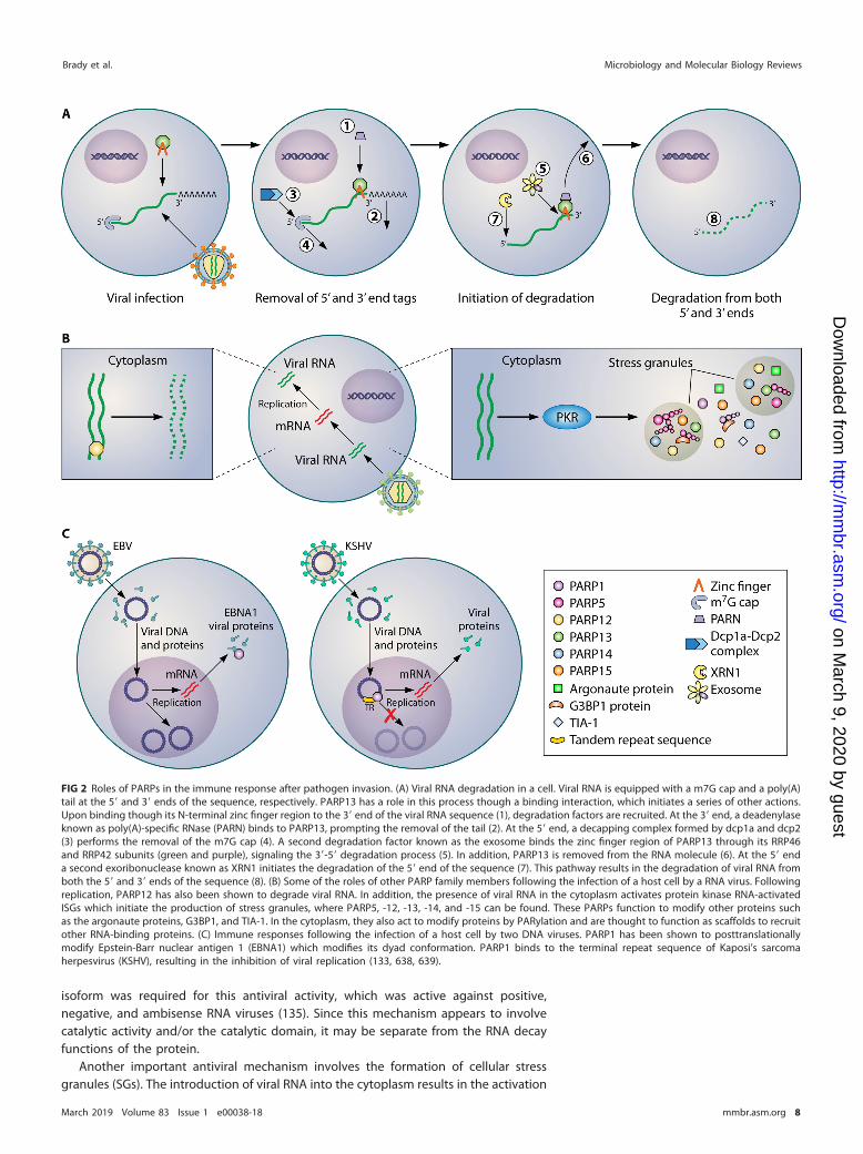
FIG 2 Roles of PARPs in the immune response after pathogen invasion. (A) Viral RNA degradation in a cell. Viral RNA is equipped with a m7G cap and a poly(A)tail at the 5= and 3= ends of the sequence, respectively. PARP13 has a role in this process though a binding interaction, which initiates a series of other actions.Upon binding though its N-terminal zinc finger region to the 3= end of the viral RNA sequence (1), degradation factors are recruited. At the 3= end, a deadenylaseknown as poly(A)-specific RNase (PARN) binds to PARP13, prompting the removal of the tail (2). At the 5= end, a decapping complex formed by dcp1a and dcp2(3) performs the removal of the m7G cap (4). A second degradation factor known as the exosome binds the zinc finger region of PARP13 through its RRP46and RRP42 subunits (green and purple), signaling the 3=-5= degradation process (5). In addition, PARP13 is removed from the RNA molecule (6). At the 5= enda second exoribonuclease known as XRN1 initiates the degradation of the 5= end of the sequence (7). This pathway results in the degradation of viral RNA fromboth the 5= and 3= ends of the sequence (8). (B) Some of the roles of other PARP family members following the infection of a host cell by a RNA virus. Followingreplication, PARP12 has also been shown to degrade viral RNA. In addition, the presence of viral RNA in the cytoplasm activates protein kinase RNA-activatedISGs which initiate the production of stress granules, where PARP5, -12, -13, -14, and -15 can be found. These PARPs function to modify other proteins suchas the argonaute proteins, G3BP1, and TIA-1. In the cytoplasm, they also act to modify proteins by PARylation and are thought to function as scaffolds to recruitother RNA-binding proteins. (C) Immune responses following the infection of a host cell by two DNA viruses. PARP1 has been shown to posttranslationallymodify Epstein-Barr nuclear antigen 1 (EBNA1) which modifies its dyad conformation. PARP1 binds to the terminal repeat sequence of Kaposi’s sarcomaherpesvirus (KSHV), resulting in the inhibition of viral replication (133, 638, 639).
Brady et al. Microbiology and Molecular Biology Reviews
March 2019 Volume 83 Issue 1 e00038-18 mmbr.asm.org 8
on March 9, 2020 by guest
http://mm
br.asm.org/
Dow
nloaded from
of protein kinase RNA (PKR)-activated SGs and the production of these membranelessorganelles, which assist in arresting proviral cellular processes such as translationalpathways. These organelles also regulate mRNA stability and have antiviral functions(reviewed in reference 138). Viruses use various strategies to disrupt or block stressgranules; for example, Ebola virus inhibits SG formation (139), influenza virus sequestersdouble-stranded RNA (dsRNA) to prevent SG initiation (140), and poliovirus 3 C pro-tease cleaves a major SG protein, Ras GTPase-activating protein-binding protein 1(G3BP1) (141). The RNA-binding PARP12 and -13 are found in stress granules, wherePARP12 is responsible for MARylation of various proteins such as the argonauteproteins, G3BP1, and TIA-1 (36). G3BP1 MARylation promotes stress granule nucleation(36). PARylation controls the sequestration of argonaute 2 (AGO2), leading to themodulation of microRNA silencing (36). PAR is able to nucleate phase transitions ofRNA-binding proteins by initiating the liquid demixing process that is required for theformation of membraneless organelles (142, 143). PARP5a, -7, and -15 have also beenlocalized to stress granules (36). Along with PARP12 and -13, PARP5a, -14, and -15 weresuggested to have regulatory roles in the production and maintenance of stressgranules (36). This is illustrated in Fig. 2B. Poly(ADP-ribose) glycosylase (PARG) alsolocalized to SGs, and its overexpression inhibited SG formation, again suggesting a keyrole of PAR in SGs (36).
Several other PARPs have been identified to have antiviral functions. PARP14 has anactive role in the development of T cells and B cells. This process is initiated byinterleukin-4, which stimulates differentiation of Th2 cells, resulting in the ADP-ribosylation of HDACs. The deacetylation of chromatin allows the transcription of genescoding for immune cell production (102). PARP9 is a binding partner of Deltex 3-like, anE3 ubiquitin ligase that ubiquitinates histone H4 and protects cells against DNAdamage. In the presence of its binding partner, PARP9 MARylates ubiquitin, preventingE3 ligase activity and providing regulation of the complex (46). The PARP9-Deltex 3-likecomplex also activates signal transducer and activator of transcription 1 (STAT1) andubiquitinates histone proteins to promote the expression of a subset of interferon-stimulated genes, thereby stimulating the innate immune response. This complex alsobinds to viral 3C proteases and ubiquitinates them for degradation (144).
Phylogenetic analyses also support a role of PARP in antiviral defense. An analysis ofPARP mutation rates showed positive selection in the catalytic domains of PARPs,suggesting a connection between the antiviral response and the activity of multiplePARPs, which is indicative of immune system defense (145, 146).
In the presence of DNA damage, PARP1 is responsible for the majority of cellularPARP activity (50, 58, 147, 148). Perhaps not surprisingly, this enzyme also performsantiviral functions. These occur through several mechanisms.
The release of viral contents into a cell initiates the host cell immune response,activating proapoptotic factors such as p53. Some viruses, such as Kaposi’s sarcomaherpesvirus (KSHV), inhibit the action of p53, arresting apoptosis through a mechanisminvolving the ORF8 protein (149). PARP1 binds the terminal repeat (TR) sequence ofKSHV, resulting in the inhibition of viral replication (Fig. 2C) (133).
Another mechanism takes advantage of PARP1’s function as a chromatin-bindingprotein. PARP1 represses the expression of retrotransposons in Drosophila and ofretroviruses in avian cells. This repression is mediated by HDACs and DNA methylases(150). Incorporation of PARP1 into chromatin causes chromatin compaction and tran-scriptional repression, which leads to retrotransposon silencing and the induction ofheterochromatin. Upon activation of PARP1, the enzyme auto-PARylates and dissoci-ates, leading to transcriptional activation and chromatin decompaction. Since otherPARP family members also interact with HDACs (102), this may represent a commonmechanism in the PARP family.
In a related manner, PARP1 has a role in HIV DNA integration. PARP1 is incorporatedinto nucleosomes in a manner that requires the N-terminal DNA-binding region and theinteraction of the C-terminal region with histone proteins (151). PARP1 participates inHIV-1 long terminal repeat function, binds to HIV trans-activation response element
PARP in Host-Pathogen Interactions Microbiology and Molecular Biology Reviews
March 2019 Volume 83 Issue 1 e00038-18 mmbr.asm.org 9
on March 9, 2020 by guest
http://mm
br.asm.org/
Dow
nloaded from
(TAR) RNA, and is required for efficient HIV-1 integration (152–154). Thus, by the samemechanism PARP1 can participate in a proviral or antiviral function, depending on thestage of the infectious cycle of the virus.
PARPs in Proviral Responses
In addition to performing antiviral functions, other cases are known in which PARPsare recruited by viruses to perform essential functions. For example, Epstein-Barrnuclear antigen 1 (EBNA1) of Epstein-Barr virus (EBV) is posttranslationally modified byPARP1, which leads to a remodeling of proteins at the dyad symmetry (DS) element(155) (Fig. 2C). PARP1 is believed to serve a dual role, both preventing DNA damage andregulating the efficiency of oriP and copy number and the replication efficiency of EBVepisomes, thus contributing to the maintenance of the virus (155).
The nucleocapsid protein of porcine reproductive and respiratory syndrome virus(PRRSV) binds to PARP1, and the interaction is critical for viral replication, since the useof a PARP inhibitor led to inhibited viral growth (156). In an intriguing parallel,Grunewald et al. have shown that the nucleocapsid proteins of both �- and�-coronaviruses are MARylated during infection (157). The function of this modification,and whether it is pro- or antiviral, is currently unknown. Adenoviral core proteins wereshown to be ADP-ribosylated, and the use of PARP inhibitors led to greatly reducedinfectivity, suggesting a role for the modification in virus decapsidation (158). PARP1 isadditionally required for efficient activity of both the H1N1 and H5N1 influenza virusRNA polymerases (159). One study suggests that a poly(ADP-ribose) glycohydrolase(PARG) isoform is degraded as a result of a gene product of herpes simplex virus 1(HSV-1), which is another indication that PARylation may be beneficial to virulence(PARG counteracts PARylation) (160). In addition, inhibiting PARP activity also inhibitsthe replication of several types of viruses, suggesting that in these cases, PARylationassists, rather than attenuates, viral replication. This has been demonstrated in severalfamilies of viruses, such as herpesviruses, adenoviruses, and arteriviruses (156, 158, 160).
An intriguing role for extracellular PAR was identified by the group of Mitchison(161). PAR was shown to activate cytokine release by macrophages, which is associatedwith activation of innate immune responses. PAR could be recognized by Toll-likereceptors 2 and 4, and inhibition of these receptors prevented cytokine release. Hence,extracellular PAR is a strong proinflammatory signal. This manner of signaling is specificto the polymer, since monomeric ADP-ribose did not elicit a response. Since PAR isprimarily an intracellular molecule, it remains to be determined how the extracellularPAR is produced. An extracellular ADP-ribosyltransferase (ART) has previously beenidentified on T cells, which might function as the source (162). Alternatively, PAR mightbe released by cells after necrotic cell death.
Several RNA viruses encode macrodomains, which have been described as the“readers and erasers” of ADP-ribosylation (163). These domains recognize and reverseMARylation and PARylation. For example, a macrodomain is conserved in coronavirusesand alphaviruses, as well as other viruses from the Hepeviridae and Togaviridae families(164–166). Viral macrodomains can remove ADP-ribose or PAR from various proteinsubstrates (167, 168). In addition, PARP10 and PARP14 were induced in response tointerferon alpha and to lipopolysaccharide (LPS), suggesting that MARylation is relevantto host-pathogen interaction (169). Since PAR is a regulator of multiple signalingpathways, conserved viral macrodomains are likely used to interfere with one or morehost cell antiviral defense mechanisms. Indeed, these proteins mediate resistance tointerferon signaling (170), promote virulence, and suppress the innate immune re-sponse during coronavirus infection (171–173). Specific protein substrates have beenidentified for a few macrodomains (169, 174–177), but many remain unidentified todate. Some macrodomains have divergent biochemical activities, such as nucleic acidbinding or protein/protein interaction, suggesting that these readers and erasers, likePARPs, are multifunctional (168, 178–180).
Due to the fact that the activity of PARPs has been shown to be both pro- andantiviral, it is clear that a simple definition of the role of PARPs in the response to viral
Brady et al. Microbiology and Molecular Biology Reviews
March 2019 Volume 83 Issue 1 e00038-18 mmbr.asm.org 10
on March 9, 2020 by guest
http://mm
br.asm.org/
Dow
nloaded from
infection is not sufficient. There are few known enzyme-specific inhibitors of PARPs,which impedes our ability to understand how they respond to viral infections; thus,future work would involve the identification of specific inhibitors for individual PARPfamily members. Recently, exciting progress has been made in this area (reviewed inreference 181).
PARP and Bacterial Pathogens
Activation of PARP has been observed upon infection of eukaryotic cells by bacterialpathogens. For example, the group A Streptococcus (GAS) pathogenicity island encodesstreptolysin (SLO) and NAD� glycohydrolase. SLO pore formation induced PARP acti-vation, while NAD� glycohydrolase activity modulated this PARP activity (182). PARPactivation has also been observed in Helicobacter pylori infection (183). Bacterialinfection was shown to promote PARP activation, through pathways related to inflam-mation (183, 184).
Interestingly, only a few bacteria have functional PARylation systems, while manyhave PAR-binding domains, PAR-degrading enzymes, and ADP-ribosylating toxins.These proteins often form operons that may constitute effector systems and mayinclude other domains, such as SLOG, Nudix, and NADAR domains. In parallel to thecase for eukaryotic cells, these systems could serve as a source of ATP or intracellularsignaling system (185).
Perina et al. performed a detailed analysis of the distribution of enzymes of PARmetabolism in bacteria with sequenced genomes and showed that PARPs are presentin six of the 30 known bacterial phyla, where they were probably acquired by horizontalgene transfer (12). Of these, one has been characterized in vitro, namely, the PARP fromHerpetosiphon aurantiacus, which was demonstrated to synthesize polymers (186). Theroles of these systems are diverse. In nitrogen-fixing bacteria, the MARylation modifi-cation regulates nitrogenase (reviewed in reference 187). In Bacillus subtilis and Strep-tomyces griseus ADP-ribosylation contributes to sporulation (188, 189), and in Strepto-myces coelicolor it regulates antibiotic production (174). The S. coelicolor SCO5461protein was experimentally demonstrated to carry out MARylation.
Plant PARPs
As in animals, plant PARPs are important for DNA damage repair (190–193). PlantPARP2 makes the greatest contribution to DNA repair pathways (194). ArabidopsisPARP1 and PARP2 interact physically, and PARP2 is the most important of these inpathogen-induced DNA damage repair and basal resistance (194). Plant PARPs alsoparticipate in transcriptional regulation (191, 195).
PARPs also play a key role in responding to abiotic stress and to infection. Forexample, early studies using PARP inhibition showed that PARP is involved in cell deathtriggered by DNA damage or oxidative stress (196). PARP was shown to affect stresstolerance (191). Silencing of PARP genes altered stress signal transduction (197).Oxidative stress, drought, and high-light resistance is increased by the knockdownof PARP1 and PARP2 or by the application of PARP inhibitors (191, 198). Thus, theroles of PARPs in responding to abiotic and biotic stress signals appear to differ.Surprisingly, PARP knockout plants were recently found to have constitutive PARPactivity and did not exhibit altered stress responses (199). It is possible that thisactivity could be mediated by the SRO (similar to RCD-one) or RCD (radical-inducedcell death) proteins, which are PARP orthologues that occur only in plants (describedbelow). PARP3, in contrast, can be induced by several factors, particularly duringdevelopment of the seed (200). PARP3 is a vital component in the storability andviability of the plant seeds (201). Chemical inhibition of PARPs was shown to improvestress tolerance and plant growth, and this correlated with changes in the levels ofstress-related metabolites such as anthocyanins (202).
PARP was induced upon Pseudomonas infection of Arabidopsis (203, 204). Furtherwork showed that PARP inhibition disrupted lignin and callose deposition, whichcomprise part of the innate immune response, and that pathogen infection activated
PARP in Host-Pathogen Interactions Microbiology and Molecular Biology Reviews
March 2019 Volume 83 Issue 1 e00038-18 mmbr.asm.org 11
on March 9, 2020 by guest
http://mm
br.asm.org/
Dow
nloaded from

PARylation reactions (203). PARylation was shown to regulate genes involved in im-mune defense (205). Correspondingly, PARPs may be required to repair pathogen-induced DNA damage (206). Most recently, Feng and coworkers demonstrated thatPARP2 participates in important reactions with DAWDLE (DDL), a forkhead-associated(FHA) domain protein. The PARylation-dependent interaction between PARP2 and DDLis essential to the function of DDL in plant immunity (207).
Pathogens in turn utilize MARylation and PARylation to antagonize plant celldefenses. Thus, MARylation of plant proteins by bacterial virulence factors was shownto interfere with the plant immune response, especially in pattern-triggered immunity(PTI) signaling pathways (208). For example, the Pseudomonas syringae type III secretionsystem (T3SS) produces ADP-ribosyltransferase effectors that act on plant RNA-bindingproteins and kinases (209, 210). The modification of the RNA-binding protein GRP7inhibits protein function in binding to pattern recognition receptor transcripts (210,211). HopF2 MARylates kinases in the PTI signal transduction pathway, inhibiting themicrobe-associated molecular pattern (MAMP)-induced response. A third effector,AvrRpm1, is predicted to have a PARP-like fold, but its transferase activity is unclear andit may serve to target PARP substrates or binding partners through molecular mimicry(212). Genes encoding other PAR-related functions such as PARG and Nudix hydrolaseswere also upregulated in response to MAMPs, suggesting the need for a programmed,regulated immune response involving PARPs and the need to avoid excessive con-sumption of NAD� (204).
The RCD (radical-induced cell death) and SRO (similar to RCD-one) proteins are PARPorthologues that are found only in plant cells. These proteins contain ART core domainsbut lack the HYE catalytic triad and are predicted to be inactive; however, this remainsto be verified (213). For example, RCD1 does not have detectable NAD binding or PARPactivity (213). However, PARP activity has been reported for SRO1 (214). These proteinsare regulators of stress responses and plant development (199, 215, 216). RCD1 isupregulated in response to light stress (217). Further, RCD1 interacts with multiple planttranscription factors that control key aspects of plant stress. For example, dehydration-responsive element-binding protein 2 A (DREB2A) is an Arabidopsis transcription factorthat is involved in the stress response to salt and drought (218, 219). RCD1 binds anddestabilizes this transcription factor. Under certain conditions, RCD1 is degraded toincrease DREB2A function (219). Arabidopsis NAC transcription factors ANAC013 andANAC046 regulate mitochondrial function (220). RCD1 appears to function in activatinga gene expression program of antioxidant response (221).
The SRO1 protein from Oryza sativa (rice) and an RNA-binding protein, OsRBD1,conferred tolerance to multiple stresses in a yeast model system (222). These proteinshave a domain structure similar to that of human PARP11 in that they contain a WWEdomain. The SRO proteins contain an additional, plant-specific domain that is impli-cated in transcription factor interaction (213, 222). This RST domain has also beenidentified in the TATA box-binding-protein-associated factor 4 (TAF4) protein, which isfound in transcription initiation complexes of eukaryotes (213). Interestingly, the O.sativa SRO1c protein mutant exhibited an expression profile and transcription factorinteraction profile distinct from those of an RCD1 mutant and also showed impairedcold tolerance (223). Thus, the specific functions of these proteins appear to be quitedifferent from those of PARPs, despite them sharing a common fold of the catalyticdomain.
Remaining Questions
Major progress has been made in identifying the involvement and functions ofPARPs, PARylation, and MARylation in the response of cells to pathogens. New ques-tions and challenges are opening up in turn. One new hurdle is the identification ofPARP substrate proteins, which would provide insight into the signaling pathways thatare activated in response to pathogens. Several experimental protocols have beendeveloped (48, 96, 224–226) and more than 2,000 ADP-ribosylation substrate proteinsidentified to date, and this area is in rapid development (113–117). Recently, new
Brady et al. Microbiology and Molecular Biology Reviews
March 2019 Volume 83 Issue 1 e00038-18 mmbr.asm.org 12
on March 9, 2020 by guest
http://mm
br.asm.org/
Dow
nloaded from
insight has been gained into the nature of PARylation consensus sequences and motifs,which to date has been elusive (96, 227). Further work is required to determine spatialand temporal factors controlling substrate protein PARylation. In addition, we knowlittle about how PARP interactions with substrate proteins and other binding partnersare mediated and the ultimate fate of these complexes in the cell. How PARP activityis disrupted in an infected cell can contribute to changes in NAD homeostasis (reviewedin reference 228 and described further below).
PARPs IN INFLAMMATION AND IMMUNITY
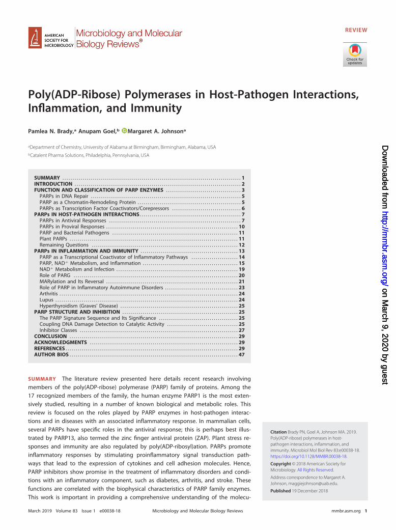
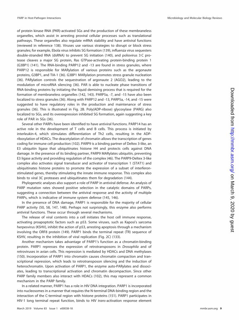
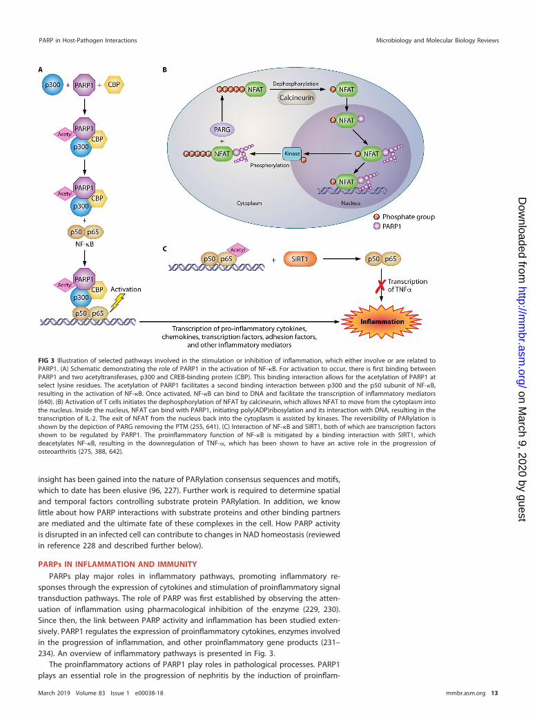
PARPs play major roles in inflammatory pathways, promoting inflammatory re-sponses through the expression of cytokines and stimulation of proinflammatory signaltransduction pathways. The role of PARP was first established by observing the atten-uation of inflammation using pharmacological inhibition of the enzyme (229, 230).Since then, the link between PARP activity and inflammation has been studied exten-sively. PARP1 regulates the expression of proinflammatory cytokines, enzymes involvedin the progression of inflammation, and other proinflammatory gene products (231–234). An overview of inflammatory pathways is presented in Fig. 3.
The proinflammatory actions of PARP1 play roles in pathological processes. PARP1plays an essential role in the progression of nephritis by the induction of proinflam-
FIG 3 Illustration of selected pathways involved in the stimulation or inhibition of inflammation, which either involve or are related toPARP1. (A) Schematic demonstrating the role of PARP1 in the activation of NF-�B. For activation to occur, there is first binding betweenPARP1 and two acetyltransferases, p300 and CREB-binding protein (CBP). This binding interaction allows for the acetylation of PARP1 atselect lysine residues. The acetylation of PARP1 facilitates a second binding interaction between p300 and the p50 subunit of NF-�B,resulting in the activation of NF-�B. Once activated, NF-�B can bind to DNA and facilitate the transcription of inflammatory mediators(640). (B) Activation of T cells initiates the dephosphorylation of NFAT by calcineurin, which allows NFAT to move from the cytoplasm intothe nucleus. Inside the nucleus, NFAT can bind with PARP1, initiating poly(ADP)ribosylation and its interaction with DNA, resulting in thetranscription of IL-2. The exit of NFAT from the nucleus back into the cytoplasm is assisted by kinases. The reversibility of PARylation isshown by the depiction of PARG removing the PTM (255, 641). (C) Interaction of NF-�B and SIRT1, both of which are transcription factorsshown to be regulated by PARP1. The proinflammatory function of NF-�B is mitigated by a binding interaction with SIRT1, whichdeacetylates NF-�B, resulting in the downregulation of TNF-�, which has been shown to have an active role in the progression ofosteoarthritis (275, 388, 642).
PARP in Host-Pathogen Interactions Microbiology and Molecular Biology Reviews
March 2019 Volume 83 Issue 1 e00038-18 mmbr.asm.org 13
on March 9, 2020 by guest
http://mm
br.asm.org/
Dow
nloaded from
matory cytokines (234) and can participate in the progression of atherogenesis byinfluencing plaque inflammation (232). Some cytokines, such as IL-6, in turn activatePARP. By stabilizing mRNA and by acting as a transcriptional activator, PARP1 has beenshown to play a role in the regulation of inflammatory gene expression (235). It wasshown that PARP1 knockout (PARP1�/�) mice were protected against several inflam-matory disorders (236, 237). Oliver and coworkers demonstrated that when the PARP1–NF-�B binding interaction was inhibited, mice that were PARP1 deficient were lesssusceptible to inflammation initiated by lipopolysaccharide (LPS) (238).
PARP overactivation leads to inflammation in several tissues; for example, PARP1activation by DNA damage caused by oxidative stress is believed to play a role indiabetic neuropathy (239, 240). The roles of PARP in inflammation have been studiedin several tissues, and its relationship to cancer and obesity has also been found.Induction of inflammation by infection with viruses, fungi, and parasites or by traumaticinjury and stroke has been observed (241, 242). PARP hyperactivation has beenidentified in several central nervous system disorders, including neuroinflammation,neurodegeneration, and ischemia. Extracellular PAR, such as could be released bynecrotic cells, was identified as a damage-associated molecular pattern that stimulatesinflammatory signaling (161).
PARP as a Transcriptional Coactivator of Inflammatory Pathways
As described above, a major role of PARP in the cell is to act as a transcriptionalcoactivator/corepressor (87–95). PARP1 regulates the function of transcription factorsinvolved in inflammatory processes, such as activating protein 2 (AP-2), yin yang 1(YY-1), transcription enhancer factor 1 (TEF-1), octamer-binding transcription factor 1(Oct-1), Myb-related protein B (B-MYB), specificity protein 1 (SP1), Ets-1, p53, and NFAT(87–95). PARP1 also acts as a coactivator of NF-�B, leading to the activation ofinflammatory signaling (243–245).
NF-�B can be activated by the action of PARP1 through various pathways. Underinflammatory conditions, the activation of NF-�B is initiated by the interaction of PARP1and two transcriptional coactivators that act as histone acetyltransferases (HATs),namely, CREB-binding protein (CBP) and p300. The formation of this complex results inthe acetylation of PARP1 at specific lysine residues. The acetylation of PARP1 is requiredfor the PARP1-CBP-p300 complex to interact with the p50 subunit of NF-�B. Thebinding between p300 and p50 activates NF-�B, initiating the transcription of proin-flammatory cytokines, chemokines, transcription factors, and other inflammatory me-diators (Fig. 3A) (110, 246, 247).
The interaction of PARP1 with SP1 appears to be proinflammatory, since thePARylation of SP1 resulted in its reduced binding to its consensus sequence. SP1 isnormally considered to promote the transcription of anti-inflammatory mediators.However, the interaction with AP-2 could inhibit or activate transcription depending onthe conditions (248). In contrast, the high-mobility-group box 1 protein (HMGB1) wasshown to be highly PARylated, and this modification enhanced its ability to preventefferocytosis, an important step in resolving inflammation (249). PARP1 was shown tolocalize to the promoters of genes responsive to NF-�B. Upon LPS stimulation, PARP1undergoes cleavage and release from the promoters, increasing the rate of transcrip-tion (250). In Drosophila, a specific protein, Charon, directs the RelB subunit of NF-�B topromoters through a specific interaction with PARP1 (251). This interaction is requiredfor the transcription of genes encoding antimicrobial defense peptides.
PARP1 was shown to favor the expression of inflammatory chemokines such as IL-1�
and TNF-� and enzymes involved in inflammation such as inducible nitric oxidesynthase (233, 234, 245, 252). For example, in a model of allergic airway inflammation,PARP was required for the production of Th2-type cytokines such as IL-5, IL-10, andgranulocyte-macrophage colony-stimulating factor (GM-CSF) (253). PARP1 plays animportant role in the inflammatory response in the brain by regulating the transcriptionof genes linked to inflammation (93). In addition, PARP induces the expression of celladhesion molecules such as selectins that favor the influx of white blood cells to tissues.
Brady et al. Microbiology and Molecular Biology Reviews
March 2019 Volume 83 Issue 1 e00038-18 mmbr.asm.org 14
on March 9, 2020 by guest
http://mm
br.asm.org/
Dow
nloaded from
Thus, PARP is required for the expression of adhesion molecules such as selectins andvascular cell adhesion molecule (VCAM) in atherogenesis (232). Also, PARP1-deficientmice undergo significant transcriptional reprogramming in the colon, with a protectiveeffect in a model of colitis. Analysis of the transcription response showed that the mostcommon gene ontology (GO) types regulated were proteolysis, protein transport, andlocalization, suggesting that PARP has other functions besides a direct effect ontranscription (254). These observations raise the question of tissue-specific PARP func-tions.
PARP1 also has a proinflammatory function in activated T cells. The interaction ofnuclear factor of activated T cells (NFAT) and PARP1 facilitates the transcription of IL-2(255). Several studies have shown that this process is dependent on the state ofphosphorylation of NFAT (255) (Fig. 3B). In inactive T cells, NFAT is heavily phosphor-ylated and resides in the cytosol. When T cells are activated, calcineurin facilitates thedephosphorylation of NFAT, allowing NFAT to enter the nucleus. In the nucleus, NFATcan bind consensus sequences, either alone or along with other transcription factors.This interaction occurs with the help of PARP1 and the PARylation of NFAT. As a result,PARP1 aids in the transcription of IL-2. Nuclear kinases facilitate the rephosphorylationof NFAT, allowing it to reenter the cytosol.
Other PARP enzymes also play roles in inflammatory processes, some of which maybe anti-inflammatory (Table 1). In addition to its antiviral functions in the regulation ofstress granule production and maintenance, PARP5b has a proinflammatory function inthe progression of cherubism (256). PARP16 participates in the unfolded-protein re-sponse (UPR) initiated by the endoplasmic reticulum. PARP16 ADP-ribosylates severalproteins and prevents the activation of activating transcription factor 6 (ATF6), therebylinking PARP16 to inflammation (257, 258).
In contrast, PARP10 is a repressor of NF-�B signaling, mediated through binding toK63-linked polyubiquitin and its substrate NEMO (259). PARP10 also acts on a numberof other cellular substrates, including glycogen synthase kinase 3B (GSK3B) and otherkinases (175).
PARP9, PARP14, and PARP15 form a family originally identified as proteins that wereoverexpressed in chemotherapy-resistant diffuse large B-cell lymphoma (260). Theseproteins have been termed the macro-PARPs or the B-aggressive lymphoma (BAL)proteins (260). PARP14, a mono(ADP-ribosyl)transferase, is a specific cofactor of STAT6that is important in B-cell and T-cell responses. PARP14 regulates the IL-4-mediatedproliferation and survival of B cells and is highly expressed in Th17 cells. Inhibition ofthis protein results in reduced Th17 cell differentiation in a model of allergic airwayinflammation (261–263). PARP14 influences the class distribution and affinity repertoireof antibodies in mice and is involved in helper T-cell development (261, 264).
Thus, at least six of the currently known 17 PARPs have demonstrated roles ininflammatory processes, through either participating in transcriptional regulation orother mechanisms.
PARP, NAD� Metabolism, and Inflammation
The PARylation and MARylation reaction consumes NAD� to produce nicotinamide(NAM) and a modified protein. Hence, the activation of PARP has a major effect onlevels of NAD� in the cell. Changes in NAD� levels can lead to changes in cellularmetabolism, homeostasis, and even cell death (reviewed in reference 265). Consump-tion of excess NAD� and ATP upon hyperactivation of PARP was originally thought tolead to cellular necrosis through a cell death process termed as parthanatos (266–268).In this mechanism, PAR acts as a signal to trigger cell death via the release ofapoptosis-inducing factor (AIF) from mitochondria (269–272). This is followed by themovement of AIF to the nucleus, which leads to the degradation of genomic DNA andcell death (269–272). NAD� levels decrease to as much as 20% to 30% of normal uponDNA damage (273, 274), and conversely, NAD� doubles in PARP1 knockout mousetissue (275). However, recent work has shown that PAR-dependent energy depletion infact occurs through inhibition of hexokinase, and therefore glycolysis, by PAR produced
PARP in Host-Pathogen Interactions Microbiology and Molecular Biology Reviews
March 2019 Volume 83 Issue 1 e00038-18 mmbr.asm.org 15
on March 9, 2020 by guest
http://mm
br.asm.org/
Dow
nloaded from
by activated PARP1 (276). This inhibition occurs through a specific PAR-binding motif(PBM) on hexokinase. Despite debate over the exact mechanism of parthanatos, it isclear that PARP activation, the activities of other enzymes in NAD� metabolism, andcellular NAD� levels must all be regulated to avoid deleterious effects.
The importance of NAD� homeostasis was originally observed in pellagra, a defi-ciency of NAD� precursors (277; reviewed in reference 228). NAD� serves as a coen-zyme for numerous enzymes in reduction-oxidation reactions and as an electron carrierin cellular intermediary metabolism. Hence, the loss of NAD� homeostasis can lead tounfavorable changes in the cell. The multifaceted roles played by NAD� in metabolismhave been established through the breadth of its function in various biological path-ways (reviewed in reference 278). Decreased cellular NAD� levels have been associatedwith aging (279–283), consumption of a high-fat diet (275, 284), diabetes (279, 285),and stress. These effects are physiologically deleterious, leading to the degeneration ofmuscle cells (286). The repletion of NAD� by providing precursors has been shown tobe beneficial in several pathogenic conditions. Nicotinic acid (NA) was shown to beeffective in treating dyslipidemia (287); however, side effects were noted, and newderivatives have been developed. Increased NAD� levels provided protection againstischemia (288–290) and against neurodegeneration (291). NAM treatment was shownto benefit in a rodent model of obesity and diabetes (285). NAM could also inhibitoxidative damage induced through �-amyloid peptide (292, 293). Repletion usingnicotinamide riboside (NR) correlated with improved muscle function, a reduction inprotein PARylation, and reductions in inflammation and fibrosis in mouse and Caeno-rhabditis elegans models of muscular dystrophy (294). NR treatment also improvedsymptoms of diabetes and diabetic neuropathy in a mouse model (295). NR is currentlyregarded as promising for therapeutics. It has been recently suggested that the key tothe selectivity of NAD� metabolism is the compartmentalization of intermediates intodifferent subcellular compartments for storage until needed for NAD�-dependentsignaling pathways (278).
Similar effects are observed upon increases in PARP activity given that theseenzymes are NAD consumers. Thus, PARP1 activity increases with age and with ahigh-calorie diet (275, 280, 296), and PARP1 knockout can protect against obesity inmice (275), correlating with the effects of NAD� levels. PARP7 activation reduced NAD�
levels and increased PARylation of proteins in liver tissue (297). Aberrant activation ofPARPs can play a role in the neurodegeneration observed in certain disorders involvingDNA damage.
In contrast, PARP inhibition increases NAD� levels (275, 298) and protects againstobesity (275, 298, 299). PARP5 knockout mice had reduced fat and body weight (300)or resistance to diabetes (301). PARP knockout in C. elegans led to an increase in lifespan (296). PARP inhibition also upregulates mitochondrial biogenesis and the un-folded protein response, which could contribute to the beneficial effects towardmetabolic disease (298).
Two other classes of enzymes consume NAD� and also strongly affect cellularmetabolism. The first is the sirtuins or SIR2 family, which are class III histone deacetylaseenzymes that function to remove acetyl modifications and other acyl groups fromproteins using NAD� as a cosubstrate (reviewed in reference 302). These enzymes acton both histone and nonhistone proteins. They function to stimulate oxidative meta-bolic pathways in mitochondria, improving resistance to oxidative stress (reviewed inreferences 265 and 303). Through this mechanism, they favor increased life span inmultiple organisms, such as yeast, C. elegans, Drosophila, and mammals (304–310). Theiraction benefits symptoms of inflammation and aging-related diseases such as cancerand cardiovascular disease. In mammals, they hold a broad spectrum of biological rolesin cellular processes and pathways that can be attributed to the cellular localization ofthe seven sirtuin family members. Through their influence on the cell cycle, apoptosis,and metabolism, they have emerged as important regulators of cancer cell proliferationand tumorigenesis (reviewed in references 311 and 312). Several sirtuin enzymes alsoexhibit ADP-ribosyltransferase activity, which is less efficient than their deacetylase
Brady et al. Microbiology and Molecular Biology Reviews
March 2019 Volume 83 Issue 1 e00038-18 mmbr.asm.org 16
on March 9, 2020 by guest
http://mm
br.asm.org/
Dow
nloaded from
activity. The physiological importance of this enzyme activity has been debated, but itmay influence DNA repair (313) and enzyme activities (314). Microbial sirtuins havebeen identified mainly in pathogenic bacteria and fungi and termed SirTMs or class Msirtuins (315). This class of enzymes exhibit more efficient ADP-ribosylation thanmammalian sirtuins and may take advantage of this activity as part of a regulatoryresponse to oxidative stress, such as during engulfment by host cells (315).
In mammalian cells, SIRT1 is found predominantly in the nucleus to remove acylgroups from histones and a number of cellular targets (316). The action of SIRT1 isprotective against osteoporosis, acting as a positive regulator of bone mass in mice(317, 318). This may be mediated by forkhead transcription factors of class O (FoxO)proteins (319) or under inflammatory conditions, through negative regulation of NF-�Bsignaling (320, 321). In addition, SIRT1 action protects against other aging-related andneurodegenerative diseases, including Alzheimer’s disease (322–326). Recently, protec-tion against neurodegenerative diseases and promotion of stem cell differentiationwere reported (327–329). SIRT1 deacetylation of p53 protected against neuronal cellapoptosis in a model of diabetic cognitive impairment (330). SIRT1 contributed tomemory enhancement in healthy animals and in Alzheimer’s disease models, throughthe regulation of neurotrophic factors, enhancement of tau degradation, and additionalmechanisms (331).
SIRT1 function has also been shown to be strongly associated with cancer andapoptosis (reviewed in reference 332). SIRT1 gene polymorphisms were shown to assistin compensating for oxidative stress during aging (333), to affect gene expression incardiovascular disease (334), and to affect obesity, fat and cholesterol metabolism, andhigh blood pressure (335–337).
SIRT2 can be found predominantly in the cytoplasm but is also present in thenucleus during certain points in the cell cycle, where it deacetylates histone substratesand cell cycle checkpoint kinases (338, 339). In mammals, this protein is highlyexpressed in the brain (340). Like SIRT1, SIRT2 is upregulated during calorie restrictionand downregulated under conditions of energy excess (341, 342). Recent applicationsto its function as a deacetylase of microtubules link it to aging brains and neuropro-tective effects (343). In addition, SIRT2 acts to suppress inflammatory signaling throughNF-�B deacetylation. SIRT2 regulates several proteins important in metabolic regulationand homeostasis, particularly lipid metabolism and regulation. SIRT2 suppresses adi-pogenesis, inhibits adipocyte differentiation by deacetylating FoxO1, and stimulatesthe degradation of ATP-citrate lyase, a key enzyme in hepatic lipogenesis (344–346).There are also roles in insulin signaling and metabolism (340).
SIRT3 to -5 are localized primarily to mitochondria and have been implicated inoxidative stress responses (347–350). SIRT3 acts as a tumor suppressor through theregulation of reactive oxygen species (ROS) (351). SIRT3 also activates FOXO3a, aregulator of ROS in the heart and negative regulator of cardiac hypertrophy (352). Onthe other hand, SIRT3 was also shown to have an oncogenic role under some circum-stances. SIRT3 is essential for maintaining mitochondrial function and is overexpressedin head-and-neck squamous cell carcinoma (HNSCC) (353), where it contributes to cellgrowth and survival (354, 355). SIRT3 may prevent cells from undergoing apoptosisunder stress conditions (356), antagonize p53 growth arrest (357), and induce resis-tance to anticancer agents (358). A SIRT3 inhibitor was able to enhance apoptosis ofHNSCC cells (359), likely by disrupting the ROS balance, supporting SIRT3 as a usefultherapeutic target for this type of cancer.
SIRT4 and SIRT5 have multiple enzymatic activities; for example, SIRT5 cleavessuccinyl, malonyl, and glutaryl groups in preference to acetyl groups (360). SIRT4 wasshown to have lipoamidase activity that negatively regulates the pyruvate dehydroge-nase complex (361, 362). Like SIRT3, SIRT4 may contribute to tumor suppression ortumorigenesis depending on conditions. SIRT4 also participates in DNA damage re-sponse pathways and contributes to genome stability, thus contributing to tumorsuppression (363, 364). However, SIRT4 was also shown to be involved in cellular stressresponses, is induced by stress, and contributes to cell survival and growth under stress
PARP in Host-Pathogen Interactions Microbiology and Molecular Biology Reviews
March 2019 Volume 83 Issue 1 e00038-18 mmbr.asm.org 17
on March 9, 2020 by guest
http://mm
br.asm.org/
Dow
nloaded from
conditions (365). In this manner, SIRT4 has been implicated in oncogenic transforma-tion and may contribute to survival and drug resistance of cancer cells (365).
SIRT5 is specific for negatively charged acyl groups, especially succinyl, malonyl, andglutaryl groups (360, 366). This activity regulates the activity of mitochondrial enzymessuch as the pyruvate dehydrogenase complex, succinate dehydrogenase, and carbam-oyl phosphate synthetase 1 (367–369). This impacts diverse metabolic pathways suchas glycolysis and the urea cycle.
SIRT6 is localized in the nucleus, where it helps to maintain genome stability andtelomere function by histone deacetylation (370). Recent findings support its ADP-ribosylation of PARP1 under oxidative stress to assist in the repair of DNA (313,371–374). SIRT6 acts as a tumor suppressor and participates in genome maintenance(370). SIRT6 is also able to cleave long-chain fatty acyl groups such as myristoyl andpalmitoyl, allowing it to regulate the secretion of tumor necrosis factor alpha (375).SIRT7 resides in the nucleus and participates in transcription (376). This protein hasbeen identified as an oncogene through its activity in transcriptional repression (377),association with the transcription factor ELK4 (371), and selective H3K18 deacetylationactivity, which has been linked to oncogenic transformation (371, 378–381) and ag-gressive cancers (378, 379). SIRT7 participates in DNA double-strand break repair and isinvolved in genome integrity maintenance and nonhomologous end-joining repair(382). However, much of its catalytic activity may still be unknown. The breadth offunctions of this family of enzymes has made them promising drug targets.
PARPs and sirtuins regulate each other in a complex manner. PARP2 acts as atranscriptional repressor of SIRT1 expression (275) and negatively regulates the SIRT1promoter. By efficiently consuming NAD, PARP1 limits SIRT1 activity (275, 293, 383,384). In contrast, SIRT1 inhibits PARP1 through its deacetylation activity and throughtranscriptional regulation. Increased PARP activity was observed in SIRT1 knockout cells(384, 385). PARP1 knockdown (pme-2 knockdown) in worms led to increased Sir2activity as well as longer life span and increased NAD� concentration (386). WhilePARP1 is a transcriptional coactivator of NF-�B and favors inflammatory processes,SIRT1 inhibits NF-�B activity by deacetylating the protein (387). SIRT1 interacts withNF-�B, resulting in the deacetylation of the latter and in the inhibition of TNF-� (Fig. 3C)(275, 388). SIRT1 and PARP1 are connected through their use of the substrate NAD�,and both are able to modify histones. The two enzymes participate in antagonistic crosstalk due to their competition for the NAD� substrate (383). Many cellular functions arelikely to be regulated by SIRT1/PARP1 reciprocal regulation.
The other important consumer of NAD� that strongly affects overall cellular NAD�
levels is the CD38/CD157 protein, an enzyme that produces the second messengersADPR and cyclic ADP-ribose (cADPR) from NAD� (389). In contrast to sirtuins, whoseactivity is affected by low levels of NAD�, CD38 plays an active role in depleting NAD�
(390). For example, cells overexpressing CD38 showed decreased [NAD�] together withdecreased expression of antioxidant proteins, likely due to reduced sirtuin activity (391).As an effect of this metabolism, CD38 knockout mice showed improved resistance toglucose intolerance and diet-induced obesity (392, 393). This correlated with increased[NAD�] in multiple tissues and increased sirtuin activity (392, 394). The roles of ADPRand cADPR as second messengers have been reviewed (395–397). CD38 is a multifunc-tional transmembrane glycoprotein and, in addition to its role in producing cADPR, alsoregulates Ca2� flux in the cytoplasm (398, 399). CD38 participates in other signaltransduction pathways such as insulin secretion (400, 401). CD38 assists in B-celldifferentiation and leukocyte adhesion and proliferation (402, 403) and associates withlipid rafts (404, 405).
CD38 expression has also been shown to correlate with aging. Overexpression ofCD38 in cancer cells leads to the development of senescence (406). CD38 inhibits thefunction of SIRT3 in the mitochondria by affecting levels of NAD� (390). CD38 expres-sion is induced by inflammatory cytokines and interferon (407, 408), which also increaseduring aging (409–413), suggesting a possible reason for CD38 increase during theaging process (414).
Brady et al. Microbiology and Molecular Biology Reviews
March 2019 Volume 83 Issue 1 e00038-18 mmbr.asm.org 18
on March 9, 2020 by guest
http://mm
br.asm.org/
Dow
nloaded from
As a result, CD38-inhibitory therapy is an area of interest for metabolic dysfunctionrelated to age (415). Specific CD38 inhibitors have been developed (416–420). Severalinhibitors were shown to have the ability to raise NAD levels and improve glucosehomeostasis and fatty acid metabolism in vivo (417, 420). CD38 was shown to play arole in certain diseases related to aging, including Alzheimer’s disease (421).
CD38 is overexpressed on cells of some hematologic malignancies (422–426) andhas been proposed as a therapeutic target for myeloma and for T-cell acute lympho-blastic leukemia treatment (405, 427). Recently, it was suggested that CD38 may be abiomarker of resistance to certain cancer therapies, and it was effectively used as asecondary target in combinational therapies for lung cancer (428). CD38-expressingcells promoted tumor growth in a murine model of esophageal cancer (429). Its effecton mitochondrial function led to an enhancement of proliferation and inhibition ofapoptosis in cervical cancer cells (430). Conversely, CD38 knockout led to suppressionof tumorigenesis in a lung cancer model (431), and CD38 overexpression was identifiedin patient samples. This suppression occurred both in wild-type mice and in anARH1-deficient background; ARH1-deficient mice are already prone to tumor develop-ment (described above).
A monoclonal antibody directed against CD38, termed daratumumab, has beenapproved for the treatment of multiple myeloma (432). This antibody was originallydeveloped using a panel of CD38-specific monoclonal antibodies generated fromhuman antibody transgenic mice (433). The antibody exhibited high efficacy in ananimal model of multiple myeloma (434). CD38 has also been developed as a target forpositron emission tomography (PET) imaging using daratumumab antibody (435, 436).Other antibodies, such as MOR202, a human antibody, and isatuximab, a humanizedantibody, are in various stages of clinical trials (437, 438). CD38 expression has alsobeen associated with HIV infection, where CD38 expression was noted on HIV-specificCD8� cytotoxic T cells (439, 440), and with autoimmune disease (441), where clinicalapplications are also possible.
NAD� Metabolism and Infection
NAD� is an essential cofactor and substrate for bacterial cellular metabolism, andpathogenic bacteria have evolved to exploit NAD� and PARP pathways. Group AStreptococcus (GAS) possesses a NAD� glycohydrolase that cleaves NAD�, yielding NA,ADPR, and cADPR, which drastically decreases NAD� and ATP concentrations and leadsto cell death. This leads to a significant reduction in innate immune system responses,and protects GAS from autophagy (442) and lysosome acidification (443), and leads toa reduction in PARP activity. The reduction in PARP activity is believed to stimulate therelease of high-mobility-group box protein 1 (HMGB1) from the nucleus to the cytosol,where it acts as an inflammatory mediator (444), is deposited in damaged tissues (445),and contributes to necrosis. Conversely, NADase-negative GAS is also able to activatePARP, by an unknown mechanism.
A few other bacterial pathogens also activate PARP upon infection. Thus, H. pyloriinfection activates PARP by PARylation (183). H. pylori infection triggers apoptosis viathe caspase-independent AIF pathway (446) and programmed necrosis (447). Bacterialinfection was shown to promote PARP activation, through pathways related to inflam-mation (183, 184).
Various pathogens exploit PARP activity to promote conditions favorable to growthand virulence. In contrast, Chlamydia trachomatis infection led to the degradation ofPARP1 through the action of a specific bacterial enzyme, chlamydial protease-likeactivity factor (CPAF) (448), and this was associated with the inhibition of HMGB1release from the nucleus along with degradation of HMGB1 (449). This would lead toa reduced inflammatory response. CPAF is downregulated during persistent infection,allowing PARP1 activation and HMGB1 release (450). Chronic inflammation thus may beobserved during persistent disease.
NAD� production through the activity of the indoleamine 2,3-dioxygenase (IDO)enzyme has been shown to assist the clearing of pathogenic infections. For example,
PARP in Host-Pathogen Interactions Microbiology and Molecular Biology Reviews
March 2019 Volume 83 Issue 1 e00038-18 mmbr.asm.org 19
on March 9, 2020 by guest
http://mm
br.asm.org/
Dow
nloaded from
IDO induced by IFN-� inhibited the replication of Staphylococcus aureus (451), andCD4� T cells inhibited the growth of Mycobacterium tuberculosis by sequesteringtryptophan, the precursor to NAD� (452). It should be noted, however, that somepathogens are NAD� auxotrophs and require NAD� obtained from the host cell toreplicate. For example, Borrelia burgdorferi and Brucella abortus, both intracellularpathogens, produce nicotinamidases that convert NAM to nicotinic acid (NA), a pre-cursor to NAD� in the Preiss–Handler pathway (453, 454). Haemophilus influenzae lacksthe Preiss-Handler pathway for NAD biosynthesis and requires NMN and NR as precur-sors (455–457). Structural data for H. influenzae NAD nucleotidase (NadN), a periplasmicenzyme, have suggested a novel mechanism for the synthesis of NAD� that involves itshydrolysis to NR, adenosine, and phosphate, followed by uptake of NR across the innermembrane and resynthesis via the NadR enzyme. A significant conformational changeis involved during catalysis, which suggests likely avenues for inhibitor development(457). These enzymes are also found in other bacteria and in parasites such asLeishmania. In Leishmania, knockout of the enzyme led to a significant decrease inNAD� content and a reduction in virulence (458). The Leishmania parasite was shownto increase intracellular NAD levels once present in the cell (459).
Role of PARG
Damage to strands of DNA activates repair pathways, which involve PARPs, to thesite of repair, where the synthesis of PAR polymers is often prompted. This biochemicalreaction is regulated by an enzyme known as poly(ADP-ribose) glycohydrolase (PARG).The reversibility of the ADP-ribosylation reaction is shown through the action of PARG,which hydrolyzes the PAR polymer (460). PARG activity is primarily responsible for thedegradation of PAR polymers whose mass accumulation is metabolically harmful to thecell, as described above (461). In addition to the disruption of NAD� metabolism,excessive PARP activity and PAR production could have unpredictable effects onRNA-binding and DNA-binding proteins through competition for binding (462) andcould cause the dysregulation of cellular organelle assembly and stability (463).
PAR polymers produced upon DNA damage are short-lived, with a half-life of lessthan 1 min. This is in contrast to constitutively produced polymers, which are longer-lived, with half-lives of �8 h (464–468). The degradation of PAR polymers occursspecifically at glycosidic bonds by hydrolysis and yields free ADP-ribose (469). PARGproduction is regulated by a single gene, producing a protein which is approximately110 kDa with a C-terminal catalytic domain (470–472). While full-length PARG remainsin the nucleus, shorter isoforms are also present and are distributed predominantly inthe cytoplasm (473–475). These isoforms vary not only in size but also in the way inwhich they are activated by biological events such as apoptosis or proteolysis of theirposttranslational modifications (PTMs). PARG nuclear localization increases upon DNAdamage, presumably in response to the high level of PARylation produced by PARPs(476, 477).
Though PAR is integral to the regulation of cellular death pathways, the role of PARGwas not well understood. Recently it has been demonstrated that following a lethaldosage of the alkylating agent N-methyl-N=-nitro-N-nitrosoguanidine (MNNG) to HeLacells, PARP1 directly interacts with PARG, even in the absence of NAD� or PAR,facilitating nonapoptotic cellular death pathways (478). A role of PARG in apoptotic celldeath has also been suggested. It is thought that the preferential binding of DNA repairfactor XRCC1 to PARylated PARP1 to help facilitate repair displaces PARG from PARP1,which triggers a cascade of apoptosis. (272, 478).
PARG has further been demonstrated to be necessary for cell survival in murinemodels, where the absence of PARG in embryonic cells proved to be lethal (461).However, in other studies, the absence of PARG was not shown to be essential forsurvival in the absence of genotoxic stress, which suggests that stress plays a pivotalrole in PARG activity (479–481). This has been supported by studies showing that in thepresence of genotoxic stress, cells lacking PARG exhibited increased cell death inaddition to the nonrepair of single- and double-strand DNA breaks (482–484). In
Brady et al. Microbiology and Molecular Biology Reviews
March 2019 Volume 83 Issue 1 e00038-18 mmbr.asm.org 20
on March 9, 2020 by guest
http://mm
br.asm.org/
Dow
nloaded from
response to replicative stress conditions, PARG is pivotal in regulating the massaccumulation of PAR, arresting the collapse of replication forks and the development ofadditional DSBs (480).
PARG was shown to be integral to the growth of Trypanosoma cruzi epimastigotes,and PARG inhibition caused changes to the cell cycle in the parasite. Knockdown ofhost cell PARG also abrogated T. cruzi infection (485). In Trypanosoma brucei, PARGknockdown led to increased sensitivity to oxidative stress (486). Hence, PARG function-ality is also essential for establishing trypanosomal infection.
A strong case can be made for the importance of the biomedical implications of theinhibition of PARG in the field of cancer therapy. For example, the absence or inhibitionof PARG has been shown to arrest the metastasis of cancer cells in the human colon andin murine models, the death of BRCA-2 tumor cells (487, 488), and cell death ofhomologous repair-deficient tumor cells (489). PARG silencing led to a reduction inPARP expression and a decrease in signaling through the phosphatidylinositol (PI)3-kinase/Akt pathway, mediated through a reduction in NF-�B (487).
The inhibition of binding between PARG and the human antigen R (HuR) RNA-binding protein helps to improve PARP inhibition therapy in pancreatic cancer cells(490). Another study presented work demonstrating that the inhibition of PARG helpedto reduce the negative effects of cellular exposure to benzo[a]pyrene, a polycyclicaromatic hydrocarbon known to induce carcinogenesis (491, 492).
The focus of PARG inhibitor therapy for cancer can be attributed to the role it hasin DNA repair, like PARP1, -2, and -3. Importantly, PARG is encoded by a single gene, incomparison to the 17 human PARP genes. As a result, all isoforms of PARG have aconserved catalytic domain, which offers a more direct target for inhibitor development(493). It has been demonstrated that PARG inhibitors offer unique pharmacologicaleffects in comparison to olaparib, the first PARP inhibitor to be accepted by the FDA(494–496). Several PARG inhibitors, both synthetic and natural, have been identified;however, their wide use is limited by problems with toxicity and cell permeability(497–499). Four specific inhibitors of PARG, ADP (hydroxymethyl)pyrrolidinediol (ADP-HPD), mono-galloyl glucose, rhodanine-based PARG inhibitor 2 (RBPI-2), and salicylan-ilide, have increased specificity to PARG and have been used extensively to helpuncover the breadth of the biological role of PARG inhibitors (488, 500–503). Recently,new smaller inhibitors, known as phenolic hydrazide hydrazones, have been synthe-sized and shown to be effective inhibitors of PARG in vitro, with 50% inhibitoryconcentration (IC50) values in the micromolar range (504).
The role of PARG in DNA repair has helped to highlight its potential as a target forinhibitor development. To date the primary focus of PARG inhibitor research has beenin cancer therapy; thus, more research investigating the additional applications of PARGinhibitor therapy is needed. Work on PARG inhibition against infection is in thepreliminary stages but is also promising.
MARylation and Its Reversal
MARylation is a common mechanism used by bacterial toxins to interfere with hostcell metabolic and regulatory pathways. ADP-ribosylating toxins transfer an ADP-ribosemoiety to amino acids such as arginine, cysteine, and asparagine to acceptor proteinsin the host cell, destroying enzyme activity. This activity is shared by a family of proteinsincluding cholera toxin, diphtheria toxin, pertussis toxin, and others (10). These toxinsare structurally and functionally related to the PARP enzymes (505). In addition to themodifications carried out by pathogens, these PTMs are also exerted by host cell PARPsfor numerous regulatory functions, as has been described above.
MARylation cannot be removed by PARG, and its reversal requires other enzymes(186). Instead, mono(ADP-ribosyl) units are acted on by two other classes of enzymes,namely the ADP-ribosylhydrolases (ARHs) and the macrodomains. The former arehomologues of the dinitrogenase reductase-activating glycohydrolase (DraG). In hu-mans, three ARHs are expressed and have functions that are yet to be fully understood.
PARP in Host-Pathogen Interactions Microbiology and Molecular Biology Reviews
March 2019 Volume 83 Issue 1 e00038-18 mmbr.asm.org 21
on March 9, 2020 by guest
http://mm
br.asm.org/
Dow
nloaded from

Though ARH2 is thought to be catalytically inactive (506–508), the other two enzymesare active in the hydrolysis of the ADP-ribosylation modification.
ARH1 is a site-specific hydrolase of MARylated proteins at arginine residues (509).This enzyme plays a role in the suppression of tumorigenesis through regulating cellproliferation and the cell cycle (510). ARH1 mutants or knockout cells with low enzymeactivity were tumorigenic, and ARH1 mutants identified in the human cancer databaselocalized to the enzyme catalytic site (511). Importantly, ARH1 knockout mouse cellscould not hydrolyze MARylated arginine, showing that ARH1 is the only cytoplasmicenzyme able to degrade this PTM (512). The enzyme also plays a role in the responseto cholera toxin. Cholera toxin is an ADP-ribosylating toxin that acts on the � subunitof the Gs protein (Gs�, a binding partner of adenylate cyclase). Activation of adenylatecyclase leads to increased cellular cyclic AMP, ion efflux from the cell via the cysticfibrosis transmembrane regulator (CFTR), and fluid loss from the intestine. In ARH1knockout mice and fibroblasts, MARylation of Gs� was increased and was longerlasting, together with increased fluid accumulation in the intestine (512). These obser-vations suggest that ARH1 is important in the host cell response to cholera toxin.
ARH3 catalyzes primarily exolytic activity, acting mainly at terminal sites of PARchains and on MARylated serine residues. Its activity varies from that of PARG, in thatit site specifically hydrolyzes PAR at chain termini, by a Mg2�-dependent mechanism,to yield ADP-ribose (513). Two amino acids, D77 and D78, are critical for hydrolaseactivity (506–508). An important physiological role of ARH3 is to protect cells fromparthanatos and to regulate PAR degradation in both the nuclei and cytoplasm (499).Thus, oxidative stress stimulates increased PAR synthesis by PARP1 and PAR cleavageby PARG in the nucleus. PAR is translocated to the cytoplasm through nuclear poresand releases mitochondrial membrane-associated AIF, which initiates parthanatos(269–272, 514). ARH3 then acts to modulate the level of PAR by degrading these freechains, starting at chain termini, after they have been translocated. Thus, ARH3 isimportant in modulating and protecting cells from parthanatos (499).
ARH3 is also able to cleave proteins that are MARylated on serine residues. Thisactivity has been observed in human cancer cells (515, 516). Serine ADP-ribosylation isa widespread and important PTM, appearing after DNA damage on histones (225, 517,518) and high-mobility group proteins, DNA repair factors, and other proteins (519,520). The sequence motifs KS and RS were shown to identify substrate proteins (516).The activity of ARH3 is interesting in that it has no activity in hydrolyzing ADP-ribosefrom arginine, cysteine, diphthamide, or asparagine bonds (506, 508, 521). ARH3, ratherthan PARG, is responsible for mitochondrial PAR degradation, suggesting a functionrelated to PAR or O-acetyl-ADP-ribose (OAADPR) metabolism in the mitochondria (522).
Both ARH1 and ARH3 are able to degrade OAADPR produced by sirtuins (507).OAADPR participates in ion channel gating (523), redox metabolism (524), chromatinregulation (525, 526), and gene silencing (525). Therefore, ARH3 has a role in signaltransduction involving both PAR and OAADPR (reviewed in reference 527).
It is also known that some macrodomain proteins have the ability to degrade theADP-ribosyl modification. The macrodomain proteins terminal ADP-ribose protein gly-cohydrolase (TARG1), MacroD1, and MacroD2 degrade mono(ADP-ribose) at glutamateand aspartate residues. TARG1 degrades MAR by a different mechanism than PARG, bybreaking the ester linkage between PAR and glutamate, an action that cannot beaccomplished by PARG (186). MacroD1 exists primarily in the mitochondria and has lowspecificity in its ADPR hydrolase activity, removing a broad spectrum of ADP-ribosylmoieties with ester linkages from proteins, DNA, and small molecules (528). MacroD2,in addition to its ADP-ribosylhydrolase function, acts mainly in the cytoplasm, where itselectively deacetylates O-acetyl-ADP-ribose (529). Several macrodomain proteins havealso been identified as degraders of PAR.
It has been shown biochemically, structurally, and phylogenetically that viral andcellular macrodomains are closely related (530). Therefore, it is not surprising that viralmacrodomains also have the ability to degrade ADP-ribosylation. Most macrodomainsfrom viruses belong to the MacroD class and are characterized by their ability to
Brady et al. Microbiology and Molecular Biology Reviews
March 2019 Volume 83 Issue 1 e00038-18 mmbr.asm.org 22
on March 9, 2020 by guest
http://mm
br.asm.org/
Dow
nloaded from
remove the entire PAR modification, similarly to TARG1 (167). The nsP3 macrodomainfrom chikungunya virus can hydrolyze the MAR modification from aspartate andglutamate, but not lysine, residues (171). Mutant viruses with reduced hydrolase activityshowed lower virulence in mice and slower replication in mammalian cells; mutantswith no activity were unable to replicate at all in mammalian or mosquito cells.Therefore, this macrodomain is essential for viral replication, in both the host and theinsect vector, and for virulence (171).
It is believed that viral macrodomains can remove the MARylation and PARylationmodification (167). The Hepeviridae, Togaviridae, and Coronaviridae families of virusesexpress conserved macrodomain proteins (531–539). Many of these macrodomainsexhibit ADP-ribosylhydrolase activity (171, 538, 540, 541). Macrodomain mutations thatreduced nsp3 ADP-ribosylhydrolase activity in vitro also led to a reduction of viral loadin infected mice and protected mice from an otherwise lethal severe acute respiratorysyndrome coronavirus (SARS-CoV) infection (173). This was attributed to changes in theinnate immune response, including an enhanced interferon and proinflammatorycytokine response. These observations show that the SARS-CoV macrodomain is in-volved in suppression of the innate immune response and cytokine repression and isimportant for virulence (173). Its exact molecular target(s) is currently unknown. Inmouse hepatitis virus (MHV), the conserved macrodomain is required for induction ofliver disease and production of the inflammatory cytokines TNF-� and IL-6 (542). Asubsequent MHV study using a macrodomain point mutant showed a drop in viral titer,cytokine and chemokine expression, and virulence with respect to those with the wildtype, supporting the role of the macrodomain in virulence and interaction with theinnate immune system (172). Other work demonstrated that mutant viruses of SARS-CoV and human coronavirus 229E (HCoV-229E) lacking macrodomain activity werehighly sensitive to interferon and showed an unusual cytokine induction pattern andslower growth, also suggesting participation of the macrodomain in resistance to theinnate immune system (170).
It has been suggested that macrodomain activity may antagonize the antiviraleffects of ADP-ribosylation (157). Most macrodomains possess conserved primarysequences, but some subclasses lack sequence conservation, resulting in novel func-tions. For example, Mycobacterium tuberculosis expresses a non-macroD-type macrodo-main which has the ability to remove ADP-ribosylation from DNA rather than proteins.In addition, it acts as an antitoxin to a mycobacterial toxin that ADP-ribosylates DNA atspecific thymidine residues (543). Coronaviruses encode additional macrodomains thatinteract with guanine quadruplexes (168, 544) and target host cell p53 for ubiquityla-tion and degradation by stabilizing the accumulation of the E3 ubiquitin ligase RCHY1(178, 180). This domain is important in the replication of the viral RNA genome (178).It is presently unclear whether the ADP-ribosylhydrolase function is shared by thesedivergent domains.
Role of PARP in Inflammatory Autoimmune Disorders
PARP inhibitors are presently being explored as therapeutics to prevent cell death,tissue damage, and dysfunction associated with aging or oxidation damage-relatedpathologies, such as cardiovascular disease, stroke, autoimmune and inflammatorydisease, and diabetes. Reactive oxygen and nitrogen species are generated in cardio-myocytes and endothelial cells during ischemia/reperfusion injury, cardiovascular ag-ing, and diabetic complications. These reactive species induce oxidative DNA damageand consequently activate PARPs (545). The effects of PARP inhibitors on inflammatoryresponses may contribute to their therapeutic effects in cancers (546). Many cancercells display dysfunctional DNA repair, and PARP inhibition can assist in preventingcellular replication (547). PARP inhibitors can play a crucial role in the treatment of bothBRCA-negative and -positive cancers and can be effectively used as therapeutics forprostate, ovarian, and breast cancer. These applications have been reviewed elsewhere(548).
PARP in Host-Pathogen Interactions Microbiology and Molecular Biology Reviews
March 2019 Volume 83 Issue 1 e00038-18 mmbr.asm.org 23
on March 9, 2020 by guest
http://mm
br.asm.org/
Dow
nloaded from
PARP inhibition has positive effects on inflammatory responses. Results published byGarcia and coworkers in 2008 demonstrated that the inhibition of PARP1 reduced theexpression of proinflammatory cytokines (549). Inhibition of PARP1 repressed theproduction of proinflammatory cytotoxic cytokines and enhanced the production ofanti-inflammatory cytokines (550–553).
PARP inhibitors were tissue-protective in animal models of multiple sclerosis, men-ingitis, arthritis, stroke, and traumatic brain injury (241, 554–560). PARP inhibitorsreduced neuroinflammation, edema, leukocyte infiltration, and the expression of ad-hesion proteins (242). PARP inhibition in leukocytes reduced inflammation via effectson the cytoskeleton and reduced leukocyte adhesion and migration across the blood-brain barrier (BBB) (561). PARP inhibitors protected the BBB and reduced its permea-bility in in vitro and in vivo models of inflammation (557). PARP1 inhibition was alsoprotective in a model of pancreatitis and reversed chronic liver injury and fibrosis (562).Studies conducted on PARP1 knockout mice further suggest that inhibitors of PARP area preventative therapy against inflammation, since the activation of PARP mediated theprogression of symptoms associated with hemorrhagic shock, such as lung inflamma-tion and heart failure (563).
PARP inhibitors are in development for treatment of other inflammatory conditions.Specific roles are seen in rheumatoid arthritis (RA), Graves’ disease, and Huntington’sdisease. Two PARP polymorphisms were identified as effective in the treatment ofGraves’ disease and Graves ophthalmopathy (564). PARP1 inhibition was also protectivein animal models of diabetic retinopathy and of Huntington’s disease (239, 565, 566).
Arthritis
Considered an autoimmune disease, rheumatoid arthritis (RA) is characterized byinflammation occurring in the joints and surrounding tissue. This results in the deteri-oration of the joint cartilage as well as the erosion of bone. This disease is observed inall populations worldwide; however, there is no clear pathogenic pathway which hasbeen identified (567). In a collagen-induced arthritis (CIA) model, PARP1�/� miceshowed lower levels of proinflammatory cytokine expression (568). This was corrobo-rated by studies in patient-derived cells (549). In mouse models, PARP inhibitionresulted in a reduction in the onset of the disease (569–571). Thus, Kröger andcoworkers demonstrated that the combination usage of a PARP inhibitor and thalido-mide, an inhibitor of tumor necrosis factor, had synergistic actions, resulting in thesuccessful inhibition of PARP (571). Inhibition of PARP arrested the progression ofpreviously established cases (571). 5-Iodo-6-amino-1,2-benzopyrone (INH2BP), a novelPARP inhibitor, showed positive dual effects on both the onset and the severity of thedisease (572). In addition, it is known that there is a positive correlation between thephorbol ester-activated burst of white blood cells and the onset and progression ofarthritis (567, 570). When PARP activity was inhibited, the development of arthritis wasprevented.
PARP inhibitors, in conjunction with small interfering RNA (siRNA) technology, haveprovided strong positive results in the treatment of RA (573). One factor that createsconcern is the possibility of DNA mutation acceleration due to long-term PARP inhibi-tion. However, PARP inhibitors are well tolerated by patients, and it is possible thatwithout aggressive treatment the inflammation in the joints can be treated successfullywith the application of dosages within human tolerance.
Lupus
Systemic lupus erythematosus is an autoimmune disease that is characterized bywidespread inflammation. Both heredity and environmental factors have been identi-fied as possible contributors (566). Abnormal metabolism of PARP has a role in thedevelopment of lupus (574). There is a decrease in the synthesis of PARP in the leukocytesof patients diagnosed with lupus, which may be a result of a defect in the transcription ofthe gene (574). In addition, there is an elevation in the levels of anti-PARP and anti-PARautoantibodies found in patients (575, 576).
Brady et al. Microbiology and Molecular Biology Reviews
March 2019 Volume 83 Issue 1 e00038-18 mmbr.asm.org 24
on March 9, 2020 by guest
http://mm
br.asm.org/
Dow
nloaded from
Hyperthyroidism (Graves’ Disease)
Graves’ disease is an autoimmune disease which is associated with hyperthyroidism.The circulation of antibodies which mimic the thyroid-stimulating hormone (TSH), bybinding to and activating the substrate, contribute to pathogenesis. Several genes andenvironmental factors have been identified (577). Polymorphism of PARP1 may be acontributor to the development of Graves’ disease (564, 578, 579).
PARP STRUCTURE AND INHIBITION
PARPs are characterized by the presence of the PARP signature sequence and a�-�-loop-�-� NAD�-binding fold. This sequence is not conserved equally among themembers of the PARP family, accounting for variations in catalytic function. There areseveral other structural variations among the members of the PARP family. Thisvariation accounts for similarity in the catalytic activity along with the versatility of theirbiological roles, cellular locales, binding partners, and substrate proteins. The mostcurrent understanding of the domain structure of the 17 human PARP proteins isillustrated in Fig. 1.
The PARP Signature Sequence and Its Significance
The PARP signature sequence is responsible for ADP-ribosyltransferase activity. It iscomposed of 70 to 100 amino acids and includes the NAD�-binding site. The catalyticactivity of PARPs (resulting in PAR formation) occurs when the ADP-ribose moiety ofNAD� is transferred to the target protein, with the release of nicotinamide. Catalysiscan be attributed to the NAD�-binding pocket, formed by � sheets and � helices,between a conserved Glu residue on one side and His on the other. NAD� binding isstabilized by a hydrogen bond between His and the O2= hydroxyl of the adenosineribose (580). Catalysis is assisted by the HYE triad, which consists of H862, Y896, andE988 (PARP1 numbering). In the triad, the glutamate residue acts as a general base,while the histidine assists in orienting NAD� for nucleophilic attack and stabilizing thetransition state and the tyrosine assists in NAD� binding (580–582). The catalytic site ofthe PARP1 ART domain in the presence of an effective inhibitor, olaparib, is shown inFig. 4A. Olaparib interacts with two residues of the catalytic triad, H862 and Y896, andforms hydrophobic interactions and hydrogen bonds with other residues in the bindingpocket (583, 584). Inhibitor classes are discussed further below.
The DNA-binding domain of PARP1 consists of tandem repeat zinc finger domainswhich bind to DNA and activate the catalytic domain (585). A study by Ali et al. revealedthe crystal structure of the human PARP1-DBD bound to a DNA break. ZnF1 and ZnF2domains of multiple PARP1 molecules form a DNA break-sensing module (586). Analternate mechanism suggests that DNA damage can also be detected by monomericPARP1, through cooperative action of flexibly linked ZnF1 and ZnF2 domains. Thismechanism proposes that the binding of ZnF2 positions ZnF1 via a fly-casting process(587, 588). PARP1 “reels in” protein domains in the same molecule by binding to DNA,triggering PARylation by unfolding an inhibitory HD subdomain (584, 587). Activationby DNA binding triggers the automodification of PARP1.
The central region of the enzyme contains a BRCT (breast cancer type 1 suscepti-bility protein [BRCA1] C-terminal) motif, which is involved in protein-protein interac-tions (589). It is suggested that automodification in this domain and nearby causes therelease of DNA repair proteins required for the processing and repair of nicks. Auto-modification reduces affinity for intact chromatin but not for nucleosomes with ex-posed DNA ends and may cause PARP1’s release of DNA and convert PARP1 to ahistone-binding protein (86).
Coupling DNA Damage Detection to Catalytic Activity
Langelier et al. reported that upon binding of PARP1 to a DNA break, the domainscollapse together, establishing interdomain contacts that distort the structure of thehelical domain (HD) (590). When in complex with DNA, partial unfolding and destabi-lization of the HD occur, and this underlies PARP1 activation (590) (Fig. 4B). In contrast
PARP in Host-Pathogen Interactions Microbiology and Molecular Biology Reviews
March 2019 Volume 83 Issue 1 e00038-18 mmbr.asm.org 25
on March 9, 2020 by guest
http://mm
br.asm.org/
Dow
nloaded from
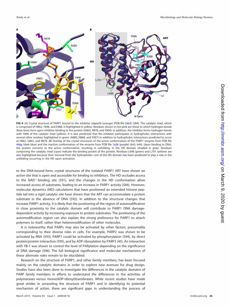
to the DNA-bound form, crystal structures of the isolated PARP1 ART have shown anactive site that is open and accessible for binding to inhibitors. The HD occludes accessto the NAD�-binding site (591), and the changes in the HD conformation allowincreased access of substrates, leading to an increase in PARP1 activity (584). However,molecular dynamics (MD) calculations that have positioned an extended histone pep-tide tail into a rigid catalytic site have shown that the ART can accommodate a proteinsubstrate in the absence of DNA (592). In addition to the structural changes thatincrease PARP1 activity, it is likely that the positioning of the region of automodificationin close proximity to the catalytic domain will contribute to PARP1 DNA damage-dependent activity by increasing exposure to protein substrates. The positioning of theautomodification region can also explain the strong preference for PARP1 to attachpolymers to itself, rather than heteromodification of other molecules.
It is noteworthy that PARPs may also be activated by other factors, presumablycorresponding to their diverse roles in cells. For example, PARP2 was shown to beactivated by RNA (593). PARP1 could be activated by phosphorylation (594), by directprotein/protein interaction (595), and by ADP-ribosylation by PARP3 (45). An interactionwith YB-1 was shown to control the level of PARylation depending on the significanceof DNA damage (596). The full biological significance and molecular mechanisms ofthese alternate roles remain to be elucidated.
Research on the structure of PARP1, and other family members, has been focusedmainly on the catalytic domains in order to explore new avenues for drug design.Studies have also been done to investigate the differences in the catalytic domains ofPARP family members in efforts to understand the differences in the activities ofpolymerases versus mono(ADP-ribosyl)transferases. While recent studies have madegreat strides in unraveling the structure of PARP1 and in identifying its potentialmechanism of action, there are significant gaps in understanding the process of
FIG 4 (A) Crystal structure of PARP1 bound to the inhibitor olaparib (orange) (PDB file 5ds3) (584). The catalytic triad, whichis comprised of H862, Y896, and E988, is highlighted in yellow. Residues shown in hot pink are those to which hydrogen bonds(blue lines) form upon inhibitor binding to the protein (G863, R878, and S904). In addition, the inhibitor forms hydrogen bondswith Y896 of the catalytic triad (yellow). It is also predicted that the inhibitor participates in hydrophobic interactions withseveral other residues highlighted in green (A880, N868, and Y907) in addition to hydrophobic interactions predicted to occurat H862, G863, and R878. (B) Overlay of the crystal structures of the active conformation of the PARP1 enzyme from PDB file4dqy (dark blue) and the inactive conformation of the enzyme from PDB file 1a26 (purple) (643, 644). Upon binding to DNA,the protein converts to the active conformation, resulting in unfolding, in the HD domain (shaded in gray). Residuescomprising the catalytic triad (cyan) indicate the binding pocket of the protein. Residues L698 (green) and L701 (yellow) arealso highlighted because their removal from the hydrophobic core of the HD domain has been predicted to play a role in theunfolding occurring in the HD upon activation.
Brady et al. Microbiology and Molecular Biology Reviews
March 2019 Volume 83 Issue 1 e00038-18 mmbr.asm.org 26
on March 9, 2020 by guest
http://mm
br.asm.org/
Dow
nloaded from

automodification, as well as the interaction between the DNA-binding and catalyticdomains (42, 590, 591). Understanding these mechanisms is critical to gaining insightinto critical cellular functions that include processes spanning the complete life cycle ofthe cell.
Inhibitor Classes
The pivotal role played by PARP in many biological processes makes it a prime targetfor inhibitory therapies. Nicotinamide (NAM) and 3-aminobenzamide (3-AB) were thefirst two inhibitors of PARP1 identified (597, 598). Benzamide and derivatives areeffective inhibitors of PARPs owing to their structural similarity to NAD� (599). How-ever, many early inhibitors were not specific to individual PARPs, had IC50 values in themicromolar range, and could sometimes have toxic side effects (600, 601). Manyinhibitors that have progressed to clinical trials are effective in treating various forms ofcancer. Yet the involvement of PARP1 in the progression of conditions such as chronicinflammation, diabetes, and neurological disorders has piqued the interest of research-ers to explore other areas (602).
In 1992, Banasik and coworkers demonstrated that many inhibitors of bothmono(ADP-ribosylation) and poly(ADP-ribosylation) exist and that the process may beregulated by a number of cellular metabolites and structural components. For example,metabolites such as unsaturated fatty acids and metabolites of tryptophan are strongand specific inhibitors of PARP1 in vitro (601). New inhibitor classes, such as isoquino-linones and quinazolinones, were discovered (601, 603). Tricyclic inhibitors, such asdiazepinoindolones, are another class that has attracted the interest of researchers.These inhibitors were synthesized to maximize spatial and atomic interactions in theNAD�-binding site of PARP1 (604). Isoquinolinones are another class of inhibitors thathave been interesting to researchers, as they have been implicated in the protectionfrom various diseases (601, 605). Since then, various studies identifying many of theseinhibitors have been conducted (601). A detailed PARP1 pharmacophore has beendeveloped (601, 603, 606–609).
Known PARP inhibitors include olaparib, rucaparib, niraparib, veliparib, simmiparib,talazoparib, and iniparib, with the first three being FDA approved (610–612). Olaparibis a bicyclic amide compound of the benzylphthalazinone class with an IC50 of 5 nM(613). This compound was shown to block tumor growth in a BRCA2-deficient xenograftmouse model and subsequently showed efficacy in several clinical trials against cancer(614). The catalytic site of the PARP1 ART domain with the binding mode of olaparib isshown in Fig. 4A. Olaparib forms hydrogen bonds with G863, R878, and S904 alongwith Y896 of the catalytic triad. In addition, hydrophobic interactions occur with A880,N868, Y907, H862, G863, and R878 (583). Veliparib and talazoparib are currently inclinical trials, while iniparib development has been discontinued (615–617).
The development of more specific PARP inhibitors is an area with rapid progress,and specific inhibitors for individual PARPs should help to elucidate their roles. It hasbecome clear that inhibitors which mimic the binding of NAD� yield poor specificity forPARP1 (607, 618). In 2012, Wahlberg et al. showed that many well-characterized andhigh-affinity PARP inhibitors have promiscuous inhibitory activity, binding to severalPARP family members (618). Hence, many recent studies focus on the identification ofinhibitors which bind to the adenine subsite, located adjacent to the NAD�-bindingpocket (618). It is hypothesized that since the function of each family member isdifferent, there should be some structural variations among PARPs. Since the ART ishighly conserved, binding to an alternate site may elicit binding specificity (619).Haikaranen and coworkers identified an inhibitor, JW55, as well as several of itsanalogues which were shown to be potent and selective tankyrase inhibitors thatbound to the adenine subsite (620). Continued development of inhibitors of thetankyrases target other binding sites, while some have been designed to target boththe adenine subsite and the NAD�-binding site, effectively tuning inhibitor specificityfor the tankyrases (621).
PARP in Host-Pathogen Interactions Microbiology and Molecular Biology Reviews
March 2019 Volume 83 Issue 1 e00038-18 mmbr.asm.org 27
on March 9, 2020 by guest
http://mm
br.asm.org/
Dow
nloaded from
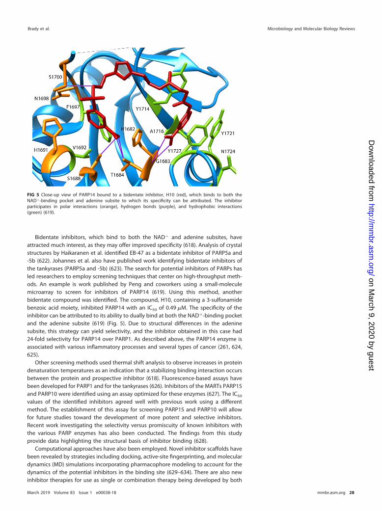
Bidentate inhibitors, which bind to both the NAD� and adenine subsites, haveattracted much interest, as they may offer improved specificity (618). Analysis of crystalstructures by Haikaranen et al. identified EB-47 as a bidentate inhibitor of PARP5a and-5b (622). Johannes et al. also have published work identifying bidentate inhibitors ofthe tankyrases (PARP5a and -5b) (623). The search for potential inhibitors of PARPs hasled researchers to employ screening techniques that center on high-throughput meth-ods. An example is work published by Peng and coworkers using a small-moleculemicroarray to screen for inhibitors of PARP14 (619). Using this method, anotherbidentate compound was identified. The compound, H10, containing a 3-sulfonamidebenzoic acid moiety, inhibited PARP14 with an IC50 of 0.49 �M. The specificity of theinhibitor can be attributed to its ability to dually bind at both the NAD�-binding pocketand the adenine subsite (619) (Fig. 5). Due to structural differences in the adeninesubsite, this strategy can yield selectivity, and the inhibitor obtained in this case had24-fold selectivity for PARP14 over PARP1. As described above, the PARP14 enzyme isassociated with various inflammatory processes and several types of cancer (261, 624,625).
Other screening methods used thermal shift analysis to observe increases in proteindenaturation temperatures as an indication that a stabilizing binding interaction occursbetween the protein and prospective inhibitor (618). Fluorescence-based assays havebeen developed for PARP1 and for the tankyrases (626). Inhibitors of the MARTs PARP15and PARP10 were identified using an assay optimized for these enzymes (627). The IC50
values of the identified inhibitors agreed well with previous work using a differentmethod. The establishment of this assay for screening PARP15 and PARP10 will allowfor future studies toward the development of more potent and selective inhibitors.Recent work investigating the selectivity versus promiscuity of known inhibitors withthe various PARP enzymes has also been conducted. The findings from this studyprovide data highlighting the structural basis of inhibitor binding (628).
Computational approaches have also been employed. Novel inhibitor scaffolds havebeen revealed by strategies including docking, active-site fingerprinting, and moleculardynamics (MD) simulations incorporating pharmacophore modeling to account for thedynamics of the potential inhibitors in the binding site (629–634). There are also newinhibitor therapies for use as single or combination therapy being developed by both
FIG 5 Close-up view of PARP14 bound to a bidentate inhibitor, H10 (red), which binds to both theNAD�-binding pocket and adenine subsite to which its specificity can be attributed. The inhibitorparticipates in polar interactions (orange), hydrogen bonds (purple), and hydrophobic interactions(green) (619).
Brady et al. Microbiology and Molecular Biology Reviews
March 2019 Volume 83 Issue 1 e00038-18 mmbr.asm.org 28
on March 9, 2020 by guest
http://mm
br.asm.org/
Dow
nloaded from
synthesis and modification of existing inhibitors (635). Recently a computationalscreening analysis identified a natural product known as ZINC67913374, which ispredicted to be an improved inhibitor compared to olaparib (583). The combination ofthese theoretical, chemical, and biophysical approaches holds great promise for thedevelopment of selective inhibitors to target PARP functions within cells and tissues.
CONCLUSION
The review presented here gives an overview of recent findings in regard to thebiological functions of the PARP family of proteins. Though the function of PARP1 is thebest studied, this review highlights functions of many of the other family memberswhich have been recently identified. Specifically, work was highlighted which impli-cated the function of this family of proteins in immune and inflammatory responses,suggesting that these enzymes are very promising for future therapeutic development.In addition, some of the most recent work pertaining to the structural and biophysicalcharacteristics of PARP enzymes has offered solutions to the challenges of overcomingpromiscuous inhibitor binding. The structural diversity and the wide array of biologicalfunctions in this class of enzymes attest to their importance in the progression ofdiseases in regard to both immune and inflammatory responses.
ACKNOWLEDGMENTSThis work was supported by National Institutes of Health grant R35GM119456 to
M.A.J. and by an NIH IRACDA MERIT Scholar award (K12 GM088010) to P.N.B.We thank members of the Johnson laboratory for helpful discussions.
REFERENCES1. D’Amours D, Desnoyers S, D’Silva I, Poirier GG. 1999. Poly(ADP-
ribosyl)ation reactions in the regulation of nuclear functions. BiochemJ 342:249 –268. https://doi.org/10.1042/bj3420249.
2. Chambon P, Weill JD, Mandel P. 1963. Nicotinamide mononucleotideactivation of new DNA-dependent polyadenylic acid synthesizing nu-clear enzyme. Biochem Biophys Res Commun 11:39 – 43.
3. Chambon P, Weil JD, Doly J, Strosser MT, Mandel P. 1966. On theformation of a novel adenylic compound by enzymatic extracts of livernuclei. Biochem Biophys Res Commun 25:638 – 643. https://doi.org/10.1016/0006-291X(66)90502-X.
4. Nishizuka Y, Ueda K, Nakazawa K, Hayaishi O. 1967. Studies on thepolymer of adenosine diphosphate ribose. I. Enzymic formation fromnicotinamide adenine dinuclotide in mammalian nuclei. J Biol Chem242:3164 –3171.
5. Sugimura T, Fujimura S, Hasegawa S, Kawamura Y. 1967. Polymeriza-tion of the adenosine 5=-diphosphate ribose moiety of NAD by rat livernuclear enzyme. Biochim Biophys Acta 138:438 – 441. https://doi.org/10.1016/0005-2787(67)90507-2.
6. Alkhatib HM, Chen DF, Cherney B, Bhatia K, Notario V, Giri C, SteinG, Slattery E, Roeder RG, Smulson ME. 1987. Cloning and expressionof cDNA for human poly(ADP-ribose) polymerase. Proc Natl Acad SciU S A 84:1224 –1228.
7. Uchida K, Morita T, Sato T, Ogura T, Yamashita R, Noguchi S, Suzuki H,Nyunoya H, Miwa M, Sugimura T. 1987. Nucleotide sequence of afull-length cDNA for human fibroblast poly(ADP-ribose) polymerase.Biochem Biophys Res Commun 148:617– 622.
8. Kurosaki T, Ushiro H, Mitsuuchi Y, Suzuki S, Matsuda M, Matsuda Y,Katunuma N, Kangawa K, Matsuo H, Hirose T. 1987. Primary structure ofhuman poly(ADP-ribose) synthetase as deduced from cDNA sequence.J Biol Chem 262:15990 –15997.
9. Koch-Nolte F, Petersen D, Balasubramanian S, Haag F, Kahlke D, WillerT, Kastelein R, Bazan F, Thiele H-G. 1996. Mouse T cell membraneproteins Rt61 and Rt62 are arginine/protein mono(ADPribosyl)trans-ferases and share secondary structure motifs with ADP-ribosylatingbacterial toxins. J Biol Chem 271:7686 –7693. https://doi.org/10.1074/jbc.271.13.7686.
10. Hottiger MO, Hassa PO, Luscher B, Schuler H, Koch-Nolte F. 2010. Towarda unified nomenclature for mammalian ADP-ribosyltransferases. TrendsBiochem Sci 35:208–219. https://doi.org/10.1016/j.tibs.2009.12.003.
11. Citarelli M, Teotia S, Lamb RS. 2010. Evolutionary history of the
poly(ADP-ribose) polymerase gene family in eukaryotes. BMC Evol Biol10:308. https://doi.org/10.1186/1471-2148-10-308.
12. Perina D, Mikoc A, Ahel J, Cetkovic H, Zaja R, Ahel I. 2014. Distributionof protein poly(ADP-ribosyl)ation systems across all domains of life.DNA Repair (Amst) 23:4 –16. https://doi.org/10.1016/j.dnarep.2014.05.003.
13. Otto H, Reche PA, Bazan F, Dittmar K, Haag F, Koch-Nolte F. 2005. Insilico characterization of the family of PARP-like poly(ADP-ribosyl)transferases (pARTs). BMC Genomics 6:139.
14. Beck C, Robert I, Reina-San-Martin B, Schreiber V, Dantzer F. 2014.Poly(ADP-ribose) polymerases in double-strand break repair: focus onPARP1, PARP2 and PARP3. Exp Cell Res 329:18 –25. https://doi.org/10.1016/j.yexcr.2014.07.003.
15. Yu W, Ginjala V, Pant V, Chernukhin I, Whitehead J, Docquier F, FarrarD, Tavoosidana G, Mukhopadhyay R, Kanduri C, Oshimura M, FeinbergAP, Lobanenkov V, Klenova E, Ohlsson R. 2004. Poly(ADP-ribosyl)ationregulates CTCF-dependent chromatin insulation. Nat Genet 36:1105–1110. https://doi.org/10.1038/ng1426.
16. Zampieri M, Guastafierro T, Calabrese R, Ciccarone F, Bacalini MG, RealeA, Perilli M, Passananti C, Caiafa P. 2012. ADP-ribose polymers localizedon Ctcf-Parp1-Dnmt1 complex prevent methylation of Ctcf target sites.Biochem J 441:645– 652. https://doi.org/10.1042/BJ20111417.
17. Zardo G, Reale A, Passananti C, Pradhan S, Buontempo S, De Matteis G,Adams RL, Caiafa P. 2002. Inhibition of poly(ADP-ribosyl)ation inducesDNA hypermethylation: a possible molecular mechanism. FASEB J 16:1319 –1321. https://doi.org/10.1096/fj.01-0827fje.
18. Chang P, Jacobson MK, Mitchison TJ. 2004. Poly(ADP-ribose) is requiredfor spindle assembly and structure. Nature 432:645– 649. https://doi.org/10.1038/nature03061.
19. Asher G, Reinke H, Altmeyer M, Gutierrez-Arcelus M, Hottiger MO,Schibler U. 2010. Poly(ADP-ribose) polymerase 1 participates in thephase entrainment of circadian clocks to feeding. Cell 142:943–953.https://doi.org/10.1016/j.cell.2010.08.016.
20. Ju BG, Solum D, Song EJ, Lee KJ, Rose DW, Glass CK, Rosenfeld MG.2004. Activating the PARP-1 sensor component of the groucho/TLE1corepressor complex mediates a CaMKinase IIdelta-dependent neuro-genic gene activation pathway. Cell 119:815– 829. https://doi.org/10.1016/j.cell.2004.11.017.
21. Nasta F, Laudisi F, Sambucci M, Rosado MM, Pioli C. 2010. Increased
PARP in Host-Pathogen Interactions Microbiology and Molecular Biology Reviews
March 2019 Volume 83 Issue 1 e00038-18 mmbr.asm.org 29
on March 9, 2020 by guest
http://mm
br.asm.org/
Dow
nloaded from
Foxp3� regulatory T cells in poly(ADP-ribose) polymerase-1 deficiency.J Immunol 184:3470 –3477. https://doi.org/10.4049/jimmunol.0901568.
22. Kotova E, Jarnik M, Tulin AV. 2009. Poly(ADP-ribose) polymerase 1 isrequired for protein localization to Cajal body. PLoS Genet 5:e1000387.https://doi.org/10.1371/journal.pgen.1000387.
23. Tulin A, Stewart D, Spradling AC. 2002. The Drosophila heterochromaticgene encoding poly(ADP-ribose) polymerase (PARP) is required tomodulate chromatin structure during development. Genes Dev 16:2108 –2119. https://doi.org/10.1101/gad.1003902.
24. Yang F, Baumann C, De La Fuente R. 2009. Persistence of histone H2AXphosphorylation after meiotic chromosome synapsis and abnormalcentromere cohesion in poly(ADP-ribose) polymerase (Parp-1) nulloocytes. Dev Biol 331:326 –338. https://doi.org/10.1016/j.ydbio.2009.05.550.
25. Panda S, Poirier GG, Kay SA. 2002. tej defines a role for poly(ADP-ribosyl)ation in establishing period length of the arabidopsis circa-dian oscillator. Dev Cell 3:51– 61.
26. Sambucci M, Laudisi F, Novelli F, Bennici E, Rosado MM, Pioli C. 2013.Effects of PARP-1 deficiency on Th1 and Th2 cell differentiation. SciWorld J 2013:1–9. https://doi.org/10.1155/2013/375024.
27. Takahashi K, Yamanaka S. 2006. Induction of pluripotent stem cellsfrom mouse embryonic and adult fibroblast cultures by defined factors.Cell 126:663– 676. https://doi.org/10.1016/j.cell.2006.07.024.
28. Di Giammartino DC, Shi Y, Manley JL. 2013. PARP1 represses PAP andinhibits polyadenylation during heat shock. Mol Cell 49:7–17. https://doi.org/10.1016/j.molcel.2012.11.005.
29. Boamah EK, Kotova E, Garabedian M, Jarnik M, Tulin AV. 2012.Poly(ADP-ribose) polymerase 1 (PARP-1) regulates ribosomal biogene-sis in Drosophila nucleoli. PLoS Genet 8:e1002442. https://doi.org/10.1371/journal.pgen.1002442.
30. Sitikov AS, Davydova EK, Bezlepkina TA, Ovchinnikov LP, Spirin AS.1984. Eukaryotic elongation factor 2 loses its non-specific affinity forRNA and leaves polyribosomes as a result of ADP-ribosylation. FEBSLett 176:406 – 410.
31. Todorova T, Bock FJ, Chang P. 2014. PARP13 regulates cellular mRNApost-transcriptionally and functions as a pro-apoptotic factor bydestabilizing TRAILR4 transcript. Nat Commun 5:5362. https://doi.org/10.1038/ncomms6362.
32. Ji Y, Tulin AV. 2012. Poly(ADP-ribose) controls DE-cadherin-dependentstem cell maintenance and oocyte localization. Nature Commun 3:760.https://doi.org/10.1038/ncomms1759.
33. Matveeva E, Maiorano J, Zhang Q, Eteleeb AM, Convertini P, Chen J,Infantino V, Stamm S, Wang J, Rouchka EC, Fondufe-Mittendorf YN.2016. Involvement of PARP1 in the regulation of alternative splicing.Cell Discov 2:15046. https://doi.org/10.1038/celldisc.2015.46.
34. Calkins AS, Iglehart JD, Lazaro J-B. 2013. DNA damage-induced inhibi-tion of rRNA synthesis by DNA-PK and PARP-1. Nucleic Acids Res41:7378 –7386. https://doi.org/10.1093/nar/gkt502.
35. Malanga M, Czubaty A, Girstun A, Staron K, Althaus FR. 2008. Poly(ADP-ribose) binds to the splicing factor ASF/SF2 and regulates its phosphor-ylation by DNA topoisomerase I. J Biol Chem 283:19991–19998. https://doi.org/10.1074/jbc.M709495200.
36. Leung AK, Vyas S, Rood JE, Bhutkar A, Sharp PA, Chang P. 2011.Poly(ADP-ribose) regulates stress responses and microRNA activity inthe cytoplasm. Mol Cell 42:489 – 499. https://doi.org/10.1016/j.molcel.2011.04.015.
37. Ji Y, Tulin AV. 2009. Poly(ADP-ribosyl)ation of heterogeneous nuclearribonucleoproteins modulates splicing. Nucleic Acids Res 37:3501–3513. https://doi.org/10.1093/nar/gkp218.
38. Hanahan D, Weinberg Robert A. 2011. Hallmarks of cancer: The nextgeneration. Cell 144:646 – 674. https://doi.org/10.1016/j.cell.2011.02.013.
39. Ricks TK, Chiu HJ, Ison G, Kim G, McKee AE, Kluetz P, Pazdur R. 2015.Successes and challenges of PARP inhibitors in cancer therapy. FrontOncol 5:222. https://doi.org/10.3389/fonc.2015.00222.
40. Bell CE, Eisenberg D. 1996. Crystal structure of diphtheria toxin boundto nicotinamide adenine dinucleotide. Biochemistry 35:1137–1149.https://doi.org/10.1021/bi9520848.
41. Domenighini M, Rappuoli R. 1996. Three conserved consensus se-quences identify the NAD-binding site of ADP-ribosylating enzymes,expressed by eukaryotes, bacteria and T-even bacteriophages. MolMicrobiol 21:667– 674.
42. Langelier M-F, Riccio AA, Pascal JM. 2014. PARP-2 and PARP-3 areselectively activated by 5= phosphorylated DNA breaks through an
allosteric regulatory mechanism shared with PARP-1. Nucleic Acids Res42:7762–7775. https://doi.org/10.1093/nar/gku474.
43. Simonin F, Hofferer L, Panzeter PL, Muller S, de Murcia G, Althaus FR.1993. The carboxyl-terminal domain of human poly(ADP-ribose) poly-merase. Overproduction in Escherichia coli, large scale purification, andcharacterization. J Biol Chem 268:13454 –13461.
44. Vyas S, Matic I, Uchima L, Rood J, Zaja R, Hay RT, Ahel I, Chang P. 2014.Family-wide analysis of poly(ADP-ribose) polymerase activity. Nat Com-mun 5:4426. https://doi.org/10.1038/ncomms5426.
45. Loseva O, Jemth AS, Bryant HE, Schuler H, Lehtio L, Karlberg T, HelledayT. 2010. Poly(ADP-ribose) polymerase-3 (PARP-3) is a mono-ADP ribo-sylase that activates PARP-1 in absence of DNA. J Biol Chem 285:8054 – 8060. https://doi.org/10.1074/jbc.M109.077834.
46. Yang CS, Jividen K, Spencer A, Dworak N, Ni L, Oostdyk LT, ChatterjeeM, Kusmider B, Reon B, Parlak M, Gorbunova V, Abbas T, Jeffery E,Sherman NE, Paschal BM. 2017. Ubiquitin modification by the E3ligase/ADP-ribosyltransferase Dtx3L/Parp9. Mol Cell 66:503–516.e5.https://doi.org/10.1016/j.molcel.2017.04.028.
47. Schreiber V, Dantzer F, Ame JC, de Murcia G. 2006. Poly(ADP-ribose):novel functions for an old molecule. Nat Rev Mol Cell Biol 7:517–528.https://doi.org/10.1038/nrm1963.
48. Altmeyer M, Messner S, Hassa PO, Fey M, Hottiger MO. 2009. Molecularmechanism of poly(ADP-ribosyl)ation by PARP1 and identification oflysine residues as ADP-ribose acceptor sites. Nucleic Acids Res 37:3723–3738. https://doi.org/10.1093/nar/gkp229.
49. Rulten SL, Fisher AEO, Robert I, Zuma MC, Rouleau M, Ju L, Poirier G,Reina-San-Martin B, Caldecott KW. 2011. PARP-3 and APLF functiontogether to accelerate nonhomologous end-joining. Mol Cell 41:33– 45.https://doi.org/10.1016/j.molcel.2010.12.006.
50. Ame JC, Rolli V, Schreiber V, Niedergang C, Apiou F, Decker P, Muller S,Hoger T, Menissier-de Murcia J, de Murcia G. 1999. PARP-2, A novelmammalian DNA damage-dependent poly(ADP-ribose) polymerase. JBiol Chem 274:17860 –17868.
51. de Murcia JM, Niedergang C, Trucco C, Ricoul M, Dutrillaux B, Mark M,Oliver FJ, Masson M, Dierich A, LeMeur M, Walztinger C, Chambon P, deMurcia G. 1997. Requirement of poly(ADP-ribose) polymerase in recov-ery from DNA damage in mice and in cells. Proc Natl Acad Sci U S A94:7303–7307.
52. Lilyestrom W, van der Woerd MJ, Clark N, Luger K. 2010. Structural andbiophysical studies of human PARP-1 in complex with damaged DNA.J Mol Biol 395:983–994. https://doi.org/10.1016/j.jmb.2009.11.062.
53. Lonskaya I, Potaman VN, Shlyakhtenko LS, Oussatcheva EA, Lyub-chenko YL, Soldatenkov VA. 2005. Regulation of poly(ADP-ribose)polymerase-1 by DNA structure-specific binding. J Biol Chem 280:17076 –17083. https://doi.org/10.1074/jbc.M413483200.
54. Fekete A, Kenesi E, Hunyadi-Gulyas E, Durgo H, Berko B, Dunai ZA,Bauer PI. 2012. The guanine-quadruplex sructure in the human c-mycgene’s promoter is converted into B-DNA form by the humanpoly(ADP-ribose)polymerase-1. PLoS One 7:e42690. https://doi.org/10.1371/journal.pone.0042690.
55. Schreiber V, Ame JC, Dolle P, Schultz I, Rinaldi B, Fraulob V, Menissier-deMurcia J, de Murcia G. 2002. Poly(ADP-ribose) polymerase-2 (PARP-2) isrequired for efficient base excision DNA repair in association withPARP-1 and XRCC1. J Biol Chem 277:23028 –23036. https://doi.org/10.1074/jbc.M202390200.
56. Nash RA, Caldecott KW, Barnes DE, Lindahl T. 1997. XRCC1 proteininteracts with one of two distinct forms of DNA ligase III. Biochemistry36:5207–5211. https://doi.org/10.1021/bi962281m.
57. Menissier de Murcia J, Ricoul M, Tartier L, Niedergang C, Huber A,Dantzer F, Schreiber V, Ame JC, Dierich A, LeMeur M, Sabatier L,Chambon P, de Murcia G. 2003. Functional interaction between PARP-1and PARP-2 in chromosome stability and embryonic development inmouse. EMBO J 22:2255–2263. https://doi.org/10.1093/emboj/cdg206.
58. Shieh WM, Amé J-C, Wilson MV, Wang Z-Q, Koh DW, Jacobson MK,Jacobson EL. 1998. Poly(ADP-ribose) polymerase null mouse cells syn-thesize ADP-ribose polymers. J Biol Chem 273:30069 –30072.
59. Fenton AL, Shirodkar P, Macrae CJ, Meng L, Koch CA. 2013. The PARP3-and ATM-dependent phosphorylation of APLF facilitates DNA double-strand break repair. Nucleic Acids Res 41:4080 – 4092. https://doi.org/10.1093/nar/gkt134.
60. Boehler C, Gauthier LR, Mortusewicz O, Biard DS, Saliou J-M, Bresson A,Sanglier-Cianferani S, Smith S, Schreiber V, Boussin F, Dantzer F. 2011.Poly(ADP-ribose) polymerase 3 (PARP3), a newcomer in cellular response
Brady et al. Microbiology and Molecular Biology Reviews
March 2019 Volume 83 Issue 1 e00038-18 mmbr.asm.org 30
on March 9, 2020 by guest
http://mm
br.asm.org/
Dow
nloaded from
to DNA damage and mitotic progression. Proc Natl Acad Sci U S A108:2783–2788. https://doi.org/10.1073/pnas.1016574108.
61. Grundy GJ, Polo LM, Zeng Z, Rulten SL, Hoch NC, Paomephan P, Xu Y,Sweet SM, Thorne AW, Oliver AW, Matthews SJ, Pearl LH, Caldecott KW.2016. PARP3 is a sensor of nicked nucleosomes and monoribosylateshistone H2B(Glu2). Nat Commun 7:12404. https://doi.org/10.1038/ncomms12404.
62. Kutuzov MM, Khodyreva SN, Amé J-C, Ilina ES, Sukhanova MV, SchreiberV, Lavrik OI. 2013. Interaction of PARP-2 with DNA structures mimickingDNA repair intermediates and consequences on activity of base exci-sion repair proteins. Biochimie 95:1208 –1215. https://doi.org/10.1016/j.biochi.2013.01.007.
63. Talhaoui I, Lebedeva NA, Zarkovic G, Saint-Pierre C, Kutuzov MM,Sukhanova MV, Matkarimov BT, Gasparutto D, Saparbaev MK, Lavrik OI,Ishchenko AA. 2016. Poly(ADP-ribose) polymerases covalently modifystrand break termini in DNA fragments in vitro. Nucleic Acids Res44:9279 –9295. https://doi.org/10.1093/nar/gkw675.
64. Belousova EA, Ishchenko AA, Lavrik OI. 2018. DNA is a new target ofParp3. Sci Rep 8:4176. https://doi.org/10.1038/s41598-018-22673-3.
65. Zarkovic G, Belousova EA, Talhaoui I, Saint-Pierre C, Kutuzov MM,Matkarimov BT, Biard D, Gasparutto D, Lavrik OI, Ishchenko AA. 2018.Characterization of DNA ADP-ribosyltransferase activities of PARP2 andPARP3: new insights into DNA ADP-ribosylation. Nucleic Acids Res46:2417–2431. https://doi.org/10.1093/nar/gkx1318.
66. Nolan NL, Butt TR, Wong M, Lambrianidou A, Smulson ME. 1980.Characterization of poly(ADP-ribose)– histone H1 complex formation inpurified polynucleosomes and chromatin. Eur J Biochem 113:15–25.
67. Boulikas T. 1990. Poly(ADP-ribosylated) histones in chromatin replica-tion. J Biol Chem 265:14638 –14647.
68. Poirier GG, de Murcia G, Jongstra-Bilen J, Niedergang C, Mandel P. 1982.Poly(ADP-ribosyl)ation of polynucleosomes causes relaxation of chro-matin structure. Proc Natl Acad Sci U S A 79:3423–3427.
69. de Murcia G, Huletsky A, Lamarre D, Gaudreau A, Pouyet J, Daune M,Poirier GG. 1986. Modulation of chromatin superstructure inducedby poly(ADP-ribose) synthesis and degradation. J Biol Chem 261:7011–7017.
70. Leduc Y, de Murcia G, Lamarre D, Poirier GG. 1986. Visualization ofpoly(ADP-ribose) synthetase associated with polynucleosomes by im-munoelectron microscopy. Biochim Biophys Acta 885:248 –255.
71. Frechette A, Huletsky A, Aubin RJ, de Murcia G, Mandel P, Lord A,Grondin G, Poirier GG. 1985. Poly(ADP-ribosyl)ation of chromatin: ki-netics of relaxation and its effect on chromatin solubility. Can JBiochem Cell Biol 63:764 –773. https://doi.org/10.1139/o85-096.
72. Tulin A, Spradling A. 2003. Chromatin loosening by poly(ADP)-ribosepolymerase (PARP) at Drosophila puff loci. Science 299:560 –562.https://doi.org/10.1126/science.1078764.
73. Ling F, Tang Y, Li M, Li QS, Li X, Yang L, Zhao W, Jin CC, Zeng Z, Liu C,Wu CF, Chen WW, Lin X, Wang YL, Threadgill MD. 2017. Mono-ADP-ribosylation of histone 3 at arginine-117 promotes proliferationthrough its interaction with P300. Oncotarget 8:72773–72787. https://doi.org/10.18632/oncotarget.20347.
74. Martinez-Zamudio RI, Ha HC. 2014. PARP1 enhances inflammatorycytokine expression by alteration of promoter chromatin structure inmicroglia. Brain Behav 4:552–565. https://doi.org/10.1002/brb3.239.
75. Liu Z, Kraus WL. 2017. Catalytic-independent functions of PARP-1 de-termine Sox2 pioneer activity at intractable genomic loci. Mol Cell65:589 – 603.e9. https://doi.org/10.1016/j.molcel.2017.01.017.
76. Sala A, La Rocca G, Burgio G, Kotova E, Di Gesù D, Collesano M, IngrassiaAMR, Tulin AV, Corona DFV. 2008. The nucleosome-remodeling ATPaseISWI is regulated by poly-ADP-ribosylation. PLoS Biol 6:2329 –2342.https://doi.org/10.1371/journal.pbio.0060252.
77. Gottschalk AJ, Trivedi RD, Conaway JW, Conaway RC. 2012. Activationof the SNF2 family ATPase ALC1 by poly(ADP-ribose) in a stableALC1·PARP1·nucleosome intermediate. J Biol Chem 287:43527– 43532.https://doi.org/10.1074/jbc.M112.401141.
78. Singh HR, Nardozza AP, Moller IR, Knobloch G, Kistemaker HAV, HasslerM, Harrer N, Blessing C, Eustermann S, Kotthoff C, Huet S, Mueller-Planitz F, Filippov DV, Timinszky G, Rand KD, Ladurner AG. 2017. Apoly-ADP-ribose trigger releases the auto-inhibition of a chromatinremodeling oncogene. Mol Cell 68:860 – 871. https://doi.org/10.1016/j.molcel.2017.11.019.
79. Lehmann LC, Hewitt G, Aibara S, Leitner A, Marklund E, Maslen SL,Maturi V, Chen Y, van der Spoel D, Skehel JM, Moustakas A, Boulton SJ,Deindl S. 2017. Mechanistic insights into autoinhibition of the onco-
genic chromatin remodeler ALC1. Mol Cell 68:847– 859. https://doi.org/10.1016/j.molcel.2017.10.017.
80. Ahel D, Horejsi Z, Wiechens N, Polo SE, Garcia-Wilson E, Ahel I, Flynn H,Skehel M, West SC, Jackson SP, Owen-Hughes T, Boulton SJ. 2009.Poly(ADP-ribose)-dependent regulation of DNA repair by the chroma-tin remodeling enzyme ALC1. Science 325:1240 –1243. https://doi.org/10.1126/science.1177321.
81. Tsuda M, Cho K, Ooka M, Shimizu N, Watanabe R, Yasui A, Nakazawa Y,Ogi T, Harada H, Agama K, Nakamura J, Asada R, Fujiike H, Sakuma T,Yamamoto T, Murai J, Hiraoka M, Koike K, Pommier Y, Takeda S, HirotaK. 2017. ALC1/CHD1L, a chromatin-remodeling enzyme, is required forefficient base excision repair. PLoS One 12:e0188320. https://doi.org/10.1371/journal.pone.0188320.
82. Strickfaden H, McDonald D, Kruhlak MJ, Haince J-F, Th’ng JPH, RouleauM, Ishibashi T, Corry GN, Ausio J, Underhill DA, Poirier GG, Hendzel MJ.2016. Poly(ADP-ribosyl)ation-dependent transient chromatin decon-densation and histone displacement following laser microirradiation. JBiol Chem 291:1789 –1802. https://doi.org/10.1074/jbc.M115.694992.
83. Martinez-Zamudio R, Ha HC. 2012. Histone ADP-ribosylation facilitatesgene transcription by directly remodeling nucleosomes. Mol Cell Biol32:2490 –2502. https://doi.org/10.1128/MCB.06667-11.
84. Wacker DA, Ruhl DD, Balagamwala EH, Hope KM, Zhang T, Kraus WL.2007. The DNA binding and catalytic domains of poly(ADP-ribose)polymerase 1 cooperate in the regulation of chromatin structure andtranscription. Mol Cell Biol 27:7475–7485. https://doi.org/10.1128/MCB.01314-07.
85. Kim MY, Mauro S, Gevry N, Lis JT, Kraus WL. 2004. NAD�-dependentmodulation of chromatin structure and transcription by nucleosomebinding properties of PARP-1. Cell 119:803– 814. https://doi.org/10.1016/j.cell.2004.11.002.
86. Muthurajan UM, Hepler MR, Hieb AR, Clark NJ, Kramer M, Yao T, LugerK. 2014. Automodification switches PARP-1 function from chromatinarchitectural protein to histone chaperone. Proc Natl Acad Sci U S A111:12752–12757. https://doi.org/10.1073/pnas.1405005111.
87. Kannan P, Yu Y, Wankhade S, Tainsky MA. 1999. PolyADP-ribose poly-merase is a coactivator for AP-2-mediated transcriptional activation.Nucleic Acids Res 27:866 – 874.
88. Zingarelli B, Hake PW, Burroughs TJ, Piraino G, O’Connor M, Denen-berg A. 2004. Activator protein-1 signalling pathway and apoptosisare modulated by poly(ADP-ribose) polymerase-1 in experimentalcolitis. Immunology 113:509 –517. https://doi.org/10.1111/j.1365-2567.2004.01991.x.
89. Oei SL, Griesenbeck J, Schweiger M, Babich V, Kropotov A, Tomilin N.1997. Interaction of the transcription factor YY1 with human poly(ADP-ribosyl) transferase. Biochem Biophys Res Commun 240:108 –111.https://doi.org/10.1006/bbrc.1997.7621.
90. Nie J, Sakamoto S, Song D, Qu Z, Ota K, Taniguchi T. 1998. Interactionof Oct-1 and automodification domain of poly(ADP-ribose) synthetase.FEBS Lett 424:27–32.
91. Cervellera MN, Sala A. 2000. Poly(ADP-ribose) polymerase is a B-MYBcoactivator. J Biol Chem 275:10692–10696.
92. Kanai M, Hanashiro K, Kim SH, Hanai S, Boulares AH, Miwa M, FukasawaK. 2007. Inhibition of Crm1-p53 interaction and nuclear export of p53by poly(ADP-ribosyl)ation. Nat Cell Biol 9:1175–1183. https://doi.org/10.1038/ncb1638.
93. Ha HC, Hester LD, Snyder SH. 2002. Poly(ADP-ribose) polymerase-1dependence of stress-induced transcription factors and associatedgene expression in glia. Proc Natl Acad Sci U S A 99:3270 –3275.https://doi.org/10.1073/pnas.052712399.
94. Legrand AJ, Choul-Li S, Spriet C, Idziorek T, Vicogne D, Drobecq H,Dantzer F, Villeret V, Aumercier M. 2013. The level of Ets-1 protein isregulated by poly(ADP-ribose) polymerase-1 (PARP-1) in cancer cells toprevent DNA damage. PLoS One 8:e55883. https://doi.org/10.1371/journal.pone.0055883.
95. Butler AJ, Ordahl CP. 1999. Poly(ADP-ribose) polymerase binds withtranscription enhancer factor 1 to MCAT1 elements to regulate muscle-specific transcription. Mol Cell Biol 19:296 –306. https://doi.org/10.1128/MCB.19.1.296.
96. Gibson BA, Zhang Y, Jiang H, Hussey KM, Shrimp JH, Lin H, Schwede F,Yu Y, Kraus WL. 2016. Chemical genetic discovery of PARP targetsreveals a role for PARP-1 in transcription elongation. Science 353:45–50.https://doi.org/10.1126/science.aaf7865.
97. Tuncel H, Tanaka S, Oka S, Nakai S, Fukutomi R, Okamoto M, Ota T,Kaneko H, Tatsuka M, Shimamoto F. 2012. PARP6, a mono(ADP-ribosyl)
PARP in Host-Pathogen Interactions Microbiology and Molecular Biology Reviews
March 2019 Volume 83 Issue 1 e00038-18 mmbr.asm.org 31
on March 9, 2020 by guest
http://mm
br.asm.org/
Dow
nloaded from
transferase and a negative regulator of cell proliferation, is involved incolorectal cancer development. Int J Oncol 41:2079 –2086. https://doi.org/10.3892/ijo.2012.1652.
98. MacPherson L, Tamblyn L, Rajendra S, Bralha F, McPherson JP, Mat-thews J. 2013. 2,3,7,8-Tetrachlorodibenzo-p-dioxin poly(ADP-ribose)polymerase (TiPARP, ARTD14) is a mono-ADP-ribosyltransferase andrepressor of aryl hydrocarbon receptor transactivation. Nucleic AcidsRes 41:1604 –1621. https://doi.org/10.1093/nar/gks1337.
99. Bindesbøll C, Tan S, Bott D, Cho T, Tamblyn L, MacPherson L, Grønning-Wang L, Nebb Hilde I, Matthews J. 2016. TCDD-inducible poly-ADP-ribose polymerase (TIPARP/PARP7) mono-ADP-ribosylates and co-activates liver X receptors. Biochem J 473:899 –910. https://doi.org/10.1042/BJ20151077.
100. Goenka S, Boothby M. 2006. Selective potentiation of Stat-dependentgene expression by collaborator of Stat6 (CoaSt6), a transcriptionalcofactor. Proc Natl Acad Sci U S A 103:4210 – 4215. https://doi.org/10.1073/pnas.0506981103.
101. Yu M, Schreek S, Cerni C, Schamberger C, Lesniewicz K, Poreba E,Vervoorts J, Walsemann G, Grotzinger J, Kremmer E, Mehraein Y,Mertsching J, Kraft R, Austen M, Luscher-Firzlaff J, Luscher B. 2005.PARP-10, a novel Myc-interacting protein with poly(ADP-ribose) poly-merase activity, inhibits transformation. Oncogene 24:1982–1993.https://doi.org/10.1038/sj.onc.1208410.
102. Mehrotra P, Riley JP, Patel R, Li F, Voss L, Goenka S. 2011. PARP-14functions as a transcriptional switch for Stat6-dependent gene activa-tion. J Biol Chem 286:1767–1776. https://doi.org/10.1074/jbc.M110.157768.
103. Juszczynski P, Kutok JL, Li C, Mitra J, Aguiar RC, Shipp MA. 2006. BAL1and BBAP are regulated by a gamma interferon-responsive bidirec-tional promoter and are overexpressed in diffuse large B-cell lympho-mas with a prominent inflammatory infiltrate. Mol Cell Biol 26:5348 –5359. https://doi.org/10.1128/MCB.02351-05.
104. Yan Q, Xu R, Zhu L, Cheng X, Wang Z, Manis J, Shipp MA. 2013. BAL1and its partner E3 ligase, BBAP, link poly(ADP-ribose) activation,ubiquitylation, and double-strand DNA repair independent of ATM,MDC1, and RNF8. Mol Cell Biol 33:845– 857. https://doi.org/10.1128/MCB.00990-12.
105. Nicolae CM, Aho ER, Vlahos AH, Choe KN, De S, Karras GI, Moldovan GL.2014. The ADP-ribosyltransferase PARP10/ARTD10 interacts with pro-liferating cell nuclear antigen (PCNA) and is required for DNA damagetolerance. J Biol Chem 289:13627–13637. https://doi.org/10.1074/jbc.M114.556340.
106. Malik N, Smulson M. 1984. A relationship between nuclear poly(ade-nosine diphosphate ribosylation) and acetylation posttranslationalmodifications. 1. Nucleosome studies. Biochemistry 23:3721–3725.
107. Verdone L, La Fortezza M, Ciccarone F, Caiafa P, Zampieri M, CasertaM. 2015. Poly(ADP-ribosyl)ation affects histone acetylation and tran-scription. PLoS One 10:e0144287. https://doi.org/10.1371/journal.pone.0144287.
108. Yang Z, Li L, Chen L, Yuan W, Dong L, Zhang Y, Wu H, Wang C. 2014.PARP-1 mediates LPS-induced HMGB1 release by macrophagesthrough regulation of HMGB1 acetylation. J Immunol 193:6114 – 6123.https://doi.org/10.4049/jimmunol.1400359.
109. Krukenberg KA, Jiang R, Steen JA, Mitchison TJ. 2014. Basal activity ofa PARP1-NuA4 complex varies dramatically across cancer cell lines. CellRep 8:1808 –1818. https://doi.org/10.1016/j.celrep.2014.08.009.
110. Hassa PO, Haenni SS, Buerki C, Meier NI, Lane WS, Owen H, Gersbach M,Imhof R, Hottiger MO. 2005. Acetylation of poly(ADP-ribose)polymerase-1 by p300/CREB-binding protein regulates coactivation ofNF-kappaB-dependent transcription. J Biol Chem 280:40450 – 40464.https://doi.org/10.1074/jbc.M507553200.
111. Reale A, Matteis GD, Galleazzi G, Zampieri M, Caiafa P. 2005. Modula-tion of DNMT1 activity by ADP-ribose polymers. Oncogene 24:13–19.https://doi.org/10.1038/sj.onc.1208005.
112. Krishnakumar R, Kraus WL. 2010. PARP-1 regulates chromatin structureand transcription through a KDM5B-dependent pathway. Mol Cell39:736 –749. https://doi.org/10.1016/j.molcel.2010.08.014.
113. Martello R, Leutert M, Jungmichel S, Bilan V, Larsen SC, Young C,Hottiger MO, Nielsen ML. 2016. Proteome-wide identification of theendogenous ADP-ribosylome of mammalian cells and tissue. Nat Com-mun 7:12917. https://doi.org/10.1038/ncomms12917.
114. Jungmichel S, Rosenthal F, Altmeyer M, Lukas J, Hottiger Michael O,Nielsen Michael L. 2013. Proteome-wide identification of poly(ADP-
ribosyl)ation targets in different genotoxic stress responses. Mol Cell52:272–285. https://doi.org/10.1016/j.molcel.2013.08.026.
115. Zhang Y, Wang J, Ding M, Yu Y. 2013. Site-specific characterization ofthe Asp- and Glu-ADP-ribosylated proteome. Nat Methods 10:981–984.https://doi.org/10.1038/nmeth.2603.
116. Vivelo CA, Wat R, Agrawal C, Tee HY, Leung Anthony KL. 2017.ADPriboDB: the database of ADP-ribosylated proteins. Nucleic AcidsRes 45:D204 –D209. https://doi.org/10.1093/nar/gkw706.
117. Rosenthal F, Nanni P, Barkow-Oesterreicher S, Hottiger MO. 2015.Optimization of LTQ-orbitrap mass spectrometer parameters for theidentification of ADP-ribosylation sites. J Proteome Res 14:4072– 4079.https://doi.org/10.1021/acs.jproteome.5b00432.
118. Gao G, Guo X, Goff SP. 2002. Inhibition of retroviral RNA production byZAP, a CCCH-type zinc finger protein. Science 297:1703–1706. https://doi.org/10.1126/science.1074276.
119. Seo G, Kincaid R, Phanaksri T, Burke J, Pare J, Cox J, Hsiang T, Krug R,Sullivan C. 2013. Reciprocal inhibition between intracellular antiviralsignaling and the RNAi machinery in mammalian cells. Cell Host Mi-crobe 14:435– 445. https://doi.org/10.1016/j.chom.2013.09.002.
120. Xuan Y, Liu L, Shen S, Deng H, Gao G. 2012. Zinc finger antiviral proteininhibits murine gammaherpesvirus 68 M2 expression and regulatesviral latency in cultured cells. J Virol 86:12431–12434. https://doi.org/10.1128/JVI.01514-12.
121. Zhu Y, Chen G, Lv F, Wang X, Ji X, Xu Y. 2011. Zinc-finger antiviralprotein inhibits HIV-1 infection by selectively targeting multiply splicedviral mRNAs for degradation. Proc Natl Acad Sci U S A 108:15834 –15839. https://doi.org/10.1073/pnas.1101676108.
122. Müller S, Möller P, Bick MJ, Wurr S, Becker S, Günther S, Kümmerer BM.2007. Inhibition of filovirus replication by the zinc finger antiviralprotein. J Virol 81:2391–2400. https://doi.org/10.1128/JVI.01601-06.
123. Bick MJ, Carroll J-WN, Gao G, Goff SP, Rice CM, MacDonald MR. 2003.Expression of the zinc-finger antiviral protein inhibits alphavirus repli-cation. J Virol 77:11555–11562.
124. Hayakawa S, Shiratori S, Yamato H, Kameyama T, Kitatsuji C, Kashigi F,Goto S, Kameoka S, Fujikura D, Yamada T, Mizutani T, Kazumata M, SatoM, Tanaka J, Asaka M, Ohba Y, Miyazaki T, Imamura M, Takaoka A. 2011.ZAPS is a potent stimulator of signaling mediated by the RNA helicaseRIG-I during antiviral responses. Nat Immunol 12:37– 44. https://doi.org/10.1038/ni.1963.
125. Mao R, Nie H, Cai D, Zhang J, Liu H, Yan R. 2013. Inhibition of hepatitisB virus replication by the host zinc finger antiviral protein. PLoS Pathog9:e1003494. https://doi.org/10.1371/journal.ppat.1003494.
126. Guo X, Ma J, Sun J, Gao G. 2007. The zinc-finger antiviral protein recruitsthe RNA processing exosome to degrade the target mRNA. Proc NatlAcad Sci U S A 104:151–156. https://doi.org/10.1073/pnas.0607063104.
127. Leung A, Todorova T, Ando Y, Chang P. 2012. Poly(ADP-ribose) regu-lates post-transcriptional gene regulation in the cytoplasm. RNA Biol9:542–548. https://doi.org/10.4161/rna.19899.
128. Bock Florian J, Todorova Tanya T, Chang P. 2015. RNA regulation bypoly(ADP-ribose) polymerases. Mol Cell 58:959 –969. https://doi.org/10.1016/j.molcel.2015.01.037.
129. Huang Z, Wang X, Gao G. 2010. Analyses of SELEX-derived ZAP-bindingRNA aptamers suggest that the binding specificity is determined byboth structure and sequence of the RNA. Protein Cell 1:752–759.https://doi.org/10.1007/s13238-010-0096-9.
130. Chen S, Xu Y, Zhang K, Wang X, Sun J, Gao G, Liu Y. 2012. Structure ofN-terminal domain of ZAP indicates how a zinc-finger protein recog-nizes complex RNA. Nat Struct Mol Biol 19:430 – 435. https://doi.org/10.1038/nsmb.2243.
131. Wang Y, Li H, Tang Q, Maul GG, Yuan Y. 2008. Kaposi’s sarcoma-associated herpesvirus ori-lyt-dependent DNA replication: involvementof host cellular factors. J Virol 82:2867–2882. https://doi.org/10.1128/JVI.01319-07.
132. Gwack Y, Nakamura H, Lee SH, Souvlis J, Yustein JT, Gygi S, Kung H-J,Jung JU. 2003. Poly(ADP-ribose) polymerase 1 and Ste20-like kinasehKFC act as transcriptional repressors for gamma-2 herpesvirus lyticreplication. Mol Cell Biol 23:8282– 8294.
133. Ohsaki E, Ueda K, Sakakibara S, Do E, Yada K, Yamanishi K. 2004.Poly(ADP-ribose) polymerase 1 binds to Kaposi’s sarcoma-associatedherpesvirus (KSHV) terminal repeat sequence and modulates KSHVreplication in latency. J Virol 78:9936 –9946. https://doi.org/10.1128/JVI.78.18.9936-9946.2004.
134. Cheong WC, Park JH, Kang HR, Song MJ. 2015. Downregulation ofpoly(ADP-ribose) polymerase 1 by a viral processivity factor facilitates
Brady et al. Microbiology and Molecular Biology Reviews
March 2019 Volume 83 Issue 1 e00038-18 mmbr.asm.org 32
on March 9, 2020 by guest
http://mm
br.asm.org/
Dow
nloaded from
lytic replication of gammaherpesvirus. J Virol 89:9676 –9682. https://doi.org/10.1128/JVI.00559-15.
135. Atasheva S, Akhrymuk M, Frolova EI, Frolov I. 2012. New PARP genewith an anti-alphavirus function. J Virol 86:8147– 8160. https://doi.org/10.1128/JVI.00733-12.
136. Goodier JL, Pereira GC, Cheung LE, Rose RJ, Kazazian HH, Jr. 2015.The broad-spectrum antiviral protein ZAP restricts human retro-transposition. PLoS Genet 11:e1005252. https://doi.org/10.1371/journal.pgen.1005252.
137. Atasheva S, Frolova EI, Frolov I. 2014. Interferon-stimulatedpoly(ADP-ribose) polymerases are potent inhibitors of cellular trans-lation and virus replication. J Virol 88:2116 –2130. https://doi.org/10.1128/JVI.03443-13.
138. McCormick C, Khaperskyy DA. 2017. Translation inhibition and stressgranules in the antiviral immune response. Nat Rev Immunol 17:647– 660. https://doi.org/10.1038/nri.2017.63.
139. Le Sage V, Cinti A, McCarthy S, Amorim R, Rao S, Daino GL, TramontanoE, Branch DR, Mouland AJ. 2017. Ebola virus VP35 blocks stress granuleassembly. Virology 502:73– 83. https://doi.org/10.1016/j.virol.2016.12.012.
140. Lu Y, Wambach M, Katze MG, Krug RM. 1995. Binding of the influenzavirus NS1 protein to double-stranded RNA inhibits the activation of theprotein kinase that phosphorylates the elF-2 translation initiation fac-tor. Virology 214:222–228.
141. White JP, Cardenas AM, Marissen WE, Lloyd RE. 2007. Inhibition ofcytoplasmic mRNA stress granule formation by a viral proteinase. CellHost Microbe 2:295–305. https://doi.org/10.1016/j.chom.2007.08.006.
142. Altmeyer M, Neelsen KJ, Teloni F, Pozdnyakova I, Pellegrino S, Grøfte M,Rask M-BD, Streicher W, Jungmichel S, Nielsen ML, Lukas J. 2015. Liquiddemixing of intrinsically disordered proteins is seeded by poly(ADP-ribose). Nat Commun 6:8088. https://doi.org/10.1038/ncomms9088.
143. Patel A, Lee HO, Jawerth L, Maharana S, Jahnel M, Hein MY, Stoynov S,Mahamid J, Saha S, Franzmann TM, Pozniakovski A, Poser I, Maghelli N,Royer LA, Weigert M, Myers EW, Grill S, Drechsel D, Hyman AA, AlbertiS. 2015. A liquid-to-solid phase transition of the ALS protein FUSaccelerated by disease mutation. Cell 162:1066 –1077. https://doi.org/10.1016/j.cell.2015.07.047.
144. Zhang Y, Mao D, Roswit WT, Jin X, Patel AC, Patel DA, Agapov E, WangZ, Tidwell RM, Atkinson JJ, Huang G, McCarthy R, Yu J, Yun NE, PaesslerS, Lawson TG, Omattage NS, Brett TJ, Holtzman MJ. 2015. PARP9-DTX3Lubiquitin ligase targets host histone H2BJ and viral 3C protease toenhance interferon signaling and control viral infection. Nat Immunol16:1215–1227. https://doi.org/10.1038/ni.3279.
145. Daugherty MD, Young JM, Kerns JA, Malik HS. 2014. Rapid evolutionof PARP genes suggests a broad role for ADP-ribosylation in host-virus conflicts. PLoS Genet 10:e1004403. https://doi.org/10.1371/journal.pgen.1004403.
146. Kerns JA, Emerman M, Malik HS. 2008. Positive selection and increasedantiviral activity associated with the PARP-containing isoform of hu-man zinc-finger antiviral protein. PLoS Genet 4:e21. https://doi.org/10.1371/journal.pgen.0040021.
147. Ding R, Pommier Y, Kang VH, Smulson M. 1992. Depletion of poly(ADP-ribose) polymerase by antisense RNA expression results in a delay inDNA strand break rejoining. J Biol Chem 267:12804 –12812.
148. Wang ZQ, Auer B, Stingl L, Berghammer H, Haidacher D, SchweigerM, Wagner EF. 1995. Mice lacking ADPRT and poly(ADP-ribosyl)ationdevelop normally but are susceptible to skin disease. Genes Dev9:509 –520.
149. Park J, Seo T, Hwang S, Lee D, Gwack Y, Choe J. 2000. The K-bZIPprotein from Kaposi’s sarcoma-associated herpesvirus interacts withp53 and represses its transcriptional activity. J Virol 74:11977–11982.https://doi.org/10.1128/JVI.74.24.11977-11982.2000.
150. Bueno MT, Reyes D, Valdes L, Saheba A, Urias E, Mendoza C, Fregoso OI,Llano M. 2013. Poly(ADP-ribose) polymerase 1 promotes transcriptionalrepression of integrated retroviruses. J Virol 87:2496 –2507. https://doi.org/10.1128/JVI.01668-12.
151. Pinnola A, Naumova N, Shah M, Tulin AV. 2007. Nucleosomal corehistones mediate dynamic regulation of poly(ADP-ribose) polymerase 1protein binding to chromatin and induction of its enzymatic activity. JBiol Chem 282:32511–32519. https://doi.org/10.1074/jbc.M705989200.
152. Kameoka M, Nukuzuma S, Itaya A, Tanaka Y, Ota K, Inada Y, Ikuta K,Yoshihara K. 2005. Poly(ADP-ribose)polymerase-1 is required for inte-gration of the human immunodeficiency virus type 1 genome near
centromeric alphoid DNA in human and murine cells. Biochem BiophysRes Commun 334:412– 417. https://doi.org/10.1016/j.bbrc.2005.06.104.
153. Kameoka M, Tanaka Y, Ota K, Itaya A, Yoshihara K. 1999. Poly(ADP-ribose) polymerase is involved in PMA-induced activation of HIV-1 inU1 cells by modulating the LTR function. Biochem Biophys Res Com-mun 262:285–289. https://doi.org/10.1006/bbrc.1999.1146.
154. Ha HC, Juluri K, Zhou Y, Leung S, Hermankova M, Snyder SH. 2001.Poly(ADP-ribose) polymerase-1 is required for efficient HIV-1 integra-tion. Proc Natl Acad Sci U S A 98:3364 –3368. https://doi.org/10.1073/pnas.051633498.
155. Tempera I, Deng Z, Atanasiu C, Chen CJ, D’Erme M, Lieberman PM.2010. Regulation of Epstein-Barr virus OriP replication by poly(ADP-ribose) polymerase 1. J Virol 84:4988 – 4997. https://doi.org/10.1128/JVI.02333-09.
156. Liu L, Lear Z, Hughes DJ, Wu W, Zhou EM, Whitehouse A, Chen H,Hiscox JA. 2015. Resolution of the cellular proteome of the nucleocap-sid protein from a highly pathogenic isolate of porcine reproductiveand respiratory syndrome virus identifies PARP-1 as a cellular targetwhose interaction is critical for virus biology. Vet Microbiol 176:109 –119. https://doi.org/10.1016/j.vetmic.2014.11.023.
157. Grunewald ME, Fehr AR, Athmer J, Perlman S. 2018. The coronavirusnucleocapsid protein is ADP-ribosylated. Virology 517:62– 68. https://doi.org/10.1016/j.virol.2017.11.020.
158. Dery CV, de Murcia G, Lamarre D, Morin N, Poirier GG, Weber J. 1986.Possible role of ADP-ribosylation of adenovirus core proteins in virusinfection. Virus Res 4:313–329.
159. Bortz E, Westera L, Maamary J, Steel J, Albrecht RA, Manicassamy B,Chase G, Martínez-Sobrido L, Schwemmle M, García-Sastre A. 2011.Host- and strain specific regulation of influenza virus polymerase ac-tivity by interacting cellular proteins. mBio 2:e00151-11. https://doi.org/10.1128/mBio.00151-11.
160. Grady SL, Hwang J, Vastag L, Rabinowitz JD, Shenk T. 2012. Herpessimplex virus 1 infection activates poly(ADP-ribose) polymerase andtriggers the degradation of poly(ADP-ribose) glycohydrolase. J Virol86:8259 – 8268. https://doi.org/10.1128/JVI.00495-12.
161. Krukenberg KA, Kim S, Tan ES, Maliga Z, Mitchison TJ. 2015. Extra-cellular poly(ADP-ribose) is a pro-inflammatory signal for macro-phages. Chem Biol 22:446 – 452. https://doi.org/10.1016/j.chembiol.2015.03.007.
162. Morrison AR, Moss J, Stevens LA, Evans JE, Farrell C, Merithew E,Lambright DG, Greiner DL, Mordes JP, Rossini AA, Bortell R. 2006. ART2,a T cell surface mono-ADP-ribosyltransferase, generates extracellularpoly(ADP-ribose). J Biol Chem 281:33363–33372. https://doi.org/10.1074/jbc.M607259200.
163. Teloni F, Altmeyer M. 2016. Readers of poly(ADP-ribose): designed tobe fit for purpose. Nucleic Acids Res 44:993–1006. https://doi.org/10.1093/nar/gkv1383.
164. Koonin EV, Gorbalenya AE, Purdy MA, Rozanov MN, Reyes GR, BradleyDW. 1992. Computer-assisted assignment of functional domains in thenonstructural polyprotein of hepatitis E virus: delineation of an addi-tional group of positive-strand RNA plant and animal viruses. Proc NatlAcad Sci U S A 89:8259 – 8263.
165. Dominguez G, Wang CY, Frey TK. 1990. Sequence of the genome RNAof rubella virus: evidence for genetic rearrangement during togavirusevolution. Virology 177:225–238.
166. Gorbalenya AE, Koonin EV, Lai MM. 1991. Putative papain-related thiolproteases of positive-strand RNA viruses. Identification of rubi- andaphthovirus proteases and delineation of a novel conserved domainassociated with proteases of rubi-, alpha- and coronaviruses. FEBS Lett288:201–205.
167. Li C, Debing Y, Jankevicius G, Neyts J, Ahel I, Coutard B, Canard B. 2016.Viral macrodomains reverse protein ADP-ribosylation. J Virol 90:8478 – 8486. https://doi.org/10.1128/JVI.00705-16.
168. Johnson MA, Chatterjee A, Neuman BW, Wuthrich K. 2010. SARS coro-navirus unique domain: three-domain molecular architecture in solu-tion and RNA binding. J Mol Biol 400:724 –742. https://doi.org/10.1016/j.jmb.2010.05.027.
169. Eckei L, Krieg S, Butepage M, Lehmann A, Gross A, Lippok B, Grimm AR,Kummerer BM, Rossetti G, Luscher B, Verheugd P. 2017. The conservedmacrodomains of the non-structural proteins of Chikungunya virus andother pathogenic positive strand RNA viruses function as mono-ADP-ribosylhydrolases. Sci Rep 7:41746. https://doi.org/10.1038/srep41746.
170. Kuri T, Eriksson KK, Putics A, Zust R, Snijder EJ, Davidson AD, Siddell SG,Thiel V, Ziebuhr J, Weber F. 2011. The ADP-ribose-1’’-mono-
PARP in Host-Pathogen Interactions Microbiology and Molecular Biology Reviews
March 2019 Volume 83 Issue 1 e00038-18 mmbr.asm.org 33
on March 9, 2020 by guest
http://mm
br.asm.org/
Dow
nloaded from
phosphatase domains of severe acute respiratory syndrome coronavi-rus and human coronavirus 229E mediate resistance to antiviral inter-feron responses. J Gen Virol 92:1899 –1905. https://doi.org/10.1099/vir.0.031856-0.
171. McPherson RL, Abraham R, Sreekumar E, Ong SE, Cheng SJ, Baxter VK,Kistemaker HA, Filippov DV, Griffin DE, Leung AK. 2017. ADP-ribosylhydrolase activity of Chikungunya virus macrodomain is criticalfor virus replication and virulence. Proc Natl Acad Sci U S A 114:1666 –1671. https://doi.org/10.1073/pnas.1621485114.
172. Fehr AR, Athmer J, Channappanavar R, Phillips JM, Meyerholz DK,Perlman S. 2015. The nsp3 macrodomain promotes virulence in micewith coronavirus-induced encephalitis. J Virol 89:1523–1536. https://doi.org/10.1128/JVI.02596-14.
173. Fehr AR, Channappanavar R, Jankevicius G, Fett C, Zhao J, Athmer J,Meyerholz DK, Ahel I, Perlman S. 2016. The conserved coronavirusmacrodomain promotes virulence and suppresses the innate immuneresponse during severe acute respiratory syndrome coronavirus infec-tion. mBio 7:e01721-16. https://doi.org/10.1128/mBio.01721-16.
174. Lalic J, Posavec Marjanovic M, Palazzo L, Perina D, Sabljic I, Žaja R, ColbyT, Pleše B, Halasz M, Jankevicius G, Bucca G, Ahel M, Matic I, CetkovicH, Luic M, Mikoc A, Ahel I. 2016. Disruption of macrodomain proteinSCO6735 increases antibiotic production in Streptomyces coelicolor. JBiol Chem 291:23175–23187. https://doi.org/10.1074/jbc.M116.721894.
175. Feijs KL, Kleine H, Braczynski A, Forst AH, Herzog N, Verheugd P, LinzenU, Kremmer E, Luscher B. 2013. ARTD10 substrate identification onprotein microarrays: regulation of GSK3beta by mono-ADP-ribosylation. Cell Commun Signal 11:5. https://doi.org/10.1186/1478-811X-11-5.
176. Forst AH, Karlberg T, Herzog N, Thorsell AG, Gross A, Feijs KL, VerheugdP, Kursula P, Nijmeijer B, Kremmer E, Kleine H, Ladurner AG, Schuler H,Luscher B. 2013. Recognition of mono-ADP-ribosylated ARTD10 sub-strates by ARTD8 macrodomains. Structure 21:462– 475. https://doi.org/10.1016/j.str.2012.12.019.
177. Carter-O’Connell I, Jin H, Morgan RK, Zaja R, David LL, Ahel I, Cohen MS.2016. Identifying family-member specific targets of mono-ARTDs usinga chemical genetics approach. Cell Rep 14:621– 631. https://doi.org/10.1016/j.celrep.2015.12.045.
178. Kusov Y, Tan J, Alvarez E, Enjuanes L, Hilgenfeld R. 2015. AG-quadruplex-binding macrodomain within the “SARS-unique domain”is essential for the activity of the SARS-coronavirus replication-transcription complex. Virology 484:313–322. https://doi.org/10.1016/j.virol.2015.06.016.
179. Paudyal S, Alfonso-Prieto M, Carnevale V, Redhu SK, Klein ML, Nichol-son AW. 2015. Combined computational and experimental analysis ofa complex of ribonuclease III and the regulatory macrodomain protein,YmdB. Proteins 83:459 – 472. https://doi.org/10.1002/prot.24751.
180. Ma-Lauer Y, Carbajo-Lozoya J, Hein MY, Muller MA, Deng W, Lei J,Meyer B, Kusov Y, von Brunn B, Bairad DR, Hunten S, Drosten C,Hermeking H, Leonhardt H, Mann M, Hilgenfeld R, von Brunn A. 2016.p53 down-regulates SARS coronavirus replication and is targeted bythe SARS-unique domain and PLpro via E3 ubiquitin ligase RCHY1. ProcNatl Acad Sci U S A 113:E5192–E5201. https://doi.org/10.1073/pnas.1603435113.
181. Steffen J, Brody J, Armen R, Pascal J. 2013. Structural implications forselective targeting of PARPs. Front Oncol 3:301. https://doi.org/10.3389/fonc.2013.00301.
182. Chandrasekaran S, Caparon MG. 2015. The Streptococcus pyogenesNAD(�) glycohydrolase modulates epithelial cell PARylation andHMGB1 release. Cell Microbiol 17:1376 –1390. https://doi.org/10.1111/cmi.12442.
183. Nossa CW, Jain P, Tamilselvam B, Gupta VR, Chen LF, Schreiber V,Desnoyers S, Blanke SR. 2009. Activation of the abundant nuclear factorpoly(ADP-ribose) polymerase-1 by Helicobacter pylori. Proc Natl AcadSci U S A 106:19998 –20003. https://doi.org/10.1073/pnas.0906753106.
184. Altmeyer M, Barthel M, Eberhard M, Rehrauer H, Hardt W-D, HottigerMO. 2010. Absence of poly(ADP-ribose) polymerase 1 delays the onsetof Salmonella enterica serovar Typhimurium-induced gut inflamma-tion. Infect Immun 78:3420 –3431. https://doi.org/10.1128/IAI.00211-10.
185. Wright RHG, Lioutas A, Le Dily F, Soronellas D, Pohl A, Bonet J, NachtAS, Samino S, Font-Mateu J, Vicent GP, Wierer M, Trabado MA, Schel-horn C, Carolis C, Macias MJ, Yanes O, Oliva B, Beato M. 2016. ADP-ribose– derived nuclear ATP synthesis by NUDIX5 is required for chro-matin remodeling. Science 352:1221–1225. https://doi.org/10.1126/science.aad9335.
186. Slade D, Dunstan MS, Barkauskaite E, Weston R, Lafite P, Dixon N, AhelM, Leys D, Ahel I. 2011. The structure and catalytic mechanism of apoly(ADP-ribose) glycohydrolase. Nature 477:616 – 620. https://doi.org/10.1038/nature10404.
187. Nordlund S, Hogbom M. 2013. ADP-ribosylation, a mechanism regulat-ing nitrogenase activity. FEBS J 280:3484 –3490. https://doi.org/10.1111/febs.12279.
188. Penyige A, Vargha G, Ensign JC, Barabas G. 1992. The possible role ofADP ribosylation in physiological regulation of sporulation in Strepto-myces griseus. Gene 115:181–185.
189. Huh JW, Shima J, Ochi K. 1996. ADP-ribosylation of proteins in Bacillussubtilis and its possible importance in sporulation. J Bacteriol 178:4935– 4941.
190. Doucet-Chabeaud G, Godon C, Brutesco C, de Murcia G, Kazmaier M.2001. Ionising radiation induces the expression of PARP-1 and PARP-2genes in Arabidopsis. Mol Genet Genomics 265:954 –963.
191. De Block M, Verduyn C, De Brouwer D, Cornelissen M. 2005. Poly(ADP-ribose) polymerase in plants affects energy homeostasis, cell death andstress tolerance. Plant J 41:95–106. https://doi.org/10.1111/j.1365-313X.2004.02277.x.
192. Zhang H, Gu Z, Wu Q, Yang L, Liu C, Ma H, Xia Y, Ge X. 2015. ArabidopsisPARG1 is the key factor promoting cell survival among the enzymesregulating post-translational poly(ADP-ribosyl)ation. Sci Rep 5:15892.https://doi.org/10.1038/srep15892.
193. Jia Q, den Dulk-Ras A, Shen H, Hooykaas PJ, de Pater S. 2013. Poly(ADP-ribose)polymerases are involved in microhomology mediated back-upnon-homologous end joining in Arabidopsis thaliana. Plant Mol Biol82:339 –351. https://doi.org/10.1007/s11103-013-0065-9.
194. Song J, Keppler BD, Wise RR, Bent AF. 2015. PARP2 Is the predominantpoly(ADP-ribose) polymerase in Arabidopsis DNA damage and immuneresponses. PLoS Genet 11:e1005200. https://doi.org/10.1371/journal.pgen.1005200.
195. Storozhenko S, Inze D, Van Montagu M, Kushnir S. 2001. Arabidopsiscoactivator ALY-like proteins, DIP1 and DIP2, interact physically withthe DNA-binding domain of the Zn-finger poly(ADP-ribose) polymer-ase. J Exp Bot 52:1375–1380.
196. Wagner D, Przybyla D, Op den Camp R, Kim C, Landgraf F, Lee KP,Würsch M, Laloi C, Nater M, Hideg E, Apel K. 2004. The genetic basis ofsinglet oxygen-induced stress responses of Arabidopsis thaliana. Sci-ence 306:1183–1185. https://doi.org/10.1126/science.1103178.
197. Vanderauwera S, De Block M, Van de Steene N, van de Cotte B, MetzlaffM, Van Breusegem F. 2007. Silencing of poly(ADP-ribose) polymerase inplants alters abiotic stress signal transduction. Proc Natl Acad Sci U S A104:15150 –15155. https://doi.org/10.1073/pnas.0706668104.
198. Amor Y, Babiychuk E, Inzé D, Levine A. 1998. The involvement ofpoly(ADP-ribose) polymerase in the oxidative stress responses inplants. FEBS Lett 440:1–7.
199. Rissel D, Heym PP, Thor K, Brandt W, Wessjohann LA, Peiter E. 2017. Nosilver bullet— canonical poly(ADP-ribose) polymerases (PARPs) are nouniversal factors of abiotic and biotic stress resistance of Arabidopsisthaliana. Front Plant Sci 8:59. https://doi.org/10.3389/fpls.2017.00059.
200. Hunt L, Holdsworth MJ, Gray JE. 2007. Nicotinamidase activity is im-portant for germination. Plant J 51:341–351. https://doi.org/10.1111/j.1365-313X.2007.03151.x.
201. Rissel D, Losch J, Peiter E. 2014. The nuclear protein poly(ADP-ribose)polymerase 3 (AtPARP3) is required for seed storability in Arabidopsisthaliana. Plant Biol 16:1058 –1064. https://doi.org/10.1111/plb.12167.
202. Schulz P, Neukermans J, Van Der Kelen K, Mühlenbock P, VanBreusegem F, Noctor G, Teige M, Metzlaff M, Hannah MA. 2012.Chemical PARP inhibition enhances growth of arabidopsis and re-duces anthocyanin accumulation and the activation of stress pro-tective mechanisms. PLoS One 7:e37287. https://doi.org/10.1371/journal.pone.0037287.
203. Adams-Phillips L, Briggs AG, Bent AF. 2010. Disruption of poly(ADP-ribosyl)ation mechanisms alters responses of Arabidopsis to bioticstress. Plant Physiol 152:267–280. https://doi.org/10.1104/pp.109.148049.
204. Adams-Phillips L, Wan J, Tan X, Dunning FM, Meyers BC, MichelmoreRW, Bent AF. 2008. Discovery of ADP-ribosylation and other plantdefense pathway elements through expression profiling of four differ-ent Arabidopsis-Pseudomonas R-avr interactions. Mol Plant MicrobeInteract 21:646 – 657. https://doi.org/10.1094/MPMI-21-5-0646.
205. Feng B, Liu C, Mvv d. O, Intorne AC, Li B, Babilonia K, de Souza Filho GA,Shan L, He P. 2015. Protein poly(ADP-ribosyl)ation regulates Arabidop-
Brady et al. Microbiology and Molecular Biology Reviews
March 2019 Volume 83 Issue 1 e00038-18 mmbr.asm.org 34
on March 9, 2020 by guest
http://mm
br.asm.org/
Dow
nloaded from
sis immune gene expression and defense responses. PLoS Genet 11:e1004936. https://doi.org/10.1371/journal.pgen.1004936.
206. Song J, Bent AF. 2014. Microbial pathogens trigger host DNA double-strand breaks whose abundance is reduced by plant defense re-sponses. PLoS Pathog 10:e1004030. https://doi.org/10.1371/journal.ppat.1004030.
207. Feng B, Ma S, Chen S, Zhu N, Zhang S, Yu B, Yu Y, Le B, Chen X,Dinesh�Kumar SP, Shan L, He P. 2016. PARylation of the forkhead�associated domain protein DAWDLE regulates plant immunity. EMBORep 17:1799 –1813. https://doi.org/10.15252/embr.201642486.
208. Feng B, Liu C, Shan L, He P. 2016. Protein ADP-ribosylation takes controlin plant– bacterium interactions. PLoS Pathog 12:e1005941. https://doi.org/10.1371/journal.ppat.1005941.
209. Wang Y, Li J, Hou S, Wang X, Li Y, Ren D, Chen S, Tang X, Zhou JM. 2010.A Pseudomonas syringae ADP-ribosyltransferase inhibits Arabidopsismitogen-activated protein kinase kinases. Plant Cell 22:2033–2044.https://doi.org/10.1105/tpc.110.075697.
210. Fu ZQ, Guo M, Jeong B-R, Tian F, Elthon TE, Cerny RL, Staiger D, AlfanoJR. 2007. A type III effector ADP-ribosylates RNA-binding proteins andquells plant immunity. Nature 447:284 –288. https://doi.org/10.1038/nature05737.
211. Jeong B-R, Lin Y, Joe A, Guo M, Korneli C, Yang H, Wang P, Yu M, CernyRL, Staiger D, Alfano JR, Xu Y. 2011. Structure function analysis of anADP-ribosyltransferase type III effector and its RNA-binding target inplant immunity. J Biol Chem 286:43272– 43281. https://doi.org/10.1074/jbc.M111.290122.
212. Cherkis KA, Temple BR, Chung EH, Sondek J, Dangl JL. 2012. AvrRpm1missense mutations weakly activate RPS2-mediated immune responsein Arabidopsis thaliana. PLoS One 7:e42633. https://doi.org/10.1371/journal.pone.0042633.
213. Jaspers P, Overmyer K, Wrzaczek M, Vainonen JP, Blomster T, SalojarviJ, Reddy RA, Kangasjarvi J. 2010. The RST and PARP-like domain con-taining SRO protein family: analysis of protein structure, function andconservation in land plants. BMC Genomics 11:170. https://doi.org/10.1186/1471-2164-11-170.
214. Liu S, Liu S, Wang M, Wei T, Meng C, Wang M, Xia G. 2014. A wheatSIMILAR TO RCD-ONE gene enhances seedling growth and abioticstress resistance by modulating redox homeostasis and maintaininggenomic integrity. Plant Cell 26:164 –180. https://doi.org/10.1105/tpc.113.118687.
215. Teotia S, Lamb RS. 2009. The paralogous genes RADICAL-INDUCEDCELL DEATH1 and SIMILAR TO RCD ONE1 have partially redundantfunctions during Arabidopsis development. Plant Physiol 151:180 –198.https://doi.org/10.1104/pp.109.142786.
216. Jaspers P, Blomster T, Brosche M, Salojarvi J, Ahlfors R, Vainonen JP,Reddy RA, Immink R, Angenent G, Turck F, Overmyer K, Kangasjarvi J.2009. Unequally redundant RCD1 and SRO1 mediate stress and devel-opmental responses and interact with transcription factors. Plant J60:268 –279. https://doi.org/10.1111/j.1365-313X.2009.03951.x.
217. Bechtold U, Richard O, Zamboni A, Gapper C, Geisler M, Pogson B,Karpinski S, Mullineaux PM. 2008. Impact of chloroplastic- andextracellular-sourced ROS on high light-responsive gene expression inArabidopsis. J Exp Bot 59:121–133. https://doi.org/10.1093/jxb/erm289.
218. Sakuma Y, Maruyama K, Qin F, Osakabe Y, Shinozaki K, Yamaguchi-Shinozaki K. 2006. Dual function of an Arabidopsis transcription factorDREB2A in water-stress-responsive and heat-stress-responsive geneexpression. Proc Natl Acad Sci U S A 103:18822–18827. https://doi.org/10.1073/pnas.0605639103.
219. Vainonen JP, Jaspers P, Wrzaczek M, Lamminmaki A, Reddy RA,Vaahtera L, Brosche M, Kangasjarvi J. 2012. RCD1-DREB2A interaction inleaf senescence and stress responses in Arabidopsis thaliana. BiochemJ 442:573–581. https://doi.org/10.1042/BJ20111739.
220. O’Shea C, Staby L, Bendsen SK, Tidemand FG, Redsted A, Willemoës M,Kragelund BB, Skriver K. 2017. Structures and short linear motif ofdisordered transcription factor regions provide clues to the interac-tome of the cellular hub protein radical-induced cell death1. J BiolChem 292:512–527. https://doi.org/10.1074/jbc.M116.753426.
221. Hiltscher H, Rudnik R, Shaikhali J, Heiber I, Mellenthin M, MeirellesDuarte I, Schuster G, Kahmann U, Baier M. 2014. The radical inducedcell death protein 1 (RCD1) supports transcriptional activation of genesfor chloroplast antioxidant enzymes. Front Plant Sci 5:475. https://doi.org/10.3389/fpls.2014.00475.
222. Sharma S, Kaur C, Singla-Pareek SL, Sopory SK. 2016. OsSRO1a interactswith RNA binding domain-containing protein (OsRBD1) and functions
in abiotic stress tolerance in yeast. Front Plant Sci 7:62. https://doi.org/10.3389/fpls.2016.00062.
223. You J, Zong W, Du H, Hu H, Xiong L. 2014. A special member of the riceSRO family, OsSRO1c, mediates responses to multiple abiotic stressesthrough interaction with various transcription factors. Plant Mol Biol84:693–705. https://doi.org/10.1007/s11103-013-0163-8.
224. Daniels CM, Ong SE, Leung AK. 2014. Phosphoproteomic approach tocharacterize protein mono- and poly(ADP-ribosyl)ation sites from cells.J Proteome Res 13:3510 –3522. https://doi.org/10.1021/pr401032q.
225. Leidecker O, Bonfiglio JJ, Colby T, Zhang Q, Atanassov I, Zaja R, PalazzoL, Stockum A, Ahel I, Matic I. 2016. Serine is a new target residue forendogenous ADP-ribosylation on histones. Nat Chem Biol 12:998 –10000. https://doi.org/10.1038/nchembio.2180.
226. Gagné J-P, Ethier C, Defoy D, Bourassa S, Langelier M-F, Riccio AA,Pascal JM, Moon K-M, Foster LJ, Ning Z, Figeys D, Droit A, Poirier GG.2015. Quantitative site-specific ADP-ribosylation profiling of DNA-dependent PARPs. DNA Repair (Amst) 30:68 –79. https://doi.org/10.1016/j.dnarep.2015.02.004.
227. Fischbach A, Kruger A, Hampp S, Assmann G, Rank L, Hufnagel M,Stockl MT, Fischer JMF, Veith S, Rossatti P, Ganz M, Ferrando-May E,Hartwig A, Hauser K, Wiesmuller L, Burkle A, Mangerich A. 2018. TheC-terminal domain of p53 orchestrates the interplay between non-covalent and covalent poly(ADP-ribosyl)ation of p53 by PARP1. NucleicAcids Res 46:804 – 822. https://doi.org/10.1093/nar/gkx1205.
228. Houtkooper RH, Cantó C, Wanders RJ, Auwerx J. 2010. The secret life ofNAD(�): an old metabolite controlling new metabolic signaling path-ways. Endocr Rev 31:194 –223. https://doi.org/10.1210/er.2009-0026.
229. Hauschildt S, Scheipers P, Bessler W, Schwarz K, Ullmer A, Flad HD,Heine H. 1997. Role of ADP-ribosylation in activated monocytes/macrophages. Adv Exp Med Biol 419:249 –252.
230. Szabó C, Lim LHK, Cuzzocrea S, Getting SJ, Zingarelli B, Flower RJ,Salzman AL, Perretti M. 1997. Inhibition of poly(ADP-ribose) synthetaseattenuates neutrophil recruitment and exerts antiinflammatory effects.J Exp Med 186:1041–1049. https://doi.org/10.1084/jem.186.7.1041.
231. Chiang J, Shen YC, Wang YH, Hou YC, Chen CC, Liao JF, Yu MC, JuanCW, Liou KT. 2009. Honokiol protects rats against eccentric exercise-induced skeletal muscle damage by inhibiting NF-kappaB inducedoxidative stress and inflammation. Eur J Pharmacol 610:119 –127.https://doi.org/10.1016/j.ejphar.2009.03.035.
232. von Lukowicz T, Hassa PO, Lohmann C, Boren J, Braunersreuther V,Mach F, Odermatt B, Gersbach M, Camici GG, Stahli BE, Tanner FC,Hottiger MO, Luscher TF, Matter CM. 2008. PARP1 is required foradhesion molecule expression in atherogenesis. Cardiovasc Res 78:158 –166. https://doi.org/10.1093/cvr/cvm110.
233. Park EM, Cho S, Frys K, Racchumi G, Zhou P, Anrather J, Iadecola C.2004. Interaction between inducible nitric oxide synthase andpoly(ADP-ribose) polymerase in focal ischemic brain injury. Stroke35:2896 –2901. https://doi.org/10.1161/01.STR.0000147042.53659.6c.
234. Jog NR, Dinnall JA, Gallucci S, Madaio MP, Caricchio R. 2009.Poly(ADP-ribose) polymerase-1 regulates the progression of auto-immune nephritis in males by inducing necrotic cell death andmodulating inflammation. J Immunol 182:7297–7306. https://doi.org/10.4049/jimmunol.0803565.
235. Galbis-Martinez M, Saenz L, Ramirez P, Parrilla P, Yelamos J. 2010.Poly(ADP-ribose) polymerase-1 modulates interferon-gamma-inducibleprotein (IP)-10 expression in murine embryonic fibroblasts by stabiliz-ing IP-10 mRNA. Mol Immunol 47:1492–1499. https://doi.org/10.1016/j.molimm.2010.01.022.
236. Boulares AH, Zoltoski AJ, Sherif ZA, Jolly P, Massaro D, Smulson ME.2003. Gene knockout or pharmacological inhibition of poly(ADP-ribose)polymerase-1 prevents lung inflammation in a murine model ofasthma. Am J Respir Cell Mol Biol 28:322–329. https://doi.org/10.1165/rcmb.2001-0015OC.
237. Pieper AA, Brat DJ, Krug DK, Watkins CC, Gupta A, Blackshaw S, VermaA, Wang Z-Q, Snyder SH. 1999. Poly(ADP-ribose) polymerase-deficientmice are protected from streptozotocin-induced diabetes. Proc NatlAcad Sci U S A 96:3059 –3064.
238. Oliver FJ, Ménissier-de Murcia J, Nacci C, Decker P, Andriantsitohaina R,Muller S, de la Rubia G, Stoclet JC, de Murcia G. 1999. Resistance toendotoxic shock as a consequence of defective NF-kappaB activation inpoly(ADP-ribose) polymerase-1 deficient mice. EMBO J 18:4446 – 4454.https://doi.org/10.1093/emboj/18.16.4446.
239. Guzyk MM, Tykhomyrov AA, Nedzvetsky VS, Prischepa IV, Grinenko TV,Yanitska LV, Kuchmerovska TM. 2016. Poly(ADP-ribose) polymerase-1
PARP in Host-Pathogen Interactions Microbiology and Molecular Biology Reviews
March 2019 Volume 83 Issue 1 e00038-18 mmbr.asm.org 35
on March 9, 2020 by guest
http://mm
br.asm.org/
Dow
nloaded from

(PARP-1) inhibitors reduce reactive gliosis and improve angiostatinlevels in retina of diabetic rats. Neurochem Res 41:2526 –2537. https://doi.org/10.1007/s11064-016-1964-3.
240. Li WJ, Shin M-K, Oh S-J. 2011. Poly(ADP-ribose) polymerase is in-volved in the development of diabetic cystopathy via regulation ofnuclear factor kappa B. Urology 77:1265.e1– e8. https://doi.org/10.1016/j.urology.2011.01.022.
241. Koedel U, Winkler F, Angele B, Fontana A, Pfister HW. 2002. Meningitis-associated central nervous system complications are mediated by theactivation of poly(ADP-ribose) polymerase. J Cereb Blood Flow Metab22:39 – 49. https://doi.org/10.1097/00004647-200201000-00005.
242. Lenzser G, Kis B, Snipes JA, Gaspar T, Sandor P, Komjati K, Szabo C,Busija DW. 2007. Contribution of poly(ADP-ribose) polymerase to post-ischemic blood-brain barrier damage in rats. J Cereb Blood Flow Metab27:1318 –1326. https://doi.org/10.1038/sj.jcbfm.9600437.
243. Castri P, Lee YJ, Ponzio T, Maric D, Spatz M, Bembry J, Hallenbeck J.2014. Poly(ADP-ribose) polymerase-1 and its cleavage products dif-ferentially modulate cellular protection through NF-kappaB-dependent signaling. Biochim Biophys Acta 1843:640 – 651. https://doi.org/10.1016/j.bbamcr.2013.12.005.
244. Vuong B, Hogan-Cann ADJ, Alano CC, Stevenson M, Chan WY, Ander-son CM, Swanson RA, Kauppinen TM. 2015. NF-�B transcriptional acti-vation by TNF� requires phospholipase C, extracellular signal-regulatedkinase 2 and poly(ADP-ribose) polymerase-1. J Neuroinflamm 12:229.https://doi.org/10.1186/s12974-015-0448-8.
245. Ba X, Gupta S, Davidson M, Garg NJ. 2010. Trypanosoma cruzi inducesthe reactive oxygen species-PARP-1-RelA pathway for up-regulation ofcytokine expression in cardiomyocytes. J Biol Chem 285:11596 –11606.https://doi.org/10.1074/jbc.M109.076984.
246. Hassa PO, Buerki C, Lombardi C, Imhof R, Hottiger MO. 2003. Transcrip-tional coactivation of nuclear factor-kappaB-dependent gene expres-sion by p300 is regulated by poly(ADP)-ribose polymerase-1. J BiolChem 278:45145– 45153. https://doi.org/10.1074/jbc.M307957200.
247. Chang W-J, Alvarez-Gonzalez R. 2001. The sequence-specific DNA bind-ing of NF-�B is reversibly rby the automodification reaction ofpoly(ADP-ribose) polymerase 1. J Biol Chem 276:47664 – 47670. https://doi.org/10.1074/jbc.M104666200.
248. Li M, Naidu P, Yu Y, Berger Nathan A, Kannan P. 2004. Dual regulationof AP-2� transcriptional activation by poly(ADP-ribose) polymerase-1.Biochem J 382:323–329. https://doi.org/10.1042/BJ20040593.
249. Davis K, Banerjee S, Friggeri A, Bell C, Abraham E, Zerfaoui M. 2012.Poly(ADP-ribosyl)ation of high mobility group box 1 (HMGB1) proteinenhances inhibition of efferocytosis. Mol Med 18:359 –369. https://doi.org/10.2119/molmed.2011.00203.
250. Erener S, Petrilli V, Kassner I, Minotti R, Castillo R, Santoro R, Hassa PO,Tschopp J, Hottiger MO. 2012. Inflammasome-activated caspase 7cleaves PARP1 to enhance the expression of a subset of NF-kappaBtarget genes. Mol Cell 46:200 –211. https://doi.org/10.1016/j.molcel.2012.02.016.
251. Ji Y, Thomas C, Tulin N, Lodhi N, Boamah E, Kolenko V, Tulin AV.2016. Charon mediates IMD-driven PARP-1-dependent immune re-sponses in Drosophila. J Immunol 197:2382–2389. https://doi.org/10.4049/jimmunol.1600994.
252. Phulwani NK, Kielian T. 2008. Poly(ADP-ribose) polymerases (PARPs) 1-3regulate astrocyte activation. J Neurochem 106:578 –590. https://doi.org/10.1111/j.1471-4159.2008.05403.x.
253. Oumouna M, Datta R, Oumouna-Benachour K, Suzuki Y, Hans C, Mat-thews K, Fallon K, Boulares H. 2006. Poly(ADP-ribose) polymerase-1inhibition prevents eosinophil recruitment by modulating Th2 cyto-kines in a murine model of allergic airway inflammation: a potentialspecific effect on IL-5. J Immunol 177:6489 – 6496. https://doi.org/10.4049/jimmunol.177.9.6489.
254. Larmonier CB, Shehab KW, Laubitz D, Jamwal DR, Ghishan FK, Kiela PR.2016. Transcriptional reprogramming and resistance to colonic muco-sal injury in poly(ADP-ribose) polymerase 1 (PARP1)-deficient mice. JBiol Chem 291:8918 – 8930. https://doi.org/10.1074/jbc.M116.714386.
255. Olabisi OA, Soto-Nieves N, Nieves E, Yang TTC, Yang X, Yu RYL, Suk HY,Macian F, Chow C-W. 2008. Regulation of transcription factor NFAT byADP-ribosylation. Mol Cell Biol 28:2860 –2871. https://doi.org/10.1128/MCB.01746-07.
256. Levaot N, Voytyuk O, Dimitriou I, Sircoulomb F, Chandrakumar A,Deckert M, Krzyzanowski PM, Scotter A, Gu S, Janmohamed S, Cong F,Simoncic PD, Ueki Y, La Rose J, Rottapel R. 2011. Loss of Tankyrase-mediated destruction of 3BP2 is the underlying pathogenic mechanism
of cherubism. Cell 147:1324 –1339. https://doi.org/10.1016/j.cell.2011.10.045.
257. Jwa M, Chang P. 2012. PARP16 is a tail-anchored endoplasmic reticu-lum protein required for the PERK and IRE1�-mediated unfolded pro-tein response. Nat Cell Biol 14:1223–1230. https://doi.org/10.1038/ncb2593.
258. Scarpa ES, Fabrizio G, Di Girolamo M. 2013. A role of intracellularmono-ADP-ribosylation in cancer biology. FEBS J 280:3551–3562.https://doi.org/10.1111/febs.12290.
259. Verheugd P, Forst AH, Milke L, Herzog N, Feijs KL, Kremmer E, Kleine H,Luscher B. 2013. Regulation of NF-kappaB signalling by the mono-ADP-ribosyltransferase ARTD10. Nat Commun 4:1683. https://doi.org/10.1038/ncomms2672.
260. Aguiar RC, Takeyama K, He C, Kreinbrink K, Shipp MA. 2005.B-aggressive lymphoma family proteins have unique domains thatmodulate transcription and exhibit poly(ADP-ribose) polymeraseactivity. J Biol Chem 280:33756 –33765. https://doi.org/10.1074/jbc.M505408200.
261. Barbarulo A, Iansante V, Chaidos A, Naresh K, Rahemtulla A, Franzoso G,Karadimitris A, Haskard DO, Papa S, Bubici C. 2013. Poly(ADP-ribose)polymerase family member 14 (PARP14) is a novel effector of theJNK2-dependent pro-survival signal in multiple myeloma. Oncogene32:4231– 4242. https://doi.org/10.1038/onc.2012.448.
262. Bachmann SB, Frommel SC, Camicia R, Winkler HC, Santoro R, Hassa PO.2014. DTX3L and ARTD9 inhibit IRF1 expression and mediate in coop-eration with ARTD8 survival and proliferation of metastatic prostatecancer cells. Mol Cancer 13:125. https://doi.org/10.1186/1476-4598-13-125.
263. Mehrotra P, Krishnamurthy P, Sun J, Goenka S, Kaplan MH. 2015.Poly-ADP-ribosyl polymerase-14 promotes T helper 17 and follicularT helper development. Immunology 146:537–546. https://doi.org/10.1111/imm.12515.
264. Cho SH, Raybuck A, Wei M, Erickson J, Nam KT, Cox RG, TrochtenbergA, Thomas JW, Williams J, Boothby M. 2013. B cell-intrinsic and-extrinsic regulation of antibody responses by PARP14, an intracellular(ADP-ribosyl)transferase. J Immunol 191:3169 –3178. https://doi.org/10.4049/jimmunol.1301106.
265. Imai S, Guarente L. 2014. NAD� and sirtuins in aging and disease.Trends Cell Biol 24:464 – 471. https://doi.org/10.1016/j.tcb.2014.04.002.
266. Ha HC, Snyder SH. 1999. Poly(ADP-ribose) polymerase is a mediator ofnecrotic cell death by ATP depletion. Proc Natl Acad Sci U S A 96:13978 –13982.
267. Berger NA, Sims JL, Catino DM, Berger SJ. 1983. Poly(ADP-ribose)polymerase mediates the suicide response to massive DNA damage:studies in normal and DNA-repair defective cells. Princess TakamatsuSymp 13:219 –226.
268. Jacobson MK, Levi V, Juarez-Salinas H, Barton RA, Jacobson EL. 1980.Effect of carcinogenic N-alkyl-N-nitroso compounds on nicotinamideadenine dinucleotide metabolism. Cancer Res 40:1797–1802.
269. Andrabi SA, Kim NS, Yu SW, Wang H, Koh DW, Sasaki M, Klaus JA,Otsuka T, Zhang Z, Koehler RC, Hurn PD, Poirier GG, Dawson VL,Dawson TM. 2006. Poly(ADP-ribose) (PAR) polymer is a death signal.Proc Natl Acad Sci U S A 103:18308 –18313. https://doi.org/10.1073/pnas.0606526103.
270. Wang Y, Kim NS, Haince JF, Kang HC, David KK, Andrabi SA, Poirier GG,Dawson VL, Dawson TM. 2011. Poly(ADP-ribose) (PAR) binding toapoptosis-inducing factor is critical for PAR polymerase-1-dependentcell death (parthanatos). Sci Signal 4:ra20. https://doi.org/10.1126/scisignal.2000902.
271. Yu SW, Andrabi SA, Wang H, Kim NS, Poirier GG, Dawson TM, DawsonVL. 2006. Apoptosis-inducing factor mediates poly(ADP-ribose) (PAR)polymer-induced cell death. Proc Natl Acad Sci U S A 103:18314 –18319. https://doi.org/10.1073/pnas.0606528103.
272. Yu S-W, Wang H, Poitras MF, Coombs C, Bowers WJ, Federoff HJ, PoirierGG, Dawson TM, Dawson VL. 2002. Mediation of poly(ADP-ribose)polymerase-1-dependent cell death by apoptosis-inducing factor. Sci-ence 297:259 –263. https://doi.org/10.1126/science.1072221.
273. Goodwin PM, Lewis PJ, Davies MI, Skidmore CJ, Shall S. 1978. The effectof gamma radiation and neocarzinostatin on NAD and ATP levels inmouse leukaemia cells. Biochim Biophys Acta 543:576 –582.
274. Skidmore CJ, Davies MI, Goodwin PM, Halldorsson H, Lewis PJ, Shall S,Zia’ee AA. 1979. The involvement of poly(ADP-ribose) polymerase inthe degradation of NAD caused by gamma-radiation and N-methyl-N-nitrosourea. Eur J Biochem 101:135–142.
Brady et al. Microbiology and Molecular Biology Reviews
March 2019 Volume 83 Issue 1 e00038-18 mmbr.asm.org 36
on March 9, 2020 by guest
http://mm
br.asm.org/
Dow
nloaded from
275. Bai P, Cantó C, Oudart H, Brunyánszki A, Cen Y, Thomas C, YamamotoH, Huber A, Kiss B, Houtkooper RH, Schoonjans K, Schreiber V, SauveAA, Menissier-de Murcia J, Auwerx J. 2011. PARP-1 inhibition increasesmitochondrial metabolism through SIRT1 activation. Cell Metab 13:461– 468. https://doi.org/10.1016/j.cmet.2011.03.004.
276. Andrabi SA, Umanah GK, Chang C, Stevens DA, Karuppagounder SS,Gagne JP, Poirier GG, Dawson VL, Dawson TM. 2014. Poly(ADP-ribose)polymerase-dependent energy depletion occurs through inhibition ofglycolysis. Proc Natl Acad Sci U S A 111:10209 –10214. https://doi.org/10.1073/pnas.1405158111.
277. Sydenstricker VP. 1958. The history of pellagra, its recognition as adisorder of nutrition and its conquest. Am J Clin Nutr 6:409 – 414.https://doi.org/10.1093/ajcn/6.4.409.
278. Nikiforov A, Kulikova V, Ziegler M. 2015. The human NAD metabolome:functions, metabolism and compartmentalization. Crit Rev BiochemMol Biol 50:284 –297. https://doi.org/10.3109/10409238.2015.1028612.
279. Yoshino J, Mills KF, Yoon MJ, Imai S. 2011. Nicotinamide mononucle-otide, a key NAD(�) intermediate, treats the pathophysiology of diet-and age-induced diabetes in mice. Cell Metab 14:528 –536. https://doi.org/10.1016/j.cmet.2011.08.014.
280. Braidy N, Guillemin GJ, Mansour H, Chan-Ling T, Poljak A, Grant R. 2011.Age related changes in NAD� metabolism oxidative stress and sirt1activity in Wistar rats. PLoS One 6:e19194. https://doi.org/10.1371/journal.pone.0019194.
281. Gomes AP, Price NL, Ling AJ, Moslehi JJ, Montgomery MK, Rajman L,White JP, Teodoro JS, Wrann CD, Hubbard BP, Mercken EM, PalmeiraCM, de Cabo R, Rolo AP, Turner N, Bell EL, Sinclair DA. 2013. DecliningNAD(�) induces a pseudohypoxic state disrupting nuclear-mitochondrial communication during aging. Cell 155:1624 –1638.https://doi.org/10.1016/j.cell.2013.11.037.
282. Khan NA, Auranen M, Paetau I, Pirinen E, Euro L, Forsstrom S, Pasila L,Velagapudi V, Carroll CJ, Auwerx J, Suomalainen A. 2014. Effectivetreatment of mitochondrial myopathy by nicotinamide riboside, avitamin B3. EMBO Mol Med 6:721–731. https://doi.org/10.1002/emmm.201403943.
283. Massudi H, Grant R, Braidy N, Guest J, Farnsworth B, Guillemin GJ. 2012.Age-associated changes in oxidative stress and NAD� metabolism inhuman tissue. PLoS One 7:e42357. https://doi.org/10.1371/journal.pone.0042357.
284. Kraus D, Yang Q, Kong D, Banks AS, Zhang L, Rodgers JT, Pirinen E,Pulinilkunnil TC, Gong F, Wang YC, Cen Y, Sauve AA, Asara JM,Peroni OD, Monia BP, Bhanot S, Alhonen L, Puigserver P, Kahn BB.2014. Nicotinamide N-methyltransferase knockdown protectsagainst diet-induced obesity. Nature 508:258 –262. https://doi.org/10.1038/nature13198.
285. Yang SJ, Choi JM, Kim L, Park SE, Rhee EJ, Lee WY, Oh KW, Park SW, ParkCY. 2014. Nicotinamide improves glucose metabolism and affects thehepatic NAD-sirtuin pathway in a rodent model of obesity and type 2diabetes. J Nutr Biochem 25:66 –72. https://doi.org/10.1016/j.jnutbio.2013.09.004.
286. Frederick DW, Loro E, Liu L, Davila A, Jr, Chellappa K, Silverman IM,Quinn WJ, III, Gosai SJ, Tichy ED, Davis JG, Mourkioti F, Gregory BD,Dellinger RW, Redpath P, Migaud ME, Nakamaru-Ogiso E, RabinowitzJD, Khurana TS, Baur JA. 2016. Loss of NAD homeostasis leads toprogressive and reversible degeneration of skeletal muscle. Cell Metab24:269 –282. https://doi.org/10.1016/j.cmet.2016.07.005.
287. Altschul R, Hoffer A, Stephen JD. 1955. Influence of nicotinic acid onserum cholesterol in man. Arch Biochem Biophys 54:558 –559.
288. Klaidman L, Morales M, Kem S, Yang J, Chang ML, Adams JD. Jr, 2003.Nicotinamide offers multiple protective mechanisms in stroke as aprecursor for NAD�, as a PARP inhibitor and by partial restoration ofmitochondrial function. Pharmacology 69:150 –157. https://doi.org/10.1159/000072668.
289. Sadanaga-Akiyoshi F, Yao H, Tanuma S, Nakahara T, Hong JS, IbayashiS, Uchimura H, Fujishima M. 2003. Nicotinamide attenuates focal isch-emic brain injury in rats: with special reference to changes in nicotin-amide and NAD� levels in ischemic core and penumbra. NeurochemRes 28:1227–1234.
290. Feng Y, Paul IA, LeBlanc MH. 2006. Nicotinamide reduces hypoxicischemic brain injury in the newborn rat. Brain Res Bull 69:117–122.https://doi.org/10.1016/j.brainresbull.2005.11.011.
291. Ieraci A, Herrera DG. 2006. Nicotinamide protects against ethanol-induced apoptotic neurodegeneration in the developing mouse brain.PLoS Med 3:0547– 0557.
292. Turunc Bayrakdar E, Uyanikgil Y, Kanit L, Koylu E, Yalcin A. 2014.Nicotinamide treatment reduces the levels of oxidative stress, apopto-sis, and PARP-1 activity in Abeta(1-42)-induced rat model of Alzhei-mer’s disease. Free Radic Res 48:146 –158. https://doi.org/10.3109/10715762.2013.857018.
293. Qin W, Yang T, Ho L, Zhao Z, Wang J, Chen L, Zhao W, Thiyagarajan M,MacGrogan D, Rodgers JT, Puigserver P, Sadoshima J, Deng H, PedriniS, Gandy S, Sauve AA, Pasinetti GM. 2006. Neuronal SIRT1 activation asa novel mechanism underlying the prevention of Alzheimer diseaseamyloid neuropathology by calorie restriction. J Biol Chem 281:21745–21754. https://doi.org/10.1074/jbc.M602909200.
294. Zhang H, Ryu D, Wu Y, Gariani K, Wang X, Luan P, D’Amico D, RopelleER, Lutolf MP, Aebersold R, Schoonjans K, Menzies KJ, Auwerx J. 2016.NAD(�) repletion improves mitochondrial and stem cell function andenhances life span in mice. Science 352:1436 –1443. https://doi.org/10.1126/science.aaf2693.
295. Trammell SAJ, Weidemann BJ, Chadda A, Yorek MS, Holmes A, CoppeyLJ, Obrosov A, Kardon RH, Yorek MA, Brenner C. 2016. Nicotinamideriboside opposes type 2 diabetes and neuropathy in mice. Sci Rep6:26933. https://doi.org/10.1038/srep26933.
296. Mouchiroud L, Houtkooper RH, Auwerx J. 2013. NAD(�) metabolism, atherapeutic target for age-related metabolic disease. Crit Rev BiochemMol Biol 48:397– 408. https://doi.org/10.3109/10409238.2013.789479.
297. Diani-Moore S, Ram P, Li X, Mondal P, Youn DY, Sauve AA, Rifkind AB.2010. Identification of the aryl hydrocarbon receptor target geneTiPARP as a mediator of suppression of hepatic gluconeogenesis by2,3,7,8-tetrachlorodibenzo-p-dioxin and of nicotinamide as a correctiveagent for this effect. J Biol Chem 285:38801–38810. https://doi.org/10.1074/jbc.M110.131573.
298. Pirinen E, Canto C, Jo YS, Morato L, Zhang H, Menzies KJ, Williams EG,Mouchiroud L, Moullan N, Hagberg C, Li W, Timmers S, Imhof R,Verbeek J, Pujol A, van Loon B, Viscomi C, Zeviani M, Schrauwen P,Sauve AA, Schoonjans K, Auwerx J. 2014. Pharmacological inhibition ofpoly(ADP-ribose) polymerases improves fitness and mitochondrialfunction in skeletal muscle. Cell Metab 19:1034 –1041. https://doi.org/10.1016/j.cmet.2014.04.002.
299. Lehmann M, Pirinen E, Mirsaidi A, Kunze FA, Richards PJ, Auwerx J,Hottiger MO. 2015. ARTD1-induced poly-ADP-ribose formation en-hances PPARgamma ligand binding and co-factor exchange. NucleicAcids Res 43:129 –142. https://doi.org/10.1093/nar/gku1260.
300. Chiang YJ, Nguyen M-L, Gurunathan S, Kaminker P, Tessarollo L,Campisi J, Hodes RJ. 2006. Generation and characterization of telomerelength maintenance in tankyrase 2-deficient mice. Mol Cell Biol 26:2037–2043. https://doi.org/10.1128/MCB.26.6.2037-2043.2006.
301. Masutani M, Suzuki H, Kamada N, Watanabe M, Ueda O, Nozaki T,Jishage K-i, Watanabe T, Sugimoto T, Nakagama H, Ochiya T, SugimuraT. 1999. Poly(ADP-ribose) polymerase gene disruption conferred miceresistant to streptozotocin-induced diabetes. Proc Natl Acad Sci U S A96:2301–2304. https://doi.org/10.1073/pnas.96.5.2301.
302. North BJ, Verdin E. 2004. Sirtuins: Sir2-related NAD-dependent proteindeacetylases. Genome Biol 5:224. https://doi.org/10.1186/gb-2004-5-5-224.
303. Guarente L. 2013. Calorie restriction and sirtuins revisited. Gene Dev27:2072–2085. https://doi.org/10.1101/gad.227439.113.
304. Imai S, Armstrong CM, Kaeberlein M, Guarente L. 2000. Transcriptionalsilencing and longevity protein Sir2 is an NAD-dependent histonedeacetylase. Nature 403:795– 800. https://doi.org/10.1038/35001622.
305. Satoh A, Brace CS, Rensing N, Cliften P, Wozniak DF, Herzog ED,Yamada KA, Imai S. 2013. Sirt1 extends life span and delays aging inmice through the regulation of Nk2 homeobox 1 in the DMH and LH.Cell Metab 18:416 – 430. https://doi.org/10.1016/j.cmet.2013.07.013.
306. Lagouge M, Argmann C, Gerhart-Hines Z, Meziane H, Lerin C, DaussinF, Messadeq N, Milne J, Lambert P, Elliott P, Geny B, Laakso M, Puig-server P, Auwerx J. 2006. Resveratrol improves mitochondrial functionand protects against metabolic disease by activating SIRT1 and PGC-1alpha. Cell 127:1109 –1122. https://doi.org/10.1016/j.cell.2006.11.013.
307. Rogina B, Helfand SL. 2004. Sir2 mediates longevity in the fly througha pathway related to calorie restriction. Proc Natl Acad Sci U S A101:15998 –16003. https://doi.org/10.1073/pnas.0404184101.
308. Wood JG, Rogina B, Lavu S, Howitz K, Helfand SL, Tatar M, Sinclair D.2004. Sirtuin activators mimic caloric restriction and delay ageing inmetazoans. Nature 430:686 – 689. https://doi.org/10.1038/nature02789.
309. Tissenbaum HA, Guarente L. 2001. Increased dosage of a sir-2 gene
PARP in Host-Pathogen Interactions Microbiology and Molecular Biology Reviews
March 2019 Volume 83 Issue 1 e00038-18 mmbr.asm.org 37
on March 9, 2020 by guest
http://mm
br.asm.org/
Dow
nloaded from
extends lifespan in Caenorhabditis elegans. Nature 410:227–230.https://doi.org/10.1038/35065638.
310. Kennedy BK, Gotta M, Sinclair DA, Mills K, McNabb DS, Murthy M, PakSM, Laroche T, Gasser SM, Guarente L. 1997. Redistribution of silencingproteins from telomeres to the nucleolus is associated with extensionof life span in S. cerevisiae. Cell 89:381–391.
311. Chalkiadaki A, Guarente L. 2015. The multifaceted functions ofsirtuins in cancer. Nat Rev Cancer 15:608 – 624. https://doi.org/10.1038/nrc3985.
312. Yang H, Bi Y, Xue L, Wang J, Lu Y, Zhang Z, Chen X, Chu Y, Yang R, WangR, Liu G. 2015. Multifaceted modulation of SIRT1 in cancer and inflam-mation. Crit Rev Oncog 20:49 – 64.
313. Mao Z, Hine C, Tian X, Van Meter M, Au M, Vaidya A, Seluanov A,Gorbunova V. 2011. SIRT6 promotes DNA repair under stress by acti-vating PARP1. Science 332:1443–1446. https://doi.org/10.1126/science.1202723.
314. Haigis MC, Mostoslavsky R, Haigis KM, Fahie K, Christodoulou DC,Murphy AJ, Valenzuela DM, Yancopoulos GD, Karow M, Blander G,Wolberger C, Prolla TA, Weindruch R, Alt FW, Guarente L. 2006. SIRT4inhibits glutamate dehydrogenase and opposes the effects of calorierestriction in pancreatic beta cells. Cell 126:941–954. https://doi.org/10.1016/j.cell.2006.06.057.
315. Rack JG, Morra R, Barkauskaite E, Kraehenbuehl R, Ariza A, Qu Y,Ortmayer M, Leidecker O, Cameron DR, Matic I, Peleg AY, Leys D, TravenA, Ahel I. 2015. Identification of a class of protein ADP-ribosylatingsirtuins in microbial pathogens. Mol Cell 59:309 –320. https://doi.org/10.1016/j.molcel.2015.06.013.
316. Vaquero A, Scher M, Lee D, Erdjument-Bromage H, Tempst P, ReinbergD. 2004. Human SirT1 interacts with histone H1 and promotes forma-tion of facultative heterochromatin. Mol Cell 16:93–105. https://doi.org/10.1016/j.molcel.2004.08.031.
317. Mercken EM, Mitchell SJ, Martin-Montalvo A, Minor RK, Almeida M,Gomes AP, Scheibye-Knudsen M, Palacios HH, Licata JJ, Zhang Y, BeckerKG, Khraiwesh H, Gonzalez-Reyes JA, Villalba JM, Baur JA, Elliott P,Westphal C, Vlasuk GP, Ellis JL, Sinclair DA, Bernier M, de Cabo R. 2014.SRT2104 extends survival of male mice on a standard diet and pre-serves bone and muscle mass. Aging Cell 13:787–796. https://doi.org/10.1111/acel.12220.
318. Zainabadi K, Liu CJ, Caldwell ALM, Guarente L. 2017. SIRT1 is apositive regulator of in vivo bone mass and a therapeutic target forosteoporosis. PLoS One 12:e0185236. https://doi.org/10.1371/journal.pone.0185236.
319. Kim HN, Han L, Iyer S, de Cabo R, Zhao H, O’Brien CA, Manolagas SC,Almeida M. 2015. Sirtuin1 suppresses osteoclastogenesis by deacety-lating FoxOs. Mol Endocrinol 29:1498 –1509. https://doi.org/10.1210/me.2015-1133.
320. Shakibaei M, Buhrmann C, Mobasheri A. 2011. Resveratrol-mediatedSIRT-1 interactions with p300 modulate receptor activator of NF-kappaB ligand (RANKL) activation of NF-kappaB signaling and inhibitosteoclastogenesis in bone-derived cells. J Biol Chem 286:11492–11505. https://doi.org/10.1074/jbc.M110.198713.
321. Edwards JR, Perrien DS, Fleming N, Nyman JS, Ono K, Connelly L, MooreMM, Lwin ST, Yull FE, Mundy GR, Elefteriou F. 2013. Silent informationregulator (Sir)T1 inhibits NF-kappaB signaling to maintain normal skel-etal remodeling. J Bone Miner Res 28:960 –969. https://doi.org/10.1002/jbmr.1824.
322. Barger JL, Kayo T, Vann JM, Arias EB, Wang J, Hacker TA, Wang Y,Raederstorff D, Morrow JD, Leeuwenburgh C, Allison DB, Saupe KW,Cartee GD, Weindruch R, Prolla TA. 2008. A low dose of dietary res-veratrol partially mimics caloric restriction and retards aging parame-ters in mice. PLoS One 3:e2264. https://doi.org/10.1371/journal.pone.0002264.
323. Kim D, Nguyen MD, Dobbin MM, Fischer A, Sananbenesi F, Rodgers JT,Delalle I, Baur JA, Sui G, Armour SM, Puigserver P, Sinclair DA, Tsai LH.2007. SIRT1 deacetylase protects against neurodegeneration in modelsfor Alzheimer’s disease and amyotrophic lateral sclerosis. EMBO J 26:3169 –3179. https://doi.org/10.1038/sj.emboj.7601758.
324. Pearson KJ, Baur JA, Lewis KN, Peshkin L, Price NL, Labinskyy N,Swindell WR, Kamara D, Minor RK, Perez E, Jamieson HA, Zhang Y, DunnSR, Sharma K, Pleshko N, Woollett LA, Csiszar A, Ikeno Y, Le Couteur D,Elliott PJ, Becker KG, Navas P, Ingram DK, Wolf NS, Ungvari Z, SinclairDA, de Cabo R. 2008. Resveratrol delays age-related deterioration andmimics transcriptional aspects of dietary restriction without extending
life span. Cell Metab 8:157–168. https://doi.org/10.1016/j.cmet.2008.06.011.
325. Graff J, Kahn M, Samiei A, Gao J, Ota KT, Rei D, Tsai LH. 2013. A dietaryregimen of caloric restriction or pharmacological activation of SIRT1 todelay the onset of neurodegeneration. J Neurosci 33:8951– 8960.https://doi.org/10.1523/JNEUROSCI.5657-12.2013.
326. Jiang M, Zheng J, Peng Q, Hou Z, Zhang J, Mori S, Ellis JL, Vlasuk GP,Fries H, Suri V, Duan W. 2014. Sirtuin 1 activator SRT2104 protectsHuntington’s disease mice. Ann Clin Transl Neurol 1:1047–1052.https://doi.org/10.1002/acn3.135.
327. Srivastava S, Haigis MC. 2011. Role of sirtuins and calorie restriction inneuroprotection: implications in Alzheimer’s and Parkinson’s diseases.Curr Pharm Des 17:3418 –3433.
328. Zhang Y, Wang J, Chen G, Fan D, Deng M. 2011. Inhibition of Sirt1promotes neural progenitors toward motoneuron differentiation fromhuman embryonic stem cells. Biochem Biophys Res Commun 404:610 – 614. https://doi.org/10.1016/j.bbrc.2010.12.014.
329. Chae HD, Broxmeyer HE. 2011. SIRT1 deficiency downregulates PTEN/JNK/FOXO1 pathway to block reactive oxygen species-induced apop-tosis in mouse embryonic stem cells. Stem Cells Dev 20:1277–1285.https://doi.org/10.1089/scd.2010.0465.
330. Shi X, Pi L, Zhou S, Li X, Min F, Wang S, Liu Z, Wu J. 2018. Activation ofsirtuin 1 attenuates high glucose-induced neuronal apoptosis bydeacetylating p53. Front Endocrinol 9:274. https://doi.org/10.3389/fendo.2018.00274.
331. Corpas R, Revilla S, Ursulet S, Castro-Freire M, Kaliman P, Petegnief V,Gimenez-Llort L, Sarkis C, Pallas M, Sanfeliu C. 2017. SIRT1 overexpres-sion in mouse hippocampus induces cognitive enhancement throughproteostatic and neurotrophic mechanisms. Mol Neurobiol 54:5604 –5619. https://doi.org/10.1007/s12035-016-0087-9.
332. Dang W. 2014. The controversial world of sirtuins. Drug Discov TodayTechnol 12:e9 – e17. https://doi.org/10.1016/j.ddtec.2012.08.003.
333. Kilic U, Gok O, Erenberk U, Dundaroz MR, Torun E, Kucukardali Y,Elibol-Can B, Uysal O, Dundar T. 2015. A remarkable age-related in-crease in SIRT1 protein expression against oxidative stress in elderly:SIRT1 gene variants and longevity in human. PLoS One 10:e0117954.https://doi.org/10.1371/journal.pone.0117954.
334. Kilic U, Gok O, Bacaksiz A, Izmirli M, Elibol-Can B, Uysal O. 2014. SIRT1gene polymorphisms affect the protein expression in cardiovasculardiseases. PLoS One 9:e90428. https://doi.org/10.1371/journal.pone.0090428.
335. Inamori T, Goda T, Kasezawa N, Yamakawa-Kobayashi K. 2013. Thecombined effects of genetic variation in the SIRT1 gene and dietaryintake of n-3 and n-6 polyunsaturated fatty acids on serum LDL-C andHDL-C levels: a population based study. Lipids Health Dis 12:4. https://doi.org/10.1186/1476-511X-12-4.
336. Shimoyama Y, Mitsuda Y, Tsuruta Y, Suzuki K, Hamajima N, Niwa T.2012. SIRTUIN 1 gene polymorphisms are associated with cholesterolmetabolism and coronary artery calcification in Japanese hemodialysispatients. J Ren Nutr 22:114 –119. https://doi.org/10.1053/j.jrn.2011.10.025.
337. Shimoyama Y, Suzuki K, Hamajima N, Niwa T. 2011. Sirtuin 1 genepolymorphisms are associated with body fat and blood pressure inJapanese. Transl Res 157:339 –347. https://doi.org/10.1016/j.trsl.2011.02.004.
338. Vaquero A, Scher MB, Lee DH, Sutton A, Cheng HL, Alt FW, Serrano L,Sternglanz R, Reinberg D. 2006. SirT2 is a histone deacetylase withpreference for histone H4 Lys 16 during mitosis. Genes Dev 20:1256 –1261. https://doi.org/10.1101/gad.1412706.
339. Dryden SC, Nahhas FA, Nowak JE, Goustin AS, Tainsky MA. 2003. Rolefor human SIRT2 NAD-dependent deacetylase activity in control ofmitotic exit in the cell cycle. Mol Cell Biol 23:3173–3185.
340. Arora A, Dey CS. 2016. SIRT2 regulates insulin sensitivity in insulinresistant neuronal cells. Biochem Biophys Res Commun 474:747–752.https://doi.org/10.1016/j.bbrc.2016.05.029.
341. Wang F, Nguyen M, Qin FX, Tong Q. 2007. SIRT2 deacetylates FOXO3ain response to oxidative stress and caloric restriction. Aging Cell6:505–514. https://doi.org/10.1111/j.1474-9726.2007.00304.x.
342. Lemos V, de Oliveira RM, Naia L, Szego E, Ramos E, Pinho S, Magro F,Cavadas C, Rego AC, Costa V, Outeiro TF, Gomes P. 2017. The NAD�-dependent deacetylase SIRT2 attenuates oxidative stress and mito-chondrial dysfunction and improves insulin sensitivity in hepatocytes.Hum Mol Genet 26:4105– 4117. https://doi.org/10.1093/hmg/ddx298.
343. Maxwell MM, Tomkinson EM, Nobles J, Wizeman JW, Amore AM, Quinti
Brady et al. Microbiology and Molecular Biology Reviews
March 2019 Volume 83 Issue 1 e00038-18 mmbr.asm.org 38
on March 9, 2020 by guest
http://mm
br.asm.org/
Dow
nloaded from
L, Chopra V, Hersch SM, Kazantsev AG. 2011. The Sirtuin 2 microtubuledeacetylase is an abundant neuronal protein that accumulates in theaging CNS. Hum Mol Genet 20:3986 –3996. https://doi.org/10.1093/hmg/ddr326.
344. Jing E, Gesta S, Kahn CR. 2007. SIRT2 regulates adipocyte differentiationthrough FoxO1 acetylation/deacetylation. Cell Metab 6:105–114.https://doi.org/10.1016/j.cmet.2007.07.003.
345. Lin R, Tao R, Gao X, Li T, Zhou X, Guan KL, Xiong Y, Lei QY. 2013.Acetylation stabilizes ATP-citrate lyase to promote lipid biosynthesisand tumor growth. Mol Cell 51:506 –518. https://doi.org/10.1016/j.molcel.2013.07.002.
346. Wang Q, Jiang L, Wang J, Li S, Yu Y, You J, Zeng R, Gao X, Rui L, Li W,Liu Y. 2009. Abrogation of hepatic ATP-citrate lyase protects againstfatty liver and ameliorates hyperglycemia in leptin receptor-deficientmice. Hepatology 49:1166 –1175. https://doi.org/10.1002/hep.22774.
347. Sidorova-Darmos E, Sommer R, Eubanks JH. 2018. The role of SIRT3 inthe brain under physiological and pathological conditions. Front CellNeurosci 12:196. https://doi.org/10.3389/fncel.2018.00196.
348. Schlicker C, Gertz M, Papatheodorou P, Kachholz B, Becker CFW, Steeg-born C. 2008. Substrates and regulation mechanisms for the humanmitochondrial sirtuins Sirt3 and Sirt5. J Mol Biol 382:790 – 801. https://doi.org/10.1016/j.jmb.2008.07.048.
349. Ahuja N, Schwer B, Carobbio S, Waltregny D, North BJ, Castronovo V,Maechler P, Verdin E. 2007. Regulation of insulin secretion by SIRT4, amitochondrial ADP-ribosyltransferase. J Biol Chem 282:33583–33592.https://doi.org/10.1074/jbc.M705488200.
350. Gertz M, Steegborn C. 2010. Function and regulation of the mitochon-drial sirtuin isoform Sirt5 in Mammalia. Biochim Biophys Acta 1804:1658 –1665. https://doi.org/10.1016/j.bbapap.2009.09.011.
351. Bell EL, Emerling BM, Ricoult SJ, Guarente L. 2011. SirT3 suppresseshypoxia inducible factor 1alpha and tumor growth by inhibiting mito-chondrial ROS production. Oncogene 30:2986 –2996. https://doi.org/10.1038/onc.2011.37.
352. Sundaresan NR, Gupta M, Kim G, Rajamohan SB, Isbatan A, Gupta MP.2009. Sirt3 blocks the cardiac hypertrophic response by augmentingFoxo3a-dependent antioxidant defense mechanisms in mice. J ClinInvest 119:2758 –2771. https://doi.org/10.1172/JCI39162.
353. Alhazzazi TY, Kamarajan P, Joo N, Huang JY, Verdin E, D’Silva NJ, KapilaYL. 2011. Sirtuin-3 (SIRT3), a novel potential therapeutic target for oralcancer. Cancer 117:1670 –1678. https://doi.org/10.1002/cncr.25676.
354. Lai CC, Lin PM, Lin SF, Hsu CH, Lin HC, Hu ML, Hsu CM, Yang MY. 2013.Altered expression of SIRT gene family in head and neck squamous cellcarcinoma. Tumour Biol 34:1847–1854. https://doi.org/10.1007/s13277-013-0726-y.
355. Li H, Feng Z, Wu W, Li J, Zhang J, Xia T. 2013. SIRT3 regulates cellproliferation and apoptosis related to energy metabolism in non-smallcell lung cancer cells through deacetylation of NMNAT2. Int J Oncol43:1420 –1430. https://doi.org/10.3892/ijo.2013.2103.
356. Sundaresan NR, Samant SA, Pillai VB, Rajamohan SB, Gupta MP. 2008.SIRT3 is a stress-responsive deacetylase in cardiomyocytes that pro-tects cells from stress-mediated cell death by deacetylation of Ku70.Mol Cell Biol 28:6384 – 6401. https://doi.org/10.1128/MCB.00426-08.
357. Li S, Banck M, Mujtaba S, Zhou MM, Sugrue MM, Walsh MJ. 2010.p53-induced growth arrest is regulated by the mitochondrial SirT3deacetylase. PLoS One 5:e10486. https://doi.org/10.1371/journal.pone.0010486.
358. Zhang L, Ren X, Cheng Y, Huber-Keener K, Liu X, Zhang Y, Yuan YS,Yang JW, Liu CG, Yang JM. 2013. Identification of Sirtuin 3, a mitochon-drial protein deacetylase, as a new contributor to tamoxifen resistancein breast cancer cells. Biochem Pharmacol 86:726 –733. https://doi.org/10.1016/j.bcp.2013.06.032.
359. Alhazzazi TY, Kamarajan P, Xu Y, Ai T, Chen L, Verdin E, Kapila YL. 2016.A novel sirtuin-3 inhibitor, LC-0296, inhibits cell survival and prolifera-tion, and promotes apoptosis of head and neck cancer cells. AnticancerRes 36:49 – 60.
360. Du J, Zhou Y, Su X, Yu JJ, Khan S, Jiang H, Kim J, Woo J, Kim JH, ChoiBH, He B, Chen W, Zhang S, Cerione RA, Auwerx J, Hao Q, Lin H. 2011.Sirt5 is a NAD-dependent protein lysine demalonylase and desucciny-lase. Science 334:806 – 809. https://doi.org/10.1126/science.1207861.
361. Mathias RA, Greco TM, Oberstein A, Budayeva HG, Chakrabarti R,Rowland EA, Kang Y, Shenk T, Cristea IM. 2014. Sirtuin 4 is a lipoami-dase regulating pyruvate dehydrogenase complex activity. Cell 159:1615–1625. https://doi.org/10.1016/j.cell.2014.11.046.
362. Rowland EA, Greco TM, Snowden CK, McCabe AL, Silhavy TJ, Cristea IM.
2017. Sirtuin lipoamidase activity is conserved in bacteria as a regulatorof metabolic enzyme complexes. mBio 8:e01096-17. https://doi.org/10.1128/mBio.01096-17.
363. Jeong SM, Xiao C, Finley LW, Lahusen T, Souza AL, Pierce K, Li YH, WangX, Laurent G, German NJ, Xu X, Li C, Wang RH, Lee J, Csibi A, CerioneR, Blenis J, Clish CB, Kimmelman A, Deng CX, Haigis MC. 2013. SIRT4 hastumor-suppressive activity and regulates the cellular metabolic re-sponse to DNA damage by inhibiting mitochondrial glutamine metab-olism. Cancer Cell 23:450 – 463. https://doi.org/10.1016/j.ccr.2013.02.024.
364. Jeong SM, Lee A, Lee J, Haigis MC. 2014. SIRT4 protein suppressestumor formation in genetic models of Myc-induced B cell lymphoma. JBiol Chem 289:4135– 4144. https://doi.org/10.1074/jbc.M113.525949.
365. Jeong SM, Hwang S, Seong RH. 2016. SIRT4 regulates cancer cellsurvival and growth after stress. Biochem Biophys Res Commun 470:251–256. https://doi.org/10.1016/j.bbrc.2016.01.078.
366. Tan M, Peng C, Anderson KA, Chhoy P, Xie Z, Dai L, Park J, Chen Y,Huang H, Zhang Y, Ro J, Wagner GR, Green MF, Madsen AS, SchmiesingJ, Peterson BS, Xu G, Ilkayeva OR, Muehlbauer MJ, Braulke T, Muhl-hausen C, Backos DS, Olsen CA, McGuire PJ, Pletcher SD, Lombard DB,Hirschey MD, Zhao Y. 2014. Lysine glutarylation is a protein posttrans-lational modification regulated by SIRT5. Cell Metab 19:605– 617.https://doi.org/10.1016/j.cmet.2014.03.014.
367. Nishida Y, Rardin MJ, Carrico C, He W, Sahu AK, Gut P, Najjar R, Fitch M,Hellerstein M, Gibson BW, Verdin E. 2015. SIRT5 regulates both cytosolicand mitochondrial protein malonylation with glycolysis as a majortarget. Mol Cell 59:321–332. https://doi.org/10.1016/j.molcel.2015.05.022.
368. Nakagawa T, Lomb DJ, Haigis MC, Guarente L. 2009. SIRT5 deacetylatescarbamoyl phosphate synthetase 1 and regulates the urea cycle. Cell137:560 –570. https://doi.org/10.1016/j.cell.2009.02.026.
369. Park J, Chen Y, Tishkoff DX, Peng C, Tan M, Dai L, Xie Z, Zhang Y,Zwaans BM, Skinner ME, Lombard DB, Zhao Y. 2013. SIRT5-mediatedlysine desuccinylation impacts diverse metabolic pathways. Mol Cell50:919 –930. https://doi.org/10.1016/j.molcel.2013.06.001.
370. Mostoslavsky R, Chua KF, Lombard DB, Pang WW, Fischer MR, Gellon L,Liu P, Mostoslavsky G, Franco S, Murphy MM, Mills KD, Patel P, Hsu JT,Hong AL, Ford E, Cheng HL, Kennedy C, Nunez N, Bronson R, Fren-dewey D, Auerbach W, Valenzuela D, Karow M, Hottiger MO, HurstingS, Barrett JC, Guarente L, Mulligan R, Demple B, Yancopoulos GD, AltFW. 2006. Genomic instability and aging-like phenotype in the absenceof mammalian SIRT6. Cell 124:315–329. https://doi.org/10.1016/j.cell.2005.11.044.
371. Barber MF, Michishita-Kioi E, Xi Y, Tasselli L, Kioi M, Moqtaderi Z,Tennen RI, Paredes S, Young NL, Chen K, Struhl K, Garcia BA, Gozani O,Li W, Chua KF. 2012. SIRT7 links H3K18 deacetylation to maintenance ofoncogenic transformation. Nature 487:114 –118. https://doi.org/10.1038/nature11043.
372. McCord RA, Michishita E, Hong T, Berber E, Boxer LD, Kusumoto R, GuanS, Shi X, Gozani O, Burlingame AL, Bohr VA, Chua KF. 2009. SIRT6stabilizes DNA-dependent protein kinase at chromatin for DNA double-strand break repair. Aging 1:109 –121. https://doi.org/10.18632/aging.100011.
373. Mao Z, Tian X, Van Meter M, Ke Z, Gorbunova V, Seluanov A. 2012.Sirtuin 6 (SIRT6) rescues the decline of homologous recombinationrepair during replicative senescence. Proc Natl Acad Sci U S A 109:11800 –11805. https://doi.org/10.1073/pnas.1200583109.
374. Kaidi A, Weinert BT, Choudhary C, Jackson SP. 2010. Human SIRT6promotes DNA-end resection through CtIP deacetylation. Science 329:1348 –1353. https://doi.org/10.1126/science.1192049.
375. Jiang H, Khan S, Wang Y, Charron G, He B, Sebastian C, Du J, Kim R, GeE, Mostoslavsky R, Hang HC, Hao Q, Lin H. 2013. SIRT6 regulatesTNF-alpha secretion through hydrolysis of long-chain fatty acyl lysine.Nature 496:110 –113. https://doi.org/10.1038/nature12038.
376. Ford E, Voit R, Liszt G, Magin C, Grummt I, Guarente L. 2006. Mamma-lian Sir2 homolog SIRT7 is an activator of RNA polymerase I transcrip-tion. Genes Dev 20:1075–1080. https://doi.org/10.1101/gad.1399706.
377. Kim JK, Noh JH, Jung KH, Eun JW, Bae HJ, Kim MG, Chang YG, Shen Q,Park WS, Lee JY, Borlak J, Nam SW. 2013. Sirtuin7 oncogenic potentialin human hepatocellular carcinoma and its regulation by the tumorsuppressors MiR-125a-5p and MiR-125b. Hepatology 57:1055–1067.https://doi.org/10.1002/hep.26101.
378. Seligson DB, Horvath S, McBrian MA, Mah V, Yu H, Tze S, Wang Q, ChiaD, Goodglick L, Kurdistani SK. 2009. Global levels of histone modifica-
PARP in Host-Pathogen Interactions Microbiology and Molecular Biology Reviews
March 2019 Volume 83 Issue 1 e00038-18 mmbr.asm.org 39
on March 9, 2020 by guest
http://mm
br.asm.org/
Dow
nloaded from
tions predict prognosis in different cancers. Am J Pathol 174:1619 –1628. https://doi.org/10.2353/ajpath.2009.080874.
379. Manuyakorn A, Paulus R, Farrell J, Dawson NA, Tze S, Cheung-Lau G,Hines OJ, Reber H, Seligson DB, Horvath S, Kurdistani SK, Guha C,Dawson DW. 2010. Cellular histone modification patterns predictprognosis and treatment response in resectable pancreaticadenocarcinoma: results from RTOG 9704. J Clin Oncol 28:1358 –1365. https://doi.org/10.1200/JCO.2009.24.5639.
380. Horwitz GA, Zhang K, McBrian MA, Grunstein M, Kurdistani SK, Berk AJ.2008. Adenovirus small e1a alters global patterns of histone modifica-tion. Science 321:1084 –1085. https://doi.org/10.1126/science.1155544.
381. Ferrari R, Pellegrini M, Horwitz GA, Xie W, Berk AJ, Kurdistani SK. 2008.Epigenetic reprogramming by adenovirus e1a. Science 321:1086 –1088.https://doi.org/10.1126/science.1155546.
382. Vazquez BN, Thackray JK, Simonet NG, Kane-Goldsmith N, Martinez-Redondo P, Nguyen T, Bunting S, Vaquero A, Tischfield JA, Serrano L.2016. SIRT7 promotes genome integrity and modulates non-homologous end joining DNA repair. EMBO J 35:1488 –1503. https://doi.org/10.15252/embj.201593499.
383. Pillai JB, Isbatan A, Imai S, Gupta MP. 2005. Poly(ADP-ribose)polymerase-1-dependent cardiac myocyte cell death during heartfailure is mediated by NAD� depletion and reduced Sir2alphadeacetylase activity. J Biol Chem 280:43121– 43130. https://doi.org/10.1074/jbc.M506162200.
384. Rajamohan SB, Pillai VB, Gupta M, Sundaresan NR, Birukov KG, SamantS, Hottiger MO, Gupta MP. 2009. SIRT1 promotes cell survival understress by deacetylation-dependent deactivation of poly(ADP-ribose)polymerase 1. Mol Cell Biol 29:4116 – 4129. https://doi.org/10.1128/MCB.00121-09.
385. Kolthur-Seetharam U, Dantzer F, McBurney MW, de Murcia G, Sassone-Corsi P. 2006. Control of AIF-mediated cell death by the functionalinterplay of SIRT1 and PARP-1 in response to DNA damage. Cell Cycle5:873– 877. https://doi.org/10.4161/cc.5.8.2690.
386. Mouchiroud L, Houtkooper RH, Moullan N, Katsyuba E, Ryu D, Cantó C,Mottis A, Jo Y-S, Viswanathan M, Schoonjans K, Guarente L, Auwerx J.2013. The NAD/sirtuin pathway modulates longevity through activa-tion of mitochondrial UPR and FOXO signaling. Cell 154:430 – 441.https://doi.org/10.1016/j.cell.2013.06.016.
387. Yeung F, Hoberg JE, Ramsey CS, Keller MD, Jones DR, Frye RA, MayoMW. 2004. Modulation of NF-kappaB-dependent transcription and cellsurvival by the SIRT1 deacetylase. EMBO J 23:2369 –2380. https://doi.org/10.1038/sj.emboj.7600244.
388. Moon MH, Jeong JK, Lee YJ, Seol JW, Jackson CJ, Park SY. 2013. SIRT1,a class III histone deacetylase, regulates TNF-alpha-induced inflamma-tion in human chondrocytes. Osteoarthr Cartilage 21:470 – 480. https://doi.org/10.1016/j.joca.2012.11.017.
389. Liu Q, Graeff R, Kriksunov IA, Jiang H, Zhang B, Oppenheimer N, Lin H,Potter BV, Lee HC, Hao Q. 2009. Structural basis for enzymatic evolutionfrom a dedicated ADP-ribosyl cyclase to a multifunctional NAD hydro-lase. J Biol Chem 284:27637–27645. https://doi.org/10.1074/jbc.M109.031005.
390. Camacho-Pereira J, Tarragó MG, Chini CCS, Nin V, Escande C, WarnerGM, Puranik AS, Schoon RA, Reid JM, Galina A, Chini EN. 2016. CD38dictates age-related NAD decline and mitochondrial dysfunctionthrough an SIRT3-dependent mechanism. Cell Metab 23:1127–1139.https://doi.org/10.1016/j.cmet.2016.05.006.
391. Hu Y, Wang H, Wang Q, Deng H. 2014. Overexpression of CD38decreases cellular NAD levels and alters the expression of proteinsinvolved in energy metabolism and antioxidant defense. J ProteomeRes 13:786 –795. https://doi.org/10.1021/pr4010597.
392. Barbosa MT, Soares SM, Novak CM, Sinclair D, Levine JA, Aksoy P, ChiniEN. 2007. The enzyme CD38 (a NAD glycohydrolase, EC 3.2.2.5) isnecessary for the development of diet-induced obesity. FASEB J 21:3629 –3639. https://doi.org/10.1096/fj.07-8290com.
393. Chiang SH, Harrington WW, Luo G, Milliken NO, Ulrich JC, Chen J, RajpalDK, Qian Y, Carpenter T, Murray R, Geske RS, Stimpson SA, Kramer HF,Haffner CD, Becherer JD, Preugschat F, Billin AN. 2015. Genetic ablationof CD38 protects against western diet-induced exercise intolerance andmetabolic inflexibility. PLoS One 10:e0134927. https://doi.org/10.1371/journal.pone.0134927.
394. Aksoy P, Escande C, White TA, Thompson M, Soares S, Benech JC, ChiniEN. 2006. Regulation of SIRT 1 mediated NAD dependent deacetylation:a novel role for the multifunctional enzyme CD38. Biochem BiophysRes Commun 349:353–359. https://doi.org/10.1016/j.bbrc.2006.08.066.
395. Lee HC, Aarhus R. 1991. ADP-ribosyl cyclase: an enzyme that cyclizesNAD� into a calcium-mobilizing metabolite. Cell Regul 2:203–209.https://doi.org/10.1091/mbc.2.3.203.
396. Chini EN. 2009. CD38 as a regulator of cellular NAD: a novel potentialpharmacological target for metabolic conditions. Curr Pharm Des 15:57– 63.
397. Malavasi F, Deaglio S, Funaro A, Ferrero E, Horenstein AL, Ortolan E,Vaisitti T, Aydin S. 2008. Evolution and function of the ADP ribosylcyclase/CD38 gene family in physiology and pathology. Physiol Rev88:841– 886. https://doi.org/10.1152/physrev.00035.2007.
398. Aarhus R, Graeff RM, Dickey DM, Walseth TF, Lee HC. 1995. ADP-ribosylcyclase and CD38 catalyze the synthesis of a calcium-mobilizing me-tabolite from NADP. J Biol Chem 270:30327–30333.
399. Guse AH, da Silva CP, Berg I, Skapenko AL, Weber K, Heyer P, Hoheneg-ger M, Ashamu GA, Schulze-Koops H, Potter BV, Mayr GW. 1999.Regulation of calcium signalling in T lymphocytes by the secondmessenger cyclic ADP-ribose. Nature 398:70 –73. https://doi.org/10.1038/18024.
400. Kato I, Yamamoto Y, Fujimura M, Noguchi N, Takasawa S, Okamoto H.1999. CD38 disruption impairs glucose-induced increases in cyclic ADP-ribose, [Ca2�]i, and insulin secretion. J Biol Chem 274:1869 –1872.
401. Takasawa S, Nata K, Yonekura H, Okamoto H. 1993. Cyclic ADP-ribose ininsulin secretion from pancreatic beta cells. Science 259:370 –373.https://doi.org/10.1126/science.8420005.
402. Deaglio S, Morra M, Mallone R, Ausiello CM, Prager E, Garbarino G,Dianzani U, Stockinger H, Malavasi F. 1998. Human CD38 (ADP-ribosylcyclase) is a counter-receptor of CD31, an Ig superfamily member. JImmunol 160:395– 402.
403. Cesano A, Visonneau S, Deaglio S, Malavasi F, Santoli D. 1998. Role ofCD38 and its ligand in the regulation of MHC-nonrestricted cytotoxic Tcells. J Immunol 160:1106 –1115.
404. Hara-Yokoyama M, Kukimoto-Niino M, Terasawa K, Harumiya S,Podyma-Inoue KA, Hino N, Sakamoto K, Itoh S, Hashii N, Hiruta Y,Kawasaki N, Mishima-Tsumagari C, Kaitsu Y, Matsumoto T, WakiyamaM, Shirouzu M, Kasama T, Takayanagi H, Utsunomiya-Tate N, Takatsu K,Katada T, Hirabayashi Y, Yokoyama S, Yanagishita M. 2012. Tetramericinteraction of the ectoenzyme CD38 on the cell surface enables itscatalytic and raft-association activities. Structure 20:1585–1595. https://doi.org/10.1016/j.str.2012.06.017.
405. Deaglio S, Vaisitti T, Billington R, Bergui L, Omede P, Genazzani AA,Malavasi F. 2007. CD38/CD19: a lipid raft-dependent signaling complexin human B cells. Blood 109:5390 –5398. https://doi.org/10.1182/blood-2006-12-061812.
406. Chini CC, Guerrico AM, Nin V, Camacho-Pereira J, Escande C, BarbosaMT, Chini EN. 2014. Targeting of NAD metabolism in pancreatic cancercells: potential novel therapy for pancreatic tumors. Clin Cancer Res20:120 –130. https://doi.org/10.1158/1078-0432.CCR-13-0150.
407. Amici SA, Young NA, Narvaez-Miranda J, Jablonski KA, Arcos J, Rosas L,Papenfuss TL, Torrelles JB, Jarjour WN, Guerau-de-Arellano M. 2018.CD38 is robustly induced in human macrophages and monocytes ininflammatory conditions. Front Immunol 9:1593. https://doi.org/10.3389/fimmu.2018.01593.
408. Lee CU, Song EK, Yoo CH, Kwak YK, Han MK. 2012. Lipopolysaccharideinduces CD38 expression and solubilization in J774 macrophage cells.Mol Cells 34:573–576. https://doi.org/10.1007/s10059-012-0263-3.
409. Alvarez-Rodriguez L, Lopez-Hoyos M, Munoz-Cacho P, Martinez-Taboada VM. 2012. Aging is associated with circulating cytokinedysregulation. Cell Immunol 273:124 –132. https://doi.org/10.1016/j.cellimm.2012.01.001.
410. Fagiolo U, Cossarizza A, Scala E, Fanales-Belasio E, Ortolani C, Cozzi E,Monti D, Franceschi C, Paganelli R. 1993. Increased cytokine productionin mononuclear cells of healthy elderly people. Eur J Immunol 23:2375–2378. https://doi.org/10.1002/eji.1830230950.
411. Riancho JA, Zarrabeitia MT, Amado JA, Olmos JM, Gonzalez MJ. 1994.Age-related differences in cytokine secretion. Gerontology 40:8 –12.https://doi.org/10.1159/000213568.
412. Ghosh S, Lertwattanarak R, Garduno Jde J, Galeana JJ, Li J, Zamarripa F,Lancaster JL, Mohan S, Hussey S, Musi N. 2015. Elevated muscle TLR4expression and metabolic endotoxemia in human aging. J Gerontol ABiol Sci Med Sci 70:232–246. https://doi.org/10.1093/gerona/glu067.
413. Kim KA, Jeong JJ, Yoo SY, Kim DH. 2016. Gut microbiota lipopolysac-charide accelerates inflamm-aging in mice. BMC Microbiol 16:9. https://doi.org/10.1186/s12866-016-0625-7.
414. Polzonetti V, Carpi FM, Micozzi D, Pucciarelli S, Vincenzetti S, Napolioni
Brady et al. Microbiology and Molecular Biology Reviews
March 2019 Volume 83 Issue 1 e00038-18 mmbr.asm.org 40
on March 9, 2020 by guest
http://mm
br.asm.org/
Dow
nloaded from
V. 2012. Population variability in CD38 activity: correlation with age andsignificant effect of TNF-alpha -308G�A and CD38 184C�G SNPs. MolGenet Metab 105:502–507. https://doi.org/10.1016/j.ymgme.2011.12.016.
415. Tarragó MG, Chini CCS, Kanamori KS, Warner GM, Caride A, de OliveiraGC, Rud M, Samani A, Hein KZ, Huang R, Jurk D, Cho DS, Boslett JJ,Miller JD, Zweier JL, Passos JF, Doles JD, Becherer DJ, Chini EN. 2018. Apotent and specific CD38 inhibitor ameliorates age-related metabolicdysfunction by reversing tissue NAD� decline. Cell Metab 27:1081–1095. https://doi.org/10.1016/j.cmet.2018.03.016.
416. Wang S, Zhu W, Wang X, Li J, Zhang K, Zhang L, Zhao YJ, Lee HC, ZhangL. 2014. Design, synthesis and SAR studies of NAD analogues as potentinhibitors towards CD38 NADase. Molecules 19:15754 –15767. https://doi.org/10.3390/molecules191015754.
417. Haffner CD, Becherer JD, Boros EE, Cadilla R, Carpenter T, Cowan D,Deaton DN, Guo Y, Harrington W, Henke BR, Jeune MR, Kaldor I,Milliken N, Petrov KG, Preugschat F, Schulte C, Shearer BG, Shearer T,Smalley TL, Jr, Stewart EL, Stuart JD, Ulrich JC. 2015. Discovery, synthe-sis, and biological evaluation of thiazoloquin(az)olin(on)es as potentCD38 inhibitors. J Med Chem 58:3548 –3571. https://doi.org/10.1021/jm502009h.
418. Zhang S, Xue X, Zhang L, Zhang L, Liu Z. 2015. Comparative analysis ofpharmacophore features and quantitative structure-activity relation-ships for CD38 covalent and non-covalent inhibitors. Chem Biol DrugDes 86:1411–1424. https://doi.org/10.1111/cbdd.12606.
419. Becherer JD, Boros EE, Carpenter TY, Cowan DJ, Deaton DN, HaffnerCD, Jeune MR, Kaldor IW, Poole JC, Preugschat F, Rheault TR, SchulteCA, Shearer BG, Shearer TW, Shewchuk LM, Smalley TL, Jr, StewartEL, Stuart JD, Ulrich JC. 2015. Discovery of 4-amino-8-quinolinecarboxamides as novel, submicromolar inhibitors of NAD-hydrolyzing enzyme CD38. J Med Chem 58:7021–7056. https://doi.org/10.1021/acs.jmedchem.5b00992.
420. Escande C, Nin V, Price NL, Capellini V, Gomes AP, Barbosa MT, O’NeilL, White TA, Sinclair DA, Chini EN. 2013. Flavonoid apigenin is aninhibitor of the NAD� ase CD38: implications for cellular NAD� me-tabolism, protein acetylation, and treatment of metabolic syndrome.Diabetes 62:1084 –1093. https://doi.org/10.2337/db12-1139.
421. Blacher E, Dadali T, Bespalko A, Haupenthal VJ, Grimm MO, HartmannT, Lund FE, Stein R, Levy A. 2015. Alzheimer’s disease pathology isattenuated in a CD38-deficient mouse model. Ann Neurol 78:88 –103.https://doi.org/10.1002/ana.24425.
422. Santonocito AM, Consoli U, Bagnato S, Milone G, Palumbo GA, DiRaimondo F, Stagno F, Guglielmo P, Giustolisi R. 2004. Flow cytometricdetection of aneuploid CD38(��) plasmacells and CD19(�)B-lymphocytes in bone marrow, peripheral blood and PBSC harvest inmultiple myeloma patients. Leuk Res 28:469 – 477. https://doi.org/10.1016/j.leukres.2003.09.015.
423. Suzuki R, Suzumiya J, Nakamura S, Aoki S, Notoya A, Ozaki S, Gondo H,Hino N, Mori H, Sugimori H, Kawa K, Oshimi K. 2004. Aggressive naturalkiller-cell leukemia revisited: large granular lymphocyte leukemia ofcytotoxic NK cells. Leukemia 18:763–770. https://doi.org/10.1038/sj.leu.2403262.
424. Keyhani A, Huh YO, Jendiroba D, Pagliaro L, Cortez J, Pierce S, PearlmanM, Estey E, Kantarjian H, Freireich EJ. 2000. Increased CD38 expressionis associated with favorable prognosis in adult acute leukemia. LeukRes 24:153–159.
425. Parry-Jones N, Matutes E, Morilla R, Brito-Babapulle V, Wotherspoon A,Swansbury GJ, Catovsky D. 2007. Cytogenetic abnormalities additionalto t(11;14) correlate with clinical features in leukaemic presentation ofmantle cell lymphoma, and may influence prognosis: a study of 60cases by FISH. Br J Haematol 137:117–124. https://doi.org/10.1111/j.1365-2141.2007.06526.x.
426. Damle RN, Wasil T, Fais F, Ghiotto F, Valetto A, Allen SL, Buchbinder A,Budman D, Dittmar K, Kolitz J, Lichtman SM, Schulman P, VinciguerraVP, Rai KR, Ferrarini M, Chiorazzi N. 1999. Ig V gene mutation status andCD38 expression as novel prognostic indicators in chronic lymphocyticleukemia. Blood 94:1840 –1847.
427. Bride KL, Vincent TL, Im SY, Aplenc R, Barrett DM, Carroll WL, Carson R,Dai Y, Devidas M, Dunsmore KP, Fuller T, Glisovic-Aplenc T, Horton TM,Hunger SP, Loh ML, Maude SL, Raetz EA, Winter SS, Grupp SA, Herm-iston ML, Wood BL, Teachey DT. 2018. Preclinical efficacy of daratu-mumab in T-cell acute lymphoblastic leukemia. Blood 131:995–999.https://doi.org/10.1182/blood-2017-07-794214.
428. Chen L, Li Y, Yi X, Gibbons DL. 2018. Targeting CD38 to improve
anti-PD-1/CTLA-4 combination therapy in lung cancer. J Clin Oncol36:144 –144. https://doi.org/10.1200/JCO.2018.36.5_suppl.144.
429. Karakasheva TA, Waldron TJ, Eruslanov E, Kim SB, Lee JS, O’Brien S,Hicks PD, Basu D, Singhal S, Malavasi F, Rustgi AK. 2015. CD38-expressing myeloid-derived suppressor cells promote tumor growth ina murine model of esophageal cancer. Cancer Res 75:4074 – 4085.https://doi.org/10.1158/0008-5472.CAN-14-3639.
430. Liao S, Xiao S, Chen H, Zhang M, Chen Z, Long Y, Gao L, Zhu G, He J,Peng S, Xiong W, Zeng Z, Li Z, Zhou M, Li X, Ma J, Wu M, Xiang J, Li G,Zhou Y. 2017. CD38 enhances the proliferation and inhibits the apop-tosis of cervical cancer cells by affecting the mitochondria functions.Mol Carcinog 56:2245–2257. https://doi.org/10.1002/mc.22677.
431. Bu X, Kato J, Hong JA, Merino MJ, Schrump DS, Lund FE, Moss J. 2018.CD38 knockout suppresses tumorigenesis in mice and clonogenicgrowth of human lung cancer cells. Carcinogenesis 39:242–251. https://doi.org/10.1093/carcin/bgx137.
432. Lokhorst HM, Plesner T, Laubach JP, Nahi H, Gimsing P, Hansson M,Minnema MC, Lassen U, Krejcik J, Palumbo A, van de Donk NW, AhmadiT, Khan I, Uhlar CM, Wang J, Sasser AK, Losic N, Lisby S, Basse L, BrunN, Richardson PG. 2015. Targeting CD38 with daratumumab mono-therapy in multiple myeloma. N Engl J Med 373:1207–1219. https://doi.org/10.1056/NEJMoa1506348.
433. Lonberg N, Taylor LD, Harding FA, Trounstine M, Higgins KM, SchrammSR, Kuo CC, Mashayekh R, Wymore K, McCabe JG. 1994. Antigen-specific human antibodies from mice comprising four distinct geneticmodifications. Nature 368:856 – 859. https://doi.org/10.1038/368856a0.
434. de Weers M, Tai YT, van der Veer MS, Bakker JM, Vink T, Jacobs DC,Oomen LA, Peipp M, Valerius T, Slootstra JW, Mutis T, Bleeker WK,Anderson KC, Lokhorst HM, van de Winkel JG, Parren PW. 2011. Dara-tumumab, a novel therapeutic human CD38 monoclonal antibody,induces killing of multiple myeloma and other hematological tumors. JImmunol 186:1840 –1848. https://doi.org/10.4049/jimmunol.1003032.
435. Ghai A, Maji D, Cho N, Chanswangphuwana C, Rettig M, Shen D,DiPersio J, Akers W, Dehdashti F, Achilefu S, Vij R, Shokeen M. 2018.Preclinical development of CD38-targeted [(89)Zr]Zr-DFO-daratumumab for imaging multiple myeloma. J Nucl Med 59:216 –222.https://doi.org/10.2967/jnumed.117.196063.
436. Ehlerding EB, England CG, Jiang D, Graves SA, Kang L, Lacognata S,Barnhart TE, Cai W. 2017. CD38 as a PET imaging target in lung cancer.Mol Pharm 14:2400 –2406. https://doi.org/10.1021/acs.molpharmaceut.7b00298.
437. Martin T, Richter J, Vij R, Cole C, Atanackovic D, Zonder J, Kaufman JL,Bensinger W, Dimopoulos MA, San Miguel J, Zimmerman T, Lendvai N,Hari P, Ocio EM, Gasparetto C, Kumar S, Hsu K, Charpentier E, StricklandSA, Mikhael J. 2015. A dose finding phase II trial of Isatuximab(SAR650984, anti-CD38 mAb) as a single agent in relapsed/refractorymultiple myeloma. Blood 126:509 –509.
438. Raab MS, Chatterjee M, Goldschmidt H, Agis H, Blau I, Einsele H,Engelhardt M, Ferstl B, Gramatzki M, Röllig C, Weisel K, Jarutat T,Weinelt D, Endell J, Boxhammer R, Peschel C. 2016. A phase I/IIa studyof the CD38 antibody MOR202 alone and in combination with poma-lidomide or lenalidomide in patients with relapsed or refractory mul-tiple myeloma. Blood 128:1152–1152.
439. Kestens L, Vanham G, Gigase P, Young G, Hannet I, Vanlangendonck F,Hulstaert F, Bach BA. 1992. Expression of activation antigens, HLA-DRand CD38, on CD8 lymphocytes during HIV-1 infection. AIDS 6:793–797.
440. Ho HN, Hultin LE, Mitsuyasu RT, Matud JL, Hausner MA, Bockstoce D,Chou CC, O’Rourke S, Taylor JM, Giorgi JV. 1993. Circulating HIV-specificCD8� cytotoxic T cells express CD38 and HLA-DR antigens. J Immunol150:3070 –3079.
441. Henriques A, Silva I, Ines L, Souto-Carneiro MM, Pais ML, Trindade H, daSilva JA, Paiva A. 2016. CD38, CD81 and BAFFR combined expression bytransitional B cells distinguishes active from inactive systemic lupuserythematosus. Clin Exp Med 16:227–232. https://doi.org/10.1007/s10238-015-0348-3.
442. O’Seaghdha M, Wessels MR. 2013. Streptolysin O and its co-toxinNAD-glycohydrolase protect group a Streptococcus from xenophagickilling. PLoS Pathog 9:e1003394. https://doi.org/10.1371/journal.ppat.1003394.
443. Bastiat-Sempe B, Love JF, Lomayesva N, Wessels MR. 2014. StreptolysinO and NAD-glycohydrolase prevent phagolysosome acidification andpromote group A Streptococcus survival in macrophages. mBio5:e01690-14. https://doi.org/10.1128/mBio.01690-14.
444. Scaffidi P, Misteli T, Bianchi ME. 2002. Release of chromatin protein
PARP in Host-Pathogen Interactions Microbiology and Molecular Biology Reviews
March 2019 Volume 83 Issue 1 e00038-18 mmbr.asm.org 41
on March 9, 2020 by guest
http://mm
br.asm.org/
Dow
nloaded from
HMGB1 by necrotic cells triggers inflammation. Nature 418:191–195.https://doi.org/10.1038/nature00858.
445. Johansson L, Snall J, Sendi P, Linner A, Thulin P, Linder A, Treutiger CJ,Norrby-Teglund A. 2014. HMGB1 in severe soft tissue infections causedby Streptococcus pyogenes. Front Cell Infect Microbiol 4:1– 8. https://doi.org/10.3389/fcimb.2014.00004.
446. Singh M, Prasad KN, Saxena A, Yachha SK. 2006. Helicobacter pyloriinduces apoptosis of T- and B-cell lines and translocates mitochondrialapoptosis-inducing factor to nucleus. Curr Microbiol 52:254 –260.https://doi.org/10.1007/s00284-005-0103-1.
447. Radin JN, González-Rivera C, Ivie SE, McClain MS, Cover TL. 2011.Helicobacter pylori VacA induces programmed necrosis in gastric epi-thelial cells. Infect Immun 79:2535–2543. https://doi.org/10.1128/IAI.01370-10.
448. Zhong G, Fan P, Ji H, Dong F, Huang Y. 2001. Identification of achlamydial protease-like activity factor responsible for the degradationof host transcription factors. J Exp Med 193:935–942.
449. Yu H, Schwarzer K, Forster M, Kniemeyer O, Forsbach-Birk V, Straube E,Rodel J. 2010. Role of high-mobility group box 1 protein and poly(ADP-ribose) polymerase 1 degradation in Chlamydia trachomatis-inducedcytopathicity. Infect Immun 78:3288 –3297. https://doi.org/10.1128/IAI.01404-09.
450. Rodel J, Grosse C, Yu H, Wolf K, Otto GP, Liebler-Tenorio E, Forsbach-Birk V, Straube E. 2012. Persistent Chlamydia trachomatis infection ofHeLa cells mediates apoptosis resistance through a Chlamydiaprotease-like activity factor-independent mechanism and induces highmobility group box 1 release. Infect Immun 80:195–205. https://doi.org/10.1128/IAI.05619-11.
451. Schroten H, Spors B, Hucke C, Stins M, Kim KS, Adam R, Daubener W.2001. Potential role of human brain microvascular endothelial cells inthe pathogenesis of brain abscess: inhibition of Staphylococcus aureusby activation of indoleamine 2,3-dioxygenase. Neuropediatrics 32:206 –210. https://doi.org/10.1055/s-2001-17375.
452. Zhang YJ, Reddy MC, Ioerger TR, Rothchild AC, Dartois V, Schuster BM,Trauner A, Wallis D, Galaviz S, Huttenhower C, Sacchettini JC, Behar SM,Rubin EJ. 2013. Tryptophan biosynthesis protects mycobacteria fromCD4 T cell-mediated killing. Cell 155:1296 –1308. https://doi.org/10.1016/j.cell.2013.10.045.
453. Preiss J, Handler P. 1958. Biosynthesis of diphosphopyridine nucleotide.I. Identification of intermediates. J Biol Chem 233:488 – 492.
454. Preiss J, Handler P. 1958. Biosynthesis of diphosphopyridine nucleotide.II. Enzymatic aspects. J Biol Chem 233:493–500.
455. Kurnasov OV, Polanuyer BM, Ananta S, Sloutsky R, Tam A, Gerdes SY,Osterman AL. 2002. Ribosylnicotinamide kinase domain of NadRprotein: identification and implications in NAD biosynthesis. J Bacteriol184:6906 – 6917.
456. Singh SK, Kurnasov OV, Chen B, Robinson H, Grishin NV, Osterman AL,Zhang H. 2002. Crystal structure of Haemophilus influenzae NadRprotein. A bifunctional enzyme endowed with NMN adenyltransferaseand ribosylnicotinimide kinase activities. J Biol Chem 277:33291–33299.https://doi.org/10.1074/jbc.M204368200.
457. Garavaglia S, Bruzzone S, Cassani C, Canella L, Allegrone G, Sturla L,Mannino E, Millo E, De Flora A, Rizzi M. 2012. The high-resolutioncrystal structure of periplasmic Haemophilus influenzae NAD nucle-otidase reveals a novel enzymatic function of human CD73 relatedto NAD metabolism. Biochem J 441:131–141. https://doi.org/10.1042/BJ20111263.
458. Gazanion E, Garcia D, Silvestre R, Gerard C, Guichou JF, Labesse G,Seveno M, Cordeiro-Da-Silva A, Ouaissi A, Sereno D, Vergnes B. 2011.The Leishmania nicotinamidase is essential for NAD� production andparasite proliferation. Mol Microbiol 82:21–38. https://doi.org/10.1111/j.1365-2958.2011.07799.x.
459. Moreira D, Rodrigues V, Abengozar M, Rivas L, Rial E, Laforge M, Li X,Foretz M, Viollet B, Estaquier J, Cordeiro da Silva A, Silvestre R. 2015.Leishmania infantum modulates host macrophage mitochondrial me-tabolism by hijacking the SIRT1-AMPK axis. PLoS Pathog 11:e1004684.https://doi.org/10.1371/journal.ppat.1004684.
460. Brochu G, Duchaine C, Thibeault L, Lagueux J, Shah GM, Poirier GG.1994. Mode of action of poly(ADP-ribose) glycohydrolase. BiochimBiophys Acta 1219:342–350.
461. Koh DW, Lawler AM, Poitras MF, Sasaki M, Wattler S, Nehls MC, StogerT, Poirier GG, Dawson VL, Dawson TM. 2004. Failure to degradepoly(ADP-ribose) causes increased sensitivity to cytotoxicity and early
embryonic lethality. Proc Natl Acad Sci U S A 101:17699 –17704. https://doi.org/10.1073/pnas.0406182101.
462. Krietsch J, Caron M-C, Gagné J-P, Ethier C, Vignard J, Vincent M,Rouleau M, Hendzel MJ, Poirier GG, Masson J-Y. 2012. PARP activationregulates the RNA-binding protein NONO in the DNA damage responseto DNA double-strand breaks. Nucleic Acids Res 40:10287–10301.https://doi.org/10.1093/nar/gks798.
463. Aguzzi A, Altmeyer M. 2016. Phase separation: linking cellular compart-mentalization to disease. Trends Cell Biol 26:547–558. https://doi.org/10.1016/j.tcb.2016.03.004.
464. Alvarez-Gonzalez R, Althaus FR. 1989. Poly(ADP-ribose) catabolism inmammalian cells exposed to DNA-damaging agents. Mutat Res 218:67–74.
465. Alvarez-Gonzalez R, Jacobson MK. 1987. Characterization of polymersof adenosine diphosphate ribose generated in vitro and in vivo. Bio-chemistry 26:3218 –3224.
466. Wielckens K, Schmidt A, George E, Bredehorst R, Hilz H. 1982. DNAfragmentation and NAD depletion. Their relation to the turnover ofendogenous mono(ADP-ribosyl) and poly(ADP-ribosyl) proteins. J BiolChem 257:12872–12877.
467. Wielckens K, George E, Pless T, Hilz H. 1983. Stimulation of poly(ADP-ribosyl)ation during Ehrlich ascites tumor cell “starvation” and suppres-sion of concomitant DNA fragmentation by benzamide. J Biol Chem258:4098 – 4104.
468. Jacobson EL, Antol KM, Juarez-Salinas H, Jacobson MK. 1983. Poly(ADP-ribose) metabolism in ultraviolet irradiated human fibroblasts. J BiolChem 258:103–107.
469. Davidovic L, Vodenicharov M, Affar EB, Poirier GG. 2001. Importance ofpoly(ADP-ribose) glycohydrolase in the control of poly(ADP-ribose)metabolism. Exp Cell Res 268:7–13. https://doi.org/10.1006/excr.2001.5263.
470. Patel Chandra N, Koh David W, Jaconson Myron K, Oliveira MA. 2005.Identification of three critical acidic residues of poly(ADP-ribose) gly-cohydrolase involved in catalysis: determining the PARG catalytic do-main. Biochem J 388:493–500. https://doi.org/10.1042/BJ20040942.
471. Meyer RG, Meyer-Ficca ML, Jacobson EL, Jacobson MK. 2003. Humanpoly(ADP-ribose) glycohydrolase (PARG) gene and the common pro-moter sequence it shares with inner mitochondrial membrane trans-locase 23 (TIM23). Gene 314:181–190.
472. Lin W, Amé J-C, Aboul-Ela N, Jacobson EL, Jacobson MK. 1997. Isolationand Characterization of the cDNA encoding bovine poly(ADP-ribose)glycohydrolase. J Biol Chem 272:11895–11901.
473. Botta D, Jacobson MK. 2010. Identification of a regulatory segment ofpoly(ADP-ribose) glycohydrolase. Biochemistry 49:7674 –7682. https://doi.org/10.1021/bi100973m.
474. Meyer RG, Meyer-Ficca ML, Whatcott CJ, Jacobson EL, Jacobson MK.2007. Two small enzyme isoforms mediate mammalian mitochondrialpoly(ADP-ribose) glycohydrolase (PARG) activity. Exp Cell Res 313:2920 –2936. https://doi.org/10.1016/j.yexcr.2007.03.043.
475. Whatcott CJ, Meyer-Ficca ML, Meyer RG, Jacobson MK. 2009. A specificisoform of poly(ADP-ribose) glycohydrolase is targeted to the mito-chondrial matrix by a N-terminal mitochondrial targeting sequence.Exp Cell Res 315:3477–3485. https://doi.org/10.1016/j.yexcr.2009.04.005.
476. Ohashi S, Kanai M, Hanai S, Uchiumi F, Maruta H, Tanuma S, Miwa M.2003. Subcellular localization of poly(ADP-ribose) glycohydrolase inmammalian cells. Biochem Biophys Res Commun 307:915–921.
477. Vilchez Larrea SC, Alonso GD, Schlesinger M, Torres HN, Flawia MM,Fernandez Villamil SH. 2011. Poly(ADP-ribose) polymerase plays a dif-ferential role in DNA damage-response and cell death pathways inTrypanosoma cruzi. Int J Parasitol 41:405– 416. https://doi.org/10.1016/j.ijpara.2010.11.008.
478. Keil C, Grobe T, Oei SL. 2006. MNNG-induced cell death is controlled byinteractions between PARP-1, poly(ADP-ribose) glycohydrolase, andXRCC1. J Biol Chem 281:34394 –34405. https://doi.org/10.1074/jbc.M606470200.
479. Cortes U, Tong WM, Coyle DL, Meyer-Ficca ML, Meyer RG, Petrilli V,Herceg Z, Jacobson EL, Jacobson MK, Wang ZQ. 2004. Depletion of the110-kilodalton isoform of poly(ADP-ribose) glycohydrolase increasessensitivity to genotoxic and endotoxic stress in mice. Mol Cell Biol24:7163–7178. https://doi.org/10.1128/MCB.24.16.7163-7178.2004.
480. Illuzzi G, Fouquerel E, Amé J-C, Noll A, Rehmet K, Nasheuer H-P, DantzerF, Schreiber V. 2014. PARG is dispensable for recovery from transientreplicative stress but required to prevent detrimental accumulation of
Brady et al. Microbiology and Molecular Biology Reviews
March 2019 Volume 83 Issue 1 e00038-18 mmbr.asm.org 42
on March 9, 2020 by guest
http://mm
br.asm.org/
Dow
nloaded from
poly(ADP-ribose) upon prolonged replicative stress. Nucleic Acids Res42:7776 –7792. https://doi.org/10.1093/nar/gku505.
481. Fujihara H, Ogino H, Maeda D, Shirai H, Nozaki T, Kamada N, Jishage K,Tanuma S, Takato T, Ochiya T, Sugimura T, Masutani M. 2009. Poly(ADP-ribose) glycohydrolase deficiency sensitizes mouse ES cells to DNAdamaging agents. Curr Cancer Drug Targets 9:953–962.
482. Erdelyi K, Bai P, Kovacs I, Szabo E, Mocsar G, Kakuk A, Szabo C, GergelyP, Virag L. 2009. Dual role of poly(ADP-ribose) glycohydrolase in theregulation of cell death in oxidatively stressed A549 cells. FASEB J23:3553–3563. https://doi.org/10.1096/fj.09-133264.
483. Fisher AE, Hochegger H, Takeda S, Caldecott KW. 2007. Poly(ADP-ribose) polymerase 1 accelerates single-strand break repair in concertwith poly(ADP-ribose) glycohydrolase. Mol Cell Biol 27:5597–5605.https://doi.org/10.1128/MCB.02248-06.
484. Ame JC, Fouquerel E, Gauthier LR, Biard D, Boussin FD, Dantzer F, deMurcia G, Schreiber V. 2009. Radiation-induced mitotic catastrophe inPARG-deficient cells. J Cell Sci 122:1990 –2002. https://doi.org/10.1242/jcs.039115.
485. Vilchez Larrea SC, Schlesinger M, Kevorkian ML, Flawiá MM, Alonso GD,Fernández Villamil SH. 2013. Host cell poly(ADP-ribose) glycohydrolaseis crucial for Trypanosoma cruzi infection cycle. PLoS One 8:e67356.https://doi.org/10.1371/journal.pone.0067356.
486. Schlesinger M, Vilchez Larrea SC, Haikarainen T, Narwal M, Venkanna-gari H, Flawiá MM, Lehtiö L, Fernández Villamil SH. 2016. DisruptedADP-ribose metabolism with nuclear poly(ADP-ribose) accumulationleads to different cell death pathways in presence of hydrogen perox-ide in procyclic Trypanosoma brucei. Parasite Vector 9:173. https://doi.org/10.1186/s13071-016-1461-1.
487. Fauzee NJ, Li Q, Wang YL, Pan J. 2012. Silencing poly(ADP-ribose)glycohydrolase (PARG) expression inhibits growth of human coloncancer cells in vitro via PI3K/Akt/NFkappa-B pathway. Pathol Oncol Res18:191–199. https://doi.org/10.1007/s12253-011-9428-1.
488. Fathers C, Drayton RM, Solovieva S, Bryant HE. 2012. Inhibition ofpoly(ADP-ribose) glycohydrolase (PARG) specifically kills BRCA2-deficient tumor cells. Cell Cycle 11:990 –997. https://doi.org/10.4161/cc.11.5.19482.
489. Gravells P, Grant E, Smith KM, James DI, Bryant HE. 2017. Specific killingof DNA damage-response deficient cells with inhibitors of poly(ADP-ribose) glycohydrolase. DNA Repair (Amst) 52:81–91. https://doi.org/10.1016/j.dnarep.2017.02.010.
490. Chand SN, Zarei M, Schiewer MJ, Kamath AR, Romeo C, Lal S, CozzitortoJA, Nevler A, Scolaro L, Londin E, Jiang W, Meisner-Kober N, PishvaianMJ, Knudsen KE, Yeo CJ, Pascal JM, Winter JM, Brody JR. 2017. Post-transcriptional regulation of PARG mRNA by HuR facilitates DNA repairand resistance to PARP inhibitors. Cancer Res 77:5011–5025. https://doi.org/10.1158/0008-5472.CAN-16-2704.
491. Gelboin HV. 1980. Benzo[alpha]pyrene metabolism, activation andcarcinogenesis: role and regulation of mixed-function oxidases andrelated enzymes. Physiol Rev 60:1107–1166. https://doi.org/10.1152/physrev.1980.60.4.1107.
492. Li X, Li X, Zhu Z, Huang P, Zhuang Z, Liu J, Gao W, Liu Y, Huang H. 2016.Poly(ADP-ribose) glycohydrolase (PARG) silencing suppresses benzo-(a)pyrene induced cell transformation. PLoS One 11:e0151172. https://doi.org/10.1371/journal.pone.0151172.
493. Stowell AIJ, James DI, Waddell ID, Bennett N, Truman C, Hardern IM,Ogilvie DJ. 2016. A high-throughput screening-compatible homoge-neous time-resolved fluorescence assay measuring the glycohydrolaseactivity of human poly(ADP-ribose) glycohydrolase. Anal Biochem 503:58 – 64. https://doi.org/10.1016/j.ab.2016.03.016.
494. Bixel K, Hays JL. 2015. Olaparib in the management of ovarian cancer.Pharmgenomics Pers Med 8:127–135. https://doi.org/10.2147/PGPM.S62809.
495. Frampton JE. 2015. Olaparib: a review of its use as maintenancetherapy in patients with ovarian cancer. BioDrugs 29:143–150. https://doi.org/10.1007/s40259-015-0125-6.
496. James DI, Smith KM, Jordan AM, Fairweather EE, Griffiths LA, HamiltonNS, Hitchin JR, Hutton CP, Jones S, Kelly P, McGonagle AE, Small H,Stowell AIJ, Tucker J, Waddell ID, Waszkowycz B, Ogilvie DJ. 2016.First-in-class chemical probes against poly(ADP-ribose) glycohydrolase(PARG) inhibit DNA repair with differential pharmacology to Olaparib.ACS Chem Biol 11:3179 –3190. https://doi.org/10.1021/acschembio.6b00609.
497. Raderschall E, Golub EI, Haaf T. 1999. Nuclear foci of mammalian
recombination proteins are located at single-stranded DNA regionsformed after DNA damage. Proc Natl Acad Sci U S A 96:1921–1926.
498. Cheng Q, Barboule N, Frit P, Gomez D, Bombarde O, Couderc B, Ren GS,Salles B, Calsou P. 2011. Ku counteracts mobilization of PARP1 and MRNin chromatin damaged with DNA double-strand breaks. Nucleic AcidsRes 39:9605–9619. https://doi.org/10.1093/nar/gkr656.
499. Mashimo M, Kato J, Moss J. 2013. ADP-ribosyl-acceptor hydrolase 3regulates poly(ADP-ribose) degradation and cell death during oxidativestress. Proc Natl Acad Sci U S A 110:18964 –18969. https://doi.org/10.1073/pnas.1312783110.
500. Falsig J, Christiansen SH, Feuerhahn S, Bürkle A, Oei SL, Keil C, Leist M.2004. Poly(ADP-ribose) glycohydrolase as a target for neuroprotectiveintervention: assessment of currently available pharmacological tools.Eur J Pharmacol 497:7–16. https://doi.org/10.1016/j.ejphar.2004.06.042.
501. Le May N, Iltis I, Ame JC, Zhovmer A, Biard D, Egly JM, Schreiber V, CoinF. 2012. Poly(ADP-ribose) glycohydrolase regulates retinoic acidreceptor-mediated gene expression. Mol Cell 48:785–798. https://doi.org/10.1016/j.molcel.2012.09.021.
502. Shahar OD, Raghu Ram EV, Shimshoni E, Hareli S, Meshorer E, GoldbergM. 2012. Live imaging of induced and controlled DNA double-strandbreak formation reveals extremely low repair by homologous recom-bination in human cells. Oncogene 31:3495–3504. https://doi.org/10.1038/onc.2011.516.
503. Farmer H, McCabe N, Lord CJ, Tutt AN, Johnson DA, Richardson TB,Santarosa M, Dillon KJ, Hickson I, Knights C, Martin NM, Jackson SP,Smith GC, Ashworth A. 2005. Targeting the DNA repair defect in BRCAmutant cells as a therapeutic strategy. Nature 434:917–921. https://doi.org/10.1038/nature03445.
504. Islam R, Koizumi F, Kodera Y, Inoue K, Okawara T, Masutani M. 2014.Design and synthesis of phenolic hydrazide hydrazones as potentpoly(ADP-ribose) glycohydrolase (PARG) inhibitors. Bioorg Med ChemLett 24:3802–3806. https://doi.org/10.1016/j.bmcl.2014.06.065.
505. Aravind L, Zhang D, de Souza RF, Anand S, Iyer LM. 2015. The naturalhistory of ADP-ribosyltransferases and the ADP-ribosylation system.Curr Top Microbiol Immunol 384:3–32. https://doi.org/10.1007/82_2014_414.
506. Oka S, Kato J, Moss J. 2006. Identification and characterization of amammalian 39-kDa poly(ADP-ribose) glycohydrolase. J Biol Chem 281:705–713. https://doi.org/10.1074/jbc.M510290200.
507. Kasamatsu A, Nakao M, Smith BC, Comstock LR, Ono T, Kato J, Denu JM,Moss J. 2011. Hydrolysis of O-acetyl-ADP-ribose isomers by ADP-ribosylhydrolase 3. J Biol Chem 286:21110 –21117. https://doi.org/10.1074/jbc.M111.237636.
508. Ono T, Kasamatsu A, Oka S, Moss J. 2006. The 39-kDa poly(ADP-ribose)glycohydrolase ARH3 hydrolyzes O-acetyl-ADP-ribose, a product of theSir2 family of acetyl-histone deacetylases. Proc Natl Acad Sci U S A103:16687–16691. https://doi.org/10.1073/pnas.0607911103.
509. Moss J, Jacobson MK, Stanley SJ. 1985. Reversibility of arginine-specificmono(ADP-ribosyl)ation: identification in erythrocytes of an ADP-ribose-L-arginine cleavage enzyme. Proc Natl Acad Sci U S A 82:5603–5607.
510. Kato J, Zhu J, Liu C, Stylianou M, Hoffmann V, Lizak MJ, Glasgow CG,Moss J. 2011. ADP-ribosylarginine hydrolase regulates cell proliferationand tumorigenesis. Cancer Res 71:5327–5335. https://doi.org/10.1158/0008-5472.CAN-10-0733.
511. Kato J, Vekhter D, Heath J, Zhu J, Barbieri JT, Moss J. 2015. Mutations ofthe functional ARH1 allele in tumors from ARH1 heterozygous mice andcells affect ARH1 catalytic activity, cell proliferation and tumorigenesis.Oncogenesis 4:e151. https://doi.org/10.1038/oncsis.2015.5.
512. Kato J, Zhu J, Liu C, Moss J. 2007. Enhanced sensitivity to cholera toxinin ADP-ribosylarginine hydrolase-deficient mice. Mol Cell Biol 27:5534 –5543. https://doi.org/10.1128/MCB.00302-07.
513. Pourfarjam Y, Ventura J, Kurinov I, Cho A, Moss J, Kim IK. 2018. Structureof human ADP-ribosyl-acceptor hydrolase 3 bound to ADP-ribose re-veals a conformational switch that enables specific substrate recogni-tion. J Biol Chem 293:12350 –12359. https://doi.org/10.1074/jbc.RA118.003586.
514. Yu SW, Wang Y, Frydenlund DS, Ottersen OP, Dawson VL, Dawson TM.2009. Outer mitochondrial membrane localization of apoptosis-inducing factor: mechanistic implications for release. ASN Neuro1:e00021. https://doi.org/10.1042/AN20090046.
515. Fontana P, Bonfiglio JJ, Palazzo L, Bartlett E, Matic I, Ahel I. 2017. SerineADP-ribosylation reversal by the hydrolase ARH3. Elife 6:e28533.https://doi.org/10.7554/eLife.28533.
PARP in Host-Pathogen Interactions Microbiology and Molecular Biology Reviews
March 2019 Volume 83 Issue 1 e00038-18 mmbr.asm.org 43
on March 9, 2020 by guest
http://mm
br.asm.org/
Dow
nloaded from
516. Abplanalp J, Leutert M, Frugier E, Nowak K, Feurer R, Kato J, KistemakerHVA, Filippov DV, Moss J, Caflisch A, Hottiger MO. 2017. Proteomicanalyses identify ARH3 as a serine mono-ADP-ribosylhydrolase. NatCommun 8:2055. https://doi.org/10.1038/s41467-017-02253-1.
517. Palazzo L, Leidecker O, Prokhorova E, Dauben H, Matic I, Ahel I. 2018.Serine is the major residue for ADP-ribosylation upon DNA damage.Elife 7:e34334. https://doi.org/10.7554/eLife.34334.
518. Bilan V, Leutert M, Nanni P, Panse C, Hottiger MO. 2017. Combininghigher-energy collision dissociation and electron-transfer/higher-energycollision dissociation fragmentation in a product-dependent manner con-fidently assigns proteomewide ADP-ribose acceptor sites. Anal Chem89:1523–1530. https://doi.org/10.1021/acs.analchem.6b03365.
519. Bonfiglio JJ, Fontana P, Zhang Q, Colby T, Gibbs-Seymour I, AtanassovI, Bartlett E, Zaja R, Ahel I, Matic I. 2017. Serine ADP-ribosylationdepends on HPF1. Mol Cell 65:932–940. https://doi.org/10.1016/j.molcel.2017.01.003.
520. Bonfiglio JJ, Colby T, Matic I. 2017. Mass spectrometry for serineADP-ribosylation? Think O-glycosylation! Nucleic Acids Res 45:6259 – 6264. https://doi.org/10.1093/nar/gkx446.
521. Mueller-Dieckmann C, Kernstock S, Lisurek M, von Kries JP, Haag F,Weiss MS, Koch-Nolte F. 2006. The structure of human ADP-ribosylhydrolase 3 (ARH3) provides insights into the reversibility ofprotein ADP-ribosylation. Proc Natl Acad Sci U S A 103:15026 –15031.https://doi.org/10.1073/pnas.0606762103.
522. Niere M, Mashimo M, Agledal L, Dolle C, Kasamatsu A, Kato J, Moss J,Ziegler M. 2012. ADP-ribosylhydrolase 3 (ARH3), not poly(ADP-ribose)glycohydrolase (PARG) isoforms, is responsible for degradation of mi-tochondrial matrix-associated poly(ADP-ribose). J Biol Chem 287:16088 –16102. https://doi.org/10.1074/jbc.M112.349183.
523. Grubisha O, Rafty LA, Takanishi CL, Xu X, Tong L, Perraud AL, Scharen-berg AM, Denu JM. 2006. Metabolite of SIR2 reaction modulates TRPM2ion channel. J Biol Chem 281:14057–14065. https://doi.org/10.1074/jbc.M513741200.
524. Tong L, Lee S, Denu JM. 2009. Hydrolase regulates NAD� metabolitesand modulates cellular redox. J Biol Chem 284:11256 –11266. https://doi.org/10.1074/jbc.M809790200.
525. Liou GG, Tanny JC, Kruger RG, Walz T, Moazed D. 2005. Assembly of theSIR complex and its regulation by O-acetyl-ADP-ribose, a product ofNAD-dependent histone deacetylation. Cell 121:515–527. https://doi.org/10.1016/j.cell.2005.03.035.
526. Martino F, Kueng S, Robinson P, Tsai-Pflugfelder M, van Leeuwen F,Ziegler M, Cubizolles F, Cockell MM, Rhodes D, Gasser SM. 2009.Reconstitution of yeast silent chromatin: multiple contact sites andO-AADPR binding load SIR complexes onto nucleosomes in vitro. MolCell 33:323–334. https://doi.org/10.1016/j.molcel.2009.01.009.
527. Tong L, Denu JM. 2010. Function and metabolism of sirtuin metaboliteO-acetyl-ADP-ribose. Biochim Biophys Acta 1804:1617–1625. https://doi.org/10.1016/j.bbapap.2010.02.007.
528. Agnew T, Munnur D, Crawford K, Palazzo L, Mikoc A, Ahel I. 2018.MacroD1 Is a promiscuous ADP-ribosyl hydrolase localized to mito-chondria. Front Microbiol 9:1–11. https://doi.org/10.3389/fmicb.2018.00020.
529. Chen D, Vollmar M, Rossi MN, Phillips C, Kraehenbuehl R, Slade D,Mehrotra PV, von Delft F, Crosthwaite SK, Gileadi O, Denu JM, Ahel I.2011. Identification of macrodomain proteins as novel O-acetyl-ADP-ribose deacetylases. J Biol Chem 286:13261–13271. https://doi.org/10.1074/jbc.M110.206771.
530. Neuvonen M, Ahola T. 2009. Differential activities of cellular and viralmacro domain proteins in binding of ADP-ribose metabolites. J MolBiol 385:212–225. https://doi.org/10.1016/j.jmb.2008.10.045.
531. Ojha NK, Lole KS. 2016. Hepatitis E virus ORF1 encoded macro domainprotein interacts with light chain subunit of human ferritin and inhibitsits secretion. Mol Cell Biochem 417:75– 85. https://doi.org/10.1007/s11010-016-2715-0.
532. Parvez MK. 2015. The hepatitis E virus ORF1 ‘X-domain’ residues forma putative macrodomain protein/Appr-1’’-pase catalytic-site, critical forviral RNA replication. Gene 566:47–53. https://doi.org/10.1016/j.gene.2015.04.026.
533. Putics A, Filipowicz W, Hall J, Gorbalenya AE, Ziebuhr J. 2005. ADP-ribose-1�-monophosphatase: a conserved coronavirus enzyme that isdispensable for viral replication in tissue culture. J Virol 79:12721–12731. https://doi.org/10.1128/JVI.79.20.12721-12731.2005.
534. Cho CC, Lin MH, Chuang CY, Hsu CH. 2016. Macro domain from MiddleEast respiratory syndrome coronavirus (MERS-CoV) is an efficient ADP-
ribose binding module: crystal structure and biochemical studies. J BiolChem 291:4894 – 4902. https://doi.org/10.1074/jbc.M115.700542.
535. Park E, Griffin DE. 2009. The nsP3 macro domain is important forSindbis virus replication in neurons and neurovirulence in mice. Virol-ogy 388:305–314. https://doi.org/10.1016/j.virol.2009.03.031.
536. Hurst-Hess KR, Kuo L, Masters PS. 2015. Dissection of amino-terminalfunctional domains of murine coronavirus nonstructural protein 3. JVirol 89:6033– 6047. https://doi.org/10.1128/JVI.00197-15.
537. De I, Fata-Hartley C, Sawicki SG, Sawicki DL. 2003. Functional analysis ofnsP3 phosphoprotein mutants of Sindbis virus. J Virol 77:13106 –13116.
538. Akhrymuk I, Frolov I, Frolova EI. 2018. Sindbis virus infection causes celldeath by nsP2-induced transcriptional shutoff or by nsP3-dependenttranslational shutoff. J Virol https://doi.org/10.1128/jvi.01388-18.
539. Keep S, Bickerton E, Armesto M, Britton P. 2018. The ADRP domain froma virulent strain of infectious bronchitis virus is not sufficient to confera pathogenic phenotype to the attenuated Beaudette strain. J GenVirol 99:1097–1102. https://doi.org/10.1099/jgv.0.001098.
540. Rosenthal F, Feijs KLH, Frugier E, Bonalli M, Forst AH, Imhof R,Winkler HC, Fischer D, Caflisch A, Hassa PO, Lüscher B, Hottiger MO.2013. Macrodomain-containing proteins are new mono-ADP-ribosylhydrolases. Nat Struct Mol Biol 20:502. https://doi.org/10.1038/nsmb.2521.
541. Jankevicius G, Hassler M, Golia B, Rybin V, Zacharias M, Timinszky G,Ladurner AG. 2013. A family of macrodomain proteins reverses cellularmono-ADP-ribosylation. Nat Struct Mol Biol 20:508 –514. https://doi.org/10.1038/nsmb.2523.
542. Eriksson KK, Cervantes-Barragán L, Ludewig B, Thiel V. 2008. Mousehepatitis virus liver pathology is dependent on ADP-ribose-1�-phosphatase, a viral function conserved in the alpha-like supergroup. JVirology 82:12325–12334. https://doi.org/10.1128/JVI.02082-08.
543. Jankevicius G, Ariza A, Ahel M, Ahel I. 2016. The toxin-antitoxin systemDarTG catalyzes reversible ADP-ribosylation of DNA. Mol Cell 64:1109 –1116. https://doi.org/10.1016/j.molcel.2016.11.014.
544. Tan J, Vonrhein C, Smart OS, Bricogne G, Bollati M, Kusov Y, Hansen G,Mesters JR, Schmidt CL, Hilgenfeld R. 2009. The SARS-unique domain(SUD) of SARS coronavirus contains two macrodomains that bindG-quadruplexes. PLoS Pathog 5:e1000428. https://doi.org/10.1371/journal.ppat.1000428.
545. Elser M, Borsig L, Hassa PO, Erener S, Messner S, Valovka T, Keller S,Gassmann M, Hottiger MO. 2008. Poly(ADP-ribose) polymerase 1 pro-motes tumor cell survival by coactivating hypoxia-inducible factor-1-dependent gene expression. Mol Cancer Res 6:282–290. https://doi.org/10.1158/1541-7786.MCR-07-0377.
546. Frizzell KM, Gamble MJ, Berrocal JG, Zhang T, Krishnakumar R, Cen Y,Sauve AA, Kraus WL. 2009. Global analysis of transcriptional regulationby poly(ADP-ribose) polymerase-1 and poly(ADP-ribose) glycohydro-lase in MCF-7 human breast cancer cells. J Biol Chem 284:33926 –33938.https://doi.org/10.1074/jbc.M109.023879.
547. Plummer R. 2011. Poly(ADP-ribose) polymerase inhibition: a new direc-tion for BRCA and triple-negative breast cancer? Breast Cancer Res13:218-218. https://doi.org/10.1186/bcr2877.
548. Sonnenblick A, de Azambuja E, Azim HA, Jr, Piccart M. 2015. An updateon PARP inhibitors—moving to the adjuvant setting. Nat Rev ClinOncol 12:27– 41. https://doi.org/10.1038/nrclinonc.2014.163.
549. García S, Bodaño A, Pablos JL, Gómez-Reino JJ, Conde C. 2008.Poly(ADP-ribose) polymerase inhibition reduces tumor necrosis factor-induced inflammatory response in rheumatoid synovial fibroblasts.Ann Rheum Dis 67:631– 637. https://doi.org/10.1136/ard.2007.077040.
550. Szabo C, Dawson VL. 1998. Role of poly(ADP-ribose) synthetase ininflammation and ischaemia-reperfusion. Trends Pharmacol Sci 19:287–298.
551. Infante J, Llorca J, Mateo I, Rodríguez-Rodríguez E, Sánchez-Quintana C,Sánchez-Juan P, Fernández-Viadero C, Peña N, Berciano J, CombarrosO. 2007. Interaction between poly(ADP-ribose) polymerase 1 and in-terleukin 1A genes is associated with Alzheimer’s disease risk. DementGeriatr Cogn 23:215–218. https://doi.org/10.1159/000099471.
552. Infante J, Sánchez-Juan P, Mateo I, Rodríguez-Rodríguez E, Sánchez-Quintana C, Llorca J, Fontalba A, Terrazas J, Oterino A, Berciano J,Combarros O. 2007. Poly(ADP-ribose) polymerase-1 (PARP-1) geneticvariants are protective against Parkinson’s disease. J Neurol Sci 256:68 –70. https://doi.org/10.1016/j.jns.2007.02.008.
553. Hassa PO, Covic M, Hasan S, Imhof R, Hottiger MO. 2001. The enzymaticand DNA binding activity of PARP-1 are not required for NF-�B coacti-
Brady et al. Microbiology and Molecular Biology Reviews
March 2019 Volume 83 Issue 1 e00038-18 mmbr.asm.org 44
on March 9, 2020 by guest
http://mm
br.asm.org/
Dow
nloaded from
vator function. J Biol Chem 276:45588 – 45597. https://doi.org/10.1074/jbc.M106528200.
554. Kauppinen TM, Suh SW, Genain CP, Swanson RA. 2005. Poly(ADP-ribose) polymerase-1 activation in a primate model of multiple sclero-sis. J Neurosci Res 81:190 –198. https://doi.org/10.1002/jnr.20525.
555. Chiarugi A. 2002. Inhibitors of poly(ADP-ribose) polymerase-1 suppresstranscriptional activation in lymphocytes and ameliorate autoimmuneencephalomyelitis in rats. Br J Pharmacol 137:761–770. https://doi.org/10.1038/sj.bjp.0704934.
556. Farez MF, Quintana FJ, Gandhi R, Izquierdo G, Lucas M, Weiner HL.2009. Toll-like receptor 2 and poly(ADP-ribose) polymerase 1 promotecentral nervous system neuroinflammation in progressive EAE. NatImmunol 10:958 –964. https://doi.org/10.1038/ni.1775.
557. Rom S, Zuluaga-Ramirez V, Dykstra H, Reichenbach NL, Ramirez SH,Persidsky Y. 2015. Poly(ADP-ribose) polymerase-1 inhibition in brainendothelium protects the blood-brain barrier under physiologic andneuroinflammatory conditions. J Cereb Blood Flow Metab 35:28 –36.https://doi.org/10.1038/jcbfm.2014.167.
558. Sun Y, Zhou L, Lv D, Liu H, He T, Wang X. 2015. Poly(ADP-ribose)polymerase 1 inhibition prevents interleukin-1beta-induced inflamma-tion in human osteoarthritic chondrocytes. Acta Biochim Biophys Sin(Shanghai) 47:422– 430. https://doi.org/10.1093/abbs/gmv033.
559. Lescot T, Fulla-Oller L, Palmier B, Po C, Beziaud T, Puybasset L, PlotkineM, Gillet B, Meric P, Marchand-Leroux C. 2010. Effect of acute poly(ADP-ribose) polymerase inhibition by 3-AB on blood-brain barrier permea-bility and edema formation after focal traumatic brain injury in rats. JNeurotrauma 27:1069 –1079. https://doi.org/10.1089/neu.2009.1188.
560. Allcock RJ, Baluchova K, Cheong KY, Price P. 2001. Haplotypic singlenucleotide polymorphisms in the central MHC gene IKBL, a potentialregulator of NF-kappaB function. Immunogenetics 52:289 –293.
561. Yu J, Zuo T, Deng W, Shi Q, Ma P, Chen C, Zhao L, Zhao K, Wang W.2016. Poly(ADP-ribose) polymerase inhibition suppresses inflammationand promotes recovery from adrenal injury in a rat model of acutenecrotizing pancreatitis. BMC Gastroenterol 16:81. https://doi.org/10.1186/s12876-016-0493-5.
562. Mukhopadhyay P, Rajesh M, Cao Z, Horvath B, Park O, Wang H, ErdelyiK, Holovac E, Wang Y, Liaudet L, Hamdaoui N, Lafdil F, Hasko G, SzaboC, Boulares AH, Gao B, Pacher P. 2014. Poly(ADP-ribose) polymerase-1is a key mediator of liver inflammation and fibrosis. Hepatology 59:1998 –2009. https://doi.org/10.1002/hep.26763.
563. Liaudet L, Soriano FG, Szabó É, Virág L, Mabley JG, Salzman AL, SzabóC. 2000. Protection against hemorrhagic shock in mice geneticallydeficient in poly(ADP-ribose)polymerase. Proc Natl Acad Sci U S A97:10203–10208. https://doi.org/10.1073/pnas.170226797.
564. Niyazoglu M, Baykara O, Koc A, Aydogdu P, Onaran I, Dellal FD,Tasan E, Sultuybek GK. 2014. Association of PARP-1, NF-kappaB,NF-kappaBIA and IL-6, IL-1beta and TNF-alpha with Graves diseaseand Graves ophthalmopathy. Gene 547:226 –232. https://doi.org/10.1016/j.gene.2014.06.038.
565. Cardinale A, Paldino E, Giampa C, Bernardi G, Fusco FR. 2015. PARP-1inhibition is neuroprotective in the R6/2 mouse model of Huntington’sdisease. PLoS One 10:e0134482. https://doi.org/10.1371/journal.pone.0134482.
566. Cerboni B, Morozzi G, Galeazzi M, Bellisai F, Micheli V, Pompucci G,Sestini S. 2009. Poly(ADP-ribose) polymerase activity in systemic lupuserythematosus and systemic sclerosis. Hum Immunol 70:487– 491.https://doi.org/10.1016/j.humimm.2009.04.021.
567. Harris ED, Jr. 1990. Rheumatoid arthritis. Pathophysiology and implica-tions for therapy. N Engl J Med 322:1277–1289. https://doi.org/10.1056/NEJM199005033221805.
568. García S, Bodaño A, González A, Forteza J, Gómez-Reino JJ, Conde C.2006. Partial protection against collagen antibody-induced arthritis inPARP-1 deficient mice. Arthritis Res Ther 8:1–9.
569. Ehrlich W, Huser H, Kroger H. 1995. Inhibition of the induction ofcollagenase by interleukin-1 beta in cultured rabbit synovial fibroblastsafter treatment with the poly(ADP-ribose)-polymerase inhibitor3-aminobenzamide. Rheumatol Int 15:171–172.
570. Miesel R, Kurpisz M, Kroger H. 1995. Modulation of inflammatoryarthritis by inhibition of poly(ADP ribose) polymerase. Inflammation19:379 –387. https://doi.org/10.1007/BF01534394.
571. Kroger H, Miesel R, Dietrich A, Ohde M, Rajnavolgyi E, Ockenfels H.1996. Synergistic effects of thalidomide and poly(ADP-ribose) polymer-ase inhibition on type II collagen-induced arthritis in mice. Inflamma-tion 20:203–215.
572. Szabo C, Virag L, Cuzzocrea S, Scott GS, Hake P, O’Connor MP, ZingarelliB, Salzman A, Kun E. 1998. Protection against peroxynitrite-inducedfibroblast injury and arthritis development by inhibition of poly(ADP-ribose) synthase. Proc Natl Acad Sci U S A 95:3867–3872. https://doi.org/10.1073/pnas.95.7.3867.
573. García S, Conde C. 2015. The role of poly(ADP-ribose) polymerase-1 inrheumatoid arthritis. Mediat Inflamm 2015:837250. https://doi.org/10.1155/2015/837250.
574. Lee JS, Haug BL, Sibley JT. 1994. Decreased mRNA levels coding forpoly(ADP-ribose) polymerase in lymphocytes of patients with SLE.Lupus 3:113–116. https://doi.org/10.1177/096120339400300210.
575. Negri C, Scovassi AI, Cerino A, Negroni M, Borzi RM, Meliconi R, FacchiniA, Montecucco CM, Astaldi Ricotti GC. 1990. Autoantibodies topoly(ADP-ribose)polymerase in autoimmune diseases. Autoimmunity6:203–209.
576. Grader-Beck T, Casciola-Rosen L, Lang TJ, Puliaev R, Rosen A, Via CS.2007. Apoptotic splenocytes drive the autoimmune response topoly(ADP-ribose) polymerase 1 in a murine model of lupus. J Immunol178:95–102. https://doi.org/10.4049/jimmunol.178.1.95.
577. Tomer Y, Huber A. 2009. The etiology of autoimmune thyroid disease:a story of genes and environment. J Autoimmun 32:231–239. https://doi.org/10.1016/j.jaut.2009.02.007.
578. Kurylowicz A, Hiromatsu Y, Jurecka-Lubieniecka B, Kula D, Kowalska M,Ichimura M, Koga H, Kaku H, Bar-Andziak E, Nauman J, Jarzab B, PloskiR, Bednarczuk T. 2007. Association of NFKB1 -94ins/del ATTG promoterpolymorphism with susceptibility to and phenotype of Graves’ disease.Genes Immun 8:532–538. https://doi.org/10.1038/sj.gene.6364418.
579. Kurylowicz A, Miskiewicz P, Bar-Andziak E, Nauman J, Bednarczuk T. 2009.Association of polymorphism in genes encoding �B inhibitors (I�B) withsusceptibility to and phenotype of Graves’ disease: a case-control study.Thyroid Res 2:10. https://doi.org/10.1186/1756-6614-2-10.
580. Marsischky GT, Wilson BA, Collier RJ. 1995. Role of glutamic acid 988 ofhuman poly-ADP-ribose polymerase in polymer formation. Evidencefor active site similarities to the ADP-ribosylating toxins. J Biol Chem270:3247–3254.
581. Blanke SR, Huang K, Wilson BA, Papini E, Covacci A, Collier RJ. 1994.Active-site mutations of the diphtheria toxin catalytic domain: role ofhistidine-21 in nicotinamide adenine dinucleotide binding and ADP-ribosylation of elongation factor 2. Biochemistry 33:5155–5161.
582. Kleine H, Poreba E, Lesniewicz K, Hassa PO, Hottiger MO, Litchfield DW,Shilton BH, Luscher B. 2008. Substrate-assisted catalysis by PARP10limits its activity to mono-ADP-ribosylation. Mol Cell 32:57–59. https://doi.org/10.1016/j.molcel.2008.08.009.
583. Li J, Zhou N, Cai P, Bao J. 2016. In silico screening identifies a novelpotential PARP1 inhibitor targeting synthetic lethality in cancer treat-ment. Int J Mol Sci 17:258. https://doi.org/10.3390/ijms17020258.
584. Dawicki-McKenna JM, Langelier MF, DeNizio JE, Riccio AA, Cao CD,Karch KR, McCauley M, Steffen JD, Black BE, Pascal JM. 2015. PARP-1activation requires local unfolding of an autoinhibitory domain. MolCell 60:755–768. https://doi.org/10.1016/j.molcel.2015.10.013.
585. Eustermann S, Videler H, Yang JC, Cole PT, Gruszka D, Veprintsev D,Neuhaus D. 2011. The DNA-binding domain of human PARP-1 interactswith DNA single-strand breaks as a monomer through its second zincfinger. J Mol Biol 407:149 –170. https://doi.org/10.1016/j.jmb.2011.01.034.
586. Ali AA, Timinszky G, Arribas-Bosacoma R, Kozlowski M, Hassa PO,Hassler M, Ladurner AG, Pearl LH, Oliver AW. 2012. The zinc-fingerdomains of PARP1 cooperate to recognize DNA strand breaks. NatStruct Mol Biol 19:685– 692. https://doi.org/10.1038/nsmb.2335.
587. Eustermann S, Wu WF, Langelier MF, Yang JC, Easton LE, Riccio AA,Pascal JM, Neuhaus D. 2015. Structural basis of detection and signalingof DNA single-strand beaks by human PARP-1. Mol Cell 60:742–754.https://doi.org/10.1016/j.molcel.2015.10.032.
588. Shoemaker BA, Portman JJ, Wolynes PG. 2000. Speeding molecularrecognition by using the folding funnel: the fly-casting mechanism.Proc Natl Acad Sci U S A 97:8868 – 8873. https://doi.org/10.1073/pnas.160259697.
589. Bork P, Hofmann K, Bucher P, Neuwald AF, Altschul SF, Koonin EV. 1997.A superfamily of conserved domains in DNA damage-responsive cellcycle checkpoint proteins. FASEB J 11:68 –76.
590. Langelier M-F, Planck JL, Roy S, Pascal JM. 2012. Structural basis forDNA-dependent poly(ADP-ribosyl)ation by human PARP-1. Science336:728 –732. https://doi.org/10.1126/science.1216338.
591. Langelier M-F, Zandarashvili L, Aguiar PM, Black BE, Pascal JM. 2018.
PARP in Host-Pathogen Interactions Microbiology and Molecular Biology Reviews
March 2019 Volume 83 Issue 1 e00038-18 mmbr.asm.org 45
on March 9, 2020 by guest
http://mm
br.asm.org/
Dow
nloaded from
NAD� analog reveals PARP-1 substrate-blocking mechanism and allo-steric communication from catalytic center to DNA-binding domains.Nat Commun 9:844. https://doi.org/10.1038/s41467-018-03234-8.
592. Messner S, Altmeyer M, Zhao H, Pozivil A, Roschitzki B, Gehrig P,Rutishauser D, Huang D, Caflisch A, Hottiger MO. 2010. PARP1 ADP-ribosylates lysine residues of the core histone tails. Nucleic Acids Res38:6350 – 6362. https://doi.org/10.1093/nar/gkq463.
593. Leger K, Bar D, Savic N, Santoro R, Hottiger MO. 2014. ARTD2 activity isstimulated by RNA. Nucleic Acids Res 42:5072–5082. https://doi.org/10.1093/nar/gku131.
594. Kauppinen TM, Chan WY, Suh SW, Wiggins AK, Huang EJ, Swanson RA.2006. Direct phosphorylation and regulation of poly(ADP-ribose)polymerase-1 by extracellular signal-regulated kinases 1/2. Proc Natl AcadSci USA 103:7136–7141. https://doi.org/10.1073/pnas.0508606103.
595. Cohen-Armon M, Visochek L, Rozensal D, Kalal A, Geistrikh I, Klein R,Bendetz-Nezer S, Yao Z, Seger R. 2007. DNA-independent PARP-1activation by phosphorylated ERK2 increases Elk1 activity: a link tohistone acetylation. Mol Cell 25:297–308. https://doi.org/10.1016/j.molcel.2006.12.012.
596. Alemasova EE, Lavrik OI. 2017. At the interface of three nucleic acids:The role of RNA-binding proteins and poly(ADP-ribose) in DNA repair.Acta Naturae 9:4 –16.
597. Clark JB, Ferris GM, Pinder S. 1971. Inhibition of nuclear NAD nucleo-sidase and poly ADP-ribose polymerase activity from rat liver by nico-tinamide and 5’-methyl nicotinamide. Biochim Biophys Acta 238:82– 85.
598. Clark JB, Pinder S. 1969. Control of the steady-state concentrations ofthe nicotinamide nucleotides in rat liver. Biochem J 114:321–330.
599. Giansanti V, Dona F, Tillhon M, Scovassi AI. 2010. PARP inhibitors: newtools to protect from inflammation. Biochem Pharmacol 80:1869 –1877.https://doi.org/10.1016/j.bcp.2010.04.022.
600. Rankin PW, Jacobson EL, Benjamin RC, Moss J, Jacobson MK. 1989.Quantitative studies of inhibitors of ADP-ribosylation in vitro and invivo. J Biol Chem 264:4312– 4317.
601. Banasik M, Komura H, Shimoyama M, Ueda K. 1992. Specific inhibitorsof poly(ADP-ribose) synthetase and mono(ADP-ribosyl)transferase. JBiol Chem 267:1569 –1575.
602. Banasik M, Stedeford T, Strosznajder RP. 2012. Natural inhibitors ofpoly(ADP-ribose) polymerase-1. Mol Neurobiol 46:55– 63. https://doi.org/10.1007/s12035-012-8257-x.
603. Watson CY, Whish WJ, Threadgill MD. 1998. Synthesis of 3-substitutedbenzamides and 5-substituted isoquinolin-1(2H)-ones and preliminaryevaluation as inhibitors of poly(ADP-ribose)polymerase (PARP). BioorgMed Chem 6:721–734.
604. Tikhe JG, Webber SE, Hostomsky Z, Maegley KA, Ekkers A, Li J, Yu XH,Almassy RJ, Kumpf RA, Boritzki TJ, Zhang C, Calabrese CR, Curtin NJ,Kyle S, Thomas HD, Wang LZ, Calvert AH, Golding BT, Griffin RJ, NewellDR. 2004. Design, synthesis, and evaluation of 3,4-dihydro-2H-[1,4]diazepino[6,7,1-hi]indol-1-ones as inhibitors of poly(ADP-ribose)polymerase. J Med Chem 47:5467–5481. https://doi.org/10.1021/jm030513r.
605. Eltze T, Boer R, Wagner T, Weinbrenner S, McDonald MC, ThiemermannC, Burkle A, Klein T. 2008. Imidazoquinolinone, imidazopyridine, andisoquinolindione derivatives as novel and potent inhibitors of thepoly(ADP-ribose) polymerase (PARP): a comparison with standard PARPinhibitors. Mol Pharmacol 74:1587–1598. https://doi.org/10.1124/mol.108.048751.
606. Banasik M, Ueda K. 1994. Inhibitors and activators of ADP-ribosylationreactions. Mol Cell Biochem 138:185–197.
607. Ferraris DV. 2010. Evolution of poly(ADP-ribose) polymerase-1 (PARP-1)inhibitors. from concept to clinic. J Med Chem 53:4561– 4584. https://doi.org/10.1021/jm100012m.
608. Purnell MR, Whish WJ. 1980. Novel inhibitors of poly(ADP-ribose) syn-thetase. Biochem J 185:775–777.
609. Griffin RJ, Srinivasan S, Bowman K, Calvert AH, Curtin NJ, Newell DR,Pemberton LC, Golding BT. 1998. Resistance-modifying agents. 5. Syn-thesis and biological properties of quinazolinone inhibitors of the DNArepair enzyme poly(ADP-ribose) polymerase (PARP). J Med Chem 41:5247–5256. https://doi.org/10.1021/jm980273t.
610. Sistigu A, Manic G, Obrist F, Vitale I. 2016. Trial watch—inhibiting PARPenzymes for anticancer therapy. Mol Cel Oncol 3:e1053594. https://doi.org/10.1080/23723556.2015.1053594.
611. Mirza MR, Pignata S, Ledermann JA. 2018. Latest clinical evidence andfurther development of PARP inhibitors in ovarian cancer. Ann Oncol29:1366 –1376. https://doi.org/10.1093/annonc/mdy174.
612. Helleday T. 2016. PARP inhibitor receives FDA breakthrough therapydesignation in castration resistant prostate cancer: beyond germlineBRCA mutations. Ann Oncol 27:755–757. https://doi.org/10.1093/annonc/mdw048.
613. Menear KA, Adcock C, Boulter R, Cockcroft X-l, Copsey L, Cranston A,Dillon KJ, Drzewiecki J, Garman S, Gomez S, Javaid H, Kerrigan F,Knights C, Lau A, Loh VM, Matthews ITW, Moore S, O’Connor MJ, SmithGCM, Martin NMB. 2008. 4-[3-(4-Cyclopropanecarbonylpiperazine-1-carbonyl)-4-fluorobenzyl]-2H-phthalazin-1-one: a novel bioavailable in-hibitor of poly(ADP-ribose) polymerase-1. J Med Chem 51:6581– 6591.https://doi.org/10.1021/jm8001263.
614. Bryant HE, Schultz N, Thomas HD, Parker KM, Flower D, Lopez E, Kyle S,Meuth M, Curtin NJ, Helleday T. 2005. Specific killing of BRCA2-deficienttumours with inhibitors of poly(ADP-ribose) polymerase. Nature 434:913–917. https://doi.org/10.1038/nature03443.
615. Mateo J, Ong M, Tan DS, Gonzalez MA, de Bono JS. 2013. Appraisinginiparib, the PARP inhibitor that never was—what must we learn?Nat Rev Clin Oncol 10:688 – 696. https://doi.org/10.1038/nrclinonc.2013.177.
616. Graybill WS, Pothuri B, Chase DM, Monk BJ. 2017. Poly(ADP-ribose)polymerase (PARP) inhibitors as treatment versus maintenance in ovar-ian carcinoma. Gynecol Oncol 146:11–15. https://doi.org/10.1016/j.ygyno.2017.03.508.
617. Papa A, Caruso D, Strudel M, Tomao S, Tomao F. 2016. Update onpoly-ADP-ribose polymerase inhibition for ovarian cancer treatment. JTransl Med 14:267. https://doi.org/10.1186/s12967-016-1027-1.
618. Wahlberg E, Karlberg T, Kouznetsova E, Markova N, Macchiarulo A,Thorsell AG, Pol E, Frostell A, Ekblad T, Oncu D, Kull B, Robertson GM,Pellicciari R, Schuler H, Weigelt J. 2012. Family-wide chemical profilingand structural analysis of PARP and tankyrase inhibitors. Nat Biotechnol30:283–288. https://doi.org/10.1038/nbt.2121.
619. Peng B, Thorsell AG, Karlberg T, Schüler H, Yao SQ. 2017. Small mole-cule microarray based discovery of PARP14 inhibitors. Angew Chem IntEd 56:248 –253. https://doi.org/10.1002/anie.201609655.
620. Haikarainen T, Waaler J, Ignatev A, Nkizinkiko Y, Venkannagari H, ObajiE, Krauss S, Lehtiö L. 2016. Development and structural analysis ofadenosine site binding tankyrase inhibitors. Bioorg Med Chem Lett26:328 –333. https://doi.org/10.1016/j.bmcl.2015.12.018.
621. Bregman H, Gunaydin H, Gu Y, Schneider S, Wilson C, DiMauro EF,Huang X. 2013. Discovery of a class of novel tankyrase inhibitors thatbind to both the nicotinamide pocket and the induced pocket. J MedChem 56:1341–1345. https://doi.org/10.1021/jm301607v.
622. Haikarainen T, Narwal M, Joensuu P, Lehtio L. 2014. Evaluation andstructural basis for the inhibition of tankyrases by PARP inhibitors. ACSMed Chem Lett 5:18 –22. https://doi.org/10.1021/ml400292s.
623. Johannes JW, Almeida L, Barlaam B, Boriack-Sjodin PA, Casella R, CroftRA, Dishington AP, Gingipalli L, Gu C, Hawkins JL, Holmes JL, Howard T,Huang J, Ioannidis S, Kazmirski S, Lamb ML, McGuire TM, Moore JE, OggD, Patel A, Pike KG, Pontz T, Robb GR, Su N, Wang H, Wu X, Zhang HJ,Zhang Y, Zheng X, Wang T. 2015. Pyrimidinone nicotinamide mimeticsas selective tankyrase and wnt pathway inhibitors suitable for in vivopharmacology. ACS Med Chem Lett 6:254 –259. https://doi.org/10.1021/ml5003663.
624. Iansante V, Choy PM, Fung SW, Liu Y, Chai J-G, Dyson J, Del Rio A,D’Santos C, Williams R, Chokshi S, Anders RA, Bubici C, Papa S. 2015.PARP14 promotes the Warburg effect in hepatocellular carcinoma byinhibiting JNK1-dependent PKM2 phosphorylation and activation. NatCommun 6:7882. https://doi.org/10.1038/ncomms8882.
625. Cho SH, Ahn AK, Bhargava P, Lee C-H, Eischen CM, McGuinness O,Boothby M. 2011. Glycolytic rate and lymphomagenesis depend onPARP14, an ADP ribosyltransferase of the B aggressive lymphoma (BAL)family. Proc Natl Acad Sci U S A 108:15972–15977. https://doi.org/10.1073/pnas.1017082108.
626. Narwal M, Fallarero A, Vuorela P, Lehtio L. 2012. Homogeneous screen-ing assay for human tankyrase. J Biomol Screen 17:593– 604. https://doi.org/10.1177/1087057112436558.
627. Venkannagari H, Fallarero A, Feijs KL, Luscher B, Lehtio L. 2013. Activity-based assay for human mono-ADP-ribosyltransferases ARTD7/PARP15and ARTD10/PARP10 aimed at screening and profiling inhibitors. Eur JPharm Sci 49:148 –156. https://doi.org/10.1016/j.ejps.2013.02.012.
628. Ekblad T, Lindgren AE, Andersson CD, Caraballo R, Thorsell AG, KarlbergT, Spjut S, Linusson A, Schuler H, Elofsson M. 2015. Towards smallmolecule inhibitors of mono-ADP-ribosyltransferases. Eur J Med Chem95:546 –551. https://doi.org/10.1016/j.ejmech.2015.03.067.
Brady et al. Microbiology and Molecular Biology Reviews
March 2019 Volume 83 Issue 1 e00038-18 mmbr.asm.org 46
on March 9, 2020 by guest
http://mm
br.asm.org/
Dow
nloaded from
629. Fatima S, Jatavath MB, Bathini R, Sivan SK, Manga V. 2014. Multiplereceptor conformation docking, dock pose clustering and 3D QSARstudies on human poly(ADP-ribose) polymerase-1 (PARP-1) inhibitors. JRecept Signal Transduct Res 34:417– 430. https://doi.org/10.3109/10799893.2014.917323.
630. Chadha N, Silakari O. 2016. Active site fingerprinting and pharmaco-phore screening strategies for the identification of dual inhibitors ofprotein kinase C and poly(ADP-ribose) polymerase-1 (PARP-1). MolDivers 20:747–761. https://doi.org/10.1007/s11030-016-9676-9.
631. Costantino G, Macchiarulo A, Camaioni E, Pellicciari R. 2001. Modelingof poly(ADP-ribose)polymerase (PARP) inhibitors. Docking of ligandsand quantitative structure-activity relationship analysis. J Med Chem44:3786 –3794.
632. Halder AK, Saha A, Saha KD, Jha T. 2015. Stepwise development ofstructure-activity relationship of diverse PARP-1 inhibitors throughcomparative and validated in silico modeling techniques and molecu-lar dynamics simulation. J Biomol Struct Dyn 33:1756 –1779. https://doi.org/10.1080/07391102.2014.969772.
633. Ekhteiari Salmas R, Unlu A, Bektas M, Yurtsever M, Mestanoglu M,Durdagi S. 2016. Virtual screening of small molecules databases fordiscovery of novel PARP-1 inhibitors: combination of in silico and invitro studies. J Biomol Struct Dyn 35:1899 –1915. https://doi.org/10.1080/07391102.2016.1199328.
634. Baptista SJ, Silva MMC, Moroni E, Meli M, Colombo G, Dinis TCP,Salvador JAR. 2017. Novel PARP-1 inhibitor scaffolds disclosed by adynamic structure-based pharmacophore approach. PLoS One 12:e0170846. https://doi.org/10.1371/journal.pone.0170846.
635. Jagtap PG, Baloglu E, Southan GJ, Mabley JG, Li H, Zhou J, van Duzer J,Salzman AL, Szabo C. 2005. Discovery of potent poly(ADP-ribose)polymerase-1 inhibitors from the modification of indeno[1,2-c]isoquinolinone. J Med Chem 48:5100 –5103. https://doi.org/10.1021/jm0502891.
636. Vyas S, Chang P. 2014. New PARP targets for cancer therapy. Nat RevCancer 14:502–509. https://doi.org/10.1038/nrc3748.
637. Li N, Chen J. 2014. ADP-ribosylation: activation, recognition, andremoval. Mol Cells 37:9 –16. https://doi.org/10.14348/molcells.2014.2245.
638. Lilley CE, Schwartz RA, Weitzman MD. 2007. Using or abusing: virusesand the cellular DNA damage response. Trends Microbiol 15:119 –126.https://doi.org/10.1016/j.tim.2007.01.003.
639. Deng Z, Atanasiu C, Zhao K, Marmorstein R, Sbodio JI, Chi NW, Lieber-man PM. 2005. Inhibition of Epstein-Barr virus OriP function bytankyrase, a telomere-associated poly-ADP ribose polymerase thatbinds and modifies EBNA1. J Virol 79:4640 – 4650. https://doi.org/10.1128/JVI.79.8.4640-4650.2005.
640. Weaver AN, Yang ES. 2013. Beyond DNA repair: additional functions ofPARP-1 in cancer. Front Oncol 3:290. https://doi.org/10.3389/fonc.2013.00290.
641. Valdor R, Schreiber V, Saenz L, Martinez T, Munoz-Suano A, Dominguez-Villar M, Ramirez P, Parrilla P, Aguado E, Garcia CF, Yelamos J. 2008.Regulation of NFAT by poly(ADP-ribose) polymerase activity in T cells.Mol Immunol 45:1863–1871. https://doi.org/10.1016/j.molimm.2007.10.044.
642. Bai P, Cantó C. 2012. The role of PARP-1 and PARP-2 enzymes inmetabolic regulation and disease. Cell Metab 16:290 –295. https://doi.org/10.1016/j.cmet.2012.06.016.
643. Hartkoorn RC, Sala C, Neres J, Pojer F, Magnet S, Mukherjee R, UplekarS, Boy-Rottger S, Altmann KH, Cole ST. 2012. Towards a new tubercu-losis drug: pyridomycin—nature’s isoniazid. EMBO Mol Med4:1032–1042. https://doi.org/10.1002/emmm.201201689.
644. Ruf A, Mennissier de Murcia J, de Murcia G, Schulz GE. 1996. Structureof the catalytic fragment of poly(AD-ribose) polymerase from chicken.Proc Natl Acad Sci U S A 93:7481–7485.
Pamlea N. Brady is a native of Winston-Salem, NC. She received her bachelor’s de-gree in chemistry from Winston-Salem StateUniversity in 2008. She then went on toattend graduate school at Louisiana StateUniversity, where she received her doctoraldegree in 2015 in the field of analyticalchemistry. Her graduate research was fo-cused on developing methods to assignamino acid residues by nuclear magneticresonance (NMR) spectroscopy and matrix-assisted laser desorption ionization mass spectrometry (MALDI-MS). Sheis currently an IRACDA MERIT postdoctoral fellow at the University ofAlabama at Birmingham, where she studies the structure and functionof coronaviral macrodomains. She enjoys investigating the diversefunctions of PAR-related enzymes in viral systems and has been work-ing in this field for 3 years.
Anupam Goel received his B.Tech. in bio-technology from Uttar Pradesh TechnicalUniversity, India. This was followed by aPh.D. in biochemistry at Montana State Uni-versity, an M.B.A. from the University of Ala-bama at Birmingham, and an M.S. in manag-ing information technology from SullivanUniversity, Louisville, KY. He has held severalpositions in the biotechnology industry, in-cluding Quality Specialist at MassMutual,Governance Specialist at TE Connectivity,Technical Consultant at Cigna Healthcare, Technical Consultant at BBVACompass, and presently Consultant (Validation and Technology Trans-fer) at Catalent Pharma Solutions. His research interests lie in under-standing and establishing targeted combination therapies in cancerwhich address areas of unmet medical need to treat difficult-to-treatcancers (for example, the clinical combination of Opdivo and Rubraca).This requires him to develop a solid understanding of the interplaybetween PARP inhibitors, checkpoint inhibitors, and current immuno-therapy techniques. Dr. Goel has worked in the area of poly(ADP-ribose)polymerases for 8 years.
Continued next page
PARP in Host-Pathogen Interactions Microbiology and Molecular Biology Reviews
March 2019 Volume 83 Issue 1 e00038-18 mmbr.asm.org 47
on March 9, 2020 by guest
http://mm
br.asm.org/
Dow
nloaded from
Margaret A. Johnson received her educa-tion at Simon Fraser University in Burnaby,Canada, investigating molecular mimicry.She received her Ph.D. in 2003, followed bypostdoctoral training from 2003 to 2009 atthe Scripps Research Institute in biomolecu-lar NMR spectroscopy and structural genom-ics. As a research scientist at the University ofAlberta in Edmonton, Canada, she devel-oped methods for combining experimentaland theoretical data for application to modelbiomolecular complexes. She is currently an Assistant Professor ofChemistry at the University of Alabama at Birmingham with researchinterests in macrodomain structure and function, poly(ADP-ribose) andnucleic acids, and the biochemistry of emerging viruses.
Brady et al. Microbiology and Molecular Biology Reviews
March 2019 Volume 83 Issue 1 e00038-18 mmbr.asm.org 48
on March 9, 2020 by guest
http://mm
br.asm.org/
Dow
nloaded from
Correction for Brady et al., “Poly(ADP-Ribose) Polymerases inHost-Pathogen Interactions, Inflammation, and Immunity”
Pamlea N. Brady,a Anupam Goel,b Margaret A. Johnsona
aDepartment of Chemistry, University of Alabama at Birmingham, Birmingham, Alabama, USAbCatalent Pharma Solutions, Philadelphia, Pennsylvania, USA
Volume 83, no. 1. e00038-18, 2018, https://doi.org/10.1128/MMBR.00038-18.Page 7, paragraph 4: Lines 2 to 6 should read “. . . PARP12 shares some of the
observed roles of PARP13 in RNA decay (36) and in the antiviral response. Like PARP13,it recognizes specific sequences in viral RNA and DNA (121, 122, 125, 137). Both proteinscontain zinc finger domains in the N-terminal region. Recognition of RNA by thesenucleic acid-binding domains is required for antiviral activity (129, 130, 137).”
Page 7, paragraph 6: Lines 1 to 7 should read “PARP7, -10, -12, and -13 are allinduced by interferons, and all can inhibit viral replication (135, 136). PARP7 andPARP10, which are MARTs, are capable of translation inhibition. These proteins formcomplexes with ribosomes that are mediated by their N-terminal RNA-binding domains(137). Translation inhibition prevents viral growth by stopping viral protein synthesis.The finding that the induction of PARP7, -10, -12, and -13 can result in the inhibition ofvirus replication is consistent with previous results supporting the importance of PARPsin the inhibition of viral replication (135, 136).”
Page 9, paragraph 1: Lines 8 to 10 should read “. . . The RNA-binding PARP12 and -13are found in stress granules, where PARPs are responsible for MARylation of variousproteins such as the argonaute proteins, G3BP1, and TIA-1 (36).”
Citation Brady PN, Goel A, Johnson MA. 2019.Correction for Brady et al., “Poly(ADP-ribose)polymerases in host-pathogen interactions,inflammation, and immunity.” Microbiol MolBiol Rev 83:e00010-19. https://doi.org/10.1128/MMBR.00010-19.
Copyright © 2019 American Society forMicrobiology. All Rights Reserved.
Published 13 March 2019
AUTHOR CORRECTION
crossm
June 2019 Volume 83 Issue 2 e00010-19 mmbr.asm.org 1Microbiology and Molecular Biology Reviews