Embed Size (px)
Citation preview
Polyacrylamide Gel Photopatterning Enables Automated ProteinImmunoblotting in a Two-Dimensional Microdevice
Mei He and Amy E. Herr*
Department of Bioengineering, UniVersity of California, Berkeley, California 94720
Received December 2, 2009; E-mail: [email protected]
Immunoblotting has revolutionized the life sciences and impor-tant aspects of diagnostic medicine.1 Protein immunoblotting iscentral to studies ranging from cellular signaling to confirmatoryserum diagnosis of HIV.2 In 1979 Towbin and colleagues intro-duced the multistage immunoblotting assay (i.e., Western blotting)to overcome the challenge of spurious binding of fluorescentlylabeled antibody probes to nontarget proteinaceous sample com-ponents, as can occur in immunohistochemistry and flow cytom-etry.3 To achieve high-specificity protein identification in a singleassay, protein immunoblotting reports analyte identity by sequen-tially assaying two characteristics: (i) protein electrophoreticmobility (µ, e.g., charge to mass ratio, size or molecular weight)and (ii) binding between resolved protein target and antibody probe.The unrivaled specificity of immunoblotting arises from thiscapability to directly map analyte mobility to the presence orabsence of an antibody-binding interaction.
While powerful and ubiquitous, conventional slab-gel immuno-blotting has numerous drawbacks, yet the approach has changedlittle since introduction. The technique suffers from a manual, low-throughput naturesoften requiring days to complete and neces-sitating interventions by a skilled operator. Slab-gel electrophoresis,the first step, requires hours. The second step, protein transfer tothe membrane, is also time consuming and suffers from variableblotting efficiency and reproducibility.4 Large molecular weightspecies are notoriously difficult to transfer.5 The last step, blottingwith antibodies, requires an incubation step and consumes sub-stantial antibody mass (∼10 µg), which is especially detrimentalas antibodies are typically costly and vary in affinity from lot-to-lot.6
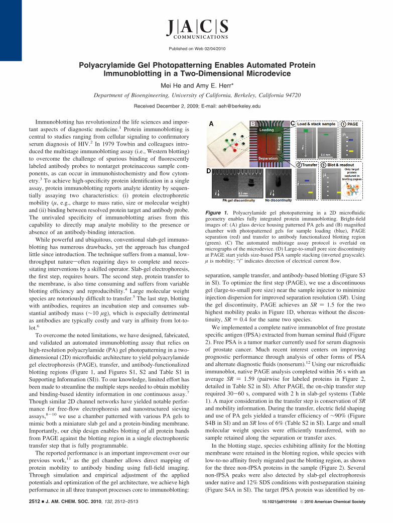
To overcome the noted limitations, we have designed, fabricated,and validated an automated immunoblotting assay that relies onhigh-resolution polyacrylamide (PA) gel photopatterning in a two-dimensional (2D) microfluidic architecture to yield polyacrylamidegel electrophoresis (PAGE), transfer, and antibody-functionalizedblotting regions (Figure 1, and Figures S1, S2 and Table S1 inSupporting Information (SI)). To our knowledge, limited effort hasbeen made to streamline the multiple steps needed to obtain mobilityand binding-based identity information in one continuous assay.7
Though similar 2D channel networks have yielded notable perfor-mance for free-flow electrophoresis and nanostructured sievingassays,8-10 we use a chamber patterned with various PA gels tomimic both a miniature slab gel and a protein-binding membrane.Importantly, our chip design enables blotting of all protein bandsfrom PAGE against the blotting region in a single electrophoretictransfer step that is fully programmable.
The reported performance is an important improvement over ourprevious work,11 as the gel chamber allows direct mapping ofprotein mobility to antibody binding using full-field imaging.Through simulation and empirical adjustment of the appliedpotentials and optimization of the gel architecture, we achieve highperformance in all three transport processes core to immunoblotting:
separation, sample transfer, and antibody-based blotting (Figure S3in SI). To optimize the first step (PAGE), we use a discontinuousgel (large-to-small pore size) near the sample injector to minimizeinjection dispersion for improved separation resolution (SR). Usingthe gel discontinuity, PAGE achieves an SR ) 1.5 for the twohighest mobility peaks in Figure 1D, whereas without the discon-tinuity, SR ) 0.4 for the same two species.
We implemented a complete native immunoblot of free prostatespecific antigen (fPSA) extracted from human seminal fluid (Figure2). Free PSA is a tumor marker currently used for serum diagnosisof prostate cancer. Much recent interest centers on improvingprognostic performance through analysis of other forms of PSAand alternate diagnostic fluids (nonserum).12 Using our microfluidicimmunoblot, native PAGE analysis completed within 36 s with anaverage SR ) 1.59 (pairwise for labeled proteins in Figure 2,detailed in Table S2 in SI). After PAGE, the on-chip transfer steprequired 30-60 s, compared with 2 h in slab-gel systems (Table1). A major consideration in the transfer step is conservation of SRand mobility information. During the transfer, electric field shapingand use of PA gels yielded a transfer efficiency of ∼90% (FigureS4B in SI) and an SR loss of 6% (Table S2 in SI). Large and smallmolecular weight species were efficiently transferred, with nosample retained along the separation or transfer axes.
In the blotting stage, species exhibiting affinity for the blottingmembrane were retained in the blotting region, while species withlow-to-no affinity freely migrated past the blotting region, as shownfor the three non-fPSA proteins in the sample (Figure 2). Severalnon-fPSA peaks were also detected by slab-gel electrophoresisunder native and 12% SDS conditions with postseparation staining(Figure S4A in SI). The target fPSA protein was identified by on-
Figure 1. Polyacrylamide gel photopatterning in a 2D microfluidicgeometry enables fully integrated protein immunoblotting. Bright-fieldimages of: (A) glass device housing patterned PA gels and (B) magnifiedchamber with photopatterned gels for sample loading (blue), PAGEseparation (red) and transfer to antibody functionalized blotting region(green). (C) The automated multistage assay protocol is overlaid onmicrographs of the microdevice. (D) Large-to-small pore size discontinuityat PAGE start yields size-based PSA sample stacking (inverted grayscale).µ is mobility; “i” indicates direction of electrical current flow.
Published on Web 02/04/2010
10.1021/ja910164d 2010 American Chemical Society2512 9 J. AM. CHEM. SOC. 2010, 132, 2512–2513
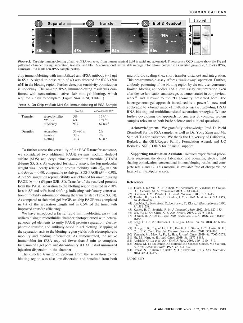
chip immunoblotting with immobilized anti-fPSA antibody (∼1 µg)in 65 s. A signal-to-noise ratio of 40 was detected for fPSA (500nM) in the blotting region. Further detection sensitivity optimizationis underway. The on-chip fPSA immunoblotting result was con-firmed with conventional native slab mini-gel blotting, whichrequired 2 days to complete (Figure S4A in SI, Table 1).
To further assess the versatility of the PAGE-transfer sequence,we considered two additional PAGE systems: sodium dodecylsulfate (SDS) and cetyl trimethylammonium bromide (CTAB)(Figure S5, SI). As expected for sizing assays, the log molecularweight was linearly related to protein mobility with RSDS
2 ) 0.96and RCTAB
2 ) 0.90, comparable to slab-gel SDS-PAGE (R2 ) 0.98).A ∼2.5% migration reproducibility was obtained for on-chip sizingPAGE (n ) 4) (Figure S5B, SI). Transfer of the resolved proteinsfrom the PAGE separation to the blotting region resulted in <10%loss in SR and <4% band shifting, indicating satisfactory conserva-tion of mobility information during the transfer step (Table S3, SI).As compared to slab mini-gel PAGE, on-chip PAGE was completedin 4% of the separation length and in 0.5% of the time, withimproved transfer efficiency.
We have introduced a facile, rapid immunoblotting assay thatutilizes a single microfluidic chamber photopatterned with hetero-geneous gel elements to unify PAGE protein separation, electro-phoretic transfer, and antibody-based in-gel blotting. Mapping ofthe separation axis to the blotting region yields both electrophoreticmobility and binding information. As demonstrated, the nativeimmunoblot for fPSA required fewer than 5 min to complete.Inclusion of a gel pore size discontinuity at PAGE start minimizedinjection dispersion in the chamber.
The directed transfer of proteins from the separation to theblotting region was also low-dispersion and benefited from both
microfluidic scaling (i.e., short transfer distance) and integration.This programmable assay affords ‘walk-away’ operation. Further,antibody-patterning of the blotting region by the end-user consumeslimited blotting antibodies and allows assay customization evenafter device fabrication and storage, as demonstrated in our previouswork11 and relevant to the 2D geometry presented here. Theheterogeneous gel approach introduced is a powerful new toolapplicable to a broad range of multistage assays, including DNA/RNA blotting and multidimensional separation strategies. We arefurther developing the approach for analysis of complex proteinsamples relevant to both basic science and clinical questions.
Acknowledgment. We gratefully acknowledge Prof. D. Peehl(Stanford) for the PSA sample, as well as Dr. Yong Zeng and Mr.Samuel Tia for assistance. We thank the University of California,Berkeley, the QB3/Rogers Family Foundation Award, and UCBerkeley NSF COINS for financial support.
Supporting Information Available: Detailed experimental proce-dures regarding the device fabrication and operation, electric fieldshaping optimization, conventional immunoblotting results, and com-plete refs 7 and 12. This material is available free of charge via theInternet at http://pubs.acs.org.
References
(1) Tissot, J. D.; Vu, D. H.; Aubert, V.; Schneider, P.; Vuadens, F.; Crettaz,D.; Duchosal, M. A. Proteomics 2002, 2, 813–824.
(2) Gershoni, J. M.; Palade, G. E. Anal. Biochem. 1983, 131, 1–15.(3) Towbin, H.; Staehelin, T.; Gordon, J. Proc. Natl. Acad. Sci. U.S.A. 1979,
76, 4350–4354.(4) Jungblut, P.; Eckerskorn, C.; Lottspeich, F.; Klose, J. Electrophoresis 1990,
11, 581–588.(5) Kurien, B. T.; Scofield, R. H. J. Immunol. Meth. 2002, 266, 127–133.(6) Wu, Y.; Li, Q.; Chen, X. Z. Nat. Protoc. 2007, 2, 3278–3284.(7) O’Neill, R. A.; et al. Proc. Natl. Acad. Sci. U.S.A. 2006, 103, 16153–
16158.(8) Zeng, Y.; He, M.; Harrison, D. J. Angew. Chem., Int. Ed. 2008, 47, 6388–
6391.(9) Huang, L. R.; Tegenfeldt, J. O.; Kraeft, J. J.; Sturm, J. C.; Austin, R. H.;
Cox, E. C. Tech. Dig. Int. Electron DeVices Meet. 2001, 363–366.(10) Yamada, M.; Mao, P.; Fu, J.; Han, J. Anal. Chem. 2009, 81, 7067–7074.(11) He, M.; Herr, A. E. Anal. Chem. 2009, 81, 8177–8184.(12) Andriole, G. L.; et al. New Engl. J. Med. 2009, 360, 1310–1319.(13) Ochoa, M. T.; Fholenhag, K.; Malmlof, K.; Sanchez-Gomez, M.; Skottner,
A. Arch. Latinoam. Nutr. 1997, 47, 331–337.(14) Cowan, S. L.; Diem, L.; Brake, M. C.; Crawford, J. T. J. Clin. Microbiol.
2004, 42, 474–477.
JA910164D
Figure 2. On-chip immunoblotting of native fPSA extracted from human seminal fluid is rapid and automated. Fluorescence CCD images show the PA gelpatterned chamber during: separation, transfer, and blot. A conventional native slab mini-gel blot allows comparison (inverted grayscale, * marks fPSA,numerals 1-3 mark non-fPSA sample peaks).
Table 1. On-Chip vs Slab Mini-Gel Immunoblotting of PSA Sample
on-chip conventional WB6
Transfer reproducibility 3% 15%13
SR loss 6% 15%14
efficiency 90% 67.8%4
Duration separation 30-60 s 2 htransfer 30 s 2 hblotting 30 s 6-8 h
J. AM. CHEM. SOC. 9 VOL. 132, NO. 8, 2010 2513
C O M M U N I C A T I O N S