Embed Size (px)
Citation preview

J. Cell Sci. 36, 1-18 (1979)Printed in Great Britain © Company of Biologists Limited J979
POLLEN-PISTIL INCOMPATIBILITY IN
PETUNIA HYBRID A: CHANGES IN THE PISTIL
FOLLOWING COMPATIBLE AND
INCOMPATIBLE INTRASPECIFIC CROSSES
M. HERRERO AND H. G. DICKINSONDepartment of Botany, Plant Science Laboratories, University of Reading,Whiteknights, Reading RG6 2AS, England
SUMMARY
The structural events in the stigma and transmitting tissue of Petunia hybrida pistils thataccompany compatible and incompatible intraspecific pollinations have been investigated indetail, together with the changes in reserve levels that also take place at this time. Many of thesephenomena may be explained in terms of 3 phases of secretion by the cells in the upper regionsof the transmitting tissue. The first, independent of pollination, results in the deposition of anintercellular matrix, rich in protein and carbohydrate. The second, triggered by pollination,although independent of the compatibility of the pollen grain, involves synthesis of moleculesbelieved to be specific to the S(incompatibility)-gene system. The third phase of secretionoccurs only following a compatible mating, and involves the transfer of stylar reserves tosupport the growth of the pollen tubes. These observations are discussed in terms of currentmodels of the incompatibility mechanism operating in Petunia.
INTRODUCTION
In plants with gametophytic control of pollen compatibility with respect to thestyle, much attention has been focussed upon the structure and physiology of thepistil. For example, the cytology of the stigma of Petunia sp. (Konar & Linskens,1966a) and that of the stylar transmitting tissue in a variety of species (van der Pluijm& Linskens, 1966; Sassen, 1974; Bell & Hicks, 1976; Cresti, Went, Pacini & Willimse,1976) has been the subject of recent intensive investigation. Likewise, the stigma andstyle of Lilium, a plant with a stylar canal rather than a transmitting tissue per se, havealso been described in detail (Rosen & Thomas, 1970). Physiological studies, however,have extended into the differences in stylar metabolism following compatible andincompatible crosses. Bredemeijer (1974) has demonstrated differences in the stylarperoxidase isozymes following such crosses in Nicotiana, while different levelsof RNA and protein synthesis have been described following crosses of differentcompatibility in Petunia by van der Donk (1974).
Structural aspects of these stylar differences have yet to be fully examined.Certainly, structural changes accompanying cross and self-pollination have beendescribed in Oenothera (Dickinson & Lawson, 1975I, but here few alterations appearto occur in the female tissue. In any event, this species possesses neither an organizedtransmitting tissue, nor a stylar canal, making localization of the site of the self-

2 M. Herrero and H. G. Dickinson
incompatibility reaction difficult. Likewise, although Lycopersicum has a compactstyle with a transmitting tissue, the many elegant ultrastructural investigations intothe differences between compatible and incompatible crosses (e.g. de Nettancourtet al. 1973) have not considered stylar changes in detail. Our knowledge of the reactionof compact styles to pollination is thus restricted to studies of the disruption of thetissue by pollen tube growth in Lychnis (Crang, 1966), and measurement of uptake ofmaterials from the style by pollen tubes in a number of species (Linskens & Esser,1959; Kroh, Miki-Hirosige, Rosen & Loewus, 1970; Kroh & Helsper, 1974). Stylarchanges may, however, be of considerable importance since little is known of thefactors that induce relatively slow growth of the incompatible tubes, when comparedwith the rate of compatible tube extension. While structural data may be few, modelsexplaining these events certainly are not. Kroes (1973), for example, suggests thatincompatible tubes lack an enzyme necessary to use the nutrients available in thestyle while, in an elegant hypothesis for Petunia, van der Donk (1975) proposes that,following a pollination-generated stimulus, synthesis of polypeptides takes place inthe style. It is the interaction between these 'pollination-polypeptides' (or a largermolecular assembly containing them) and the tube that results in the 'compatible' or'incompatible' style of growth.
In an attempt to align some of these hypotheses with structural events, we describehere the changes in the stigma and style accompanying compatible and incompatibleintraspecific pollination in Petunia.
MATERIALS AND METHODS
Plant material
Seedlings homozygous with respect to the incompatibility genes were raised by means ofbud pollination of clones Ka3 and T211 (genotypes 22 and 33 respectively), the seeds of whichwere originally kindly supplied by Professor H. F. Linskens. Following growth and floweringin the greenhouse, individual flowers were detached for the pollination studies. These experi-ments were carried out in a well-lit air-conditioned chamber, maintained at a constant tempera-ture of 25 °C.
Studies of pollen tube growth
Pollen tube growth was measured using material stained with aniline blue examined under aLeitz epi-UV irradiance system fitted to a Leitz Dialux microscope. Styles were preparedfollowing a modifications of Linskens & Esser's (1957) technique. Material was first softenedfor 1 min at 100 °C in a 5 % aqueous solution of sodium sulphite, and then stained in a O'l %solution of aniline blue in 01 N K3PO4, for 5 min. The tissues were examined mounted in thestain.
Determination of starch levels
With the aid of a sharp scalpel short sections were excised from different regions of the styleand, using light pressure, the transmitting tissue contained in the section extruded onto aclean slide. Difficulties were encountered in extracting the very top of the transmitting tissue,that which interfaces with the stigma. However, by shaving off the stigmatic papillae with amicroscalpel it was possible to expose, and subsequently express, the top layer of the trans-mitting tissue.
Starch in these cells was then stained with a saturated solution of iodine in potassium iodide

Self-incompatibility in Petunia 3
for 5 min at room temperature. Following rinsing in 0-3 M Sorensen phosphate buffer, thetransmitting tissue was mounted in glycerol, squashed carefully, and the preparation sealedwith rubber solution. The intensity of the stain was measured using a Vickers M85 scanningmicrodensitometer, operating at a wavelength of 500 ran. The method of preparation produceda monolayer of stained cells, and readings were taken of a constant area of this monolayer,limited by the field of the densitometer 'mask'. Each field contained between 4 and 6 cells. Itmust be emphasized that the readings resulting from this work can in no way be linked stoichio-metrically with the starch in the tissue, but they do provide a useful indication of the levels ofthis reserve. In some of the results it was considered helpful to express the data as 'amountsper flower'. To obtain this measure, the average was taken of the readings from each level inthe style.
Preparation for light and electron microscopy
For enzyme cytochemistry, fresh material was sectioned at 5 /im in a Slee cryostat andstained to reveal the location of acid phosphatase (Knox & Heslop-Harrison, 1970), esterase(Pearse, i960) and peroxidase (Jensen, 1962).
Tissue for electron microscopy was prepared according to Dickinson & Lawson (1975). Toidentify protein in this material, sections were digested with protease (Dickinson & Potter,1975). Thick sections (1-5 fim) of these Epon blocks were also cut for light microscopy. Car-bohydrate was localized in these sections using treatment with periodic acid and Schiff's reagent(Feder & O'Brien, 1968).
RESULTS
The stigmaAt anthesis, degeneration of the stigmatic tissue takes place completely indepen-
dently of pollination and indeed, the compatibility of that pollination. Prior to thisevent, electron micrographs reveal the papillae not only to contain reserves of starchand saturated lipid, but also plastids (see Figs. 1, 2), dictyosomes, associated vesicles,microbodies and mitochondria. As degeneration of these commences, large dropletsof lipid become evident inside the cytoplasm of a proportion of the cells (see Fig. 3).The plasma membranes of these cells then rupture, releasing the lipid to coalescewith other extracellular droplets (see Figs. 4, 5), and the remaining cytoplasmiccontent to form small islets of degenerate cytoplasm that float in the lipid (see Fig. 3).Cytochemical tests, however, show the stigmatic extract to contain sugars and also acidphosphatase. This enzyme is not distributed evenly, but apparently contained in drop-lets in the stigmatic fluid (see Fig. 6). No reaction was observed with the cytochemicaltests for either peroxidase or esterase.
It is into this lipid-rich fluid that the pollen grain alights. Within 30 min it hasgerminated, and the pollen tube begun to grow between the papillae (see Figs. 7, 8)into the stigma.
The cells of the transmitting tissueThe cells of this region present the features of normal somatic meristematic plant
cells, albeit with rather thick walls, some of which may measure up to 0-5 /tm in width.Apart from the conventional cell contents, the cytoplasm of these cells possess reservesof lipid and starch, considerable numbers of dictyosomes, mitochondria, and a largeendoplasmic reticulum (see Fig. 9). Frequently, these cells contain microbodies. Close

M. Herrero and H. G. Dickinson
Self-incompatibility in Petunia 5
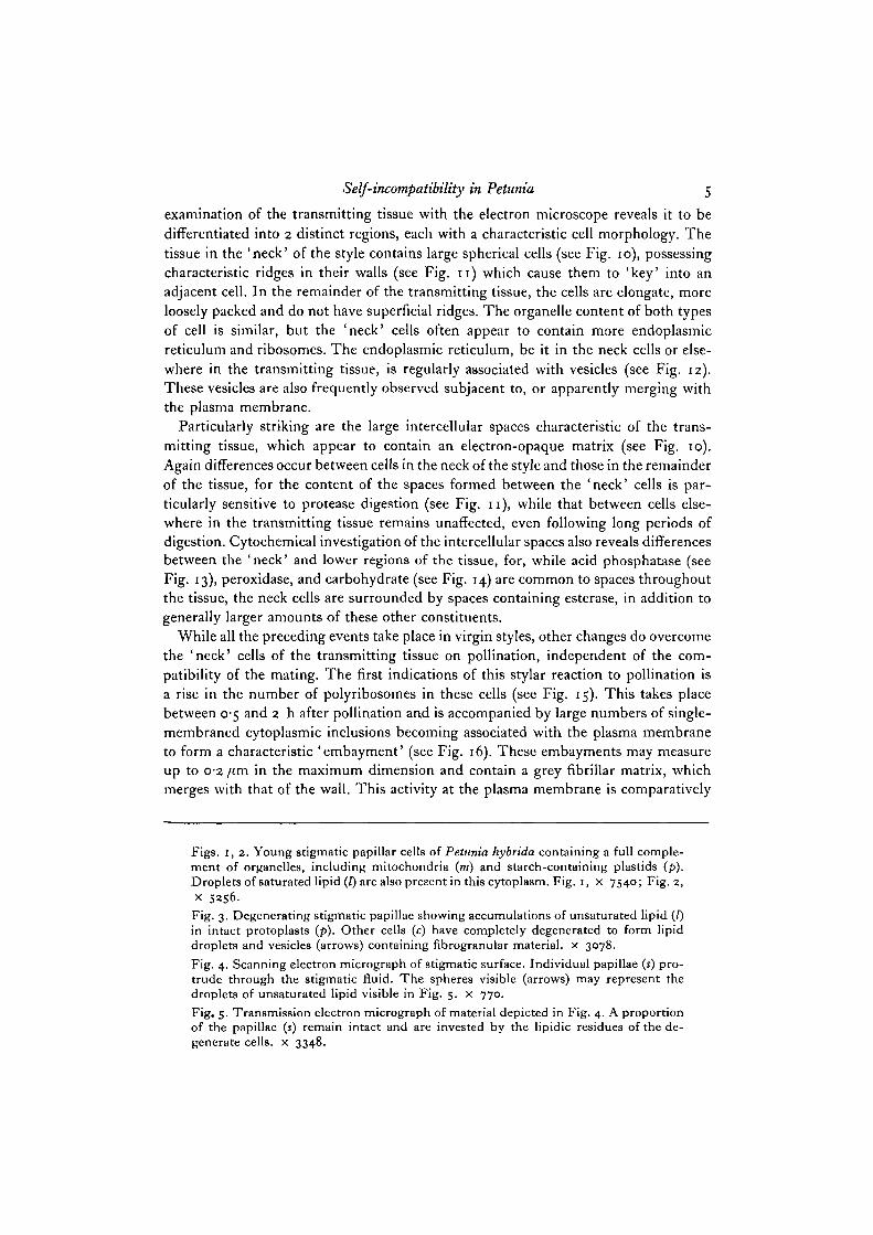
examination of the transmitting tissue with the electron microscope reveals it to bedifferentiated into 2 distinct regions, each with a characteristic cell morphology. Thetissue in the 'neck' of the style contains large spherical cells (see Fig. 10), possessingcharacteristic ridges in their walls (see Fig. 11) which cause them to 'key' into anadjacent cell. In the remainder of the transmitting tissue, the cells are elongate, moreloosely packed and do not have superficial ridges. The organelle content of both typesof cell is similar, but the 'neck' cells often appear to contain more endoplasmicreticulum and ribosomes. The endoplasmic reticulum, be it in the neck cells or else-where in the transmitting tissue, is regularly associated with vesicles (see Fig. 12).These vesicles are also frequently observed subjacent to, or apparently merging withthe plasma membrane.
Particularly striking are the large intercellular spaces characteristic of the trans-mitting tissue, which appear to contain an electron-opaque matrix (see Fig. 10).Again differences occur between cells in the neck of the style and those in the remainderof the tissue, for the content of the spaces formed between the 'neck' cells is par-ticularly sensitive to protease digestion (see Fig. n ) , while that between cells else-where in the transmitting tissue remains unaffected, even following long periods ofdigestion. Cytochemical investigation of the intercellular spaces also reveals differencesbetween the 'neck' and lower regions of the tissue, for, while acid phosphatase (seeFig. 13), peroxidase, and carbohydrate (see Fig. 14) are common to spaces throughoutthe tissue, the neck cells are surrounded by spaces containing esterase, in addition togenerally larger amounts of these other constituents.
While all the preceding events take place in virgin styles, other changes do overcomethe 'neck' cells of the transmitting tissue on pollination, independent of the com-patibility of the mating. The first indications of this stylar reaction to pollination isa rise in the number of polyribosomes in these cells (see Fig. 15). This takes placebetween 0-5 and 2 h after pollination and is accompanied by large numbers of single-membraned cytoplasmic inclusions becoming associated with the plasma membraneto form a characteristic 'embayment' (see Fig. 16). These embayments may measureup to 0-2 /im in the maximum dimension and contain a grey fibrillar matrix, whichmerges with that of the wall. This activity at the plasma membrane is comparatively
Figs. 1, 2. Young 8tigmatic papillar cells of Petunia hybrida containing a full comple-ment of organelles, including mitochondria (»/) and starch-containing plastids (p).Droplets of saturated lipid (i) are also present in this cytoplasm. Fig. i, x 7540; Fig. 2,x 5256.Fig. 3. Degenerating stigmatic papillae showing accumulations of unsaturated lipid (/)in intact protoplasts (p). Other cells (c) have completely degenerated to form lipiddroplets and vesicles (arrows) containing fibrogranular material, x 3078.Fig. 4. Scanning electron micrograph of stigmatic surface. Individual papillae (s) pro-trude through the stigmatic fluid. The spheres visible (arrows) may represent thedroplets of unsaturated lipid visible in Fig. 5. x 770.Fig. 5. Transmission electron micrograph of material depicted in Fig. 4. A proportionof the papillae (s) remain intact and are invested by the lipidic residues of the de-generate cells, x 3348.

M. Herrero and H. G. Dickinson

Self-incompatibility in Petunia y
short-lived and, within about 4 h, this membrane has returned to its originalaspect.
All these events occur before the arrival of the pollen tubes, which are at this timegrowing at a rate of about 150/tm/h through stigmatic tissue. Close to these extendingpollen tubes many of the 'neck' cells of the transmitting tissue appear necrotic,containing dark granular cytoplasm, large numbers of vesicles, and disorganizedplastids (see Fig. 17). In addition, cytoplasmic fragments may frequently be observedin the intercellular spaces dividing these cells (see Fig. 18). These necrotic cells, iftested some 24 h after pollination, react most strikingly with protease, appearing tobe almost totally sensitive to the enzyme (see Fig. 19).
The stylar cells following self- and cross-pollinations
Although the cells of all regions of the transmitting tissue appear to react in-dependently of the compatibility of the pollen tube approaching them, their behaviourfollowing its passage differs considerably according to the nature of the mating.
Following a compatible cross, for example, cells are characterized by large vacuolesinvested by a thin peripheral layer of cytoplasm. This cytoplasm is no longer rich inreserves, but instead contains a nucleus, few mitochondria and scattered dictyosomes(see Figs. 20, 21). The transmitting tissue cells of self-pollinated flowers, on the otherhand, resemble those of unpollinated flowers. Little depletion of reserves is indicatedby the micrographs (see Fig. 22), and the cytoplasm: vacuole ratio remains largelyunchanged.
Starch metabolism during pollination
While the changes above may be easily observed qualitatively, a quantitative measureof the events is not so simply obtained. Nevertheless, microdensitometry of preparationsin which the starch has been stained (see Fig. 23) provides a quantitative representationof levels of this reserve.
From such results, it is clear that in unpollinated flowers no change occurs in the
Fig. 6. Light-microscopic preparation treated to reveal acid phosphatase. The enzymeis localized in small droplets which occur both on the papillar surfaces (s) and in thelarge spaces between the loosely packed cells of the stigma, x 1176.Fig. 7. Scanning electron micrograph of a pollen grain germinating on the stigma.The pollen tube (t) has penetrated the stigmatic surface (arrow). A papilla (s) is alsovisible, as is a tube (ij) from another grain, x 1534.Fig. 8. Transmission electron micrograph of material shown in Fig. 7. The pollentube (t) is growing down between stigmatic papillae (s) invested by droplets (arrows)of the lipidic remains of degenerated cells, x 6016.Fig. 9. Transmitting tissue cell of Petunia showing a starch-containing plastid (/>),mitochondria (m), a dictyosome (d) and elements of the endoplasmic reticulum(arrows), x 18165.Fig. 10. The ' neck' region of the transmitting tissue. The cells of this region are spheri-cal and possess unusual 'key' junctions (arrows) with their neighbours (shown betterin Fig. 11). Note also the electron-opaque intercellular spaces, x i960.

M. Herrero and H. G. Dickinson

Self-incompatibility in Petunia 9
starch content of the transmitting tissue until the final degeneration of the style. Aftera cross-pollination, however, a massive decrease in starch levels takes place, while,following a self-mating, there is only a slight depletion of the reserve (see Table 1).
In order to investigate a possible relationship between the passage of the pollentubes and depletion of the starch, the number of pollen tubes at different levels in thestyles was expressed as a percentage of the number of pollen grains present, and thispercentage considered in terms of the starch levels in the entire pistil. When theseresults are plotted (see Fig. 24) a clear relationship may be detected in compatiblecrosses, the starch of the stylar cells decreasing with the passage of the pollen tubesthrough the adjacent intercellular spaces. Following incompatible crosses, this inverserelationship is not nearly as marked.
Table 1. Levels of IKI-stainable material, expressed as arbitrarymicrodensitometer units per flower. {The standard errors are shown in parentheses.)
Unpollinated Self-pollinated Cross-pollinated
24 h 20-7910-38 180110-83 I I -6I±O-O8
48 h 20-9711-31 I 6 - 5 7 ± I - 6 6 1-8610-46
This difference in starch metabolism between tubes of differing compatibility isparticularly well underlined by a series of readings taken at the neck of the trans-mitting tissue, subjacent to the stigma, when pollen tube distribution is almostidentical (see Fig. 25 A, E). Starch levels in the selfed styles are far in excess of thosefound following cross-pollination.
In addition to these effects of pollination, measurement of starch content at 4 levelsat 24 and 48 h after pollination indicates that in styles pollinated with grains of
Fig. 11. Protease-digested material similar to that shown in Fig. 10. The loss of electronopacity of the intercellular spaces is striking. The 'key' junctions between cells(arrows) are also conspicuous in this micrograph, x 4504.Fig. 12. Portion of cell in ' neck' region of the transmitting tissue. Elements of the endo-plasmic reticulum are evident («) as are associated vesicles (arrow), x 25500.Fig. 13. Light-microscopic preparation of 'neck' region of transmitting tissue, treatedto reveal acid phosphatase. Note the presence of this enzyme in the intercellularspaces, x 1178.Fig. 14. As Fig. 13, but material reacted to show the location of carbohydrate. Starch(arrows) is visible within the cells, but the intercellular matrix is also PAS-positive.x 761.Fig. 15. Cells of the 'neck' region of the transmitting tissue 2 h after pollination.Large numbers of polyribosomes (arrows) are present and the embayments (e) arealso visible, x 27675.Fig. 16. As Fig. 15. The embayments (e) of the plasma membrane are particularlystriking, as is their fibrillar content. X 14287.Fig. 17. Pollen tube (i) growing through the 'neck' region of the transmitting tissue.Near the tube some of the cells («) appear disorganized and necrotic, while others (c)display a normal aspect, x 5771.

M. Herrero and H. G. Dickinson

Self-incompatibility in Petunia i1
either compatability, a detectable 'wave' of starch synthesis precedes the tube tip inits passage through the transmitting tissue (see Fig. 25 c, D).
DISCUSSION
The maturing stigma and first events after pollination
The final stages in stigmatic maturation which appear to involve the 'degeneration'of the papillae (Dumas, 1975) are clearly independent of pollination and, indeed, thecompatibility of pollination. Konar & Linskens (19666) have proposed that the stigmaacts purely as a location for the germination of pollen and is not involved in itsnutrition per se, and there is little from the present investigation to indicate otherwise.
However, while the pollen grains are germinating in this stigmatic exudate, strikingchanges appear to be induced in the subjacent regions of the transmitting tissue.These start with a marked increase in polyribosome number in the cells, and continuewith the formation of the characteristic 'embayments'. While conclusive evidence is,of course, unavailable, these profiles do indicate a transfer of cytoplasmically syn-thesized material into the carbohydrate and pectin rich intercellular spaces (Kroh,1973; Kroh & van Bakel, 1973; Sassen, 1974). The nature of the stimulus that travelsto these cells of the stylar neck and triggers these changes is not clear. Linskens &Spanjers (1973) have recorded a difference in the electrical resistance of the pistilafter pollination, but it remains equally possible that these changes are induced by aflow of chemical messenger. These events must also be accompanied by biochemicalchanges, such as the activation of the glutamic dehydrogenase described by Roggen
(1967).Biochemical studies (van der Donk, 1975) have indicated pollination to stimulate
the production of 'recognition' polypeptides, specific to the incompatibility (S)genotype of the plant. It is striking that this aggregation of ribosomes into polysomesand the apparent secretion of materials by the cells in the neck of the transmitting
Fig. 18. 'Neck' region of transmitting tissue during passage of pollen tubes. Intactcells (c) are present, but also visible is a degenerate protoplast (n) and cytoplasmicfragments (/). x 5769.Fig. 19. Degenerate protoplast (n) as shown in Fig. 18, following digestion withprotease. This cell is almost totally sensitive to the enzyme, whereas intact cells (c)are not. x 7185.Fig. 20. Tangential section of a transmitting tissue cell following the passage ofcompatible pollen tubes. Note the absence of reserves from this protoplast, x 9585.Fig. 21. As Fig. 20, but median section revealing the large vacuoles (v) of these cells,x 6140.Fig. 22. Transmitting tissue cell following the passage of incompatible pollen tubes.Note the presence of starch (si) in the plastids and droplets of lipid ([) free in thecytoplasm, x 9060.Fig. 23. Light-microscopic preparation of lower region of the transmitting tissuetreated with PAS to reveal the presence of starch. Although the cell walls react andthe intercellular regions are slightly sensitive, the main staining is in the starch grainsof the stylar cells, x 934.

12 M. Herrero and H. G. Dickinson
100
2 0 -
Stigma
Style
100 -
8 0 -
r 60-
: 4 0 -
2 0 -
- 25
- 2 0
_cu-15 3
- 10 j j
- 5
Stigma 0-25 0-5 0-75
Style
Fig. 24. The number of pollen tubes and levels of starch at different points in thestyle 24 h after pollination following compatible (A), and incompatible (B) pollinations.The pollen tube number (•) is expressed as a percentage of the grains on the stigmasurface. The levels of starch ( • ) are in arbitrary microdensitometer units.

Self-incompatibility in Petunia
171]
Fig. 25. Levels of IKI-stained starch, expressed in arbitrary microdensitometerunits, in unpollinated (D), selfed (^), and crossed (3§) flowers at 4 levels in thepistil, 24 (A,B,C,D) and 48 (E,F,G,H) h after pollination. The pollen tube numbers aregiven in brackets under the lower axes. The regions of the pistil from which measure-ments were taken are as follows: A, E, stigma; B, F, top quarter of style; c, G,second quarter; and D, H, third quarter.
CEL 36

14 M. Herrero and H. G. Dickinson
tissue is so closely synchronized with the proposed synthesis of the s-specific poly-peptides, all these events occurring in the style ahead of the advancing pollen tubes.
Once the pollen tubes reach the transmitting tissue, there is little doubt that somecellular degeneration takes place. This has been reported in Gossypium (Jensen &Fisher, 1969) and would appear to be a general feature of pollen tube growth, irres-pective of its compatibility. Although results have yet to be analysed statistically,more transmitting tissue cells appear to degenerate in the vicinity of the growingpollen tubes than elsewhere in the tissue. The fate of the cytoplasm released into theintercellular space by the rupture of these protoplasts is not yet clear, but it may oftenbe seen to invest the pollen tubes.
The metabolism of starch over the course of pollination
There is little doubt that a slight increase in stylar starch synthesis is stimulated bypollination itself. As with the previous cytoplasmic effects, it is not yet clear how thestimulus for this process is transmitted from the stigma. Other changes in the car-bohydrate metabolism of the pistil also occur at this point, for Tupy (1961), workingon Nicotiana, has reported an increase in glucose and fructose levels at the ovary 1 dayafter pollination, combined with a decline in the stigmatic sugars.
The pronounced drop in starch levels accompanying the growth of compatible pollentubes is not unexpected. Starch is clearly an energy reserve, and if the tube is growingheterotrophically, depletion of such reserves should certainly occur. These observationsare consistent with those of Linskens (1955) who reported free sugars to decrease tobelow half their pre-pollination levels following the passage of the pollen tubes, andof Roggen (1967) who demonstrated induction of enzymes for carbohydrate metabolismin the vicinity of the growing pollen tubes. The uses to which these extracellularcarbohydrates may be put are many. O'Kelley (1955) describes their utilization inrespiration, while Kessler, Feingold & Hassid (i960) have, in studies in vitro, describedtheir employment in the synthesis of sucrose, callose and starch. In an investigationinto the fate of the intracellular pectic substances, Kroh et al. (1970) and Kroh &Helsper (1974), reported their incorporation into the pollen tube wall.
The lack of starch mobilization following the passage of incompatible tubes is notso easily understood. This does not simply result from fewer tubes generally growingin such circumstances, for areas at the top of the transmitting tissue containingequivalent numbers of pollen tubes exhibit differences in starch metabolism depen-dent upon the compatibility of the cross. Again, biochemical investigations tend tosupport these observations; Linskens (1953, 1955) has reported a decrease in res-piratory rate of pistils 12 h following self-pollination, and also differences betweenselfed and crossed styles in their endogenous levels of 'glucan-hydrolases' (Linskenset al. 1969). Taken in toto this evidence indicates that, in incompatible tubes, majorpathways of carbohydrate metabolism are either blocked, or alternatively not activated.

Self-incompatibility in Petunia 15
The structural differences in the transmitting tissue following compatible or incompatiblepollinations
Since the difference in pollen tube number normally encountered between com-patible and incompatible crosses might cause effects that would be incorrectlyinterpreted, results are only discussed from the upper regions of the transmittingtissue where numbers of pollen tubes are equal irrespective of their compatibilityand true stylar changes are thus most conspicuous.
The very different aspects displayed by transmitting tissue permeated by compatibletubes, and that containing incompatible tubes may almost be fully explained in termsof the utilization of reserves. The tissue with incompatible tubes looks very similarto that of pollinated flowers prior to the passage of the pollen tubes, while that incontact with compatible tubes is deficient in reserves in the form of starch and lipid,and contains far fewer microbodies.
Such a 'degeneration' of stylar tissue following the passage of compatible pollentubes was reported by Crang (1966) to occur in Lychnis. In another species, Lilium,Crang (1969) found degeneration to occur in the parenchymatous cells investing thestylar canal cells, although these latter cells seemed not to be affected. While thiswork did not indicate clearly the cause of this degeneration, Crang (1969) proposedit to result from either enzymic secretion by the pollen tubes, or the stimulation bythe pollen tube of 'autolytic bodies' in the stylar cells.
Although Rosen & Thomas (1970) confirmed that no ultrastructural changes over-came the stylar canal cells of Lilium on pollination, they reported an increase insecretory activity by these cells. Likewise, Yamada (1956) described the loss of cyto-plasmic organization by the parenchymatous cells after pollination in Lilium, andpointed to the coincident arrival of a mucilagenous substance on the surface of thecanal cells. The fact that starch and lipid were the first components of the cytoplasmto disappear from the cells of Lychnis and Lilium led Crang (1969) to propose thatthese reserves were utilized in the nutrition of the pollen tubes. In Gossypium, on theother hand, the position appears to differ, for Jensen & Fisher (1969) report themaintenance of stylar starch and lipid levels over the course of pollination. Resultsfrom the present investigation, however, concur well with those of Crang (1969),Yamada (1965), Rosen & Thomas (1970) and indicate that compatible pollen tubesstimulate the mobilization of stylar reserves, and subsequently utilize these productsin the course of their metabolism.
Whilst this difference in metabolism of reserves between compatible and incom-patible tubes is doubtless of significance, it is perhaps the events immediately priorto, and following pollination that are possibly most important to our understandingof the incompatibility system in Petunia. In an autoradiographic investigation,Labarca & Loewus (1973) reported that secretion by cells of the transmitting tissueinto the intercellular space was independent of pollination, results supported by theelectron-microscopic work of Sassen (1974) which indicated a release of intercellularsubstances throughout maturation of the style, reaching a maximum prior to anthesis.In a more recent investigation Cresti et al. (1976) have examined the formation of

16 M. Herrero and H. G. Dickinson
the intercellular matrix in Lycopersicum, and report that first secreted are pecticsubstances followed, after conspicuous activity of the endoplasmic reticulum andassociated polyribosomes, by the formation of a small amount of protein.
While our results agree with this in part, there is little doubt that there are at least3 phases of synthesis, one massive and prior to pollination, one stimulated by pollina-tion and involving the 'embayments', and a final phase of secretion when mobilizedstylar reserves are transferred to the pollen tube. The second phase occurs prior tothe passage of the pollen tubes and is independent of compatibility, while the thirdoccurs during pollen tube growth, and depends upon compatibility. These events mayclearly be explained in terms of the model of van der Donk (1975) in which the S geneacts as a ' master-gene', switching on a battery of stylar genes that result in the supportof pollen tube growth. In this connexion, it is noteworthy that little starch and lipidappears to be mobilized in transmitting tissue cells on self-pollination.
Alternatively, the differences observed between compatible and incompatible tubesmay result from heterotrophic and autotrophic styles of growth. This concept is notnew, for Rosen & Gawlick (1966) suggested, from work on Lilium, that changes theyobserved in incompatible tubes could be explained in terms of an inability to switchfrom an autotrophic to heterotrophic form of growth. Although this proposal hasreceived much consideration in the literature (Kroes, 1973; Vasil, 1974), it has littlesupport from the autoradiographic work of Kroh et al. (1970), where incompatibletubes were shown to take up more labelled precursor from the style than compatibletubes. This work, though far from conclusive, and the results of the study presentedhere would perhaps indicate that a metabolic deficiency lies within the incompatibletube itself.
Whatever model of pollen tube metabolism is finally adopted, the fact remains thatincompatible pollen tubes do germinate, stimulate stylar metabolism and must utilizea portion, albeit very small, of the stylar reserves. At first sight this might appear toto be an inefficient use of stylar reserves, but under poor cross-pollination conditionssuch a mechanism enables self-pollination to ' prime' the style for the growth of thefew cross-pollen tubes available.
The precise point of recognition presumably occurs when the pollen tube makescontact with S-specific polypeptides, or a larger molecular assembly containing them.The nature of these molecules and, indeed, their receptors in the pollen tube are notknown. It is probably in species in which the contents of the stylar canal may beextracted without physiological damage to the surrounding cells, that the search forthese recognition molecules may most profitably be carried out.
Thanks are due to the ARC and OECD for financial support, to the Royal Society forthe provision of photomicrographic equipment, and to Ursula Potter for help with theillustrations.
REFERENCESBELL, J. & HICKS, G. (1976). Transmitting tissue in the pistil of Tobacco. Light and electron
microscopic observations. Planta 131, 187-200.

Self-incompatibility in Petunia 17
BREDEMEIJER, G. M. M. (1974). Peroxidase activity and peroxidase isozyme composition inself-pollinated, cross-pollinated and unpollinated styles of Nicotiana alata. Acta bot. neerl.23 (2). 149-157-
CRANG, R. E. (1966). A fine structural study of in vivo pollen tube penetration in Lychnis alba.Trans. Am. microsc. Soc. 85 (4), 564-570.
CRANG, R. E. (1969). Pollination effects on style degradation in Lilium philippinense. Trans.Am. microsc. Soc. 88 (2), 294-299.
CRESTI, M., WENT, J. L. VAN, PACLNI, E. & WILLEMSE, M. T. M. (1976). Ultrastructure oftransmitting tissue of Lycopersicum peruvianum. Style development and histochemistry.Planla 132, 305-312.
DICKINSON, H. G. & LAWSON, J. (1975). Pollen tube growth in the stigma of Oenothera organensisfollowing compatible and incompatible intraspecific pollinations. Proc. R. Soc. B 188, 327-^^.
DICKINSON, H. G. & POTTER, U. (1975). Post-meiotic nucleocytoplasmic interaction in Pinusbanksiana: The secretion of RNA by the nucleus. Planta 122, 99-104.
DONK, J. A. W. M. VAN DER (1974). Gene activity and the incompatibility reaction in Petunia.In Fertilisation in Higher Plants (ed. H. F. Linskens), pp. 279-283. Amsterdam: North-Holland.
DONK, J. A. W. M. VAN DER (1975). Recognition and gene expression during the incompatibilityreaction in Petunia hybrida L. Molec. gen. Genet. 141, 305-317.
DUMAS, C. (1975). Le Stigmate et la Sicrition stigmatique (Etude histophysiologique, cytologique etbiochemiqiie de VActiviti glandulaire lipophile.) Thesis, University Claude-Bernard Lyon,France.
FEDER, N. & O'BRIEN, T. P. (1968). Plant microtechnique some principles and new methods.Am.y. Bot. ss, 123-142.
JENSEN, W. A. (1962). In Botanical Histochemistry, p. 349. San Francisco and London: Freeman.JENSEN, W. H. & FISHER, D. B. (1969). Cotton embryogenesis: The tissue of the stigma and
style and their relation to the pollen tube. Planta 84, 97-121.KESSLER, G., FEINGOLD, D. S. & HASSID, W. Z. (i960). Utilisation of exogenous sugar for
biosynthesis of carbohydrates in germinating pollen. PI. Physiol., Lancaster 35, 505-509.KNOX, R. B. & HESLOP-HARRISON, J. (1970). Pollen wall proteins: Localization and enzymic
activity. J. Cell Sci. 6, 1-27.KONAR, R. N. & LINSKENS, H. F. (1966a). The morphology and anatomy of the stigma of
Petunia hybrida. Planta 71, 356-371.KONAR, R. N. & LINSKENS, H. F. (1966Z)). Physiology and biochemistry of the stigma fluid of
Petunia hybrida. Planta 71, 372-387.KROES, H. W. (1973). An enzyme theory of self-incompatibility. Incompatibility Newslett. 2,
5-i4-KROH, M. (1973). Nature of the intercellular substances of stylar transmitting tissue. In
Biogenesis of Plant Cell Wall polysaccharides (ed. F. Loewus), pp. 195-205. New York:Academic Press.
KROH, M. & BAKEL, C. H. J. VAN (1973). Incorporation of label into the intercellular substanceof stylar transmitting tissue from Petunia pistil labelled with tritiated myo-inositol. Anelectromicroscopic autoradiographic study. Acta bot. neerl. 22 (2), 106—111.
KROH, M. & HELSPER, J. P. F. G. (1974). Transmitting tissue and pollen tube growth. InFertilization in Higher Plants (ed. H. F. Linskens), pp. 167-179. Amsterdam: North-Holland.
KROH, N., MIKI-HIROSIGE, H., ROSEN, W. & LOEWUS, F. (1970). Incorporation of label intopollen tube walls from myo-inositol labelled Lilium longiflorum pistils. PI. Physiol.,Lancaster45, 92-94-
LABARCA, C. & LOEWUS, F. (1973). The nutritional role of pistil exudate in pollen tube wallsformation in Lilium longiflorum. II. Production and utilization of exudate from stigma andstigma canal. PL Physiol., Lancaster 52, 87-92.
LINSKENS, H. F. (1953). Physiologische und chemische Unterschiede zwischen selbst- undfremdbestaubten Petunien-Griffeln. Naturwissen schaften 40, 28-29.
LINSKENS, H. F. (1955). Physiologische Untersuchungen der Pollenschauch-Hemmung selbst-steriler Petunien. Z. Bot. 43, 1-44.
LINSKENS, H. F. & ESSER, K. (1957). Ueber eine specifische Anafarbung der Pollenschlaucheund die Zahl kallose-propten nach Selbstung und Fremdung. Natunvissenshaften 44, 16.
18 M. Herrero and H. G. Dickinson
LISKENS, H. F. & ESSER, K. (1959). Stoffaufnahme der Pollenschliiuche aus dem Leitgewebedes Griffels. Proc. K. ned. Akad. Wet. C62, 150-154.
LINSKENS, H. F., HAVEZ, R., LINDEH, R., SALDEN, M., RANDOUX, A., LANIEZ, D. & CUSTAUT, D.(1969). Etude des glycanne-hydrolases en cours de la croissance du pollen chez Petuniahybrida auto-incompatible. C.r. hebd. Stone. Acad. Set., Paris B 269, 1855-1857.
LINSKENS, H. F. & SPANJERS, A. W. (1973). Changes of the electrical potential in the transmittingtissue of Petunia styles after cross- and self-pollination. Incompatibility Ncwslett. 3, 81-85.
NETTANCOURT, D. DE, DEVREUX, M., BOZZINI, A., CRESTI, M., PACINI, E. & SARFATTI, G. (1973).Ultrastructural aspects of the self-incompatibility mechanism in Lycopersicum peruvianumMill. J. Cell Set. 12, 403-419.
O'KELLEY, J. C. (1955). External carbohydrates in growth and respiration of pollen tubes 'invitro'. Am.jf. Bot. 42, 322-327.
PEARSE, A. G. E. (i960). In Histochemistry, Theoretical and Applied, 2nd ed., p. 462. London:Little, Brown, Boston and Churchill.
PLUIJM, J. VAN DER & LINSKENS, H. F. (1966). Feinsrruktur der Pollenschauche im Griffel vonPetunia. Zuchter 36, 220-224.
ROGGEN, H. P. J. R. (1967). Changes in enzyme activities during the progame phase in Petuniahybrida. Acta. bot. neerl. 16, 1-31.
ROSEN, W. G. & GAWLICK, S. R. (1966). Relation of lily pollen tube structure to pistil in-compatibility and mode of nutrition. In Electron Microscopy (Proc. VI int. Congr. ElectronMicrosc., Kyoto), pp. 313-314. Tokyo: Mazuren.
ROSEN, W. G. & THOMAS, H. R. (1970). Secretory cells of lily pistils. 1. Fine structure andfunction. Am. J. Bot. 57 (9), 1108-1114.
SASSEN, M. M. A. (1974). The stylar transmitting tissue. Acta bot. neerl. 23, 99-108.TUPY, J. (1961). Changes in glucose and fructose level in Nicotiana alata styles and ovaries
accompanying compatible pollen tube growth. Biologia PL 3, 1-14.VASIL, I. K. (1974). The histology and physiology of pollen germination and pollen tube growth
on the stigma and in the style. In Fertilisation in Higher Plants (ed. H. F. Linskens), pp. 105-118. Amsterdam: North-Holland.
YAMADA, Y. (1965). Studies on the histological and cytological changes in the tissues of thepistil after pollination. Jap. Jour. Bot. 19 (3), 69-82.
{Received 27 July 1978)