Embed Size (px)
Citation preview

Department of Microbiology
Quality Manual
Policy # MI_TECH
Page 1 of 154
Version: 1.3 CURRENT
Section: Bacteriology Procedures Subject Title: Technical Procedure Manual
Prepared by QA Committee
Issued by: Laboratory Manager Revision Date: 11/29/2019
Approved by Laboratory Director:
Microbiologist-in-Chief
Next Review Date: 5/1/2020
Uncontrolled When Printed
UNIVERSITY HEALTH NETWORK/MOUNT SINAI HOSPITAL, DEPARTMENT OF MICROBIOLOGY
NOTE: This document is Uncontrolled When Printed.
Any documents appearing in paper form that do not state "CONTROLLED COPY " in red print are not controlled and should be checked against the document (titled as above) on the server prior to use.
Management System\UHN_Mount Sinai Hospital Microbiology\Standard Operating Procedures\Bacteriology Procedures\
TABLE OF CONTENTS
ALA (RAPID PORPHYRIN TEST) ....................................................................................................... 4
ANAEROBIC/CAMPYLOBACTER JAR SET UP ............................................................................... 6
Anaerobic Jar ....................................................................................................................................... 6 Campylobacter Jar ............................................................................................................................... 7
API Test Strips ......................................................................................................................................... 8
IDENTIFICATION OF CORYNEBACTERIUM (API CORYNE) ................................................... 8 IDENTIFICATION OF ENTEROBACTERIACEAE (API 20E) ..................................................... 13 IDENTIFICATION OF NON-ENTERIC GRAM-NEGATIVE RODS (API 20NE) ....................... 21
SYSTEM FOR IDENTIFICATION OF NEISSERIA & HAEMOPHILUS (API NH) .................... 26 IDENTIFICATION OF STREPTOCOCCACEAE (API 20 Strep) .................................................. 30
API WEBSITE for Identification Profile ........................................................................................... 35
BACITRACIN DISK TEST .................................................................................................................. 36
BILE ESCULIN TEST .......................................................................................................................... 38
BILE SOLUBILITY TEST.................................................................................................................... 39
CATALASE TEST ................................................................................................................................ 40
CETRIMIDE PSEUDOMONAS SELECTIVE AGAR ........................................................................ 42
CRYPTOCOCCAL ANTIGEN ............................................................................................................. 43
E. coli O157 LATEX TEST (OXOID ................................................................................................... 49
GERM TUBE TEST .............................................................................................................................. 53
INDOLE TEST ...................................................................................................................................... 55
KOEHLER ILLUMINATION............................................................................................................... 57
KOH STRING TEST ............................................................................................................................. 58
LAP TEST ............................................................................................................................................. 60
MOTILITY TEST MEDIUM ................................................................................................................ 62
MUG TEST (PGUA) ............................................................................................................................. 65
ONPG-PHENYLALANINE-MOTILITY MEDIUM (ONPG-PAM) ................................................... 69
Department of Microbiology
Quality Manual
Policy # MI_TECH
Page 2 of
154
Version: 1.3 CURRENT
Section: Bacteriology Procedures Subject Title: Technical Procedure Manual
UNIVERSITY HEALTH NETWORK/MOUNT SINAI HOSPITAL, DEPARTMENT OF MICROBIOLOGY
NOTE: This document is Uncontrolled When Printed.
Any documents appearing in paper form that do not state "CONTROLLED COPY” in red print are not controlled and should be checked against the
document (titled as above) on the server prior to use. Management System\UHN_Mount Sinai Hospital Microbiology\Standard Operating Procedures\Bacteriology Procedures\
OPTOCHIN SENSITIVITY TEST ....................................................................................................... 71
ORNITHINE DECARBOXYLASE ...................................................................................................... 73
OXIDASE (API STRIP) ........................................................................................................................ 75
OXIDASE (SPOT TEST DROPPER) ................................................................................................... 77
PASTOREX STAPH PLUS TEST ........................................................................................................ 78
PHADEBACT MONOCLONAL GC.................................................................................................... 80
PLATE STREAKING METHODS ....................................................................................................... 82
PRO-AMP GLU-AMP TESTS .............................................................................................................. 84
PYR TEST ............................................................................................................................................. 86
QUANTITATION OF ORGANISMS & CELLS ON SMEARS & CULTURE .................................. 88
RapID ANA II SYSTEM ....................................................................................................................... 90
RapID MGP TEST ................................................................................................................................. 92
RapID VP TEST .................................................................................................................................... 94
SALMONELLA SEROLOGICAL AGGLUTINATION TEST ........................................................... 96
SHIGELLA SEROLOGICAL TESTING.............................................................................................. 99
SHIGELLA WELLCOLEX LATEX AGGLUTINATION ................................................................ 103
SPOT INDOLE .................................................................................................................................... 106
STAINING METHODS ...................................................................................................................... 108
ROUTINE STAINS ......................................................................................................................... 108 GRAM STAIN ............................................................................................................................. 108
GRAM STAINING MACHINE-MIDAS III OPERATION ....................................................... 110 BACTO 3-STEP GRAM STAIN PROCEDURE ........................................................................ 113 ACRIDINE ORANGE STAIN .................................................................................................... 115
EOSINOPHIL STAIN ................................................................................................................. 117 STAINS FOR GRAM POSITIVE BRANCHING / BEADED BACILLI ...................................... 118
ACID FAST STAIN FOR MYCOBACTERIA (KINYOUN) .................................................... 118 ACID FAST STAIN FOR NOCARDIA (MODIFED KINYOUN) ............................................ 120 REPORTING FOR GRAM POSITIVE BRACHING / BEADED BACILLI............................. 122 FLUOROCHROME STAIN for MYCOBACTERIUM ............................................................. 123
FUNGAL STAINS .......................................................................................................................... 125

Department of Microbiology
Quality Manual
Policy # MI_TECH
Page 3 of
154
Version: 1.3 CURRENT
Section: Bacteriology Procedures Subject Title: Technical Procedure Manual
UNIVERSITY HEALTH NETWORK/MOUNT SINAI HOSPITAL, DEPARTMENT OF MICROBIOLOGY
NOTE: This document is Uncontrolled When Printed.
Any documents appearing in paper form that do not state "CONTROLLED COPY” in red print are not controlled and should be checked against the
document (titled as above) on the server prior to use. Management System\UHN_Mount Sinai Hospital Microbiology\Standard Operating Procedures\Bacteriology Procedures\
FUNGI-FLUOR STAIN ........................................................................................................... 125 INDIA INK .................................................................................................................................. 128 LACTOPHENOL ANILINE BLUE (LPCB) .............................................................................. 130
STAPHAUREX TEST......................................................................................................................... 132
STREPTOCOCCAL GROUPING ...................................................................................................... 134
STRING TEST..................................................................................................................................... 136
THERMONUCLEASE TEST ............................................................................................................. 141
TRIBUTYRIN TEST ........................................................................................................................... 143
TSI (TRIPLE SUGAR IRON) ............................................................................................................. 144
TUBE COAGULASE TEST ............................................................................................................... 147
UREA SLANT ..................................................................................................................................... 149
Record of Edited Revisions .................................................................................................................. 151
ΒCARBA TEST…………..…………………………………………………...…Susceptibility Manual
βLACTA TEST …. ….………………………………………………………...…
BETA-LACTAMASE TESTING ........................................................................... Susceptibility Manual
DENKA MRSA SCREEN ...................................................................................... Susceptibility Manual

Department of Microbiology
Quality Manual
Policy # MI_TECH
Page 4 of
154
Version: 1.3 CURRENT
Section: Bacteriology Procedures Subject Title: Technical Procedure Manual
UNIVERSITY HEALTH NETWORK/MOUNT SINAI HOSPITAL, DEPARTMENT OF MICROBIOLOGY
NOTE: This document is Uncontrolled When Printed.
Any documents appearing in paper form that do not state "CONTROLLED COPY” in red print are not controlled and should be checked against the
document (titled as above) on the server prior to use. Management System\UHN_Mount Sinai Hospital Microbiology\Standard Operating Procedures\Bacteriology Procedures\
ALA (RAPID PORPHYRIN TEST)
Principle
This test is used for rapidly detecting porphyrin as a means of speciating Haemophilus species.
Enzymes which convert ALA (delta - aminolevulinic acid) to porphyrins in the biosynthesis of hemin (X
factor) are produced by Haemophilus parainfluenzae but not by H. influenzae. The production of
porphyrins is detected by examination with an ultra-violet (UV) light.
Reagents
BBL TAXO Differentiation Disks ALA. (Store refrigerated in the dark. Allow 10-15 minutes for the
container to reach room temperature before opening).
Sterile distilled water
Other Materials
Petri dish
Inoculating loop
Gauze
Long-wave UV lamp
Forceps
Procedure
1. Place one ALA disk for each organism to be tested on the inside of a Petri dish using forceps.
2. Moisten each disk with one drop of sterile water.
3. Rub a loopful of the test organism onto the moistened disk holding it in place with sterile forceps.
4. Saturate gauze with water, squeeze out any excess and place it in the petri dish as far away from
the disks as possible.
5. Incubate at 35oC.
6. Examine at hourly intervals for 6 hours by removing the top of the petri dish and exposing the
disks to UV light in a darkened room. NB: Wear UV safety goggles when using the UV light.

Department of Microbiology
Quality Manual
Policy # MI_TECH
Page 5 of
154
Version: 1.3 CURRENT
Section: Bacteriology Procedures Subject Title: Technical Procedure Manual
UNIVERSITY HEALTH NETWORK/MOUNT SINAI HOSPITAL, DEPARTMENT OF MICROBIOLOGY
NOTE: This document is Uncontrolled When Printed.
Any documents appearing in paper form that do not state "CONTROLLED COPY” in red print are not controlled and should be checked against the
document (titled as above) on the server prior to use. Management System\UHN_Mount Sinai Hospital Microbiology\Standard Operating Procedures\Bacteriology Procedures\
Interpretation
A. Positive: Orange-red fluorescence
B. Negative: No fluorescence observed
Precautions
1. Use for differentiating Haemophilus spp. only.
2. Best results are obtained when a heavy inoculum is used.
3. ALA is light sensitive. Disks must be protected from light.
Quality Control
Test the following positive and negative controls each time an unknown is tested:
Positive: H. parainfluenza (ATCC 7901)
Negative: H. influenzae (ATCC 35056)
Reference
Remel A.L.A. Disk packaage insert, 2010.

Department of Microbiology
Quality Manual
Policy # MI_TECH
Page 6 of
154
Version: 1.3 CURRENT
Section: Bacteriology Procedures Subject Title: Technical Procedure Manual
UNIVERSITY HEALTH NETWORK/MOUNT SINAI HOSPITAL, DEPARTMENT OF MICROBIOLOGY
NOTE: This document is Uncontrolled When Printed.
Any documents appearing in paper form that do not state "CONTROLLED COPY” in red print are not controlled and should be checked against the
document (titled as above) on the server prior to use. Management System\UHN_Mount Sinai Hospital Microbiology\Standard Operating Procedures\Bacteriology Procedures\
ANAEROBIC/CAMPYLOBACTER JAR SET UP
Anaerobic Jar
1. Anaerobic plates are kept in the nitrogen holding box until there is enough for a jar/container
or until the end of the day.
2. Place the inoculated plates (max 14/jar or 10/container), biological indicators and anaerobic
indicator strip into an anaerobic jar/container/
3. Tear open an anerobic foil sachet at the tear-nick indicated and remove the anaerobic paper
sachet from within.
4. Immediately place the paper sachet in the jar down the side of the plates.
5. Close and seal jar/container (no catalyst required).
Note: The time between opening the foil sachet and sealing the jar should not exceed one
minute.
Note: Jar and lid must be labelled with the same number.
6. Label jar/container with date and place in walk in incubator.
Control Testing
An anaerobic indicator is added to each jar as it is set up to visually check that anaerobic conditions
have been achieved and maintained. Check the jar after 2 hours incubation to make sure the indicator
does not indicate oxygen present.
Biological Indicator
Inoculate a quarter anaerobic plate with the following test organisms:
Bacteroides fragilis ATCC 25285: growth
Clostridium sordellii ATCC 9714: growth
Clostridium difficile ATCC 9089: growth
Pseudomonas aeruginosa ATCC 27853: no growth
Record results in the daily Anaerobic Jar QC worklist in LIS.

Department of Microbiology
Quality Manual
Policy # MI_TECH
Page 7 of
154
Version: 1.3 CURRENT
Section: Bacteriology Procedures Subject Title: Technical Procedure Manual
UNIVERSITY HEALTH NETWORK/MOUNT SINAI HOSPITAL, DEPARTMENT OF MICROBIOLOGY
NOTE: This document is Uncontrolled When Printed.
Any documents appearing in paper form that do not state "CONTROLLED COPY” in red print are not controlled and should be checked against the
document (titled as above) on the server prior to use. Management System\UHN_Mount Sinai Hospital Microbiology\Standard Operating Procedures\Bacteriology Procedures\
Campylobacter Jar
1. Campylobacter plates are kept in the CO2 incubator until there is enough for a jar or until 4
p.m. (Any late cultures will be set up at the end of the shift).
2. Place a dampened paper towel into the bottom of an anaerobic jar/container.
3. Place the inoculated plates (max 14/jar or 10/container) into the jar/container. Include a plate
freshly inoculated with the control organism.
4. Tear open a CampyGen foil sachet at the tear-nick indicated and remove the CampyGen paper
sachet from within.
5. Immediately place the paper sachet in the jar down the side of the plates or in the pocket of a
container.
6. Close and seal jar/container (no catalyst required).
Note: The time between opening the foil sachet and sealing the jar should not exceed one
minute.
Note: Jar and lid must be labelled with the same number.
7. Label jar with date and place in 42oC incubator.
Biological Indicator
Inoculate a campylobacter agar plate with the following test organism:
Campylobacter jejuni ATCC 29428: growth
Record results in the daily Campylobacter Jar QC worklist in LIS.
Note: The technologist on the enteric bench is responsible for the daily subculturing of the
control organism (3 new plates). One newly subcultured plate will be incubated with the
reincubate culture jar. The old control plate and the remaining 2 newly subcultured
plates will be kept in the CO2 incubator until the end of the day.
The 2 new subcultured plates are for setting up new jars. If more are needed, the technicians
will subculture new plates from the old control plate.

Department of Microbiology
Quality Manual
Policy # MI_TECH
Page 8 of
154
Version: 1.3 CURRENT
Section: Bacteriology Procedures Subject Title: Technical Procedure Manual
UNIVERSITY HEALTH NETWORK/MOUNT SINAI HOSPITAL, DEPARTMENT OF MICROBIOLOGY
NOTE: This document is Uncontrolled When Printed.
Any documents appearing in paper form that do not state "CONTROLLED COPY” in red print are not controlled and should be checked against the
document (titled as above) on the server prior to use. Management System\UHN_Mount Sinai Hospital Microbiology\Standard Operating Procedures\Bacteriology Procedures\
API Test Strips
IDENTIFICATION OF CORYNEBACTERIUM (API CORYNE)
Principle
The API CORYNE system facilitates the 24 hour identification of C. jeikeium (CDC Group JK),
other medically important Corynebacteria, Rhodococcus equi, Listeria species, Erysipelothrix
rhusiopathiae, Actinomyces pyogenes, Arcanobacterium haemolyticum, Brevibacterium species and
Gardnerella vaginalis.
The API CORYNE strip consists of 20 microtubes containing dehydrated substrates for the
demonstration of enzymatic activity or the fermentation of carbohydrates (CHO). The addition of a
dense test suspension of bacteria rehydrates the enzymatic substrates. The metabolic end products
produced during incubation are detected through spontaneous coloured reactions or by the addition of
reagents.
The fermentation tests are inoculated with an enrichment medium (containing pH indicator) which
reconstitutes the CHO substrates. Fermentation of CHO is detected by colour change in the pH
indicator.
Materials
API Coryne strips - store at 2 - 80C
GP medium - store at 2 - 80C
McFarland Standard #6
Nitrate A - store at 2 - 300C (Room Temperature)
Nitrate B - store at 2 - 80C
Zym A - store at 2 - 80C in the dark
Zym B - store at 2 - 80C in the dark
PYZ - store at 2 - 80C in the dark
H2O2 - store at 2 - 80C
Mineral oil
Sterile saline 3 ml
Department of Microbiology
Quality Manual
Policy # MI_TECH
Page 9 of
154
Version: 1.3 CURRENT
Section: Bacteriology Procedures Subject Title: Technical Procedure Manual
UNIVERSITY HEALTH NETWORK/MOUNT SINAI HOSPITAL, DEPARTMENT OF MICROBIOLOGY
NOTE: This document is Uncontrolled When Printed.
Any documents appearing in paper form that do not state "CONTROLLED COPY” in red print are not controlled and should be checked against the
document (titled as above) on the server prior to use. Management System\UHN_Mount Sinai Hospital Microbiology\Standard Operating Procedures\Bacteriology Procedures\
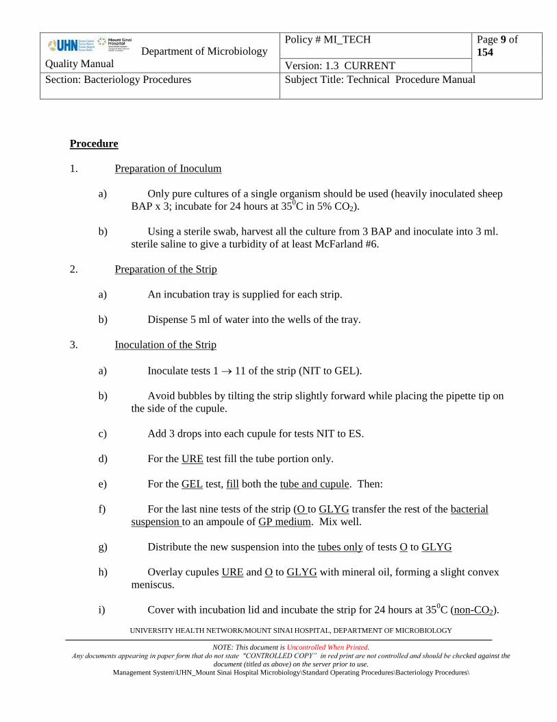
Procedure
1. Preparation of Inoculum
a) Only pure cultures of a single organism should be used (heavily inoculated sheep
BAP x 3; incubate for 24 hours at 350C in 5% CO2).
b) Using a sterile swab, harvest all the culture from 3 BAP and inoculate into 3 ml.
sterile saline to give a turbidity of at least McFarland #6.
2. Preparation of the Strip
a) An incubation tray is supplied for each strip.
b) Dispense 5 ml of water into the wells of the tray.
3. Inoculation of the Strip
a) Inoculate tests 1 11 of the strip (NIT to GEL).
b) Avoid bubbles by tilting the strip slightly forward while placing the pipette tip on
the side of the cupule.
c) Add 3 drops into each cupule for tests NIT to ES.
d) For the URE test fill the tube portion only.
e) For the GEL test, fill both the tube and cupule. Then:
f) For the last nine tests of the strip (O to GLYG transfer the rest of the bacterial
suspension to an ampoule of GP medium. Mix well.
g) Distribute the new suspension into the tubes only of tests O to GLYG
h) Overlay cupules URE and O to GLYG with mineral oil, forming a slight convex
meniscus.
i) Cover with incubation lid and incubate the strip for 24 hours at 350C (non-CO2).

Department of Microbiology
Quality Manual
Policy # MI_TECH
Page 10 of
154
Version: 1.3 CURRENT
Section: Bacteriology Procedures Subject Title: Technical Procedure Manual
UNIVERSITY HEALTH NETWORK/MOUNT SINAI HOSPITAL, DEPARTMENT OF MICROBIOLOGY
NOTE: This document is Uncontrolled When Printed.
Any documents appearing in paper form that do not state "CONTROLLED COPY” in red print are not controlled and should be checked against the
document (titled as above) on the server prior to use. Management System\UHN_Mount Sinai Hospital Microbiology\Standard Operating Procedures\Bacteriology Procedures\
Interpretation
REACTIONS TABLE
TESTS REACTONS NEGATIVE
RESULTS
POSITIVE
RESULTS
NIT Addition of NIT A + NIT B
(10 min)
NIT NITrate reduction Colourless
Very pale pink
Dark pink
Red
PYZ PYZ (10 min)
PYZ PYraZinamidase Colourless
Very pale brown
Very pale orange
Brown
Orange
PyrA BNAG Addition of ZYM A + ZYM B (10 min)
PyrA Pyrrolidonyl Arylamidase Colourless
Pale orange
Orange
PAL Alkaline Phosphatase Colourless
Beige-pale purple
Pale orange
Purple
GUR Beta GlucURonidase Colourless
Pale grey
Pale beige
Blue
GAL Beta GALactosidase Colourless
Beige-pale purple
Purple
GLU Alpha GLUcosidase Colourless
Beige-pale purple
Pale green
Purple
BNAG N-Acetyl-B Glucosaminidase Colourless
Beige-pale purple
Pale brown
Pale grey
Brown
Department of Microbiology
Quality Manual
Policy # MI_TECH
Page 11 of
154
Version: 1.3 CURRENT
Section: Bacteriology Procedures Subject Title: Technical Procedure Manual
UNIVERSITY HEALTH NETWORK/MOUNT SINAI HOSPITAL, DEPARTMENT OF MICROBIOLOGY
NOTE: This document is Uncontrolled When Printed.
Any documents appearing in paper form that do not state "CONTROLLED COPY” in red print are not controlled and should be checked against the
document (titled as above) on the server prior to use. Management System\UHN_Mount Sinai Hospital Microbiology\Standard Operating Procedures\Bacteriology Procedures\
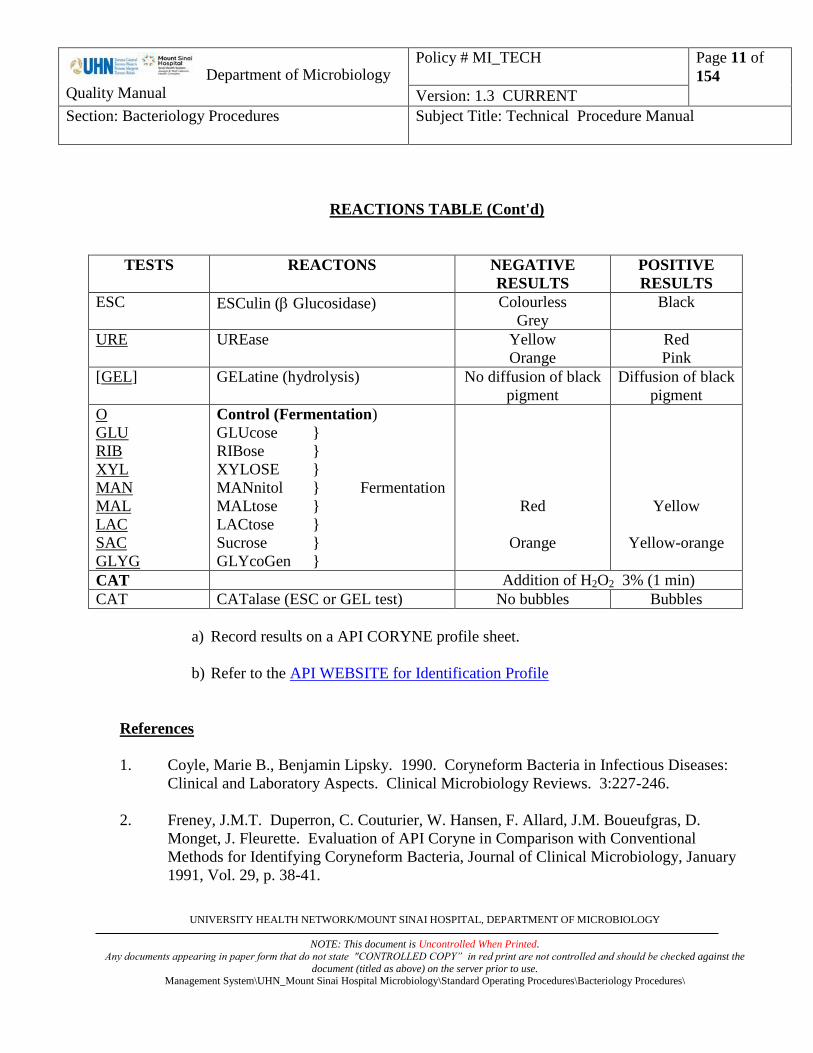
REACTIONS TABLE (Cont'd)
TESTS REACTONS NEGATIVE
RESULTS
POSITIVE
RESULTS
ESC ESCulin ( Glucosidase) Colourless
Grey
Black
URE UREase Yellow
Orange
Red
Pink
[GEL] GELatine (hydrolysis) No diffusion of black
pigment
Diffusion of black
pigment
O
GLU
RIB
XYL
MAN
MAL
LAC
SAC
GLYG
Control (Fermentation)
GLUcose }
RIBose }
XYLOSE }
MANnitol } Fermentation
MALtose }
LACtose }
Sucrose }
GLYcoGen }
Red
Orange
Yellow
Yellow-orange
CAT Addition of H2O2 3% (1 min)
CAT CATalase (ESC or GEL test) No bubbles Bubbles
a) Record results on a API CORYNE profile sheet.
b) Refer to the API WEBSITE for Identification Profile
References
1. Coyle, Marie B., Benjamin Lipsky. 1990. Coryneform Bacteria in Infectious Diseases:
Clinical and Laboratory Aspects. Clinical Microbiology Reviews. 3:227-246.
2. Freney, J.M.T. Duperron, C. Couturier, W. Hansen, F. Allard, J.M. Boueufgras, D.
Monget, J. Fleurette. Evaluation of API Coryne in Comparison with Conventional
Methods for Identifying Coryneform Bacteria, Journal of Clinical Microbiology, January
1991, Vol. 29, p. 38-41.

Department of Microbiology
Quality Manual
Policy # MI_TECH
Page 12 of
154
Version: 1.3 CURRENT
Section: Bacteriology Procedures Subject Title: Technical Procedure Manual
UNIVERSITY HEALTH NETWORK/MOUNT SINAI HOSPITAL, DEPARTMENT OF MICROBIOLOGY
NOTE: This document is Uncontrolled When Printed.
Any documents appearing in paper form that do not state "CONTROLLED COPY” in red print are not controlled and should be checked against the
document (titled as above) on the server prior to use. Management System\UHN_Mount Sinai Hospital Microbiology\Standard Operating Procedures\Bacteriology Procedures\
3. Murray P.A., et al. Manual of Clinical Microbiology, 7th
ed., 1999.

Department of Microbiology
Quality Manual
Policy # MI_TECH
Page 13 of
154
Version: 1.3 CURRENT
Section: Bacteriology Procedures Subject Title: Technical Procedure Manual
UNIVERSITY HEALTH NETWORK/MOUNT SINAI HOSPITAL, DEPARTMENT OF MICROBIOLOGY
NOTE: This document is Uncontrolled When Printed.
Any documents appearing in paper form that do not state "CONTROLLED COPY” in red print are not controlled and should be checked against the
document (titled as above) on the server prior to use. Management System\UHN_Mount Sinai Hospital Microbiology\Standard Operating Procedures\Bacteriology Procedures\
IDENTIFICATION OF ENTEROBACTERIACEAE (API 20E)
Principle
The API 20E system facilitates the 24-hour identification of Enterobacteriaceae as well as 24 or
48-hour identification of other Gram negative bacteria.
The API 20E strip consists of microtubes containing dehydrated substrates for the demonstration
of enzymatic activity and carbohydrate (CHO) fermentation. The substrates are reconstituted by
adding a bacterial suspension. After incubation, the metabolic end products are detected by
indicator systems or the addition of reagents. CHO fermentation is detected by colour change in
the pH indicator.
Materials
API 20E strips - store at 2-80C
0.85% sterile saline
Nitrate A - store at 2-80C
Nitrate B - store at 2-80C
Mineral oil
Zinc dust
Kovacs Reagent
Voges - Proskauer Reagents
Ferric Chloride Store at 2-80C
H2O2
Oxidase Reagent
OF Dextrose ID of non-
Motility Medium Enterobacteriaceae
Procedure
1. Preparation of Inoculum
a) Add 5 ml. of 0.85% saline to a sterile test tube.
b) Using a sterile inoculating loop, carefully touch the centre of a well isolated colony
(2-3 mm. Diameter) and thoroughly emulsify in the saline.

Department of Microbiology
Quality Manual
Policy # MI_TECH
Page 14 of
154
Version: 1.3 CURRENT
Section: Bacteriology Procedures Subject Title: Technical Procedure Manual
UNIVERSITY HEALTH NETWORK/MOUNT SINAI HOSPITAL, DEPARTMENT OF MICROBIOLOGY
NOTE: This document is Uncontrolled When Printed.
Any documents appearing in paper form that do not state "CONTROLLED COPY” in red print are not controlled and should be checked against the
document (titled as above) on the server prior to use. Management System\UHN_Mount Sinai Hospital Microbiology\Standard Operating Procedures\Bacteriology Procedures\
2. Preparation of the Strip
a) An incubation tray and lid is supplied for each strip.
b) Dispense 5 ml of water in to the tray.
3. Inoculation of the Strip
a) Remove the cap from the tube containing the bacterial suspension and insert a 5 ml.
Pasteur pipette.
b) Tilt the API 20E incubation tray and fill the tube section of the microtubes by
placing the pipette tip against the side of the cupule.
Note: The ADH, LDC, ODC, H2S, AND URE reactions can be interpreted best if
these microtubes are slightly underfilled.
c) Fill both the TUBE and CUPULE section of [CIT], [VP] and [GEL] tubes.
d) After inoculation, completely fill the cupule section of the ADH, LDC, ODC,
H2S and URE tubes with mineral oil.
e) Using the excess bacterial suspension, inoculate an agar slant or plate (non-selective
media such as nutrient agar, blood agar or tryptic (trypticase) soy agar is suggested)
as a purity check and for oxidase testing, serology, and/or additional biochemical
testing. Incubate the slant or plate for 18-24 hours at 350C.
4. Incubation of the Strip
a) After inoculation, place the plastic lid on the tray and incubate the strip for 18-24
hours at 350C in a non-CO2 incubator.
b) Weekend incubation: The biochemical reactions of the API 20E should be read
after 18-24 hours incubation. If the strips cannot be read after 24 hours incubation
at 350C, the strips should be removed from the incubator and stored at 2-8
0C
(refrigerator) until the reactions can be read.
5. Reading the Strip
a) After 18 hours of incubation and before 24 hours incubation, record all reactions
not requiring the addition of reagents.

Department of Microbiology
Quality Manual
Policy # MI_TECH
Page 15 of
154
Version: 1.3 CURRENT
Section: Bacteriology Procedures Subject Title: Technical Procedure Manual
UNIVERSITY HEALTH NETWORK/MOUNT SINAI HOSPITAL, DEPARTMENT OF MICROBIOLOGY
NOTE: This document is Uncontrolled When Printed.
Any documents appearing in paper form that do not state "CONTROLLED COPY” in red print are not controlled and should be checked against the
document (titled as above) on the server prior to use. Management System\UHN_Mount Sinai Hospital Microbiology\Standard Operating Procedures\Bacteriology Procedures\
b) If the GLU tube is negative (blue or green), do not add reagents. Reincubate a
further 18-24 hours.

Department of Microbiology
Quality Manual
Policy # MI_TECH
Page 16 of
154
Version: 1.3 CURRENT
Section: Bacteriology Procedures Subject Title: Technical Procedure Manual
UNIVERSITY HEALTH NETWORK/MOUNT SINAI HOSPITAL, DEPARTMENT OF MICROBIOLOGY
NOTE: This document is Uncontrolled When Printed.
Any documents appearing in paper form that do not state "CONTROLLED COPY” in red print are not controlled and should be checked against the
document (titled as above) on the server prior to use. Management System\UHN_Mount Sinai Hospital Microbiology\Standard Operating Procedures\Bacteriology Procedures\
c) If the GLU is positive (yellow):
i. Perform the oxidase test.
A portion of the growth from the agar slate or plate, inoculated from the 20E
bacterial suspension, should be rubbed onto filter paper to which a drop of
oxidase reagent (1% tetramethyl-p-phenylenediamine dihydrochloride) has
been added. The area where the growth has been added will turn dark
purple within 10 seconds if the reaction is positive and will be colourless or
light purple if negative.
Note: (a) Nichrome wire loops should NOT be used in performing
the oxidase test. Nichrome wire can cause a false positive
reaction.
(b) The oxidase test should NOT be performed using bacterial
growth from selective media such as MacConkey, EMB, etc.
Note: (a) Before addition of reagents, observe GLU tube (positive or
negative) for bubbles.
(b) The nitrate reduction and indole tests must be performed last
since these reactions release gaseous products which interfere
with the interpretation of other tests on the strip. The plastic
incubation lid should not be replaced after the addition of
these reagents.
ii. Add the reagents to TDA and VP tubes. If positive, the TDA reactions will
be immediate, whereas the VP reaction may be delayed up to 10 minutes.
iii. The Kovacs' reagent should then be added to the IND tube.
iv. The Nitrate Reduction test should be performed on all oxidase positive
organisms. The reagents should be added to the GLU tube after the Kovacs
Reagent has been added to the IND tube.

Department of Microbiology
Quality Manual
Policy # MI_TECH
Page 17 of
154
Version: 1.3 CURRENT
Section: Bacteriology Procedures Subject Title: Technical Procedure Manual
UNIVERSITY HEALTH NETWORK/MOUNT SINAI HOSPITAL, DEPARTMENT OF MICROBIOLOGY
NOTE: This document is Uncontrolled When Printed.
Any documents appearing in paper form that do not state "CONTROLLED COPY” in red print are not controlled and should be checked against the
document (titled as above) on the server prior to use. Management System\UHN_Mount Sinai Hospital Microbiology\Standard Operating Procedures\Bacteriology Procedures\
Interpretation
a) Record results on a API 20E analytical profile sheet.
b) The tests are separated into groups of three. The following numerical value is assigned to
each reaction recorded:
1- positive reaction in the first test of the group
2- positive reaction in the second test of the group
4- positive reaction in any test
0- negative reaction in any test
c) Refer to the API WEBSITE for Identification Profile
Reference
1. Murray P.A., et al. Manual of Clinical Microbiology, 7th
ed., 1999.

Department of Microbiology
Quality Manual
Policy # MI_TECH
Page 18 of
154
Version: 1.3 CURRENT
Section: Bacteriology Procedures Subject Title: Technical Procedure Manual
UNIVERSITY HEALTH NETWORK/MOUNT SINAI HOSPITAL, DEPARTMENT OF MICROBIOLOGY
NOTE: This document is Uncontrolled When Printed.
Any documents appearing in paper form that do not state "CONTROLLED COPY” in red print are not controlled and should be checked against the
document (titled as above) on the server prior to use. Management System\UHN_Mount Sinai Hospital Microbiology\Standard Operating Procedures\Bacteriology Procedures\
SUMMARY OF RESULTS - 18-24 HOUR PROCEDURE
TUBE INCUBATION POSITIVE NEGATIVE COMMENTS
ONPG Yellow Colourless (1) Any shade of yellow is a positive
reaction. (2) VP tube, before the addition of reagents,
can be used a negative control.
ADH 18-24 hr
36-48 hr
Red or Orange
Red
Yellow
Yellow or Orange
Orange reactions occurring at 36-48 hours
should be interpreted as negative.
LDC 18-24 hr
36-48 hr
Red or Orange
Red
Yellow
Yellow or Orange
Any shade of orange within 18-24 hours is a
positive reaction.
At 36-48 hours, orange decarboxylase reactions should be interpreted as negative.
ODC 18-34 hr
36-48 hr
Red or Orange
Red
Yellow
Yellow or Orange
Orange reactions occurring at 36-48 hours
should be interpreted as negative.
CIT Turquoise or
Dark Blue
Light Green
Or Yellow
(1) Both the tube and cupule should be filled.
(2) Reaction is read in the aerobic (cupule) area.
H2S Black Deposit No Black Deposit (1) H2S production may range from a heavy
black deposit to a very thin black line around the tube bottom. Carefully
examine the bottom of the tube before
considering the reaction negative. (2) A "browning" of the medium is a
negative reaction unless a black deposit
is present. "Browning" occurs with TDA positive organisms.
URE 18-24 hr 36-48 hr
Red or Orange Red
Yellow Yellow or Orange
A method of lower sensitivity has been chosen. Klebsiella, Proteus and Yersinia routinely give
positive reactions.
TDA Add 1 drop 10% Ferric chloride.
Brown-Red
Yellow
(1) Immediate reaction.
(2) Indole positive organisms may produce a
golden orange colour due to indole production. This is a negative reaction.
IND Add 1 drop Kovacs Reagent
Red Ring
Yellow
(1) The reaction should read within 2
minutes after the addition of the Kovacs reagents and the results recorded.
(2) After several minutes, the HCl present in
Kovacs reagent may react with the plastic of the cupule resulting in a change from a
negative (yellow) colour to a brownish-
red. This is a negative reaction.

Department of Microbiology
Quality Manual
Policy # MI_TECH
Page 19 of
154
Version: 1.3 CURRENT
Section: Bacteriology Procedures Subject Title: Technical Procedure Manual
UNIVERSITY HEALTH NETWORK/MOUNT SINAI HOSPITAL, DEPARTMENT OF MICROBIOLOGY
NOTE: This document is Uncontrolled When Printed.
Any documents appearing in paper form that do not state "CONTROLLED COPY” in red print are not controlled and should be checked against the
document (titled as above) on the server prior to use. Management System\UHN_Mount Sinai Hospital Microbiology\Standard Operating Procedures\Bacteriology Procedures\
SUMMARY OF RESULTS - 18-24 HOUR PROCEDURE (cont'd)
TUBE INCUBATION POSITIVE NEGATIVE COMMENTS
VP Add 1 drop of 40% Potassium Hydroxide, then 1 drop of alpha-
napthol.
(1) Wait 10 minutes before considering the
reaction negative. (2) A pale pink colour which appears
immediately after the addition of reagents but which turns dark `pink or red after 10
minutes should be interpreted as positive.
Motility may be observed by hanging drop or
wet mount preparation.
Red Colourless
GEL Diffusion of the
pigment
No diffusion (1) The solid gelatin particles may spread
throughout the tube after inoculation. Unless diffusion occurs, the reaction is
negative. (2) Any degree of diffusion is a positive
reaction.
GLU
MAN
INO SOR
RHA
SAC MEL
AMY ARA
Yellow Or Gray
Yellow
Blue or Blue-Green
Blue or
Blue-Green
COMMENTS FOR ALL CARBOHYDRATES
Fermentation (Enterobacteriaceae, Aeromonas, Vibrio)
(1) Fermentation of the carbohydrates begins
in the most anaerobic portion (bottom) of the tube. Therefore, these reactions should
be read from the bottom of the tube to the
top. (2) A yellow colour at the bottom of the tube
only indicates a weak or delayed positive
reaction.
Oxidation (Other Gram-negatives)
(1) Oxidative utilization of the carbohydrates begins in the most aerobic portion (top) of
the tube. Therefore, these reactions should
be read from the top to the bottom of the tube.
(2) A yellow colour in the upper portion of the tube and blue in the bottom of the tube
indicate oxidative utilization of the sugar.
This reaction should be considered positive only for non-Enterobacteriaceae
gram negative rods. This is a negative
reaction for fermentative organisms such as Enterobacteriaceae.

Department of Microbiology
Quality Manual
Policy # MI_TECH
Page 20 of
154
Version: 1.3 CURRENT
Section: Bacteriology Procedures Subject Title: Technical Procedure Manual
UNIVERSITY HEALTH NETWORK/MOUNT SINAI HOSPITAL, DEPARTMENT OF MICROBIOLOGY
NOTE: This document is Uncontrolled When Printed.
Any documents appearing in paper form that do not state "CONTROLLED COPY” in red print are not controlled and should be checked against the
document (titled as above) on the server prior to use. Management System\UHN_Mount Sinai Hospital Microbiology\Standard Operating Procedures\Bacteriology Procedures\
SUMMARY OF RESULTS - 18- 24 HOUR PROCEDURE (cont'd)
TUBE INCUBATION POSITIVE NEGATIVE COMMENTS
GLU
After reading GLU reaction, add 2 drops 0.8% sulfanilic acid and 2 drops 0.5% N. N-dimethyl-alpha-naphthylamine
(1) Before addition of reagents, observe GLU tube (positive or negative) for bubbles.
Bubbles are indicative of reduction of nitrate to the nitrogenous (N2) state.
(2) A positive reaction may take 2-3 minutes
for the red colour to appear.
(3) Confirm a negative test by adding zinc
dust or 20 mesh granular zinc. A pink-
orange colour after 10 minutes confirms a negative reaction. A yellow colour
indicates reduction of nitrates to the
nitrogenous (N2) state.
NO2
N2 gas
Red
Bubbles: Yellow
after reagents
and zinc
Yellow
Orange after
reagents and
zinc
MAN
INO SOR
Catalase
After reading carbohydrate reaction, add 1 drop 1.5% H2O2
(1) Bubbles may take 1-2 minutes to appear.
(2) Best results will be obtained if the test is run in tubes which have no gas from
fermentation.
Bubbles
No bubbles

Department of Microbiology
Quality Manual
Policy # MI_TECH
Page 21 of
154
Version: 1.3 CURRENT
Section: Bacteriology Procedures Subject Title: Technical Procedure Manual
UNIVERSITY HEALTH NETWORK/MOUNT SINAI HOSPITAL, DEPARTMENT OF MICROBIOLOGY
NOTE: This document is Uncontrolled When Printed.
Any documents appearing in paper form that do not state "CONTROLLED COPY” in red print are not controlled and should be checked against the
document (titled as above) on the server prior to use. Management System\UHN_Mount Sinai Hospital Microbiology\Standard Operating Procedures\Bacteriology Procedures\
IDENTIFICATION OF NON-ENTERIC GRAM-NEGATIVE RODS (API 20NE)
Principle
The API 20NE system facilitates the identification of non-fastidious Gram-negative rods not
belonging to the Enterobacteriaceae within 48 hours.
The API 20NE strip consists of microtubes containing dehydrated media and substrates. The media
microtubes containing conventional tests are inoculated with a bacterial suspension which
reconstitutes the media. After incubation, the metabolic end products are detected by indicator systems
or the addition of reagents. The substrate microtubes contain assimilation tests and are inoculated with
a minimal medium. If the bacteria are capable of utilizing the corresponding substrate, then they will
grow.
Materials
API 20NE strips - store at 2-80C
0.85% sterile saline
Mineral oil
Zinc dust
AUX Medium }
James Reagent }
Nitrate 1 - store at 2-80C } Store at 2-8
0C
Nitrate 2 - store at 2-80C }
Oxidase Reagent
Procedure
1. Preparation of Inoculum
a) Add 2 ml. of 0.85% saline to a sterile test tube.
b) Using a sterile inoculating loop, carefully touch the centre of a well isolated colony
(2-3 mm. Diameter) and thoroughly emulsify in the saline. The suspension turbidity
should be equal to a 0.5 McFarland standard.
2. Preparation of the Strip
a) An incubation tray and lid are supplied for each strip.
b) Dispense 5 ml of distilled water in to the tray.

Department of Microbiology
Quality Manual
Policy # MI_TECH
Page 22 of
154
Version: 1.3 CURRENT
Section: Bacteriology Procedures Subject Title: Technical Procedure Manual
UNIVERSITY HEALTH NETWORK/MOUNT SINAI HOSPITAL, DEPARTMENT OF MICROBIOLOGY
NOTE: This document is Uncontrolled When Printed.
Any documents appearing in paper form that do not state "CONTROLLED COPY” in red print are not controlled and should be checked against the
document (titled as above) on the server prior to use. Management System\UHN_Mount Sinai Hospital Microbiology\Standard Operating Procedures\Bacteriology Procedures\
4. Inoculation of the Strip
a) Remove the cap from the tube containing the bacterial suspension and insert a
sterile pipette.
b) Tilt the API 20NE incubation tray and fill the TUBE section of the NO3 to PNPG
microtubes by placing the pipette tip against the side of the cupule.
c) Open an ampule of AUX Medium and add 200 uL of the bacterial suspension to the
ampule. Mix well with a pipette while avoiding the formation of air bubbles.
d) Using the AUX Medium bacterial suspension, fill both the TUBE and CUPULE
section of [GLU] to [PAC]. Do not overfill the cupules. Fill to a flat or slightly
convex meniscus.
e) After inoculation, completely fill the CUPULE section of the 3 underlined tests,
GLU, ADH and URE tubes with mineral oil.
f) Using the excess bacterial suspension, inoculate an agar slant or plate (non-selective
media such as nutrient agar, blood agar or tryptic (trypticase) soy agar is suggested)
as a purity check and for oxidase testing, and/or additional biochemical testing.
Incubate the slant or plate with the API 20NE strip.
4. Incubation of the Strip
a) After inoculation, place the plastic lid on the tray and incubate the strip for 24 hours
at 300C in a non-CO2 incubator.
5. Reading the Strip
a) After 24 hours incubation, record all reactions not requiring the addition of
reagents.
b) Perform the oxidase test.
A portion of the growth from the agar slate or plate, inoculated from the 20NE
bacterial suspension, should be rubbed onto filter paper to which a drop of oxidase
reagent (1% tetramethyl-p-phenylenediamine dihydrochloride) has been added.
The area where the growth has been added will turn dark purple within 10 seconds
if the reaction is positive and will be colourless or light purple if negative.
Note: (a) Nichrome wire loops should NOT be used in performing the oxidase
test. Nichrome wire can cause a false positivereaction.
Department of Microbiology
Quality Manual
Policy # MI_TECH
Page 23 of
154
Version: 1.3 CURRENT
Section: Bacteriology Procedures Subject Title: Technical Procedure Manual
UNIVERSITY HEALTH NETWORK/MOUNT SINAI HOSPITAL, DEPARTMENT OF MICROBIOLOGY
NOTE: This document is Uncontrolled When Printed.
Any documents appearing in paper form that do not state "CONTROLLED COPY” in red print are not controlled and should be checked against the
document (titled as above) on the server prior to use. Management System\UHN_Mount Sinai Hospital Microbiology\Standard Operating Procedures\Bacteriology Procedures\
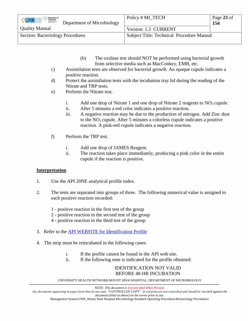
(b) The oxidase test should NOT be performed using bacterial growth
from selective media such as MacConkey, EMB, etc.
c) Assimilation tests are observed for bacterial growth. An opaque cupule indicates a
positive reaction.
d) Protect the assimilation tests with the incubation tray lid during the reading of the
Nitrate and TRP tests.
e) Perform the Nitrate test.
i. Add one drop of Nitrate 1 and one drop of Nitrate 2 reagents to NO3 cupule.
ii. After 5 minutes a red color indicates a positive reaction.
iii. A negative reaction may be due to the production of nitrogen. Add Zinc dust
to the NO3 cupule. After 5 minutes a colorless cupule indicates a positive
reaction. A pink-red cupule indicates a negative reaction.
f) Perform the TRP test.
i. Add one drop of JAMES Reagent.
ii. The reaction takes place immediately, producing a pink color in the entire
cupule if the reaction is positive.
Interpretation
1. Use the API 20NE analytical profile index.
2. The tests are separated into groups of three. The following numerical value is assigned to
each positive reaction recorded:
1 - positive reaction in the first test of the group
2 - positive reaction in the second test of the group
4 - positive reaction in the third test of the group
3. Refer to the API WEBSITE for Identification Profile
4. The strip must be reincubated in the following cases:
i. If the profile cannot be found in the API web site.
ii. If the following note is indicated for the profile obtained:
IDENTIFICATION NOT VALID
BEFORE 48-HR INCUBATION

Department of Microbiology
Quality Manual
Policy # MI_TECH
Page 24 of
154
Version: 1.3 CURRENT
Section: Bacteriology Procedures Subject Title: Technical Procedure Manual
UNIVERSITY HEALTH NETWORK/MOUNT SINAI HOSPITAL, DEPARTMENT OF MICROBIOLOGY
NOTE: This document is Uncontrolled When Printed.
Any documents appearing in paper form that do not state "CONTROLLED COPY” in red print are not controlled and should be checked against the
document (titled as above) on the server prior to use. Management System\UHN_Mount Sinai Hospital Microbiology\Standard Operating Procedures\Bacteriology Procedures\
iii. If the strip is to be reincubated, remove the reagents from the NO3 and TRP
cupules and then cover these tests with mineral oil.
iv. Reincubate the strip for another 24 hours at 30oC in a non-CO2 incubator.
v. Read all the tests again, except for NO3, TRP and GLU.
READING TABLE
TESTS SUBSTRATES REACTONS/ENZYMES NEGATIVE
RESULTS
POSITIVE
RESULTS
N03 Potassium
nitrate
NITrate reduction to
nitrites NIT 1 + NIT 2 / 5 min
colourless pink-red
NITrates to nitrogen Zn / 5 min
pink colourless
TRP tryptophane indole production JAMES / immediate
colourless / pink
pale green / yellow
GLU glucose Acidification blue to green yellow
ADH arginine arginine dihydrolase yellow orange/pink/red
URE urea Urease yellow orange/pink/red
ESC esculin hydrolysis (-glucosidase) yellow grey/brown/black
GEL gelatine
(with India ink)
hydrolysis (protease) no pigment
diffusion
diffusion of black
pigment
PNPG p-nitrophenyl--D-
galactopyranoside
-galactosidase colourless
yellow
[GLU] glucose Assimilation transparent opaque
[ARA] arabinose Assimilation transparent opaque
[MNE] mannose Assimilation transparent opaque

Department of Microbiology
Quality Manual
Policy # MI_TECH
Page 25 of
154
Version: 1.3 CURRENT
Section: Bacteriology Procedures Subject Title: Technical Procedure Manual
UNIVERSITY HEALTH NETWORK/MOUNT SINAI HOSPITAL, DEPARTMENT OF MICROBIOLOGY
NOTE: This document is Uncontrolled When Printed.
Any documents appearing in paper form that do not state "CONTROLLED COPY” in red print are not controlled and should be checked against the
document (titled as above) on the server prior to use. Management System\UHN_Mount Sinai Hospital Microbiology\Standard Operating Procedures\Bacteriology Procedures\
[MAN] mannitol Assimilation transparent opaque
[NAG] N-acetyl-glucosamine Assimilation transparent opaque
[MAL] maltose Assimilation transparent opaque
[GNT] gluconate Assimilation transparent opaque
[CAP] caprate Assimilation transparent opaque
[ADI] adipate Assimilation transparent opaque
[MLT] malate Assimilation transparent opaque
[CIT] citrate Assimilation transparent opaque
[PAC] phenyl-acetate Assimilation transparent opaque
OX see oxidase test cytochrome oxidase colorless/
light purple
dark purple
Quality Control
To be performed on receipt of every new lot of strip by the QC bench technologist.
Reference
Reference Package Insert - api 20NE system for the identification, bioMerieux Inc., Missouri
USA.
Department of Microbiology
Quality Manual
Policy # MI_TECH
Page 26 of
154
Version: 1.3 CURRENT
Section: Bacteriology Procedures Subject Title: Technical Procedure Manual
UNIVERSITY HEALTH NETWORK/MOUNT SINAI HOSPITAL, DEPARTMENT OF MICROBIOLOGY
NOTE: This document is Uncontrolled When Printed.
Any documents appearing in paper form that do not state "CONTROLLED COPY” in red print are not controlled and should be checked against the
document (titled as above) on the server prior to use. Management System\UHN_Mount Sinai Hospital Microbiology\Standard Operating Procedures\Bacteriology Procedures\
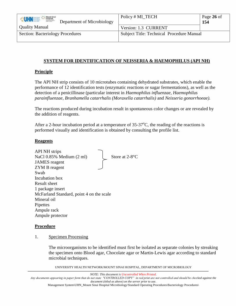
SYSTEM FOR IDENTIFICATION OF NEISSERIA & HAEMOPHILUS (API NH)
Principle
The API NH strip consists of 10 microtubes containing dehydrated substrates, which enable the
performance of 12 identification tests (enzymatic reactions or sugar fermentations), as well as the
detection of a penicillinase (particular interest in Haemophilus influenzae, Haemophilus
parainfluenzae, Branhamella catarrhalis (Moraxella catarrhalis) and Neisseria gonorrhoeae).
The reactions produced during incubation result in spontaneous color changes or are revealed by
the addition of reagents.
After a 2-hour incubation period at a temperature of 35-37oC, the reading of the reactions is
performed visually and identification is obtained by consulting the profile list.
Reagents
API NH strips
NaCl 0.85% Medium (2 ml) Store at 2-8°C
JAMES reagent
ZYM B reagent
Swab
Incubation box
Result sheet
1 package insert
McFarland Standard, point 4 on the scale
Mineral oil
Pipettes
Ampule rack
Ampule protector
Procedure
1. Specimen Processing
The microorganisms to be identified must first be isolated as separate colonies by streaking
the specimen onto Blood agar, Chocolate agar or Martin-Lewis agar according to standard
microbial techniques.

Department of Microbiology
Quality Manual
Policy # MI_TECH
Page 27 of
154
Version: 1.3 CURRENT
Section: Bacteriology Procedures Subject Title: Technical Procedure Manual
UNIVERSITY HEALTH NETWORK/MOUNT SINAI HOSPITAL, DEPARTMENT OF MICROBIOLOGY
NOTE: This document is Uncontrolled When Printed.
Any documents appearing in paper form that do not state "CONTROLLED COPY” in red print are not controlled and should be checked against the
document (titled as above) on the server prior to use. Management System\UHN_Mount Sinai Hospital Microbiology\Standard Operating Procedures\Bacteriology Procedures\
2. Preparation of Strip
Each strip is composed of 10 cupules. Each cupule has an open and closed area (cupule
and tube). An incubation tray is supplied for each strip. It serves as a support and
individual chamber while both protecting the strip from contaminants in the air and
assuring the humid atmosphere necessary to avoid dehydration during incubation.
Remove the strip from its individual packaging
Place the strip in the incubation box
Discard the desiccant sachet
Record the specimen number on the flat portion of the tray (do not record the number on
the lid as it may be misplaced during handling).
3. Preparation of the Inoculum
Open an ampule of NaCl 0.85% Medium (2 ml) with the ampule protector.
Using a swab, pick up a few well-isolated colonies and prepare a suspension with a
turbidity equivalent to 4 McFarland, ensuring it is well mixed.
The suspension should be used immediately after preparation.
4. Inoculation of the Strip
Distribute the prepared bacterial suspension into the cupules, avoiding the formation of
bubbles (tilt the strip slightly forwards and place the tip of the pipette or PSIpette against
the side of the cupule):
- Only fill the tube part of the first 7 microtubes (PEN to URE): about 50 l.
- Fill tube and cupule of the last 3 microtubes LIP/ProA, PAL/GGT, GAL/IND:
about 150 l, avoiding the formation of a convex meniscus.
Cover the first 7 tests (PEN to URE) with mineral oil (underlined tests).
NOTE: The quality of the filling is very important: tubes which are insufficiently or
excessively full may cause false positive or false negative results.
Close the incubation box.
Incubate for 2 hours at 35-37oC in aerobic conditions.
5.Incubation
Incubate for 2 hours at 35-37oC in aerobic conditions.

Department of Microbiology
Quality Manual
Policy # MI_TECH
Page 28 of
154
Version: 1.3 CURRENT
Section: Bacteriology Procedures Subject Title: Technical Procedure Manual
UNIVERSITY HEALTH NETWORK/MOUNT SINAI HOSPITAL, DEPARTMENT OF MICROBIOLOGY
NOTE: This document is Uncontrolled When Printed.
Any documents appearing in paper form that do not state "CONTROLLED COPY” in red print are not controlled and should be checked against the
document (titled as above) on the server prior to use. Management System\UHN_Mount Sinai Hospital Microbiology\Standard Operating Procedures\Bacteriology Procedures\
6.Reading the Strip
Refer to the Reactions Table for a description of how to read the reactions.
Note all spontaneous reactions (PEN to GAL) and record them as + or -.
Add 1 drop of ZYM B reagent to microtubes 8 and 9: LIP/ProA and PAL/GGT.
Add 1 drop of JAMES reagent to microtube 10: GAL/IND.
Wait 2 minutes then read the reactions by referring to the Reading Table in this package
insert and record them on the result sheet.
- If the LIP reaction is positive (blue pigment), interpret the ProA reaction as
negative, whether the ZYM B reagent has been added or not.
- If, after a 2-hour incubation period, several reactions (fermentation, penicillinase)
are doubtful, re-incubate the strip for another 2 hours and read the reactions again
(the enzymatic tests should not be re-read in this case).
Reactions Table
TESTS REACTIONS SUBSTRATES QTY
(mg)
RESULTS
NEGATIVE POSITIVE
1) PEN
PENicillinase
Penicillin G
1.36
Blue
(penicillinase absent)
Yellow
Yellow-green
Yellow-blue (penicillinase present
2) GLU 3) FRU
4) MAL
5) SAC
GLUcose (Acidification) FRUctose (Acidification)
MALtose (Acidification)
SACcharose/Sucrose (Acidification)
Glucose Fructose
Maltose
Sucrose
0.5 0.1
0.1
0.5
Red
Red-orange
Yellow
Orange
6) ODC
Ornithine DeCarboxylase
Ornithine
0.55
Yellow-green Grey-green
Blue
7) URE
UREease
Urea
0.41
Yellow
Pink-violet
8a) LIP
LIPase
5-bromo-3-indoxyl-
caprate
0.033
Colorless
Pale grey
Blue
(+precipitate)
9a) PAL
Alkaline Phosphatase
Para-Nitrophenyl-
phosphate 2CHA
0.038
Colorless
Pale yellow
Yellow
10a) GAL
Beta GALactosidaase
Para-Nitrophenyl-BD
galactopyranoside
0.04
Colorless
Yellow

Department of Microbiology
Quality Manual
Policy # MI_TECH
Page 29 of
154
Version: 1.3 CURRENT
Section: Bacteriology Procedures Subject Title: Technical Procedure Manual
UNIVERSITY HEALTH NETWORK/MOUNT SINAI HOSPITAL, DEPARTMENT OF MICROBIOLOGY
NOTE: This document is Uncontrolled When Printed.
Any documents appearing in paper form that do not state "CONTROLLED COPY” in red print are not controlled and should be checked against the
document (titled as above) on the server prior to use. Management System\UHN_Mount Sinai Hospital Microbiology\Standard Operating Procedures\Bacteriology Procedures\
Reactions Table (Cont'd)
TESTS REACTIONS SUBSTRATES QTY
(mg)
RESULTS
NEGATIVE POSITIVE
8b) ProA
Proline Arylamidase If LIP is +. ProA is always -
Proline-4-methoxy-
naphthylamide
0.056
ZYM B / 3 min
Yellow Pale orange
(brown if LIP +)
Orange
9b) GGT
Gamma Glutamyl Transferase
Gamma glutamyl
4-methoxy-
naphthylamide
0.049
ZYM B / 3 min
Yellow
Pale orange (yellow-orange if PAL +)
Orange
10b) IND
INDole
Tryptophane
0.036
JAMES / 3 min
Colorless
Pink
Interpretation
a) Record results on a API NH profile sheet.
b) Refer to the API WEBSITE for Identification Profile
Quality Control
To be performed on receipt of every new lot of strip by the QC bench technologist.
QC organisms to be used:
Neisseria gonorrhoea ATCC 31426
Haemophilus influenzae ATCC 10211
Branhamella catarrhalis ATCC 23246
Haemophilus paraphrophilus ATCC 49917
Reference
Reference Package Insert - api NH system for the identification of Neisseria and Haemophilus
bioMerieux Inc., Missouri USA.

Department of Microbiology
Quality Manual
Policy # MI_TECH
Page 30 of
154
Version: 1.3 CURRENT
Section: Bacteriology Procedures Subject Title: Technical Procedure Manual
UNIVERSITY HEALTH NETWORK/MOUNT SINAI HOSPITAL, DEPARTMENT OF MICROBIOLOGY
NOTE: This document is Uncontrolled When Printed.
Any documents appearing in paper form that do not state "CONTROLLED COPY” in red print are not controlled and should be checked against the
document (titled as above) on the server prior to use. Management System\UHN_Mount Sinai Hospital Microbiology\Standard Operating Procedures\Bacteriology Procedures\
IDENTIFICATION OF STREPTOCOCCACEAE (API 20 Strep)
Principle
API 20 Strep facilitates the group or species identification of most streptococci and enterococci,
and those most common related organisms.
The API 20 Strep consists of 20 microtubes containing dehydrated substrates for the demonstration
of enzymatic activity or the fermentation of sugars. The enzymatic tests are inoculated with a
dense suspension of organisms, made from a pure culture, which is used to reconstitute the
enzymatic substrates. During incubation, metabolism produces colour changes that are either
spontaneous or revealed by the addition of reagents. The fermentation tests are inoculated with an
enriched medium which rehydrates the sugar substrates. Fermentation of carbohydrates is detected
by a shift in the PH indicator.
Materials
API 20 Strep strips
API GP medium, 2 ml Store at 2-80C
Sterile distilled water, 2 ml
Mineral oil
NIN – Ninhydrin
ZYM A Reagent Store at 2-80C
ZYM B Reagent
Voges - Proskauer Reagents
Procedure
6. Preparation of Inoculum
a) Add 2 ml of sterile distilled water without additives to a sterile test tube.
b) Using a sterile swab, make a dense suspension with a turbidity greater than
4 McFarland standard from a fresh, pure culture; two plates may be necessary for an
adequate inoculum. This suspension must be used immediately after preparation.
Department of Microbiology
Quality Manual
Policy # MI_TECH
Page 31 of
154
Version: 1.3 CURRENT
Section: Bacteriology Procedures Subject Title: Technical Procedure Manual
UNIVERSITY HEALTH NETWORK/MOUNT SINAI HOSPITAL, DEPARTMENT OF MICROBIOLOGY
NOTE: This document is Uncontrolled When Printed.
Any documents appearing in paper form that do not state "CONTROLLED COPY” in red print are not controlled and should be checked against the
document (titled as above) on the server prior to use. Management System\UHN_Mount Sinai Hospital Microbiology\Standard Operating Procedures\Bacteriology Procedures\
7. Preparation of the Strip
a) An incubation tray and lid is supplied for each strip.
b) Dispense 5 ml of water in to the tray.
c) Label the tray with the patient name and order number.
8. Inoculation of the Strip
a) Remove the cap from the tube containing the bacterial suspension.
b) When inoculating the API 20 Strep strip, avoid the formation of bubbles (tilt the
strip slightly forwards and place the tip of a Pasteur pipette against the side of the
cupule):
The first half of the strip (tests VP to ADH) will be inoculated with this suspension
a) For tests VP to LAP, distribute approximately 100µl into each cupule.
b) For the ADH test: fill the tube only.
For the second half of the strip (RIB to GLYG):
a) Carefully open an ampule of API GP Medium and transfer the rest of the
suspension (approximately 0.5 ml) into it. Mix well.
b) Distribute this new suspension into the tubes only.
c) Fill the cupule of the underlined tests (ADH to GLYG) with mineral oil to form a
convex meniscus.
d) Place the lid on the tray.
e) Using the excess bacterial suspension, inoculate a blood agar plate as a purity
check. Incubate blood agar plate in CO2 overnight.
9. Incubation of the Strip
a) Incubate the strip at 360C in aerobic conditions for 4 to 4½ hours to obtain a first
reading and for 24 hours to obtain a second reading if required.
10. Reading the Strip
a) After 4 hours of incubation: add the reagents:
VP test: 1 drop each of VP 1 and VP 2
HIP test: 2 drops of NIN
PYRA, aGAL, ßGUR, ßGAL, PAL and LAP tests:
Department of Microbiology
Quality Manual
Policy # MI_TECH
Page 32 of
154
Version: 1.3 CURRENT
Section: Bacteriology Procedures Subject Title: Technical Procedure Manual
UNIVERSITY HEALTH NETWORK/MOUNT SINAI HOSPITAL, DEPARTMENT OF MICROBIOLOGY
NOTE: This document is Uncontrolled When Printed.
Any documents appearing in paper form that do not state "CONTROLLED COPY” in red print are not controlled and should be checked against the
document (titled as above) on the server prior to use. Management System\UHN_Mount Sinai Hospital Microbiology\Standard Operating Procedures\Bacteriology Procedures\
1 drop each of ZYM A and ZYM B
Wait 10 minutes; record the reactions on the API 20 Strep analytical profile sheet
Reincubation is necessary in the following cases:
- low discrimination;
- unacceptable or doubtful profile;
- or if the following comment is given for the profile:
IDENTIFICATION NOT VALID
BEFORE 24 HOURS INCUBATION
In this case, after 24 hours, reread the reactions ESC, ADH and RIB to
GLYG. Do not reread the enzymatic reactions (HIP, PYRA, aGAL, ßGUR,
ßGAL, PAL, LAP) and VP
Record the reactions on the API 20 Strep analytical profile sheet

Department of Microbiology
Quality Manual
Policy # MI_TECH
Page 33 of
154
Version: 1.3 CURRENT
Section: Bacteriology Procedures Subject Title: Technical Procedure Manual
UNIVERSITY HEALTH NETWORK/MOUNT SINAI HOSPITAL, DEPARTMENT OF MICROBIOLOGY
NOTE: This document is Uncontrolled When Printed.
Any documents appearing in paper form that do not state "CONTROLLED COPY” in red print are not controlled and should be checked against the
document (titled as above) on the server prior to use. Management System\UHN_Mount Sinai Hospital Microbiology\Standard Operating Procedures\Bacteriology Procedures\
Interpretation
SUMMARY OF RESULTS – 4 to 24 HOUR PROCEDURE
TESTS RESULTS
NEGATIVE POSITIVE
VP
Add VP 1 + VP2/ wait 10 minutes
Colourless Pink-Red
HIP
Add NIN/wait 10 minutes
Colourless/Pale blue
Bluish-grey
Dark blue/Violet
ESC
4 hours 24 hours 4 hours 24 hours
Colourless
Pale yellow
Colourless
Pale yellow
Light grey
Black
Grey
Black
PYRA
Add ZYM A + ZYM B/ wait 10 minutes (PYRA to LAP)
Colourless or very pale orange Orange
aGAL Colourless Violet
ßGUR Colourless Blue
ßGAL Colourless or very pale violet Violet
PAL Colourless or very pale violet Violet
LAP Colourless Orange
ADH Yellow Red
RIB
4 hours 24 hours 4 hours 24 hours
Red Orange/Red Orange/Yellow Yellow
ARA Red Orange/Red Orange/Yellow Yellow
MAN Red Orange/Red Orange/Yellow Yellow
SOR Red Orange/Red Orange/Yellow Yellow
LAC Red Orange/Red Orange/Yellow Yellow
TRE Red Orange/Red Orange/Yellow Yellow
INU Red Orange/Red Orange/Yellow Yellow
RAF Red Orange/Red Orange/Yellow Yellow
AMD Red Orange/Red Orange/Yellow Yellow
GLYG Red or Orange Bright Yellow
d) The tests are separated into groups of three. The following numerical value is assigned to
each reaction recorded:
3- positive reaction in the first test of the group

Department of Microbiology
Quality Manual
Policy # MI_TECH
Page 34 of
154
Version: 1.3 CURRENT
Section: Bacteriology Procedures Subject Title: Technical Procedure Manual
UNIVERSITY HEALTH NETWORK/MOUNT SINAI HOSPITAL, DEPARTMENT OF MICROBIOLOGY
NOTE: This document is Uncontrolled When Printed.
Any documents appearing in paper form that do not state "CONTROLLED COPY” in red print are not controlled and should be checked against the
document (titled as above) on the server prior to use. Management System\UHN_Mount Sinai Hospital Microbiology\Standard Operating Procedures\Bacteriology Procedures\
4- positive reaction in the second test of the group
4- positive reaction in any test
1- negative reaction in any test
e) Refer to the API WEBSITE for Identification Profile

Department of Microbiology
Quality Manual
Policy # MI_TECH
Page 35 of
154
Version: 1.3 CURRENT
Section: Bacteriology Procedures Subject Title: Technical Procedure Manual
UNIVERSITY HEALTH NETWORK/MOUNT SINAI HOSPITAL, DEPARTMENT OF MICROBIOLOGY
NOTE: This document is Uncontrolled When Printed.
Any documents appearing in paper form that do not state "CONTROLLED COPY” in red print are not controlled and should be checked against the
document (titled as above) on the server prior to use. Management System\UHN_Mount Sinai Hospital Microbiology\Standard Operating Procedures\Bacteriology Procedures\
API WEBSITE for Identification Profile
1. Read API biochemical strip as per reading procedure.
2. Go to API website:
https://apiweb.biomerieux.com/servlet/Authenticate?action=prepareLogin
Login: See Seniors
3. Enter reactions or profile number
4. Obtain identification
5. Copy the identification and percent probability to the LIS workcard.

Department of Microbiology
Quality Manual
Policy # MI_TECH
Page 36 of
154
Version: 1.3 CURRENT
Section: Bacteriology Procedures Subject Title: Technical Procedure Manual
UNIVERSITY HEALTH NETWORK/MOUNT SINAI HOSPITAL, DEPARTMENT OF MICROBIOLOGY
NOTE: This document is Uncontrolled When Printed.
Any documents appearing in paper form that do not state "CONTROLLED COPY” in red print are not controlled and should be checked against the
document (titled as above) on the server prior to use. Management System\UHN_Mount Sinai Hospital Microbiology\Standard Operating Procedures\Bacteriology Procedures\
BACITRACIN DISK TEST
Principle
This is a screening test for the presumptive identification of Group A Streptococci which are
susceptible to 0.04U bacitracin. Other beta-haemolytic streptococci are usually resistant to this
concentration of bacitracin.
Reagents
Bacto Differentiation Disks Bacitracin (0.04U). Store refrigerated.
5% Sheep Blood Agar (BA)
Other Materials
Culture loop
Cotton swabs
Forceps
Procedure
1. Inoculate the surface of the BA with the suspect beta haemolytic Streptococcus. Streak for
confluent growth.
2. Using aseptic technique, place a bacitracin disk onto the inoculated surface.
3. Incubate in O2 at 35oC X 18-24 hr.
Interpretation
Susceptible: any zone of inhibition around the disk (Presumptive Group A Streptococcus).
Resistant: growth up to the edge of the disk
Precautions
1. Other beta-haemolytic streptococci may be susceptible to bacitracin. Therefore this test can
be used only for the presumptive identification of Gp. A Strep.

Department of Microbiology
Quality Manual
Policy # MI_TECH
Page 37 of
154
Version: 1.3 CURRENT
Section: Bacteriology Procedures Subject Title: Technical Procedure Manual
UNIVERSITY HEALTH NETWORK/MOUNT SINAI HOSPITAL, DEPARTMENT OF MICROBIOLOGY
NOTE: This document is Uncontrolled When Printed.
Any documents appearing in paper form that do not state "CONTROLLED COPY” in red print are not controlled and should be checked against the
document (titled as above) on the server prior to use. Management System\UHN_Mount Sinai Hospital Microbiology\Standard Operating Procedures\Bacteriology Procedures\
Quality Control
Test with known susceptible and resistant control strains weekly.
Susceptible: Gp.A Strep. (ATCC 19615)
Resistant: Gp.B Strep. (ATCC 13813)
Reference
1. Difco Differentiation Disks Bacitracin package insert.

Department of Microbiology
Quality Manual
Policy # MI_TECH
Page 38 of
154
Version: 1.3 CURRENT
Section: Bacteriology Procedures Subject Title: Technical Procedure Manual
UNIVERSITY HEALTH NETWORK/MOUNT SINAI HOSPITAL, DEPARTMENT OF MICROBIOLOGY
NOTE: This document is Uncontrolled When Printed.
Any documents appearing in paper form that do not state "CONTROLLED COPY” in red print are not controlled and should be checked against the
document (titled as above) on the server prior to use. Management System\UHN_Mount Sinai Hospital Microbiology\Standard Operating Procedures\Bacteriology Procedures\
BILE ESCULIN TEST
Principle
This test determines the ability of an organism to grow in the presence of bile and to hydrolyze the
glycoside esculin to esculetin and glucose. The test is used to presumptively identify Group D
Streptococci.
Materials
Bile esculin agar slant / plate – Store at 2-8°C
Culture loop
Procedure
1. Heavily inoculate a bile esculin slant / plate with the suspect organism.
2. Incubate in O2 at 35oC for 18-24 hr.
Interpretation
Positive: Presence of a dark brown to black colour on the slant.
Negative: No blackening of the medium. Growth may occur, but this does not indicate esculin
splitting.
Quality Control
Each new lot of media should be tested with known control strains.
Positive: E. faecalis (ATCC 29212)
Negative: Gp.B Strep. (ATCC 13813)
No Growth: Gp.A Strep. (ATCC 19615)
References
1. MacFaddin JF, Biochemical Tests for Identification of Medical Bacteria, 2nd ed., Williams
and Wilkins, Baltimore MD, 1980, p4-12.

Department of Microbiology
Quality Manual
Policy # MI_TECH
Page 39 of
154
Version: 1.3 CURRENT
Section: Bacteriology Procedures Subject Title: Technical Procedure Manual
UNIVERSITY HEALTH NETWORK/MOUNT SINAI HOSPITAL, DEPARTMENT OF MICROBIOLOGY
NOTE: This document is Uncontrolled When Printed.
Any documents appearing in paper form that do not state "CONTROLLED COPY” in red print are not controlled and should be checked against the
document (titled as above) on the server prior to use. Management System\UHN_Mount Sinai Hospital Microbiology\Standard Operating Procedures\Bacteriology Procedures\
BILE SOLUBILITY TEST
Principle
Tests the ability of alpha haemolytic streptococci to lyse in the presence of bile salts. This test is used
for the identification of Streptococcus pneumoniae.
Reagents
BBL Spot Test dropper (10% sodium desoxycholate). Store at room temperature.
Procedure
1. Hold the dropper upright and squeeze gently to crush the glass ampoule inside the dispenser.
2. Place 1 drop of the reagent directly on isolated colonies of suspected S. pneumoniae.
3. Keep the plates very level to prevent the reagent from running and washing a non-
pneumucoccal colony away, producing a false positive result.
4. Incubate at room temperature on the bench for 15-30 minutes until the reagent drys. Do not
invert the plate; leave the lid ajar.
5. Examine the colonies for lysis.
Interpretation
Positive (bile soluble): Lysis of the colonies.
Negative (bile insoluble): No lysis of colonies.
Quality Control
Test with known positive and negative control strains weekly.
Positive: S. pneumoniae (ATCC 6303)
Negative: S. sanguinis (ATCC 10556)
References
1. Murray PA, et al. Manual of Clinical Microbiology, 7th ed., 1999; p. 1665.
2. BBL Desoxycholate Reagent Droppers package insert, June 2010.

Department of Microbiology
Quality Manual
Policy # MI_TECH
Page 40 of
154
Version: 1.3 CURRENT
Section: Bacteriology Procedures Subject Title: Technical Procedure Manual
UNIVERSITY HEALTH NETWORK/MOUNT SINAI HOSPITAL, DEPARTMENT OF MICROBIOLOGY
NOTE: This document is Uncontrolled When Printed.
Any documents appearing in paper form that do not state "CONTROLLED COPY” in red print are not controlled and should be checked against the
document (titled as above) on the server prior to use. Management System\UHN_Mount Sinai Hospital Microbiology\Standard Operating Procedures\Bacteriology Procedures\
CATALASE TEST
Principle
Detects the presence of the enzyme catalase which hydrolyzes H2O2 to produce H2O and O2. This test
is used to differentiate Staphylococci (catalase positive) from Streptococci (catalase negative).
Reagents
Hydrogen peroxide (H2O2), 3%
Store in a dark bottle and avoid any undue exposure to light.
Keep refrigerated at all times when not in use.
Other Materials
Clean glass microscope slides
Plastic culture loop or wooden applicator stick
Procedure
1. Pick a colony from an 18-24 hr culture and place it on a clean glass slide. Avoid carry over of
blood agar which can cause false positives.
2. Put one drop of 3% H2O2 over the organism on the slide. Do not reverse the order of the
procedure as false positive results may occur. Do not mix.
3. Observe for immediate bubbling (gas liberation) and record the result.
4. Discard the slide into a discard container.
Interpretation
Positive test: Immediate bubbling, easily observed (02 formed)
Negative test: No bubbling
Precautions
1. Carry over of blood agar must be avoided.
2. Growth for testing must be from an 18-24 hr culture.
3. 3% H2O2 is caustic - avoid exposure to skin. If H2O2 does get on the skin, immediately flood
the area with 70% ethyl alcohol, not water.

Department of Microbiology
Quality Manual
Policy # MI_TECH
Page 41 of
154
Version: 1.3 CURRENT
Section: Bacteriology Procedures Subject Title: Technical Procedure Manual
UNIVERSITY HEALTH NETWORK/MOUNT SINAI HOSPITAL, DEPARTMENT OF MICROBIOLOGY
NOTE: This document is Uncontrolled When Printed.
Any documents appearing in paper form that do not state "CONTROLLED COPY” in red print are not controlled and should be checked against the
document (titled as above) on the server prior to use. Management System\UHN_Mount Sinai Hospital Microbiology\Standard Operating Procedures\Bacteriology Procedures\
4. Aerosols may be released by the bubbling of the O2.
5. H2O2 is unstable and breaks down easily on exposure to light. The solution must be kept
refrigerated in the dark.
Quality Control
H2O2 is very unstable and should be tested daily or immediately prior to its use.
Positive: S. aureus (ATCC 25923)
Negative: E. faecalis (ATCC 29212)
References
1. MacFaddin JF, Biochemical Tests for Identification of Medical Bacteria, 2nd ed., Williams
and Wilkins, Baltimore MD., 1980, p51-58.
2. Murray PA, et al. Manual of Clinical Microbiology, 7th ed., 1999; pp 426-427.
Department of Microbiology
Quality Manual
Policy # MI_TECH
Page 42 of
154
Version: 1.3 CURRENT
Section: Bacteriology Procedures Subject Title: Technical Procedure Manual
UNIVERSITY HEALTH NETWORK/MOUNT SINAI HOSPITAL, DEPARTMENT OF MICROBIOLOGY
NOTE: This document is Uncontrolled When Printed.
Any documents appearing in paper form that do not state "CONTROLLED COPY” in red print are not controlled and should be checked against the
document (titled as above) on the server prior to use. Management System\UHN_Mount Sinai Hospital Microbiology\Standard Operating Procedures\Bacteriology Procedures\
CETRIMIDE PSEUDOMONAS SELECTIVE AGAR
Principle
Cetrimide Selective Agar is used for the identification of Pseudomonas aeruginosa. Cetrimide is a
compound that has germicidal activity against most organisms except Pseudomonas aeruginosa.
Also pigment production is enhanced on this media.
Procedure
1. Divide the plate into approximately 8 pie shaped divisions.
2. Streak the test organism (pure culture) onto one of the pie shaped divisions.
3. Incubate at 350C for 18 - 24 hours.
Interpretation
Pseudomonas aeruginosa will grow on this media and will be pigmented a pale green to dark blue-
green colour. All other organisms will not grow or will be non-pigmented.
Quality Control
Test with positive and negative controls each time the test is set up.
Positive: Pseudomonas aeruginosa (ATCC 27853)
Negative: Escherichia coli (ATCC 25922)
Reference
1. PML Technical Manual, 1990.

Department of Microbiology
Quality Manual
Policy # MI_TECH
Page 43 of
154
Version: 1.3 CURRENT
Section: Bacteriology Procedures Subject Title: Technical Procedure Manual
UNIVERSITY HEALTH NETWORK/MOUNT SINAI HOSPITAL, DEPARTMENT OF MICROBIOLOGY
NOTE: This document is Uncontrolled When Printed.
Any documents appearing in paper form that do not state "CONTROLLED COPY” in red print are not controlled and should be checked against the
document (titled as above) on the server prior to use. Management System\UHN_Mount Sinai Hospital Microbiology\Standard Operating Procedures\Bacteriology Procedures\
CRYPTOCOCCAL ANTIGEN
Principle
Latex particles coated with anti-cryptococcal globulin (ACGR) reacts with cryptococcal polysaccharide
antigen (in CSF or serum) causing a visible agglutination.
Specimen Collection and Processing
5 mL of blood is collected in a serum separator tube and separated by centrifugation. The serum is
removed to a vial and refrigerated until testing. Specimens are stored at -20oC after testing.
Spinal fluid is collected in clean, sterile, centrifuge tubes. Specimens are stored refrigerated after testing.
Note: Fungus culture should also be set up.
Reagents
Meridian CALAS (Cryptococcal Antigen Latex Agglutination System)
GBDA - Glycine buffered diluent with albumin.
AGC - Antiglobulin control. Rehydrate with 1.5 mL dH2O.
NC - Negative control. Rehydrate with 2.5 mL dH2O and inactivate the negative at 56oC for 30
minutes.
CAC - Cryptococcal antigen control (Positive control).
Pronase - Rehydrate with 2.5 mL dH2O, can be stored at 2oC-8
oC for approximately 1 month.
Detect Latex (ACGR) - Anti-Cryptococcal globulin reagent.
Control Latex (NGR) - Normal globulin reagent.
Note: Ensure that all reconstituted vials are thoroughly dissolved before use
All reagents are stored refrigerated. Do not interchange reagents with a kit having a different lot number.
Allow reagents to warm to room temperature before use. Mix gently before use.
Department of Microbiology
Quality Manual
Policy # MI_TECH
Page 44 of
154
Version: 1.3 CURRENT
Section: Bacteriology Procedures Subject Title: Technical Procedure Manual
UNIVERSITY HEALTH NETWORK/MOUNT SINAI HOSPITAL, DEPARTMENT OF MICROBIOLOGY
NOTE: This document is Uncontrolled When Printed.
Any documents appearing in paper form that do not state "CONTROLLED COPY” in red print are not controlled and should be checked against the
document (titled as above) on the server prior to use. Management System\UHN_Mount Sinai Hospital Microbiology\Standard Operating Procedures\Bacteriology Procedures\
Other Materials
100 oC heating block
56oC heating block
x 0.1 mL pipettes
Rotator
Small serologic test tubes
Test tube rack
Marking pen
Applicator sticks
Eppendorf 5415R Centrifuge
The following are provided by Meridian:
Capillary pipettes
Slide cards
Procedure
Specimen preparation
1. Store specimen refrigerated if testing is not done immediately.
2. Inactivate serum by mixing 200 µL of serum and 200 µL of pronase solution in a 12 x 75 mm tube
and incubate at 56oC for 15 minutes. Further inactivate by heating at 100
oC for 5 minutes. This
constitutes a 1:2 dilution.
3. Centrifuge CSF at 1000g/3300 rpm in the Eppendorf 5415R centrifuge for 15 minutes. Inactivate the
supernatant in a boiling water bath for 5 minutes.

Department of Microbiology
Quality Manual
Policy # MI_TECH
Page 45 of
154
Version: 1.3 CURRENT
Section: Bacteriology Procedures Subject Title: Technical Procedure Manual
UNIVERSITY HEALTH NETWORK/MOUNT SINAI HOSPITAL, DEPARTMENT OF MICROBIOLOGY
NOTE: This document is Uncontrolled When Printed.
Any documents appearing in paper form that do not state "CONTROLLED COPY” in red print are not controlled and should be checked against the
document (titled as above) on the server prior to use. Management System\UHN_Mount Sinai Hospital Microbiology\Standard Operating Procedures\Bacteriology Procedures\
Performing the test
Note: Controls must be run each time a patient specimen is tested.
1. Set up and label the slide as follows:
2. Gently resuspend the latex particles in the Detect Latex (ACGR) and Control Latex(NGR) reagents.
Rock each reagent just prior to use.
3. Place one drop of Detect Latex (ACGR) or Control Latex (NGR) into the designated spots.
4. Place 25 μL (one drop) of the cryptococcal antigen control (CAC) into the designated rings.
Repeat with the negative control (NC) and anti-globulin control (AGC)
5. Place 25 μL of specimen in the designated rings.
6. Using a separate applicator stick, mix the contents of each ring thoroughly, spreading the contents
over the entire surface area.
7. Place the slide on the rotator and rotate at 125 rpm for 5 minutes.
8. Read the reactions immediately.
Detec
Latex
(NGR)
Control
Latex
(ACGR)
CAC AGC NC TEST
(POS (Anti-globulin (Negative
Control) Control) Control)
Not
Used

Department of Microbiology
Quality Manual
Policy # MI_TECH
Page 46 of
154
Version: 1.3 CURRENT
Section: Bacteriology Procedures Subject Title: Technical Procedure Manual
UNIVERSITY HEALTH NETWORK/MOUNT SINAI HOSPITAL, DEPARTMENT OF MICROBIOLOGY
NOTE: This document is Uncontrolled When Printed.
Any documents appearing in paper form that do not state "CONTROLLED COPY” in red print are not controlled and should be checked against the
document (titled as above) on the server prior to use. Management System\UHN_Mount Sinai Hospital Microbiology\Standard Operating Procedures\Bacteriology Procedures\
9. Rate the agglutination as follows: document agglutination results on worksheet.
A second technologist must double check/verify all results. Document verification on worksheet.
1+ = fine granulation against a milky background
2+ = small but definite clumps against a slightly cloudy background
3+ = large and small clumps against a clear background
4+ = large clumps against a clear background
Positive Test: > 2+ agglutination (small but definite clumps or larger against a slightly
cloudy or clear background)
Negative Test: < 1+ agglutination (fine granulation against a milky background) or a
homogenous suspension of particles with no visible
clumping)
Detect Latex (ACGR) + Positive Control = Positive
Detect Latex (ACGR) + Negative Control = Negative
Control Latex (AGC) + Anti-globulin Control = Positive
Control Latex (AGC) + Positive Control = Negative
Control Latex (AGC) + Negative Control = Negative
10. Patient specimens showing > 2+ agglutination in EITHER Detect Latex or Control Latex should
be titrated against both Detect Latex (ACGR) and Control Latex (AGC) reagents.
a) Prepare two-fold serial dilutions of the specimen using 200 µL of GBDA in each of 8
test tubes labelled as follows:
Tube 1 2 3 4 5 6 7 8
Serum 1:4 1:8 1:16 1:32 1:64 1:128 1:256 1:512
CSF 1:2 1:4 1:8 1:16 1:32 1:64 1:128 1:256

Department of Microbiology
Quality Manual
Policy # MI_TECH
Page 47 of
154
Version: 1.3 CURRENT
Section: Bacteriology Procedures Subject Title: Technical Procedure Manual
UNIVERSITY HEALTH NETWORK/MOUNT SINAI HOSPITAL, DEPARTMENT OF MICROBIOLOGY
NOTE: This document is Uncontrolled When Printed.
Any documents appearing in paper form that do not state "CONTROLLED COPY” in red print are not controlled and should be checked against the
document (titled as above) on the server prior to use. Management System\UHN_Mount Sinai Hospital Microbiology\Standard Operating Procedures\Bacteriology Procedures\
b) Transfer one drop of each dilution into 2 rings.
c) Add one drop of Detect Latex (ACGR) to one ring of each dilution.
d) Add one drop of Control Latex (NGR) to each of the other rings.
e) Mix using separate applicator sticks.
f) Place the slide on the rotator and rotate at 125 rpm for 5 minutes.
g) Rate the agglutination.
h) If tube #8 gives an agglutination of 2+ or greater, the specimen must be further serially
diluted until a titre may be obtained.
Interpretation of results
Negative: Negative result in initial screening tests against Detect Latex(ACGR).
Positive: The titre is reported as the highest dilution showing a 2+ or greater reaction with Detect
Latex (ACGR) and negative with Control Latex (NGR).
Positive with Nonspecific Interference:
The titre with Detect Latex (ACGR) is at least 4 fold higher
than the titre with Control Latex (NGR). (E.g. Detection
Latex titer 1:32, Control Latex titer 1:8 or lower)
Uninterpretable test:
The titre with Detect Latex (ACGR) is less than 4-fold greater than the titre with
Control Latex (NGR).
(E.g. Detection Latex 1:32, Control latex 1:16)

Department of Microbiology
Quality Manual
Policy # MI_TECH
Page 48 of
154
Version: 1.3 CURRENT
Section: Bacteriology Procedures Subject Title: Technical Procedure Manual
UNIVERSITY HEALTH NETWORK/MOUNT SINAI HOSPITAL, DEPARTMENT OF MICROBIOLOGY
NOTE: This document is Uncontrolled When Printed.
Any documents appearing in paper form that do not state "CONTROLLED COPY” in red print are not controlled and should be checked against the
document (titled as above) on the server prior to use. Management System\UHN_Mount Sinai Hospital Microbiology\Standard Operating Procedures\Bacteriology Procedures\
Reporting
Telephone all positive reports.
Negative Report: "Cryptococcal antigen not detected by latex agglutination."
Positive Report: "Cryptococcal antigen detected at a titre of by latex agglutination."
Positives with titre <1:4 add:
}CRLO “Titre of < 1:4 may be due to a very low level of antigen or non-specific
cross-reactivity. Results should be interpreted within the context of the clinical
signs, symptoms, and history of the patient.
Positive with Nonspecific Interference: “Cryptococcal antigen detected with nonspecific
Interference.”
Uninterpretable Report: “Cryptococcal antigen test invalid due to nonspecific interference.”
Quality Control
The pattern of reactions for the controls must be as follows.
Detect Latex (ACGR) + N/A -
Controlt Latex (NGR) - + -
CAC AGC NC
Failure to obtain this pattern indicates that the test must be repeated and the patient test results cannot be
reported.
References
Product Insert, 1986 Rev 7/01 Meridian Diagnostics Inc., 3471 River Hills Dr., Cincinnati, Ohio 45244.
(513)-271-3700.

Department of Microbiology
Quality Manual
Policy # MI_TECH
Page 49 of
154
Version: 1.3 CURRENT
Section: Bacteriology Procedures Subject Title: Technical Procedure Manual
UNIVERSITY HEALTH NETWORK/MOUNT SINAI HOSPITAL, DEPARTMENT OF MICROBIOLOGY
NOTE: This document is Uncontrolled When Printed.
Any documents appearing in paper form that do not state "CONTROLLED COPY” in red print are not controlled and should be checked against the
document (titled as above) on the server prior to use. Management System\UHN_Mount Sinai Hospital Microbiology\Standard Operating Procedures\Bacteriology Procedures\
E. coli O157 LATEX TEST (OXOID)
Principle
The Latex test will demonstrate by slide agglutination, E. coli strains possessing the O157 antigen.
Sorbitol MacConkey Agar (SMAC) should be used as the primary screen. Non-sorbital
fermenting colonies (NSF) are tested with the latex reagents, to determine if the isolate belongs to
the O157 serogroup, and is therefore a potential vero-cytotoxin (VT) producing strain.
Reagents
DR621M Test Latex - consists of latex particles sensitized with specific rabbit antibody
reactive with the O157 antigen.
DR622M Control Latex - consists of latex particles sensitized with pre-immune rabbit
globulins.
DR623M - positive control suspension
DR624M - negative control suspension
Storage
Do not freeze. Store at 20C - 8
0C. Do not use kit beyond the expiry date.
Procedure
NSF colonies may be taken from SMAC plates or alternatively NSF isolates may be inoculated
onto non-selective agar media for testing.
For best results, sub NSF to a Blood Agar plate prior to testing.
It is necessary to test up to 10 separate NSF colonies to ensure a high probability of detection from
mixed cultures.
1) Bring the latex reagents to room temperature. Make sure the latex suspensions are mixed by
vigorous shaking. Expel any latex from the dropper pipette for complete mixing.

Department of Microbiology
Quality Manual
Policy # MI_TECH
Page 50 of
154
Version: 1.3 CURRENT
Section: Bacteriology Procedures Subject Title: Technical Procedure Manual
UNIVERSITY HEALTH NETWORK/MOUNT SINAI HOSPITAL, DEPARTMENT OF MICROBIOLOGY
NOTE: This document is Uncontrolled When Printed.
Any documents appearing in paper form that do not state "CONTROLLED COPY” in red print are not controlled and should be checked against the
document (titled as above) on the server prior to use. Management System\UHN_Mount Sinai Hospital Microbiology\Standard Operating Procedures\Bacteriology Procedures\
2) Dispense 1 drop of the Test latex onto a circle of the black slide. Place it close to the edge of
the circle.
Department of Microbiology
Quality Manual
Policy # MI_TECH
Page 51 of
154
Version: 1.3 CURRENT
Section: Bacteriology Procedures Subject Title: Technical Procedure Manual
UNIVERSITY HEALTH NETWORK/MOUNT SINAI HOSPITAL, DEPARTMENT OF MICROBIOLOGY
NOTE: This document is Uncontrolled When Printed.
Any documents appearing in paper form that do not state "CONTROLLED COPY” in red print are not controlled and should be checked against the
document (titled as above) on the server prior to use. Management System\UHN_Mount Sinai Hospital Microbiology\Standard Operating Procedures\Bacteriology Procedures\
3) Add some loopfuls or a pasteur pipette drop of saline to the circle. Ensure that the latex and
saline do not mix at this stage.
4) Using a loop, pick off a portion of the colony to be tested and carefully emulsify in the saline
drop.
5) Mix the Test latex and suspension together and spread to cover most of the reaction area using
the loop. Flame the loop.
6) Rock the slide in a circular motion, observing for agglutination. Do not rock the card for more
than 1 minute and do not use a magnifying glass.
7) If no agglutination occurs, then proceed to test other NSF colonies if these are present.
8) If agglutination with the test reagent does occur, then it is necessary to test a further portion of
the colony with the control reagent to ensure that the isolate is not an auto agglutinating
strain.
9) When finished, dispose of the reaction slide into disinfectant.
Interpretation
a) Positive result - Agglutination of the Test latex occurs within 1 minute.
No agglutination of the Control latex. *Perform
biochemical tests to confirm that the organism is an E. coli
strain.
b) Negative result - no agglutination of the Test latex.
c) Non-interpretable result - clumping of the Control latex.
References
1. Borczyk A., Lior H., Crebin B. 1987. Int. J. Food. Microbiol. 4, 347-349.
2. Konowalchuk J., Speirs J. and Stavric S. 1977. Infect. Immune. 18, 775-779.
3. Scotland S., Day N. and Rowe B. 1980. FEMS Microbiol. Lett. 7, 15-17.

Department of Microbiology
Quality Manual
Policy # MI_TECH
Page 52 of
154
Version: 1.3 CURRENT
Section: Bacteriology Procedures Subject Title: Technical Procedure Manual
UNIVERSITY HEALTH NETWORK/MOUNT SINAI HOSPITAL, DEPARTMENT OF MICROBIOLOGY
NOTE: This document is Uncontrolled When Printed.
Any documents appearing in paper form that do not state "CONTROLLED COPY” in red print are not controlled and should be checked against the
document (titled as above) on the server prior to use. Management System\UHN_Mount Sinai Hospital Microbiology\Standard Operating Procedures\Bacteriology Procedures\
4. Centers for Disease Control. 1982. Morbid Mortal Wkly. 31, 580-585.
5. Karmali M., Steel B., Petric M. and Lim C. 1983. Lancet 8, 619-620.
6. Johnson W., Lior H. and Bezanson. 1983. Lancet 8, 76.
7. March S. and Ratnam. 1986. J. Clin. Microbiol. 23, 869-872.
8. Krishnan C., Fitzgerald V., Dakin S. and Behme R. 1987. J. Clin. Microbiol. 25, 1043-1047.

Department of Microbiology
Quality Manual
Policy # MI_TECH
Page 53 of
154
Version: 1.3 CURRENT
Section: Bacteriology Procedures Subject Title: Technical Procedure Manual
UNIVERSITY HEALTH NETWORK/MOUNT SINAI HOSPITAL, DEPARTMENT OF MICROBIOLOGY
NOTE: This document is Uncontrolled When Printed.
Any documents appearing in paper form that do not state "CONTROLLED COPY” in red print are not controlled and should be checked against the
document (titled as above) on the server prior to use. Management System\UHN_Mount Sinai Hospital Microbiology\Standard Operating Procedures\Bacteriology Procedures\
GERM TUBE TEST
Principle
This is a rapid test for the presumptive identification of C. albicans.
Reagents
Bovine serum
A small volume to be used as a working solution may be stored
at 2 to 8oC. Stock solution can be dispensed into small
tubes and stored at -20oC.
Other Materials
Clean glass microscope slides
Glass coverslips
Vitek tubes (13 x 100 mm)
Pasteur pipettes
Procedure
1. Put 3 drops of serum into a small Vitek tube.
2. Using a Pasteur pipette, touch a colony of yeast and gently emulsify it in the serum. The
pipette can be left in the tube.
3. Incubate at 37oC for 2-4 hours but no longer.
4. Transfer a drop of the serum to a slide for examination.
5. Coverslip and examine microscopically using x 40 objective.
Interpretation
Germ tubes are appendages half the width and 3 to 4 times the length of the yeast cell from which
they arise. There is no constriction between the yeast cell and the germination tube.
Positive test: presence of short lateral filaments (germ tubes)
Negative test: yeast cells only

Department of Microbiology
Quality Manual
Policy # MI_TECH
Page 54 of
154
Version: 1.3 CURRENT
Section: Bacteriology Procedures Subject Title: Technical Procedure Manual
UNIVERSITY HEALTH NETWORK/MOUNT SINAI HOSPITAL, DEPARTMENT OF MICROBIOLOGY
NOTE: This document is Uncontrolled When Printed.
Any documents appearing in paper form that do not state "CONTROLLED COPY” in red print are not controlled and should be checked against the
document (titled as above) on the server prior to use. Management System\UHN_Mount Sinai Hospital Microbiology\Standard Operating Procedures\Bacteriology Procedures\
Precaution
C. tropicalis may form pseudohyphae which may be falsely interpreted as germ tubes.
Quality Control
Set up known controls each time a test is run.
Positive: C. albicans (ATCC 10231)
Negative: C. tropicalis (ATCC 13803)
Reference
1. Murray PA, et al. Manual of Clinical Microbiology, 7th ed., 1999; pp. 1189-1191.
Department of Microbiology
Quality Manual
Policy # MI_TECH
Page 55 of
154
Version: 1.3 CURRENT
Section: Bacteriology Procedures Subject Title: Technical Procedure Manual
UNIVERSITY HEALTH NETWORK/MOUNT SINAI HOSPITAL, DEPARTMENT OF MICROBIOLOGY
NOTE: This document is Uncontrolled When Printed.
Any documents appearing in paper form that do not state "CONTROLLED COPY” in red print are not controlled and should be checked against the
document (titled as above) on the server prior to use. Management System\UHN_Mount Sinai Hospital Microbiology\Standard Operating Procedures\Bacteriology Procedures\
INDOLE TEST
Principle
Bacteria that produce the enzyme tryptophanase will deaminate tryptophan to indole, pyruvic acid
and ammonia in the presence of a co-enzyme pyridoxal phosphate.
Indole combines with Ehrlich's / Kovac's reagent to form a red-coloured complex.
Materials
Test A: Filter paper strips impregnated with Ehrlich's reagent.
Test B: Kovac's reagent
2% Tryptone broth (Difco, Oxoid)
Store Kovac’s reagent at 2-8°C
Method
A: Filter paper strips are suspended over tubes of ONPG / PAM media, and incubated at 350C
overnight.
Interpretation
Positive test - development of red colour on the strip
Negative test - white-yellow colour
B: 1. Inoculate the tryptone broth, and incubate at 350C overnight
2. Add a few drops of Kovac's reagent to the broth.
Interpretation
Positive test - red colour in the upper layer
Negative test - light-yellow colour in the upper layer

Department of Microbiology
Quality Manual
Policy # MI_TECH
Page 56 of
154
Version: 1.3 CURRENT
Section: Bacteriology Procedures Subject Title: Technical Procedure Manual
UNIVERSITY HEALTH NETWORK/MOUNT SINAI HOSPITAL, DEPARTMENT OF MICROBIOLOGY
NOTE: This document is Uncontrolled When Printed.
Any documents appearing in paper form that do not state "CONTROLLED COPY” in red print are not controlled and should be checked against the
document (titled as above) on the server prior to use. Management System\UHN_Mount Sinai Hospital Microbiology\Standard Operating Procedures\Bacteriology Procedures\
Quality Control
Test the following positive and negative controls weekly:
Positive: Proteus vulgaris (ATCC 13315)
Negative: Klebsiella pneumoniae (ATCC 13883)
Reference
1. Murray, PA, et al. Manual of Clinical Microbiology 7th
ed. 1999

Department of Microbiology
Quality Manual
Policy # MI_TECH
Page 57 of
154
Version: 1.3 CURRENT
Section: Bacteriology Procedures Subject Title: Technical Procedure Manual
UNIVERSITY HEALTH NETWORK/MOUNT SINAI HOSPITAL, DEPARTMENT OF MICROBIOLOGY
NOTE: This document is Uncontrolled When Printed.
Any documents appearing in paper form that do not state "CONTROLLED COPY” in red print are not controlled and should be checked against the
document (titled as above) on the server prior to use. Management System\UHN_Mount Sinai Hospital Microbiology\Standard Operating Procedures\Bacteriology Procedures\
KOEHLER ILLUMINATION
The microscope should be set up using Koehler illumination for all parasitology examinations.
This ensures that all the light from the lamp is being focused onto the specimen and that the field
to be examined is evenly illuminated.
Procedure
1. Turn the lamp on.
2. Bring the condenser up to the top position, with the top lens swung in.
3. Open the condenser diaphragm.
4. Place a specimen on the stage and focus with the 10x objective.
5. Close the field diaphragm.
6. Lower the condenser until the edge of the field diaphragm is in sharp focus.
7. Center the field diaphragm image with condenser centering screws.
8. Open the field diaphragm until the edge just disappears from view.
9. Remove one eyepiece.
10. Looking down the eyepiece tube, close the condenser diaphragm until the illumination is
approximately 2/3 full.
11. Replace the eyepiece.
12. Repeat for each objective lens when changed.
Reference
1. Baron E., Finegold S.M., Bailey & Scott's Diagnostic Microbiology, 8th
ed., The C.V.
Mosby Company, p64.

Department of Microbiology
Quality Manual
Policy # MI_TECH
Page 58 of
154
Version: 1.3 CURRENT
Section: Bacteriology Procedures Subject Title: Technical Procedure Manual
UNIVERSITY HEALTH NETWORK/MOUNT SINAI HOSPITAL, DEPARTMENT OF MICROBIOLOGY
NOTE: This document is Uncontrolled When Printed.
Any documents appearing in paper form that do not state "CONTROLLED COPY” in red print are not controlled and should be checked against the
document (titled as above) on the server prior to use. Management System\UHN_Mount Sinai Hospital Microbiology\Standard Operating Procedures\Bacteriology Procedures\
KOH STRING TEST
Principle
The formation of a string (DNA) in 3% KOH indicates that the isolate is a gram negative organism.
Reagents
3% KOH
Other Materials
Glass slides
Culture loop
Procedure
1. Place a drop of 3% KOH onto a glass slide.
2. Emulsify in KOH a loopful of the culture from a BA incubated for 18-24 hr.
3. Continue to mix the suspension for 60 sec and by slowly lifting the loop; observe for the
formation of a string.
Interpretation
Positive: formation of a string within 60 seconds
Negative: failure to form a string
Precautions
1. False positive and false negative results may occur.
Quality Control
Known controls should be tested each time the test is performed.
Positive: P. aeruginosa (ATCC 27853)
Negative: S. aureus (ATCC 25923)
Department of Microbiology
Quality Manual
Policy # MI_TECH
Page 59 of
154
Version: 1.3 CURRENT
Section: Bacteriology Procedures Subject Title: Technical Procedure Manual
UNIVERSITY HEALTH NETWORK/MOUNT SINAI HOSPITAL, DEPARTMENT OF MICROBIOLOGY
NOTE: This document is Uncontrolled When Printed.
Any documents appearing in paper form that do not state "CONTROLLED COPY” in red print are not controlled and should be checked against the
document (titled as above) on the server prior to use. Management System\UHN_Mount Sinai Hospital Microbiology\Standard Operating Procedures\Bacteriology Procedures\
References
1. Murray PA et al. Manual of Clinical Microbiology, 7th ed., 1999; p. 1671.

Department of Microbiology
Quality Manual
Policy # MI_TECH
Page 60 of
154
Version: 1.3 CURRENT
Section: Bacteriology Procedures Subject Title: Technical Procedure Manual
UNIVERSITY HEALTH NETWORK/MOUNT SINAI HOSPITAL, DEPARTMENT OF MICROBIOLOGY
NOTE: This document is Uncontrolled When Printed.
Any documents appearing in paper form that do not state "CONTROLLED COPY” in red print are not controlled and should be checked against the
document (titled as above) on the server prior to use. Management System\UHN_Mount Sinai Hospital Microbiology\Standard Operating Procedures\Bacteriology Procedures\
LAP TEST
Principle
LAP (Leucine-β-naphthylamide) impregnated disks serve as a substrate for the detection of
Leucine aminopeptidase. Following the hydrolysis of the substrate by the enzyme the resulting β-
naphthylamine produces a red colour upon the addition of cinnamaldehyde reagent. This test is
usually used, in conjunction with other tests, for the identification of streptococci and other
catalase negative gram positive cocci.
Reagents
LAP discs
Cinnamaldehyde reagent (0.01% p-dimethylamino-cinnamaldehyde)
(Disks and reagents are both in LAP kit, Store at 2-8°C)
Glass slide
Inoculating loop
Forceps
Sterile distilled water
Procedure
1. Place a LAP disk onto a glass slide and moisten it with one drop of sterile distilled water.
2. Rub a loopful of the culture onto the moistened disk holding it in place with sterile forceps.
3. Leave at room temperature for 5 minutes.
4. After 5 minutes, add 1 drop of cinnamaldehyde reagent.
Interpretation
Positive: red colour within one minute
Negative: no colour change or slight yellow colour
Quality Control
Test known positive and negative controls each time an unknown is run.
Positive: E. faecalis (ATCC 29212)
Negative: Leuconostoc (ATCC 8923)

Department of Microbiology
Quality Manual
Policy # MI_TECH
Page 61 of
154
Version: 1.3 CURRENT
Section: Bacteriology Procedures Subject Title: Technical Procedure Manual
UNIVERSITY HEALTH NETWORK/MOUNT SINAI HOSPITAL, DEPARTMENT OF MICROBIOLOGY
NOTE: This document is Uncontrolled When Printed.
Any documents appearing in paper form that do not state "CONTROLLED COPY” in red print are not controlled and should be checked against the
document (titled as above) on the server prior to use. Management System\UHN_Mount Sinai Hospital Microbiology\Standard Operating Procedures\Bacteriology Procedures\
Reference
Remel LAP test package insert July 2010.

Department of Microbiology
Quality Manual
Policy # MI_TECH
Page 62 of
154
Version: 1.3 CURRENT
Section: Bacteriology Procedures Subject Title: Technical Procedure Manual
UNIVERSITY HEALTH NETWORK/MOUNT SINAI HOSPITAL, DEPARTMENT OF MICROBIOLOGY
NOTE: This document is Uncontrolled When Printed.
Any documents appearing in paper form that do not state "CONTROLLED COPY” in red print are not controlled and should be checked against the
document (titled as above) on the server prior to use. Management System\UHN_Mount Sinai Hospital Microbiology\Standard Operating Procedures\Bacteriology Procedures\
MOTILITY TEST MEDIUM
Principle
Motility Test Medium is a semi-solid agar designed to demonstrate motility by diffusion.
Motility Test Medium is a modification of the formula of Tittsler and Sandhoizer. The medium
contains small amounts of agar and gelatin, as well as triphenyltetrazolium chloride (TTC). TTC is a
soluble compound which is taken up by the bacterial cells. Once the substance has been absorbed by
the bacteria, it is reduced, releasing the acid formazan, a highly pigmented red, insoluble compound.
Organisms are stabbed into the medium with an inoculating wire. If the organisms are motile, they
will diffuse into the soft medium laterally from the line of inoculation, resulting in a diffuse, pink
colour throughout the medium. Non-motile organisms grow along the line of inoculation only,
producing a pinkish-red line with no diffusion.
Storage
Upon receipt store at 2 - 8oC, away from direct light. Media should not be used if there are signs of
contamination, deterioration (shrinking or discoloration), or if the expiration date has passed.
Limitations
Motility tests often show a false-negative reaction. The organism may be weakly motile, or the
flagella may be damaged due to heating, shaking, or other trauma. Hanging drop motility may be
performed from an inoculated tryptone broth incubated for 2-4 hours to confirm motility results.
Consult appropriate microbiological texts for procedure.
TTC may be inhibitory to some fastidious bacteria.
Most motility of bacteria should be interpreted at 35oC: however, certain bacteria such as Yersinia
enterocolitica demonstrate the best motility at 25oC.
Organisms that require oxygen for growth, such as Pseudomonas aeruginosa, will produce a
spreading film on the surface of the medium, and will not fan out from the inoculation line where
oxygen is depleted.

Department of Microbiology
Quality Manual
Policy # MI_TECH
Page 63 of
154
Version: 1.3 CURRENT
Section: Bacteriology Procedures Subject Title: Technical Procedure Manual
UNIVERSITY HEALTH NETWORK/MOUNT SINAI HOSPITAL, DEPARTMENT OF MICROBIOLOGY
NOTE: This document is Uncontrolled When Printed.
Any documents appearing in paper form that do not state "CONTROLLED COPY” in red print are not controlled and should be checked against the
document (titled as above) on the server prior to use. Management System\UHN_Mount Sinai Hospital Microbiology\Standard Operating Procedures\Bacteriology Procedures\
Procedure
1. Tube Method
Prior to inoculation, the medium should be brought to room temperature. Inoculate selected
colonies of a pure 18 to 24 hour culture, or from a turbid broth culture 4-8 hours old. Using a
straight needle, stab the centre of the medium about 1/4" from the top. Incubate the tubes with
the caps loose at 35oC (see "Limitations") for 18 - 24 hours. Observe for motility.
2. Plate Method
If using a multipoint inoculation system, make a pour plate form the 18 ml tube by gently
melting the agar in a boiling water bath and dispensing the liquid medium into a sterile petri
dish. Prepare the inoculum by touching the top of one or two well isolated colonies and
inoculating into a broth. Stab the inoculum into the medium using the modified pins of a
replicator or by using a straight needle. Incubate aerobically at 350C (see "Limitations") for
16-18 hours. Examine for the presence of a pink diffusion from the point of inoculation.
Interpretation
Positive: A diffuse pink colour occurring throughout the medium.
Negative: A pinkish red line at the stab site with no diffusion.
Quality Control
Test the following positive and negative control organisms on receipt of each shipment:
Positive: Escherichia coli (ATCC 25922)
Negative: Klebsiella pneumoniae (ATCC 13883)
References
1. Finegold, S.M., and E. J. Baron, Bailey and Scott's Diagnostic Microbiology, 7th
ed., C.V.
Mosby, St. Louis, 1986. Koneman, E.W., et al., Color Atlas and Textbook of Diagnostic
Microbiology, J.B. Lippincott, Philadelphia, 1979. Lennette, E.H., et al., Manual of
Clinical Microbiology, 4th
ed., American Society for Microbiology, Washington, D.C.,
1985.

Department of Microbiology
Quality Manual
Policy # MI_TECH
Page 64 of
154
Version: 1.3 CURRENT
Section: Bacteriology Procedures Subject Title: Technical Procedure Manual
UNIVERSITY HEALTH NETWORK/MOUNT SINAI HOSPITAL, DEPARTMENT OF MICROBIOLOGY
NOTE: This document is Uncontrolled When Printed.
Any documents appearing in paper form that do not state "CONTROLLED COPY” in red print are not controlled and should be checked against the
document (titled as above) on the server prior to use. Management System\UHN_Mount Sinai Hospital Microbiology\Standard Operating Procedures\Bacteriology Procedures\
2. MacFaddin, J.F., Biochemical Tests for Identification of Medical Bacteria, Williams and
Wilkins, Baltimore, 1980. Tittsler R.P., and L.A. Sandhoizer, J. Bacteriol., 31:575, 1936.

Department of Microbiology
Quality Manual
Policy # MI_TECH
Page 65 of
154
Version: 1.3 CURRENT
Section: Bacteriology Procedures Subject Title: Technical Procedure Manual
UNIVERSITY HEALTH NETWORK/MOUNT SINAI HOSPITAL, DEPARTMENT OF MICROBIOLOGY
NOTE: This document is Uncontrolled When Printed.
Any documents appearing in paper form that do not state "CONTROLLED COPY” in red print are not controlled and should be checked against the
document (titled as above) on the server prior to use. Management System\UHN_Mount Sinai Hospital Microbiology\Standard Operating Procedures\Bacteriology Procedures\
MUG TEST (PGUA)
Principle
If an organism produces the enzyme glucuronidase it will break down the substrate ortho-nitrophenyl-
beta-glucuronide liberating the ortho-nitrophenyl producing a yellow colour. This test is used, in
conjunction with others, for the identification of E. coli.
Reagents and Materials
PGUA tablets – Store at 2-8°C
13x100mm tubes
Tryptone water
Kovac's reagent – Store at 2-8°C
Procedure
1. Prepare a dense suspension of the test organism (lactose-fermenter only) in 0.25 mL of the
tryptone water.
2. Add 1 PGUA tablet to the tube.
3. Incubate at 36oC for 4 hours.
4. Examine the tube for development of a yellow colour.
5. Add 1 drop of Kovac's Indole reagent and observe for the development of a red colour.
Interpretation
MUG positive: Yellow colour
MUG negative: Colourless
Indole positive: Red colour after addition of Kovac's
Indole negative: Kovac's remains yellow
MUG INDOLE INTERPRETATION / ACTOIN
+ + report as E. coli
- + set up VITEK Identification
+ - set up VITEK Identification
- - set up VITEK Identification

Department of Microbiology
Quality Manual
Policy # MI_TECH
Page 66 of
154
Version: 1.3 CURRENT
Section: Bacteriology Procedures Subject Title: Technical Procedure Manual
UNIVERSITY HEALTH NETWORK/MOUNT SINAI HOSPITAL, DEPARTMENT OF MICROBIOLOGY
NOTE: This document is Uncontrolled When Printed.
Any documents appearing in paper form that do not state "CONTROLLED COPY” in red print are not controlled and should be checked against the
document (titled as above) on the server prior to use. Management System\UHN_Mount Sinai Hospital Microbiology\Standard Operating Procedures\Bacteriology Procedures\
Precautions
1. E. coli O157:H7 and non-motile strains which produce verotoxin is MUG test negative.
Quality Control
The following controls are tested weekly:
MUG INDOLE
E. coli (ATCC 25922) + +
P. mirabilis (ATCC 12453) - -
Reference
1. Prolab package insert

Department of Microbiology
Quality Manual
Policy # MI_TECH
Page 67 of
154
Version: 1.3 CURRENT
Section: Bacteriology Procedures Subject Title: Technical Procedure Manual
UNIVERSITY HEALTH NETWORK/MOUNT SINAI HOSPITAL, DEPARTMENT OF MICROBIOLOGY
NOTE: This document is Uncontrolled When Printed.
Any documents appearing in paper form that do not state "CONTROLLED COPY” in red print are not controlled and should be checked against the
document (titled as above) on the server prior to use. Management System\UHN_Mount Sinai Hospital Microbiology\Standard Operating Procedures\Bacteriology Procedures\
ONPG TEST
Principle
This test is used to demonstrate the presence or absence of the enzyme B-galactosidase using the
substrate ortho-nitrophenyl-D-galactopyranoside in order to differentiate lactose-fermenting from non
lactose-fermenting organisms and in the identification of B. cepacia.
Reagents
ONPG disks (Store refrigerated 2-8°C)
Sterile saline
Other materials
Sterile tube (13 x 100 mm)
Bacteriology loop
Sterile graduated Pasteur pipette
Procedure
1. Place an ONPG disk into a sterile tube and add 0.2 mL saline.
2. Heavily inoculate the tube with a loopful of the test isolate.
3. Incubate at 35oC for up to 4 hours.
Interpretation
Positive: yellow colour within 4 hours
Negative: colourless at 4 hours
Precautions
1. A heavy inoculum is necessary to obtain a high concentration of enzyme.
Quality Control
Test with known positive and negative controls each time the test is performed.
Positive: E. coli (ATCC 25922)

Department of Microbiology
Quality Manual
Policy # MI_TECH
Page 68 of
154
Version: 1.3 CURRENT
Section: Bacteriology Procedures Subject Title: Technical Procedure Manual
UNIVERSITY HEALTH NETWORK/MOUNT SINAI HOSPITAL, DEPARTMENT OF MICROBIOLOGY
NOTE: This document is Uncontrolled When Printed.
Any documents appearing in paper form that do not state "CONTROLLED COPY” in red print are not controlled and should be checked against the
document (titled as above) on the server prior to use. Management System\UHN_Mount Sinai Hospital Microbiology\Standard Operating Procedures\Bacteriology Procedures\
Negative: P. vulgaris (ATCC 13315)
References
1. MacFaddin JF, Biochemical Tests for Identification of Medical Bacteria, 2nd ed., Williams
and Wilkins, Baltimore MD., 1980, p120-128.

Department of Microbiology
Quality Manual
Policy # MI_TECH
Page 69 of
154
Version: 1.3 CURRENT
Section: Bacteriology Procedures Subject Title: Technical Procedure Manual
UNIVERSITY HEALTH NETWORK/MOUNT SINAI HOSPITAL, DEPARTMENT OF MICROBIOLOGY
NOTE: This document is Uncontrolled When Printed.
Any documents appearing in paper form that do not state "CONTROLLED COPY” in red print are not controlled and should be checked against the
document (titled as above) on the server prior to use. Management System\UHN_Mount Sinai Hospital Microbiology\Standard Operating Procedures\Bacteriology Procedures\
ONPG-PHENYLALANINE-MOTILITY MEDIUM (ONPG-PAM)
Principle
This test is used to determine an organism's motility, its ability to ferment lactose and produce
phenylalanine deaminase. The medium is primarily used as a screening procedure for the detection of
enteric pathogens.
Reagents
ONPG-PAM tube
10% Ferric chloride
Other materials
Culture wire
Procedure
1. Inoculate the ONPG-PAM tube by stabbing the centre of it to the bottom of the tube.
2. Incubate the tube in O2 at 35oC X 18 hours.
3. Read the tube for ONPG, motility and indole.
4. Add 2 drops of 10% ferric chloride solution and read the phenylalanine result.
Interpretation
ONPG: Positive: yellow
Negative: no colour change
Motility: Positive: diffuse growth from line of inoculum
Negative: growth does not spread from line of inoculum
Phenylalanine (PPA): Positive: green
Negative: yellow/brown

Department of Microbiology
Quality Manual
Policy # MI_TECH
Page 70 of
154
Version: 1.3 CURRENT
Section: Bacteriology Procedures Subject Title: Technical Procedure Manual
UNIVERSITY HEALTH NETWORK/MOUNT SINAI HOSPITAL, DEPARTMENT OF MICROBIOLOGY
NOTE: This document is Uncontrolled When Printed.
Any documents appearing in paper form that do not state "CONTROLLED COPY” in red print are not controlled and should be checked against the
document (titled as above) on the server prior to use. Management System\UHN_Mount Sinai Hospital Microbiology\Standard Operating Procedures\Bacteriology Procedures\
Quality Control
Test with control organisms each time a new batch of media is prepared.
ONP
G
Motility PPA
K. pneumoniae (ATCC
13883)
+ - -
P. vulgaris (ATCC 13315) - + +
References
1. Murray, PA, et al. 1999. Manual of Clinical Microbiology, 7th ed., American Society for
Microbiology, Washington, D.C

Department of Microbiology
Quality Manual
Policy # MI_TECH
Page 71 of
154
Version: 1.3 CURRENT
Section: Bacteriology Procedures Subject Title: Technical Procedure Manual
UNIVERSITY HEALTH NETWORK/MOUNT SINAI HOSPITAL, DEPARTMENT OF MICROBIOLOGY
NOTE: This document is Uncontrolled When Printed.
Any documents appearing in paper form that do not state "CONTROLLED COPY” in red print are not controlled and should be checked against the
document (titled as above) on the server prior to use. Management System\UHN_Mount Sinai Hospital Microbiology\Standard Operating Procedures\Bacteriology Procedures\
OPTOCHIN SENSITIVITY TEST
Principle
This test is used to determine an organism's susceptibility to the chemical Optochin
(ethylhydrocupreine hydrochloride) for the presumptive identification of S. pneumoniae.
Reagents
Bacto Optochin Disks (5 µg disk) Store refrigerated at 2-8°C
5% Sheep Blood Agar (BA)
Other Materials
Culture loop
Forceps
Cotton swabs
Procedure
1. Inoculate the suspected alpha haemolytic colony onto a BA to obtain confluent growth.
2. Using aseptic technique place an Optochin disk onto the surface of the inoculated agar. Press
down with forceps.
3. Incubate at 35oC in CO2 for 18-24 hours.
Interpretation
Susceptible: Zone of inhibition of at least 14 mm
Resistant: Zone of inhibition less than 14 mm
Quality Control
Test with known susceptible and resistant control strains weekly:
Susceptible: S. pneumoniae (ATCC 6303)
Resistant: Viridans Strep. (LPTP 8610)
Department of Microbiology
Quality Manual
Policy # MI_TECH
Page 72 of
154
Version: 1.3 CURRENT
Section: Bacteriology Procedures Subject Title: Technical Procedure Manual
UNIVERSITY HEALTH NETWORK/MOUNT SINAI HOSPITAL, DEPARTMENT OF MICROBIOLOGY
NOTE: This document is Uncontrolled When Printed.
Any documents appearing in paper form that do not state "CONTROLLED COPY” in red print are not controlled and should be checked against the
document (titled as above) on the server prior to use. Management System\UHN_Mount Sinai Hospital Microbiology\Standard Operating Procedures\Bacteriology Procedures\
References
1. MacFaddin JF, Biochemical Tests for Identification of Medical Bacteria, 2nd ed., Williams and
Wilkins, Baltimore MD., 1980, p245-249.
2. BD BBL package insert, July 2010.

Department of Microbiology
Quality Manual
Policy # MI_TECH
Page 73 of
154
Version: 1.3 CURRENT
Section: Bacteriology Procedures Subject Title: Technical Procedure Manual
UNIVERSITY HEALTH NETWORK/MOUNT SINAI HOSPITAL, DEPARTMENT OF MICROBIOLOGY
NOTE: This document is Uncontrolled When Printed.
Any documents appearing in paper form that do not state "CONTROLLED COPY” in red print are not controlled and should be checked against the
document (titled as above) on the server prior to use. Management System\UHN_Mount Sinai Hospital Microbiology\Standard Operating Procedures\Bacteriology Procedures\
ORNITHINE DECARBOXYLASE
Principle
This test is used to determine an organism's ability to decarboxylate or hydrolyze the amino acid -
ornithine, forming an amine - putrescine that produces an alkaline pH. It is primarily used for the
differentiation of S. lugdunensis from other coagulase-negative staphylococci.
Reagents
Mǿller’s ornithine Decarboxylase (1% ornithine) with bromcresol purple. OXOID. Store refrigerated
2-8°C.
Mineral Oil
Other Materials
Culture loop
Procedure
1. Inoculate one or two colonies of a 18 to 24 hour culture of staphylococci into the ornithine
decarboxylase tube.
2. Overlay the inoculated tube with approximately 1 mL of mineral oil.
3. Incubate at 35oC in O2 for 18-24 hours.
4. Observe for purple colour.
Interpretation
Positive: Turbid, purple colour (alkaline)
Negative: Bright yellow colour (acidic)
Quality Control
Test with known positive and negative control strains weekly:
Positive: S. lugdunensis (ATCC 700328)
Negative: S. aureus (ATCC 25923)

Department of Microbiology
Quality Manual
Policy # MI_TECH
Page 74 of
154
Version: 1.3 CURRENT
Section: Bacteriology Procedures Subject Title: Technical Procedure Manual
UNIVERSITY HEALTH NETWORK/MOUNT SINAI HOSPITAL, DEPARTMENT OF MICROBIOLOGY
NOTE: This document is Uncontrolled When Printed.
Any documents appearing in paper form that do not state "CONTROLLED COPY” in red print are not controlled and should be checked against the
document (titled as above) on the server prior to use. Management System\UHN_Mount Sinai Hospital Microbiology\Standard Operating Procedures\Bacteriology Procedures\
References
1. MacFaddin JF, Biochemical Tests for Identification of Medical Bacteria, 2nd ed., Williams and
Wilkins, Baltimore MD., 1980.
2. Izenberg H.D. 2003. Decarboxylase-Dihydrolase Tests 3.17.15, in Clinical Microbiology
Procedures Handbook, 2nd
ed. Vol.1 ASM Press, Washington, D.C.

Department of Microbiology
Quality Manual
Policy # MI_TECH
Page 75 of
154
Version: 1.3 CURRENT
Section: Bacteriology Procedures Subject Title: Technical Procedure Manual
UNIVERSITY HEALTH NETWORK/MOUNT SINAI HOSPITAL, DEPARTMENT OF MICROBIOLOGY
NOTE: This document is Uncontrolled When Printed.
Any documents appearing in paper form that do not state "CONTROLLED COPY” in red print are not controlled and should be checked against the
document (titled as above) on the server prior to use. Management System\UHN_Mount Sinai Hospital Microbiology\Standard Operating Procedures\Bacteriology Procedures\
OXIDASE (API STRIP)
Principle
This test determines whether an isolate produces oxidase enzymes. This test is mainly used, in
conjunction with other tests, for the identification of gram negative organisms and Bacillus species.
Reagents
API Oxidase Reagent
1. 0.2% Aqueous ascorbic acid: Reconstitute ascorbic acid with 25 ml sterile distilled water.
This solution may be refrigerated 2-8°C for up to 28 days. The expiry date must be written on
the bottle.
2. N,N,N,-Tetramethyl-p-phenylenediamine-dihydrochloride:
Reconstitute with 5 ml of the 0.2% aqueous ascorbic acid. It is recommended that this be re-
constituted 4-5 hours before use. This solution may be refrigerated for up to 7 days at 2 - 8oC.
The expiry date must be written on the bottle.
Other Materials
API filter paper
API oxidase tray
Wooden applicator stick
Procedure
1. Place a filter paper in the oxidase tray and moisten entire paper with oxidase reagent. Allow to
air dry. May be used for up to 1 week
2. Transfer a portion of the colony to the filter paper using a wooden applicator stick.
3. Observe for 30 seconds.
Interpretation
Positive: Development of a purple colour within 30 seconds
Negative: No colour change
Precautions Nichrome wire may cause false positive reactions.

Department of Microbiology
Quality Manual
Policy # MI_TECH
Page 76 of
154
Version: 1.3 CURRENT
Section: Bacteriology Procedures Subject Title: Technical Procedure Manual
UNIVERSITY HEALTH NETWORK/MOUNT SINAI HOSPITAL, DEPARTMENT OF MICROBIOLOGY
NOTE: This document is Uncontrolled When Printed.
Any documents appearing in paper form that do not state "CONTROLLED COPY” in red print are not controlled and should be checked against the
document (titled as above) on the server prior to use. Management System\UHN_Mount Sinai Hospital Microbiology\Standard Operating Procedures\Bacteriology Procedures\
Quality Control
Test daily with known positive and negative controls.
Positive: P. aeruginosa (ATCC 27853)
Negative: K. pneumoniae (ATCC 13883)
References
1. API Oxidase package insert 3/80.
2. MacFaddin JF, Biochemical Tests for Identification of Medical Bacteria, 2nd ed., Williams
and Wilkins, Baltimore MD, 1980, p249-260.
Department of Microbiology
Quality Manual
Policy # MI_TECH
Page 77 of
154
Version: 1.3 CURRENT
Section: Bacteriology Procedures Subject Title: Technical Procedure Manual
UNIVERSITY HEALTH NETWORK/MOUNT SINAI HOSPITAL, DEPARTMENT OF MICROBIOLOGY
NOTE: This document is Uncontrolled When Printed.
Any documents appearing in paper form that do not state "CONTROLLED COPY” in red print are not controlled and should be checked against the
document (titled as above) on the server prior to use. Management System\UHN_Mount Sinai Hospital Microbiology\Standard Operating Procedures\Bacteriology Procedures\
OXIDASE (SPOT TEST DROPPER)
Principle
This test determines whether an isolate produces oxidase enzymes and is used for the identification of
Neisseria species isolated from primary plates.
Reagents
Spot Test dropper. Store at room temperature
Procedure
1. Hold the dropper upright and squeeze gently to crush the glass ampule inside the dispenser.
2. Add 2 - 3 drops directly to the colonies to be tested and observe for 30 seconds.
Interpretation
Positive: Development of a purple colour within 30 seconds
Negative: No colour change
Note: Colonies which are positive must be subcultured immediately since prolonged
exposure will result in death of the organisms.
Quality Control
Test daily with known positive and negative controls.
P. aeruginosa (ATCC 27853) : positive
E. coli (ATCC 25922) : negative
References
1. Difco Spot Text Oxidase reagent package insert 1985.
2. MacFaddin JF, Biochemical Tests for Identification of Medical Bacteria, 2nd ed., Williams
and Wilkins, Baltimore MD, 1980, p249-260.

Department of Microbiology
Quality Manual
Policy # MI_TECH
Page 78 of
154
Version: 1.3 CURRENT
Section: Bacteriology Procedures Subject Title: Technical Procedure Manual
UNIVERSITY HEALTH NETWORK/MOUNT SINAI HOSPITAL, DEPARTMENT OF MICROBIOLOGY
NOTE: This document is Uncontrolled When Printed.
Any documents appearing in paper form that do not state "CONTROLLED COPY” in red print are not controlled and should be checked against the
document (titled as above) on the server prior to use. Management System\UHN_Mount Sinai Hospital Microbiology\Standard Operating Procedures\Bacteriology Procedures\
PASTOREX STAPH PLUS TEST
Principle
A rapid slide latex agglutination test for the detection of clumping factor, capsular polysaccharide
and protein A produced by most strains of S. aureus.
Reagents and Materials
Pastorex test latex suspension (store refrigerated 2-8°C)
Disposable reaction cards
Plastic stick
Procedure
1. Confirm the identification of a suspect Staphylococcus by Gram stain and catalase test.
2. Allow the latex reagent to warm to room temperature before use.
3. Shake the reagent so that all of the particles are re-suspended.
4. Dispense one drop of latex test reagent in one of the circles on the reaction card.
5. Dispense one drop of negative control reagent in another circle on the card.
6. Emulsify 1 to 3 colonies into the test latex with a loop for 10 seconds.
7. Repeat step 6 for the negative control reagent.
8. Gently rock the card for 30 seconds and look for clumping.
9. Discard the card into a discard container.
Interpretation
Positive test: Clumping within 30 seconds with the test latex particles only.
Negative test: No clumping in either latex.
Uninterpretable test: Clumping in the negative control.
Precautions
1. False positive results may occur after 40 seconds.
2. False positive agglutination can occur with organisms other than staphylococci.

Department of Microbiology
Quality Manual
Policy # MI_TECH
Page 79 of
154
Version: 1.3 CURRENT
Section: Bacteriology Procedures Subject Title: Technical Procedure Manual
UNIVERSITY HEALTH NETWORK/MOUNT SINAI HOSPITAL, DEPARTMENT OF MICROBIOLOGY
NOTE: This document is Uncontrolled When Printed.
Any documents appearing in paper form that do not state "CONTROLLED COPY” in red print are not controlled and should be checked against the
document (titled as above) on the server prior to use. Management System\UHN_Mount Sinai Hospital Microbiology\Standard Operating Procedures\Bacteriology Procedures\
Quality Control
Test known positive and negative controls daily.
Positive: S. aureus (ATCC 29213)
Negative: S. epidermidis (ATCC 12228)
References
1. Pastorex Staph Plus package insert Jan 2015..

Department of Microbiology
Quality Manual
Policy # MI_TECH
Page 80 of
154
Version: 1.3 CURRENT
Section: Bacteriology Procedures Subject Title: Technical Procedure Manual
UNIVERSITY HEALTH NETWORK/MOUNT SINAI HOSPITAL, DEPARTMENT OF MICROBIOLOGY
NOTE: This document is Uncontrolled When Printed.
Any documents appearing in paper form that do not state "CONTROLLED COPY” in red print are not controlled and should be checked against the
document (titled as above) on the server prior to use. Management System\UHN_Mount Sinai Hospital Microbiology\Standard Operating Procedures\Bacteriology Procedures\
PHADEBACT MONOCLONAL GC
Principle
Phadebact Monoclonal GC is a co-agglutination test. It is intended for the identification of
Neisseria gonorrhoeae and to distinguish between serogroups WI and WII/WIII.
The WI and WII/WIII reagents are composed of two pools of murine monoclonal antibodies
reacting with group specific parts of a membrane protein called Protein I. The monoclonal
antibodies are bound to Protein A on the surface of non-viable staphylococci. When a sample
containing N gonorrhoeae is mixed with the reagent, specific antigens on the surface of the
gonococci bind to the corresponding specific antibodies. A co-agglutination lattice is formed.
Reagents and Materials
WI Gonococcal Reagents (blue) - Store at 2 - 8oC
WII/WIII Reagents (blue) 1 vial each - Store at 2 - 8oC
Disposable slides
Disposable droppers
0.5ml saline in a tube
Procedure
Using suspected colonies of Neisseria gonorrhoeae make a light suspension in 0.5ml saline
Heat the suspension in boiling water for 5 minutes
Let cool to room temperature
Dispense one drop of the WI and one drop of the WII/WIII onto slide
Dispense one drop of the heat treated suspension to the WI and one drop to the WII/WII
Using fresh loop for each, mix gently and thoroughly
Rock the slide and read within 1minute. Observe for coagglutination
Interpretation
Positive test: Coagglutination within 1 minute in either WI or WII/WIII
Negative test: No coagglutination in either W1 or WII/WIII
Quality Control:

Department of Microbiology
Quality Manual
Policy # MI_TECH
Page 81 of
154
Version: 1.3 CURRENT
Section: Bacteriology Procedures Subject Title: Technical Procedure Manual
UNIVERSITY HEALTH NETWORK/MOUNT SINAI HOSPITAL, DEPARTMENT OF MICROBIOLOGY
NOTE: This document is Uncontrolled When Printed.
Any documents appearing in paper form that do not state "CONTROLLED COPY” in red print are not controlled and should be checked against the
document (titled as above) on the server prior to use. Management System\UHN_Mount Sinai Hospital Microbiology\Standard Operating Procedures\Bacteriology Procedures\
Positive: Neisseria gonorrhoeae ATCC 19424 (serogroup WI)
Neisseria gonorrhoeae ATCC 49498 (serogroup WII/WIII)
Negative: Neisseria meningitidis ATCC 13090
Note:
By simultaneous use of both reagents (WI and WII/WIII) in testing an unknown sample, there is a
built-in negative control since mixed infections are rare. If the unknown bacteria belong to N.
gonorrhoeae group WI, co-agglutination occurs with the WI. Gonococcal Reagent and the WII/III
Gonococcal Reagent will show a negative result as per manufacturer’s guidelines.
Reference:
Phadebact Monoclonal GC
MKL Diagnostics package insert March 2012.

Department of Microbiology
Quality Manual
Policy # MI_TECH
Page 82 of
154
Version: 1.3 CURRENT
Section: Bacteriology Procedures Subject Title: Technical Procedure Manual
UNIVERSITY HEALTH NETWORK/MOUNT SINAI HOSPITAL, DEPARTMENT OF MICROBIOLOGY
NOTE: This document is Uncontrolled When Printed.
Any documents appearing in paper form that do not state "CONTROLLED COPY” in red print are not controlled and should be checked against the
document (titled as above) on the server prior to use. Management System\UHN_Mount Sinai Hospital Microbiology\Standard Operating Procedures\Bacteriology Procedures\
PLATE STREAKING METHODS
Blood Agar and MacConkey Agar for Urine Cultures
1 uL disposable loop
Inoculate in one continuous streak down the middle of the plate. With the same loop, streak out the
entire plate at 90o to the initial inoculum. Streak a minimum of 15 lines.
1o inoculum
Martin-Lewis Agar
Inoculate plate with specimen swab in a "Z" pattern across the plate (with continuous rotation of the
swab while inoculating). Streak the entire plate with a sterile loop at 90o to the initial inoculum. Streak
a minimum of 15 lines.
1o inoculum

Department of Microbiology
Quality Manual
Policy # MI_TECH
Page 83 of
154
Version: 1.3 CURRENT
Section: Bacteriology Procedures Subject Title: Technical Procedure Manual
UNIVERSITY HEALTH NETWORK/MOUNT SINAI HOSPITAL, DEPARTMENT OF MICROBIOLOGY
NOTE: This document is Uncontrolled When Printed.
Any documents appearing in paper form that do not state "CONTROLLED COPY” in red print are not controlled and should be checked against the
document (titled as above) on the server prior to use. Management System\UHN_Mount Sinai Hospital Microbiology\Standard Operating Procedures\Bacteriology Procedures\
Isoplater Streaking
Manual Streaking
Inoculate specimen with swab or loop onto the entire first quadrant of the agar plate. Use a sterile
loop and streak out the second, third and fourth quadrants as per diagram:
1 2
Use a sterile loop
3 4
Growth Quantitation:
+ / -
1 +
2 +
3 +

Department of Microbiology
Quality Manual
Policy # MI_TECH
Page 84 of
154
Version: 1.3 CURRENT
Section: Bacteriology Procedures Subject Title: Technical Procedure Manual
UNIVERSITY HEALTH NETWORK/MOUNT SINAI HOSPITAL, DEPARTMENT OF MICROBIOLOGY
NOTE: This document is Uncontrolled When Printed.
Any documents appearing in paper form that do not state "CONTROLLED COPY” in red print are not controlled and should be checked against the
document (titled as above) on the server prior to use. Management System\UHN_Mount Sinai Hospital Microbiology\Standard Operating Procedures\Bacteriology Procedures\
PRO-AMP GLU-AMP TESTS
Principle
Rapid chromogenic tests for the identification of pathogenic Neisseria
Reagents
Pro-Amp tablets
Glu-Amp tablets
Fast Blue BB solution
Sterile Saline
Other Materials
Sterile Tubes (13 x 100mm)
Procedure
1. Suspend the growth from Choc media in 2 tubes of 0.25 ml saline to achieve the turbidity > #2
McFarland standard.
2. Add 1 tablet to the respective tube.
3. Incubate at 36oC x 4 hours.
4. After incubation add 3 drops of Fast Blue BB solution to each tube and read results after 10
minutes.
Interpretation
Positive: Orange/salmon colour
Negative: Yellow colour
Organism Glu-Amp Pro-Amp
N. gonorrhoeae - +
N. meningitidis + v
Quality Control
Test with control organisms when test is run:

Department of Microbiology
Quality Manual
Policy # MI_TECH
Page 85 of
154
Version: 1.3 CURRENT
Section: Bacteriology Procedures Subject Title: Technical Procedure Manual
UNIVERSITY HEALTH NETWORK/MOUNT SINAI HOSPITAL, DEPARTMENT OF MICROBIOLOGY
NOTE: This document is Uncontrolled When Printed.
Any documents appearing in paper form that do not state "CONTROLLED COPY” in red print are not controlled and should be checked against the
document (titled as above) on the server prior to use. Management System\UHN_Mount Sinai Hospital Microbiology\Standard Operating Procedures\Bacteriology Procedures\
N. gonorrhoeae (ATCC 43069)
N. menigitidis (ATCC 13090)
Reference
1. Pro lab package insert, February 1985.

Department of Microbiology
Quality Manual
Policy # MI_TECH
Page 86 of
154
Version: 1.3 CURRENT
Section: Bacteriology Procedures Subject Title: Technical Procedure Manual
UNIVERSITY HEALTH NETWORK/MOUNT SINAI HOSPITAL, DEPARTMENT OF MICROBIOLOGY
NOTE: This document is Uncontrolled When Printed.
Any documents appearing in paper form that do not state "CONTROLLED COPY” in red print are not controlled and should be checked against the
document (titled as above) on the server prior to use. Management System\UHN_Mount Sinai Hospital Microbiology\Standard Operating Procedures\Bacteriology Procedures\
PYR TEST
Principle
PYR (L-pyrrolidonyl-β-naphthylamide) impregnated disks serve as a substrate for the detection of
pyrrolidonyl peptidase. Following the hydrolysis of the substrate by the enzyme the resulting β-
naphthylamine produces a red colour upon the addition of cinnamaldehyde reagent. This test is
used, in conjunction with others, for the identification of catalase negative, gram positive cocci
including Enterococci and Group A Streptococci.
Reagents
Dry slide PYR
Cinnamaldehyde reagent (0.01% p-dimethylamino-cinnamaldehyde)
(Slide and reagents are both in PYR kit, store at room temperature.)
Glass slide
Inoculating loop
Forceps
Sterile distilled water
Procedure
1. Aseptically cut a piece of dry slide and place on a glass slide. Do not touch the reaction area.
2. Moisten it with one drop of sterile distilled water.
3. Rub a loopful of the culture onto the moistened slide holding it in place with sterile forceps.
4. Leave at room temperature for 2 minutes.
5. After 2 minutes, add 1 drop of cinnamaldehyde reagent.
Interpretation
Positive: Pink or cherry red colour within one minute
Negative: No colour change or slight yellow colour
Quality Control
Test knows positive and negative controls each time an unknown is run.
Positive: Group A streptococcus (ATCC 19615)

Department of Microbiology
Quality Manual
Policy # MI_TECH
Page 87 of
154
Version: 1.3 CURRENT
Section: Bacteriology Procedures Subject Title: Technical Procedure Manual
UNIVERSITY HEALTH NETWORK/MOUNT SINAI HOSPITAL, DEPARTMENT OF MICROBIOLOGY
NOTE: This document is Uncontrolled When Printed.
Any documents appearing in paper form that do not state "CONTROLLED COPY” in red print are not controlled and should be checked against the
document (titled as above) on the server prior to use. Management System\UHN_Mount Sinai Hospital Microbiology\Standard Operating Procedures\Bacteriology Procedures\
Negative: Group B streptococcus (ATCC 13813)
Reference BD BBL Dryslide PYR package insert June 2010.

Department of Microbiology
Quality Manual
Policy # MI_TECH
Page 88 of
154
Version: 1.3 CURRENT
Section: Bacteriology Procedures Subject Title: Technical Procedure Manual
UNIVERSITY HEALTH NETWORK/MOUNT SINAI HOSPITAL, DEPARTMENT OF MICROBIOLOGY
NOTE: This document is Uncontrolled When Printed.
Any documents appearing in paper form that do not state "CONTROLLED COPY” in red print are not controlled and should be checked against the
document (titled as above) on the server prior to use. Management System\UHN_Mount Sinai Hospital Microbiology\Standard Operating Procedures\Bacteriology Procedures\
QUANTITATION OF ORGANISMS & CELLS ON SMEARS & CULTURE
Microscopic:
Gram Smear: Report as:
± <1 per oil immersion field Few
+ 1 - 5 per oil immersion field 1+
++ 5 - 10 per oil immersion field 2+
+++ >10 per oil immersion field 3+
Fluorochome Smear for Mycobacterium Report as:
Concentrated Smear (Competency Smears -IQMH/CAP)
< 3 per smear No AFB seen
3-9 per smear Few
1-9 per ten 250X fields 1+
1-9 per 250X field 2+
10-90 per 250X field 3+
>90 per 250X field 4+
Culture:
Report as:
± few colonies in primary inoculum Scant growth
+ confluent growth in primary inoculum Light growth
++ growth up to 2nd quadrant Moderate growth
+++ growth in or >3rd quadrant Heavy growth
Size of colonies:
lg - large
med - medium
sm - small
tiny - tiny
ppt - pinpoint

Department of Microbiology
Quality Manual
Policy # MI_TECH
Page 89 of
154
Version: 1.3 CURRENT
Section: Bacteriology Procedures Subject Title: Technical Procedure Manual
UNIVERSITY HEALTH NETWORK/MOUNT SINAI HOSPITAL, DEPARTMENT OF MICROBIOLOGY
NOTE: This document is Uncontrolled When Printed.
Any documents appearing in paper form that do not state "CONTROLLED COPY” in red print are not controlled and should be checked against the
document (titled as above) on the server prior to use. Management System\UHN_Mount Sinai Hospital Microbiology\Standard Operating Procedures\Bacteriology Procedures\
References
H.D. Izenberg. 2003. in Clinical Microbiology Procedures Handbook, 2nd
ed. Vol.1 ASM
Press, Washington, D.C
Acid-fast Bacilli (AFB): Change in Reporting the Enumeration of AFB Smears, Labstract,
Ontario Public Health Laboratories, January 2007

Department of Microbiology
Quality Manual
Policy # MI_TECH
Page 90 of
154
Version: 1.3 CURRENT
Section: Bacteriology Procedures Subject Title: Technical Procedure Manual
UNIVERSITY HEALTH NETWORK/MOUNT SINAI HOSPITAL, DEPARTMENT OF MICROBIOLOGY
NOTE: This document is Uncontrolled When Printed.
Any documents appearing in paper form that do not state "CONTROLLED COPY” in red print are not controlled and should be checked against the
document (titled as above) on the server prior to use. Management System\UHN_Mount Sinai Hospital Microbiology\Standard Operating Procedures\Bacteriology Procedures\
RapID ANA II SYSTEM
Principle
The RapID ANA II System is a qualitative micromethod employing conventional and
chromogenic substrate for the identification of medically important anaerobic bacteria of human
origin.
The tests used in it are based upon the microbial degradation of specific substrate detected by
various indicator systems. The reactions are a combination of conventional tests and single-
substrate chromogenic tests.
Materials
1. RapID ANA II panels – Store at 2 - 8oC
2. Suspension fluid – Store at 2 - 8oC
3. Kovacs spot indole reagent – Store at 2 - 8oC
4. RapID ANA II reagent – Store at 2 - 8oC
5. RapID ANA ID forms
Procedure
Make an equivalent McFarland #3 turbidity suspension of 18-24 hours AnO2 culture (not more
than 72 hours) in the supplied suspension fluid. Mix it thoroughly - can be used up to 15 minutes.
Inoculate an agar (BA FAA) plate for purity and incubate for 24 hours anaerobically. Peel the lid
off the panel marked "peel to inoculate". Using the Pasteur pipette, transfer the entire contents into
the right upper corner of the panel. Seal the panel. Level the contents in the panel and slowly tilt
the panel so that every chamber receives an equal amount of suspension. Incubate the panel at
least four hours (not more than six hours) in non-CO2 incubator at 35-370C. After the incubation
period, read the panel prior to adding the reagents and write results on the ID form. Add the
reagents as per instructions. Allow 30 seconds but not more than two minutes. Read it and score
on the form.
Interpretation and Identification
Please follow the guidelines from the manufacturer and see http://remel.com/eric/
for identification code.

Department of Microbiology
Quality Manual
Policy # MI_TECH
Page 91 of
154
Version: 1.3 CURRENT
Section: Bacteriology Procedures Subject Title: Technical Procedure Manual
UNIVERSITY HEALTH NETWORK/MOUNT SINAI HOSPITAL, DEPARTMENT OF MICROBIOLOGY
NOTE: This document is Uncontrolled When Printed.
Any documents appearing in paper form that do not state "CONTROLLED COPY” in red print are not controlled and should be checked against the
document (titled as above) on the server prior to use. Management System\UHN_Mount Sinai Hospital Microbiology\Standard Operating Procedures\Bacteriology Procedures\
See seniors for login information.
See RapID ANA II System Insert #iii08-1/94.

Department of Microbiology
Quality Manual
Policy # MI_TECH
Page 92 of
154
Version: 1.3 CURRENT
Section: Bacteriology Procedures Subject Title: Technical Procedure Manual
UNIVERSITY HEALTH NETWORK/MOUNT SINAI HOSPITAL, DEPARTMENT OF MICROBIOLOGY
NOTE: This document is Uncontrolled When Printed.
Any documents appearing in paper form that do not state "CONTROLLED COPY” in red print are not controlled and should be checked against the
document (titled as above) on the server prior to use. Management System\UHN_Mount Sinai Hospital Microbiology\Standard Operating Procedures\Bacteriology Procedures\
RapID MGP TEST
Principle
Rapid MGP Medium is a 5 hour test for the differentiation of Enterococcus faecium and E.
faecalis from Enterococcus gallinarum and E. casseliflavus based on the ability to acidify the
carbohydrate methyl-glucopyranoside (MGP).
Reagents
Rapid MGP Medium (Hardy Diagnostics) Store at 2-8°C
Bacteriology loop
Procedure
1. Using a sweep of colonies from an 18-24 hour pure culture of the organism to be tested, stab
the MGP media with the loop. There should be a visible cell paste on the loop as the media
is inoculated.
2. Incubate aerobically at 350C for 5 hours.
3. Observe for the development of a yellow colour along the stab line indicating a positive test.
4. Reincubate weak reactions for 24 hours.
Interpretation
Positive: yellow colour along stab line
Negative: colour remains blue
E. casseliflavus +
E. gallinarum +
E. faecalis -
E. faecium -

Department of Microbiology
Quality Manual
Policy # MI_TECH
Page 93 of
154
Version: 1.3 CURRENT
Section: Bacteriology Procedures Subject Title: Technical Procedure Manual
UNIVERSITY HEALTH NETWORK/MOUNT SINAI HOSPITAL, DEPARTMENT OF MICROBIOLOGY
NOTE: This document is Uncontrolled When Printed.
Any documents appearing in paper form that do not state "CONTROLLED COPY” in red print are not controlled and should be checked against the
document (titled as above) on the server prior to use. Management System\UHN_Mount Sinai Hospital Microbiology\Standard Operating Procedures\Bacteriology Procedures\
Quality Control
Positive and negative controls are run each time the test is set up.
Positive: E. casseliflavus (ATCC 12755)
Negative: E. faecalis (ATCC 19966)
Reference
1. Hardy Diagnostics package insert 1999.

Department of Microbiology
Quality Manual
Policy # MI_TECH
Page 94 of
154
Version: 1.3 CURRENT
Section: Bacteriology Procedures Subject Title: Technical Procedure Manual
UNIVERSITY HEALTH NETWORK/MOUNT SINAI HOSPITAL, DEPARTMENT OF MICROBIOLOGY
NOTE: This document is Uncontrolled When Printed.
Any documents appearing in paper form that do not state "CONTROLLED COPY” in red print are not controlled and should be checked against the
document (titled as above) on the server prior to use. Management System\UHN_Mount Sinai Hospital Microbiology\Standard Operating Procedures\Bacteriology Procedures\
RapID VP TEST
Purpose
To aid in the identification of S. anginosus group
Media
MR - VP broth – Store at 2-8°C
Procedure
1. Transfer approximately 0.2 ml of VP broth into a sterile 13 x 100 test tube.
2. Using a sterile inoculating wire inoculate the test organism heavily into the broth.
3. Incubate the tube at 350C for 5 hours.
4. After incubation add 1 drop of alpha-naphthol and 1 drop of 40% KOH.
5. Shake the tube gently for one minute to expose the medium to air. Allow 10-15 minutes for
reaction to develop.
Interpretation
Positive - Red colour
Negative - No colour change within 10-15 minutes
Precautions
The order of adding reagents are important; alpha-naphthol followed by 40% KOH.
Quality Control
Quality control must be performed on each new lot of Rapid VP reagent before being put into general
use and once weekly with the following organisms:
S. pyogenes (ATCC 19615): Negative
Streptococcus species Group F (ATCC 12392): Positive

Department of Microbiology
Quality Manual
Policy # MI_TECH
Page 95 of
154
Version: 1.3 CURRENT
Section: Bacteriology Procedures Subject Title: Technical Procedure Manual
UNIVERSITY HEALTH NETWORK/MOUNT SINAI HOSPITAL, DEPARTMENT OF MICROBIOLOGY
NOTE: This document is Uncontrolled When Printed.
Any documents appearing in paper form that do not state "CONTROLLED COPY” in red print are not controlled and should be checked against the
document (titled as above) on the server prior to use. Management System\UHN_Mount Sinai Hospital Microbiology\Standard Operating Procedures\Bacteriology Procedures\
Reference
Ruoff, K.L., Ferraro, M.J. 1986. Presumptive identification of S. milleri in 5h J Clin Microbiol
24:495-497.

Department of Microbiology
Quality Manual
Policy # MI_TECH
Page 96 of
154
Version: 1.3 CURRENT
Section: Bacteriology Procedures Subject Title: Technical Procedure Manual
UNIVERSITY HEALTH NETWORK/MOUNT SINAI HOSPITAL, DEPARTMENT OF MICROBIOLOGY
NOTE: This document is Uncontrolled When Printed.
Any documents appearing in paper form that do not state "CONTROLLED COPY” in red print are not controlled and should be checked against the
document (titled as above) on the server prior to use. Management System\UHN_Mount Sinai Hospital Microbiology\Standard Operating Procedures\Bacteriology Procedures\
SALMONELLA SEROLOGICAL AGGLUTINATION TEST
I. Introduction:
Remel Salmonella serological agglutination is a slide agglutination test using somatic (O),
flagella (H) or capsular (Vi) antisera to aid in the identification of Salmonella species.
Antibodies in the serum will agglutinate with homologous bacterial antigens from culture
colonies.
II. Reagent:
1. Somatic (O) antisera
2. Flagella (H) antisera
3. Capsular (Vi) antisera
Sera should be stored at 2 to 8°C and used until the expiration date shown on the bottle
label.
On storage, some sera become slightly turbid and does not necessarily indicate
deterioration and normally will not interfere with results. Gross turbidity indicates
contamination and should be discarded.
III. Materials:
1. Glass slide
2. Bacteriology loop
3. Sterile saline
4. China marker
IV. Procedure:
Testing should be performed on suspect colonies from a fresh culture (18-24hours) sub
cultured on a non-selective agar e.g. BA, TSI. Do not use MAC or HEK plates to perform
agglutination test.
1. With a China marker, divide two slides into two sections or make 2 circles.
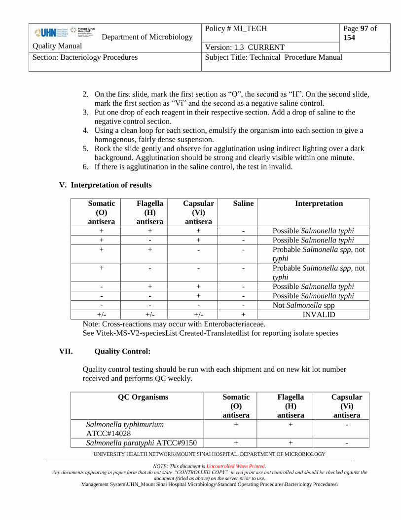
Department of Microbiology
Quality Manual
Policy # MI_TECH
Page 97 of
154
Version: 1.3 CURRENT
Section: Bacteriology Procedures Subject Title: Technical Procedure Manual
UNIVERSITY HEALTH NETWORK/MOUNT SINAI HOSPITAL, DEPARTMENT OF MICROBIOLOGY
NOTE: This document is Uncontrolled When Printed.
Any documents appearing in paper form that do not state "CONTROLLED COPY” in red print are not controlled and should be checked against the
document (titled as above) on the server prior to use. Management System\UHN_Mount Sinai Hospital Microbiology\Standard Operating Procedures\Bacteriology Procedures\
2. On the first slide, mark the first section as “O”, the second as “H”. On the second slide,
mark the first section as “Vi” and the second as a negative saline control.
3. Put one drop of each reagent in their respective section. Add a drop of saline to the
negative control section.
4. Using a clean loop for each section, emulsify the organism into each section to give a
homogenous, fairly dense suspension.
5. Rock the slide gently and observe for agglutination using indirect lighting over a dark
background. Agglutination should be strong and clearly visible within one minute.
6. If there is agglutination in the saline control, the test in invalid.
V. Interpretation of results
Somatic
(O)
antisera
Flagella
(H)
antisera
Capsular
(Vi)
antisera
Saline Interpretation
+ + + - Possible Salmonella typhi
+ - + - Possible Salmonella typhi
+ + - - Probable Salmonella spp, not
typhi
+ - - - Probable Salmonella spp, not
typhi
- + + - Possible Salmonella typhi
- - + - Possible Salmonella typhi
- - - - Not Salmonella spp
+/- +/- +/- + INVALID
Note: Cross-reactions may occur with Enterobacteriaceae.
See Vitek-MS-V2-speciesList Created-Translatedlist for reporting isolate species
VII. Quality Control:
Quality control testing should be run with each shipment and on new kit lot number
received and performs QC weekly.
QC Organisms Somatic
(O)
antisera
Flagella
(H)
antisera
Capsular
(Vi)
antisera
Salmonella typhimurium
ATCC#14028
+ + -
Salmonella paratyphi ATCC#9150 + + -

Department of Microbiology
Quality Manual
Policy # MI_TECH
Page 98 of
154
Version: 1.3 CURRENT
Section: Bacteriology Procedures Subject Title: Technical Procedure Manual
UNIVERSITY HEALTH NETWORK/MOUNT SINAI HOSPITAL, DEPARTMENT OF MICROBIOLOGY
NOTE: This document is Uncontrolled When Printed.
Any documents appearing in paper form that do not state "CONTROLLED COPY” in red print are not controlled and should be checked against the
document (titled as above) on the server prior to use. Management System\UHN_Mount Sinai Hospital Microbiology\Standard Operating Procedures\Bacteriology Procedures\
Salmonella typhi ATCC#19430 - + +
E.coli ATCC #25922 - - -
VIII. Reference:
Remel Salmonella Polyvalent Agglutination Sera insert, June 2013.

Department of Microbiology
Quality Manual
Policy # MI_TECH
Page 99 of
154
Version: 1.3 CURRENT
Section: Bacteriology Procedures Subject Title: Technical Procedure Manual
UNIVERSITY HEALTH NETWORK/MOUNT SINAI HOSPITAL, DEPARTMENT OF MICROBIOLOGY
NOTE: This document is Uncontrolled When Printed.
Any documents appearing in paper form that do not state "CONTROLLED COPY” in red print are not controlled and should be checked against the
document (titled as above) on the server prior to use. Management System\UHN_Mount Sinai Hospital Microbiology\Standard Operating Procedures\Bacteriology Procedures\
SHIGELLA SEROLOGICAL TESTING
I. Introduction:
Serological testing is performed to identify Shigella species. Testing is performed by a slide
agglutination test using (O) antisera.
II. Reagent:
Remel Shigella Polyvalent Agglutination Sera:
1. Shigella dysenteriae polyvalent (1-10)
2. Shigella flexneri polyvalent (1-6, x, y)
3. Shigella sonnei phase 1 & 2
4. Shigella boydii polyvalent 1 (1-6)
5. Shigella boydii polyvalent 2 (7-11)
6. Shigella boydii polyvalent 3 (12-15)
Sera should be stored at 2-8°C and used until the expiration date shown on the bottle label.
III. Materials:
1. Glass slide
2. Bacteriology loop
3. Sterile saline
4. China marker
IV. Procedure:
Testing should be performed on suspect colonies from a fresh culture (18-24hours) sub
cultured on a non-selective agar e.g. BA, TSI. Do not use MAC or HEK plates to perform
agglutination test.
1. With a China-marker, divide 6 slides into two. Make a circle in each section.
2. Put a drop of saline in each circle.
3. Using a loop, emulsify the organism in each drop of saline to give a homogenous, fairly
dense suspension.
4. Add one drop of undiluted antiserum of each sera to one of the suspensions and mix. The
last suspension serves as a control.

Department of Microbiology
Quality Manual
Policy # MI_TECH
Page 100 of
154
Version: 1.3 CURRENT
Section: Bacteriology Procedures Subject Title: Technical Procedure Manual
UNIVERSITY HEALTH NETWORK/MOUNT SINAI HOSPITAL, DEPARTMENT OF MICROBIOLOGY
NOTE: This document is Uncontrolled When Printed.
Any documents appearing in paper form that do not state "CONTROLLED COPY” in red print are not controlled and should be checked against the
document (titled as above) on the server prior to use. Management System\UHN_Mount Sinai Hospital Microbiology\Standard Operating Procedures\Bacteriology Procedures\
5. Rock the slide gently and observe for agglutination using indirect lighting over a dark
background. Agglutination should be strong and clearly visible within one minute.
6. If there is agglutination in the saline control, the test is invalid.
7. If the isolate is biochemically a Shigella species, but fails to agglutinate with the grouping
sera, send isolate to PHOL for confirmation.

Department of Microbiology
Quality Manual
Policy # MI_TECH
Page 101 of
154
Version: 1.3 CURRENT
Section: Bacteriology Procedures Subject Title: Technical Procedure Manual
UNIVERSITY HEALTH NETWORK/MOUNT SINAI HOSPITAL, DEPARTMENT OF MICROBIOLOGY
NOTE: This document is Uncontrolled When Printed.
Any documents appearing in paper form that do not state "CONTROLLED COPY” in red print are not controlled and should be checked against the
document (titled as above) on the server prior to use. Management System\UHN_Mount Sinai Hospital Microbiology\Standard Operating Procedures\Bacteriology Procedures\
V. Interpretation of Results:
Shigella
dysenteriae
polyvalent
(1-10)
Shigella
flexneri
polyvalent
(1-6, x, y)
Shigella
sonnei
phase 1
& 2
Shigella
boydii
polyvalent
1 (1-6)
Shigella
boydii
polyvalent
2 (7-11)
Shigella
boydii
polyvalent
3 (12-15)
Saline Interpretation
+ - - - - - - Shigella
dysenteriae
(1-10)
- + - - - - - Shigella
flexneri
(1-6, x, y)
- - + - - - - Shigella
sonnei phase
1 & 2
- - - + - - - Shigella
boydii (1-6)
- - - - + - - Shigella
boydii (7-11)
- - - - - + -
Shigella
boydii
(12-15)
- - - - - - - Negative
+/- +/- +/- +/- +/- +/- + INVALID
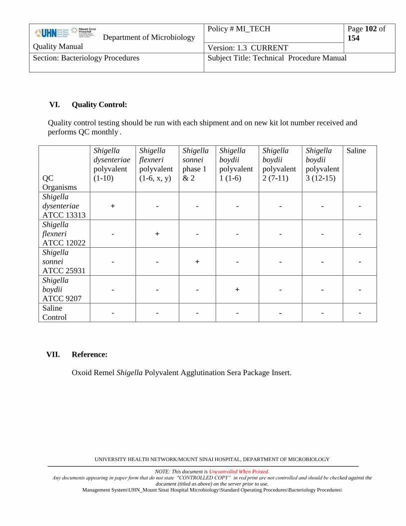
Department of Microbiology
Quality Manual
Policy # MI_TECH
Page 102 of
154
Version: 1.3 CURRENT
Section: Bacteriology Procedures Subject Title: Technical Procedure Manual
UNIVERSITY HEALTH NETWORK/MOUNT SINAI HOSPITAL, DEPARTMENT OF MICROBIOLOGY
NOTE: This document is Uncontrolled When Printed.
Any documents appearing in paper form that do not state "CONTROLLED COPY” in red print are not controlled and should be checked against the
document (titled as above) on the server prior to use. Management System\UHN_Mount Sinai Hospital Microbiology\Standard Operating Procedures\Bacteriology Procedures\
VI. Quality Control:
Quality control testing should be run with each shipment and on new kit lot number received and
performs QC monthly .
QC
Organisms
Shigella
dysenteriae
polyvalent
(1-10)
Shigella
flexneri
polyvalent
(1-6, x, y)
Shigella
sonnei
phase 1
& 2
Shigella
boydii
polyvalent
1 (1-6)
Shigella
boydii
polyvalent
2 (7-11)
Shigella
boydii
polyvalent
3 (12-15)
Saline
Shigella
dysenteriae
ATCC 13313
+ - - - - - -
Shigella
flexneri
ATCC 12022
- + - - - - -
Shigella
sonnei
ATCC 25931
- - + - - - -
Shigella
boydii
ATCC 9207
- - - + - - -
Saline
Control - - - - - - -
VII. Reference:
Oxoid Remel Shigella Polyvalent Agglutination Sera Package Insert.

Department of Microbiology
Quality Manual
Policy # MI_TECH
Page 103 of
154
Version: 1.3 CURRENT
Section: Bacteriology Procedures Subject Title: Technical Procedure Manual
UNIVERSITY HEALTH NETWORK/MOUNT SINAI HOSPITAL, DEPARTMENT OF MICROBIOLOGY
NOTE: This document is Uncontrolled When Printed.
Any documents appearing in paper form that do not state "CONTROLLED COPY” in red print are not controlled and should be checked against the
document (titled as above) on the server prior to use. Management System\UHN_Mount Sinai Hospital Microbiology\Standard Operating Procedures\Bacteriology Procedures\
SHIGELLA WELLCOLEX LATEX AGGLUTINATION
I. Introduction:
Wellcolex Colour Shigella provides a simple, rapid qualitative procedure for detection and
species identification of shigella present on solid culture media.
II. Reagent:
1. Latex reagent 1
2. Latex reagent 2
3. Red Positive Control
4. Blue Positive Control
Reagent kot to be sotred at 2 to 8°C and used until the expiration date shown on the box.
III. Materials:
Materials provided:
1. Disposable sampling sticks
2. Disposable reaction cards
3. Disposable sample dispensers
4. Disposable suspension tubes
Material required but not provided:
1. 0.45% sterile saline
2. The test should be performed on a flatbed rotator operating at approximately 80 rpm.
IV. Procedure:
1. Use disposable sample dispensers dispense 0.5 ml saline in the disposable suspension
tubes.
2. From an overnight culture pick few colonies average-sized (1 to 2 mm) suspected
Shigella colonies from the culture plate using the flat end of a sampling stick and
emulsify the bacteria in the saline.
3. Resuspend latex reagent 1 and 2 by shaking vigorously for a few seconds.

Department of Microbiology
Quality Manual
Policy # MI_TECH
Page 104 of
154
Version: 1.3 CURRENT
Section: Bacteriology Procedures Subject Title: Technical Procedure Manual
UNIVERSITY HEALTH NETWORK/MOUNT SINAI HOSPITAL, DEPARTMENT OF MICROBIOLOGY
NOTE: This document is Uncontrolled When Printed.
Any documents appearing in paper form that do not state "CONTROLLED COPY” in red print are not controlled and should be checked against the
document (titled as above) on the server prior to use. Management System\UHN_Mount Sinai Hospital Microbiology\Standard Operating Procedures\Bacteriology Procedures\
Dispense one drop of each latex reagent into a separate circle on a reaction card without
bubbles.
4. Transfer one drop of bacterial suspension to each of the two circles.
5. Using a sampling stick, mix the contents of each circle and spread to cover the whole
area of the circle.
6. Place the card on the flat bed rotator and run at 80 + 5 rpm for 2 minutes.
V. Interpretation of results
Colour of
clumps
Reagent 1 Reagent 2
Red Shigella sonnei Shigella dysenteriae
Blue Shigella flexneri Shigella boydii
None Negative
Purple Not interpretable
(further isolation procedures required)
Positive reaction report:
Shigella species,
“~presumptive identification, confirmation to follow.”
“~NOTE: Occasionally, some E. coli may identify as presumptive Shigella species using our current
in-lab methodology; confirmation by Public Health Lab reference methodology to follow.”
VII. Quality Control:
Quality control testing should be run with each shipment and on new kit lot number received and
performs QC weekly.
Positive Control Procedure:
1. Into 4 separate reaction circles, dispense 2 drops of latex reagent 1 and 2 (one drop per circle).
2. Add one drop of each positive control to 2 of the reaction circle (one containing latex reagent 1
and one containing latex reagent 2).
3. Mix the reagents using a separate disposable sampling stick for each positive control.
4. Rotate the card at 150 + 5 rpm for 2 minutes on a rotator.
Department of Microbiology
Quality Manual
Policy # MI_TECH
Page 105 of
154
Version: 1.3 CURRENT
Section: Bacteriology Procedures Subject Title: Technical Procedure Manual
UNIVERSITY HEALTH NETWORK/MOUNT SINAI HOSPITAL, DEPARTMENT OF MICROBIOLOGY
NOTE: This document is Uncontrolled When Printed.
Any documents appearing in paper form that do not state "CONTROLLED COPY” in red print are not controlled and should be checked against the
document (titled as above) on the server prior to use. Management System\UHN_Mount Sinai Hospital Microbiology\Standard Operating Procedures\Bacteriology Procedures\
The color of the agglutinated latex in reagent 1 and reagent 2 should correspond to the color of the
positive control (blue or red).
Negative Control Procedure:
Repeat the test procedure using saline instead of the test sample. There should be no significant
agglutination.
VIII. Reference:
1. Oxoid Wellcolex Colour Shigella insert
Department of Microbiology
Quality Manual
Policy # MI_TECH
Page 106 of
154
Version: 1.3 CURRENT
Section: Bacteriology Procedures Subject Title: Technical Procedure Manual
UNIVERSITY HEALTH NETWORK/MOUNT SINAI HOSPITAL, DEPARTMENT OF MICROBIOLOGY
NOTE: This document is Uncontrolled When Printed.
Any documents appearing in paper form that do not state "CONTROLLED COPY” in red print are not controlled and should be checked against the
document (titled as above) on the server prior to use. Management System\UHN_Mount Sinai Hospital Microbiology\Standard Operating Procedures\Bacteriology Procedures\
SPOT INDOLE
Principle
Spot indole reagent is for determination of the ability of micororganisms to spit indole from the
tryptophan molecule by the spot test method.
Reagents and Materials
Inoculating loop
Spot Indole Reagent – store product in at 2-30°C
Filter paper
Procedure
1. Dispense 1 of 2 drops of reagent onto a piece of filter paper.
2. Smear growth from culture onto saturated filter paper
3. Observe for blue colour within 1-3 minutes
Interpretation
Positive Test – Blue colour within 3 minutes
Negative Test – Pink colour
Precautions
Product should not be used if reagent colour has changed
Test colonies from media without glucose
Do not test from MAC or EBM agar. May produce false positives.
Certain strains of Proteus vulgaris, Providencia spp., and Aeromonas spp. Will give false
negative results.

Department of Microbiology
Quality Manual
Policy # MI_TECH
Page 107 of
154
Version: 1.3 CURRENT
Section: Bacteriology Procedures Subject Title: Technical Procedure Manual
UNIVERSITY HEALTH NETWORK/MOUNT SINAI HOSPITAL, DEPARTMENT OF MICROBIOLOGY
NOTE: This document is Uncontrolled When Printed.
Any documents appearing in paper form that do not state "CONTROLLED COPY” in red print are not controlled and should be checked against the
document (titled as above) on the server prior to use. Management System\UHN_Mount Sinai Hospital Microbiology\Standard Operating Procedures\Bacteriology Procedures\
Quality Control
Positive: E. coli ATCC 25922
Negative: Proteus mirabilis ATCC 12453
Reference
Remel Thermo Scientific package insert July 2012.

Department of Microbiology
Quality Manual
Policy # MI_TECH
Page 108 of
154
Version: 1.3 CURRENT
Section: Bacteriology Procedures Subject Title: Technical Procedure Manual
UNIVERSITY HEALTH NETWORK/MOUNT SINAI HOSPITAL, DEPARTMENT OF MICROBIOLOGY
NOTE: This document is Uncontrolled When Printed.
Any documents appearing in paper form that do not state "CONTROLLED COPY” in red print are not controlled and should be checked against the
document (titled as above) on the server prior to use. Management System\UHN_Mount Sinai Hospital Microbiology\Standard Operating Procedures\Bacteriology Procedures\
STAINING METHODS
ROUTINE STAINS
GRAM STAIN
Principle
Bacteria can be recognized as gram positive (blue-black/purple) if they retain the primary dye complex
of crystal violet and iodine in the face of attempted decolourization, or as gram negative (pink) if
decolourization occurs as shown by the cell accepting the counterstain safranin.
Generally the mechanism of the Gram stain is: The fixed bacteria are stained with the
triphenylmethane dye, crystal violet. Next the smear is flooded with Grams solution which
oxidatively forms an insoluble complex with the crystal violet. The smear is then flooded with the
organic solvent, acetone-alcohol. Depending on cell permeability the crystal violet-iodine complex
will be washed from Gram negative bacteria in solvent but not from Gram positive bacteria. Upon
counterstaining with safranin, organisms which had been discolorized by the ethanol (Gram negative)
will stain pink. Gram positive organisms which retained the crystal violet will appear blue-
black/purple microscopically.
Materials
Methanol
Crystal violet solution
Grams Iodine solution
Acetone alcohol
Safranin solution
Store solutions at room temperature. Refer to MSDS for storage requirements of alcohols.
Procedure
1. Prepare the film on the slide and allow to air dry.
DO NOT HEAT TO DRY FILM.
2. When film is dry, flood slide with methanol to fix smear. Let smear dry completely before
staining.
3. Flood slide with crystal violet - leave 1 minute.

Department of Microbiology
Quality Manual
Policy # MI_TECH
Page 109 of
154
Version: 1.3 CURRENT
Section: Bacteriology Procedures Subject Title: Technical Procedure Manual
UNIVERSITY HEALTH NETWORK/MOUNT SINAI HOSPITAL, DEPARTMENT OF MICROBIOLOGY
NOTE: This document is Uncontrolled When Printed.
Any documents appearing in paper form that do not state "CONTROLLED COPY” in red print are not controlled and should be checked against the
document (titled as above) on the server prior to use. Management System\UHN_Mount Sinai Hospital Microbiology\Standard Operating Procedures\Bacteriology Procedures\
4. Wash gently with water.
5. Flood slide with Grams Iodine - leave 1 minute.
6. Wash iodine from slide with acetone-alcohol mixture. Add a few more drops of acetone-
alcohol until no more colour comes from film - usually 30 seconds.
7. Wash gently with water.
8. Flood slide with safranin - leave 1 minute.
9. Wash gently with water. Clean back of slide with tissue and place slide in tray.
Precaution
1. At no time should the film (smear) be exposed to too much heat. When the specimen is still
wet, heat causes coagulation of the protein resulting in heavy overstaining which cannot be
removed by the decolourizer. A thick smear will also show more tendency to "lift off" during
staining.
2. Rinsing the Grams Iodine off with the decolorizer gives more stability to the CV-GI complex
and false over decolorizing will not take place.
3. Flooding a hot slide with crystal violet will cause the stain to precipitate and make
decolourizing much more difficult.
Note: Gram positive bacilli which are beaded or branching should be stained using the Modifed
Kinyoun and non-modified KINYOUN
Quality Control
Run control slides concurrently with unknowns at least once daily using known smears containing
Gram positive and Gram negative bacteria.

Department of Microbiology
Quality Manual
Policy # MI_TECH
Page 110 of
154
Version: 1.3 CURRENT
Section: Bacteriology Procedures Subject Title: Technical Procedure Manual
UNIVERSITY HEALTH NETWORK/MOUNT SINAI HOSPITAL, DEPARTMENT OF MICROBIOLOGY
NOTE: This document is Uncontrolled When Printed.
Any documents appearing in paper form that do not state "CONTROLLED COPY” in red print are not controlled and should be checked against the
document (titled as above) on the server prior to use. Management System\UHN_Mount Sinai Hospital Microbiology\Standard Operating Procedures\Bacteriology Procedures\
GRAM STAINING MACHINE-MIDAS III OPERATION
Principle
Adapting the principle of Gram Stain, the MIDAS III will carry fixed smears through different
staining solutions for a pre-set time to produced a Gram stained smear.
Materials
Staining Rack
Methanol
Crystal violet solution
Gram’s Iodine solution
Acetone alcohol (50%/50%)
Safranin solution
Store solutions at room temperature. Refer to MSDS for storage requirements of alcohols.
Instrument Programming Procedure
1. Press “Program”
2. Follow the instructions on the screen and program the duration of each staining step as follow:
For Program 1 (thin smears)
Step Station Reagent Time Set
1 1 Crystal Violet 1 minute
2 5 Water 1 minute; flow rate 1500
3 2 Gram’s Iodine 1 minute
4 5 Water 1 minute; flow rate 1500
5 3 Acetone Alcohol 20 seconds
6 5 Water 1 minute; flow rate 1500
7 4 Safranin 1 minute
8 5 Water 1 minute; flow rate 1500
9 6 Dry bath 2 minutes
Department of Microbiology
Quality Manual
Policy # MI_TECH
Page 111 of
154
Version: 1.3 CURRENT
Section: Bacteriology Procedures Subject Title: Technical Procedure Manual
UNIVERSITY HEALTH NETWORK/MOUNT SINAI HOSPITAL, DEPARTMENT OF MICROBIOLOGY
NOTE: This document is Uncontrolled When Printed.
Any documents appearing in paper form that do not state "CONTROLLED COPY” in red print are not controlled and should be checked against the
document (titled as above) on the server prior to use. Management System\UHN_Mount Sinai Hospital Microbiology\Standard Operating Procedures\Bacteriology Procedures\
For Program 2 (thick smears):
Step Station Reagent Time Set
1 1 Crystal Violet 1 minute
2 5 Water 1 minute; flow rate 1500
3 2 Gram’s Iodine 1 minute
4 5 Water 1 minute; flow rate 1500
5 3 Acetone Alcohol 45 seconds
6 5 Water 1 minute; flow rate 1500
7 4 Safranin 1.5 minutes
8 5 Water 1 minute; flow rate 1500
9 6 Dry bath 2 minutes
3. Press “End” to save program.
Procedure
Daily Set-up:
1. Remove staining station covers.
2. Discard old stains.
3. Wash containers with tap water.
4. Fill containers with fresh stains/decolourizer.
5. Record stain change in LIS micqc.
Staining Procedure:
1. Fix dry smears with methanol and air dry completely.
2. Load fixed smears on MIDAS III slide holder
3. Load holder onto the prongs of MIDAS III.
4. Press “RUN”, then “1” for regular thin smears OR “2” for thicker sputum smears.
5. Press “Enter”
6. Remove smear holder when staining is complete.
Quality Control
Run control slides concurrently with unknowns at least once daily using known smears containing
Gram positive and Gram negative bacteria.

Department of Microbiology
Quality Manual
Policy # MI_TECH
Page 112 of
154
Version: 1.3 CURRENT
Section: Bacteriology Procedures Subject Title: Technical Procedure Manual
UNIVERSITY HEALTH NETWORK/MOUNT SINAI HOSPITAL, DEPARTMENT OF MICROBIOLOGY
NOTE: This document is Uncontrolled When Printed.
Any documents appearing in paper form that do not state "CONTROLLED COPY” in red print are not controlled and should be checked against the
document (titled as above) on the server prior to use. Management System\UHN_Mount Sinai Hospital Microbiology\Standard Operating Procedures\Bacteriology Procedures\
Reference
MIDAS III operation manual.

Department of Microbiology
Quality Manual
Policy # MI_TECH
Page 113 of
154
Version: 1.3 CURRENT
Section: Bacteriology Procedures Subject Title: Technical Procedure Manual
UNIVERSITY HEALTH NETWORK/MOUNT SINAI HOSPITAL, DEPARTMENT OF MICROBIOLOGY
NOTE: This document is Uncontrolled When Printed.
Any documents appearing in paper form that do not state "CONTROLLED COPY” in red print are not controlled and should be checked against the
document (titled as above) on the server prior to use. Management System\UHN_Mount Sinai Hospital Microbiology\Standard Operating Procedures\Bacteriology Procedures\
BACTO 3-STEP GRAM STAIN PROCEDURE
Principle
To be used for problem smears to determine the Gram reaction of organisms.
Materials
3-Step Stabilized Iodine Technique
Bacto Gram Crystal Violet
Bacto Stabilized Gram Iodine
Bacto 3-Step Gram Safranin-S
3-Step Technical Iodine Technique
Bacto Gram Crystal Violet
Bacto Gram Iodine
Bacto 3-Step Gram Safranin-T
Microscope slides
Bunsen burner or methanol
Bacteriological loop
Swabs
Blotting paper
Microscope with oil immersion lens
Bactrol Gram Slide
Bactrol Disks
Department of Microbiology
Quality Manual
Policy # MI_TECH
Page 114 of
154
Version: 1.3 CURRENT
Section: Bacteriology Procedures Subject Title: Technical Procedure Manual
UNIVERSITY HEALTH NETWORK/MOUNT SINAI HOSPITAL, DEPARTMENT OF MICROBIOLOGY
NOTE: This document is Uncontrolled When Printed.
Any documents appearing in paper form that do not state "CONTROLLED COPY” in red print are not controlled and should be checked against the
document (titled as above) on the server prior to use. Management System\UHN_Mount Sinai Hospital Microbiology\Standard Operating Procedures\Bacteriology Procedures\
Procedure
1. Flood the fixed smear with primary stain (Bacto Gram Crystal Violet) and stain for 1 minute.
2. Remove the primary stain by gently washing with cold tap water.
3. Flood the slide with mordant (Bacto Stabilized Gram Iodine or Bacto Gram Iodine (traditional
formulation) and retain on the slide for 1 minute. (Refer to LIMITATIONS OF THE
PROCEDURE, #5)
4. Wash off the mordant with decolourizer / counterstain (Bacto 3-Step Gram Safranin-S or Bacto
3-Step Gram Safranin-T). (NOTE: Do not wash off iodine with water). Add more
decolourizer / counterstain solution to the slide and stain 20-50 seconds.
5. Remove the decolourizer / counterstain solution by gently washing the slide with cold tap
water.
6. Blot with blotting paper or paper towel or allow to air dry.
7. Examine the smear under an oil immersion lens.
Interpretation
REACTION 3-STEP TECHNIQUE
using either Bacto Gram Safranin-S or Bacto
Gram Safranin-T
Gram-positive
Gram-negative
Purple-black
to purple cells
Red-pink to
fuchsia cells
Quality Control
Run controls daily using 18-24 hour cultures of known gram-positive and gram-negative
microorganisms.

Department of Microbiology
Quality Manual
Policy # MI_TECH
Page 115 of
154
Version: 1.3 CURRENT
Section: Bacteriology Procedures Subject Title: Technical Procedure Manual
UNIVERSITY HEALTH NETWORK/MOUNT SINAI HOSPITAL, DEPARTMENT OF MICROBIOLOGY
NOTE: This document is Uncontrolled When Printed.
Any documents appearing in paper form that do not state "CONTROLLED COPY” in red print are not controlled and should be checked against the
document (titled as above) on the server prior to use. Management System\UHN_Mount Sinai Hospital Microbiology\Standard Operating Procedures\Bacteriology Procedures\
ACRIDINE ORANGE STAIN
Principle
Acridine orange is a fluorescent dye which will bind to the nucleic acid of bacteria and other cells. It is
recommended for use for the detection of microorganisms in direct smears. It is useful for the rapid
screening of specimens from normally sterile sites (eg. CSF) and blood smears, or smears containing
proteinaceous material where differentiation of organisms from background material may be difficult.
Reagents
Acridine Orange spot test dropper. Stored at room temperature
Absolute Methanol
Procedure
1. Prepare a smear of the specimen to be stained.
2. Allow to air dry.
3. Fix with methanol for 1 to 2 minutes.
4. Hold the dropper upright and squeeze gently to crush the glass ampoule inside the dispenser.
5. Flood the slide with the acridine orange and stain for 2 minutes.
6. Rinse thoroughly with tap water and allow to air dry.
7. Examine with a fluorescent microscope using low and oil immersion objectives.
Interpretation
Bacteria and fungus stain bright orange. The background appears black to yellow green. Leukocytes will
stain yellow, orange and red.

Department of Microbiology
Quality Manual
Policy # MI_TECH
Page 116 of
154
Version: 1.3 CURRENT
Section: Bacteriology Procedures Subject Title: Technical Procedure Manual
UNIVERSITY HEALTH NETWORK/MOUNT SINAI HOSPITAL, DEPARTMENT OF MICROBIOLOGY
NOTE: This document is Uncontrolled When Printed.
Any documents appearing in paper form that do not state "CONTROLLED COPY” in red print are not controlled and should be checked against the
document (titled as above) on the server prior to use. Management System\UHN_Mount Sinai Hospital Microbiology\Standard Operating Procedures\Bacteriology Procedures\
Quality Control
Stain a smear of Streptococcus pneumoniae (ATCC 6303) each time the test is performed.
References
1. BD Acridine Orange Stain package insert. June 2010.

Department of Microbiology
Quality Manual
Policy # MI_TECH
Page 117 of
154
Version: 1.3 CURRENT
Section: Bacteriology Procedures Subject Title: Technical Procedure Manual
UNIVERSITY HEALTH NETWORK/MOUNT SINAI HOSPITAL, DEPARTMENT OF MICROBIOLOGY
NOTE: This document is Uncontrolled When Printed.
Any documents appearing in paper form that do not state "CONTROLLED COPY” in red print are not controlled and should be checked against the
document (titled as above) on the server prior to use. Management System\UHN_Mount Sinai Hospital Microbiology\Standard Operating Procedures\Bacteriology Procedures\
EOSINOPHIL STAIN
Principle
A stain for the detection of eosinophils in clinical specimens
Reagents
Methanol
AJP Scientific Eosinophil stain:
Solution I - Eosin Y
Solution II - Buffer Ph 6.5
Solution III - Methylene Blue
Stored at room temperature
Store reagents at room temperature. Store alcohols as per MSDS.
Procedure
1. Make a thin smear and spread evenly.
2. Fix slide by air drying or with methanol
3. Cover slide with solution I and leave for 30 seconds.
4. Add solution II to cover slide. Mix gently and allow to stain for 3 to 5 minutes.
5. Wash off with tap water and drain.
6. Cover slide with solution III and immediately wash off with tap water. Drain and air dry.
Interpretation
Eosinophils are stained with red cytoplasm and bright red granules.
Reference
1. A.J.P. Scientific INC package insert.

Department of Microbiology
Quality Manual
Policy # MI_TECH
Page 118 of
154
Version: 1.3 CURRENT
Section: Bacteriology Procedures Subject Title: Technical Procedure Manual
UNIVERSITY HEALTH NETWORK/MOUNT SINAI HOSPITAL, DEPARTMENT OF MICROBIOLOGY
NOTE: This document is Uncontrolled When Printed.
Any documents appearing in paper form that do not state "CONTROLLED COPY” in red print are not controlled and should be checked against the
document (titled as above) on the server prior to use. Management System\UHN_Mount Sinai Hospital Microbiology\Standard Operating Procedures\Bacteriology Procedures\
STAINS FOR GRAM POSITIVE BRANCHING / BEADED BACILLI
ACID FAST STAIN FOR MYCOBACTERIA (KINYOUN)
Principle
To stain Mycobacteria present in specimens and cultures.
Mycobacteria are different to stain with common aniline dyes. However, they will stain with basic
fuchsin. Once stained, they retain the dye despite treatment with mineral acids i.e. HCl H2SO4. This
property of acid fastness may be due to a lipd fraction called mycolic acid. Mycobacteria also exhibit
degrees of resistance to decolourization with alcohol.
Materials
Kinyoun Carbol fuchsin
3% HCl in 95% ethanol
Brilliant green
Store reagents at room temperature. Store alcohols as per MSDS.
Procedure
1. Prepare smear over an area of 2-3 sq. cm.
2. Heat fix smear with methanol.
3. Place slide on stain rack and allow to cool. Flood with Kinyoun Carbol fuschsin for 5 min.
4. Rinse off stain with water.
5. Decolourize with 3% acid alcohol for 3 mins.
6. Rinse with water.
7. Repeat decolourization for 1-2 mins. or until no red appears.
8. Rinse with water.

Department of Microbiology
Quality Manual
Policy # MI_TECH
Page 119 of
154
Version: 1.3 CURRENT
Section: Bacteriology Procedures Subject Title: Technical Procedure Manual
UNIVERSITY HEALTH NETWORK/MOUNT SINAI HOSPITAL, DEPARTMENT OF MICROBIOLOGY
NOTE: This document is Uncontrolled When Printed.
Any documents appearing in paper form that do not state "CONTROLLED COPY” in red print are not controlled and should be checked against the
document (titled as above) on the server prior to use. Management System\UHN_Mount Sinai Hospital Microbiology\Standard Operating Procedures\Bacteriology Procedures\
9. Flood slide 3-4 mins. with Brilliant green.
10. Rinse with water.
11. Air dry. DO NOT BLOT.
Microscopy
Place a drop of oil between the specimen and coverslip and oil again on top. Smears are examined with
oil immersion lens. The coverslip prevents cross contamination.
Interpretation
The filaments of Mycobacterium species appear red-stained against a green background.
Quality Control
Mycobacterium Control Slide (Remel R40144) set up simultaneously with the processing of clinical
specimens.
Control Results:
Positive Control: Red-stained bacilli
Negative Control: Blue-green-stained bacilli
References
Baker, F.J., Breach, M.R. 1980. Medical Microbiological Technique, p. 15
Murray, PA. et al. Manual of Clinical Microbiology, 8th
edition, 2003 ASM, Washington, D.C.
Department of Microbiology
Quality Manual
Policy # MI_TECH
Page 120 of
154
Version: 1.3 CURRENT
Section: Bacteriology Procedures Subject Title: Technical Procedure Manual
UNIVERSITY HEALTH NETWORK/MOUNT SINAI HOSPITAL, DEPARTMENT OF MICROBIOLOGY
NOTE: This document is Uncontrolled When Printed.
Any documents appearing in paper form that do not state "CONTROLLED COPY” in red print are not controlled and should be checked against the
document (titled as above) on the server prior to use. Management System\UHN_Mount Sinai Hospital Microbiology\Standard Operating Procedures\Bacteriology Procedures\
ACID FAST STAIN FOR NOCARDIA (MODIFED KINYOUN)
Principle
Nocardia species possess the unique characteristic of resisting decolorization with 1%sulfuric acid.
Materials
1. Carbol-fuchsin
Basic fuchsin solution (3 g basic fuchsin in 100 mL 95% ethyl alcohol)
10 mL
Phenol 5% aqueous 90 mL
2. Decolourizer (1% sulfuric acid)
H2SO4 (concentrated) 1 mL
Distilled water 99 mL
3. Methylene blue
Methylene blue 0.3 g
Distilled water 100 mL
Store reagents at room temperature.
Procedure
Prepare 2 smears: one for the Modified Kinyoun (procedure as follows), the other slide to be stained
in parallel with KINYOUN method. * Do not make the smears too thick.
Retrieve a Modified Kinyoun control smear for parallel staining.
1. Shake stains before use
2. Fix the smear by methanol, allot to air dry.
3. Flood the slide with Carbol fuchsin and allow to stand for 5 minutes
4. Rinse Carbol Fuchsin off the slide with tap water for 5 seconds
5. Wash the slide with 50% ethanol using the squirt bottle. This should be done for 5-7 seconds
and all the film from the dye should be washed off.
6. Rinse slide with tap water for 5 seconds
7. Decolorize the slide by adding 1% sulfuric acid for 2 minutes. Place the slide on the rack over
the sink and add fresh 1% sulfuric acid every 30 seconds - at 0s, 30s, 60s, 90s)
8. Rinse with tap water for 5 seconds

Department of Microbiology
Quality Manual
Policy # MI_TECH
Page 121 of
154
Version: 1.3 CURRENT
Section: Bacteriology Procedures Subject Title: Technical Procedure Manual
UNIVERSITY HEALTH NETWORK/MOUNT SINAI HOSPITAL, DEPARTMENT OF MICROBIOLOGY
NOTE: This document is Uncontrolled When Printed.
Any documents appearing in paper form that do not state "CONTROLLED COPY” in red print are not controlled and should be checked against the
document (titled as above) on the server prior to use. Management System\UHN_Mount Sinai Hospital Microbiology\Standard Operating Procedures\Bacteriology Procedures\
9. Flood the slide with methylene blue for 1 minute to counter stain
10. Rinse off methylene blue dye with tap water and then let slide air dry
Microscopy
Place a drop of oil between the specimen and coverslip and oil again on top. Smears are examined with
oil immersion lens. The coverslip prevents cross contamination.
Interpretation
MODIFIED Kinyoun stain: the filaments of Nocardia species and Rhodococcus appear red-stained
against a blue background.
Kinyoun stain: the filaments of Nocardia species and Rhodococcus should NOT show red-stained
bacilli.
Quality Control
Positive control: Nocardia species.
Negative control: Streptomyces species.
References
Murray, PA. et al. Manual of Clinical Microbiology, 8th
edition, 2003 ASM, Washington, D.C.

Department of Microbiology
Quality Manual
Policy # MI_TECH
Page 122 of
154
Version: 1.3 CURRENT
Section: Bacteriology Procedures Subject Title: Technical Procedure Manual
UNIVERSITY HEALTH NETWORK/MOUNT SINAI HOSPITAL, DEPARTMENT OF MICROBIOLOGY
NOTE: This document is Uncontrolled When Printed.
Any documents appearing in paper form that do not state "CONTROLLED COPY” in red print are not controlled and should be checked against the
document (titled as above) on the server prior to use. Management System\UHN_Mount Sinai Hospital Microbiology\Standard Operating Procedures\Bacteriology Procedures\
REPORTING FOR GRAM POSITIVE BRACHING / BEADED BACILLI
If initial Gram smear shows:
1+ pus cells
3+ gram positive bacilli, branching
or
3+ gram positive bacilli , beaded
Stain new smear with Modified Kinyoun and KINYOUN for further characterization
Reporting:
Report as GRAM Test Comment:
Both modified and non-modified Kinyoun stains = negative:
Both Modified acid-fast and acid-fast stains: negative.
This suggests possible Actnomyces species. Culture confirmation to follow.
Both modified and non-modified Kinyoun stains = positive:
Modified acid-fast stain: positive. Acid-fast stain: positive. This suggests possible Mycobacterium
species. Culture confirmation to follow.
Modified Kinyoun stain = positive; non-modified Kinyoun stain = negative:
Modified acid-fast stain: positive. Acid-fast stain: negative. This suggests possible Nocardia species.
Culture confirmation to follow.

Department of Microbiology
Quality Manual
Policy # MI_TECH
Page 123 of
154
Version: 1.3 CURRENT
Section: Bacteriology Procedures Subject Title: Technical Procedure Manual
UNIVERSITY HEALTH NETWORK/MOUNT SINAI HOSPITAL, DEPARTMENT OF MICROBIOLOGY
NOTE: This document is Uncontrolled When Printed.
Any documents appearing in paper form that do not state "CONTROLLED COPY” in red print are not controlled and should be checked against the
document (titled as above) on the server prior to use. Management System\UHN_Mount Sinai Hospital Microbiology\Standard Operating Procedures\Bacteriology Procedures\
FLUOROCHROME STAIN for MYCOBACTERIUM
Principle
Mycobacteria possess the unique characteristic of resisting decolorization with acid alcohol as well as
staining poorly, if at all with Gram stain.
Materials
BD TB Fluorescent Stain Kit (REF 212521):
Auramine stain solution
Decolorizer solution (Hydrochloric acid/Isopropanol)
Potassium permanganate solution (0.5%)
Store reagents at room temperature.
Procedure
1. Place heat fixed smear on a staining rack.
2. Flood the smear with the thoroughly mixed auramine solution and let stand for 20 minutes.
3. Rinse the smear with tap water.
4. Decolorize the smear with decolorizer for 2-3 minutes.
5. Rinse with tap water.
6. Flood the smear with Potassium permanganate counterstain and let stand for 4-5 minutes.
Excessive treatment (>5 min.) should be avoided as it may reduce the fluorescence of
stained bacilli.
7. Rinse with tap water and air dry.
8. Examine slide under the UV microscope.

Department of Microbiology
Quality Manual
Policy # MI_TECH
Page 124 of
154
Version: 1.3 CURRENT
Section: Bacteriology Procedures Subject Title: Technical Procedure Manual
UNIVERSITY HEALTH NETWORK/MOUNT SINAI HOSPITAL, DEPARTMENT OF MICROBIOLOGY
NOTE: This document is Uncontrolled When Printed.
Any documents appearing in paper form that do not state "CONTROLLED COPY” in red print are not controlled and should be checked against the
document (titled as above) on the server prior to use. Management System\UHN_Mount Sinai Hospital Microbiology\Standard Operating Procedures\Bacteriology Procedures\
Interpretation
Acid fast organisms: reddish-orange fluorescence.
Confirm all positive results with a second fluorochrome stained smear
Quality Control
Mycobacterium Control Slide (Oxoid Cat. No. NRE-0454) - stain simultaneously with the processing of
clinical specimens.
Expected Control Results:
Positive Control: bacilli with reddish-orange fluorescence
Negative Control: no fluorescence
Reference
Murray, PA. et al. Manual of Clinical Microbiology, 8th
edition, 2003 ASM, Washington, D.C.
BD TB Fluorescent Stain Kit T (REF 212521), Difco-BBL package insert

Department of Microbiology
Quality Manual
Policy # MI_TECH
Page 125 of
154
Version: 1.3 CURRENT
Section: Bacteriology Procedures Subject Title: Technical Procedure Manual
UNIVERSITY HEALTH NETWORK/MOUNT SINAI HOSPITAL, DEPARTMENT OF MICROBIOLOGY
NOTE: This document is Uncontrolled When Printed.
Any documents appearing in paper form that do not state "CONTROLLED COPY” in red print are not controlled and should be checked against the
document (titled as above) on the server prior to use. Management System\UHN_Mount Sinai Hospital Microbiology\Standard Operating Procedures\Bacteriology Procedures\
FUNGAL STAINS
FUNGI-FLUOR STAIN
Purpose
The Fungi-Fluor stain is used for the rapid identification of various fungal elements in fresh or
frozen clinical specimens.
Principle
The active, colourless, fluorescing dye in the staining solution is Cellufluor which is the disodium salt
of 4,4’-bis[4-anilino-6-bis-(2-hydroxyethel) amino-s-triazin-2-ylamino]-2,2'-stilbenedisulfonic acid.
Fungi-Fluor staining solution is a 0.05% solution of this dye in deionized water with potassium
hydroxide added as a clearing agent. The Fungi-Fluor counter staining solution B is an aqueous
solution of Evans Blue dye used to reduce background fluorescence. Cellufluor binds nonspecifically
to beta-linked polysaccharides found in chitin and cellulose which are present in fungal cell walls.
When exposed to long wave UV light, fungal cell walls will fluoresce.
NB: Collagen, elastin, cotton fibres, plant material, some cells, cell inclusions and parasite cyst
forms (e.g. Acanthamoeba) may fluoresce.
Materials
Staining Solution A
Counterstaining Solution B
Absolute alcohol
Water
Fluorescent Microscope (Leitz Ortholux with G filter module exciting filter BP 350-460, suppression
filter LP515 or equivalent)
Precautions
1. Store in a dark or opaque bottle, tightly sealed, at room temperature.

Department of Microbiology
Quality Manual
Policy # MI_TECH
Page 126 of
154
Version: 1.3 CURRENT
Section: Bacteriology Procedures Subject Title: Technical Procedure Manual
UNIVERSITY HEALTH NETWORK/MOUNT SINAI HOSPITAL, DEPARTMENT OF MICROBIOLOGY
NOTE: This document is Uncontrolled When Printed.
Any documents appearing in paper form that do not state "CONTROLLED COPY” in red print are not controlled and should be checked against the
document (titled as above) on the server prior to use. Management System\UHN_Mount Sinai Hospital Microbiology\Standard Operating Procedures\Bacteriology Procedures\
2. Avoid eye or skin contact: use gloves and protective glasses.
Procedure
1. Prepare smear of specimen and allow to air dry.
2. Fix on the rack with absolute methanol for 5 minutes until dry. Fixed smears can be held
indefinitely until ready to stain and examine.
3. Add one drop of Fungi-Fluor solution A (Cellufluor) for 1 minute.
4. Rinse gently with tap water.
5. Apply coverslip to wetted slide and examine with the fluorescent microscope using the
designated filter. If there is a delay, add fresh distilled water to the coverslip just prior to
examination.
6. Optional for thicker smears. Add few drops of the counterstain Fungi-Fluor solution B. Rinse
gently with tap water and then proceed as in step 5 above.
NB: Gram stained smears can be overstained with Fungi-Fluor after removing immersion oil with
alcohol. Similarly, Fung-Fluor stained slides may be overstained with other stains such as
GMS, PAS, Geimsa, etc.
Quality Control
Stain a smear of Candida albicans daily.
Interpretation
Use 25x or 40x objective.
Fungal cell walls will fluoresce apple-green. Observe for characteristic morphology to differentiate
from artifacts and background. When the couterstain is used, fungi will appear yellow-green against a
red-orange background.

Department of Microbiology
Quality Manual
Policy # MI_TECH
Page 127 of
154
Version: 1.3 CURRENT
Section: Bacteriology Procedures Subject Title: Technical Procedure Manual
UNIVERSITY HEALTH NETWORK/MOUNT SINAI HOSPITAL, DEPARTMENT OF MICROBIOLOGY
NOTE: This document is Uncontrolled When Printed.
Any documents appearing in paper form that do not state "CONTROLLED COPY” in red print are not controlled and should be checked against the
document (titled as above) on the server prior to use. Management System\UHN_Mount Sinai Hospital Microbiology\Standard Operating Procedures\Bacteriology Procedures\
Appearance of other structures / organisms:
i) Fungal elements - intense peripheral staining with characteristic morphology.
ii) Pneumocystis carinii - fainter staining cyst wall (5-7 m diameter) and intensely
staining internal "been-shaped" or "double-parenthesis-
like" structures with apposed sides flattened.
iii) Acanthamoeba sp. cysts - intensely staining double wall with wrinkled outer wall
(10-25 m diameter)
References
1. Manufacturers' Instructions (Data Sheet #316). Fungi-Fluor kit - Polysciences, Inc., Sept
2012.
2. V.S. Baselski et al. "Rapid Detection of Pneumocystis carinii in Bronchoalveolar Lavage
Samples by Using Cellufluor Staining". J. Clin. Micro. 28:393-394, Feb. 1990.
3. D. H. Larone. Medically Important Fungi. A Guide to Identification. 3rd
ed. 1995, ASM Press.

Department of Microbiology
Quality Manual
Policy # MI_TECH
Page 128 of
154
Version: 1.3 CURRENT
Section: Bacteriology Procedures Subject Title: Technical Procedure Manual
UNIVERSITY HEALTH NETWORK/MOUNT SINAI HOSPITAL, DEPARTMENT OF MICROBIOLOGY
NOTE: This document is Uncontrolled When Printed.
Any documents appearing in paper form that do not state "CONTROLLED COPY” in red print are not controlled and should be checked against the
document (titled as above) on the server prior to use. Management System\UHN_Mount Sinai Hospital Microbiology\Standard Operating Procedures\Bacteriology Procedures\
INDIA INK
Purpose
The procedure is applicable only to suspected positive Crytococcal cultures.
Procedure
1. For suspected Crytococcal cultures, make a wet preparation using saline on a clean glass slide,
then add a small drop of India Ink and mix.
2. Apply a large coverslip ((22 x 40 mm) over the mixture and press it gently to obtain a thin mount.
3. If India Ink is too thick (dark), dilute it by 50% with saline.
4. Allow the preparation to stand for few minutes to settle.
5. Scan under low power in reduced light. Switch to high power if necessary.
Interpretation
The mucoid capsule appears as a clear halo that surrounds the yeast cell or lies between the cell wall
and the surrounding black mass of India Ink particles. Capsules may be broad or narrow. The yeast
cells may be round, oval or elongate. Buds may be absent, single or rarely multiple and may be
detached from the mother cell but enclosed in a common capsule attached.
Reference
Murray PA, et al. Manual of Clinical Microbiology 7th
ed., 1999, ASM Press.

Department of Microbiology
Quality Manual
Policy # MI_TECH
Page 129 of
154
Version: 1.3 CURRENT
Section: Bacteriology Procedures Subject Title: Technical Procedure Manual
UNIVERSITY HEALTH NETWORK/MOUNT SINAI HOSPITAL, DEPARTMENT OF MICROBIOLOGY
NOTE: This document is Uncontrolled When Printed.
Any documents appearing in paper form that do not state "CONTROLLED COPY” in red print are not controlled and should be checked against the
document (titled as above) on the server prior to use. Management System\UHN_Mount Sinai Hospital Microbiology\Standard Operating Procedures\Bacteriology Procedures\
India Ink positive for Cryptococcus
Artefacts produced by reaction of India Ink (False Positive)

Department of Microbiology
Quality Manual
Policy # MI_TECH
Page 130 of
154
Version: 1.3 CURRENT
Section: Bacteriology Procedures Subject Title: Technical Procedure Manual
UNIVERSITY HEALTH NETWORK/MOUNT SINAI HOSPITAL, DEPARTMENT OF MICROBIOLOGY
NOTE: This document is Uncontrolled When Printed.
Any documents appearing in paper form that do not state "CONTROLLED COPY” in red print are not controlled and should be checked against the
document (titled as above) on the server prior to use. Management System\UHN_Mount Sinai Hospital Microbiology\Standard Operating Procedures\Bacteriology Procedures\
LACTOPHENOL ANILINE BLUE (LPCB)
Purpose
To determine the morphology of the conidiogenous cells and the conidia that they give rise to in order
to identify a filamentous fungus.
Principle
LPCB contains lactic acid as a clearing agent, phenol as a disinfectant, glycerol to prevent drying and
Aniline Blue which is the dye that stains fungi. LPCB is a wet preparation.
Procedure
1) TEASE PREP
The test must be performed in the Laminar Airflow Biosafety Cabinet. First, observe the gross
morphology of the colony carefully to determine whether or not the culture is mouldy, granular or
a mixture of both. It is important to prepare the LPCB preparation by "teasing" the fungus not
"chopping" it.
Materials required for LPCB staining:
1. LPCB reagent
2. Probe to get the specimen
3. Teasing needles
4. Glass slides
5. Coverslips
6. Lead or wax pencils
7. Disinfectant bucket
8. Electric incinerator
9. Clear nail polish
10. Slide tray
Tease Prep Procedure:
1. Sterilize the loop and the needles in the incinerator and allow them to cool.
2. Label slide, place 2 drops of LPCB reagent on the slide.

Department of Microbiology
Quality Manual
Policy # MI_TECH
Page 131 of
154
Version: 1.3 CURRENT
Section: Bacteriology Procedures Subject Title: Technical Procedure Manual
UNIVERSITY HEALTH NETWORK/MOUNT SINAI HOSPITAL, DEPARTMENT OF MICROBIOLOGY
NOTE: This document is Uncontrolled When Printed.
Any documents appearing in paper form that do not state "CONTROLLED COPY” in red print are not controlled and should be checked against the
document (titled as above) on the server prior to use. Management System\UHN_Mount Sinai Hospital Microbiology\Standard Operating Procedures\Bacteriology Procedures\
3. Cut a small piece of the fungus from a granular or colored part of the colony, somewhere away
from the central part towards the periphery and place the piece of the fungus in the LPCB in
the upside down position.
4. Hold the thallus with the needle and gently tease the inverted side of the specimen into the
staining (LPCB) fluid.
5. After enough teasing, remove all the solid particles and the agar from the mixture and discard
in the disinfectant container.
6. Put a coverslip gently onto the LPCB preparation and hold the slide preparation briefly over
the incinerator opening. Heating the slide will help to stain the cell wall of the fungi and kill
the spores on the surface of the slide.
7. Seal the preparation with nail polish if necessary for a permanent mount.
8. Examine under the light microscope using the low power objective.
2) SCOTCH TAPE PREP FROM PLATE CULTURE
(Not done on suspected dimorphic fungi)
1. Place a drop of LPCB onto a clean glass slide, make a circle with Vaseline.
2. Take a small piece of clear scotch tape and loop back on itself with sticky sides out.
3. Hold the tip of the loop securely with forceps.
4. Press the sticky side firmly to the surface of the fungal colony.
5. Pull the tape gently away from the colony.
6. Open up the tape strip and place it on the drop of LPCB on the glass slide, making sure
that the entire sticky side adheres to the slide. Add cover slip..
7. Examine under the light microscope.
Reference
Davise H. Larone. Medically Important Fungi. A Guide to Identification. 3rd ed. 1995, ASM Press.
Martha E. Kern. Medical Mycology. 1985.

Department of Microbiology
Quality Manual
Policy # MI_TECH
Page 132 of
154
Version: 1.3 CURRENT
Section: Bacteriology Procedures Subject Title: Technical Procedure Manual
UNIVERSITY HEALTH NETWORK/MOUNT SINAI HOSPITAL, DEPARTMENT OF MICROBIOLOGY
NOTE: This document is Uncontrolled When Printed.
Any documents appearing in paper form that do not state "CONTROLLED COPY” in red print are not controlled and should be checked against the
document (titled as above) on the server prior to use. Management System\UHN_Mount Sinai Hospital Microbiology\Standard Operating Procedures\Bacteriology Procedures\
STAPHAUREX TEST
Principle
A rapid slide latex test for the detection of clumping factor and protein A produced by most strains of
S. aureus.
Reagents and Materials
Staphaureux latex suspension (store refrigerated 2-8°C)
Disposable reaction cards
Culture loop or wooden applicator stick
Procedure
1. Confirm the identification of a suspect Staphylococcus by Gram stain and catalase test.
2. Allow the latex reagent to warm to room temperature before use.
3. Shake the reagent so that all of the particles are re-suspended.
4. Dispense one drop of latex reagent onto the reaction card.
5. Add 1-3 colonies to the drop, mix well with a loop or wooden applicator stick.
6. Rock the slide for 20 seconds and look for clumping.
7. Discard the slide into a discard container.
Interpretation
Positive test: Clumping within 20 seconds with the sensitized latex particles.
Negative test: No clumping
Precautions
1. False positive results may occur after 20 seconds.
2. False positive agglutination can occur with E. coli and
C. albicans

Department of Microbiology
Quality Manual
Policy # MI_TECH
Page 133 of
154
Version: 1.3 CURRENT
Section: Bacteriology Procedures Subject Title: Technical Procedure Manual
UNIVERSITY HEALTH NETWORK/MOUNT SINAI HOSPITAL, DEPARTMENT OF MICROBIOLOGY
NOTE: This document is Uncontrolled When Printed.
Any documents appearing in paper form that do not state "CONTROLLED COPY” in red print are not controlled and should be checked against the
document (titled as above) on the server prior to use. Management System\UHN_Mount Sinai Hospital Microbiology\Standard Operating Procedures\Bacteriology Procedures\
Quality Control
Test known positive and negative controls daily:
Positive: S. aureus (ATCC 25923)
Negative: S. epidermidis (ATCC 12228)
References
1. Staphaurex Package insert, July 1992.

Department of Microbiology
Quality Manual
Policy # MI_TECH
Page 134 of
154
Version: 1.3 CURRENT
Section: Bacteriology Procedures Subject Title: Technical Procedure Manual
UNIVERSITY HEALTH NETWORK/MOUNT SINAI HOSPITAL, DEPARTMENT OF MICROBIOLOGY
NOTE: This document is Uncontrolled When Printed.
Any documents appearing in paper form that do not state "CONTROLLED COPY” in red print are not controlled and should be checked against the
document (titled as above) on the server prior to use. Management System\UHN_Mount Sinai Hospital Microbiology\Standard Operating Procedures\Bacteriology Procedures\
STREPTOCOCCAL GROUPING
Principle
This test is used to determine the Lancefield group of an isolate. Latex particles labelled with specific
group antisera will agglutinate in the presence of the corresponding antigen after extraction with nitrous
acid.
Reagents
Pro-lab Streptococcal grouping Latex kit. Store at 2-8°C.
Other Materials
Droppers
Disposable slides
Wooden stirring sticks
13x100 mm test tubes
Procedure
1. Label one test tube for each isolate.
2. Add 2 drops of Extraction Reagent 1 to each tube.
3. Suspend 4 beta-haemolytic colonies in the Extraction Reagent 1.
4. Shake tube to mix.
5. Add 2 drops of Extraction Reagent 2 to each tube.
6. Shake tube to mix.
7. Add 2 drops of Extraction Reagent 3 to each tube. Mix
8. Dispense one drop of each latex suspension to be tested onto separate circles on the test card.
9. Using a pasteur pipette, add one drop of extract to the latex suspension.
10. Mix the latex and extract with the wooden stick using the complete area of the circle.
11. Gently rock the card for 2 minutes and look for agglutination.

Department of Microbiology
Quality Manual
Policy # MI_TECH
Page 135 of
154
Version: 1.3 CURRENT
Section: Bacteriology Procedures Subject Title: Technical Procedure Manual
UNIVERSITY HEALTH NETWORK/MOUNT SINAI HOSPITAL, DEPARTMENT OF MICROBIOLOGY
NOTE: This document is Uncontrolled When Printed.
Any documents appearing in paper form that do not state "CONTROLLED COPY” in red print are not controlled and should be checked against the
document (titled as above) on the server prior to use. Management System\UHN_Mount Sinai Hospital Microbiology\Standard Operating Procedures\Bacteriology Procedures\
Interpretation
Positive: Strong visible agglutination within 2 minutes.
Negative: Milky appearance without visible agglutination.
Precautions
1. False positive reactions have been known to occur with organisms from unrelated genera e.g.
E. coli, Klebsiella sp., Pseudomonas sp
Quality Control
Test reagents are checked weekly.
Each test should be tested with at least one extra grouping latex suspension as a negative control.
Reference
1. Pro-lab Streptococcal Grouping package insert.

Department of Microbiology
Quality Manual
Policy # MI_TECH
Page 136 of
154
Version: 1.3 CURRENT
Section: Bacteriology Procedures Subject Title: Technical Procedure Manual
UNIVERSITY HEALTH NETWORK/MOUNT SINAI HOSPITAL, DEPARTMENT OF MICROBIOLOGY
NOTE: This document is Uncontrolled When Printed.
Any documents appearing in paper form that do not state "CONTROLLED COPY” in red print are not controlled and should be checked against the
document (titled as above) on the server prior to use. Management System\UHN_Mount Sinai Hospital Microbiology\Standard Operating Procedures\Bacteriology Procedures\
STRING TEST
Principle
Hypermucoviscous K. pneumoniae (also known as hypervirulent K. pneumoniae) are associated with
severe disease. In the mid-1980s and 1990s, reports were first published from Taiwan of a hypervirulent
K. pneumoniae strain associated with community-onset pyogenic liver abscesses with a propensity for
metastatic spread to distant sites including the eye and central nervous system causing endophthalmitis
and meningitis in otherwise healthy individuals. Reports from other Asian Pacific Rim countries (Korea,
Vietnam, and Japan) followed. Since then, reports in North America, South America, the Caribbean,
Europe, and elsewhere have occurred. The syndrome is now recognized worldwide. Association with
abscess formation at sites other than the liver is now recognized. Anecdotal reports suggest K.
pneumoniae may undergo relapse months to years after their original infection. (Virulence 2013)
The string test is used to detect hypermucoviscous phenotype associated with hypervirulent strains of
Klebsiella penumoniae phenotype . However, it is not clear if all hypervirulent strains are string test
positive. Whether non-virulent strains may possess a positive string test has also not been well studied.
Of note, having a hypermucoviscous phenotype does not necessarily equate to having a mucoid
phenotype. (BMJ 2013) (Virulence 2013)
Detection of rmpA: (regulator of mucoid phenotype A), a gene known as an extracapsular polysaccharide
synthesis regulator, has been shown to be present in most but not all hypermucoviscous/hypervirulent K.
pneumoniae. It is responsible for the overproduction of extracellular polysaccharide, distinct from
capsule production. Other regulator genes able to switch on and off the hypermucoviscosity phenotype
may also play a role. (CID 2006)
K1 and K2 capsular serotypes are common in hypermucoviscous K. pneumoniae strains but these are not
specific as non-hypermucoviscous strains may also possess these serotypes nor are they sensitive as
hypermucoviscous strains may have non-K1/K2 serotypes. (AAC 2014)
Experts advise clinicians dealing with K. pneumoniae bacteremia from an unknown focus make an effort
to identify the hypermucoviscosity status of the strain. If isolates are positive for hypermucoviscosity by
means of the string test or rmpA (regulator of mucoid phenotype A), it is advised that a detailed search for
the underlying infectious origin, particularly abscess formation at metastatic sites be conducted, involving
a new thorough physical examination and appropriate imaging tests. (CID 2006) (Virulence 2013)

Department of Microbiology
Quality Manual
Policy # MI_TECH
Page 137 of
154
Version: 1.3 CURRENT
Section: Bacteriology Procedures Subject Title: Technical Procedure Manual
UNIVERSITY HEALTH NETWORK/MOUNT SINAI HOSPITAL, DEPARTMENT OF MICROBIOLOGY
NOTE: This document is Uncontrolled When Printed.
Any documents appearing in paper form that do not state "CONTROLLED COPY” in red print are not controlled and should be checked against the
document (titled as above) on the server prior to use. Management System\UHN_Mount Sinai Hospital Microbiology\Standard Operating Procedures\Bacteriology Procedures\
Materials
10ul standard bacteriologic loop
Precautions
Isolate subcultured from the freezer must be subculture twice before testing to avoid false-negative string
test results (CID 2006))
Procedure
String test is performed on all K. pneumonia isolates from blood cultures and sterile sites.
1. Obtain a fresh culture on 5% sheep blood agar incubated at 37°C overnight
if isolate is being subcultured from the freezer, subculture twice before testing to avoid
false-negative string test results (CID 2006))
2. Using a standard 1ul bacteriology loop, touch a colony from the culture and hold for 1 second,
then lift upwards and stretch a mucoviscous string from a colony.
3. Measure string formation in mm
4. Send the isolate to NML for “K. pneumoniae hypermucoviscosity PCR” testing (includes rmpA
detection and serotyping) for:
An isolate testing positive for string test
An isolate testing negative for string test but specifically requesting testing for rmpA
and serotyping
An isolate upon microbiologists approval
Interpretation
Positive: Formation of a viscous string >5mm in length.
Negative: Formation of a viscous string <5mm in length or No String Formation
Reporting
Negative - Testing for rmpA and serotyping was not specifically requested):
Report Klebsiella pneumoniae isolated.

Department of Microbiology
Quality Manual
Policy # MI_TECH
Page 138 of
154
Version: 1.3 CURRENT
Section: Bacteriology Procedures Subject Title: Technical Procedure Manual
UNIVERSITY HEALTH NETWORK/MOUNT SINAI HOSPITAL, DEPARTMENT OF MICROBIOLOGY
NOTE: This document is Uncontrolled When Printed.
Any documents appearing in paper form that do not state "CONTROLLED COPY” in red print are not controlled and should be checked against the
document (titled as above) on the server prior to use. Management System\UHN_Mount Sinai Hospital Microbiology\Standard Operating Procedures\Bacteriology Procedures\
Negative - Testing for rmpA and serotyping was specifically requested):
Report: Klebsiella pneumoniae isolated with comment:
“This isolate has a NEGATIVE String Test not consistent with a hypermucoviscous K.
pneumoniae phenotype but the negative likelihood ratio of this test has not been
established.
Further characterization from the National Microbiology Laboratory to follow as
requested.”
“K. pneumonia hypermucoviscosity PCR resulting from NML:
Report: Klebsiella pneumoniae isolated
“This isolate has a NEGATIVE String Test not consistent with a hypermucoviscous
K. pneumoniae phenotype but the negative likelihood ratio of this test has not been
established.
Further characterization from the National Microbiology Laboratory is as follows:
magA (K1) PCR: {insert results from NML}
rmpA PCR: {insert results from NML}
K2 PCR: {insert results from NML}
K5 PCR: {insert results from NML}
as reported by the National Microbiology Laboratory (NML),
1015 Arlington St. Winnipeg, MB. Canada, R3E 3R2
NML Specimen No. {insert number on NML report}
Consultation with medical microbiology or infectious diseases is advised.”
Positive: Report Klebsiella pneumoniae isolated with comment:
“This isolate has a POSITIVE String Test consistent with a hypermucoviscous K.
pneumoniae phenotype.
Further characterization from the National Microbiology Laboratory to follow.
Consultation with medical microbiology or infectious diseases is advised.”
“K. pneumoniae hypermucoviscosity PCR resulting from NML:

Department of Microbiology
Quality Manual
Policy # MI_TECH
Page 139 of
154
Version: 1.3 CURRENT
Section: Bacteriology Procedures Subject Title: Technical Procedure Manual
UNIVERSITY HEALTH NETWORK/MOUNT SINAI HOSPITAL, DEPARTMENT OF MICROBIOLOGY
NOTE: This document is Uncontrolled When Printed.
Any documents appearing in paper form that do not state "CONTROLLED COPY” in red print are not controlled and should be checked against the
document (titled as above) on the server prior to use. Management System\UHN_Mount Sinai Hospital Microbiology\Standard Operating Procedures\Bacteriology Procedures\
Report: Klebsiella pneumoniae isolated with comment:
“This isolates has a POSITIVE String Test which is consistent with a
hypermucoviscous K. pneumoniae phenotype.
Further characterization from the National Microbiology Laboratory is as follows:
magA (K1) PCR: {insert results from NML}
rmpA PCR: {insert results from NML}
K2 PCR: {insert results from NML}
K5 PCR: {insert results from NML}
as reported by the National Microbiology Laboratory (NML),
1015 Arlington St. Winnipeg, MB. Canada, R3E 3R2
NML Specimen No. {insert number on NML report}
Consultation with medical microbiology or infectious diseases is advised.”
Quality Control
Test the following positive and negative controls each time an unknown is tested:
Positive: K. pneumoniae (ATCC 13383)
Negative: K. pneumoniae (CAP98D)
Reference
Hadano Y. String Test, BMJ Case Rep 2013. doi:10.1136/bcr-2012-008328
Liu, Ying Mei et al. Clinical and Molecular Characteristics of Emerging Hypervirulent Klebsiella
pneumoniae Bloodstream Infections in Mainland China, Antimicrobial Agents and Chemotherapy,
September 2014, Vol. 58:9, p. 5379 –5385.
Shon, Alyssa S. Bajwal, Rajinder P S. Russo, Thomas A. Hypervirulent (hypermucoviscous) Klebsiella
pneumonia: A new and dangerous breed, Virulence, Vol. 4:2, p. 107-118.
Yu, Wen-Liang et at. Association between rmpA and magA Genes and Clinical Syndromes Caused by
Klebsiella pneumoniae in Taiwan, Clinical Infectious Diseases, May 2006; Vol. 42, p. 1351–1358.

Department of Microbiology
Quality Manual
Policy # MI_TECH
Page 140 of
154
Version: 1.3 CURRENT
Section: Bacteriology Procedures Subject Title: Technical Procedure Manual
UNIVERSITY HEALTH NETWORK/MOUNT SINAI HOSPITAL, DEPARTMENT OF MICROBIOLOGY
NOTE: This document is Uncontrolled When Printed.
Any documents appearing in paper form that do not state "CONTROLLED COPY” in red print are not controlled and should be checked against the
document (titled as above) on the server prior to use. Management System\UHN_Mount Sinai Hospital Microbiology\Standard Operating Procedures\Bacteriology Procedures\
Fang, Chi-Tai. Chuang, Yi-Ping. Shun, Chia-Tung. Chang, Shan-Chwen. Wang, Jin-Town. A Novel
Virulence Gene in Klebsiella pneumoniae Strains Causing Primary Liver Abscess and Septic Metastatic
Complications, Journal of Experimental Medicine, March 2004, Vol. 199:5 p. 697-705.

Department of Microbiology
Quality Manual
Policy # MI_TECH
Page 141 of
154
Version: 1.3 CURRENT
Section: Bacteriology Procedures Subject Title: Technical Procedure Manual
UNIVERSITY HEALTH NETWORK/MOUNT SINAI HOSPITAL, DEPARTMENT OF MICROBIOLOGY
NOTE: This document is Uncontrolled When Printed.
Any documents appearing in paper form that do not state "CONTROLLED COPY” in red print are not controlled and should be checked against the
document (titled as above) on the server prior to use. Management System\UHN_Mount Sinai Hospital Microbiology\Standard Operating Procedures\Bacteriology Procedures\
THERMONUCLEASE TEST
Principle
Staphylococcus aureus contains a heat-stable thermonuclease and coagulase negative staphylococcus
does not. This is a rapid test to differentiate between the two organisms.
Materials
Toluidine blue-O DNA plate (Q-Lab). Store plates at 2-8°C.
13x100 mm tube with white cap
pasteur pipettes
Procedure
1. Dispense 2 - 3 mL of blood broth from BacT/Alert bottle showing gram positive cocci in clusters
in the direct Gram stain into a sterile capped 13x100 mm tube.
2. Place tube in heating block, 1000C for 15 minutes.
3. Let cool to room temperature.
4. Centrifuge at approximately 2500 rpm for 3 minutes.
5. Inoculate a pre-warmed (35oC for 1 hour) toluidine blue-O DNA plate by filling wells (cut well
with the end of a pasteur pipette) with 2 drops of the supernatant.
6. Incubate the plate at 35oC in the upright position (agar side down).
7. Inspect the plate at, 1 hour, 2 hours and 4 hours and again after overnight incubation if negative at
4 hours.
8. Always run negative and positive control wells with each plate each day.
Interpretation
Positive: Pink zone of clearing at the edge of the well with a darker blue ring at the outer periphery
of the zone; indicates thermonuclease activity
Negative: No zone or a small clear zone around the well

Department of Microbiology
Quality Manual
Policy # MI_TECH
Page 142 of
154
Version: 1.3 CURRENT
Section: Bacteriology Procedures Subject Title: Technical Procedure Manual
UNIVERSITY HEALTH NETWORK/MOUNT SINAI HOSPITAL, DEPARTMENT OF MICROBIOLOGY
NOTE: This document is Uncontrolled When Printed.
Any documents appearing in paper form that do not state "CONTROLLED COPY” in red print are not controlled and should be checked against the
document (titled as above) on the server prior to use. Management System\UHN_Mount Sinai Hospital Microbiology\Standard Operating Procedures\Bacteriology Procedures\
Quality Control
1. Inoculate 5 day negative patient BacT/Alert bottles with 0.5 mL of a slightly turbid suspension of
(a) S. aureus (ATCC 25923) and (b) S. epidermidis (ATCC 12228) in trypticase soy broth.
2. Incubate the bottles overnight at 36oC on the shaker.
3. Remove 3 - 6 mL of the broth-blood from the bottles and process in the same manner as the
patient specimens (steps 1 to 4). Always QC new controls before use with patient specimen
3. Supernatants may be kept refrigerated for up to 1 month for use as controls.
Reference
1. Rafner, H.B., & Stretton C.W. 1985. Thermonuclease test for same day identification of S.
aureus in blood cultures. J. Clin. Microbiol. 21:995-996.

Department of Microbiology
Quality Manual
Policy # MI_TECH
Page 143 of
154
Version: 1.3 CURRENT
Section: Bacteriology Procedures Subject Title: Technical Procedure Manual
UNIVERSITY HEALTH NETWORK/MOUNT SINAI HOSPITAL, DEPARTMENT OF MICROBIOLOGY
NOTE: This document is Uncontrolled When Printed.
Any documents appearing in paper form that do not state "CONTROLLED COPY” in red print are not controlled and should be checked against the
document (titled as above) on the server prior to use. Management System\UHN_Mount Sinai Hospital Microbiology\Standard Operating Procedures\Bacteriology Procedures\
TRIBUTYRIN TEST
Principle
A rapid chromogenic test for the identification of M. catarrhalis
Reagents
Prolab Tributyrin (TRIB) tablets
Store at 8-28°C
Sterile saline
Other Materials
Sterile tubes (13 x 100 mm)
Procedures
1. Suspend the suspect colonies in 0.25 mL (6 drops) saline to achieve the turbidity >#2 McFarland
standard.
2. Add 1 tablet to the tube.
3. Incubate at 350C x 4 hours.
4. Examine the tube for development of a yellow colour.
Interpretation
Positive: Yellow/yellow orange colour
Negative: Red
Quality Control
Test the following organism weekly:
Positive: M. catarrhalis (ATCC 8176)
Negative: N. gonorrhoeae (ATCC 43069)
References ROSCO Diagnostica

Department of Microbiology
Quality Manual
Policy # MI_TECH
Page 144 of
154
Version: 1.3 CURRENT
Section: Bacteriology Procedures Subject Title: Technical Procedure Manual
UNIVERSITY HEALTH NETWORK/MOUNT SINAI HOSPITAL, DEPARTMENT OF MICROBIOLOGY
NOTE: This document is Uncontrolled When Printed.
Any documents appearing in paper form that do not state "CONTROLLED COPY” in red print are not controlled and should be checked against the
document (titled as above) on the server prior to use. Management System\UHN_Mount Sinai Hospital Microbiology\Standard Operating Procedures\Bacteriology Procedures\
TSI (TRIPLE SUGAR IRON)
Principle
To determine the ability of an organism to attack a specific carbohydrate incorporated in a basal growth
medium, with or without the production of gas, along with the determination of possible hydrogen sulfide
(H2S) production. This test is used, in conjunction with others, for the identification of enteric pathogens.
Materials
TSI Slant – Store at 2-8°C
Inoculating wire or sterile glass pasteur pipette.
Procedure
1. Using an inoculating wire, dip into the previously inoculated TSB.
2. Stab the butt of the TSI to within 1/4 inch from bottom, draw out and fishtail over slant. Do not
tighten cap.
3. Incubate O2, 35oC X 18-24 hours.
Interpretation
Carbohydrate utilization:
1. Fermentation of glucose only
(a) Slant: red colour (alkaline reaction)
(b) Butt: yellow colour (acid reaction)
2. Fermentation of glucose and sucrose and/or lactose
(a) Slant: yellow colour (acid reaction)
(b) Butt: yellow colour (acid reaction)
3. Neither glucose nor lactose nor sucrose fermented
(a) Slant: red colour (alkaline reaction)
(b) Butt: (i) Aerobic organism
(a) No growth
(b) No colour change
(ii) Facultative organism
red colour (alkaline reaction)

Department of Microbiology
Quality Manual
Policy # MI_TECH
Page 145 of
154
Version: 1.3 CURRENT
Section: Bacteriology Procedures Subject Title: Technical Procedure Manual
UNIVERSITY HEALTH NETWORK/MOUNT SINAI HOSPITAL, DEPARTMENT OF MICROBIOLOGY
NOTE: This document is Uncontrolled When Printed.
Any documents appearing in paper form that do not state "CONTROLLED COPY” in red print are not controlled and should be checked against the
document (titled as above) on the server prior to use. Management System\UHN_Mount Sinai Hospital Microbiology\Standard Operating Procedures\Bacteriology Procedures\
Gas production:
1. Aerogenic:
(a) Gas production: CO2 and H2
(b) Evident by one of the following:
(i) a single gas bubble
(ii) bubbles in the medium
(iii) splitting of medium
(iv) complete displacement of the medium from bottom of the tube leaving a
clear area
(v) slight indentation of medium from the side of the tube
2. Anaerogenic:
No gas production
H2S production:
The presence of a black precipitate (ferrous sulfide) is evident by:
(i) A black colour spread throughout the entire butt masking the
acidity; may even be a slight evidence on the slant
(ii) A black ring near the top of the butt area
(iii) A black precipitate scattered throughout the butt but not entirely
masking the acidity present
Summary:
The ways of recording the TSI reactions are listed below. Remember that the slant is first,
followed by the butt reaction.
acid/acid +/+
acid/acid/gas +/+ with gas
acid/acid/gas/H2S +/+ with H2S
alkaline/acid -/+
alkaline/acid/gas -/+ with gas
alkaline/acid/gas/H2S -/+ with gas and H2S
alkaline/acid/H2S -/+ with H2S
alkaline/alkaline -/-

Department of Microbiology
Quality Manual
Policy # MI_TECH
Page 146 of
154
Version: 1.3 CURRENT
Section: Bacteriology Procedures Subject Title: Technical Procedure Manual
UNIVERSITY HEALTH NETWORK/MOUNT SINAI HOSPITAL, DEPARTMENT OF MICROBIOLOGY
NOTE: This document is Uncontrolled When Printed.
Any documents appearing in paper form that do not state "CONTROLLED COPY” in red print are not controlled and should be checked against the
document (titled as above) on the server prior to use. Management System\UHN_Mount Sinai Hospital Microbiology\Standard Operating Procedures\Bacteriology Procedures\
Precautions
1. The TSI tube should be read within 18-24 hr. If read earlier, a false +/+ reaction may occur; if
after 24 hr, a false -/-reaction may occur.
2. An H2S organism may produce so much black precipitate that the acidity in the butt is completely
masked. If H2S is produced, an acid condition exists in the butt.
3. There is no inhibitor in this medium, therefore any organism may grow. Be certain that the
organism tested is a catalase positive, gram negative bacillus.
4. S. typhi usually produces a ring of H2S near the surface of the butt. Occasionally the amount of
H2S produced is so small that it will not be detected in TSI, but will show up in SIM media.
5. Some organisms produce such an abundance of gas that the medium may be completely displaced
by gas, resulting in the medium being blown up into the cap of the tube. Use caution to avoid
contamination.
6. Do not tighten the cap of a TSI tube. A free exchange of air is necessary to enhance the alkaline
reaction of the slant.
Quality Control
Test the media each time it is prepared using the following organisms:
E. coli: (ATCC 25922) : +/+
P. mirabilis: (ATCC 12453) : -/+/H2S
P. aeruginosa: (ATCC 27853) : -/-
References
1. MacFaddin JF, Biochemical Tests for Identification of Medical Bacteria, 2nd ed., Williams and
Wilkins, Baltimore MD, 1980, p183-194.
Department of Microbiology
Quality Manual
Policy # MI_TECH
Page 147 of
154
Version: 1.3 CURRENT
Section: Bacteriology Procedures Subject Title: Technical Procedure Manual
UNIVERSITY HEALTH NETWORK/MOUNT SINAI HOSPITAL, DEPARTMENT OF MICROBIOLOGY
NOTE: This document is Uncontrolled When Printed.
Any documents appearing in paper form that do not state "CONTROLLED COPY” in red print are not controlled and should be checked against the
document (titled as above) on the server prior to use. Management System\UHN_Mount Sinai Hospital Microbiology\Standard Operating Procedures\Bacteriology Procedures\
TUBE COAGULASE TEST
Principle
This test is used to speciate staphylococci by determining the ability of an isolate to clot plasma by
producing the enzyme coagulase.
Reagents
Rabbit plasma (pre dispensed) – Stored at -20°C. Thaw before use.
Other Materials
Culture loop or wooden applicator stick
Procedure
1. Emulsify a large loopful of a pure colony of Staphylococcus into the plasma.
2. Incubate at 35oC for 4 hr, observing every 30 minutes for clot formation.
3. If there is no visible clot at the end of 4 hours, leave at room temperature overnight and observe
for clot formation.
Interpretation
Positive: Clot formation
Negative: No clot formation
Precautions
1) When observing the tube, do not shake or agitate the tube.

Department of Microbiology
Quality Manual
Policy # MI_TECH
Page 148 of
154
Version: 1.3 CURRENT
Section: Bacteriology Procedures Subject Title: Technical Procedure Manual
UNIVERSITY HEALTH NETWORK/MOUNT SINAI HOSPITAL, DEPARTMENT OF MICROBIOLOGY
NOTE: This document is Uncontrolled When Printed.
Any documents appearing in paper form that do not state "CONTROLLED COPY” in red print are not controlled and should be checked against the
document (titled as above) on the server prior to use. Management System\UHN_Mount Sinai Hospital Microbiology\Standard Operating Procedures\Bacteriology Procedures\
Quality Control
Each time a coagulase test is performed; known positive and negative cultures must be tested.
Positive: S. aureus (ATCC 25923)
Negative: S. epidermidis (ATCC 12228)
References
1. MacFaddin, J.F., Biochemical Tests for Identification of Medical Bacteria, 2nd ed., Williams and
Wilkins, Baltimore MD, 1980, pgs. 64-77

Department of Microbiology
Quality Manual
Policy # MI_TECH
Page 149 of
154
Version: 1.3 CURRENT
Section: Bacteriology Procedures Subject Title: Technical Procedure Manual
UNIVERSITY HEALTH NETWORK/MOUNT SINAI HOSPITAL, DEPARTMENT OF MICROBIOLOGY
NOTE: This document is Uncontrolled When Printed.
Any documents appearing in paper form that do not state "CONTROLLED COPY” in red print are not controlled and should be checked against the
document (titled as above) on the server prior to use. Management System\UHN_Mount Sinai Hospital Microbiology\Standard Operating Procedures\Bacteriology Procedures\
UREA SLANT
Principle
To determine the ability of an organism to split urea by the action of the enzyme urease forming two
molecules of ammonia with resulting alkalinity
Materials
Urea Slant – Store at 2-8°C
Bacteriology loop
Procedure
1. From one isolated colony, heavily inoculate the urea slant.
2. Incubate O2, 350C.
3. Read at 3 hours and again at 18-24 hours.
Interpretation
Positive: Intense pink-red colour.
Rapidly positive: 1 to 6 hours (Proteus spp.)
Delayed positive: 18 hours
Negative: No colour change
Precautions
Urea test media rely on the demonstration of alkalinity, thus are not specific for urease. The utilization of
peptones or other proteins may cause an increase in pH.
Quality Control
Controls should be set up weekly.
P. mirabilis (ATCC 12453): Positive - 4 hours
K. pneumoniae (ATCC 13883): Weak positive - 18 hours
E. coli (ATCC 25922): Negative

Department of Microbiology
Quality Manual
Policy # MI_TECH
Page 150 of
154
Version: 1.3 CURRENT
Section: Bacteriology Procedures Subject Title: Technical Procedure Manual
UNIVERSITY HEALTH NETWORK/MOUNT SINAI HOSPITAL, DEPARTMENT OF MICROBIOLOGY
NOTE: This document is Uncontrolled When Printed.
Any documents appearing in paper form that do not state "CONTROLLED COPY” in red print are not controlled and should be checked against the
document (titled as above) on the server prior to use. Management System\UHN_Mount Sinai Hospital Microbiology\Standard Operating Procedures\Bacteriology Procedures\
References
1. MacFaddin JF, Biochemical Tests for Identification of Medical Bacteria, 2nd
ed., Williams and
Wilkins, Baltimore MD, 1980, p298-308.

Department of Microbiology
Quality Manual
Policy # MI_TECH
Page 151 of
154
Version: 1.3 CURRENT
Section: Bacteriology Procedures Subject Title: Technical Procedure Manual
UNIVERSITY HEALTH NETWORK/MOUNT SINAI HOSPITAL, DEPARTMENT OF MICROBIOLOGY
NOTE: This document is Uncontrolled When Printed.
Any documents appearing in paper form that do not state "CONTROLLED COPY” in red print are not controlled and should be checked against the
document (titled as above) on the server prior to use. Management System\UHN_Mount Sinai Hospital Microbiology\Standard Operating Procedures\Bacteriology Procedures\
Record of Edited Revisions
Manual Section Name: Technical Manual
Page Number / Item Date of Revision Signature of
Approval
Annual Review August 3, 2003 Dr. T. Mazzulli
Annual Review May 16, 2004 Dr. T. Mazzulli
Annual Review April 20, 2005 Dr. T. Mazzulli
Annual Review April 10, 2006 Dr. T. Mazzulli
API website login change March 14, 2007 Dr. T. Mazzulli
Annual Review April 11, 2007 Dr. T. Mazzulli
Gen-Probe for S. aureus ID added April 11, 2007 Dr. T. Mazzulli
Gram Staining machine MIDAS III added April 11, 2007 Dr. T. Mazzulli
Fluorochrome stain for Mycobacterium – name of stains
added to the procedure steps.
April 11, 2007 Dr. T. Mazzulli
Page number changes April 11, 2007 Dr. T. Mazzulli
Added quantitation of AFB smear to Quantitation of
Smear and Culture
April 11, 2007 Dr. T. Mazzulli
Annual Review April 11, 2007 Dr. T. Mazzulli
Operation of Gen-Probe Luminometer Instructions
changed
July 30, 2007 Dr. T. Mazzulli
Optochin Test – change from MHB to BA July 30, 2007 Dr. T. Mazzulli
ACCUPROBE S. aureus Operation of GEN-PROBE
luminometer change
Dec 4, 2007 Dr. T. Mazzulli
Change Gram quantitation for + to report as “Few” April 1, 2008 Dr. T. Mazzulli
New procedure change for Streptococcus grouping April 29, 2008 Dr. T. Mazzulli
Anaerobic jar organisms – wrong QC organism listed –
changed
April 29, 2008 Dr. T. Mazzulli
Annual Review April 29, 2008 Dr. T. Mazzulli
Added API strep strip April 29, 2008 Dr. T. Mazzulli
Removed ACCUPROBE for S. aureus ID April 29, 2008 Dr. T. Mazzulli
Removed CRYSTAL for MRSA ID April 29, 2008 Dr. T. Mazzulli
Cryptococcal Antigen revised April 29, 2008 Dr. T. Mazzulli
Gram Stain revised July 27, 2010 Dr. T. Mazzulli
Annual Review April 29, 2009 Dr. T. Mazzulli
Annual Review April 29, 2010 Dr. T. Mazzulli
Department of Microbiology
Quality Manual
Policy # MI_TECH
Page 152 of
154
Version: 1.3 CURRENT
Section: Bacteriology Procedures Subject Title: Technical Procedure Manual
UNIVERSITY HEALTH NETWORK/MOUNT SINAI HOSPITAL, DEPARTMENT OF MICROBIOLOGY
NOTE: This document is Uncontrolled When Printed.
Any documents appearing in paper form that do not state "CONTROLLED COPY” in red print are not controlled and should be checked against the
document (titled as above) on the server prior to use. Management System\UHN_Mount Sinai Hospital Microbiology\Standard Operating Procedures\Bacteriology Procedures\
Annual Review April 29, 2011 Dr. T. Mazzulli
Added Phadebact Monoclonal December 12, 2011 Dr. T. Mazzulli
Annual Review April 20, 2012 Dr. T. Mazzulli
Annual Review May 31, 2013 Dr. T. Mazzulli
Shigella Latex Agglutination added December 16, 2913 Dr. T. Mazzulli
For N. gonorrhoeae, confirmed ID by API NH before
reporting
December 16, 2913 Dr. T. Mazzulli
Added reporting for characterization of beaded or
branching gpb
December 16, 2913 Dr. T. Mazzulli
Do Modified Kinyoun and non Modifies Kinyoun stain
for all beaded or branching Gram positive bacilli
June 17, 2014 Dr. T. Mazzulli
Removed Kinyoun and added a second fluorochrome
stained smear as confirmation stain for positive
fluorochrome stained smear
June 17, 2014 Dr. T. Mazzulli
Reformat headers/footers
Removed sections:
1)Anaerobic Id Potency Disk 2) SPS disk 3) Gene Probe 4) Gonogen 5) Hippurate Test 6) Neisseria Id sugars 7) Rapid Yeast Plus Test 8) SIM 9) Tetrazolium Reduction Test 10) Xylose Fermentation Changes: -Anaerobic jars –using C. sordellii not C. novyii -Campy 42C removed “walk in” from type of incubator -Strep strip API changed copule to cupule -rapid VP changed milleri to Strep. Anginosus group. -Kinyoun: changed 1 hour fixing smear to fix on heating block. Crypto Ag: -change from water bath to heating block -corrected centrifugation speed; details on how to rate positive and negative test, Added nonspecific interpretation and reporting comment.
Annual Review
July 25, 2014 Dr. T. Mazzulli
Salmonella serology agglutination test added March 10, 2015 Dr. T. Mazzulli

Department of Microbiology
Quality Manual
Policy # MI_TECH
Page 153 of
154
Version: 1.3 CURRENT
Section: Bacteriology Procedures Subject Title: Technical Procedure Manual
UNIVERSITY HEALTH NETWORK/MOUNT SINAI HOSPITAL, DEPARTMENT OF MICROBIOLOGY
NOTE: This document is Uncontrolled When Printed.
Any documents appearing in paper form that do not state "CONTROLLED COPY” in red print are not controlled and should be checked against the
document (titled as above) on the server prior to use. Management System\UHN_Mount Sinai Hospital Microbiology\Standard Operating Procedures\Bacteriology Procedures\
Revised decolourization step for Modified Kinyoun
Addition of mycology staining methods
Removed High level link and replaced with Blacta link in
TOC
Annual Review
March 11, 2015 Dr. T. Mazzulli
Added String test September 29, 2015 Dr. T. Mazzulli
Update of wording of stains for GP branching bacilli
-updated QC slide for kinyoun stain from to Oxoid from
microbiologics
-kinyoun background from blue to green
-Removed reporting phrase for gpb beaded/branching
“Further characterization of gram positive bacilli,
branching / Further characterization of gram positive
bacilli”
October 20, 2015 Dr. T. Mazzulli
String Test p.123 Procedure
- changed look from 10ul loop to 1ul loop.
-Added to hold loop on colony for 1 second then lift
upward
December 3, 2015 Dr. T. Mazzulli
Added Remel Shigella Serology testing March 1, 2016 Dr. T. Mazzulli
Annual Review
Updated MSH logo in header
Updated references
Included containers with jars to anaerobic and campy
plate set up.
Bile solubility, updated negative control to S. sanguinis
ATCC 10556
Catalase updated negative control from GAS to
E.faecalis ATCC 29212
0157 serology: added for best results to sub NSF to BA
plate prior to testing
Pastorex Staph Plus test: clumping within 30sec
Archive: PRO-AMP-GLU-AMP tests for Neisseria
PYR: change from disk to BD BBL dry slide
Kinyoun QC slide changed from oxoid to remel
Tube coagulase reagent updated to pre-dispensed (non
reconstituted.
Added spot indole procedure.
Updated references.
March 6, 2016 Dr. T. Mazzulli

Department of Microbiology
Quality Manual
Policy # MI_TECH
Page 154 of
154
Version: 1.3 CURRENT
Section: Bacteriology Procedures Subject Title: Technical Procedure Manual
UNIVERSITY HEALTH NETWORK/MOUNT SINAI HOSPITAL, DEPARTMENT OF MICROBIOLOGY
NOTE: This document is Uncontrolled When Printed.
Any documents appearing in paper form that do not state "CONTROLLED COPY” in red print are not controlled and should be checked against the
document (titled as above) on the server prior to use. Management System\UHN_Mount Sinai Hospital Microbiology\Standard Operating Procedures\Bacteriology Procedures\
Updated all staining procedures to methanol fixation and
added methanol to staining materials.
“When film is dry, flood slide with methanol to fix
smear. Let smear dry completely before staining.”
Removed heat fixation for all staining methods.
April 12, 2016 Dr. T. Mazzulli
Updated Shigella serological procedure to include “When
to test” statement under procedure: Testing should be
performed on suspect colonies from a fresh culture (18-
24hours) sub cultured on a non-selective agar e.g. BA,
TSI. Do not use MAC or HEK plates to perform
agglutination test.
April 25, 2016 Dr. T. Mazzulli
Welcollex test:
Saline modified from 0.85% to 0.45%.
150RPM changed to 80RPM
September 7, 2016 Dr. T. Mazzulli
Annual Review March 6, 2016 Dr. T. Mazzulli
Modified Kinyoun staining procedure updated, including
ethanol step.
April 5, 2017 Dr. T. Mazzulli
Modified Cryptococcus Antigen: Addition of
documenting agglutination on worksheet, addition of
having second verifier with documentation and
agglutinating visual photo added showing degrees of
grading agglutination.
May 17, 2017 Dr. T. Mazzulli
Updated Rapid ANA Eric website/login information
Corrected Crytpo antigen volumes from 500ul
serum/pronase to 200ul as per package instructions.
August 15, 2017 Dr. T. Mazzulli
Annual Review
Updated Cryptococcus positive comment for titres <=1:4
April 3, 2018 Dr. T. Mazzulli
Removed API ID and password. Refer to seniors May 9, 2018 Dr. T. Mazzulli
Storage temperature completed for reagents. May 28, 2018 Dr. T. Mazzulli