Embed Size (px)
Citation preview
IN BRIEF
Polarization of Subsidiary Cell Division in MaizeStomatal Complexes
Stomatal development is a useful model to
study division asymmetry in plants (Facette
and Smith, 2012). In maize (Zea mays), the
stomatal complex consists of a pair of
guard cells bracketed by a pair of subsidiary
cells, which help regulate aperture size. The
guardmother cell (GMC) is thought to trigger
the asymmetric division of adjacent sub-
sidiary mother cells (SMCs), with the smaller
resulting daughter cells being positioned
next to the GMC (Stebbins and Shah,
1960). PAN1, a Leu-rich repeat–receptor-
like kinase with a catalytically inactive
kinase domain, promotes the asymmetric
division of SMCs in maize (Cartwright et al.,
2009). A recent study suggests that PAN1
recruits Type I ROP GTPases to the SMC
surface at the point of contact with the
GMC, where the ROPs direct polarized
F-actin accumulation and nuclear polariza-
tion (Humphries et al., 2011).
Zhang et al. (pages 4577–4589) further
investigated the mechanisms underlying
asymmetric cell division in maize SMCs
by analyzing pan2, which, like pan1, is
defective in subsidiary cell formation. They
generated a series of pan;pan2 double
mutants and found that the mutants had
a synergistic phenotype, exhibiting more
aberrant subsidiary cells than the sum of
defects in the single mutants. Therefore,
pan1 and pan2 appear to act cooperatively
in subsidiary cell formation.
Next, the authors sought to identify the
underlying molecular cause of the pan2
mutation. A quantitative proteomic analy-
sis of membranes isolated from the base
of unexpanded maize leaves identified
a set of proteins with reduced abundance
in panmutants. The protein that was most
depleted in pan2 and pan1;pan2 double
mutants was another Leu-rich repeat–
receptor-like kinase. Mapping and se-
quencing of four pan2 mutant alleles
confirmed that the identified protein was
indeed PAN2, and an in vitro kinase assay
demonstrated that PAN2 also lacked ki-
nase activity.
The authors then examined PAN2 local-
ization during asymmetric cell division.
Immunolocalization revealed that, similar to
PAN1, PAN2 localized to the surface of
wild-type SMCs, at the point of contact
with recently formed GMCs (see figure),
and remained in this spot throughout SMC
division. Whereas PAN1 was mislocalized
in pan2, PAN2 was correctly localized in
pan1. Thus, PAN2 is necessary for the
polarized localization of PAN1 and acts
genetically upstream of PAN1. However,
yeast two-hybrid and reciprocal coimmu-
noprecipitation experiments showed that
PAN1 and 2 do not physically interact.
This study places PAN2 at or near the top of
the signaling cascade that mediates division
asymmetry in SMCs. It will be interesting to
determine if these proteins are the direct
recipients of the signals from the GMCs that
trigger SMC division.
Kathleen L. Farquharson
Science Editor
REFERENCES
Cartwright, H.N., Humphries, J.A., and
Smith, L.G. (2009). PAN1: A receptor-like
protein that promotes polarization of an asym-
metric cell division in maize. Science 323:
649–651.
Facette, M.R., and Smith, L.G. (October 5,
2012). Division polarity in developing stomata.
Curr. Opin. Plant Biol. http://dx.doi.org/10.1016/
j.pbi.2012.09.013.
Humphries, J.A., Vejlupkova, Z., Luo, A.,
Meeley, R.B., Sylvester, A.W., Fowler, J.E.,
and Smith, L.G. (2011). ROP GTPases act
with the receptor-like protein PAN1 to polarize
asymmetric cell division in maize. Plant Cell
23: 2273–2284.
Stebbins, G.L., and Shah, S.S. (1960). Devel-
opmental studies of cell differentiation in the
epidermis of monocotyledons. II. Cytological
features of stomatal development in the
Gramineae. Dev. Biol. 2: 477–500.
Zhang, X., Facette, M., Humphries, J.A.,
Shen, Z., Park, Y., Sutimantanapi, D.,
Sylvester, A.W., Briggs, S.P., and Smith,
L.G. (2012). Identification of PAN2 by quan-
titative proteomics as a leucine-rich repeat–
receptor-like kinase acting upstream of PAN1
to polarize cell division in maize. Plant Cell
24: 4577–4589.
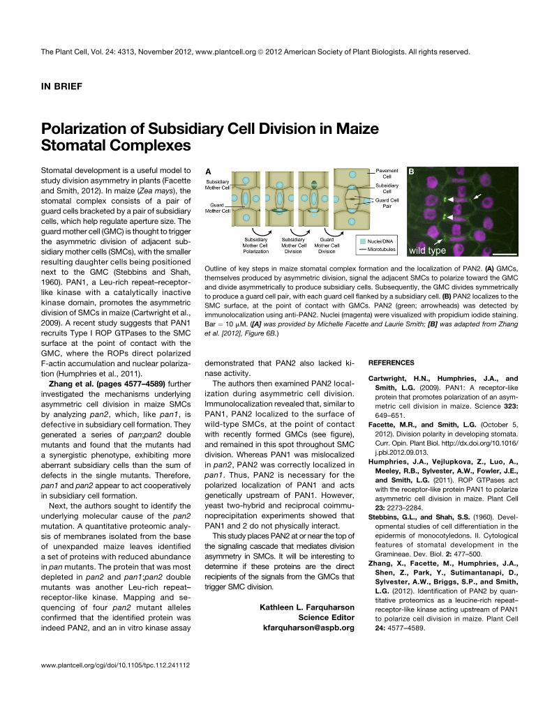
Outline of key steps in maize stomatal complex formation and the localization of PAN2. (A) GMCs,
themselves produced by asymmetric division, signal the adjacent SMCs to polarize toward the GMC
and divide asymmetrically to produce subsidiary cells. Subsequently, the GMC divides symmetrically
to produce a guard cell pair, with each guard cell flanked by a subsidiary cell. (B) PAN2 localizes to the
SMC surface, at the point of contact with GMCs. PAN2 (green; arrowheads) was detected by
immunolocalization using anti-PAN2. Nuclei (magenta) were visualized with propidium iodide staining.
Bar ¼ 10 mM. ([A] was provided by Michelle Facette and Laurie Smith; [B] was adapted from Zhang
et al. [2012], Figure 6B.)
www.plantcell.org/cgi/doi/10.1105/tpc.112.241112
The Plant Cell, Vol. 24: 4313, November 2012, www.plantcell.org ã 2012 American Society of Plant Biologists. All rights reserved.
Identification of PAN2 by Quantitative Proteomics asa Leucine-Rich Repeat–Receptor-Like Kinase ActingUpstream of PAN1 to Polarize Cell Division in MaizeC W OA
Xiaoguo Zhang,a,1 Michelle Facette,a John A. Humphries,a,2 Zhouxin Shen,a Yeri Park,a Dena Sutimantanapi,a
Anne W. Sylvester,b Steven P. Briggs,a and Laurie G. Smitha,3
a Section of Cell and Developmental Biology, University of California at San Diego, La Jolla, California 92093bDepartment of Molecular Biology, University of Wyoming, Laramie, Wyoming 82071
Mechanisms governing the polarization of plant cell division are poorly understood. Previously, we identified pangloss1(PAN1) as a leucine-rich repeat–receptor-like kinase (LRR-RLK) that promotes the polarization of subsidiary mother cell(SMC) divisions toward the adjacent guard mother cell (GMC) during stomatal development in maize (Zea mays). Here, weidentify pangloss2 (PAN2) as a second LRR-RLK promoting SMC polarization. Quantitative proteomic analysis identifieda PAN2 candidate by its depletion from membranes of pan2 single and pan1;pan2 double mutants. Genetic mapping andsequencing of mutant alleles confirmed the identity of this protein as PAN2. Like PAN1, PAN2 has a catalytically inactivekinase domain and accumulates in SMCs at sites of GMC contact before nuclear polarization. The timing of polarized PAN1and PAN2 localization is very similar, but PAN2 acts upstream because it is required for polarized accumulation of PAN1 but isindependent of PAN1 for its own localization. We find no evidence that PAN2 recruits PAN1 to the GMC contact site viaa direct or indirect physical interaction, but PAN2 interacts with itself. Together, these results place PAN2 at the top ofa cascade of events promoting the polarization of SMC divisions, potentially functioning to perceive or amplify GMC-derivedpolarizing cues.
INTRODUCTION
Asymmetric cell divisions, which give rise to daughters withdistinct developmental fates, are an important mechanism forthe generation of cell diversity during plant development (Abrashand Bergmann, 2009; Menke and Scheres, 2009). Such divisionsare often physically asymmetric as well, producing daughters withdistinct sizes and/or shapes. Many observations suggest mech-anistic links between physical and developmental asymmetry(Gallagher and Smith, 2000; Song et al., 2008; Dong et al., 2009).Moreover, orientation of division polarity is crucial for properplacement of the daughter cells within developing tissues to createfunctional cellular arrangements. Thus, polarization of cell divisionis a process of fundamental importance for plant development.
In preparation for a physically asymmetric plant cell division,the mother cell polarizes, which involves actin-dependent mi-gration of the premitotic nucleus into the future division plane
where the preprophase band later forms (Rasmussen et al.,2011). Premitotic division polarity may be determined by intrinsiccues (preexisting spatial landmarks within the mother cell) orextrinsic cues (spatial cues originating from outside the mothercell; Facette and Smith, 2012). After entry into mitosis, the di-viding nucleus is retained within the future division plane, andthe cell plate is ultimately attached there at the conclusion of cy-tokinesis through interactions between the cortical division site andthe expanding phragmoplast/cell plate (Rasmussen et al., 2011).In plants, where pathways and most proteins known to govern
division polarity in animal cells (reviewed in Gönczy, 2008) appearto be lacking, relatively little is known in mechanistic terms abouthow division asymmetry is achieved. Stomatal development hasprovided a useful focus for studies of division asymmetry. InArabidopsis thaliana, asymmetric divisions create stomatal pre-cursor cells while generating a pattern that ensures a minimum ofone nonstomatal cell separating neighboring stomata. Ligand–receptor interactions act through a mitogen-activated protein ki-nase signaling cascade to regulate the occurrence and orientationof stomate-forming asymmetric divisions (Pillitteri and Torii, 2012).BREAKING OF ASYMMETRY IN THE STOMATAL LINEAGE (BASL)and POLAR LOCALIZATION DURING ASYMMETRIC DIVISIONAND REDISTRIBUTION (POLAR) act downstream to promotedivision polarity (Dong et al., 2009; Pillitteri et al., 2011).In maize (Zea mays), an invariant sequence of asymmetric and
symmetric divisions generates stomatal complexes consistingof a pair of guard cells flanked by a pair of subsidiary cells thatregulate stomatal aperture. The first asymmetric division gen-erates a guard mother cell (GMC), which is believed to signal itslateral neighbors, the subsidiary mother cells (SMCs), to divide
1Current address: Department of Agronomy, University of Florida,Gainesville, FL 32611.2 Current address: School of Botany, University of Melbourne, Parkville,Victoria 3010, Australia.3 Address correspondence to [email protected] author responsible for distribution of materials integral to the findingspresented in this article in accordance with the policy described in theInstructions for Authors (www.plantcell.org) is: Laurie Smith ([email protected])C Some figures in this article are displayed in color online but in black andwhite in the print edition.W Online version contains Web-only data.OAOpen Access articles can be viewed online without a subscription.www.plantcell.org/cgi/doi/10.1105/tpc.112.104125
The Plant Cell, Vol. 24: 4577–4589, November 2012, www.plantcell.org ã 2012 American Society of Plant Biologists. All rights reserved.
asymmetrically in an orientation that positions the smaller daughter(the subsidiary cell) adjacent to the GMC (Farquharson, 2012;Stebbins and Shah, 1960). Asymmetric SMC divisions are pre-ceded by localized accumulation of cortical F-actin at the SMC-GMC interface and migration of the premitotic SMC nucleus tothat site (Galatis and Apostolakos, 2004). Subsequently, theGMC divides symmetrically to form a pair of guard cells flankedby the subsidiary cells.
Prior work has identified a leucine-rich repeat–receptor-like kinase(LRR-RLK), pangloss1 (PAN1), which promotes the polarizationof SMC divisions and thus might function as a receptor forGMC-derived polarizing cues (Cartwright et al., 2009). Shortlyafter GMC formation, PAN1 localizes asymmetrically in SMCs,accumulating at the site of GMC contact prior to nuclear po-larization to that site. More recently, Type I rho of plants (ROP)GTPases were shown to act downstream of PAN1 to promoteSMC polarization (Humphries et al., 2011). Partial loss of Type IROP function results in mild SMC polarization defects, and ropmutations dramatically enhance the pan1 phenotype. Like PAN1,Type I ROPs localize at the SMC surface as a patch at the site ofGMC contact but ROP patches form later than PAN1 patches.PAN1 appears to recruit ROPs through a physical interaction asindicated by coimmunoprecipitation of PAN1 and ROPs. Pheno-types resulting from partial loss of ROP function or depolarizationof ROP indicate that polarized accumulation of ROPs leads tolocalized accumulation of F-actin and nuclear polarization, but thelinks between ROPs and these downstream events are unclear.
Here, we use a quantitative proteomic approach to identifya second LRR-RLK promoting SMC polarization, pangloss2(PAN2). Analysis of PAN2 reveals that its localization and functionare similar to PAN1, but PAN2 acts upstream of PAN1. Thus,PAN2 is the earliest acting component of the SMC-polarizingmechanism identified to date.
RESULTS
PAN1 and PAN2 Interact Genetically
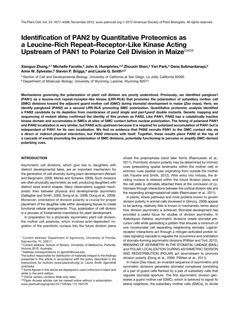
As described previously (Cartwright et al., 2009), pan2 mutationshave similar effects on stomatal subsidiary cell formation com-pared with pan1. In this study, 22 to 32% of stomatal subsidiariesformed aberrantly in mutants homozygous for either of two dif-ferent mutant alleles of each gene (Figures 1A, arrowheads, and1B). To explore the functional relationship between pan1 andpan2, we generated double mutants homozygous for three dif-ferent combinations of pan1 and pan2 alleles. Like the singlemutants, the overall morphology of double mutant plants was notmarkedly different from the wild type. However, as shown inFigures 1A and 1B, all double mutants had a synergistic pheno-type with a high frequency of aberrant subsidiary cells that was farmore than the sum of the frequencies seen in the correspondingsingle mutants.
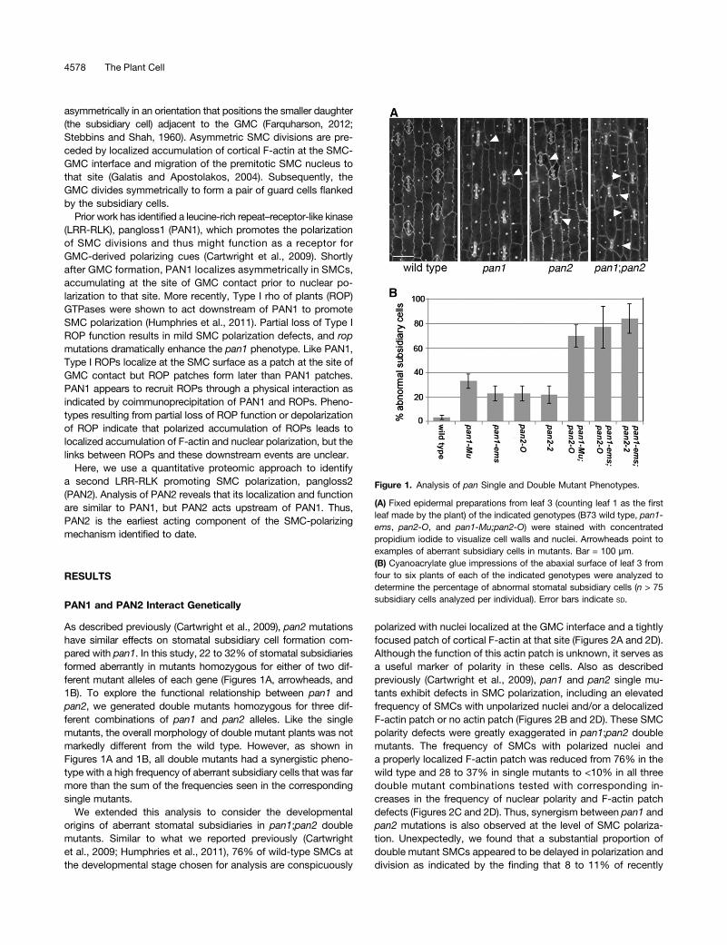
We extended this analysis to consider the developmentalorigins of aberrant stomatal subsidiaries in pan1;pan2 doublemutants. Similar to what we reported previously (Cartwrightet al., 2009; Humphries et al., 2011), 76% of wild-type SMCs atthe developmental stage chosen for analysis are conspicuously
polarized with nuclei localized at the GMC interface and a tightlyfocused patch of cortical F-actin at that site (Figures 2A and 2D).Although the function of this actin patch is unknown, it serves asa useful marker of polarity in these cells. Also as describedpreviously (Cartwright et al., 2009), pan1 and pan2 single mu-tants exhibit defects in SMC polarization, including an elevatedfrequency of SMCs with unpolarized nuclei and/or a delocalizedF-actin patch or no actin patch (Figures 2B and 2D). These SMCpolarity defects were greatly exaggerated in pan1;pan2 doublemutants. The frequency of SMCs with polarized nuclei anda properly localized F-actin patch was reduced from 76% in thewild type and 28 to 37% in single mutants to <10% in all threedouble mutant combinations tested with corresponding in-creases in the frequency of nuclear polarity and F-actin patchdefects (Figures 2C and 2D). Thus, synergism between pan1 andpan2 mutations is also observed at the level of SMC polariza-tion. Unexpectedly, we found that a substantial proportion ofdouble mutant SMCs appeared to be delayed in polarization anddivision as indicated by the finding that 8 to 11% of recently
Figure 1. Analysis of pan Single and Double Mutant Phenotypes.
(A) Fixed epidermal preparations from leaf 3 (counting leaf 1 as the firstleaf made by the plant) of the indicated genotypes (B73 wild type, pan1-ems, pan2-O, and pan1-Mu;pan2-O) were stained with concentratedpropidium iodide to visualize cell walls and nuclei. Arrowheads point toexamples of aberrant subsidiary cells in mutants. Bar = 100 µm.(B) Cyanoacrylate glue impressions of the abaxial surface of leaf 3 fromfour to six plants of each of the indicated genotypes were analyzed todetermine the percentage of abnormal stomatal subsidiary cells (n > 75subsidiary cells analyzed per individual). Error bars indicate SD.
4578 The Plant Cell
divided GMCs were flanked by SMCs that had not yet divided(Figures 2C, asterisk, and 2E). This was not observed in wild-type leaves, and rarely if ever (<2% of GMCs) in any of the pansingle mutants (Figure 2E). This finding further indicates syner-gism between pan1 and pan2 in double mutants and suggestsa previously unrecognized role for both genes in regulating thetiming of SMC division. The synergistic phenotypes observed inpan1;pan2 double mutants indicate that pan1 and pan2 actcooperatively to promote subsidiary cell formation or are par-tially redundant.
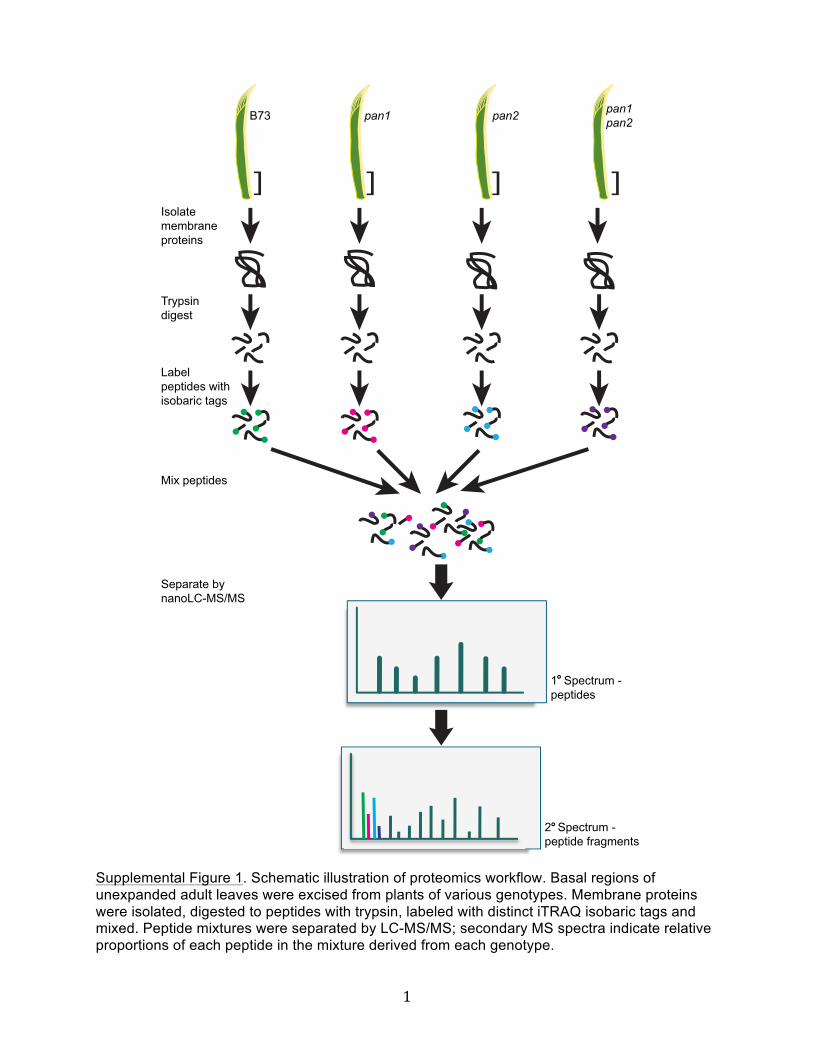
Quantitative Proteomic Analysis Reveals Changes inMembrane Protein Accumulation in pan Single andDouble Mutants
To identify proteins potentially functioning in common pathwayswith PAN1 and PAN2, we performed a comparative proteomicanalysis using the isobaric tags for relative and absolute quanti-tation (iTRAQ) method (see Supplemental Figure 1 online). Rea-soning that proteins of greatest interest were likely to be membranelocalized, we isolated membrane proteins from the basal portionsof unexpanded maize leaves (enriched in dividing cells, includingthose dividing to form stomata) from wild- type, pan1, pan2, andpan1;pan2 mutant plants. Tryptic peptides from each of the fourmembrane protein preparations were labeled with distinct iTRAQtags and then mixed. The tags have identical masses, but afterfragmentation they can be distinguished from one another by massspectrometry, permitting assessment of the proportion of eachpeptide in the mixture derived from each genotype.Peptides mapping to a total of 13,101 possible proteins were
identified by mass spectrometry (see Supplemental Data Set 1online). These proteins were assigned to 5438 groups repre-senting a minimal set of proteins to which the identified peptidescould belong (see Methods for further explanation of groups).These protein groups (henceforth referred to simply as “proteins”)are listed in Supplemental Data Set 2 online. A mutant:wild typeratio of spectral counts was calculated for each protein (meanratio for six biological replicates) to identify those whose abundancewas reproducibly altered in mutant extracts relative to the wild type.Notably, PAN1 ratios were the most reduced of any protein in bothpan1 single and pan1;pan2 double mutants (mutant:wild typeratios 0.11 and 0.05, respectively; Table 2; also see SupplementalData Set 3 online). This is consistent with our earlier finding thatPAN1 is undetectable in pan1 mutants by immunoblotting(Cartwright et al., 2009) and demonstrates that our methods arecapable of revealing differences in protein abundance in panmutants relative to the wild type. Defining changed proteins asthose showing at least a 1.5-fold increase or decrease with anassociated P value (determined from a Student’s t test on the ln-transformed values) # 0.1 in at least one of the mutants, wefound that the abundance of 253 of the 5438 identified proteinswas changed (see Supplemental Data Set 3 online).Since pan1 and pan2 are loss-of-function mutants, we rea-
soned that proteins whose abundance is reduced in pan mu-tants are more likely to be closely linked functionally to PANsthan those whose abundance is increased and therefore fo-cused subsequent analyses on the reduced proteins. The 120proteins from Supplemental Data Set 3 online that are reduced
Figure 2. Analysis of SMC Polarization and Division Defects in panSingle and Double Mutants.
(A) to (C) F-actin (green) and propidium iodide–stained nuclei (magenta)in developing stomata of B73 wild type (A), pan2-2 single mutants (B),and pan1-ems:pan2-2 double mutants (C). Arrowheads in (A) and (B) lieon top of GMCs and point to normal actin patches in adjacent SMCs;arrows in (B) and (C) point to areas of ectopic cortical actin accumulationin mutant SMCs. Asterisk marks a divided GMC flanked by undividedSMCs. Polarized and unpolarized SMC nuclei are marked P and U,respectively. Bar = 10 µm.(D) Quantification of the proportion of SMCs with polarized (P) or un-polarized (U) nuclei and actin patch status (>300 cells and four plantsanalyzed for each genotype). Error bars represent SD.(E) Quantification of the occurrence of divided GMCs flanked by un-divided SMCs (as illustrated by the example in [C]) in plants of eachgenotype indicated. This analysis used the same collection of images asthat presented in (D); error bars represent SD.
Identification of PAN2 as a LRR-RLK 4579
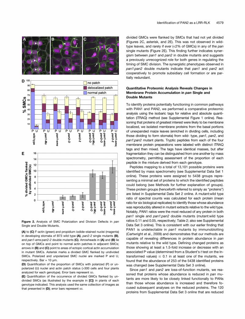
at least 1.5-fold are classified in the Venn diagram in Figure 3according to the genotypes in which they were reduced. Notsurprisingly, in view of the synergistic phenotype of pan1;pan2double mutants, the majority of the proteins that are decreasedin double mutants are not decreased in either of the single mu-tants. Unexpectedly, in spite of their very similar stomatal de-velopment defects, there is only modest overlap between the setof proteins reduced in pan1 versus pan2 single mutants, evenwhen one considers only those that are also depleted in thedouble mutant (Figure 3). This finding suggests that PAN1 andPAN2 have many distinct functions but does not reveal whetherthey have distinct functions in the SMC, since that is only one ofmany cell types included in the tissue sample analyzed.
A hypergeometric test was performed to determine whetherproteins depleted in the mutants belong predominantly to partic-ular functional classes, using Map Man bins (Thimm et al., 2004) toassign each protein to a functional class (see Supplemental Table1 online for a comprehensive report for all Map Man bins andgenotypes). Proteins in the Map Man bin “Miscellaneous” weresignificantly overrepresented among those depleted in pan1 singlemutants relative to all proteins identified (Table 1). Notably, five ofthe seven “miscellaneous” proteins depleted in pan1 are glyco-syltransferases (possibly involved in pectin or hemicellulose bio-synthesis) or callose-degrading glucanases, suggesting a possibleunique role for PAN1 in cell wall assembly. Proteins in the MapMan bins “Transport” and the “Cell” sub-bin “vesicle transport”were significantly overrepresented among those depleted in pan1;pan2 double mutants compared with all proteins identified (Table1), suggesting defects in membrane transport and vesicle traf-ficking in double mutants. Interestingly, targeted vesicle traffickingis important for achieving polarized distribution of a variety ofmembrane transport proteins (Dettmer and Friml, 2011). Thus,PAN1 and PAN2 may be important for polarized trafficking ofmembrane transport proteins, and this may be related to the cellpolarity defects seen in pan mutants. Proteins in the Map Man bin
“Signaling” were also overrepresented among proteins depleted inpan single and double mutants compared with all proteins iden-tified, although the enrichment was only statistically significant forpan1;pan2 double mutants (Table 1). This observation suggestsroles for PAN proteins in signaling, which is of great interest in viewof the identity of PAN1 as a receptor-like protein.Table 2 presents selected examples of individual proteins with
signaling functions decreased in various combinations of panmutants. Interestingly, ROP4 (the only type I ROP identified inour analysis) was significantly depleted in pan1;pan2 doublemutants. We previously showed that type I ROPs function withPAN1 to establish SMC polarity, and by immunoblotting wedemonstrated that ROPs are depleted from a Triton-insolublemembrane fraction of pan1 leaf extracts compared with the wildtype (Humphries et al., 2011). Thus, our findings for ROP4 in theproteomic analysis (where a different nonionic detergent wasused to wash the membrane fraction prior to analysis) parallelthe immunoblotting results, albeit with significant depletion de-tected only in double mutants; the degree of depletion in pan1single mutants may simply be too small to detect by iTRAQ com-parisons. As shown in Table 2, other signaling proteins depleted inpan single and double mutants include a variety of kinases, aphosphatase related to POLTERGEIST, which is required for po-larization of asymmetric cell division in Arabidopsis (Song et al.,2008), a protein kinase C substrate, and a transducin family protein.Investigating the functional relationships of these proteins to PANswill be interesting topics for future work.
Identification of PAN2 as a Catalytically InactiveLRR-RLK Family Protein
In pan2 mutants, the most significantly depleted protein identifiedby the quantitative proteomic analysis was a LRR-RLK encodedby GRMZM2G034572_T01 (Table 2); this LRR-RLK was alsoamong the most depleted proteins in pan1;pan2 double mutants.This gene encoding this protein is located near the tip of chro-mosome 2 in bin 2.10, closely matching the location where pan2was mapped using a mass spectrometry–based single nucleotidepolymorphism (SNP) genotyping method (see Supplemental DataSet 4 online). To test the possibility that GRMZM2G034572_T01is pan2, we sequenced the exons of this gene in pan2 mutants.The pan2-O allele described previously (Cartwright et al., 2009)was found to contain a missense mutation changing a conservedSer to Phe (S211 > F) near the N terminus of the protein (Figure4A). Three independent ethyl methanesulfonate–induced alleles ofpan2 that we isolated in a screen for noncomplementation ofpan2-O were all found to contain premature stop codon muta-tions at different sites within the gene (Figure 4A). These findingsestablish that GRMZM2G034572_T01 is pan2.A previously published transcription profiling study employing
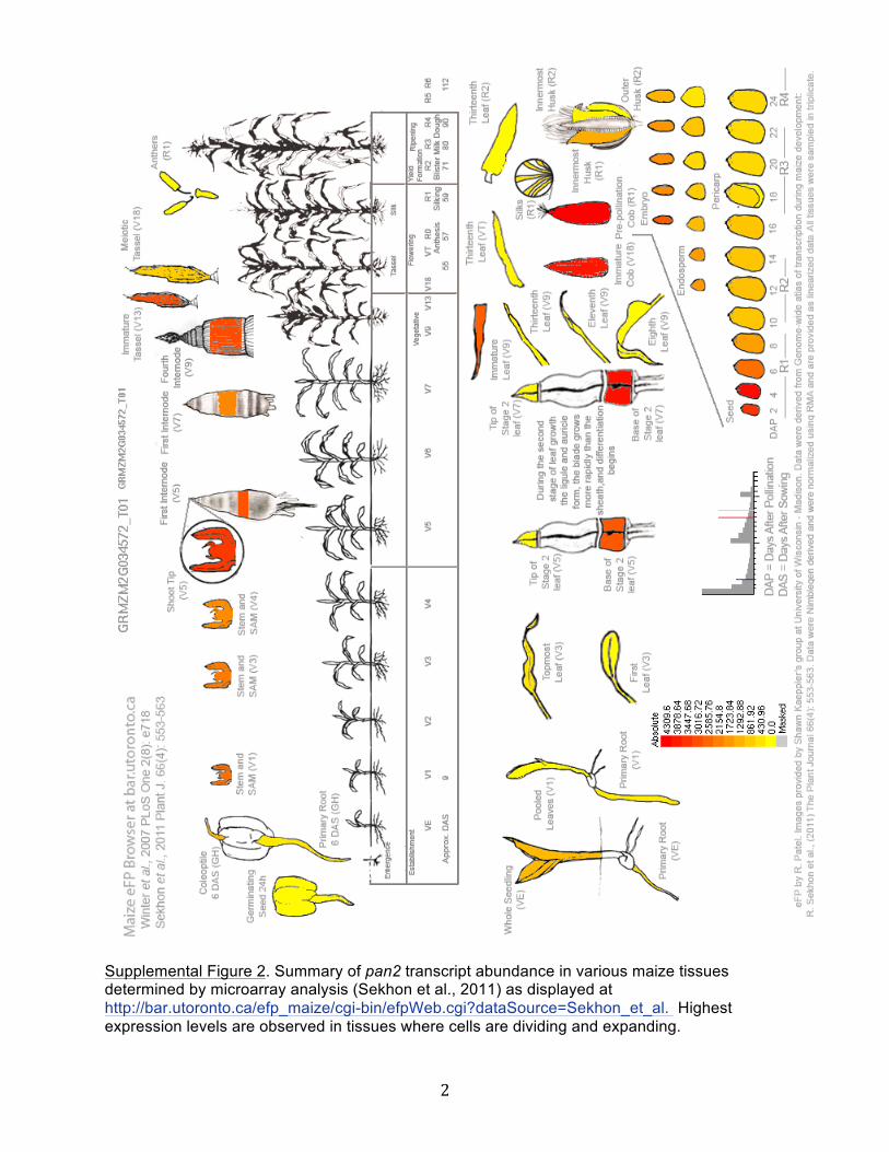
short read sequencing demonstrated that like pan1, maximumpan2mRNA levels are observed at the base of the developing leafwhere cell divisions occur (including SMC divisions) (Figure 4B; Liet al., 2010). A microarray study surveying many maize tissuesand developmental stages (Sekhon et al., 2011) confirmed that indeveloping leaves, pan2 is most highly expressed in the basal celldivision zone (see Supplemental Figure 2 online). Sekhon et al.(2011) further demonstrated that like pan1 (Cartwright et al., 2009),
Figure 3. Venn Diagram Illustrating the Overlaps in Sets of ProteinsDepleted in pan Mutant Membrane Preparations.
Proteins included in this analysis are those listed in Supplemental DataSet 3 online that were found to be reduced at least 1.5-fold relative to thewild type (regardless of P value) in one or more pan mutants. However,depletion of at least 1.5-fold with an associated P value #0.1 in at leastone mutant genotype was required for inclusion of the protein inSupplemental Data Set 3 online.[See online article for color version of this figure.]
4580 The Plant Cell
pan2 is expressed in many other maize tissues in addition todeveloping leaves, including expanding stems, immature tasselsand ears, and developing seeds (see Supplemental Figure 2online). In general, pan2 is expressed in tissues where cells aredividing and expanding, suggesting that pan2 has other func-tions in addition to promoting SMC polarization.
Like PAN1, PAN2 belongs to the LRR-RLK subfamily III (Shiuand Bleecker, 2001), but to a different major clade within thissubfamily. PAN2 is larger (predicted molecular weight of 115 kD)than PAN1 (68 kD) due mainly to a much larger extracellulardomain with 20 predicted LRR motifs compared with five forPAN1 (Figure 4A). There are no published analyses of the func-tions or properties of the closest relative of PAN2 in Arabidopsisor other plants. PAN1 lacks several amino acids that are con-served in catalytically active kinases and is catalytically inactivein vitro (Cartwright et al., 2009). As illustrated in SupplementalFigure 3 online, PAN2 also lacks key features expected of a cat-alytically active kinase (Manning et al., 2002), in particulara GXGXXG consensus sequence in the G-loop region of sub-domain I (GRSSHG in PAN2), an HRDmotif in subdomain VI (HGNin PAN2), and a DFG motif in subdomain 7 (DYC in PAN2). In-terestingly, the kinase domain of the closest relative of PAN2 inArabidopsis (At4g20940) shares these same deviations from the
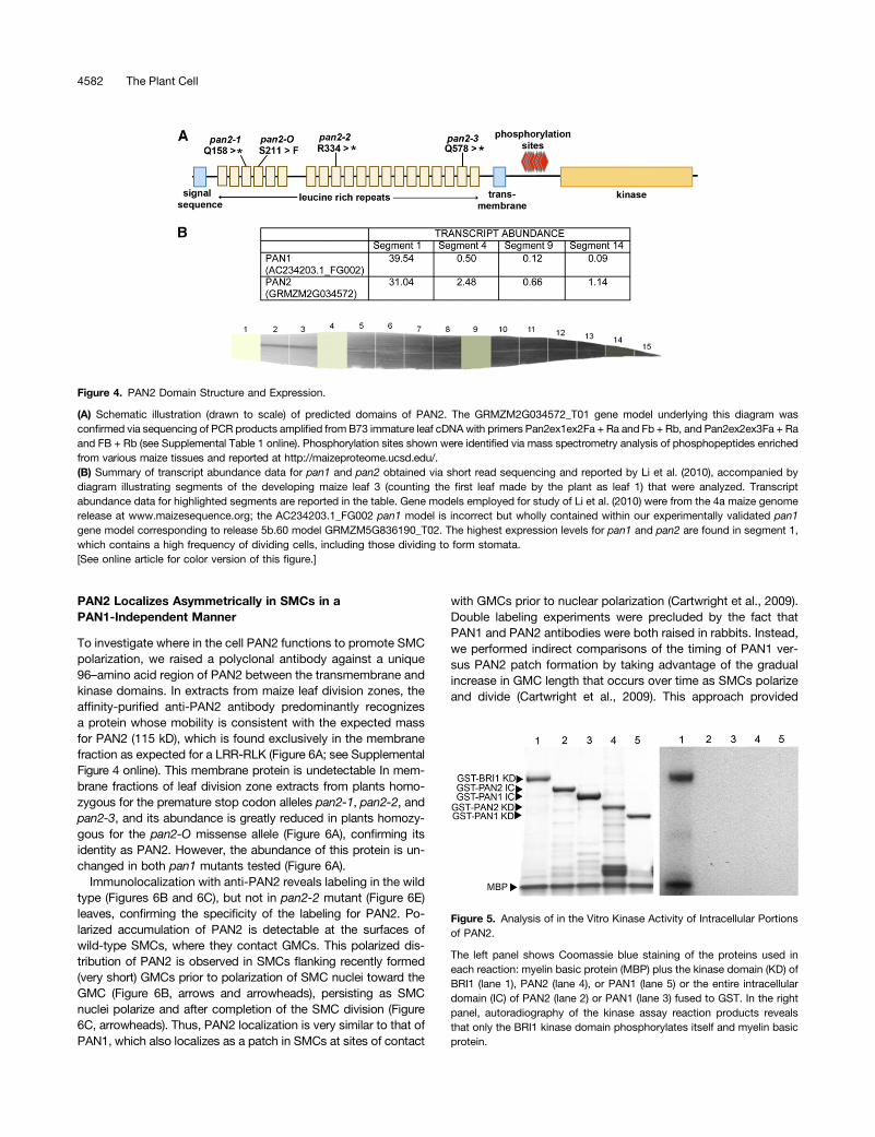
expectations for a catalytically active kinase (see SupplementalFigure 3 online).To test the enzymatic activity of the PAN2 kinase domain, in
vitro kinase assays were performed with the intracellular domainof PAN2 fused to glutathione S-transferase (GST; expressedand purified from Escherichia coli ). A GST-PAN1 intracellulardomain fusion protein was included as a negative control(Cartwright et al., 2009) and GST-BRI1 as a positive control(Friedrichsen et al., 2000). Under conditions in which the BRI1kinase domain phosphorylated itself and the artificial substratemyelin basic protein, the PAN2 intracellular domain exhibited nokinase activity (Figure 5). Inspection of an atlas of proteotypes(http://maizeproteome.ucsd.edu/) revealed that PAN2 has sev-eral sites of phosphorylation between its transmembrane andkinase domains (Figure 4A), suggesting that this juxtamembraneregion has a regulatory function, possibly negatively regulatingthe enzymatic activity of the kinase domain. Therefore, wetested the kinase domain of PAN2 alone (lacking the juxta-membrane region) along with the corresponding fragment ofPAN1 but found that these, too, exhibited no kinase activity invitro (Figure 5). Thus, consistent with analysis of the amino acidsequence, the kinase domain of PAN2 lacks detectable kinaseactivity.
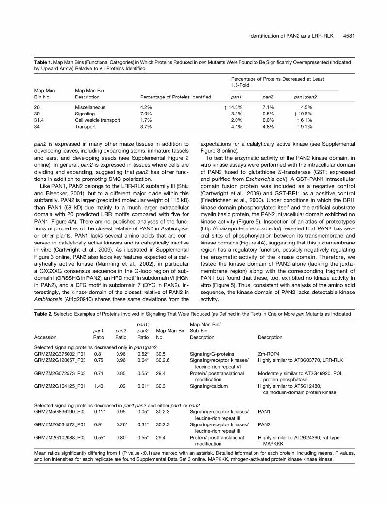
Table 1.Map Man Bins (Functional Categories) in Which Proteins Reduced in panMutants Were Found to Be Significantly Overrepresented (Indicatedby Upward Arrow) Relative to All Proteins Identified
Map ManBin No.
Map Man BinDescription
Percentage of Proteins Decreased at Least1.5-Fold
Percentage of Proteins Identified pan1 pan2 pan1;pan2
26 Miscellaneous 4.2% ↑ 14.3% 7.1% 4.5%30 Signaling 7.0% 8.2% 9.5% ↑ 10.6%31.4 Cell vesicle transport 1.7% 2.0% 0.0% ↑ 6.1%34 Transport 3.7% 4.1% 4.8% ↑ 9.1%
Table 2. Selected Examples of Proteins Involved in Signaling That Were Reduced (as Defined in the Text) in One or More pan Mutants as Indicated
Accessionpan1Ratio
pan2Ratio
pan1;pan2Ratio
Map Man BinNo.
Map Man Bin/Sub-BinDescription Description
Selected signaling proteins decreased only in pan1;pan2GRMZM2G375002_P01 0.81 0.96 0.52* 30.5 Signaling/G-proteins Zm-ROP4GRMZM2G120657_P03 0.75 0.96 0.64* 30.2.6 Signaling/receptor kinases/
leucine-rich repeat VIHighly similar to AT3G03770, LRR-RLK
GRMZM2G072573_P03 0.74 0.85 0.55* 29.4 Protein/ posttranslationalmodification
Moderately similar to AT2G46920, POLprotein phosphatase
GRMZM2G104125_P01 1.40 1.02 0.61* 30.3 Signaling/calcium Highly similar to AT5G12480,calmodulin-domain protein kinase
Selected signaling proteins decreased in pan1;pan2 and either pan1 or pan2GRMZM5G836190_P02 0.11* 0.95 0.05* 30.2.3 Signaling/receptor kinases/
leucine-rich repeat IIIPAN1
GRMZM2G034572_P01 0.91 0.26* 0.31* 30.2.3 Signaling/receptor kinases/leucine-rich repeat III
PAN2
GRMZM2G102088_P02 0.55* 0.80 0.55* 29.4 Protein/ posttranslationalmodification
Highly similar to AT2G24360, raf-typeMAPKKK
Mean ratios significantly differing from 1 (P value <0.1) are marked with an asterisk. Detailed information for each protein, including means, P values,and ion intensities for each replicate are found Supplemental Data Set 3 online. MAPKKK, mitogen-activated protein kinase kinase kinase.
Identification of PAN2 as a LRR-RLK 4581
PAN2 Localizes Asymmetrically in SMCs in aPAN1-Independent Manner
To investigate where in the cell PAN2 functions to promote SMCpolarization, we raised a polyclonal antibody against a unique96–amino acid region of PAN2 between the transmembrane andkinase domains. In extracts from maize leaf division zones, theaffinity-purified anti-PAN2 antibody predominantly recognizesa protein whose mobility is consistent with the expected massfor PAN2 (115 kD), which is found exclusively in the membranefraction as expected for a LRR-RLK (Figure 6A; see SupplementalFigure 4 online). This membrane protein is undetectable In mem-brane fractions of leaf division zone extracts from plants homo-zygous for the premature stop codon alleles pan2-1, pan2-2, andpan2-3, and its abundance is greatly reduced in plants homozy-gous for the pan2-O missense allele (Figure 6A), confirming itsidentity as PAN2. However, the abundance of this protein is un-changed in both pan1 mutants tested (Figure 6A).
Immunolocalization with anti-PAN2 reveals labeling in the wildtype (Figures 6B and 6C), but not in pan2-2 mutant (Figure 6E)leaves, confirming the specificity of the labeling for PAN2. Po-larized accumulation of PAN2 is detectable at the surfaces ofwild-type SMCs, where they contact GMCs. This polarized dis-tribution of PAN2 is observed in SMCs flanking recently formed(very short) GMCs prior to polarization of SMC nuclei toward theGMC (Figure 6B, arrows and arrowheads), persisting as SMCnuclei polarize and after completion of the SMC division (Figure6C, arrowheads). Thus, PAN2 localization is very similar to that ofPAN1, which also localizes as a patch in SMCs at sites of contact
with GMCs prior to nuclear polarization (Cartwright et al., 2009).Double labeling experiments were precluded by the fact thatPAN1 and PAN2 antibodies were both raised in rabbits. Instead,we performed indirect comparisons of the timing of PAN1 ver-sus PAN2 patch formation by taking advantage of the gradualincrease in GMC length that occurs over time as SMCs polarizeand divide (Cartwright et al., 2009). This approach provided
Figure 4. PAN2 Domain Structure and Expression.
(A) Schematic illustration (drawn to scale) of predicted domains of PAN2. The GRMZM2G034572_T01 gene model underlying this diagram wasconfirmed via sequencing of PCR products amplified from B73 immature leaf cDNAwith primers Pan2ex1ex2Fa + Ra and Fb + Rb, and Pan2ex2ex3Fa + Raand FB + Rb (see Supplemental Table 1 online). Phosphorylation sites shown were identified via mass spectrometry analysis of phosphopeptides enrichedfrom various maize tissues and reported at http://maizeproteome.ucsd.edu/.(B) Summary of transcript abundance data for pan1 and pan2 obtained via short read sequencing and reported by Li et al. (2010), accompanied bydiagram illustrating segments of the developing maize leaf 3 (counting the first leaf made by the plant as leaf 1) that were analyzed. Transcriptabundance data for highlighted segments are reported in the table. Gene models employed for study of Li et al. (2010) were from the 4a maize genomerelease at www.maizesequence.org; the AC234203.1_FG002 pan1 model is incorrect but wholly contained within our experimentally validated pan1gene model corresponding to release 5b.60 model GRMZM5G836190_T02. The highest expression levels for pan1 and pan2 are found in segment 1,which contains a high frequency of dividing cells, including those dividing to form stomata.[See online article for color version of this figure.]
Figure 5. Analysis of in the Vitro Kinase Activity of Intracellular Portionsof PAN2.
The left panel shows Coomassie blue staining of the proteins used ineach reaction: myelin basic protein (MBP) plus the kinase domain (KD) ofBRI1 (lane 1), PAN2 (lane 4), or PAN1 (lane 5) or the entire intracellulardomain (IC) of PAN2 (lane 2) or PAN1 (lane 3) fused to GST. In the rightpanel, autoradiography of the kinase assay reaction products revealsthat only the BRI1 kinase domain phosphorylates itself and myelin basicprotein.
4582 The Plant Cell
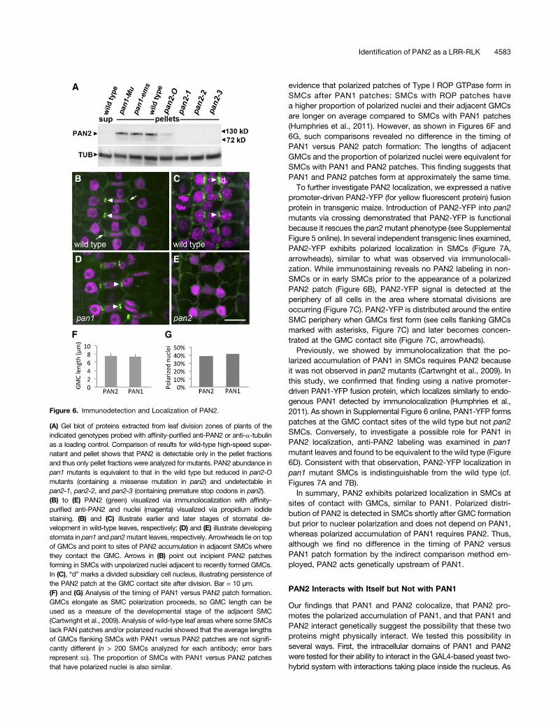
evidence that polarized patches of Type I ROP GTPase form inSMCs after PAN1 patches: SMCs with ROP patches havea higher proportion of polarized nuclei and their adjacent GMCsare longer on average compared to SMCs with PAN1 patches(Humphries et al., 2011). However, as shown in Figures 6F and6G, such comparisons revealed no difference in the timing ofPAN1 versus PAN2 patch formation: The lengths of adjacentGMCs and the proportion of polarized nuclei were equivalent forSMCs with PAN1 and PAN2 patches. This finding suggests thatPAN1 and PAN2 patches form at approximately the same time.To further investigate PAN2 localization, we expressed a native
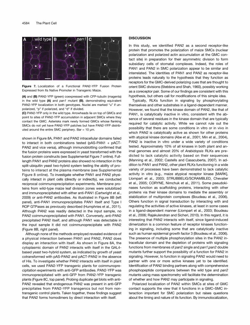
promoter-driven PAN2-YFP (for yellow fluorescent protein) fusionprotein in transgenic maize. Introduction of PAN2-YFP into pan2mutants via crossing demonstrated that PAN2-YFP is functionalbecause it rescues the pan2mutant phenotype (see SupplementalFigure 5 online). In several independent transgenic lines examined,PAN2-YFP exhibits polarized localization in SMCs (Figure 7A,arrowheads), similar to what was observed via immunolocali-zation. While immunostaining reveals no PAN2 labeling in non-SMCs or in early SMCs prior to the appearance of a polarizedPAN2 patch (Figure 6B), PAN2-YFP signal is detected at theperiphery of all cells in the area where stomatal divisions areoccurring (Figure 7C). PAN2-YFP is distributed around the entireSMC periphery when GMCs first form (see cells flanking GMCsmarked with asterisks, Figure 7C) and later becomes concen-trated at the GMC contact site (Figure 7C, arrowheads).Previously, we showed by immunolocalization that the po-
larized accumulation of PAN1 in SMCs requires PAN2 becauseit was not observed in pan2 mutants (Cartwright et al., 2009). Inthis study, we confirmed that finding using a native promoter-driven PAN1-YFP fusion protein, which localizes similarly to endo-genous PAN1 detected by immunolocalization (Humphries et al.,2011). As shown in Supplemental Figure 6 online, PAN1-YFP formspatches at the GMC contact sites of the wild type but not pan2SMCs. Conversely, to investigate a possible role for PAN1 inPAN2 localization, anti-PAN2 labeling was examined in pan1mutant leaves and found to be equivalent to the wild type (Figure6D). Consistent with that observation, PAN2-YFP localization inpan1 mutant SMCs is indistinguishable from the wild type (cf.Figures 7A and 7B).In summary, PAN2 exhibits polarized localization in SMCs at
sites of contact with GMCs, similar to PAN1. Polarized distri-bution of PAN2 is detected in SMCs shortly after GMC formationbut prior to nuclear polarization and does not depend on PAN1,whereas polarized accumulation of PAN1 requires PAN2. Thus,although we find no difference in the timing of PAN2 versusPAN1 patch formation by the indirect comparison method em-ployed, PAN2 acts genetically upstream of PAN1.
PAN2 Interacts with Itself but Not with PAN1
Our findings that PAN1 and PAN2 colocalize, that PAN2 pro-motes the polarized accumulation of PAN1, and that PAN1 andPAN2 interact genetically suggest the possibility that these twoproteins might physically interact. We tested this possibility inseveral ways. First, the intracellular domains of PAN1 and PAN2were tested for their ability to interact in the GAL4-based yeast two-hybrid system with interactions taking place inside the nucleus. As
Figure 6. Immunodetection and Localization of PAN2.
(A) Gel blot of proteins extracted from leaf division zones of plants of theindicated genotypes probed with affinity-purified anti-PAN2 or anti-a-tubulinas a loading control. Comparison of results for wild-type high-speed super-natant and pellet shows that PAN2 is detectable only in the pellet fractionsand thus only pellet fractions were analyzed for mutants. PAN2 abundance inpan1 mutants is equivalent to that in the wild type but reduced in pan2-Omutants (containing a missense mutation in pan2) and undetectable inpan2-1, pan2-2, and pan2-3 (containing premature stop codons in pan2).(B) to (E) PAN2 (green) visualized via immunolocalization with affinity-purified anti-PAN2 and nuclei (magenta) visualized via propidium iodidestaining. (B) and (C) illustrate earlier and later stages of stomatal de-velopment in wild-type leaves, respectively; (D) and (E) illustrate developingstomata in pan1 and pan2mutant leaves, respectively. Arrowheads lie on topof GMCs and point to sites of PAN2 accumulation in adjacent SMCs wherethey contact the GMC. Arrows in (B) point out incipient PAN2 patchesforming in SMCs with unpolarized nuclei adjacent to recently formed GMCs.In (C), “d” marks a divided subsidiary cell nucleus, illustrating persistence ofthe PAN2 patch at the GMC contact site after division. Bar = 10 µm.(F) and (G) Analysis of the timing of PAN1 versus PAN2 patch formation.GMCs elongate as SMC polarization proceeds, so GMC length can beused as a measure of the developmental stage of the adjacent SMC(Cartwright et al., 2009). Analysis of wild-type leaf areas where some SMCslack PAN patches and/or polarized nuclei showed that the average lengthsof GMCs flanking SMCs with PAN1 versus PAN2 patches are not signifi-cantly different (n > 200 SMCs analyzed for each antibody; error barsrepresent SD). The proportion of SMCs with PAN1 versus PAN2 patchesthat have polarized nuclei is also similar.
Identification of PAN2 as a LRR-RLK 4583
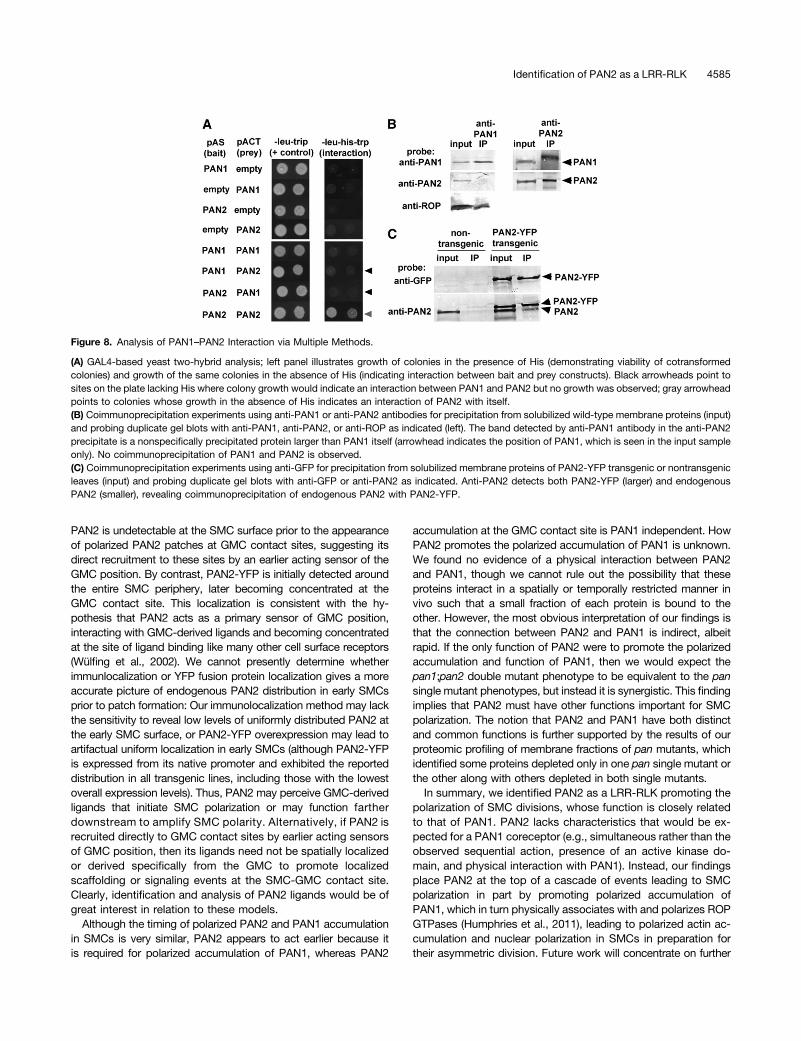
shown in Figure 8A, PAN1 and PAN2 intracellular domains failedto interact in both combinations tested (pAS-PAN1 + pACT-PAN2 and vice versa), although immunoblotting confirmed thatthe fusion proteins were expressed in yeast transformed with thefusion protein constructs (see Supplemental Figure 7 online). Full-length PAN1 and PAN2 proteins also showed no interaction in thesplit-ubiquitin yeast two-hybrid system testing the ability of pro-teins to interact at the plasma membrane (see SupplementalFigure 8 online). To investigate whether PAN1 and PAN2 physi-cally interact in plant cells (directly or indirectly), we conductedreciprocal coimmunoprecipitation experiments. Membrane pro-teins from wild-type maize leaf division zones were solubilizedand immunoprecipitated with either anti-PAN1 (Cartwright et al.,2009) or anti-PAN2 antibodies. As illustrated in Figure 8B (leftpanel), anti-PAN1 immunoprecipitates PAN1 itself and Type IROP GTPases as previously described (Humphries et al., 2011).Although PAN2 was readily detected in the input sample, noPAN2 coimmunoprecipitated with PAN1. Conversely, anti-PAN2precipitated PAN2 itself, and although PAN1 was detectable inthe input sample it did not coimmunoprecipitate with PAN2(Figure 8B, right panel).
Although none of the methods employed revealed evidence ofa physical interaction between PAN1 and PAN2, PAN2 doesdisplay an interaction with itself. As shown in Figure 8A, thecytoplasmic domain of PAN2 interacts with itself in the GAL4-based yeast two-hybrid system, as indicated by growth of yeastcotransformed with pAS-PAN2 and pACT-PAN2 in the absenceof His. To investigate whether PAN2 interacts with itself in plantcells, we used PAN2-YFP transgenic plants for coimmunopre-cipitation experiments with anti-GFP antibodies. PAN2-YFP wasimmunoprecipitated with anti-GFP from PAN2-YFP transgenicplants (Figure 8C, top panel). Probing duplicate gel blots with anti-PAN2 revealed that endogenous PAN2 was present in anti-GFPprecipitates from PAN2-YFP transgenics but not from non-transgenic control plants. Taken together, these findings suggestthat PAN2 forms homodimers by direct interaction with itself.
DISCUSSION
In this study, we identified PAN2 as a second receptor-likeprotein that promotes the polarization of maize SMCs (nuclearpolarization and polarized actin accumulation at the GMC con-tact site) in preparation for their asymmetric division to formsubsidiary cells of stomatal complexes. Indeed, the roles ofPAN1 and PAN2 in SMC polarization appear to be similar andinterrelated. The identities of PAN1 and PAN2 as receptor-likeproteins leads naturally to the hypothesis that they function asreceptors for the GMC-derived polarizing cues that are thought toorient SMC divisions (Stebbins and Shah, 1960), possibly workingas a coreceptor pair. Some of our findings are consistent with thishypothesis, but others call for modifications of this simple idea.Typically, RLKs function in signaling by phosphorylating
themselves and other substrates in a ligand-dependent manner.However, we found that the kinase domain of PAN2, like that ofPAN1, is catalytically inactive in vitro, consistent with the ab-sence of several residues in the kinase domain that are typicallyrequired for catalytic activity. While we cannot rule out thepossibility that there are some conditions in vitro or in vivo inwhich PAN2 is catalytically active as shown for other proteinswith atypical kinase domains (Abe et al., 2001; Min et al., 2004),PAN2 is inactive in vitro under a wide variety of conditionstested. Approximately 10% of all kinases in both plant and an-imal genomes and almost 20% of Arabidopsis RLKs are pre-dicted to lack catalytic activity based on their sequences(Manning et al., 2002; Castells and Casacuberta, 2007). In ad-dition to PAN1 and PAN2, other plant RLKs functioning in a widevariety of processes have been demonstrated to lack catalyticactivity in vitro (e.g., maize atypical receptor kinase [MARK],Llompart et al., 2003; STRUBBELIG/SCRAMBLED, Chevalieret al., 2005; CORYNE, Nimchuk et al., 2011). Some pseudoki-nases function as scaffolding proteins, interacting with otherproteins via their kinase domains to mediate the assembly orstabilization of multiprotein complexes (Boudeau et al., 2006).Others function in signal transduction by interacting with andregulating the activities of active kinases, at least in some casesin a ligand-dependent manner (Llompart et al., 2003; Boudeauet al., 2006; Rajakulendran and Sicheri, 2010). In this regard, it isinteresting that PAN2 interacts with itself, since ligand-induceddimerization is a common feature of receptor kinases function-ing in signaling, including some that are catalytically inactivesuch as human epidermal growth factor 3 (Boudeau et al., 2006).The presence of multiple phosphorylation sites in the PAN2 in-tracellular domain and the depletion of proteins with signalingfunctions from membranes of pan2 single and pan1;pan2 doublemutants further support the possibility of a function for PAN2 insignaling. However, to function in signaling PAN2 would need topartner with one or more active kinases yet to be identified.Identification of PAN2 binding partners along with comprehensivephosphopeptide comparisons between the wild type and pan2mutants using mass spectrometry will facilitate the determinationof whether and how PAN2 may participate in signaling.Polarized localization of PAN2 within SMCs at sites of GMC
contact supports the view that it functions in a GMC–SMC in-teraction important for SMC polarization but raises questionsabout the timing and nature of its function. By immunolocalization,
Figure 7. Localization of a Functional PAN2-YFP Fusion ProteinExpressed from Its Native Promoter in Transgenic Maize.
(A) and (B) PAN2-YFP (green) coexpressed with CFP-tubulin (magenta)in the wild type (A) and pan1 mutant (B), demonstrating equivalentPAN2-YFP localization in both genotypes. Nuclei are marked “u” if un-polarized, “p” if polarized, and “d” if divided.(C) PAN2-YFP only in the wild type. Arrowheads lie on top of GMCs andpoint to sites of PAN2-YFP accumulation in adjacent SMCs where theycontact the GMC. Asterisks mark newly formed GMCs whose flankingSMCs do not yet have PAN2-YFP patches but have PAN2-YFP distrib-uted around the entire SMC periphery. Bar = 10 µm.
4584 The Plant Cell
PAN2 is undetectable at the SMC surface prior to the appearanceof polarized PAN2 patches at GMC contact sites, suggesting itsdirect recruitment to these sites by an earlier acting sensor of theGMC position. By contrast, PAN2-YFP is initially detected aroundthe entire SMC periphery, later becoming concentrated at theGMC contact site. This localization is consistent with the hy-pothesis that PAN2 acts as a primary sensor of GMC position,interacting with GMC-derived ligands and becoming concentratedat the site of ligand binding like many other cell surface receptors(Wülfing et al., 2002). We cannot presently determine whetherimmunlocalization or YFP fusion protein localization gives a moreaccurate picture of endogenous PAN2 distribution in early SMCsprior to patch formation: Our immunolocalization method may lackthe sensitivity to reveal low levels of uniformly distributed PAN2 atthe early SMC surface, or PAN2-YFP overexpression may lead toartifactual uniform localization in early SMCs (although PAN2-YFPis expressed from its native promoter and exhibited the reporteddistribution in all transgenic lines, including those with the lowestoverall expression levels). Thus, PAN2 may perceive GMC-derivedligands that initiate SMC polarization or may function fartherdownstream to amplify SMC polarity. Alternatively, if PAN2 isrecruited directly to GMC contact sites by earlier acting sensorsof GMC position, then its ligands need not be spatially localizedor derived specifically from the GMC to promote localizedscaffolding or signaling events at the SMC-GMC contact site.Clearly, identification and analysis of PAN2 ligands would be ofgreat interest in relation to these models.
Although the timing of polarized PAN2 and PAN1 accumulationin SMCs is very similar, PAN2 appears to act earlier because itis required for polarized accumulation of PAN1, whereas PAN2
accumulation at the GMC contact site is PAN1 independent. HowPAN2 promotes the polarized accumulation of PAN1 is unknown.We found no evidence of a physical interaction between PAN2and PAN1, though we cannot rule out the possibility that theseproteins interact in a spatially or temporally restricted manner invivo such that a small fraction of each protein is bound to theother. However, the most obvious interpretation of our findings isthat the connection between PAN2 and PAN1 is indirect, albeitrapid. If the only function of PAN2 were to promote the polarizedaccumulation and function of PAN1, then we would expect thepan1;pan2 double mutant phenotype to be equivalent to the pansingle mutant phenotypes, but instead it is synergistic. This findingimplies that PAN2 must have other functions important for SMCpolarization. The notion that PAN2 and PAN1 have both distinctand common functions is further supported by the results of ourproteomic profiling of membrane fractions of pan mutants, whichidentified some proteins depleted only in one pan single mutant orthe other along with others depleted in both single mutants.In summary, we identified PAN2 as a LRR-RLK promoting the
polarization of SMC divisions, whose function is closely relatedto that of PAN1. PAN2 lacks characteristics that would be ex-pected for a PAN1 coreceptor (e.g., simultaneous rather than theobserved sequential action, presence of an active kinase do-main, and physical interaction with PAN1). Instead, our findingsplace PAN2 at the top of a cascade of events leading to SMCpolarization in part by promoting polarized accumulation ofPAN1, which in turn physically associates with and polarizes ROPGTPases (Humphries et al., 2011), leading to polarized actin ac-cumulation and nuclear polarization in SMCs in preparation fortheir asymmetric division. Future work will concentrate on further
Figure 8. Analysis of PAN1–PAN2 Interaction via Multiple Methods.
(A) GAL4-based yeast two-hybrid analysis; left panel illustrates growth of colonies in the presence of His (demonstrating viability of cotransformedcolonies) and growth of the same colonies in the absence of His (indicating interaction between bait and prey constructs). Black arrowheads point tosites on the plate lacking His where colony growth would indicate an interaction between PAN1 and PAN2 but no growth was observed; gray arrowheadpoints to colonies whose growth in the absence of His indicates an interaction of PAN2 with itself.(B) Coimmunoprecipitation experiments using anti-PAN1 or anti-PAN2 antibodies for precipitation from solubilized wild-type membrane proteins (input)and probing duplicate gel blots with anti-PAN1, anti-PAN2, or anti-ROP as indicated (left). The band detected by anti-PAN1 antibody in the anti-PAN2precipitate is a nonspecifically precipitated protein larger than PAN1 itself (arrowhead indicates the position of PAN1, which is seen in the input sampleonly). No coimmunoprecipitation of PAN1 and PAN2 is observed.(C) Coimmunoprecipitation experiments using anti-GFP for precipitation from solubilized membrane proteins of PAN2-YFP transgenic or nontransgenicleaves (input) and probing duplicate gel blots with anti-GFP or anti-PAN2 as indicated. Anti-PAN2 detects both PAN2-YFP (larger) and endogenousPAN2 (smaller), revealing coimmunoprecipitation of endogenous PAN2 with PAN2-YFP.
Identification of PAN2 as a LRR-RLK 4585
elucidation of the pathway(s) in which PAN2 and PAN1 function topromote the premitotic polarization of SMCs.
METHODS
Primers
The sequences of all primers used in this study are provided inSupplemental Table 2 online.
Plants and Genetics
All mutants analyzed in this study segregate as single gene recessives,and all alleles were backcrossed into the B73 inbred wild-type back-ground at least three times for use in the experiments presented. pan1-Muand pan1-ems were previously described; both are null alleles with noPAN1 detectable via immunoblot analysis of homozygous mutants(Cartwright et al., 2009). pan2-O arose from an ethyl methanesulfonatemutagenesis with previous phenotypic description by Gallagher andSmith (2000) and Cartwright et al. (2009). pan2 was mapped to the tip ofchromosome 2 (bin 2.10) via bulked segregant analysis of F2 familiessegregating pan2-O after a single backcross to B73, Mo17, and W22inbred backgrounds. Genomic DNA extracted from pools of mutant andwild-type sibling tissue were used for PCR with a panel of 2076 primerpairs, and the products analyzed on an automated Sequenom massspectrometry platform to identify markers linked to pan2 as described byLiu et al. (2010). Results for all markers and genetic backgrounds arepresented in Supplemental Data Set 4 online. An F1 noncomplementationscreen was performed to isolate additional pan2 mutant alleles. Briefly,ethyl methanesulfonate–mutagenized pollen was crossed onto pan2-Ohomozygous female ears as previously described (Neuffer, 1994), and theF1 progeny were screened for the presence of the pan2 mutant phe-notype. Rare plants displaying the mutant phenotype were outcrossed toB73 wild-type plants. F1 progeny not inheriting pan2-O (as determined byanalysis of genetic markers closely linked to pan2) were selfed to produceplants homozygous for the new pan2 alleles (pan2-1, -2, and -3), whichall exhibited the pan2 phenotype. To identify mutations in pan2, exonsof GRMZM2G034572_T01 were amplified from genomic DNA of plantshomozygous for pan2-O, pan2-1, pan2-2, and pan2-3 via PCR withM2g34572 series primers listed in Supplemental Table 2 online, and PCRproducts were directly sequenced.
Phenotypic Analysis
For routine scoring of mutant phenotypes and quantitative analyses ofsubsidiary cell defects, imprints of the abaxial surfaces of mature leaf 3 or4 (calling the first leaf made by the plant leaf 1) weremade in cyanoacrylateglue and examined on a stereomicroscope or on a compoundmicroscopeat 310 magnification with bright-field or differential interference contrastoptics. To visualize nuclei and cell walls (Figure 1A), mature leaf 3 tissuewas fixed and stained with 100 µg/mL propidium iodide as describedpreviously (Hunter et al., 2012), mounted in water, and imaged via con-focal microscopy. Actin and nuclei were visualized via confocal mi-croscopy of fixed tissues excised from the basal 1 cm of unexpanded leaf3 or 4 stained with Alexafluor 488-phalloidin (Molecular Probes/Invitrogen)and 10 µg/mL propidium iodide as previously described (Cartwright et al.,2009). Images were analyzed in a double blind manner for actin patchesand nuclear position in SMCs.
Confocal Microscopy and Image Processing
Confocal microscopy was performed using a custom-assembled spin-ning disk microscope system described previously (Walker et al., 2007).
Image processing was performed using Metamorph version 7.0r1, NIHImage J, or Adobe Photoshop, applying only linear adjustments to pixelvalues.
Preparation and Analysis of Membrane Proteins viaMass Spectrometry
A detailed description of peptide preparation, mass spectrometry, andanalysis of mass spectra is provided in Supplemental Methods 1 (inSupplemental Data online). Briefly, membrane proteins were isolatedfrom the cell division zone at the bases of unexpanded leaves, detergentextracted, reduced, alkylated, and trypsin digested. Following detergentremoval via chromatography, peptides were labeled with iTRAQ reagents(AB SCIEX) and separated via online two-dimensional HPLC prior toacquisition of tandemmass spectra on a LTQ linear ion trap tandemmassspectrometer (Thermo Electron). Spectra were searched using SpectrumMill vB04.00 (Agilent) against themaize (Zeamays) 5a.59 working gene setdatabase (136,770 protein sequences). A 1:1 concatenated forward-reverse database of 273,540 proteins was constructed to calculate thefalse discovery rate (FDR). Cutoff scores were set to obtain a FDR of<0.1% at the unique peptide level.
Peptides mapping to a total of 13,101 proteins were identified (proteinlevel FDR = 0.3%, reported in Supplemental Data Set 1 online) from themaize 5a working gene set, but many of these peptides could not beuniquely assigned to a single protein. This total of 13,101 includes allproteins to which such shared peptides could belong and is almostcertainly an overestimate of the true number of different proteins detectedin a given sample. To obtain a more realistic estimate, groups of proteinsto which individual shared peptides mapped were identified. Within sucha group (collection of proteins sharing the peptide sequence), the proteinhaving the highest number of different peptides mapped to it (or, in thecase of ties, the longest protein) was assigned as a group leader. Al-ternatively, a group consists of one protein if it shares no peptides with anyother protein. Groups, identified by their leaders, are listed in SupplementalData Set 2 online and correspond to the 5438 proteins discussed inResults.
Generation and Purification of PAN2-Specific Antibody
A 96–amino acid fragment of PAN2 corresponding to amino acids 675 to770 (the region between the transmembrane and kinase domains), whichdisplayed low sequence identity with other LRR-LRKs or other proteins inmaize, was used to generate a PAN2-specific antibody. The corre-sponding coding sequence was amplified from B73 leaf division zonecDNA with PCR primers PAN2pip96F and R (see Supplemental Table 1online) and cloned into the BamHI and XhoI sites of pET28a (Novagen) inframewith the 6HIS tag. The fusion protein was induced in Escherichia colistrain BL21 with 0.5 mM isopropyl-b-D-thio-galactoside at 28°C for 6 hand purified on nickel-nitrilotriacetic acid (Ni-NTA) resin (Novagen) ac-cording to the manufacturer’s protocols. The His tag was removed usinga ThrombinCleanCleave kit (Sigma-Aldrich) according to themanufacturer’sinstructions. Three milligrams of the purified PAN2 fragment was used toproduce polyclonal antibodies in rabbits by Pacific Immunology. For affinitypurification, the same protein fragment was coupled to beads using anAminoLink Plus immobilization kit (Pierce), and purification was performedaccording to the manufacturer’s protocols.
Immunolocalization
Immunolocalization of PAN2 was performed in leaf tissue excised fromthe basal 1 to 3 cm of unexpanded leaves of 2-to 4-week-old plants asdescribed previously (Cartwright et al., 2009) using affinity-purified anti-PAN2 at 0.5 to 2 µg/mL and tyramide-based signal amplification with
4586 The Plant Cell
Invitrogen TSA kit #12 (Alexa Fluor 488) according to the manufacturer’sprotocol. Following antibody staining, tissues were stained with 10 µg/mLpropidium iodide (Sigma-Aldrich) in PBS to label nuclei prior to mountingin Vectashield (Vector Laboratories) for confocal microscopy.
Production of PAN2-YFP Transgenic Plants
A 10.5-kb genomic DNA fragment including the entire pan2 coding region(minus the stop codon) and 3.5 kb of 59 sequence was amplified from B73genomic DNA with primers PAN2-3GWp1 and PAN2-3GWp4. A 1.5-kbfragment immediately 39 of the pan2 coding region was amplified fromB73 genomic DNA with PAN2-3GWp3 and PAN2-3GWp2. Citrine YFPwas amplified as described previously (Mohanty et al., 2009). These threefragmentswere assembled in pDONR221 (Invitrogen) to insert YFP in framewith PAN2 at its C terminus with the 39 pan2 flanking sequence down-stream using a MultiSite Gateway three-fragment vector construction kit(Invitrogen) following the manufacturer’s instructions. The sequence of thePAN2-YFP coding region was verified using primers of the PAN2seq serieslisted in Supplemental Table 2 online. An error-free PAN2-YFP constructwas recombined into the binary vector pAM1006 and introduced intomaizevia Agrobacterium tumefaciens–mediated transformation at the Iowa StateUniversity Plant Transformation Facility (http://www.agron.iastate.edu/ptf/)as described at http://maize.jcvi.org/cellgenomics/protocol.shtml. Primarytransformants were crossed to B73 to produce T1 progeny used forimmunoprecipitation experiments, and T1s were crossed with plantsexpressing CFP-TUB (described at http://maize.jcvi.org/cellgenomics/protocol.shtml) to produce T2 progeny used for the imaging experimentspresented in Figure 7. Primary transformants were also crossed to pan1-ems and pan2-2 mutants, and the T1 progeny backcrossed again tomutants, to produce homozygous pan1 and pan2 mutants expressingPAN2-YFP. Images presented are representative of multiple independentevents that were examined by microscopy.
Yeast Two-Hybrid Analysis
For use in the split ubiquitin yeast two-hybrid interaction system (Grefenet al., 2007), full-length pan1 cDNA was amplified from a maize leaf di-vision zone cDNA preparation with primers PAN1-GWF and -GWR andcloned into pDONR207 using BP Clonase (Invitrogen) to generatepDONR-PAN1. Full-length pan2 was amplified with PAN2-TOPO-F and-R from full-length pan2 cDNA clone pSK-PAN2 constructed as illustratedin Supplemental Figure 9 online. The PCR product was cloned into vectorpENTR/TOPO-D (Invitrogen) according to manufacturer’s directions togenerate pENTR-PAN2. Using the Gateway recombination system,pMetYC_GW (Lalonde et al., 2010) was used to generate PAN1-CUB andPAN2-CUB, and pXN25_GW, a close relative of pXN22_GW (Lalondeet al., 2010), was used to generate PAN1-NUB and PAN2-NUB. Yeasttransformation was performed as described previously (Gietz andWoods,2002), and the split ubiquitin assay was performed according to Lalondeet al. (2010).
For use in the GAL4-based yeast two-hybrid interaction system, cDNAfragments encoding intracellular portions of PAN1 and PAN2 were clonedinto yeast two-hybrid vectors viaGatewaycloning. Primers P1SOL_pENTR_Fand P1_STOP_pENTR_R were used to amplify the cDNA segment encodingamino acids 308 to 662 of PAN1 from pDONR-PAN1 (described above).Primers P2SOL_pENTR_F and P2STOP_pENTR_R were used to amplify thecDNA segment encoding amino acids 675 to 1075 of PAN2 from pSK-PAN2(described above). PCR products were cloned to the entry vector pENTR/D-TOPO (Invitrogen). Gateway recombination was used to transfer the insertsfrom these entry clones to destination vectors pASGW-attR and pACTGW-attR (Nakayama et al., 2002). Transformation of these plasmids into yeaststrain AH109 was performed as described by Gietz and Woods (2002). Atleast four independent cotransformed colonies from two separate
experiments were tested for each construct to determine their ability togrow in the absence of His.
Protein Gel Blot Analysis and Immunoprecipitation
Immunoblotting experiments used maize leaf tissues enriched in dividingcells (the basal 2 cm of leaves remaining on 3- to 4-week-old maize plantsafter removal of all leaves with fully or partially expanded sheaths). Mem-brane fractions of extracts from these tissues were prepared, separated viaSDS-PAGE, and analyzed via immunoblotting as previously described(Cartwright et al., 2009) with the following modifications: microsomalfractions were resuspended in PBS containing 0.1% SDS and sonicatedfor 10 s to aid resuspension prior to freezing aliquots at 280°C; aliquotswere boiled in SDS loading buffer with 100 mM DTT for 10 min prior toloading on 4 to 20%gradient polyacrylamide gels (Mini-Protean TGX, Bio-Rad); and 0.5 g SDS was added per liter of transfer buffer to facilitatetransfer of PAN2 to the membrane. PAN2 was detected with affinity-purified anti-PAN2 at 1 µg/mL; tubulin was detected with mouse mono-clonal anti-a-tubulin clone B-5-1-2 (Sigma-Aldrich) at 0.2 µg/mL.
Yeast protein extracts were prepared for immunoblotting (seeSupplemental Figure 7 online) from cotransformed colonies as describedin the Clontech Yeast Protocols Handbook (Protocol number PT3024-1, ver-sionPR973283, July 2009; http://www.clontech.com/xxclt_ibcGetAttachment.jsp?cItemId=17602andminisite=10020andsecItmId=14852).Separation, trans-fer, and detection of proteins was performed as described by Cartwright et al.(2009) using rabbit anti-GAL4 binding domain (SC-577; Santa Cruz Bio-technology) diluted 1:600, rabbit anti-HA epitope tag polyclonal antibody(Pierce PA1-985) diluted 1:500, and affinity-purified anti-PAN2 at 1 µg/mL.
Immunoprecipitation experiments were performed using membranefractions of maize leaf tissue enriched in dividing cells prepared as above,after solubilizing themembrane proteins as described previously (Chinchillaet al., 2007). Immunoprecipitation with anti-PAN1 (Cartwright et al., 2009)and anti-PAN2 was performed as previously described (Humphries et al.,2011). Immunoprecipitation of PAN2-YFP was performed using magneticbeads covalently coupled to anti-GFP antibody (Miltenyi µMACS system)according to the manufacturer’s instructions. Precipitated proteins wereremoved from the beads by boiling in SDS loading buffer, separated viaSDS-PAGE, and analyzed via immunoblotting as described above, de-tecting with anti-PAN1 at 2.5 µg/mL, anti-PAN2 at 1 µg/mL, anti-ROP2/4/9(described in Humphries et al., 2011) at 1 µg/mL, and rabbit anti-GFP serum(Invitrogen A-6455) diluted 1:1000.
Analysis of in Vitro Kinase Activity
Portions of pan1 encoding the intracellular region (Arg-301 to Gly-662)and kinase domain (Arg-421 to Gly-662) were amplified by PCR using theprimers PAN1-KD-F and -R, and PAN1-IC-F and -R, respectively. Sim-ilarly, portions of the pan2 gene encoding the intracellular region (Lys-676to Ile-1080) and kinase domain (Arg-759 to Ile-1080) were amplified usingthe primers PAN2-KD-F and -R and PAN2-IC-F and -R, respectively (seeSupplemental Table 2 online). The amplified fragments were cloned intoBamHI and XhoI sites of pGEX-4T3 (GE Life Sciences) to generateN-terminal GST fusions. The positive control construct (BRI1-JKC GST fu-sion)was providedby JoanneChory (Salk Institute). GST fusion proteinswereinduced in E. coli strain BL21 with 0.5 mM isopropyl-b-D-thio-galactoside at28°C and purified from cell lysates using GST bind columns (Novagen)according to themanufacturer’s protocol. Tomeasure kinase activity, purifiedGST fusion proteinsweremixedwith 0.5mg/mLmyelin basic protein (Sigma-Aldrich), incubated for 1 h at room temperature in kinase buffer (50 mM Tris-Cl, 0 to 20 mMMgCl2, 0 to 20 mMMnCl2, 0 to 40 mMCaCl2, 2 mMDTT, 1%glycerol, and 10 µCi [g-32P]ATP). Reaction products were separated onNuPAGE 4 to 12% Bis-Tris gels (Invitrogen) and visualized via CoomassieBrilliant Blue staining and autoradiography.
Identification of PAN2 as a LRR-RLK 4587
Accession Numbers
pan gene and PAN protein sequences can be found at http://www.maizesequence.org as GRMZM2G034572_T01 and _P01 (PAN2) andGRMZM5G836190_T02 and _P02 (PAN1) (release 5b.60).
Supplemental Data
The following materials are available in the online version of this article.
Supplemental Figure 1. Schematic Illustration of Proteomics Workflow.
Supplemental Figure 2. Summary of pan2 Transcript Abundance inVarious Maize Tissues Determined by Microarray Analysis.
Supplemental Figure 3. Alignment of the Kinase Domains of PAN2and Other LRR-RLKs.
Supplemental Figure 4. Full Extent of Protein Gel Blot Probed withAnti-PAN2 Antibody Displayed in Cropped View in Figure 6A.
Supplemental Figure 5. Rescue of pan2 Mutant Phenotype by PAN2-YFP.
Supplemental Figure 6. Localization of PAN1-YFP Expressed fromthe Native pan1 Promoter in Wild-Type and pan2 Mutant Leaves.
Supplemental Figure 7. Protein Gel Blot Analysis of Yeast Trans-formed with PAN1 and PAN2 Intracellular Domain Constructs Used forTwo-Hybrid Analysis.
Supplemental Figure 8. Analysis of PAN1 and PAN2 Interaction atthe Yeast Plasma Membrane Using the Split-Ubiquitin Two-HybridSystem.
Supplemental Figure 9. Schematic Illustration of Construction ofa Full-Length pan2 cDNA Clone.
Supplemental Table 1. Distribution of Changed Proteins among MapMan Bins.
Supplemental Table 2. Primers Used in This Study.
Supplemental Methods 1.
Supplemental Data Set 1. All 13,143 Possible Proteins Identified inSix Biological Replicates of iTRAQ Experiments.
Supplemental Data Set 2. Subset of Supplemental Data Set 1Composed of 5438 Group Leaders.
Supplemental Data Set 3. Proteins Changed in pan1, pan2, and/orpan1 pan2 Mutants.
Supplemental Data Set 4. pan2 Mapping Data Generated via BulkedSegregant Analysis of Alleles at 2076 Marker Loci Analyzed on aSequenom Mass Spectrometry Platform.
ACKNOWLEDGMENTS
We thank Travis Alexander (University of California San Diego) for helpwith quantitative analysis of double mutants, Lauren Clark for isolation ofpan2-1, pan2-2, and pan2-3, Adam Zona (University of California SanDiego) for analysis of complementation of the pan2 phenotype byPAN2-YFP, Heather Cartwright (University of California San Diego) forinitial analysis of pan1;pan2 mutants, Erik Vollbrecht and Pat Schnable(Iowa State University) for Sequenom mapping, and Anding Luo(University of Wyoming) for help with construction of PAN2-YFP. Thiswork was funded by National Science Foundation Grants IOS-0843704and IOS-1147265 to L.G.S., National Science Foundation Grant DBI-0924023 to S.P.B., and National Science Foundation Grant DBI-0501862to A.W.S.
AUTHOR CONTRIBUTIONS
X.Z., M.F., J.A.H., Z.S., Y.P., and D.S. performed the research andanalyzed the data. X.Z., M.F., J.A.H., L.G.S., Z.S., S.P.B., and A.W.S.designed the research. L.G.S. wrote the article with input from thecoauthors.
Received August 14, 2012; revised October 4, 2012; accepted October30, 2012; published November 28, 2012.
REFERENCES
Abe, Y., Matsumoto, S., Wei, S., Nezu, K., Miyoshi, A., Kito, K.,Ueda, N., Shigemoto, K., Hitsumoto, Y., Nikawa, J., andEnomoto, Y. (2001). Cloning and characterization of a p53-relatedprotein kinase expressed in interleukin-2-activated cytotoxic T-cells,epithelial tumor cell lines, and the testes. J. Biol. Chem. 276: 44003–44011.
Abrash, E.B., and Bergmann, D.C. (2009). Asymmetric cell divisions:A view from plant development. Dev. Cell 16: 783–796.
Boudeau, J., Miranda-Saavedra, D., Barton, G.J., and Alessi, D.R.(2006). Emerging roles of pseudokinases. Trends Cell Biol. 16:443–452.
Cartwright, H.N., Humphries, J.A., and Smith, L.G. (2009). PAN1: Areceptor-like protein that promotes polarization of an asymmetriccell division in maize. Science 323: 649–651.
Castells, E., and Casacuberta, J.M. (2007). Signalling throughkinase-defective domains: The prevalence of atypical receptor-likekinases in plants. J. Exp. Bot. 58: 3503–3511.
Chevalier, D., Batoux, M., Fulton, L., Pfister, K., Yadav, R.K.,Schellenberg, M., and Schneitz, K. (2005). STRUBBELIG definesa receptor kinase-mediated signaling pathway regulating organdevelopment in Arabidopsis. Proc. Natl. Acad. Sci. USA 102:9074–9079.
Chinchilla, D., Zipfel, C., Robatzek, S., Kemmerling, B., Nürnberger, T.,Jones, J.D., Felix, G., and Boller, T. (2007). A flagellin-induced complexof the receptor FLS2 and BAK1 initiates plant defence. Nature 448:497–500.
Dettmer, J., and Friml, J. (2011). Cell polarity in plants: When two dothe same, it is not the same. Curr. Opin. Cell Biol. 23: 686–696.
Dong, J., MacAlister, C.A., and Bergmann, D.C. (2009). BASL controlsasymmetric cell division in Arabidopsis. Cell 137: 1320–1330.
Facette, M.R., and Smith, L.G. (2012). Division polarity in developingstomata. Curr. Opin. Plant Biol., vol. 15 (http://dx.doi.org/10.1016/j.pbi.2012.09.013).
Farquharson, K. (2012). Polarization of Subsidiary Cell Division inMaize Stomatal Complexes. Plant Cell 24: 4313.
Friedrichsen, D.M., Joazeiro, C.A., Li, J., Hunter, T., and Chory, J.(2000). Brassinosteroid-insensitive-1 is a ubiquitously expressedleucine-rich repeat receptor serine/threonine kinase. Plant Physiol.123: 1247–1256.
Galatis, B., and Apostolakos, P. (2004). The role of the cytoskeletonin the morphogenesis and function of stomatal complexes. NewPhytol. 161: 613–639.
Gallagher, K., and Smith, L.G. (2000). Roles for polarity and nucleardeterminants in specifying daughter cell fates after an asymmetriccell division in the maize leaf. Curr. Biol. 10: 1229–1232.
Gietz, R.D., and Woods, R.A. (2002). Transformation of yeast by lithiumacetate/single-stranded carrier DNA/polyethylene glycol method. Meth-ods Enzymol. 350: 87–96.
4588 The Plant Cell
Gönczy, P. (2008). Mechanisms of asymmetric cell division: Flies andworms pave the way. Nat. Rev. Mol. Cell Biol. 9: 355–366.
Grefen, C., Lalonde, S., and Obrdlik, P. (2007). Split-ubiquitin sys-tem for identifying protein-protein interactions in membrane andfull-length proteins. Curr. Protoc. Neurosci. 5: 27.
Humphries, J.A., Vejlupkova, Z., Luo, A., Meeley, R.B., Sylvester,A.W., Fowler, J.E., and Smith, L.G. (2011). ROP GTPases act withthe receptor-like protein PAN1 to polarize asymmetric cell divisionin maize. Plant Cell 23: 2273–2284.
Hunter, C.T., Kirienko, D.H., Sylvester, A.W., Peter, G.F., McCarty,D.R., and Koch, K.E. (2012). Cellulose Synthase-Like D1 is integralto normal cell division, expansion, and leaf development in maize.Plant Physiol. 158: 708–724.
Lalonde, S., et al. (2010). A membrane protein/signaling protein in-teraction network for Arabidopsis version AMPv2. Front Physiol 1: 24.
Li, P., et al. (2010). The developmental dynamics of the maize leaftranscriptome. Nat. Genet. 42: 1060–1067.
Liu, S., Chen, H.D., Makarevitch, I., Shirmer, R., Emrich, S.J.,Dietrich, C.R., Barbazuk, W.B., Springer, N.M., and Schnable,P.S. (2010). High-throughput genetic mapping of mutants via quanti-tative single nucleotide polymorphism typing. Genetics 184: 19–26.
Llompart, B., Castells, E., Río, A., Roca, R., Ferrando, A., Stiefel,V., Puigdomenech, P., and Casacuberta, J.M. (2003). The directactivation of MIK, a germinal center kinase (GCK)-like kinase, byMARK, a maize atypical receptor kinase, suggests a new mecha-nism for signaling through kinase-dead receptors. J. Biol. Chem.278: 48105–48111.
Manning, G., Whyte, D.B., Martinez, R., Hunter, T., and Sudarsanam, S.(2002). The protein kinase complement of the human genome. Science298: 1912–1934.
Menke, F.L., and Scheres, B. (2009). Plant asymmetric cell division,vive la différence! Cell 137: 1189–1192.
Min, X., Lee, B.H., Cobb, M.H., and Goldsmith, E.J. (2004). Crystalstructure of the kinase domain of WNK1, a kinase that causes ahereditary form of hypertension. Structure 12: 1303–1311.
Mohanty, A., Yang, Y., Luo, A., Sylvester, A.W., and Jackson, D.(2009). Methods for generation and analysis of fluorescent protein-tagged maize lines. Methods Mol. Biol. 526: 71–89.
Nakayama, M., Kikuno, R., and Ohara, O. (2002). Protein-proteininteractions between large proteins: Two-hybrid screening using afunctionally classified library composed of long cDNAs. GenomeRes. 12: 1773–1784.
Neuffer, M.G. (1994). Mutagenesis. In The Maize Handbook, M. Freelingand V. Walbot, eds (New York: Springer-Verlag), pp. 212–219.
Nimchuk, Z.L., Tarr, P.T., and Meyerowitz, E.M. (2011). An evolu-tionarily conserved pseudokinase mediates stem cell production inplants. Plant Cell 23: 851–854.
Pillitteri, L.J., Peterson, K.M., Horst, R.J., and Torii, K.U. (2011).Molecular profiling of stomatal meristemoids reveals new compo-nent of asymmetric cell division and commonalities among stem cellpopulations in Arabidopsis. Plant Cell 23: 3260–3275.
Pillitteri, L.J., and Torii, K.U. (2012). Mechanisms of stomatal de-velopment. Annu. Rev. Plant Biol. 63: 591–614.
Rajakulendran, T., and Sicheri, F. (2010). Allosteric protein kinaseregulation by pseudokinases: Insights from STRAD. Sci. Signal. 3: pe8.
Rasmussen, C.G., Humphries, J.A., and Smith, L.G. (2011). De-termination of symmetric and asymmetric division planes in plantcells. Annu. Rev. Plant Biol. 62: 387–409.
Sekhon, R.S., Lin, H., Childs, K.L., Hansey, C.N., Buell, C.R., deLeon, N., and Kaeppler, S.M. (2011). Genome-wide atlas of tran-scription during maize development. Plant J. 66: 553–563.
Shiu, S.H., and Bleecker, A.B. (2001). Receptor-like kinases fromArabidopsis form a monophyletic gene family related to animal re-ceptor kinases. Proc. Natl. Acad. Sci. USA 98: 10763–10768.
Song, S.K., Hofhuis, H., Lee, M.M., and Clark, S.E. (2008). Key di-visions in the early Arabidopsis embryo require POL and PLL1phosphatases to establish the root stem cell organizer and vascularaxis. Dev. Cell 15: 98–109.
Stebbins, G.L., and Shah, S.S. (1960). Developmental studies of celldifferentiation in the epidermis of monocotyledons. II. Cytologicalfeatures of stomatal development in the Gramineae. Dev. Biol. 2:477–500.
Thimm, O., Bläsing, O., Gibon, Y., Nagel, A., Meyer, S., Krüger, P.,Selbig, J., Müller, L.A., Rhee, S.Y., and Stitt, M. (2004). MAPMAN:A user-driven tool to display genomics data sets onto diagrams ofmetabolic pathways and other biological processes. Plant J. 37:914–939.
Walker, K.L., Müller, S., Moss, D., Ehrhardt, D.W., and Smith, L.G.(2007). Arabidopsis TANGLED identifies the division plane throughoutmitosis and cytokinesis. Curr. Biol. 17: 1827–1836.
Wülfing, C., Tskvitaria-Fuller, I., Burroughs, N., Sjaastad, M.D.,Klem, J., and Schatzle, J.D. (2002). Interface accumulation ofreceptor/ligand couples in lymphocyte activation: Methods, mech-anisms, and significance. Immunol. Rev. 189: 64–83.
Identification of PAN2 as a LRR-RLK 4589
1
Supplemental Figure 1. Schematic illustration of proteomics workflow. Basal regions of unexpanded adult leaves were excised from plants of various genotypes. Membrane proteins were isolated, digested to peptides with trypsin, labeled with distinct iTRAQ isobaric tags and mixed. Peptide mixtures were separated by LC-MS/MS; secondary MS spectra indicate relative proportions of each peptide in the mixture derived from each genotype.
B73 pan1 pan2 pan1pan2
] ]]]Isolatemembraneproteins
Trypsin digest
Labelpeptides withisobaric tags
Mix peptides
Separate by nanoLC-MS/MS
1 Spectrum - peptides
2 Spectrum - peptide fragments
2
Supplemental Figure 2. Summary of pan2 transcript abundance in various maize tissues determined by microarray analysis (Sekhon et al., 2011) as displayed at http://bar.utoronto.ca/efp_maize/cgi-bin/efpWeb.cgi?dataSource=Sekhon_et_al. Highest expression levels are observed in tissues where cells are dividing and expanding.
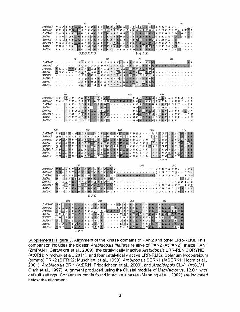
3
Supplemental Figure 3. Alignment of the kinase domains of PAN2 and other LRR-RLKs. This comparison includes the closest Arabidopsis thaliana relative of PAN2 (AtPAN2), maize PAN1 (ZmPAN1; Cartwright et al., 2009), the catalytically inactive Arabidopsis LRR-RLK CORYNE (AtCRN; Nimchuk et al., 2011), and four catalytically active LRR-RLKs: Solanum lycopersicum (tomato) PRK2 (SlPRK2; Muschietti et al., 1998), Arabidopsis SERK1 (AtSERK1; Hecht et al., 2001), Arabidopsis BRI1 (AtBRI1; Friedrichsen et al., 2000), and Arabidopsis CLV1 (AtCLV1; Clark et al., 1997). Alignment produced using the Clustal module of MacVector vs. 12.0.1 with default settings. Consensus motifs found in active kinases (Manning et al., 2002) are indicated below the alignment.
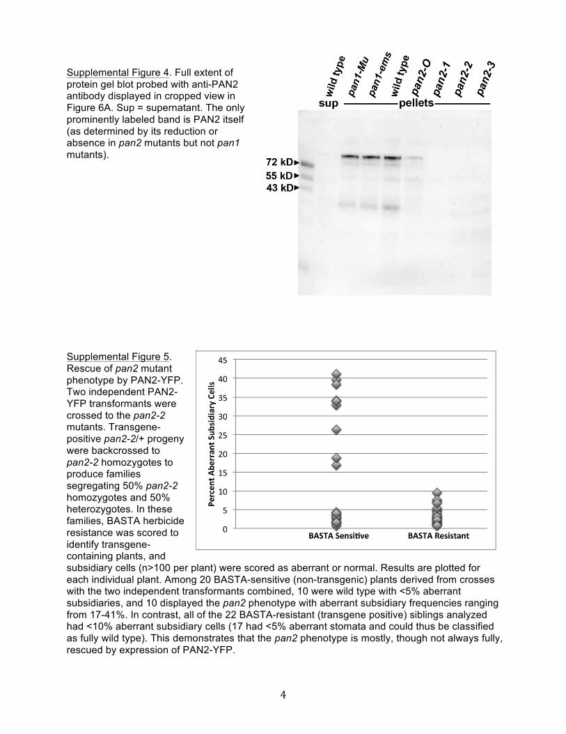
4
Supplemental Figure 4. Full extent of protein gel blot probed with anti-PAN2 antibody displayed in cropped view in Figure 6A. Sup = supernatant. The only prominently labeled band is PAN2 itself (as determined by its reduction or absence in pan2 mutants but not pan1 mutants). Supplemental Figure 5. Rescue of pan2 mutant phenotype by PAN2-YFP. Two independent PAN2-YFP transformants were crossed to the pan2-2 mutants. Transgene-positive pan2-2/+ progeny were backcrossed to pan2-2 homozygotes to produce families segregating 50% pan2-2 homozygotes and 50% heterozygotes. In these families, BASTA herbicide resistance was scored to identify transgene-containing plants, and subsidiary cells (n>100 per plant) were scored as aberrant or normal. Results are plotted for each individual plant. Among 20 BASTA-sensitive (non-transgenic) plants derived from crosses with the two independent transformants combined, 10 were wild type with <5% aberrant subsidiaries, and 10 displayed the pan2 phenotype with aberrant subsidiary frequencies ranging from 17-41%. In contrast, all of the 22 BASTA-resistant (transgene positive) siblings analyzed had <10% aberrant subsidiary cells (17 had <5% aberrant stomata and could thus be classified as fully wild type). This demonstrates that the pan2 phenotype is mostly, though not always fully, rescued by expression of PAN2-YFP.
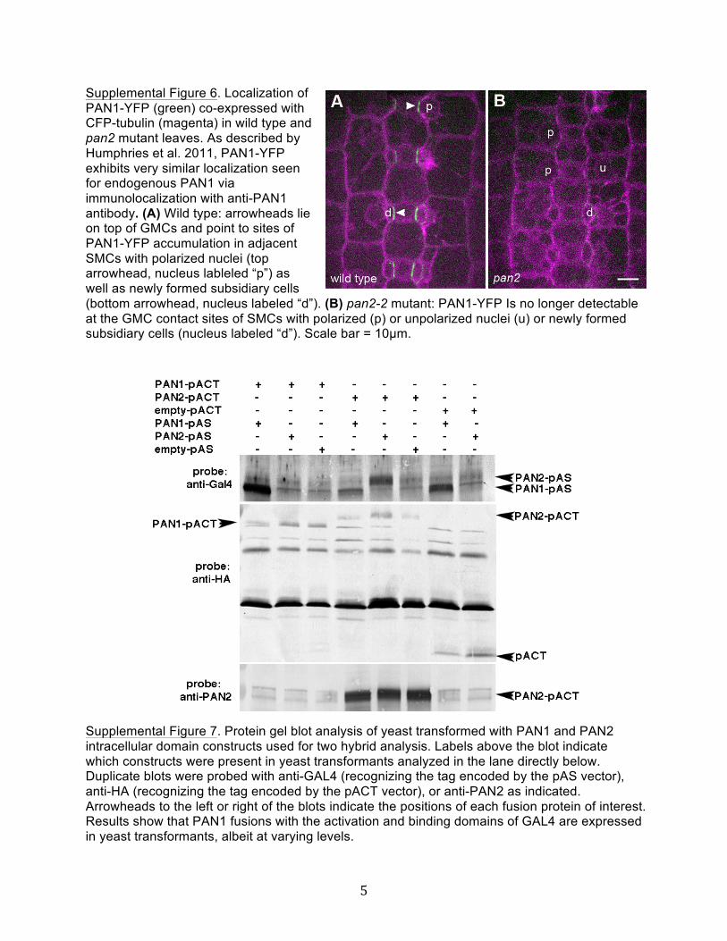
5
Supplemental Figure 6. Localization of PAN1-YFP (green) co-expressed with CFP-tubulin (magenta) in wild type and pan2 mutant leaves. As described by Humphries et al. 2011, PAN1-YFP exhibits very similar localization seen for endogenous PAN1 via immunolocalization with anti-PAN1 antibody. (A) Wild type: arrowheads lie on top of GMCs and point to sites of PAN1-YFP accumulation in adjacent SMCs with polarized nuclei (top arrowhead, nucleus lableled “p”) as well as newly formed subsidiary cells (bottom arrowhead, nucleus labeled “d”). (B) pan2-2 mutant: PAN1-YFP Is no longer detectable at the GMC contact sites of SMCs with polarized (p) or unpolarized nuclei (u) or newly formed subsidiary cells (nucleus labeled “d”). Scale bar = 10µm. Supplemental Figure 7. Protein gel blot analysis of yeast transformed with PAN1 and PAN2 intracellular domain constructs used for two hybrid analysis. Labels above the blot indicate which constructs were present in yeast transformants analyzed in the lane directly below. Duplicate blots were probed with anti-GAL4 (recognizing the tag encoded by the pAS vector), anti-HA (recognizing the tag encoded by the pACT vector), or anti-PAN2 as indicated. Arrowheads to the left or right of the blots indicate the positions of each fusion protein of interest. Results show that PAN1 fusions with the activation and binding domains of GAL4 are expressed in yeast transformants, albeit at varying levels.
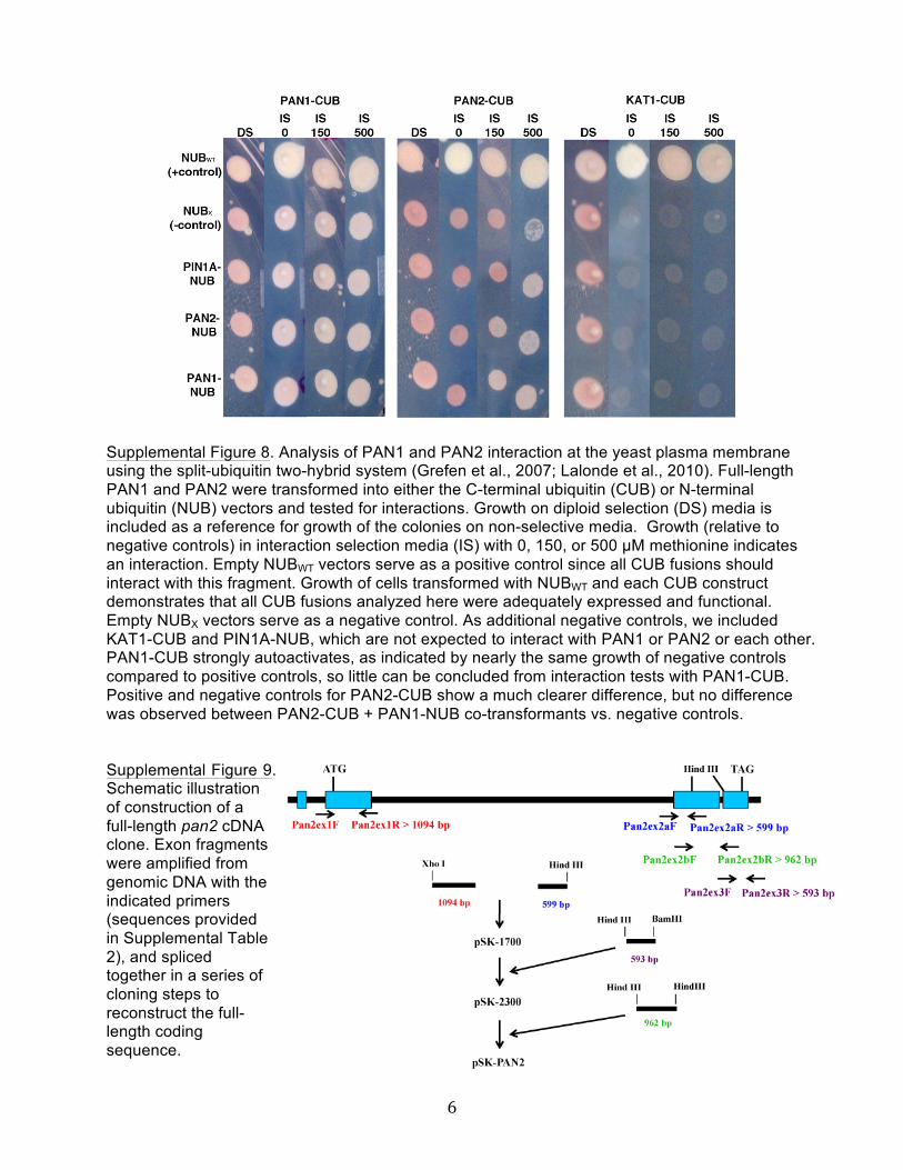
6
Supplemental Figure 8. Analysis of PAN1 and PAN2 interaction at the yeast plasma membrane using the split-ubiquitin two-hybrid system (Grefen et al., 2007; Lalonde et al., 2010). Full-length PAN1 and PAN2 were transformed into either the C-terminal ubiquitin (CUB) or N-terminal ubiquitin (NUB) vectors and tested for interactions. Growth on diploid selection (DS) media is included as a reference for growth of the colonies on non-selective media. Growth (relative to negative controls) in interaction selection media (IS) with 0, 150, or 500 µM methionine indicates an interaction. Empty NUBWT vectors serve as a positive control since all CUB fusions should interact with this fragment. Growth of cells transformed with NUBWT and each CUB construct demonstrates that all CUB fusions analyzed here were adequately expressed and functional. Empty NUBX vectors serve as a negative control. As additional negative controls, we included KAT1-CUB and PIN1A-NUB, which are not expected to interact with PAN1 or PAN2 or each other. PAN1-CUB strongly autoactivates, as indicated by nearly the same growth of negative controls compared to positive controls, so little can be concluded from interaction tests with PAN1-CUB. Positive and negative controls for PAN2-CUB show a much clearer difference, but no difference was observed between PAN2-CUB + PAN1-NUB co-transformants vs. negative controls. Supplemental Figure 9. Schematic illustration of construction of a full-length pan2 cDNA clone. Exon fragments were amplified from genomic DNA with the indicated primers (sequences provided in Supplemental Table 2), and spliced together in a series of cloning steps to reconstruct the full-length coding sequence.
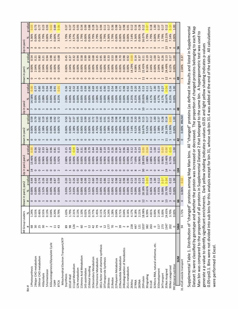
7
!""#$%&'(
#)*+,*
%-#
.&/
0#10#(+023#(+04#
5(#10#(+023(+04
#.&/
0#10#(+04#
5(#10#(+04#
.&/
0#10#(+02#
5(#10#(+02#
610#7#
7#8#
7#8#
(9:+"'*#
7#8#
(9:+"'*#
7#8#
(9:+"'*#
7#8#
(9:+"'*#
7#8#
(9:+"'*#
7#8#
(9:+"'*#
2#;<
&=&->0=<*-1-#
?@#
2A4B8#
C#CACC8#
CABB#
2B#
2DAB?8
#CACC#
C#CACC8#
CAEF#
G#2BAE@8
#CACC#
C#CACC8#
CAEB#
D#EAD?8#
CACD#
4#H+I&%#JKL#M
*=+N&"1-M#
DC#
CAEE8#
C#CACC8#
CA?F#
C#CACC8#
CAE?#
C#CACC8#
CAGF#
C#CACC8#
CAGG#
2#4ACB8#
CA42#
C#CACC8#
CAGD#
D#H10&%#JKL#M
*=+N&"1-M#
4E#
CAB?8#
C#CACC8#
CAGB#
C#CACC8#
CA?4#
C#CACC8#
CA@4#
C#CACC8#
CA@C#
C#CACC8#
CA@C#
C#CACC8#
CAGG#
B#$">O&">-1-#
DC#
CAEE8#
2#2AE48#
CA4?#
2#CAF?8#
CADD#
2#4AD@8#
CA2F#
2#4AC@8#
CA4C#
2#4ACB8#
CA42#
2#2AGF8#
CA4D#
E#P*%M
*0=+1&0#
G#CA2D8#
C#CACC8#
CAF4#
C#CACC8#
CA@G#
C#CACC8#
CAFE#
C#CACC8#
CAFB#
C#CACC8#
CAFB#
C#CACC8#
CAFD#
?#$"'O&0*
&Q*0
*-1-R$
">&S>"+=*#J>O"*#
4#CACB8#
C#CACC8#
CAF@#
C#CACC8#
CAF?#
C#CACC8#
CAF@#
C#CACC8#
CAF@#
C#CACC8#
CAF@#
2#2AGF8#
CAC4#
G#L;;
#22#
CA4C8#
C#CACC8#
CA@G#
C#CACC8#
CA@2#
C#CACC8#
CAF4#
C#CACC8#
CAF2#
C#CACC8#
CAF2#
C#CACC8#
CA@F#
@#TJ
!#
BD#
CAGF8#
C#CACC8#
CAEF#
4#2AF48#
CA2E#
C#CACC8#
CAG4#
D#?A4E8#
CAC2#
C#CACC8#
CA?@#
4#DAEG8#
CAC?#
F#H1=&O<&
0,%1+"#U"*O=%&0#T%+0-(&%=R!T;#
->0=<*
-1-#
@F#
2A?D8#
4#DACD8#
CA4C#
D#4A@@8#
CA2E#
C#CACC8#
CAEC#
2#4AC@8#
CAD?#
C#CACC8#
CABE#
2#2AGF8#
CADG#
2C#J*""#V
+""#
F@#
2AGF8#
2#2AE48#
CADG#
2#CAF?8#
CA4F#
C#CACC8#
CABG#
C#CACC8#
CAB4#
2#4ACB8#
CADG#
2#2AGF8#
CADG#
22#)1(1,#H*=+N&"1-M#
24?#
4AD28#
4#DACD8#
CA4?#
C#CACC8#
CACF#
2#4AD@8#
CADG#
C#CACC8#
CAD4#
C#CACC8#
CAD4#
4#DAEG8#
CA4D#
24#W9M
*=+N&"1-M#
G#CA2D8#
C#CACC8#
CAF4#
C#CACC8#
CA@G#
C#CACC8#
CAFE#
C#CACC8#
CAFB#
C#CACC8#
CAFB#
C#CACC8#
CAFD#
2D#!M10&#!O1,#H*=+N&"1-M#
?C#
2A2C8#
C#CACC8#
CAB@#
C#CACC8#
CAD2#
C#CACC8#
CA?D#
C#CACC8#
CAEF#
C#CACC8#
CAE@#
2#2AGF8#
CADB#
2B#X9+--1M1"+Y&
0#2#
CAC48#
C#CACC8#
CAFF#
C#CACC8#
CAF@#
C#CACC8#
CAFF#
C#CACC8#
CAFF#
C#CACC8#
CAFF#
C#CACC8#
CAFF#
2E#H*=+"#K+0,"10Q#
24#
CA448#
2#2AE48#
CA2D#
C#CACC8#
CAGF#
C#CACC8#
CAF2#
C#CACC8#
CAFC#
C#CACC8#
CAFC#
C#CACC8#
CA@@#
2?#X*
O&0,
+%>#H*=+N&"1-M#
B4#
CAGG8#
C#CACC8#
CA?C#
C#CACC8#
CABB#
C#CACC8#
CAG4#
C#CACC8#
CA?F#
C#CACC8#
CA?@#
2#2AGF8#
CA4@#
2G#K&%M&0
*#H*=+N&"1-M#
GF#
2ABE8#
4#DACD8#
CA2@#
2#CAF?8#
CADB#
4#BAG?8#
CA2C#
2#4AC@8#
CADE#
2#4ACB8#
CADE#
2#2AGF8#
CADG#
2@#J&9Z+O=&%#+0,
#:1=+M
10*#->0=<*
-1-#
?#CA228#
C#CACC8#
CAFD#
C#CACC8#
CA@F#
C#CACC8#
CAFE#
C#CACC8#
CAFE#
C#CACC8#
CAFE#
C#CACC8#
CAFB#
2F#T*=%+(>%%&"*#X>0=<*
-1-#
24#
CA448#
C#CACC8#
CA@?#
2#CAF?8#
CA2F#
C#CACC8#
CAF2#
C#CACC8#
CAFC#
C#CACC8#
CAFC#
2#2AGF8#
CA22#
4C#X=%*--#
2GG#
DA4B8#
C#CACC8#
CA22#
B#DA@E8#
CA2F#
2#4AD@8#
CADE#
2#4AC@8#
CADD#
2#4ACB8#
CADD#
D#EAD?8#
CA2G#
42#[*,
&S#
GC#
2A4@8#
C#CACC8#
CAB4#
C#CACC8#
CA4?#
C#CACC8#
CAE@#
C#CACC8#
CAEB#
C#CACC8#
CAED#
2#2AGF8#
CAD?#
44#;&">+M10#H
*=+N&"1-M#
4#CACB8#
C#CACC8#
CAF@#
C#CACC8#
CAF?#
C#CACC8#
CAF@#
C#CACC8#
CAF@#
C#CACC8#
CAF@#
C#CACC8#
CAF@#
4D#W'O"*&Y
,*#H
*=+N&"1-M#
DF#
CAG28#
C#CACC8#
CA?4#
C#CACC8#
CABG#
C#CACC8#
CAGB#
C#CACC8#
CAG2#
C#CACC8#
CAGC#
C#CACC8#
CA?G#
4B#61&,*
Q%+,+Y
&0#&Z#\
*0&N
1&YO-#
4#CACB8#
C#CACC8#
CAF@#
C#CACC8#
CAF?#
C#CACC8#
CAF@#
C#CACC8#
CAF@#
C#CACC8#
CAF@#
C#CACC8#
CAF@#
4E#O29M
*=+N&"1-M#
F#CA2?8#
C#CACC8#
CAFC#
C#CACC8#
CA@B#
C#CACC8#
CAFD#
C#CACC8#
CAF4#
C#CACC8#
CAF4#
C#CACC8#
CAF2#
4?#H1-O#
44@#
BA2G8#
D#BAEE8#
CA4D#
?#EAGG8#
CA24#
D#GA2B8#
CA2?#
D#?A4E8#
CA2F#
G#2BA4F8
#CACC#
B#GA2B8#
CA24#
4G#[W!#
BBG#
@A2@8#
?#FACF8#
CA2?#
@#GA?F8#
CA2B#
B#FAE48#
CA4C#
D#?A4E8#
CA4C#
4#BAC@8#
CA2B#
D#EAD?8#
CA2?#
4@#.W!#
242#
4A428#
C#CACC8#
CA4D#
2#CAF?8#
CA4D#
C#CACC8#
CADF#
C#CACC8#
CADB#
2#4ACB8#
CADG#
C#CACC8#
CA4@#
4F#;%&=*10#
2CDG#
2@AF@8
#24#
2@A2@8
#CA2D#
DC#
4@A@E8
#CACC#
G#2?A?G8
#CA2E#
2C#
4CA@D8
#CA2D#
22#
44ABE8
#CA22#
F#2?ACG8
#CA24#
DC#-1Q0+"10Q#
D@4#
?AFF8#
G#2CA?28
#CACF#
D#4A@@8#
CACB#
B#FAE48#
CA2G#
2#4AC@8#
CA22#
B#@A2?8#
CA2F#
2#2AGF8#
CACG#
D2#J*""#
4?D#
BA@28#
B#?AC?8#
CA2@#
D#4A@@8#
CA2B#
C#CACC8#
CA24#
4#BA2G8#
CA4G#
2#4ACB8#
CA44#
2#2AGF8#
CA2@#
D4#H1O%&#[W!]#0+='%+"#+0Y
-*0-*]#*=OA#
C#CACC8#
C#CACC8#
2ACC#
C#CACC8#
2ACC#
C#CACC8#
2ACC#
C#CACC8#
2ACC#
C#CACC8#
2ACC#
C#CACC8#
2ACC#
DD#.*:*"&(
M*0
=#22G#
4A2B8#
D#BAEE8#
CA24#
B#DA@E8#
CA22#
2#4AD@8#
CADG#
4#BA2G8#
CA2F#
2#4ACB8#
CADG#
2#2AGF8#
CADG#
DB#T%+0-(&
%=#
4GD#
EACC8#
?#FACF8#
CACG#
2#CAF?8#
CACD#
4#BAG?8#
CA4@#
4#BA2G8#
CA4G#
4#BAC@8#
CA4?#
2#2AGF8#
CA2G#
DE#W&=#!--1Q0*
,#2DBG#
4BA?E8
#2D#
2FAGC8
#CAC@#
2B#
2DAB?8
#CACC#
22#
4?A2F8
#CA2B#
@#2?A?G8
#CAC?#
24#
4BABF8
#CA2D#
2D#
4DA428
#CA24#
FF#W&=#O+=*Q&%1^*,
#4C4#
DAGC8#
D#BAEE8#
CA44#
G#?AGD8#
CACE#
E#22AFC8
#CAC2#
D#?A4E8#
CA2?#
D#?A248#
CA2G#
B#GA2B8#
CA2C#
FF2#H10*%+"#0'=%1Y&
0#C#
CACC8#
C#CACC8#
2ACC#
C#CACC8#
2ACC#
C#CACC8#
2ACC#
C#CACC8#
2ACC#
C#CACC8#
2ACC#
C#CACC8#
2ACC#
!"#$
%&'&$
$$''$
$$$$
()&$
$$$$
&*$
$$$$
&+$
$$$$
&,$
$$$$
%'$
$$$$
D2AB#J*""A:*-1O"*#=%+0-(&
%=#
FB#
2AG48#
B#?AC?8#
CAC4#
C#CACC8#7W
5H_#
2#4ACB8#
CADG#
X'((
"*M*0
=+"#T+N"*#2A#.
1-=%1N'Y
&0#&Z#`O<+0Q*,a#(%&=*10-#+M
&0Q#H+(#H
+0#N10-A##!
""#`O<+0Q*,
a#(%&=*10-#b+-#,*
c0*,
#10#[*-'"=-#+0,
#"1-=*,
#10#X'(
("*M
*0=+"#
.+=+-*=#Dd#/
*%*#O"+--1c*
,#N>#Q*0
&=>(*#+0,#/<*
=<*%#=<*
#(%&=*10#/+-#10O%*+-*,#&%#,*O%*+-*,
A##T<*#(%&(
&%Y&
0#&Z#O<+0Q*,
#(%&=*10-#N*
"&0Q10Q#=&#*+O<#H+(#
H+0#N10#/+-#O&M
(+%*,#=&#=<*
#(%&(&
%Y&0
#&Z#+""#(%&=*10-#10#X'(
("*M
*0=+"#.
+=+-*=#4#=<+=#N*
"&0Q*,
#=&#=<*#-+M*#N10A##!
#<>(*%Q*&M
*=%1O#=*-=#/+-#'-*,#=&#
Q*0*
%+=*#+#(9:+"'*
#=,*0
YZ>#-1Q01cO+0=#*0%1O<M
*0=-A##.+%e#>*""&/#-<+,10Q#10,1O+=*-#(9:+"'*-#fCACE#+0,
#"1Q<=#>*""&/#-<+,10Q#10,1O+=*-#(9:+"'*-#
CACEg(
fCA2A#6
10-#/*%*#0&
=#,1:1,*
,#10=&#-'N
#N10-#Z&%#=<1-#+0+">-1-#*SO*(=#Z&
%#N10#D2]#/
<&-*#-'N
9N10#D2AB#/+-#+,,
*,#+=#=<*#*0
,#&Z#=<*
#=+N"*A##!""#O+"O'"+Y
&0-#
/*%*#(*
%Z&%M*,
#10#USO*"A###
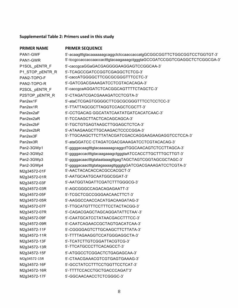
8
Supplemental Table 2: Primers used in this study
PRIMER NAME PRIMER SEQUENCE PAN1-GWF 5’-acaagtttgtacaaaaaagcaggctctccaaccaccatgGCGGCGGTTCTGGCGGTCCTGGTGT-3’ PAN1-GWR 5’-tccgccaccaccaaccactttgtacaagaaagctgggtaGCCGATCCGGTCGAGGCTCTCGGCGA-3’
P1SOL_pENTR_F 5’-caccgcaGGaGACGAGGGGAAGGAGTCCGGCAA-3’ P1_STOP_pENTR_R 5’-TCAGCCGATCCGGTCGAGGCTCTCG-3’ PAN2-TOPO-F 5’-caccATGGGGCTTCGCGCGGGTTTCCTC-3’
PAN2-TOPO-R 5’-GATCGACGAAAGATCCTCGTACACAGA-3’
P2SOL_pENTR_F 5’-caccgcaAGGATCTCACGGCAGTTTTCTAGCTC-3’
P2STOP_pENTR_R 5’-CTAGATCGACGAAAGATCCTCGTA-3’ Pan2ex1F 5’-ataCTCGAGTGGGGCTTCGCGCGGGTTTCCTCCTCC-3’ Pan2ex1R 5’-TTATTAGCGCTTAGGTCCAGCTCGCTT-3’ Pan2ex2aF 5’-CCTGACAG GGCATATCAATATGATCACATCAAC-3’ Pan2ex2aR 5’-TCCAAGCTTACTCACAGCAGCA-3’ Pan2ex2bF 5’-TGCTGTGAGTAAGCTTGGAGCTCTCA-3’ Pan2ex2bR 5’-ATAAGAAGCTTGCAAGACTCCCCGGA-3’ Pan2ex3F 5’-TTGCAAGCTTCTTATACGATCGACCAGGAAGAAGAGGTCCTCCA-3’
Pan2ex3R 5’-ataGGATCC CTAGATCGACGAAAGATCCTCGTACACAG-3’ Pan2-3GWp1 5’-ggggacaagtttgtacaaaaaagcaggctTGGCAACAGTCTCCTTAGCA-3’ Pan2-3GWp2 5’-ggggaccactttgtacaagaaagctgggtaATCCACCTTGCTTTGCTTGT-3’ Pan2-3GWp3 5’-ggggacaactttgtataataaagttgagTAGCTAGTCGGTAGCGCTAGC-3’ Pan2-3GWp4 5’-ggggacaactttgtatagaaaagttgggtgGATCGACGAAAGATCCTCGTA-3’ M2g34572-01F 5’-AACTACACACCACGCCACGCT-3’
M2g34572-01R 5’-AATGCAATGCAATGGCGGAT-3’
M2g34572-03F 5’-AATGGTAGATTCGATCTTTGGGCG-3’
M2g34572-03R 5’-AGCGGGCCAGACAGAGAATT-3’ M2g34572-05F 5’-TCGCTCGCCGGGAACAACTTCT-3’ M2g34572-05R 5’-AAGGCCAACCACATGACAAGATAG-3’ M2g34572-07F 5’-TTGCATGTTTCCTTTCCTACTACGG-3’ M2g34572-07R 5’-CAGACGAGCTAGCAGGATATTCTAA’-3’ M2g34572-09F 5’-CAATGCATCCTATAACGACCTTTCC-3’ M2g34572-09R 5’-CAATCAGAACCGCTAGTGACATCAA-3’ M2g34572-11F 5’-CGGGGAGTCTTGCAAGCTTCTTATA-3’ M2g34572-11R 5’-TTTTAGAAGGTCCATGGGAGGCTA-3’ M2g34572-13F 5’-TCATCTTGTCGGATTACGTCG-3’ M2g34572-13R 5’-TTCATGCCCTTCACAGCCT-3’
M2g34572-15F 5’-ATGGCCTCGGACTCTGAGAGCAA-3’ M2g34572-‐15R 5’-CTAACGAAACGTCGTGAGTGAAAG-3’ M2g34572-16F 5’-GCCTATCCTTTCCTGGTTCCTCAT-3’ M2g34572-16R 5’-TTTTCCACCTGCTGACCCAGAT’3’ M2g34572-17F 5’-GGCAACAACCTCTCGGGC-3’
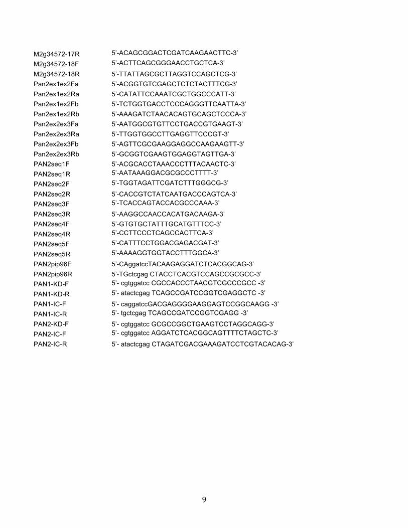
9
M2g34572-17R 5’-ACAGCGGACTCGATCAAGAACTTC-3’
M2g34572-18F 5’-ACTTCAGCGGGAACCTGCTCA-3’
M2g34572-18R 5’-TTATTAGCGCTTAGGTCCAGCTCG-3’ Pan2ex1ex2Fa 5’-ACGGTGTCGAGCTCTCTACTTTCG-3’ Pan2ex1ex2Ra 5’-CATATTCCAAATCGCTGGCCCATT-3’ Pan2ex1ex2Fb 5’-TCTGGTGACCTCCCAGGGTTCAATTA-3’ Pan2ex1ex2Rb 5’-AAAGATCTAACACAGTGCAGCTCCCA-3’ Pan2ex2ex3Fa 5’-AATGGCGTGTTCCTGACCGTGAAGT-3’ Pan2ex2ex3Ra 5’-TTGGTGGCCTTGAGGTTCCCGT-3’ Pan2ex2ex3Fb 5’-AGTTCGCGAAGGAGGCCAAGAAGTT-3’ Pan2ex2ex3Rb 5’-GCGGTCGAAGTGGAGGTAGTTGA-3’ PAN2seq1F 5’-ACGCACCTAAACCCTTTACAACTC-3’ PAN2seq1R 5’-AATAAAGGACGCGCCCTTTT-3’
PAN2seq2F 5’-TGGTAGATTCGATCTTTGGGCG-3’
PAN2seq2R 5’-CACCGTCTATCAATGACCCAGTCA-3’ PAN2seq3F 5’-TCACCAGTACCACGCCCAAA-3’
PAN2seq3R 5’-AAGGCCAACCACATGACAAGA-3’ PAN2seq4F 5’-GTGTGCTATTTGCATGTTTCC-3’ PAN2seq4R 5’-CCTTCCCTCAGCCACTTCA-3’
PAN2seq5F 5’-CATTTCCTGGACGAGACGAT-3’
PAN2seq5R 5’-AAAAGGTGGTACCTTTGGCA-3’
PAN2pip96F 5’-CAggatccTACAAGAGGATCTCACGGCAG-3’ PAN2pip96R 5’-TGctcgag CTACCTCACGTCCAGCCGCGCC-3’ PAN1-KD-F 5’- cgtggatcc CGCCACCCTAACGTCGCCCGCC -3’
PAN1-KD-R 5’- atactcgag TCAGCCGATCCGGTCGAGGCTC -3’
PAN1-IC-F 5’- caggatccGACGAGGGGAAGGAGTCCGGCAAGG -3’ PAN1-IC-R 5’- tgctcgag TCAGCCGATCCGGTCGAGG -3’
PAN2-KD-F 5’- cgtggatcc GCGCCGGCTGAAGTCCTAGGCAGG-3’ PAN2-IC-F 5’- cgtggatcc AGGATCTCACGGCAGTTTTCTAGCTC-3’
PAN2-IC-R 5’- atactcgag CTAGATCGACGAAAGATCCTCGTACACAG-3’
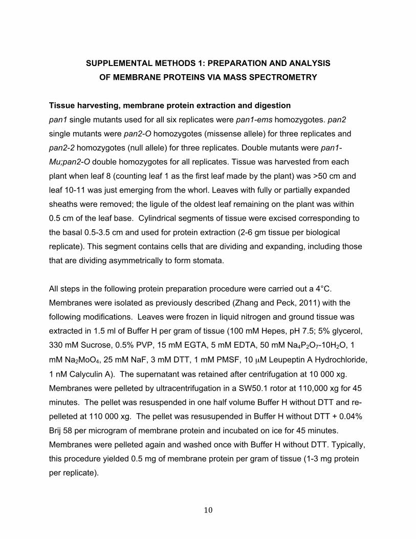
10
SUPPLEMENTAL METHODS 1: PREPARATION AND ANALYSIS
OF MEMBRANE PROTEINS VIA MASS SPECTROMETRY Tissue harvesting, membrane protein extraction and digestion pan1 single mutants used for all six replicates were pan1-ems homozygotes. pan2
single mutants were pan2-O homozygotes (missense allele) for three replicates and
pan2-2 homozygotes (null allele) for three replicates. Double mutants were pan1-
Mu;pan2-O double homozygotes for all replicates. Tissue was harvested from each
plant when leaf 8 (counting leaf 1 as the first leaf made by the plant) was >50 cm and
leaf 10-11 was just emerging from the whorl. Leaves with fully or partially expanded
sheaths were removed; the ligule of the oldest leaf remaining on the plant was within
0.5 cm of the leaf base. Cylindrical segments of tissue were excised corresponding to
the basal 0.5-3.5 cm and used for protein extraction (2-6 gm tissue per biological
replicate). This segment contains cells that are dividing and expanding, including those
that are dividing asymmetrically to form stomata.
All steps in the following protein preparation procedure were carried out a 4°C.
Membranes were isolated as previously described (Zhang and Peck, 2011) with the
following modifications. Leaves were frozen in liquid nitrogen and ground tissue was
extracted in 1.5 ml of Buffer H per gram of tissue (100 mM Hepes, pH 7.5; 5% glycerol,
330 mM Sucrose, 0.5% PVP, 15 mM EGTA, 5 mM EDTA, 50 mM Na4P2O7-10H2O, 1
mM Na2MoO4, 25 mM NaF, 3 mM DTT, 1 mM PMSF, 10 µM Leupeptin A Hydrochloride,
1 nM Calyculin A). The supernatant was retained after centrifugation at 10 000 xg.
Membranes were pelleted by ultracentrifugation in a SW50.1 rotor at 110,000 xg for 45
minutes. The pellet was resuspended in one half volume Buffer H without DTT and re-
pelleted at 110 000 xg. The pellet was resusupended in Buffer H without DTT + 0.04%
Brij 58 per microgram of membrane protein and incubated on ice for 45 minutes.
Membranes were pelleted again and washed once with Buffer H without DTT. Typically,
this procedure yielded 0.5 mg of membrane protein per gram of tissue (1-3 mg protein
per replicate).
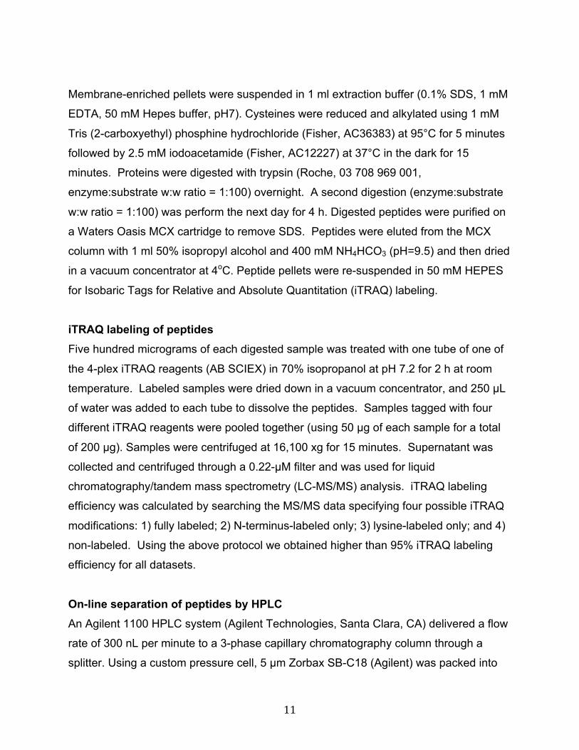
11
Membrane-enriched pellets were suspended in 1 ml extraction buffer (0.1% SDS, 1 mM
EDTA, 50 mM Hepes buffer, pH7). Cysteines were reduced and alkylated using 1 mM
Tris (2-carboxyethyl) phosphine hydrochloride (Fisher, AC36383) at 95°C for 5 minutes
followed by 2.5 mM iodoacetamide (Fisher, AC12227) at 37°C in the dark for 15
minutes. Proteins were digested with trypsin (Roche, 03 708 969 001,
enzyme:substrate w:w ratio = 1:100) overnight. A second digestion (enzyme:substrate
w:w ratio = 1:100) was perform the next day for 4 h. Digested peptides were purified on
a Waters Oasis MCX cartridge to remove SDS. Peptides were eluted from the MCX
column with 1 ml 50% isopropyl alcohol and 400 mM NH4HCO3 (pH=9.5) and then dried
in a vacuum concentrator at 4oC. Peptide pellets were re-suspended in 50 mM HEPES
for Isobaric Tags for Relative and Absolute Quantitation (iTRAQ) labeling.
iTRAQ labeling of peptides Five hundred micrograms of each digested sample was treated with one tube of one of
the 4-plex iTRAQ reagents (AB SCIEX) in 70% isopropanol at pH 7.2 for 2 h at room
temperature. Labeled samples were dried down in a vacuum concentrator, and 250 µL
of water was added to each tube to dissolve the peptides. Samples tagged with four
different iTRAQ reagents were pooled together (using 50 µg of each sample for a total
of 200 µg). Samples were centrifuged at 16,100 xg for 15 minutes. Supernatant was
collected and centrifuged through a 0.22-µM filter and was used for liquid
chromatography/tandem mass spectrometry (LC-MS/MS) analysis. iTRAQ labeling
efficiency was calculated by searching the MS/MS data specifying four possible iTRAQ
modifications: 1) fully labeled; 2) N-terminus-labeled only; 3) lysine-labeled only; and 4)
non-labeled. Using the above protocol we obtained higher than 95% iTRAQ labeling
efficiency for all datasets.
On-line separation of peptides by HPLC An Agilent 1100 HPLC system (Agilent Technologies, Santa Clara, CA) delivered a flow
rate of 300 nL per minute to a 3-phase capillary chromatography column through a
splitter. Using a custom pressure cell, 5 µm Zorbax SB-C18 (Agilent) was packed into
12
fused silica capillary tubing (200 µm ID, 360 µm OD, 20 cm long) to form the first
reverse phase column (RP1). A 5 cm long strong cation exchange (SCX) column
packed with 5 µm PolySulfoethyl (PolyLC, Inc.) was connected to RP1 using a zero
dead volume 1-µm filter (Upchurch, M548) attached to the exit of the RP1 column. A
fused silica capillary (100 µm ID, 360 µm OD, 20 cm long) packed with 5 µm Zorbax
SB-C18 (Agilent) was connected to SCX as the analytical column (the second reverse
phase column). The electro-spray tip of the fused silica tubing was pulled to a sharp tip
with the inner diameter smaller than 1 µm using a laser puller (Sutter P-2000). The
peptide mixtures were loaded onto the RP1 using the custom pressure cell. Columns
were not re-used. Peptides were first eluted from the RP1 to the SCX column using a 0
to 80% acetonitrile gradient for 150 minutes. The peptides were fractionated by the SCX
column using a series of salt gradients (from 10 mM to 1 M ammonium acetate for 20
minutes), followed by high resolution reverse phase separation using an acetonitrile
gradient of 0 to 80% for 120 minutes. It takes 3 days (38 salt fractions) for each full
proteome analysis.
Tandem mass spectrometry analysis Spectra were acquired using a LTQ linear ion trap tandem mass spectrometer (Thermo
Electron Corporation, San Jose, CA) employing automated, data-dependent acquisition.
The mass spectrometer was operated in positive ion mode with a source temperature of
150oC. A high voltage of 2,000V was applied between the electro-spray tip and the
entrance of the ion transfer tube. The full MS scan range of 400-2000 m/z was divided
into three smaller scan ranges (400-800, 800-1050, 1050-2000) to improve the dynamic
range. Both CID (Collision Induced Dissociation) and PQD (Pulsed-Q Dissociation)
scans of the same parent ion were collected for protein identification and quantitation.
A precursor isolation window of 4 Da was used for both CID and PQD. The collision
energy of CID and PQD was 35% and 30%, respectively. The activation Q of CID and
PQD was 0.25 and 0.7, respectively. The activation time of CID and PQD was 30 ms
and 0.15 ms, respectively. Each MS scan was followed by four pairs of CID-PQD
MS/MS scans of the most intense ions from the parent MS scan. A dynamic exclusion
of 1 minute was used to improve the duty cycle of MS/MS scans.
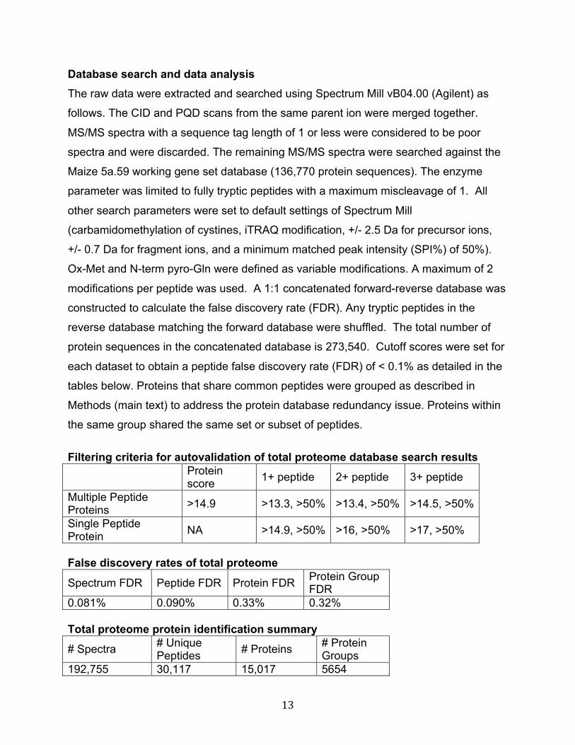
13
Database search and data analysis The raw data were extracted and searched using Spectrum Mill vB04.00 (Agilent) as
follows. The CID and PQD scans from the same parent ion were merged together.
MS/MS spectra with a sequence tag length of 1 or less were considered to be poor
spectra and were discarded. The remaining MS/MS spectra were searched against the
Maize 5a.59 working gene set database (136,770 protein sequences). The enzyme
parameter was limited to fully tryptic peptides with a maximum miscleavage of 1. All
other search parameters were set to default settings of Spectrum Mill
(carbamidomethylation of cystines, iTRAQ modification, +/- 2.5 Da for precursor ions,
+/- 0.7 Da for fragment ions, and a minimum matched peak intensity (SPI%) of 50%).
Ox-Met and N-term pyro-Gln were defined as variable modifications. A maximum of 2
modifications per peptide was used. A 1:1 concatenated forward-reverse database was
constructed to calculate the false discovery rate (FDR). Any tryptic peptides in the
reverse database matching the forward database were shuffled. The total number of
protein sequences in the concatenated database is 273,540. Cutoff scores were set for
each dataset to obtain a peptide false discovery rate (FDR) of < 0.1% as detailed in the
tables below. Proteins that share common peptides were grouped as described in
Methods (main text) to address the protein database redundancy issue. Proteins within
the same group shared the same set or subset of peptides.
Filtering criteria for autovalidation of total proteome database search results Protein
score 1+ peptide 2+ peptide 3+ peptide
Multiple Peptide Proteins >14.9 >13.3, >50% >13.4, >50% >14.5, >50%
Single Peptide Protein NA >14.9, >50% >16, >50% >17, >50%
False discovery rates of total proteome
Spectrum FDR Peptide FDR Protein FDR Protein Group FDR
0.081% 0.090% 0.33% 0.32% Total proteome protein identification summary
# Spectra # Unique Peptides # Proteins # Protein
Groups 192,755 30,117 15,017 5654
14
Quantification by iTRAQ mass tagging reagent Protein iTRAQ intensities were calculated by summing the peptide iTRAQ intensities
from each protein group. Peptides shared among different protein groups were
removed before quantitation. A minimal total iTRAQ reporter ion intensity (sum of all
four channels) of 100 was used to filter out low intensity spectra. Isotope impurities of
iTRAQ reagents were corrected using correction factors provided by the manufacturer.
Median normalization was performed to normalize the protein iTRAQ reporter intensities
in which the log ratios between different iTRAQ tags (115/114, 116/114, 117/114) were
adjusted globally such that the median log ratio is zero. Quantitative analysis was
performed on the normalized protein iTRAQ intensities. Protein ratios between treated
and non-treated samples were calculated by taking the ratios of the total iTRAQ
intensities from the corresponding iTRAQ reporters. T-test (two tailed, paired) of the ln
ratios was used to calculate the p-values.
Data Analysis All “reduced” proteins listed in Supplemental Dataset 3 were classified in the Venn
diagram shown in Figure 3. Note that for this diagram, proteins were classified as
“reduced” if they were decreased at least 1.5 fold regardless of the associated p-value
(but had to have a significant reduction in at least one genotype to qualify for inclusion
in Supplemental Dataset 3). The Venn diagram was constructed using Venny
(http://bioinfogp.cnb.csic.es/tools/venny/index.html) and diagrams were redrawn in
Adobe Illustrator. To determine functional enrichments, proteins were classified by
MapMan Bin (ignoring sub-bins) (Thimm et al., 2004); accessed at
http://mapman.gabipd.org/web/guest/mapmanstore) using mapping file
Zm_B73_5b_FGS_cds_2011. Since this mapping file contains only filtered set genes
from genome release 5b, while we used the larger working set from release 5a to
identify proteins, those proteins in the dataset with no corresponding MapMan bin in the
mapping file were assigned to a new bin (99). A hypergeometric test comparing proteins
that were increased or decreased in each of the mutants relative to all proteins identified
(Supplemental Dataset 2) was performed in Microsoft Excel.
15
REFERENCES Cartwright, H.N., Humphries, J.A., and Smith, L.G. (2009). PAN1: a receptor-like protein that promotes polarization of an asymmetric cell division in maize. Science 323: 649–651. Clark, S.E., Williams, R.W., and Meyerowitz, E.M. (1997). The CLAVATA1 gene encodes a putative receptor kinase that controls shoot and floral meristem size in Arabidopsis. Cell 89: 575–585. Friedrichsen, D.M., Joazeiro, C.A., Li, J., Hunter, T., and Chory, J. (2000). Brassinosteroid-insensitive-1 is a ubiquitously expressed leucine-rich repeat receptor serine/threonine kinase. Plant Physiol 123: 1247–1256. Grefen, C., Lalonde, S., and Obrdlik, P. (2007). Split-ubiquitin system for identifying protein-protein interactions in membrane and full-length proteins. Curr Protoc Neurosci Chapter 5: Unit 5.27. Humphries, J.A. et al. (2011). ROP GTPases act with the receptor-like protein PAN1 to polarize asymmetric cell division in maize. Plant Cell 23: 2273–2284. Lalonde, S. et al. (2010). A membrane protein/signaling protein interaction network for Arabidopsis version AMPv2. Front Physiol 1: 24. Manning, G., Whyte, D.B., Martinez, R., Hunter, T., and Sudarsanam, S. (2002). The protein kinase complement of the human genome. Science 298: 1912–1934. Muschietti, J., Eyal, Y., and McCormick, S. (1998). Pollen tube localization implies a role in pollen-pistil interactions for the tomato receptor-like protein kinases LePRK1 and LePRK2. Plant Cell 10: 319–330. Nimchuk, Z.L., Tarr, P.T., and Meyerowitz, E.M. (2011). An evolutionarily conserved pseudokinase mediates stem cell production in plants. Plant Cell 23: 851–854. Sekhon, R.S. et al. (2011). Genome-wide atlas of transcription during maize development. Plant J 66: 553–563. Thimm, O. et al. (2004). MAPMAN: a user-driven tool to display genomics data sets onto diagrams of metabolic pathways and other biological processes. Plant J 37: 914–939. Zhang, Z.J., and Peck, S.C. (2011). Simplified enrichment of plasma membrane proteins for proteomic analyses in Arabidopsis thaliana. Proteomics 11: 1780–1788.