1
Point-spread function model for fluorescence macroscopy imaging Praveen Pankajakshan 1 , Alain Dieterlen 2 , Gilbert Engler 3 , Zvi Kam 4 , Laure Blanc- Féraud 5 , Josiane Zerubia 5 , Jean-Christophe Olivo-Marin 1 1 Quantitative Image Analysis Unit, Institute Pasteur, 25 rue du Docteur Roux, 75724 Paris, France. 2 Laboratoire MIPS - LAB.EL, Université de Haute-Alsace, 61, rue Albert Camus, 68093 Mulhouse, France. 3 IBSV Unit, INRA 400 Route des Chappes, 06903 Sophia Antipolis, France. 4 Molecular Cell Biology, Weizmann Institute of Science, Rehovot 76100, Israel. 5 Ariana Project-team, INRIA, 2004 route des Lucioles, 06902 Sophia Antipolis Cedex, France. In recent years, imaging techniques have emerged in all areas of life sciences in order to study basic cellular, biochemical, physiological and pathological processes. Innovative imaging technologies are deployed to produce images of biological systems at the cellular, organ and whole body levels, either independently or in combination. Although, the last decade has seen a growth in new imaging systems involving smaller samples (e.g. fluorescent microscopes), fluorescent macroscopes were recently commercialized. Such an imaging system allows the observation of relatively large samples (up to a couple of centimeters), to acquire data in three dimensions and to also perform time-lapse imaging. However, one of the main shortcomings of fluorescence macroscopy is that the observed images are affected by field aberrations. In this case, the point-spread function (PSF) varies within the lateral field and is proportional to the distance from the center of the field of view. This is because; the zoom system in a macroscope cannot achieve the condition of lateral invariance for all magnifications. A computational approach to compensate the aberration often relies on an accurate model/knowledge of the PSF [1, 2]. The PSF can be defined either theoretically using a scalar diffraction model or experimentally by acquiring a three-dimensional image of a fluorescent bead that is positioned so as to approximate a point object of unit intensity located in the slide. As the experimental PSF is difficult to obtain for fluorescence macroscopes and can change with slight deviations from the physical conditions, in this paper, we model the PSF using a scalar diffraction approach [3]. The pupil function is modeled by chopping it, vignette, as a result of two limiting optical apertures not brought together to the same conjugated plane. The proportionality constant relates linearly to the distance between the two limiting apertures. We also compare our developed model with that obtained experimentally and show the validity of our hypothesis. Figure 1: Lateral maximum intensity projection 4μm fluorescent bead images (left), and a preliminary theoretically calculated point-spread function (right) for a macroscope fit with an objective 5X/0.5. REFERENCES [1] Z. Kam, B. Hanser, M. G. L. Gustafsson, D. A. Agard & J. W. Sedat (2001). ”Computational adaptive optics for live three-dimensional biological imaging”, Proc. Natl. Acad. Sci. 98(7):3790-3795. [2] B. M. Hanser, et al. (2003). “Phase retrieval for high-numerical-aperture optical systems”. Opt. Lett. 28(10):801- 803. [3] P. Pankajakshan (2009). “Blind deconvolution for confocal laser scanning microscopy”. PhD thesis. University of Nice Sophia-Antipolis.

Point-spread function model for fluorescence macroscopy .... Praveen_Mifobio.pdf · Point-spread function model for fluorescence macroscopy imaging Praveen Pankajakshan1, Alain Dieterlen2,

Embed Size (px)

Citation preview

Point-spread function model for fluorescence macroscopy imaging

Praveen Pankajakshan1, Alain Dieterlen2, Gilbert Engler3, Zvi Kam4, Laure Blanc-Féraud5, Josiane Zerubia5, Jean-Christophe Olivo-Marin1

1Quantitative Image Analysis Unit, Institute Pasteur, 25 rue du Docteur Roux, 75724 Paris, France.

2Laboratoire MIPS - LAB.EL, Université de Haute-Alsace, 61, rue Albert Camus, 68093 Mulhouse, France. 3IBSV Unit, INRA 400 Route des Chappes, 06903 Sophia Antipolis, France.

4Molecular Cell Biology, Weizmann Institute of Science, Rehovot 76100, Israel. 5Ariana Project-team, INRIA, 2004 route des Lucioles, 06902 Sophia Antipolis Cedex, France.

In recent years, imaging techniques have emerged in all areas of life sciences in order to study basic cellular, biochemical, physiological and pathological processes. Innovative imaging technologies are deployed to produce images of biological systems at the cellular, organ and whole body levels, either independently or in combination. Although, the last decade has seen a growth in new imaging systems involving smaller samples (e.g. fluorescent microscopes), fluorescent macroscopes were recently commercialized. Such an imaging system allows the observation of relatively large samples (up to a couple of centimeters), to acquire data in three dimensions and to also perform time-lapse imaging. However, one of the main shortcomings of fluorescence macroscopy is that the observed images are affected by field aberrations. In this case, the point-spread function (PSF) varies within the lateral field and is proportional to the distance from the center of the field of view. This is because; the zoom system in a macroscope cannot achieve the condition of lateral invariance for all magnifications. A computational approach to compensate the aberration often relies on an accurate model/knowledge of the PSF [1, 2]. The PSF can be defined either theoretically using a scalar diffraction model or experimentally by acquiring a three-dimensional image of a fluorescent bead that is positioned so as to approximate a point object of unit intensity located in the slide. As the experimental PSF is difficult to obtain for fluorescence macroscopes and can change with slight deviations from the physical conditions, in this paper, we model the PSF using a scalar diffraction approach [3]. The pupil function is modeled by chopping it, vignette, as a result of two limiting optical apertures not brought together to the same conjugated plane. The proportionality constant relates linearly to the distance between the two limiting apertures. We also compare our developed model with that obtained experimentally and show the validity of our hypothesis.

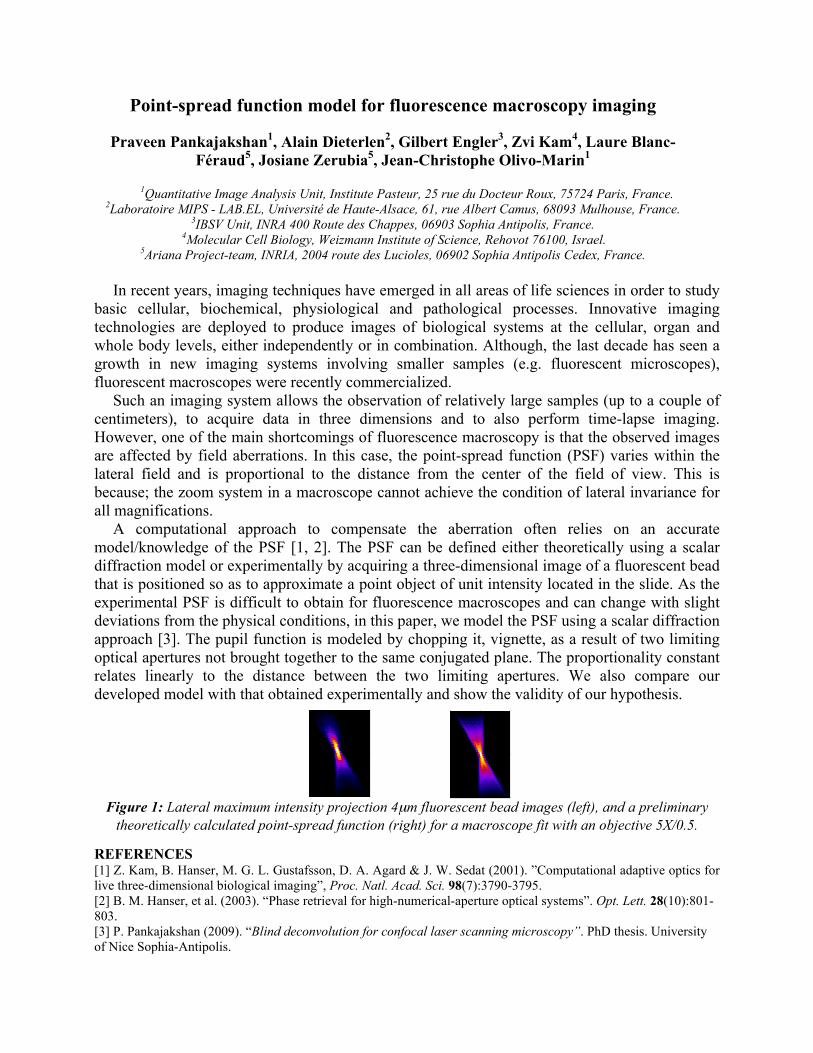
Figure 1: Lateral maximum intensity projection 4µm fluorescent bead images (left), and a preliminary

theoretically calculated point-spread function (right) for a macroscope fit with an objective 5X/0.5. REFERENCES [1] Z. Kam, B. Hanser, M. G. L. Gustafsson, D. A. Agard & J. W. Sedat (2001). ”Computational adaptive optics for live three-dimensional biological imaging”, Proc. Natl. Acad. Sci. 98(7):3790-3795. [2] B. M. Hanser, et al. (2003). “Phase retrieval for high-numerical-aperture optical systems”. Opt. Lett. 28(10):801-803. [3] P. Pankajakshan (2009). “Blind deconvolution for confocal laser scanning microscopy”. PhD thesis. University of Nice Sophia-Antipolis.