Embed Size (px)
Citation preview
PM&R Research Day 2005!
A PILOT STUDY-A PILOT STUDY-
DEFINING CORTICAL REORGANIZATION DEFINING CORTICAL REORGANIZATION WITH THE USE OF CONSTRAINT INDUCED WITH THE USE OF CONSTRAINT INDUCED MOVEMENT THERAPY MOVEMENT THERAPY
Presentation prepared by:
William Carey Scott, D.O.
Jennifer Elizabeth Marks, D.O.
Edward Babigumira, M.D.
Investigators FH. Siddiqui, MB.ChB, HH. Stonnington, MD,
G.Glynn, MD, R. Rojas, MD, K. Kurtz Burke, MD, E. Babigumira, MD, W.C. Scott, DO, J.E. Marks, DO, L. Lemen, PhD, C.Cusick, PhD, KK.Ramsdell, MS, LOTR, E.Taylor, PhD, LOTR, R. Jacobs, LOTR, CHT, MI. Mehmood, BS, D.Mercante, PhD, K. Andras, BA, MOTS; H.F. Devillier,BS, MOTS; S. Montreuil, BS, MOTS
What is CIMT? Constraint Induced
Movement Therapy
Involves intensive training of the hemiparetic limb, while restricting use of the unaffected limb.

Literature Review of CIMT
CIMT OriginsOgden R., Franz SI. On cerebral motor
control: the recovery from experimentally induced hemiplegia. Psychobiology 1917: 1:33-49.
Constraint was attempted on primates with pyramidal tract lesions
Arch Phys Med Rehabil. 1993 Apr;74(4):347-54.Technique to improve chronic motor deficit after stroke.
Taub E, Miller NE, Novack TA, Cook EW 3rd, Fleming WC, Nepomuceno CS, Connell JS, Crago JE.
The unaffected upper extremity of chronic stroke patients was restrained in a sling during waking hours for 14 days; on ten of those days, these patients were given six hours of practice in using the impaired upper extremity
The restraint subjects improved on each of the laboratory measures of motor function used--in most cases markedly
These gains were maintained during a two-year period of follow-up
Arch Phys Med Rehabil. 2001 Apr;82(4):524-8.Constraint-induced motor relearning after stroke: a naturalistic case report.
Sabari JS, Kane L, Flanagan SR, Steinberg A.
The patient sustained a right midpontine vascular infarct and fell simultaneously, fracturing her right humerus. Orthopedic intervention for the fracture mirrored the protocol suggested by proponents of CIMT by immobilizing her right arm. Her significant recovery of left arm use over a 1-year period was more extensive than what would be typically expected after the type of cerebral infarct she incurred. Her case provides the first evidence in the literature that supports the principles of CIMT when it is applied immediately poststroke.
Pediatr Rehabil. 2002 Jul-Sep;5(3):125-31.The effectiveness of constraint induced movement therapy in two young children with hemiplegia.
Glover JE, Mateer CA, Yoell C, Speed S.
Case reports for two hemiplegic children, ages 19 and 38 months, each of whom underwent a trial of CIMT. Both children made significant gains in upper arm function that were reflected in a variety of domains, including aspects of everyday functional limb use. Gains persisted to variable degrees and some unexpected new gains were noted following cessation of CIMT.

Pay attention now…
J Rehabil Med. 2003 May;(41 Suppl):41-5.Constraint-induced movement therapy: some thoughts about theories and evidence.
Van der Lee JH.
In this review four randomized clinical trials are presented systematically.
It is concluded that the learned non-use theory requires further exploration and that the evidence regarding the effectiveness of CIMT is not yet conclusive.
Arch Phys Med Rehabil. 2002 Oct;83(10):1374-7.Longer versus shorter daily constraint-induced movement therapy of chronic hemiparesis: an exploratory study.
Sterr A, Elbert T, Berthold I, Kolbel S, Rockstroh B, Taub E.
To evaluate and compare the effects of 3-hour versus 6-hour daily training sessions in constraint-induced movement therapy (CIMT).
CIMT (14 consecutive days; constraint of unaffected hand for a target of 90% of waking hours) with either 6 hours (6h/d group, n=7) or 3 hours (3h/d group, n=8) of shaping training with the affected hand per day.
The 3-hour CIMT training schedule significantly improved motor function in chronic hemiparesis, but it was less effective than the 6-hour training schedule.

Ahh…very interesting.
Clin Rehabil. 2004 Feb;18(1):110-4.Constraint-induced movement therapy: time for a little restraint?
Siegert RJ, Lord S, Porter K.
Examined selected articles and related publications concerning CIMT.
Considerable evidence from case studies and case series has accumulated but only a limited number of randomized controlled trials (RCTs) exist.
CIMT may hold considerable promise, but independent, large-scale, multicentre RCTs comparing its effectiveness with conventional therapy of equal intensity are required.
Our CIMT Study: Purpose The functional benefits gained by stroke patients
through the use of constraint induced movement therapy have been well documented.
Little information is known regarding the reorganization of cortical processes resulting from functional treatment and its implication within the dynamic of neuronal plasticity.
With the advent of functional magnetic resonance imaging, there exists an opportunity to utilize this technology to further our understanding of this phenomenon.
PurposeUsing functional MRI, the present study
aims to contribute further information in regard to cortical reorganization, in addition, define a temporal window to optimize the benefit of constraint induced motor therapy.
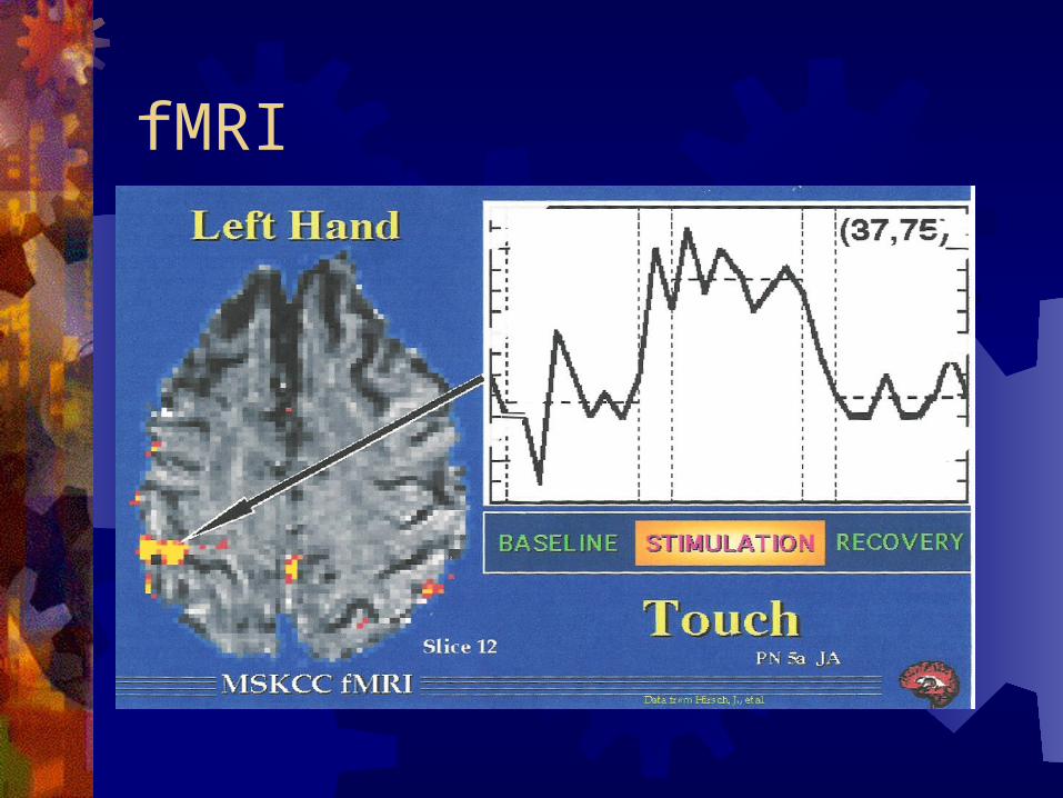
fMRI Functional MRI is based on the increase in blood
flow to the local vasculature that accompanies neural activity in the brain
Magnetic resonance imaging can be used to map changes in brain hemodynamics that correspond to mental operations. This is postulated to extend traditional anatomical imaging to include maps of human brain function
fMRI provides high resolution, noninvasive reports of neural activity detected by a blood oxygen level dependent signal
fMRI
fMRI Rapidly emerging body of literature documents
corresponding findings between fMRI and conventional electrophysiological techniques to localize specific functions of the human brain (Atlas, et al, 1996; Puce, et al, 1995; Burgess, 1995; Detre, et al, 1995; George, et al, 1995; Ives, et al, 1993).
Consequently, the number of medical and research centers with fMRI capabilities and investigational programs continues to escalate.

I wish it was still Mardi Gras…
Neurorehabil Neural Repair. 2002 Dec;16(4):326-38.Motor recovery and cortical reorganization after constraint-induced movement therapy in stroke patients: a preliminary study.
Schaechter JD, Kraft E, Hilliard TS, Dijkhuizen RM, Benner T, Finklestein SP, Rosen BR, Cramer SC.
The goal of this study was to assess motor cortical reorganization after CIMT using functional magnetic resonance imaging (fMRI).
4 incompletely recovered chronic stroke patients treated with CIMT underwent motor function testing and fMRI. Five age-matched normal subjects were also imaged.
Motor function testing showed that patients made significant gains in functional use of the stroke-affected upper extremity (detected by the Motor Activity Log) and significant reductions in motor impairment (detected by the Fugl-Meyer Stroke Scale and the Wolf Motor Function Test) immediately after CIMT, and these effects persisted at 6-month follow-up.
These data provide preliminary evidence that gains in motor function produced by CIMT in chronic stroke patients may be associated with a shift in laterality of motor cortical activation toward the undamaged hemisphere.
Early pioneers who worked on cortical reorganization
Study Objectives 1. To evaluate the efficacy of CIMT after hemiplegia,
and to study the time period of cortical reorganization 2. To study additional/different cortical reorganization
changes that take place in many chronic hemiplegic patients
3. To study the efficacy of CIMT and cortical reorganization in post one year hemiplegia and post 4-5 years of hemiplegia
4. To determine the best time to utilize CIMT effectively
5. To study whether there is a difference in outcome because of gender, side of lesion, or age
Hypotheses: 1. There will be a difference in cortical
reorganization after two weeks of CIMT in post one year and post 4-5 years of hemiplegia
2. Cortical reorganization occurs in different locations depending on chronicity
3. There will be no difference in functional outcome and cortical reorganization by:a). Gender
b). Side of lesion, i.e. right or left hemiplegia
Study Design Inclusion Criteria: 1. Male and female between 40 to 80 years of
age 2. Willing to to attend two weeks of CIMT classes 3. Subject with only one stroke 4. Upper limb hemiplegia as a result of their first
stroke 5. Affected limb has > or = to 20 degrees of
wrist extension, and their thumb and at least 2 fingers 10 degrees

Inclusion criteria (cont’d) 6. Minimal spasticity of affected upper extremity
(two or less on Ashworth scale) 7. Sufficient stability to walk when unaffected arm is
immobilized 8. Willing to stop the use of the unaffected good
arm at least 90% of waking hours 9. Ability to perform wheelchair transfers
independently with the unaffected arm immobilized 10. Ability to walk 5 feet without an AD 11. Ability to stand at table/counter without an AD
at least 15-20 minutes
Exclusion criteria 1. Any underlying medical condition that may restrict
the subject to participate in CIMT 2. Conditions that may pose an increased risk for a
second stroke 3. Scheduled elective surgery in the next 6 months 4. Life expectancy less than 3 months 5. Treatment with other investigational devices/agents
within the previous 30 days, or planned use of other investigational agents or devices
6. Metal implants in the head, or any movable fragments in the soft tissue
Exclusion criteria (cont’d) 7. Claustrophobia 8. Implanted electromechanical devices, such
as cardiac pacemaker or neurostimulator 9. Possibility of becoming pregnant 10. Large tattoos/tattooed eyeliner/permanent
cosmetics 11. MMSE score less than 24 12. Non-MRI compatible vascular clips, or
endovascular coils

Study Design - BASELINEWolf Function TestMMSESubject’s Health Assessment
QuestionnaireFinger/hand motor task MRI
Study Design- CIMTTotal of ten training sessions over a two
week periodEach subject required to continue the
given task at home at least 4 hours/dayRestricted use of unaffected upper
extremity will be achieved by wearing a resting hand splint during 90% of waking hours
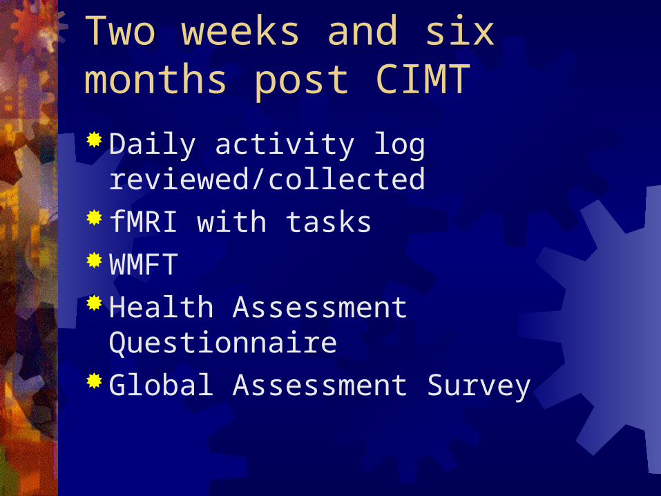
Two weeks and six months post CIMTDaily activity log reviewed/collected fMRI with tasksWMFTHealth Assessment QuestionnaireGlobal Assessment Survey
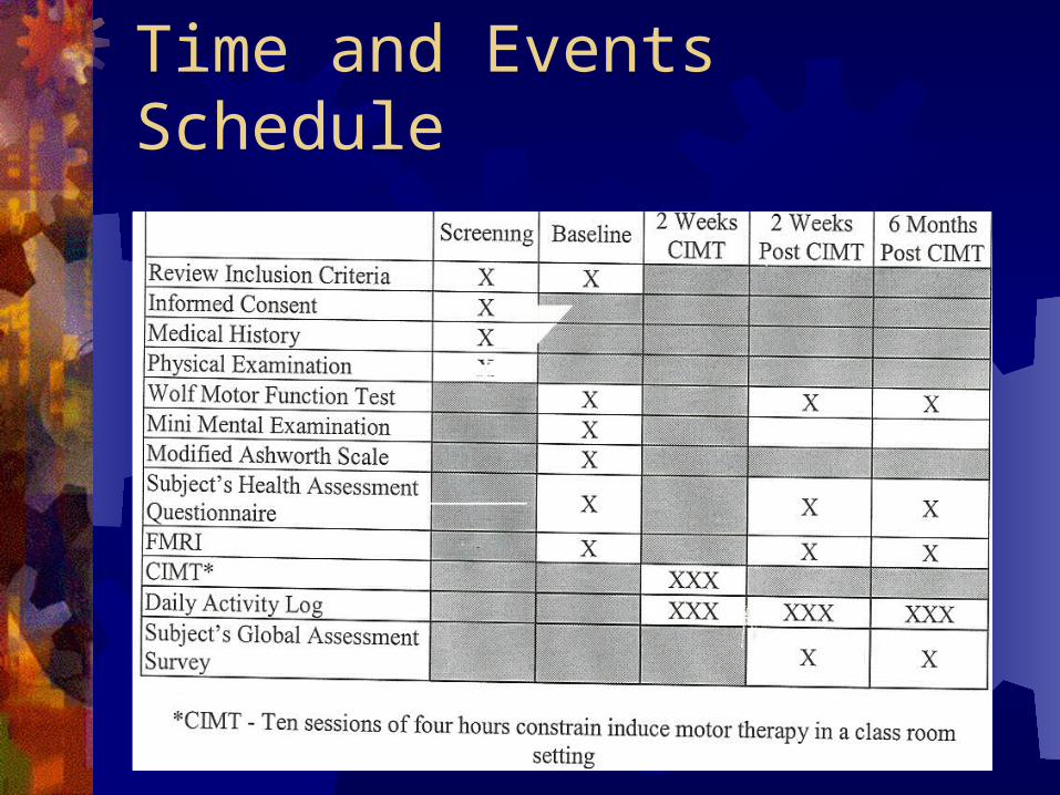
Time and Events Schedule
Wolf Motor Function Test Arch Phys Med Rehabil. 2001 Jun;82(6):750-5.
The reliability of the wolf motor function test for assessing upper extremity function after stroke.
Morris DM, Uswatte G, Crago JE, Cook EW 3rd, Taub E.
Division of Physical Therapy, University of Alabama, Birmingham
Wolf motor function Reliability Study
To examine the reliability of the Wolf Motor Function Test (WMFT) for assessing upper extremity motor function in adults with hemiplegia
A sample of convenience of 24 subjects with chronic hemiplegia (onset >1yr), showing moderate motor impairment
WMFT includes 15 functional tasks. Performances were timed and rated by using a 6-point functional ability scale
Conclusion - The WMFT is an instrument with high interrater reliability, internal consistency, test-retest reliability, and adequate stability
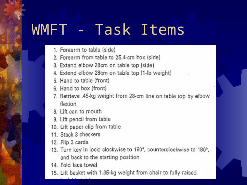
WMFT - Task Items
I can’t take much more!

Results – Full spectrum of results pending.
Results…..so far. COPM – Canadian Occupational Performance
Measure COPM is a test for self perceived change in
occupational performance over time. Test-retest reliability for COPM for performance and
satisfaction is rated at 80% and 89%respectively.
Data was analyzed with standard descriptive statistics based on performance and satisfaction at 2 weeks and 6 months post CIMT.
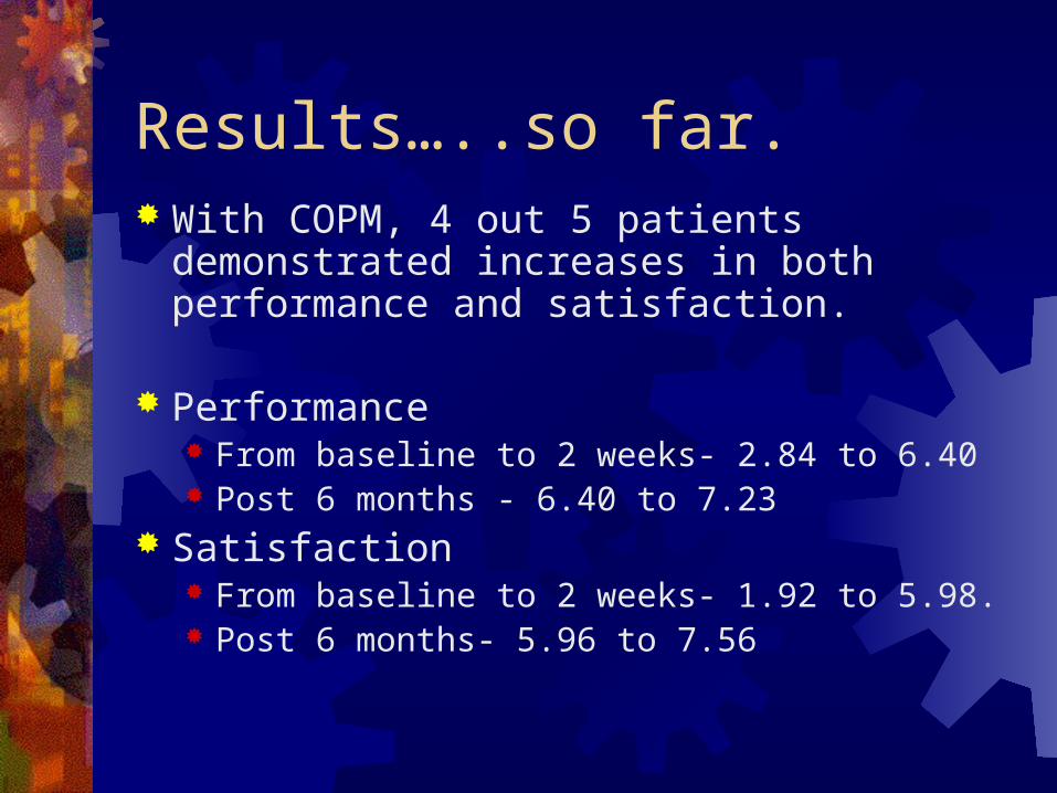
Results…..so far. With COPM, 4 out 5 patients demonstrated
increases in both performance and satisfaction.
Performance
From baseline to 2 weeks- 2.84 to 6.40 Post 6 months - 6.40 to 7.23
Satisfaction From baseline to 2 weeks- 1.92 to 5.98. Post 6 months- 5.96 to 7.56
Limitations of Study Convenience sample precluded randomization Lack of comparison and control groups Limited sample size No blinding procedures Variable compliance with limb restriction 90% of working hours A functional task performed at different frequencies appears different on fMRI – Is
change truly a reflection of cortical reorganization or of variability in frequency? Isolation of selected functional tasks is difficult
Looking at the fMRI, is cortical reorganization being interpreted for what may be activation of compensatory muscles?
Does cortical reorganization = neuronal plasticity? Do changes in blood flow as measured by fMRI correlate with cortical
reorganization or neuronal plasticity Many unaccounted variables which surely contribute to the outcome
Type of stroke not delineated Location of stroke not delineated Effect of handedness poorly understood Comorbidities and social factors
More Work To Do The next recruitment phase to begin soon
(two year study) NIH more interested in functional outcomes
versus radiological outcomes (initial data had already established cortical reorganization)
Plan to make proposal to NIH for functional outcome study and apply for funding

FIN