Embed Size (px)
Citation preview
© 2015. Published by The Company of Biologists Ltd.
PML induces compaction, TRF2 depletion and DNA damage signaling
at telomeres and promotes their alternative lengthening
Sarah Osterwald1,*, Katharina I. Deeg1,*, Inn Chung1,2, Daniel Parisotto1,3, Stefan Wörz4,
Karl Rohr4, Holger Erfle5 and Karsten Rippe1,‡
1 Research Group Genome Organization & Function, Deutsches Krebsforschungszentrum
(DKFZ) & BioQuant, Heidelberg, Germany
2 present address: Molecular Biology Program, Memorial Sloan-Kettering Cancer Center,
New York, USA
3 present address: Biochemistry Center (BZH), University of Heidelberg, Heidelberg,
Germany
4 Department of Bioinformatics and Functional Genomics, Biomedical Computer Vision
Group, University of Heidelberg & DKFZ, BioQuant, IPMB, Heidelberg, Germany
5 ViroQuant-CellNetworks RNAi Screening Facility, University of Heidelberg & BioQuant,
Heidelberg, Germany
* These authors contributed equally to the work
‡ Correspondence should be addressed to Karsten Rippe (email: [email protected],
telephone: +49-6221-54-51376, fax: +49-6221-54-51487)
Keywords: Alternative lengthening of telomeres (ALT) / PML nuclear bodies / DNA repair
Jour
nal o
f Cel
l Sci
ence
Acc
epte
d m
anus
crip
t
JCS Advance Online Article. Posted on 23 April 2015

ABSTRACT
The alternative lengthening of telomeres (ALT) mechanism allows cancer cells to escape
senescence and apoptosis in the absence of active telomerase. A characteristic feature of
this pathway is the assembly of ALT-associated PML nuclear bodies (APBs) at telomeres.
We dissected the role of APBs in a human ALT cell line by RNA interference with an
automated 3D fluorescence microscopy platform in conjunction with advanced 3D image
analysis. We identified 29 proteins that affected APB formation that were involved in
telomere and chromatin organization, protein sumoylation and DNA repair. By integrating
and extending these findings we found that APB formation induced clustering of telomere
repeats, telomere compaction and concomitant depletion of the shelterin protein TRF2.
These APB-dependent changes correlated with the induction of a DNA damage response
at colocalizing telomeres via a strong enrichment of the phosphorylated form of the ataxia
telangiectasia mutated (ATM) kinase. Accordingly, we propose that APBs promote
telomere maintenance by inducing a DNA damage response in ALT-positive tumor cells
via changing the telomeric chromatin state to trigger ATM phosphorylation.
Jour
nal o
f Cel
l Sci
ence
Acc
epte
d m
anus
crip
t

INTRODUCTION
The gradual shortening of telomeres during replication eventually triggers growth arrest
and senescence and thus provides an important tumor suppressor mechanism (d'Adda di
Fagagna et al., 2003; Harley et al., 1990). Cancer cells overcome this proliferation limit by
activating a telomere maintenance mechanism. In most cases telomerase is re-activated,
which can extend the telomere repeat sequence TTAGGG (Shay and Bacchetti, 1997).
However, 10-15% of tumors employ an alternative lengthening of telomeres (ALT)
mechanism to elongate their chromosomal ends via DNA recombination and repair
processes in the absence of telomerase (Bryan et al., 1997). ALT tumors are typically
characterized by large heterogeneity in telomere length within one cell (Bryan et al.,
1995), the occurrence of extrachromosomal telomeric repeats (ECTRs) (Wang et al.,
2004), mutations of the chromatin remodeler ATRX (Heaphy et al., 2011), genome
instability (Lovejoy et al., 2012), increased telomeric recombination (Londono-Vallejo et
al., 2004), and the presence of ALT-associated promyelocytic leukemia (PML) nuclear
bodies (APBs) (Chung et al., 2012; Yeager et al., 1999). APBs are defined as complexes
of PML nuclear bodies (PML-NBs) with telomeric DNA in telomerase-negative cells
(Yeager et al., 1999), and their ectopic assembly in ALT-positive cells induces telomere
lengthening by promoting repair-associated DNA synthesis (Chung et al., 2011). A
number of proteins involved in ALT have been identified such as the telomeric shelterin
complex (Jiang et al., 2007), the small ubiquitin-like modifier (SUMO) E3 ligase MMS21
(Potts and Yu, 2007), several DNA repair proteins (Nabetani and Ishikawa, 2011), as well
as heterochromatin protein 1 (HP1) (Jiang et al., 2009). However, the molecular details of
the ALT pathway have remained elusive.
Here, we applied a three-dimensional confocal laser scanning microscopy (CLSM)
screening platform and quantitative image analysis to evaluate changes in the nuclear
organization of APBs, PML-NBs, and telomeres at high precision based on the analysis of
more than 20 million images. With this approach we were able to characterize features of
single telomeres in their native cellular context and compared the effect of small
interfering RNA (siRNA)-mediated knockdown of ~100 genes with a comprehensive set of
image based readouts. Our results reveal that depletion of APBs by long-term PML
knockdown led to telomere shortening and a reduction of ECTRs. In addition, we found
that PML induced clustering and compaction of colocalizing telomere repeats and
simultaneously reduced binding of the telomeric repeat binding factor 2 (TRF2). These
changes in telomere organization correlated with the activation of the ataxia telangiectasia
mutated (ATM) kinase in APBs. Based on these findings we propose a model for APB-
mediated telomere lengthening in ALT-positive cells and tumors.
Jour
nal o
f Cel
l Sci
ence
Acc
epte
d m
anus
crip
t

RESULTS
PML knockdown induces telomere shortening and reduces ECTRs
PML is the central structural component for forming PML-NBs and APBs. In the absence
of PML, other PML-NB components such as SP100 and SUMO do not assemble into a
nuclear subcompartment (Ishov et al., 1999; Tavalai et al., 2006; Zhong et al., 2000).
Accordingly, we investigated the role of PML protein in ALT with an inducible stable
knockdown of PML in the ALT-positive human U2OS osteosarcoma cell line that targeted
a sequence common to the seven PML isoforms. Immunofluorescence analysis using a
pan PML antibody that detects all isoforms was conducted and evaluated with our
previously developed quantitative automated 3D confocal image acquisition and analysis
platform (Osterwald et al., 2012; Wörz et al., 2010) (supplementary material Fig. S1). The
results showed that the number of PML-NBs was reduced by 99.3 ± 0.1% after 72 h of
knockdown (p < 0.001, Fig. 1A). The knockdown completely suppressed APB formation
as quantified by colocalizations between PML and telomeres (–3.5 ± 0.3 APBs/cell or –
99.7 ± 0.3%, p < 0.001, Table 1). The disappearance of APBs was accompanied by a
reduction of C-circles, which are ALT-specific partially single-stranded telomeric
(CCCTAA)n DNA circles (–87.5 ± 4.1%, p < 0.001, Fig. 1C). At the same time, the number
of detectable telomere repeat foci per cell was significantly increased (+6.5 ± 2.6,
p < 0.001, Table 1). The reduced fluorescence intensities of the Cy3-labeled telomere
repeats after 72 h revealed that high-intensity telomere repeat signals disintegrated into
several low-intensity telomere repeat foci (median: –12.8 ± 2.0%, p < 0.001, Fig. 1D).
Thus, on average, one telomere repeat focus in APBs separated upon resolution of one
APB into 2-3 distinct telomere repeat foci.
To assess whether PML is needed for telomere elongation in ALT cells, we performed a
long-term PML knockdown in U2OS cells for 30 days corresponding to about the same
number of population doublings. The knockdown led to a significant decrease of the
telomere repeat signal (median: –24.9 ± 1.7%, p < 0.001, Fig. 1B, 1D) as detected by
interphase quantitative fluorescence in situ hybridization (Q-FISH), which was more
pronounced than the short-term effect observed after 72 h of knockdown. This reduction
of telomere content upon long-term PML knockdown was confirmed by telomere repeat
quantitative PCR (supplementary material Fig. S2A). In addition, terminal restriction
fragment (TRF) analysis after 2, 4 and 6 weeks of PML knockdown revealed that PML
knockdown induced telomere shortening (supplementary material Fig. S2B). Next, we
performed Q-FISH on metaphase chromosomes of uninduced and induced PML
knockdown cells (Fig. 1E, Table 2). This method is well established to detect and quantify
ECTRs and has been used in a number of previous studies (Episkopou et al., 2014;
Hande et al., 2001; Kamranvar et al., 2013; Kamranvar and Masucci, 2011; Tokutake et
Jour
nal o
f Cel
l Sci
ence
Acc
epte
d m
anus
crip
t

al., 1998). It is noted that we refer here to those ECTRs that are detected via our PNA
FISH probe against the G-rich telomere repeat sequence. This group of ECTRs is distinct
from the single-stranded C-rich C-circles measured by rolling circle amplification
according to the method of Henson et al. (Henson et al., 2009) that would not give rise to
a signal in our telomere repeat FISH assay. The intensity of telomere repeats associated
with chromosomes was significantly reduced after 30 days of PML knockdown (median: –
18.6 ± 9.6%, p < 0.001, Table 2) and considerably more telomere-free ends were
detected (52.5 ± 26.5%, p < 0.05, Table 2). The number of detectable ECTRs – defined
as telomere repeat signals per metaphase spread that were not associated with
chromosomes – was reduced by 59.8 ± 10.2% (p < 0.001, Table 2). In general, ECTRs
had a lower median repeat intensity than the telomeres. They accounted for only about
7% of the total telomere repeat intensity per uninduced control cell, thus representing a
small fraction of the total telomere repeat signal (Table 2). This finding agrees well with a
recent study that also quantified the contribution of ECTRs to total telomere repeat
content in ALT cells both by Q-FISH and by extraction of extrachromosomal DNA and
subsequent southern blotting (Episkopou et al., 2014). Accordingly, we conclude that the
telomere repeat signal measured in interphase FISH experiments (Table 2) mainly
originated from telomeres and only to a small fraction from ECTRs.
In summary, interphase and metaphase Q-FISH, TRF analysis and telomere quantitative
PCR consistently revealed telomere shortening upon PML knockdown that was
accompanied by a reduction of ECTRs including both C-circles and G-rich ECTRs.
An RNAi screen with automated 3D image analysis identifies 29 proteins involved in
APB formation
Having shown that depletion of APBs by PML knockdown led to telomere shortening, we
set out to identify factors that disrupt PML assembly into APBs and thus could affect the
ALT pathway. We conducted an RNA interference (RNAi) screen by quantitative
automated 3D confocal imaging and subsequent analysis of telomere, PML-NB and APB
features as described previously in further detail (Osterwald et al., 2012; Wörz et al.,
2010). Briefly, ~100 candidate proteins were knocked down by two independent siRNAs
(supplementary Table S1 online). Then the number, volume, intensity and density (defined
as intensity per volume) of telomere repeats and PML-NBs and their colocalizations, the
APBs, were determined from the automated analysis of more than 20 million images
(supplementary material Fig. S1). In this manner, we were able to reliably quantify
changes in APB formation and telomere organization at the level of single telomeres with
high precision to dissect the function of APBs.
Jour
nal o
f Cel
l Sci
ence
Acc
epte
d m
anus
crip
t
From our RNAi screen we identified 29 proteins involved in APB formation (supplementary
Table S2 online). Only those proteins that showed a significant change of more than 10%
in the number of APBs (p < 0.05) for two different siRNAs in at least three independent
experiments were selected as hits. Other ‘non-hit’ proteins that did not meet these
relatively strict requirements were classified into two groups (supplementary Table S2
online): Targets were both siRNAs consistently did not show a significant effect on the
number of APBs were considered as not being related to APB formation. The remaining
proteins had one out of two siRNAs showing a significant effect and represents
candidates for being related to APB formation. However, these were not further
investigated here. The knockdown efficiency of selected hits as well as non-hits was
analyzed by quantitative real-time PCR (supplementary material Fig. S3A) unless already
previously validated (supplementary Table S1 online). Based on the associated biological
processes according to gene ontology (GO) annotation, the APB effector proteins were
grouped into telomere organization, protein sumoylation, DNA repair and chromatin
organization (Table 3).
As inferred from our previous work, the number of APBs displays little dependence on cell
cycle state in normally proliferating U2OS cells (Osterwald et al., 2012), although the
number of PML-NBs was higher during S-phase in this cell line (Dellaire et al., 2006). For
other cell lines a higher number of APBs has been reported after inducing a cell cycle
arrest in G2/M phase for human ovarian surface epithelium (HOSE) cells (Grobelny et al.,
2000) or in G0/G1 phase for IIICF/c and GM847 cell lines (Jiang et al., 2007). Accordingly,
we addressed the question if the siRNA knockdowns conducted here were associated
with significant changes in the cell cycle distribution. The integrated background corrected
DAPI intensities per cell nucleus were computed for a given sample from the confocal
image stacks and used to obtain the relative cellular DNA content as described previously
(Fejes Tóth et al., 2004; Osterwald et al., 2012). From this quantification the cell cycle
distribution was determined by applying an identical predefined gating of the DNA content
histograms for G1, S and G2/M phase and computing the corresponding percentage of
cells within each group (supplementary material Fig. S3B). For the majority of knockdown
experiments no significant change of the cell cycle distribution was observed under our
experimental conditions (supplementary Table S2 online). For six targets (BLM, CDKN1A,
FEN1, LSD1, MORC3 and UBC9) a significant change in the range of 10 percentage
points of cells in G1 or G2/M, but not in S phase was observed for one of the siRNAs in
comparison to control siRNA. However, the direction of the measured change in the
number of APBs for siRNAs targeting FEN1, UBC9 and CDKN1A did not correspond to
that observed previously in cell cycle arrest experiments with IIICF/c, GM847 or HOSE
cells (Grobelny et al., 2000; Jiang et al., 2007). Thus, we conclude that in general the
Jour
nal o
f Cel
l Sci
ence
Acc
epte
d m
anus
crip
t

changes in the number of PML NBs and APBs measured here for the U2OS cell line upon
siRNA-mediated protein knockdown were not due to changes of the cell cycle distribution.
Changes in chromatin compaction affect TRF2 binding to telomere repeats, APB
formation and C-circle levels
Next, we evaluated the linkage between telomeric chromatin organization and APB
formation. Interestingly, knockdown of several factors involved in heterochromatin
formation, as for example the histone methyltransferase SUV4-20H2 (Benetti et al., 2007;
Schotta et al., 2004), heterochromatin protein 1 (HP1) (Jiang et al., 2011; Jiang et al.,
2009; Verschure et al., 2005), or the histone demethylase LSD1 (Shi et al., 2004),
reduced the number of APBs. In contrast, the number of APBs increased upon RNAi-
mediated knockdown of high mobility group nucleosome binding domain 5 (HMGN5),
which counteracts the chromatin-condensing activity of linker histones (Rochman et al.,
2009). Thus, the targeting of various chromatin modifiers had a significant effect on APB
formation.
This prompted us to further investigate the role of the telomeric chromatin state for the
ALT pathway. We evaluated differences in telomere compaction as reflected by the
telomere repeat density. This parameter was derived from telomere FISH images by
dividing the intensity of a Cy3-labeled focal telomere repeat by its volume. To evaluate
whether changes in telomere repeat compaction affected TRF2 binding to telomeres, we
determined the ratio of TRF2 density to telomere repeat density from colocalizing TRF2
immunofluorescence and telomere FISH signals. The measured TRF2 to telomere repeat
ratio was then corrected for the slightly reduced accessibility of the TRF2 antibody to
telomere repeats of higher density as described in supplementary material Fig. S4. In this
manner we were able to compare TRF2 binding to telomere repeats at specific telomeres
within one cell for different treatments. Next, we induced chromatin decondensation by
treatment with the histone deacetylase inhibitor SAHA (Bradner et al., 2010; Choudhary et
al., 2009; Fejes Tóth et al., 2004). Treatment with SAHA significantly reduced the
telomere repeat density (p < 0.001, Fig. 2A). The observed reduction in telomere repeat
density after SAHA treatment was accompanied by a small, but statistically significant
increase in TRF2 binding per telomere repeat (p < 0.01, Fig. 2B). Furthermore, SAHA
strongly decreased the number of APBs per cell (–49.4 ± 6.9%, p < 0.001, Fig. 2C) and C-
circle levels (–45.3 ± 10.3%, p < 0.001, Fig. 2D).
We next employed a previously introduced technique to induce the de novo formation of
ectopic APBs by recruiting GFP-tagged proteins to three telomeres in U2OS cells with
stably integrated lac operator (lacO) arrays (Chung et al., 2011; Jegou et al., 2009). As
controls, GFP alone was recruited (Fig. 2E) and a cell line with pericentric lacO array
Jour
nal o
f Cel
l Sci
ence
Acc
epte
d m
anus
crip
t

integration sites was used. HMGN5 as a factor that decondenses chromatin was recruited
and compared to HP1as a protein that promotes heterochromatin formation
(Fig. 2F, G). The capability of the two proteins to promote APB formation was monitored
by the enrichment of endogenous PML protein at the telomeric lacO arrays. Recruitment
of HMGN5 resulted in strong chromatin decondensation at the telomeric lacO arrays as
assessed by the formation of extended structures with irregular shape, which has
previously been reported for non-telomeric lacO arrays (Rochman et al., 2009). No APBs
could be detected at the telomeres of these cells, whereas 24 ± 5% of lacO sequences
were associated with APBs in the control cells (p < 0.001, Fig. 2F). In contrast, recruiting
GFP-HP1 to the telomeric lacO arrays induced the subsequent enrichment of
endogenous PML protein at the telomeres in a highly efficient manner, yielding 76 ± 11%
of colocalization (p < 0.001, Fig. 2G). PML enrichment upon HP1 recruitment could be
due to SUMO mediated interactions as discussed previously (Lang et al., 2010) or occur
via SP100 (Seeler et al., 1998), a known interaction partner of PML. Notably, PML
enrichment was accompanied with the induction of repair-associated DNA synthesis as
concluded from the increased levels of the phosphorylated histone variant H2A.X
(H2A.X) and of incorporated 5-bromo-2'-deoxyuridine (BrdU) (Fig. 2H, I). This effect was
specific for telomeric lacO arrays. Recruitment of HP1 to pericentric lacO arrays had no
significant effect on the level of H2A.X (Fig. 2J). Taken together, decondensation of
telomeric chromatin inhibited APB formation while a compacted chromatin state was
found to be compatible with both APB formation as well as repair-associated DNA
synthesis at telomeres.
Telomere repeat density is increased in APBs while TRF2 binding to telomeres is
decreased
To investigate differences in the level of compaction at single telomeres in unperturbed
ALT cells, we analyzed the telomere repeat density in APBs as compared to telomere
repeat foci that were not located in APBs. The median telomere repeat density in APBs
was 2.6 ± 0.1-fold higher as compared to telomeres outside APBs (p < 0.001, Fig. 3A). To
distinguish if APBs induce compaction of telomeric chromatin or if they assemble at
preexisting highly dense telomeres, the effect of short-term PML knockdown on the
telomere repeat density was evaluated (Table 1). The median telomere repeat density
was significantly reduced by 9.9 ± 3.4% after PML knockdown. This indicates that the
increased compaction of telomere repeats was induced by APBs and was maintained only
as long as PML was present.
Next, we evaluated if the APB-mediated increase of the telomere repeat density
influenced TRF2 binding to these telomeres. The ratio of TRF2 per telomere repeat was
Jour
nal o
f Cel
l Sci
ence
Acc
epte
d m
anus
crip
t

strongly decreased in APBs as compared to telomeres that were not located in APBs
(median: –35.2 ± 4.9%, p < 0.001, Fig. 3B). Thus, telomeres in APBs had less TRF2
bound per telomere repeat. Interestingly, telomere repeat density was inversely correlated
with TRF2 binding: The least dense telomeres had 2.6 ± 0.7-fold more TRF2 bound per
telomere repeat as compared to the densest telomere (p < 0.001, Fig. 3C). This value
includes the above-mentioned correction for differences in antibody accessibility
(supplementary material Fig. S4G). To confirm that PML was required for the reduced
binding of TRF2 at colocalizing telomeres, we measured TRF2 binding to telomere
repeats before and after PML knockdown. Notably, PML knockdown increased the ratio of
TRF2 bound per telomere repeat by about 40% (Table 1). No change of the mean
integrated TRF2 immunofluorescence signal per cell was detected, indicating that TRF2
levels per cell remained unaffected. Thus, we conclude that APBs induced a compaction
of telomeric chromatin that correlated with reduced binding of TRF2 per telomere repeat.
The SUMO E3 ligase MMS21 and the poly(ADP-ribose) polymerase 2 modulate TRF2
binding to telomeres in APBs
Next, we investigated if knockdown of proteins involved in post-translational modifications
of shelterin proteins affected the binding of TRF2 to telomeres in APBs. The ratio of TRF2
to telomere repeat signal, i.e. the coverage of telomere repeats with TRF2, was affected
inside APBs upon knockdown of two different post-translational modifiers of TRF2, which
were reported to be enriched in APBs (Dantzer et al., 2004; Potts and Yu, 2007):
(i) Knockdown of the SUMO E3 ligase MMS21, which sumoylates the shelterin
components TRF1, TRF2, TIN2, and RAP1 (Potts and Yu, 2007), increased TRF2 binding
to telomeres in APBs by 18.1 ± 1.9% (p < 0.001). The opposite effect on the ratio of TRF2
to telomere repeats was observed for the knockdown of the SUMO protease SENP6,
while other SENPs had no effect or decreased TRF2 binding to telomere repeats.
(ii) Knockdown of PARP-2 increased the ratio of TRF2 to telomere repeat signal in APBs
by 9.3 ± 0.3% (p < 0.001) without affecting the telomere repeat density. Thus, post-
translational modifiers of TRF2 that are known to be present in APBs can affect the ratio
of TRF2 that is bound per telomere repeat in APBs.
APBs induce enrichment of phosphorylated ATM at high-density telomeres
TRF2 is the main repressor of DNA damage response (DDR) at telomeres, since it inhibits
the autophosphorylation of the ATM kinase and its concomitant dissociation into
monomers, the presumed active form of the kinase (Bakkenist and Kastan, 2003; Takai et
al., 2003). As TRF2 binding to telomeres was strongly reduced in APBs (Fig. 3B), we
addressed the question whether this leads to the activation of ATM by
autophosphorylation. Consistent with a previous report (Stagno D'Alcontres et al., 2007),
Jour
nal o
f Cel
l Sci
ence
Acc
epte
d m
anus
crip
t
the phosphorylated form of ATM (p-ATM) colocalized with APBs in U2OS cells (Fig. 4A,
row 1). A quantitative analysis revealed that p-ATM was significantly enriched at
telomeres associated with PML (Fig. 4B): While only about 2% of all telomeres colocalized
with p-ATM, this fraction was significantly increased among telomeres in APBs. About
15% of all APBs colocalized with p-ATM indicating that DDR was only activated in a
specific subset of APBs (Fig. 4B). The median telomere repeat density in APBs with p-
ATM was about 2.4 times higher than the density in APBs without p-ATM and even about
5.7 times higher than at telomeres that were not located in APBs (Fig. 4C). As shown in
Figure 3C, telomeres with the highest telomere repeat density had the lowest levels of
bound TRF2. Thus, we conclude that ATM was only activated at telomeres in APBs with
the highest telomere repeat densities and the lowest TRF2 levels.
To distinguish if APBs enrich phosphorylated ATM at telomeres or if APBs form at
telomeres where DDR is already activated, we analyzed the p-ATM distribution after one
week of PML knockdown. The knockdown of PML increased the total number of p-ATM
foci (+102 ± 7%, p < 0.001, Fig. 4A, D), which is likely to reflect the previously reported
general role of PML in DNA repair (Bernardi and Pandolfi, 2007). However, while in
control cells about one fourth of all p-ATM foci were found at telomeres, the number of p-
ATM foci at telomeres was strongly reduced after PML knockdown (–90 ± 8%, p < 0.001,
Fig. 4D). This indicates that ATM became activated at telomeres after APB formation
rather than inducing the formation of APBs at telomeres where DDR was already initiated.
In support of this conclusion, ATM knockdown had no significant effect on the number of
APBs (supplementary Table S2 online). As reported previously, ATM interacts with the
MRN “damage sensor” complex, which leads to the recruitment of other repair proteins
like MDC1 and 53BP1 (Derheimer and Kastan, 2010). Although these proteins are known
to colocalize with APBs (Jiang et al., 2007), their knockdown had no effect on the number
of APBs in our screen (supplementary Table S2 online), implying that functional DDR was
not necessary for APB formation.
Next, we evaluated if APB-induced ATM phosphorylation was necessary for telomere
elongation by inhibiting ATM for 4 weeks with KU-55933 (Hickson et al., 2004). This
treatment reduced the number and density of p-ATM foci by 45.2 ± 2.1% and 48.2 ± 3.6%,
respectively (p < 0.001). Furthermore, C-circles were reduced after treatment with the
ATM inhibitor (–33.6 ± 10.8%, p < 0.05, Fig. 4E). Notably, the median fluorescence
intensity of the Cy3-labeled telomere repeats (–12.9 ± 2.7%, p < 0.001, Fig. 4F)
decreased without significantly affecting the number of APBs per cell (+2.8 ± 3.5%,
p > 0.1). Thus, ATM inhibition was correlated with a loss of telomere repeats, but did not
affect APB formation. This indicates that ATM activation in APBs promoted subsequent
DNA repair-associated telomere elongation.
Jour
nal o
f Cel
l Sci
ence
Acc
epte
d m
anus
crip
t

DISCUSSION
In the present study we investigated the link between APB formation, TRF2 binding to
telomeres and telomere lengthening in the ALT-positive U2OS cell line. Based on our
findings we propose a model for APB-mediated telomere lengthening that involves the
following main steps and integrates findings from previous studies (Fig. 5): (1) Formation
of a PML subcompartment at telomeres induces telomeric chromatin compaction and
clustering of telomeres, possibly also ECTRs, as proposed previously (Cho et al., 2014;
Draskovic et al., 2009). (2) As a result of APB formation, TRF2 becomes partly depleted at
associated telomeres. This process could involve the activity of posttranslational
modifications of TRF2 such as sumoylation by MMS21 or poly-ADP ribosylation by PARP-
2 in line with previous studies (Dantzer et al., 2004; Potts and Yu, 2007). (3) The reduced
TRF2 density triggers autophosphorylation of ATM in APBs and DDR according to the
previously identified role of TRF2 as an inhibitor of ATM (Denchi and de Lange, 2007;
Karlseder et al., 2004). (4) Telomeres are elongated by repair-associated DNA synthesis
and recombination events that are promoted by telomere clustering in APBs. (4) As APBs
disassemble, repair and recombination factors dislocate, telomeres are released and
telomere density decreases again. Altogether this leads to a re-enrichment of TRF2 that
protects the extended telomeres from chromosomal fusions by non-homologous end
joining (NHEJ).
Several lines of evidence support this model. An important role of APBs was established
from the quantitative evaluation of the effect of PML knockdown in the ALT-positive U2OS
cell line. Short-term PML knockdown for 72 h led to an almost complete loss of APBs,
while the number of detectable telomere repeat foci increased by a factor of 2-3 for every
APB that disappeared. This suggests clustering of two or three telomere repeat foci in one
APB. Our Q-FISH interphase analysis did not reveal whether the additional telomere
repeat foci observed after PML knockdown were telomeres or ECTRs. However, Q-FISH
with a C-rich PNA probe on metaphase spreads showed that ECTRs accounted only for a
relatively small fraction of the total telomere repeat intensity per cell. Furthermore, the
number of detectable ECTRs was strongly reduced after PML knockdown. Thus, we
conclude that the additional telomere repeat foci that appear after 72 h of PML knockdown
arise mostly from telomeres. However, it is possible that ECTRs also contribute to the
telomere repeat clusters inside APBs. Consistent with the view that APBs promote
telomere clustering, it has been reported that telomeres attach to the surface of artificially
enlarged APBs (Draskovic et al., 2009), and that damaged telomeres preferentially cluster
with telomeres that are associated with PML in ALT-positive cells (Cho et al., 2014).
Notably, long-term PML knockdown induced telomere shortening and significantly
increased the number of chromosomal ends where a telomere repeat signal was absent.
Jour
nal o
f Cel
l Sci
ence
Acc
epte
d m
anus
crip
t

This demonstrates that PML is crucial for telomere elongation in ALT cells and confirms
previous conclusions (Chung et al., 2011; Jiang et al., 2005). While ALT inhibition by other
means has been employed previously (Jiang et al., 2005; Potts and Yu, 2007), our study
is the first to reveal the crucial contribution of PML by showing a telomere shortening upon
its knockdown.
Having established the importance of PML for the ALT mechanism, we investigated the
formation of APBs and their function in the ALT pathway with an automated quantitative
3D image acquisition and analysis approach in conjunction with RNAi-mediated
knockdown. The quantification of individual telomeres and APBs from a total of more than
20 million images allowed us to identify 29 factors involved in APB formation and to
elucidate the subsequent effects on telomere organization with unprecedented precision.
The mechanism by which these factors operate is likely to involve direct effects that
promote telomeric APB assembly as well as indirect effects related to DNA damage and
its repair. Cell cycle effects appear to be less relevant in this context since only very few
siRNAs had significant effects on the cell cycle distribution. We would like to note that
proteins where the two targeting siRNAs showed inconsistent effects were not considered
as hits in our screen (supplementary Table S2 online). They may nevertheless be related
to APB formation and the ALT mechanism as exemplified by the ataxia telangiectasia-
and RAD3-related (ATR) protein for which only one out of two siRNAs significantly
reduced the number of APBs in our study. Yet, a recent paper showed that knockdown or
inhibition of ATR specifically inhibits the ALT pathway and also reduces the number of
APB-positive U2OS cells (Flynn et al., 2015). Knockdown of other DNA repair proteins like
the MRN complex components Rad50 and NBS1, mediator of DNA-damage checkpoint 1
(MDC1), tumor protein p53 binding protein 1 (53BP1), breast cancer protein 1 (BRCA1),
and RAD51 did not affect the number of APBs for both siRNAs used, indicating that
functional DDR and DNA repair pathways are not essential for APB formation
(supplementary Table S2 online). For 53BP1 and RAD51 knockdown this is consistent
with previous reports (Jiang et al., 2007; Potts and Yu, 2007). With respect to knockdown
of RAD50 and NBS1, there is a disagreement with a previous study that reported a
reduction in APB-positive IIICF/c cells upon knockdown of MRN components (Jiang et al.,
2007). One reason could be that the abovementioned work used methionine restriction-
induced cell cycle arrest to artificially enrich the number of APBs. This treatment per se
could have an impact on either APB formation or ALT. Accordingly, the effect of protein
knockdown might be different from what is observed under the conditions used here for
U2OS cells. A role of DNA repair proteins downstream of APB formation is also supported
by our previous findings (Chung et al., 2011). Some repair proteins were inefficient in
inducing the de novo formation of APBs, but instead were recruited to pre-assembled
Jour
nal o
f Cel
l Sci
ence
Acc
epte
d m
anus
crip
t

APBs. It is noted that the above results do not exclude that DNA damage may also
promote the assembly of PML at telomeres in ALT-negative cells as reported previously
(Hsu et al., 2012; Slatter et al., 2012).
The role of the telomeric chromatin state with respect to APB formation and telomere
elongation in ALT cells has been discussed controversially. In telomerase-positive mice it
was reported that knockout of several chromatin modifiers involved in heterochromatin
formation resulted in APB formation, and increased recombination at telomeres (Benetti et
al., 2007; Garcia-Cao et al., 2004; Gonzalo et al., 2006). To which extend these finding
apply to human cells is unclear, since several lines of evidence point to differences in
telomere biology between humans and mice (Calado and Dumitriu, 2013). Furthermore, a
number of findings demonstrate that induction of a condensed heterochromatic state can
even promote DNA repair and/or homologous recombination (Ayrapetov et al., 2014;
Geuting et al., 2013). A recent study of DDR signaling in U2OS cells is particularly
informative on this issue (Burgess et al., 2014). It shows that chromatin compaction is an
integral part of DDR signaling and follows a transient chromatin expansion step.
We found here that APB assembly in U2OS cells was inhibited by an ‘open’ telomeric
chromatin state, as the knockdown of several repressive chromatin modifiers as well as
chromatin decondensation by HDAC inhibition or HMGN5 recruitment resulted in a
significant reduction of the number of APBs (Fig. 2C, F). Previous work in ALT-positive
IIICF/c cells showed that HP1α and γ are needed for APB formation under methionine
restriction and hypothesized that HP1-mediated chromatin compaction is required for APB
formation (Jiang et al., 2009). It was concluded that compacted telomeric DNA inside
APBs would counteract telomere–telomere recombination. Here, we show that recruitment
of HP1 to telomeres is compatible with PML-induced DNA repair synthesis (Fig. 2H-I).
This is in line with studies demonstrating the importance of HP1 for DNA repair and
recombination that are discussed in several reviews (Cann and Dellaire, 2011; Dinant and
Luijsterburg, 2009; Soria et al., 2012). Recently it has been reported that chromatin
compaction is globally reduced at ALT telomeres in comparison to telomeres in
telomerase positive cells (Episkopou et al., 2014). Our work focused on analyzing the
compaction of single telomeres within an ALT cell line and revealed differences in
telomere repeat densities in relation to their association with PML (Fig. 3). In particular, we
found that telomere repeats in APBs were more compact and bound less TRF2 than
telomere repeats outside of APBs (Fig. 3B). Interestingly, the high telomere repeat
densities observed in APBs correlated with the activation of a DDR response via ATM
phosphorylation.
Previous reports already speculated that partial telomere deprotection might be important
for the repair-based ALT mechanism (Cesare et al., 2009; Cesare and Reddel, 2008;
Jour
nal o
f Cel
l Sci
ence
Acc
epte
d m
anus
crip
t
Nabetani et al., 2004). In particular, a lack of TRF2 at ALT telomeres has been proposed,
since the ratio of total TRF2 levels to the amount of telomeric DNA was found to be
significantly lower in ALT-positive cell lines compared to telomerase positive cell lines
(Cesare et al., 2009). Here, we specifically compared the ratio of TRF2 density to
telomere repeat density derived from colocalizing TRF2 immunofluorescence and
telomere FISH signals at single telomeres in the ALT-positive U2OS cell line. This
approach allowed us to reveal differences in TRF2 binding to telomeres with or without
APBs. Based on this comparison and the fact that PML knockdown led to reduced
telomere repeat density and increased binding of TRF2, we propose that APBs are able to
induce compaction of telomeric chromatin and temporally reduce TRF2 levels at these
telomeres.
A mechanism that could lead to a reduced binding of TRF2 to the telomere repeats in
APBs are posttranslational modifications of TRF2 by the SUMO E3 ligase MMS21 and the
poly(ADP-ribose) polymerase 2 that have both been found to be enriched in APBs
(Dantzer et al., 2004; Potts and Yu, 2007). In line with a previous study (Potts and Yu,
2007), knockdown of these proteins reduced APB formation in our RNAi screen. The
relevance of sumoylation of shelterin and PML-NB components for PML-NB and APB
formation has been described in a number of previous studies (Brouwer et al., 2009;
Chung et al., 2011; Hattersley et al., 2011; Lang et al., 2010; Potts and Yu, 2007; Yu et
al., 2010). Here, we additionally found that MMS21 knockdown increased TRF2 binding to
telomeres in APBs, while knockdown of the SUMO protease SENP6 resulted in a
decrease. Thus, our results support the previous hypothesis that recruitment of MMS21 to
APBs leads to shelterin destabilization at these telomeres, possibly by interfering with
TRF2 dimerization (Potts and Yu, 2007). Interestingly, also knockdown of poly (ADP-
ribose) polymerase 2 (PARP-2), another posttranslational modifier of TRF2, increased the
ratio of TRF2 to telomere repeats. It is known that PARP-2 covalently modifies the
dimerization domain of TRF2 and non-covalently binds poly(ADP-ribose) to the MYB
domain, which decreases the DNA binding affinity of TRF2 (Dantzer et al., 2004). Thus,
the enrichment of MMS21 and PARP-2 in APBs could reduce the level of TRF2 bound to
telomeres in APBs by interfering with TRF2 dimerization and DNA binding.
Short-term TRF2 depletion has previously been shown to increase the rate of telomeric
sister chromatid exchanges (T-SCEs) (Zeng et al., 2009). However, TRF2 is also
important for t-loop formation and prevents HR-induced t-loop deletions and chromosome
fusions by NHEJ (Wang et al., 2004). In addition, long-term depletion of TRF2 in ALT cells
leads to chromosome fusions by NHEJ, induction of senescence, and telomere shortening
due to uncontrolled recombination (Stagno D'Alcontres et al., 2007). Thus, we
hypothesize that ALT cells depend on partial telomere deprotection to drive telomere
Jour
nal o
f Cel
l Sci
ence
Acc
epte
d m
anus
crip
t
recombination. At the same time, they need to prevent an extensive loss of TRF2, which
would lead to telomere attrition and chromosome fusions as discussed previously (Cesare
and Reddel, 2010). Based on the results described here, we conclude that APBs induce
the formation of this ‘intermediate-state’ at colocalizing telomeres.
A previous report showed that ATM is constitutively activated in ALT cells and colocalizes
with APBs (Stagno D'Alcontres et al., 2007). Here, we show that ATM was preferentially
activated in APBs that contained the densest telomere repeats. These highly dense
telomere repeats had reduced levels of TRF2 bound per repeat. In addition, previous
studies found that TRF2 inhibits ATM by directly interacting with the region containing
serine 1981, a residue whose autophosphorylation is necessary for the activation of this
kinase (Denchi and de Lange, 2007; Karlseder et al., 2004). Accordingly, we propose that
the reduction of TRF2 binding due to APB formation triggers ATM activation specifically at
telomeres in APBs. The events subsequent to the DNA damage response downstream of
ATM like recruitment of other DNA repair proteins and DNA repair synthesis as detected
by BrdU incorporation at APBs have been addressed in our work that exploits ectopic
APB assembly (Chung et al., 2011). Other studies reported that multiple dysfunctional
telomeres in ALT positive cells colocalize with APB-like foci (Cesare et al., 2009) and that
the phosphorylated histone H2AX (γH2AX), a molecular marker of double-strand breaks
(DSBs) is found at some APBs (Nabetani et al., 2004). Here, we extended these
observations by showing that PML knockdown reduced the number of telomeres
colocalizing with p-ATM, while the total number of detectable p-ATM foci was increased
(Fig. 4D). Thus, ATM was activated at telomeres after APBs were formed as opposed to a
mechanism according to which APBs assemble at telomeres where a DDR was already
initiated. In addition, we found that inhibition of ATM did not affect the number of APBs,
but decreased C-circle levels and reduced telomere repeat content, presumably due to a
suppressed DDR at telomeres in APBs. In support of these results, it was previously
reported that ATM activity in ALT cells is not required for APB formation, but for telomeric
DNA synthesis (Nabetani et al., 2004). Inhibiting the latter process in ALT cells does not
have an immediate effect on cell viability and proliferation (Jiang et al., 2005; Potts and
Yu, 2007). Consistently, ATM inhibition in U2OS cells does hardly affect their survival on
the time scale of several days as apparent from the experiments conducted in the context
of Fig. 4 and in agreement with the findings from another study (Flynn et al., 2015).
In summary, our results demonstrate that PML induces compaction and confined TRF2
depletion at colocalizing telomeres and promotes telomere lengthening by initiating DNA
damage signaling. Thus, APBs exert a central function for the disease phenotype of ALT-
positive tumors.
Jour
nal o
f Cel
l Sci
ence
Acc
epte
d m
anus
crip
t

METHODS
Plasmids
For the inducible PML knockdown, a dsDNA oligo consisting of a miRNA against PML
was cloned into the pcDNA6.2-GW/EmGFPmiR vector (Invitrogen). The complete miRNA
and EmGFP coding sequence were then cloned into the inducible pT-Rex-DEST30 vector
(Invitrogen). Sequences of the dsDNA oligos for PML knockdown: top oligo: 5’-TGC TGT
CTT GGA TAC AGC TGC ATC TTG TTT TGG CCA CTG ACT GAC AAG ATG CAT GTA
TCC AAG A-3’, bottom oligo: 5’-CCT GTC TTG GAT ACA TGC ATC TTG TCA GTC AGT
GGC CAA AAC AAG ATG CAG CTG TAT CCA AGA C-3’. The fluorescence three-hybrid
system for recruiting GFP-tagged proteins to lacO arrays through GBP-LacI and GBP-
LacI-RFP was provided by Chromotek (Munich, Germany). The pEGFP-N2-mHMGN5
vector was kindly provided by Michael Bustin (Rochman et al., 2009). The pEGFP-HP1
plasmid was obtained by amplifying the human HP1 cDNA sequence by PCR with an
upstream forward primer containing a BspEI restriction site and a downstream reverse
primer containing a BamHI restriction site. The PCR product was then cloned in pEGFP-
C1 (Clontech, Palo Alto, CA).
Cell culture work
Human U2OS osteosarcoma cells (ATCC) and the U2OS cell clones with integrated lacO
arrays, F6B2 and F42B8 (Jegou et al., 2009), were cultured in DMEM medium (GIBCO)
containing 10% FBS (PAA) and 2 mM L-Glutamine (PAA). The cell line stably expressing
PML miRNA and EmGFP was constructed by cotransfection of the inducible pT-Rex-
DEST30 vector containing a PML miRNA and EmGFP (Invitrogen) together with the Tet
repressor-coding vector pcDNA6/TR (Invitrogen). Selection was conducted with G418 and
Blasticidin, and stable cell clones were picked and cultured for 10 days. The surviving cell
clones were splitted and one fraction was maintained in doxycycline-free medium. For
these cells, complete repression of the miRNA was ensured by analyzing GFP expression
levels. The other fraction was induced with medium containing 1 µg/ml doxycycline
(Sigma) for 24 h. The cell clone with the best repression in the uninduced state and best
expression upon induction was used. The efficiency of PML knockdown was assessed by
immunofluorescence against PML after 72 h of induction. For long-term PML knockdown,
cells were cultured in medium containing 1 µg/ml Doxycycline. Control cells were
maintained in Doxycycline-free medium. For the screening application, 80 000 cells were
seeded per slide on Lab-Tek chambered cover glasses (Thermo Scientific) and fixed after
72 h. For recruitment assays, cells were transfected using Effectene (Qiagen) according
to the manufacturer’s instructions and fixed after 24 h. For inhibition of histone
deacetylases, cells were treated with 2 µM SAHA (Millipore) for 24 h and fixed afterwards.
Jour
nal o
f Cel
l Sci
ence
Acc
epte
d m
anus
crip
t

ATM was inhibited using 10 µM of the inhibitor KU-55933 (Hickson et al., 2004)
(Calbiochem).
Immunofluorescence and fluorescence in situ hybridization (FISH)
After fixation with 4% paraformaldehyde in PBS for 12 min and washing thrice with PBS,
cells were permeabilized for 5 min with 0.1% Triton X-100 in PBS. After three PBS
washes, cells were blocked for 1 h with 10% goat serum in PBS and afterwards incubated
with primary antibody in 10% goat serum in PBS for 1 h. Cells were then washed thrice
with PBS containing 0.002% NP40 (v/v). Subsequent staining with the appropriate
secondary antibodies conjugated with fluorescent dyes was conducted for 1 h in 10% goat
serum in PBS. After washing the cells three times with PBS, cells were mounted with
ProLong Gold (Invitrogen) containing 4',6-diamidino-2’-phenylindole (DAPI). Dilutions of
the following antibodies were used: mouse anti-TRF2 (1:100, 4A794, Calbiochem), mouse
anti-ATM phosphoSer 1981 (1:100, #MAB3806, Millipore), mouse anti-Cy3/Cy5 (1:500,
#ab52060, Abcam), rabbit anti-phospho-H2A.X(Ser139) (1:100, #07-164, Millipore), rabbit
anti-PML (1:100, #sc-5621, Santa Cruz Biotechnology), mouse anti-BrdU (1:50, B44, BD
Biosciences), goat anti-mouse-Alexa488 (1:300, Invitrogen), goat anti-mouse-Alexa568
(1:300, Invitrogen), goat anti-rabbit-Alexa488 (1:300, Invitrogen) and goat anti-rabbit-
Alexa633 (1:200, Invitrogen).
For 5-bromo-2’-deoxyuridine (BrdU) staining, cells were seeded, transfected and
incubated for 2 days. After adding 100 µM BrdU (Sigma-Aldrich) to the medium for 2 h,
cells were fixed and permeabilized with PBS containing 0.2% Triton X100 (v/v). Cells were
denatured with 1.5 N HCl for 30 min, blocked, and stained with an antibody against BrdU
as described above.
For telomere FISH, cells were washed thrice with PBS and fixed with 4%
paraformaldehyde in PBS for 12 min. After 5 min permeabilization with 0.2% (v/v)
Triton X100/PBS, cells were dehydrated by a series of ethanol washes (70, 85, and 100%
ethanol) for 2 min each. After air-drying, the samples were incubated with Cy3-labeled
(CCCTAA)3 PNA probe (0.1 µM, Panagene Inc.) in 75% formamide/20 mM NaCl, 20 mM
Tris, 0.1% BSA, pH 7.4. Samples were denatured at 80°C for 3 min and hybridized over
night at 30°C. Slides were then washed consecutively with 70% formamide/10 mM Tris
pH 7.4, 2x SSC, 0.1x SSC at 55°C and 0.05% Tween-20/2x SSC (v/v). Subsequent
immunofluorescence was conducted as described above. Quantitative FISH on
metaphase spreads (Q-FISH) was performed as described previously (Poon and
Lansdorp, 2001).
Jour
nal o
f Cel
l Sci
ence
Acc
epte
d m
anus
crip
t
Fluorescence microscopy and image analysis
Confocal fluorescence images were acquired with a Leica TCS SP5 DMI6000 confocal
laser scanning microscope (oil immersion objective lens, 63x, 1.4 NA). The automated
screening was conducted as described previously (Osterwald et al., 2012). For manual
image acquisition, images were acquired with the Leica TCS SP5 DMI6000 confocal laser
scanning microscope using the LAS AF software and parameters as described above.
The automated image analysis was performed using a 3D model-based segmentation
approach (Osterwald et al., 2012; Wörz et al., 2010).
The relative frequency distributions in Fig. 1D, 2A-C, 3A-B and 4F were obtained by
binning the data and plotting the relative frequencies of telomeres or cells in each bin
together with the corresponding SEM as data points connected by lines. The analysis of
metaphase telomere FISH was done with the automated image analysis pipeline
described above. Interphase cells and telomere repeat foci not associated with
chromosomes (ECTRs) were excluded from the analysis. The manual analysis of
microscopy images was done with the ImageJ software (http://rsbweb.nih.gov/ij). For the
analysis of the recruitment efficiency to lacO arrays, spots were counted as colocalizing if
the signal at the lacO array was at least two-fold above the background and comprised at
least two pixels with a size of 200 nm.
C-circle assay
The C-circle assay was performed as described previously (Henson et al., 2009). Briefly,
DNA was isolated from 1 × 10⁶ cells using the QIAamp DNA Mini Kit (Qiagen). DNA was
quantified using a Qubit Fluorometer (Life Technologies). Genomic DNA (20 ng) was
digested with 12.5 U/μg HinfI and RsaI restriction enzymes (both Roche) and 5 000 ng/μg
RNase A (Dnase-free; Thermo Fisher Scientific) for 2 h at 37°C. The digested DNA (10 µl)
was combined with 10 µl 1× 29 Buffer, 7.5 U 29 DNA polymerase (both NEB),
0.2 mg/ml BSA, 0.1% (v/v) Tween 20, 1 mM each dATP, dGTP and dTTP and incubated
for 8 h at 30°C and then at 65°C for 20 min. After adding 40 µl 2x SSC, the sample was
dot-blotted onto a 2xSSC-soaked Roti-Nylon plus membrane (pore size 0.45 µm, Carl
Roth). The membrane was baked for 20 min at 120°C and hybridized and developed
using the TeloTAGGG Telomere Length Assay Kit (Roche). Intensities of C-circle dot blots
were analyzed and background-corrected using Image Lab 4.1 (BioRad).
TRF analysis and telomere quantitative PCR
Genomic DNA was purified using the Gentra Puregene Cell Kit (Qiagen). For terminal
restriction-fragment (TRF) analysis 5 µg of purified DNA was digested with HinfI and RsaI
overnight. The digested DNA was resolved on a 0.6% agarose gel (Biozym Gold Agarose)
Jour
nal o
f Cel
l Sci
ence
Acc
epte
d m
anus
crip
t
in 1X TAE buffer using the CHEF-DRII pulsed-field gel electrophoresis system (Bio-Rad)
with the following settings: 4 V/cm, initial switch time 1 sec, final switch time 6 sec, 13 h
duration. Southern blotting and chemiluminescent detection was performed using the
TeloTAGGG Telomere Length Assay Kit (Roche) according to the manufacturer’s
instructions. The blot was visualized with a ChemiDoc MP imaging system (Bio-Rad).
Approximate mean TRF lengths were quantified using ImageJ and according to the
following equation: Σ (ODi) /Σ (ODi/Li), where ODi is the optical density at position i and Li
is the TRF length at position i.
Telomere repeat quantitative PCR was conducted essentially as described previously
(Cawthon, 2002; O'Callaghan et al., 2008). In short, 5 or 10 ng DNA, 1X LightCycler 480
SYBR Green I Master (Roche), 500 nM forward primer, 500 nM reverse primer were
analyzed per 10 μl reaction. The primer sequences were: telo fwd, CGG TTT GTT TGG
GTT TGG GTT TGG GTT TGG GTT TGG GTT; telo rev, GGC TTG CCT TAC CCT TAC
CCT TAC CCT TAC CCT TAC CCT; 36B4 fwd, AGC AAG TGG GAA GGT GTA ATC C;
36B4 rev, CCC ATT CTATCA TCA ACG GGT ACA A. Cycling conditions (for both
telomere and 36B4 products) were 10 min at 95°C, followed by 40 cycles of 95°C for 15 s
and 60°C for 1 min. A standard curve was used to determine relative quantities of
telomere repeats (T) and single copy gene (S, 36B4 gene). The T/S ratio was calculated
and normalized to a reference T/S ratio.
RNA interference
Transfected cell microarrays were produced as previously described (Erfle et al., 2007).
Repetitions of a 4x4 array were printed on each Lab-Tek resulting in 384 spots with
24 replicates for each siRNA. A gene was considered as a hit if knockdown with two
different siRNAs consistently affected the number of APBs by more than 10% (p < 0.05).
The experiments were conducted in triplicates and 500 to 1500 cells were analyzed per
siRNA. Sequences of all siRNAs (silencer select siRNAs, Ambion) as well as reported
knockdown efficiencies if available can be found in supplementary Table S1 online. The
knockdown efficiencies of selected siRNAs that were important for further conclusions,
namely CBX3, HDAC7, MRE11, NBS1, NSBP1, PARP2, RAD50, RAP1A, SENP6,
SUV420H2 and TRF2 was validated by real-time quantitative PCR (supplementary
material Fig. S3A). Values were normalized against β-actin. Primer sequences are
provided in supplementary material Table S3. GO terms for biological processes
associated with hits were identified by using the gene ontology website
(http://geneontology.org).
Jour
nal o
f Cel
l Sci
ence
Acc
epte
d m
anus
crip
t

Cell cycle analysis
To analyze the effect of each siRNA on the cell cycle, the background-corrected
integrated DAPI intensities that were obtained in the automated high-content confocal
screening were normalized and histograms were plotted (supplementary material
Fig. S3B). As described previously, the distributions obtained in this manner correlate well
with FACS profiles (Fejes Tóth et al., 2004; Osterwald et al., 2012). Gates, e.g. minimum
and maximum DAPI intensity thresholds, were defined to obtain and compare the relative
percentage of cells in G1, S and G2/M phase for each siRNA transfection. The same
binning and gating was used for all conditions. The percentage of cells in each cell cycle
phase was obtained from at least three replicates for each siRNA transfection. These data
were used to calculate changes in the percentage of cells in G1, S and G2/M phase
induced by each siRNA relative to control siRNA and the corresponding s.e.m..
Statistical analysis
The statistical analysis was conducted using the R software (http://www.r-project.org) as
described previously (Osterwald et al., 2012). Errors bars always represent the s.e.m. of
at least three independent experiments, unless stated otherwise. A Kolmogorov-Smirnov
test was used to assess the significance of siRNA-related effects (supplementary material
Table S2) and for the evaluation of interphase and metaphase FISH results with respect
to changes in telomere repeat intensities or densities as well as TRF2 / telomere repeat
ratios. Welch’s t-test was applied for the analysis of changes in cell cycle distribution, C-
circle levels, the number of telomere-free ends, ECTRs and the percentage of ECTR
intensity of total telomere intensity. For the analysis of the recruitment efficiency to lacO
arrays, the percentage of lacO arrays with colocalization was determined with the
indicated value n giving the number of lacO arrays evaluated. Error bars were calculated
as √n, which yields the standard deviation for a Poisson distribution. In order to determine
whether the percentages of colocalization after recruiting the proteins of interest were
significantly different from the ones obtained in the controls, the two-sided Fisher’s exact
test was used to calculate p-values.
Acknowledgements
We thank Nina Beil, Fabian Erdel, Delia Braun, Jürgen Reymann, Jana Molitor, Jan-
Philipp Mallm and Brian Luke for help and discussions, and Michael Bustin for plasmid
vectors. The work of KRi, KRo und HE was funded within project CancerTelSys
(01ZX1302) in the E:med program of the German Federal Ministry of Education and
Research (BMBF). The ViroQuant-CellNetworks RNAi Screening Facility was supported
by the CellNetworks-Cluster of Excellence (EXC81).
Jour
nal o
f Cel
l Sci
ence
Acc
epte
d m
anus
crip
t

Author Contribution
SO, KID and KRi designed the experiments. SO, KID, IC and DP performed experiments.
SW and KRo together with SO established the automated confocal imaging analysis
platform. HE provided materials for reverse siRNA transfection and contributed to the
establishment of the automated confocal image acquisition platform. SO, KID, IC, DP and
KRi analyzed experiments and interpreted results. SO, KID, IC and KRi wrote the
manuscript.
Conflict of Interest
The authors declare that they have no conflict of interest.
Jour
nal o
f Cel
l Sci
ence
Acc
epte
d m
anus
crip
t

REFERENCES
Ayrapetov, M. K., Gursoy-Yuzugullu, O., Xu, C., Xu, Y. and Price, B. D. (2014). DNA
double-strand breaks promote methylation of histone H3 on lysine 9 and transient
formation of repressive chromatin. Proc Natl Acad Sci U S A 111, 9169-74.
Bakkenist, C. J. and Kastan, M. B. (2003). DNA damage activates ATM through
intermolecular autophosphorylation and dimer dissociation. Nature 421, 499-506.
Benetti, R., Gonzalo, S., Jaco, I., Schotta, G., Klatt, P., Jenuwein, T. and Blasco, M.
A. (2007). Suv4-20h deficiency results in telomere elongation and derepression of
telomere recombination. J. Cell Biol. 178, 925-36.
Bernardi, R. and Pandolfi, P. P. (2007). Structure, dynamics and functions of
promyelocytic leukaemia nuclear bodies. Nat Rev Mol Cell Biol 8, 1006-16.
Bradner, J. E., West, N., Grachan, M. L., Greenberg, E. F., Haggarty, S. J., Warnow,
T. and Mazitschek, R. (2010). Chemical phylogenetics of histone deacetylases.
Nat. Chem. Biol. 6, 238-243.
Brouwer, A. K., Schimmel, J., Wiegant, J. C., Vertegaal, A. C., Tanke, H. J. and Dirks,
R. W. (2009). Telomeric DNA Mediates De Novo PML Body Formation. Mol. Biol.
Cell 20, 4804-4815.
Bryan, T. M., Englezou, A., Dalla-Pozza, L., Dunham, M. A. and Reddel, R. R. (1997).
Evidence for an alternative mechanism for maintaining telomere length in human
tumors and tumor-derived cell lines. Nat. Med. 3, 1271-4.
Bryan, T. M., Englezou, A., Gupta, J., Bacchetti, S. and Reddel, R. R. (1995).
Telomere elongation in immortal human cells without detectable telomerase
activity. Embo J 14, 4240-8.
Burgess, R. C., Burman, B., Kruhlak, M. J. and Misteli, T. (2014). Activation of DNA
damage response signaling by condensed chromatin. Cell Rep 9, 1703-17.
Calado, R. T. and Dumitriu, B. (2013). Telomere dynamics in mice and humans. Semin
Hematol 50, 165-74.
Cann, K. L. and Dellaire, G. (2011). Heterochromatin and the DNA damage response:
the need to relax. Biochem Cell Biol 89, 45-60.
Jour
nal o
f Cel
l Sci
ence
Acc
epte
d m
anus
crip
t

Cawthon, R. M. (2002). Telomere measurement by quantitative PCR. Nucleic Acids Res
30, e47.
Cesare, A. J., Kaul, Z., Cohen, S. B., Napier, C. E., Pickett, H. A., Neumann, A. A. and
Reddel, R. R. (2009). Spontaneous occurrence of telomeric DNA damage
response in the absence of chromosome fusions. Nat Struct Mol Biol 16, 1244-51.
Cesare, A. J. and Reddel, R. R. (2008). Telomere uncapping and alternative lengthening
of telomeres. Mech Ageing Dev 129, 99-108.
Cesare, A. J. and Reddel, R. R. (2010). Alternative lengthening of telomeres: models,
mechanisms and implications. Nat. Rev. Genet. 11, 319-30.
Cho, N. W., Dilley, R. L., Lampson, M. A. and Greenberg, R. A. (2014).
Interchromosomal Homology Searches Drive Directional ALT Telomere Movement
and Synapsis. Cell 159, 108-121.
Choudhary, C., Kumar, C., Gnad, F., Nielsen, M. L., Rehman, M., Walther, T. C.,
Olsen, J. V. and Mann, M. (2009). Lysine acetylation targets protein complexes
and co-regulates major cellular functions. Science 325, 834-40.
Chung, I., Leonhardt, H. and Rippe, K. (2011). De novo assembly of a PML nuclear
subcompartment occurs through multiple pathways and induces telomere
elongation. J. Cell Sci. 124, 3603-18.
Chung, I., Osterwald, S., Deeg, K. I. and Rippe, K. (2012). PML body meets telomere:
the beginning of an ALTernate ending? Nucleus 3, 263-75.
d'Adda di Fagagna, F., Reaper, P. M., Clay-Farrace, L., Fiegler, H., Carr, P., Von
Zglinicki, T., Saretzki, G., Carter, N. P. and Jackson, S. P. (2003). A DNA
damage checkpoint response in telomere-initiated senescence. Nature 426, 194-8.
Dantzer, F., Giraud-Panis, M. J., Jaco, I., Ame, J. C., Schultz, I., Blasco, M., Koering,
C. E., Gilson, E., Menissier-de Murcia, J., de Murcia, G. et al. (2004).
Functional interaction between poly(ADP-Ribose) polymerase 2 (PARP-2) and
TRF2: PARP activity negatively regulates TRF2. Mol. Cell Biol. 24, 1595-607.
Jour
nal o
f Cel
l Sci
ence
Acc
epte
d m
anus
crip
t
Dellaire, G., Ching, R. W., Dehghani, H., Ren, Y. and Bazett-Jones, D. P. (2006). The
number of PML nuclear bodies increases in early S phase by a fission mechanism.
J. Cell Sci. 119, 1026-33.
Denchi, E. L. and de Lange, T. (2007). Protection of telomeres through independent
control of ATM and ATR by TRF2 and POT1. Nature 448, 1068-71.
Derheimer, F. A. and Kastan, M. B. (2010). Multiple roles of ATM in monitoring and
maintaining DNA integrity. FEBS Lett 584, 3675-81.
Dinant, C. and Luijsterburg, M. S. (2009). The emerging role of HP1 in the DNA damage
response. Mol Cell Biol 29, 6335-40.
Draskovic, I., Arnoult, N., Steiner, V., Bacchetti, S., Lomonte, P. and Londono-
Vallejo, A. (2009). Probing PML body function in ALT cells reveals spatiotemporal
requirements for telomere recombination. Proc. Natl. Acad. Sci. USA 106, 15726-
15731.
Episkopou, H., Draskovic, I., Van Beneden, A., Tilman, G., Mattiussi, M., Gobin, M.,
Arnoult, N., Londono-Vallejo, A. and Decottignies, A. (2014). Alternative
Lengthening of Telomeres is characterized by reduced compaction of telomeric
chromatin. Nucleic Acids Res 42, 4391-405.
Erfle, H., Neumann, B., Liebel, U., Rogers, P., Held, M., Walter, T., Ellenberg, J. and
Pepperkok, R. (2007). Reverse transfection on cell arrays for high content
screening microscopy. Nat. Protoc. 2, 392-9.
Fejes Tóth, K., Knoch, T. A., Wachsmuth, M., Stöhr, M., Frank-Stöhr, M., Bacher, C.
P., Müller, G. and Rippe, K. (2004). Trichostatin A induced histone acetylation
causes decondensation of interphase chromatin. J. Cell Sci. 117, 4277-4287.
Flynn, R. L., Cox, K. E., Jeitany, M., Wakimoto, H., Bryll, A. R., Ganem, N. J., Bersani,
F., Pineda, J. R., Suva, M. L., Benes, C. H. et al. (2015). Alternative lengthening
of telomeres renders cancer cells hypersensitive to ATR inhibitors. Science 347,
273-7.
Jour
nal o
f Cel
l Sci
ence
Acc
epte
d m
anus
crip
t

Garcia-Cao, M., O'Sullivan, R., Peters, A. H., Jenuwein, T. and Blasco, M. A. (2004).
Epigenetic regulation of telomere length in mammalian cells by the Suv39h1 and
Suv39h2 histone methyltransferases. Nat Genet 36, 94-9.
Geuting, V., Reul, C. and Lobrich, M. (2013). ATM release at resected double-strand
breaks provides heterochromatin reconstitution to facilitate homologous
recombination. PLoS Genet 9, e1003667.
Gonzalo, S., Jaco, I., Fraga, M. F., Chen, T., Li, E., Esteller, M. and Blasco, M. A.
(2006). DNA methyltransferases control telomere length and telomere
recombination in mammalian cells. Nat. Cell Biol. 8, 416-24.
Grobelny, J. V., Godwin, A. K. and Broccoli, D. (2000). ALT-associated PML bodies are
present in viable cells and are enriched in cells in the G(2)/M phase of the cell
cycle. J Cell Sci 113 Pt 24, 4577-85.
Hande, M. P., Balajee, A. S., Tchirkov, A., Wynshaw-Boris, A. and Lansdorp, P. M.
(2001). Extra-chromosomal telomeric DNA in cells from Atm(-/-) mice and patients
with ataxia-telangiectasia. Hum Mol Genet 10, 519-28.
Harley, C. B., Futcher, A. B. and Greider, C. W. (1990). Telomeres shorten during
ageing of human fibroblasts. Nature 345, 458-60.
Hattersley, N., Shen, L., Jaffray, E. G. and Hay, R. T. (2011). The SUMO protease
SENP6 is a direct regulator of PML nuclear bodies. Mol Biol Cell 22, 78-90.
Heaphy, C. M., de Wilde, R. F., Jiao, Y., Klein, A. P., Edil, B. H., Shi, C., Bettegowda,
C., Rodriguez, F. J., Eberhart, C. G., Hebbar, S. et al. (2011). Altered telomeres
in tumors with ATRX and DAXX mutations. Science 333, 425.
Henson, J. D., Cao, Y., Huschtscha, L. I., Chang, A. C., Au, A. Y., Pickett, H. A. and
Reddel, R. R. (2009). DNA C-circles are specific and quantifiable markers of
alternative-lengthening-of-telomeres activity. Nat Biotechnol 27, 1181-5.
Hickson, I., Zhao, Y., Richardson, C. J., Green, S. J., Martin, N. M., Orr, A. I., Reaper,
P. M., Jackson, S. P., Curtin, N. J. and Smith, G. C. (2004). Identification and
characterization of a novel and specific inhibitor of the ataxia-telangiectasia
mutated kinase ATM. Cancer Res. 64, 9152-9.
Jour
nal o
f Cel
l Sci
ence
Acc
epte
d m
anus
crip
t

Hsu, J. K., Lin, T. and Tsai, R. Y. (2012). Nucleostemin prevents telomere damage by
promoting PML-IV recruitment to SUMOylated TRF1. J. Cell Biol. 197, 613-24.
Ishov, A. M., Sotnikov, A. G., Negorev, D., Vladimirova, O. V., Neff, N., Kamitani, T.,
Yeh, E. T., Strauss, J. F., 3rd and Maul, G. G. (1999). PML is critical for ND10
formation and recruits the PML-interacting protein daxx to this nuclear structure
when modified by SUMO-1. J Cell Biol 147, 221-34.
Jegou, T., Chung, I., Heuvelmann, G., Wachsmuth, M., Görisch, S. M., Greulich-
Bode, K., Boukamp, P., Lichter, P. and Rippe, K. (2009). Dynamics of telomeres
and promyelocytic leukemia nuclear bodies in a telomerase negative human cell
line. Mol. Biol. Cell 20, 2070-2082.
Jiang, W. Q., Nguyen, A., Cao, Y., Chang, A. C. and Reddel, R. R. (2011). HP1-
mediated formation of alternative lengthening of telomeres-associated PML bodies
requires HIRA but not ASF1a. PLoS ONE 6, e17036.
Jiang, W. Q., Zhong, Z. H., Henson, J. D., Neumann, A. A., Chang, A. C. and Reddel,
R. R. (2005). Suppression of alternative lengthening of telomeres by Sp100-
mediated sequestration of the MRE11/RAD50/NBS1 complex. Mol. Cell. Biol. 25,
2708-21.
Jiang, W. Q., Zhong, Z. H., Henson, J. D. and Reddel, R. R. (2007). Identification of
candidate alternative lengthening of telomeres genes by methionine restriction and
RNA interference. Oncogene 26, 4635-4647.
Jiang, W. Q., Zhong, Z. H., Nguyen, A., Henson, J. D., Toouli, C. D., Braithwaite, A.
W. and Reddel, R. R. (2009). Induction of alternative lengthening of telomeres-
associated PML bodies by p53/p21 requires HP1 proteins. J. Cell Biol. 185, 797-
810.
Kamranvar, S. A., Chen, X. and Masucci, M. G. (2013). Telomere dysfunction and
activation of alternative lengthening of telomeres in B-lymphocytes infected by
Epstein-Barr virus. Oncogene 32, 5522-30.
Jour
nal o
f Cel
l Sci
ence
Acc
epte
d m
anus
crip
t

Kamranvar, S. A. and Masucci, M. G. (2011). The Epstein-Barr virus nuclear antigen-1
promotes telomere dysfunction via induction of oxidative stress. Leukemia 25,
1017-25.
Karlseder, J., Hoke, K., Mirzoeva, O. K., Bakkenist, C., Kastan, M. B., Petrini, J. H.
and de Lange, T. (2004). The telomeric protein TRF2 binds the ATM kinase and
can inhibit the ATM-dependent DNA damage response. PLoS Biol. 2, E240.
Lang, M., Jegou, T., Chung, I., Richter, K., Munch, S., Udvarhelyi, A., Cremer, C.,
Hemmerich, P., Engelhardt, J., Hell, S. W. et al. (2010). Three-dimensional
organization of promyelocytic leukemia nuclear bodies. J Cell Sci 123, 392-400.
Londono-Vallejo, J. A., Der-Sarkissian, H., Cazes, L., Bacchetti, S. and Reddel, R. R.
(2004). Alternative lengthening of telomeres is characterized by high rates of
telomeric exchange. Cancer Res. 64, 2324-7.
Lovejoy, C. A., Li, W., Reisenweber, S., Thongthip, S., Bruno, J., de Lange, T., De, S.,
Petrini, J. H., Sung, P. A., Jasin, M. et al. (2012). Loss of ATRX, genome
instability, and an altered DNA damage response are hallmarks of the alternative
lengthening of telomeres pathway. PLoS Genet. 8, e1002772.
Nabetani, A. and Ishikawa, F. (2011). Alternative lengthening of telomeres pathway:
recombination-mediated telomere maintenance mechanism in human cells. J
Biochem 149, 5-14.
Nabetani, A., Yokoyama, O. and Ishikawa, F. (2004). Localization of hRad9, hHus1,
hRad1, and hRad17 and caffeine-sensitive DNA replication at the alternative
lengthening of telomeres-associated promyelocytic leukemia body. J Biol Chem
279, 25849-57.
O'Callaghan, N., Dhillon, V., Thomas, P. and Fenech, M. (2008). A quantitative real-
time PCR method for absolute telomere length. Biotechniques 44, 807-9.
Osterwald, S., Worz, S., Reymann, J., Sieckmann, F., Rohr, K., Erfle, H. and Rippe,
K. (2012). A three-dimensional colocalization RNA interference screening platform
to elucidate the alternative lengthening of telomeres pathway. Biotechnol. J. 7,
103-16.
Jour
nal o
f Cel
l Sci
ence
Acc
epte
d m
anus
crip
t

Poon, S. S. and Lansdorp, P. M. (2001). Quantitative fluorescence in situ hybridization
(Q-FISH). Curr Protoc Cell Biol Chapter 18, Unit 18 4.
Potts, P. R. and Yu, H. (2007). The SMC5/6 complex maintains telomere length in ALT
cancer cells through SUMOylation of telomere-binding proteins. Nat. Struct. Mol.
Biol. 14, 581-90.
Rochman, M., Postnikov, Y., Correll, S., Malicet, C., Wincovitch, S., Karpova, T. S.,
McNally, J. G., Wu, X., Bubunenko, N. A., Grigoryev, S. et al. (2009). The
interaction of NSBP1/HMGN5 with nucleosomes in euchromatin counteracts linker
histone-mediated chromatin compaction and modulates transcription. Mol Cell 35,
642-56.
Schotta, G., Lachner, M., Sarma, K., Ebert, A., Sengupta, R., Reuter, G., Reinberg, D.
and Jenuwein, T. (2004). A silencing pathway to induce H3-K9 and H4-K20
trimethylation at constitutive heterochromatin. Genes Dev. 18, 1251-62.
Seeler, J. S., Marchio, A., Sitterlin, D., Transy, C. and Dejean, A. (1998). Interaction of
SP100 with HP1 proteins: a link between the promyelocytic leukemia-associated
nuclear bodies and the chromatin compartment. Proc Natl Acad Sci U S A 95,
7316-21.
Shay, J. W. and Bacchetti, S. (1997). A survey of telomerase activity in human cancer.
Eur. J. Cancer 33, 787-91.
Shi, Y., Lan, F., Matson, C., Mulligan, P., Whetstine, J. R., Cole, P. A., Casero, R. A.
and Shi, Y. (2004). Histone demethylation mediated by the nuclear amine oxidase
homolog LSD1. Cell 119, 941-53.
Slatter, T. L., Tan, X., Yuen, Y. C., Gunningham, S., Ma, S. S., Daly, E., Packer, S.,
Devenish, C., Royds, J. A. and Hung, N. A. (2012). The alternative lengthening
of telomeres pathway may operate in non-neoplastic human cells. J. Pathol. 226,
509-518.
Soria, G., Polo, S. E. and Almouzni, G. (2012). Prime, repair, restore: the active role of
chromatin in the DNA damage response. Mol Cell 46, 722-34.
Jour
nal o
f Cel
l Sci
ence
Acc
epte
d m
anus
crip
t

Stagno D'Alcontres, M., Mendez-Bermudez, A., Foxon, J. L., Royle, N. J. and
Salomoni, P. (2007). Lack of TRF2 in ALT cells causes PML-dependent p53
activation and loss of telomeric DNA. J Cell Biol 179, 855-67.
Takai, H., Smogorzewska, A. and de Lange, T. (2003). DNA damage foci at
dysfunctional telomeres. Curr. Biol. 13, 1549-56.
Tavalai, N., Papior, P., Rechter, S., Leis, M. and Stamminger, T. (2006). Evidence for a
role of the cellular ND10 protein PML in mediating intrinsic immunity against
human cytomegalovirus infections. J Virol 80, 8006-18.
Tokutake, Y., Matsumoto, T., Watanabe, T., Maeda, S., Tahara, H., Sakamoto, S.,
Niida, H., Sugimoto, M., Ide, T. and Furuichi, Y. (1998). Extra-chromosomal
telomere repeat DNA in telomerase-negative immortalized cell lines. Biochem
Biophys Res Commun 247, 765-72.
Verschure, P. J., van der Kraan, I., de Leeuw, W., van der Vlag, J., Carpenter, A. E.,
Belmont, A. S. and van Driel, R. (2005). In vivo HP1 targeting causes large-scale
chromatin condensation and enhanced histone lysine methylation. Mol. Cell Biol.
25, 4552-64.
Wang, R. C., Smogorzewska, A. and de Lange, T. (2004). Homologous recombination
generates T-loop-sized deletions at human telomeres. Cell 119, 355-68.
Wörz, S., Sander, P., Pfannmöller, M., Rieker, R. J., Joos, S., Mechtersheimer, G.,
Boukamp, P., Lichter, P. and Rohr, K. (2010). 3D Geometry-based quantification
of colocalizations in multi-channel 3D microscopy images of human soft tissue
tumors. IEEE Trans. on Medical Imaging 29, 1474-1484.
Yeager, T. R., Neumann, A. A., Englezou, A., Huschtscha, L. I., Noble, J. R. and
Reddel, R. R. (1999). Telomerase-negative immortalized human cells contain a
novel type of promyelocytic leukemia (PML) body. Cancer Res. 59, 4175-4179.
Yu, J., Lan, J., Wang, C., Wu, Q., Zhu, Y., Lai, X., Sun, J., Jin, C. and Huang, H.
(2010). PML3 interacts with TRF1 and is essential for ALT-associated PML bodies
assembly in U2OS cells. Cancer Lett 291, 177-86.
Jour
nal o
f Cel
l Sci
ence
Acc
epte
d m
anus
crip
t

Zeng, S., Xiang, T., Pandita, T. K., Gonzalez-Suarez, I., Gonzalo, S., Harris, C. C. and
Yang, Q. (2009). Telomere recombination requires the MUS81 endonuclease. Nat.
Cell Biol. 11, 616-23.
Zhong, S., Muller, S., Ronchetti, S., Freemont, P. S., Dejean, A. and Pandolfi, P. P.
(2000). Role of SUMO-1-modified PML in nuclear body formation. Blood 95, 2748-
52.
Jour
nal o
f Cel
l Sci
ence
Acc
epte
d m
anus
crip
t

FIGURES
Figure 1. PML knockdown induces loss of telomere repeats and reduces ECTRs.
The effect of an inducible PML knockdown on ALT features was evaluated after different
time points in comparison to uninduced U2OS cells. (A-B) CLSM images of uninduced
Jour
nal o
f Cel
l Sci
ence
Acc
epte
d m
anus
crip
t

and induced cells after 72 h (A) and 30 days (B) of PML knockdown stained by FISH with
a Cy3-labeled telomere repeat probe and immunofluorescence against PML. Images of
uninduced and induced cells were acquired with identical microscope settings,
respectively. Scale bars are 10 µm. (C) C-circle levels in uninduced and induced PML
knockdown U2OS cells after 1 week of induction. As control, ALT-negative HeLa cells and
uninduced PML knockdown U2OS cells without addition of polymerase were used. Error
bars represent s.e.m. (n = 6). (D) Relative frequency distributions of the fluorescence
intensity of the Cy3-labeled telomere repeat probe measured at individual telomeres in
uninduced cells and after PML knockdown for 72 h and 30 days, respectively. The
difference in telomere repeat intensity after 72 h and 30 days of PML knockdown in
comparison to uninduced cells is statistically significant (p < 0.001, Kolmogorov-Smirnov
test). At least 20 000 telomeres were analyzed for each experiment. Error bars represent
s.e.m. (E) Metaphase spreads of uninduced and induced PML knockdown U2OS cells
after 30 days of induction. Telomeres were visualized using telomeric FISH probes.
Asterisks indicate ECTRs and arrowheads telomere-free ends. Images were acquired with
identical microscope settings. Scale bars are 10 µm.
Jour
nal o
f Cel
l Sci
ence
Acc
epte
d m
anus
crip
t
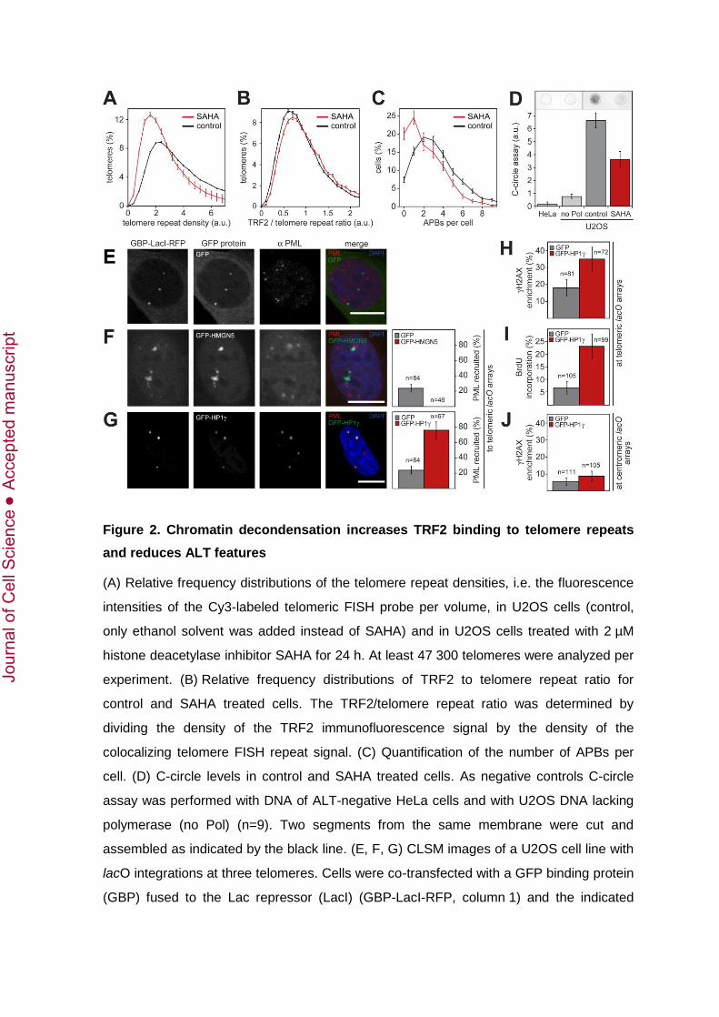
Figure 2. Chromatin decondensation increases TRF2 binding to telomere repeats
and reduces ALT features
(A) Relative frequency distributions of the telomere repeat densities, i.e. the fluorescence
intensities of the Cy3-labeled telomeric FISH probe per volume, in U2OS cells (control,
only ethanol solvent was added instead of SAHA) and in U2OS cells treated with 2 µM
histone deacetylase inhibitor SAHA for 24 h. At least 47 300 telomeres were analyzed per
experiment. (B) Relative frequency distributions of TRF2 to telomere repeat ratio for
control and SAHA treated cells. The TRF2/telomere repeat ratio was determined by
dividing the density of the TRF2 immunofluorescence signal by the density of the
colocalizing telomere FISH repeat signal. (C) Quantification of the number of APBs per
cell. (D) C-circle levels in control and SAHA treated cells. As negative controls C-circle
assay was performed with DNA of ALT-negative HeLa cells and with U2OS DNA lacking
polymerase (no Pol) (n=9). Two segments from the same membrane were cut and
assembled as indicated by the black line. (E, F, G) CLSM images of a U2OS cell line with
lacO integrations at three telomeres. Cells were co-transfected with a GFP binding protein
(GBP) fused to the Lac repressor (LacI) (GBP-LacI-RFP, column 1) and the indicated
Jour
nal o
f Cel
l Sci
ence
Acc
epte
d m
anus
crip
t
GFP fusion protein (column 2), and immunostained for endogenous PML protein to
quantify PML enrichment (column 3). Scale bars are 10 µm. (E) As a control, GFP was
recruited to the lacO arrays that visualized these as condensed spots. (F) Recruitment of
GFP-HMGN5 to the telomeric lacO arrays. The bars in the graph represent the average
percentage of colocalizations of recruited GFP-HMGN5 with endogenous PML in
comparison to control (GFP recruitment). (G) Recruitment of GFP-HP1 to the telomeric
lacO arrays. The bars in the graph represent the average percentage of colocalizations of
recruited GFP-HP1 with endogenous PML in comparison to control. (H) Quantification of
the phosphorylated histone variant H2A.X, a marker of double-strand break repair, at
telomeric lacO arrays upon GFP-HP1 recruitment. (I) Quantification of BrdU
incorporation as a marker for DNA synthesis upon recruitment of GFP-HP1Only cells
with less than 3 BrdU foci were analyzed to exclude S-phase cells with replication-
dependent BrdU incorporation. (J) Same as in panel H, but at centromeric lacO arrays.
(A)-(J) Error bars represent s.e.m.
Jour
nal o
f Cel
l Sci
ence
Acc
epte
d m
anus
crip
t

Figure 3. Telomere repeat density is increased in APBs, while TRF2 binding to
telomeres is decreased
(A) Relative frequency distributions of telomere repeat densities in U2OS cells in- and
outside of APBs. A total of 7 487 telomeres in APBs and 130 025 telomeres that were not
in APBs were analyzed. (B) Relative frequency distributions of the TRF2 to telomere
repeat ratios of telomeres in relation to PML localization. The slightly reduced accessibility
of the TRF2 antibody due to compaction of the telomere repeats was corrected as
described in supplementary material Fig. S4. (C) Normalized median TRF2 to telomere
repeat ratio in dependence of telomere repeat densities. (A)-(C) Error bars represent
s.e.m.
Jour
nal o
f Cel
l Sci
ence
Acc
epte
d m
anus
crip
t
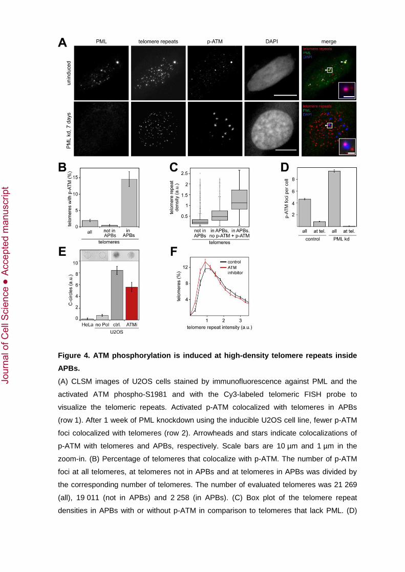
Figure 4. ATM phosphorylation is induced at high-density telomere repeats inside
APBs.
(A) CLSM images of U2OS cells stained by immunofluorescence against PML and the
activated ATM phospho-S1981 and with the Cy3-labeled telomeric FISH probe to
visualize the telomeric repeats. Activated p-ATM colocalized with telomeres in APBs
(row 1). After 1 week of PML knockdown using the inducible U2OS cell line, fewer p-ATM
foci colocalized with telomeres (row 2). Arrowheads and stars indicate colocalizations of
p-ATM with telomeres and APBs, respectively. Scale bars are 10 µm and 1 µm in the
zoom-in. (B) Percentage of telomeres that colocalize with p-ATM. The number of p-ATM
foci at all telomeres, at telomeres not in APBs and at telomeres in APBs was divided by
the corresponding number of telomeres. The number of evaluated telomeres was 21 269
(all), 19 011 (not in APBs) and 2 258 (in APBs). (C) Box plot of the telomere repeat
densities in APBs with or without p-ATM in comparison to telomeres that lack PML. (D)
Jour
nal o
f Cel
l Sci
ence
Acc
epte
d m
anus
crip
t
Effect of PML knockdown on the number of p-ATM foci in total and at telomeres. (E) C-
circle levels after inhibition of ATM with 10 µM inhibitor KU-55933 for 48 h. ALT-negative
HeLa cells and U2OS cells without addition of polymerase were used as negative
controls. The bars represent the average of 6 independent experiments. Two segments
from the same membrane were cut and assembled as indicated by the black line. (F)
Distribution of the telomere repeat intensities after 4 weeks of ATM inhibition as compared
to control cells. At least 10 000 telomeres were analyzed per treatment. (B)-(F) Error bars
represent s.e.m.
Jour
nal o
f Cel
l Sci
ence
Acc
epte
d m
anus
crip
t

Figure 5. Model for APB-mediated telomere elongation in ALT-positive cells.
The formation of a PML subcompartment at telomeres induces telomeric chromatin
compaction and clustering of telomeres, possibly also ECTRs, as proposed previously
(Cho et al., 2014; Draskovic et al., 2009). As a result of APB formation, TRF2 becomes
partly depleted at associated telomeres. This process could involve the activity of
posttranslational modifications of TRF2 such as sumoylation by MMS21 or poly-ADP
ribosylation by PARP-2 in line with previous studies (Dantzer et al., 2004; Potts and Yu,
2007). These changes in telomere features trigger autophosphorylation of ATM in APBs
and DDR according to the previously identified role of TRF2 as an inhibitor of ATM
(Denchi and de Lange, 2007; Karlseder et al., 2004). The subsequent recruitment of other
DDR and DNA repair proteins to APBs promotes telomere elongation. Upon APB
disassembly, repair and recombination factors dislocate, telomeres are released and
telomere density decreases again. As a result, TRF2 binding is increased leading to the
re-establishment of a fully protected telomere.
Jour
nal o
f Cel
l Sci
ence
Acc
epte
d m
anus
crip
t

Table 1. ALT features after PML knockdown for 72 h.
** p < 0.001 (Kolmogorov-Smirnov test)
† Telomere repeat density is the integrated signal intensity of a telomere labeled by FISH divided by the volume of that telomere.
‡ TRF2 to telomere repeat ratio is the density of the TRF2 immunofluorescence signal divided by the telomere repeat density of
the colocalizing telomere FISH signal.
APBs per cell Telomere repeat
foci per cell Telomere repeat density (a.u.) †
Telomere volume (10-3 µm3)
TRF2 to telomere repeat ratio ‡
Uninduced 3.4 ± 0.3 62.6 ± 3.1 15.9 ± 0.3 12.5 ± 0.1 0.65 ± 0.01
Induced PML knockdown
0.01 ± 0.01 69.1 ± 1.8 14.3 ± 0.4 11.8 ± 0.1 0.91 ± 0.01
Difference –3.5 ± 0.3 6.5 ± 2.6 –1.6 ± 0.6 –0.7 ± 0.1 0.26 ± 0.01
Difference (%) –99.7 ± 0.3 ** 10.8 ± 4.6 ** –9.9 ± 3.4 ** –5.5 ± 1.1 ** 41.9 ± 8.2 **
Jour
nal o
f Cel
l Sci
ence
Acc
epte
d m
anus
crip
t

Table 2. Quantitative telomere FISH analysis on metaphase spreads after 30 days PML knockdown.
Median telomere repeat intensity
without ECTRs (a.u.)
Median telomere repeat intensity
with ECTRs (a.u.)
Telomere-free ends (%)
ECTRs per metaphase
spread
Telomere repeat
intensity of ECTRs (a.u.)
ECTR fraction of total telomere
repeat intensity (%)
Uninduced 122.7 ± 9.1 119.1 ± 9.8 12.0 ± 1.1 19.1 ± 2.4 78.2 ± 9.1 6.6 ± 2.3
Induced PML knockdown
99.9 ± 9.1 99.5 ± 9.4 18.3 ± 2.7 7.7 ± 1.7 93.6 ± 16.3 2.7 ± 0.8
Difference (%) –18.6 ± 9.6 **, † –16.5 ± 10.5 **, † 52.5 ± 26.5 *, ‡ –59.8 ± 10.2 **, ‡ 19.6 ± 14.4 *, † –59.6 ± 18.3 ‡
* p < 0.05
** p < 0.001
† Kolmogorov-Smirnov test was used for statistical analysis.
‡ Welch’s t-test was used for statistical analysis.
Jour
nal o
f Cel
l Sci
ence
Acc
epte
d m
anus
crip
t

Table 3. Proteins identified in the RNAi screen that significantly affected the number of APBs per cell.
siRNA phenotype Telomere
organization Protein sumoylation DNA repair
Chromatin organization
Other biological processes
Less APBs TIN2
MMS21†
SUMO1†/2/3
UBC9
BLM‡
ERCC4‡
FEN1‡
PARP2
XRCC6‡
DNMT1
HDAC7
HP1
LSD1
SUV420H2
CDKN1A
MORC3
NR2C2
NR2F2
PML
TOP3A
More APBs POT1
TRF1* SENP6
FANCD2
FANCL
PCNA‡
RPA1‡
RPA2‡
HMGN5
A protein was considered as a hit in the screen if knockdown by two different siRNAs consistently changed the number of APBs by > 10% (p <
0.05). According to associated biological processes defined by gene ontology (GO), hits were grouped in telomere organization (GO:0032200),
protein sumoylation (GO:0016925), DNA repair (GO:0006281) and chromatin organization (GO:0006325). Hits that are annotated with more
than one of these GO terms are indicated.
* TRF1 is also annotated with the GO term “chromatin organization”.
† MMS21 und SUMO1 are also annotated with the GO term “DNA repair”.
‡ These proteins are also annotated with the GO term “telomere organization”.
Jour
nal o
f Cel
l Sci
ence
Acc
epte
d m
anus
crip
t