Embed Size (px)
Citation preview

Pt
BLa
Ub
c
d
e
Rf
h
•
•
•
•
a
ARRAA
KSPNA
0h
Colloids and Surfaces A: Physicochem. Eng. Aspects 441 (2014) 77– 83
Contents lists available at ScienceDirect
Colloids and Surfaces A: Physicochemical andEngineering Aspects
jo ur nal ho me page: www.elsev ier .com/ locate /co lsur fa
luronic-coated silver nanoprisms: Synthesis, characterization andheir antibacterial activity
ogdan Martaa, Endre Jakabb,c, Monica Potaraa, Timea Simona, Florica Imre-Lucacie,ucian Barbu-Tudoranf, Octavian Popescub,d, Simion Astileana,∗
Nanobiophotonics and Laser Microspectroscopy Center, Interdisciplinary Research Institute in Bio-Nano-Sciences and Faculty of Physics, Babes-Bolyainiversity, M Kogalniceanu 1, 40084 Cluj-Napoca, RomaniaMolecular Biology Center, Interdisciplinary Research Institute in Bio-Nano-Sciences, Babes-Bolyai University, T. Laurian 42, 400271 Cluj-Napoca, RomaniaHungarian Department of Biology and Geology, Faculty of Biology and Ecology, Babes-Bolyai University, Clinicilor 5-7, 400006 Cluj-Napoca, RomaniaInstitute of Biology, Romanian Academy, Spl. Independentei 296, 060031 Bucharest, RomaniaPhysico-Chemical Analyses Center, Interdisciplinary Research Institute in Bio-Nano-Sciences, Babes-Bolyai University, T. Laurian 42, 400271 Cluj-Napoca,omaniaElectron Microscopy Center, Faculty of Biology and Geology, Babes-Bolyai University, Clinicilor 5-7, 400006 Cluj-Napoca, Romania
i g h l i g h t s
Synthesis and characterization of sil-ver nanoprisms.Obtaining pluronic-coated silvernanoprisms and characterizingthem.Finding the optimal concentration ofpluronic for biomedical applications.Silver-pluronic nanocompositesshow a strong bactericidal effectagainst Staphylococcus aureus.
g r a p h i c a l a b s t r a c t
r t i c l e i n f o
rticle history:eceived 4 July 2013eceived in revised form 22 August 2013ccepted 29 August 2013vailable online xxx
eywords:ilver nanoprisms
a b s t r a c t
The aim of this study is to investigate the antibacterial efficiency of pluronic-coated silver nanoparticleswhen, instead of commonly explored spherical nanoparticles, anisotropic nanoparticles (nanoprisms)enveloped in a biocompatible polymer are used. In the first step we successfully synthesized silvernanoprisms with edge length between 30 and 50 nm and thickness between 4 and 6 nm exhibiting well-resolved localized surface plasmon resonances (LSPR) at 615, 443 and 335 nm. To endow with colloidaland morphological stability required in biological environment, the as-prepared silver nanoprisms werecoated with pluronic F-127—a synthetic triblock copolymer consisting of PEO (poly(ethylene oxide))–PPO
luronicanocompositentibacterial activity
(poly(propylene oxide))–PEO (poly(ethylene oxide)) chains. The bactericidal effects of pluronic-coatedsilver nanoprisms were evaluated against two methicillin-resistant Staphylococcus aureus (MRSA) strains(UCLA 8076 and 1190R) by determining the minimum inhibitory concentration (MIC) and the minimumbactericidal concentration (MBC). The strong antibacterial activity we found can be explained by consid-ering that triangular nanoprisms exhibit crystalline facets of higher reactivity and, presumably, a higher
their
rate of ions release from∗ Corresponding author. Tel.: +40264405300; fax: +40264591906.E-mail address: [email protected] (S. Astilean).
927-7757/$ – see front matter © 2013 Elsevier B.V. All rights reserved.ttp://dx.doi.org/10.1016/j.colsurfa.2013.08.076
tips and edges compared to other shapes of nanoparticles.© 2013 Elsevier B.V. All rights reserved.
1. Introduction
Medicine has met an impressive advance in the last few decadesas new drugs and cures were created for almost any affection,allowing a faster recovery and reducing its symptoms and effectson the human body. However, simultaneously with the high

7 : Physi
eth(tacbmaciotom
ttabIsadmnmvoabatdstdsc
atmtnwccfcssc(ttaeatpcAra
8 B. Marta et al. / Colloids and Surfaces A
xposure rate to drugs, some microorganisms became resistant tohem, as for example the case of some particular bacteria found inospital environment [1]. Such a bacterium is Staphylococcus aureusS. aureus), a Gram-positive bacterium which is frequently found onhe skin. S. aureus is the most common strain of the staphylococci,nd it can cause a series of skin infections such as folliculitis, furun-ulosis or abscesses. In some cases S. aureus can penetrate the skinarrier, and it can cause a lot of serious conditions like pneumonia,eningitis, bacteremia or sepsis [2]. Particularly, the strains of S.
ureus bacteria known as “methicillin resistant S. aureus” (MRSA)an survive in the presence of penicillin-like antibiotics, makingnfection with MRSA more difficult to treat and thus more danger-us. Hence, it is a need to develop new treatment methods for theseypes of bacteria, therapies based not only on antibiotics but also onther means to attack the bacterium cell to which it cannot adapt orust develop different defense mechanisms than to classical drugs.A promising perspective is emerging from the research in nano-
echnology which focuses on designing nanoparticles with con-rollable biochemical properties, including targeted or enhancedntibacterial activity. For instance, silver nanoparticles have longeen known to exhibit bacteriostatic and bactericidal effects [1].t is largely recognized that silver ions or silver compounds attackimultaneously a broad range of targets in the bacterial organismsnd, therefore, microbes are unable to develop resistance as theyo against conventional antibiotics [3]. In this regard, the develop-ent of new and more efficient nanocomposites containing silver
anoparticles is vital in a world where new and more resistanticrobial agents appear. Regarding the antibacterial activity of sil-
er nanoparticles, there have been proposed different mechanismsf toxicity. As for example, it has been shown that the antimicrobialctivity of nanoparticles is related to the action of the ions releasedy the nanoparticles [1]. A mechanism explaining the antimicrobialctivity of silver nanoparticles has been also proposed suggestinghe formation of free radicals which induce the bacterial membraneamage, resulting in cell death [4]. Other studies have shown thatilver nanoparticles interact with the cell membrane and some ofhem also penetrate the bacterial cell wall, thereby causing theeath of bacteria [5,6]. This interaction was found to depend oneveral parameters such as the nanoparticles’ size [1], shape [3],oncentration [3] and stability in the growth medium [7].
The synthesis of silver nanoparticles which exhibit strongntibacterial effects and good stability in biological media is cer-ainly an important task. In biological media, conditions differ very
uch from the conditions in the colloidal solution where nanopar-icles are dispersed. Most likely when introduced in such media,anoparticles would undergo physical and chemical changes, thatould decrease if not nullify their effects. Aggregation is the most
ommonly met process which appears as a result of the surfaceharge neutralization of nanoparticles and leads to conglomerateormation. In order to confer them stability to physical and chemicalhanges in the medium, polymers or proteins are used to encap-ulate such nanoparticles. [8]. They usually meet two roles: (i) totabilize the nanoparticles and make them resistant to physico-hemical changes that may occur in the medium they are, andii) to make the nanoparticles more body-friendly, to minimizeheir toxic impact on the human body and to allow them to passhrough the barriers of the organism unimpeded. Some polymersre active agents, having antibacterial properties themselves, oth-rs through their structure allow certain groups to attach to them,nd hence they can be used for specific targeting [9]. Therefore,he use in antibacterial applications of nanocomposites combiningolymers and nanoparticles is very promising, since the polymer
an add new properties and enhance the effect of nanoparticles.s for example, combining metallic nanoparticles with “stimuliesponsive” (e.g. temperature, pH etc.) polymers is a currentlyvailable strategy to produce multifunctional nanomaterials whichcochem. Eng. Aspects 441 (2014) 77– 83
hold enormous potential in life science applications [10–12]. Sucha polymer is Pluronic, a synthetic block co-polymer, consistingof PEO (poly(ethylene oxide))–PPO (poly(propylene oxide))–PEO(poly(ethylene oxide)) chains. The PEO–PPO–PEO triblock copoly-mers have amphiphilic characteristics (PPO being lipophilic, whilePEO is hydrophilic) and can self-assemble into micelles to forma variety of close packed structures. The micellization processis influenced by the concentration of pluronic and the tempera-ture. The fact that pluronic shows thermoreversible micellizationaround body temperature makes it suitable for medical applica-tions such as drug delivery, gene therapy or tissue engineering [13].Pluronic can also be used in the steric stabilization of nanoparti-cles, binding to their surface and forming a thin protective layerby hydrophobic association of the PPO blocks [14]. Additionally,the capping Pluronic layer can be further exploited to integratemultiple functionalities by encapsulating small molecules, suchas drugs, fluorescent labels or Raman reporters. Our group hasrecently reported that pluronic-gold nanohybrids can be usedas effective drug carriers for photodynamic therapy of cancercells [15]. In another study we demonstrated that encapsula-tion of gold nanoparticles in pluronic represents an appropriateapproach to produce highly stable and biocompatible nanoma-terials [16]. Considering the interesting optical and biologicalproperties already demonstrated, here we focused on the prepa-ration of pluronic-coated silver nanoprisms and investigation oftheir antibacterial efficiency. Our effort to produce silver nanopar-ticles of particular shape is motivated by the fact that beyond thedesigned bactericidal action such nanoparticles confer versatilitytoward the diagnostic and treatment of other affections [17]. Basedon the above considerations, we point out that the use of triblockcopolymer pluronic for nanoparticles stabilization is not a trivialoption, having in mind that this polymer shows thermoreversiblegelation around body temperature, feature that could be furtherused for drug delivery and controlled release [13].
The aim of the present study is to investigate the antibacte-rial efficiency of pluronic-coated silver nanoparticles against twomethicillin-resistant S. aureus (MRSA) strains using nanoparticlesof triangular shape (nanoprisms) instead of commonly explored,spherical nanoparticles. It has been previously described that sil-ver nanoparticles have specifically a low efficiency against S. aureus,often related to the thick peptidoglycan membrane of the Gram-positive bacteria [18]. On the other hand there is evidence thatthe bactericidal effect may be enhanced by the specific shape ofnanoparticles [3]. It is therefore rational to consider that triangu-lar nanoprisms should exhibit stronger effect due to presumablyhigher reactivity and rate of ions release at their tips and edges.
The organization of this paper is as follows. The first sec-tion presents the synthesis of silver nanoprisms in solutionfollowed by their stabilization with pluronic. The preparedpluronic-coated silver nanoprisms are characterized by UV–vis-NIRspectroscopy, transmission electron microscopy (TEM) and Zetapotential measurements. The final section presents the results onantibacterial activity of pluronic-coated silver nanoprisms againsttwo methicillin-resistant S. aureus (MRSA) strains (UCLA 8076 and1190R). The minimum inhibitory concentration (MIC) and the mini-mum bactericidal concentration (MBC) reveal a strong antibacterialeffect of the pluronic-coated silver nanoprisms, which point out therole of their particular shape.
2. Experimental details
2.1. Materials
Pluronic F-127 (12,600 MW) and silver nitrate (AgNO3) werepurchased from Sigma-Aldrich. Sodium borohydride (NaBH4) and

: Physi
tH(Aw1(
2
rc3phmpsov3pr
aosu2spat
2
rbmnpcawsMsTaa1Scawea4
2
tL
B. Marta et al. / Colloids and Surfaces A
risodium citrate (Na3C6H5O7·2H2O) were supplied by Merck.ydrogen peroxide (H2O2) was obtained from Cristal Chim
Bucuresti). All chemicals were used without further purification.ll reagents employed were of analytical grade and the solutionsere prepared using ultrapure water with a resistivity of at least
8 M� cm. All glassware used was cleaned with aqua regia solutionHCl:HNO3 3:1) followed by rinsing with ultrapure water.
.2. Synthesis of pluronic-coated silver nanoprisms
Silver nanoprisms were synthesized based on a previouslyeported method [19]. Briefly, aqueous solutions of trisodiumitrate (TSC, 14 ml, 1.25 × 10⁻² M), silver nitrate (AgNO3, 35 ml,.75 × 10−4 M), hydrogen peroxide (H2O2, 35 ml, 5 × 10⁻² M),otassium bromide (KBr, 455 �l, 1 × 10⁻² M) and sodium boro-ydride (NaBH4, 17.5 ml, 5.0 × 10⁻³ M) were combined in theentioned order, mixing them together on a magnetic stirring
late at room temperature. During the mixing process the solutionhowed some consistent optical changes, indicating the formationf silver nanoparticles. The process was sustained until no furtherisible color change in the colloid appeared (after approximately
min of stirring). After the synthesis process was completed, theroduct was purified by centrifugation at 7500 rpm for 17 min andesuspended in ultrapure water.
The preparation of pluronic-coated silver nanoprisms waschieved in a two-step capping method developed in our lab-ratory. An aqueous solution of 10 mM pluronic F-127 wastep-by-step added to the colloidal solution of silver nanoprismsp to a final concentration of 0.8 mM. The samples were kept4 h at room temperature to allow the polymer to bind onto theilver nanoprisms surfaces. For antibacterial activity assays, theluronic-coated silver nanoprisms were purified by centrifugationnd resuspended in ultrapure water to obtain the desired concen-rations.
.3. Sample characterization
UV–vis-NIR extinction spectra of the silver colloids wereecorded with a Jasco V-670 spectrophotometer, over a rangeetween 190 and 1100 nm. A JOEL-JEM 1100 transmission electronicroscope (TEM) was used to study the morphology of the silver
anoparticles. The samples used for TEM measurements were pre-ared by placing a drop of colloidal dispersion onto carbon-coatedopper grids. Samples were left to dry at room temperature. Theverage size and distribution of silver nanoprisms in TEM imagesere determined using Image J software [20]. Zeta potential mea-
urements were performed at 25 ◦C using a Zetasizer Nano ZS fromalvern. Disposable capillary cells (DTS1061) filled with 750 �l
ilver colloid dispersion (pH 7) were used for the measurements.he concentration of silver in colloidal solution was determined bytomic absorption spectroscopy (Avanta PM, GBC-Australia). Thenalysis was performed using Ag atomic spectroscopy standard000 mg l−1 acidic solution (AgNO3 in 0.5 M HNO3) from LGCtandards GmbH (Wesel, Germany). For measurement, a hollowathode Ag lamp (Model P851 HCL) was used. The instrument oper-ted at a lamp current of 4.0 mA, wavelength of 328.1 nm, and slitidth of 0.2 nm. A linear calibration curve was generated before
ach measurement using the “linear least squares” built-in method,n excellent correlation being obtained (R2 = 1.0) for the 0.25 to.0 mg l−1 domain. Each sample was measured in triplicate.
.4. Bacterial strains and culture conditions
Two methicillin-resistant S. aureus (MRSA) strains were used inhis study, UCLA 8076, originating from the University of California,os Angeles, and 1190 R, obtained from the Semmelweis University,
cochem. Eng. Aspects 441 (2014) 77– 83 79
Budapest. The cultures were maintained on Mueller Hinton agar(Fluka, Buchs, Switzerland). The bacteria were cultured overnightin 5 ml Mueller Hinton broth (Fluka, Buchs, Swizerland) in a Cer-tomat BS-T incubation shaker (Sartorius Stedim Biotech, Aubagne,France) at 37 ◦C, 150 rpm until the culture reached an OD6 0 0 of1.0 (Spekol UV VIS 3.02, Analytic Jena, Jena, Germany), correspond-ing to 109 CFU ml−1. Before incubation with pluronic-coated silvernanoprisms the cultures were diluted to 107 CFU ml−1 with sterilebroth.
2.5. Antibacterial efficiency tests
The plates used in the microdilution experiments were preparedby mixing different quantities of cation-adjusted Mueller Hintonbroth supplemented with 2% NaCl (from 45 to 90 �l, in 10 steps of5 �l) with different quantities of pluronic-coated silver nanoprisms(from 50 to 5 �l, in 10 steps of 5 �l) in order to achieve a final volumeof 95 �l in all samples. The wells were inoculated with 5 �l bacte-rial suspension of 107 CFU ml−1. The final bacteria concentrationwas 105 CFU ml−1, and the final concentrations of the pluronic-coated silver nanoprisms were: 61, 55, 49, 43, 37, 31, 24, 18, 12and 6 �g ml−1. Two additional tests were performed for pluronic F-127 and AgNO3 solutions. The final pluronic concentrations were:169, 150, 131, 112, 94, 75, 56, 37 and 19 �g ml−1. The final concen-trations of the AgNO3 inoculated were: 40, 36, 32, 28, 24, 20, 16, 12,8, respectively, 4 �g ml−1. Two control samples were also prepared,a negative control, containing only sterile broth, and a positive con-trol containing 5 �l bacteria suspension and 95 �l sterile broth. Theinoculated microdilution plates were covered with a plastic lid andincubated at 37 ◦C for 24 h. Bacterial growth was evaluated visually,and the minimum inhibitory concentration (MIC) was recorded asthe lowest concentration of the pluronic-coated silver nanoprismsthat completely inhibits bacterial growth. For evaluation of theminimum bactericidal concentration (MBC), 100 �l aliquots weretaken from wells without any visible bacterial growth and platedonto Mueller Hinton agar plates. After incubation, colony formingunits (CFUs), corresponding to the number of surviving cells, werecounted and digital images of the plates were captured. The MBCwas determined as the lowest concentration of pluronic-coatedsilver nanoprisms that inhibits colony formation.
3. Results and discussions
3.1. Synthesis and characterization of the colloidal silvernanoprisms
The growth of silver nanoprisms was first revealed by the spec-tacularly color changes in the solution over time, from colorlessto yellow, then orange, pink, mauve, and finally a deep blue.The visible color relies on resonant light interaction with silvernanoparticles via excitation of surface plasmons with contribu-tion from light scattering and absorption [21]. This phenomenon isknown as localized surface plasmon resonance (LSPR) and is char-acterized by strong UV–vis-NIR extinction bands. As the color ofcolloidal silver nanoparticles is strongly dependent on their sizeand shape, such color evolution should be connected to the mor-phological transformation of silver nanoparticles, which evolve insolution from quasi-spherical to triangular shape.
Detailed characterizations of the silver nanoparticles mor-phology were obtained by combining information from LSPRspectroscopy and TEM analysis. Fig. 1 shows a representative
picture of the resulted colloidal solution together with its corre-sponding UV–vis-NIR extinction spectrum collected at the end ofreaction. The LSPR spectrum exhibits three peaks correspondingto different modes of plasmon excitation. In literature it is well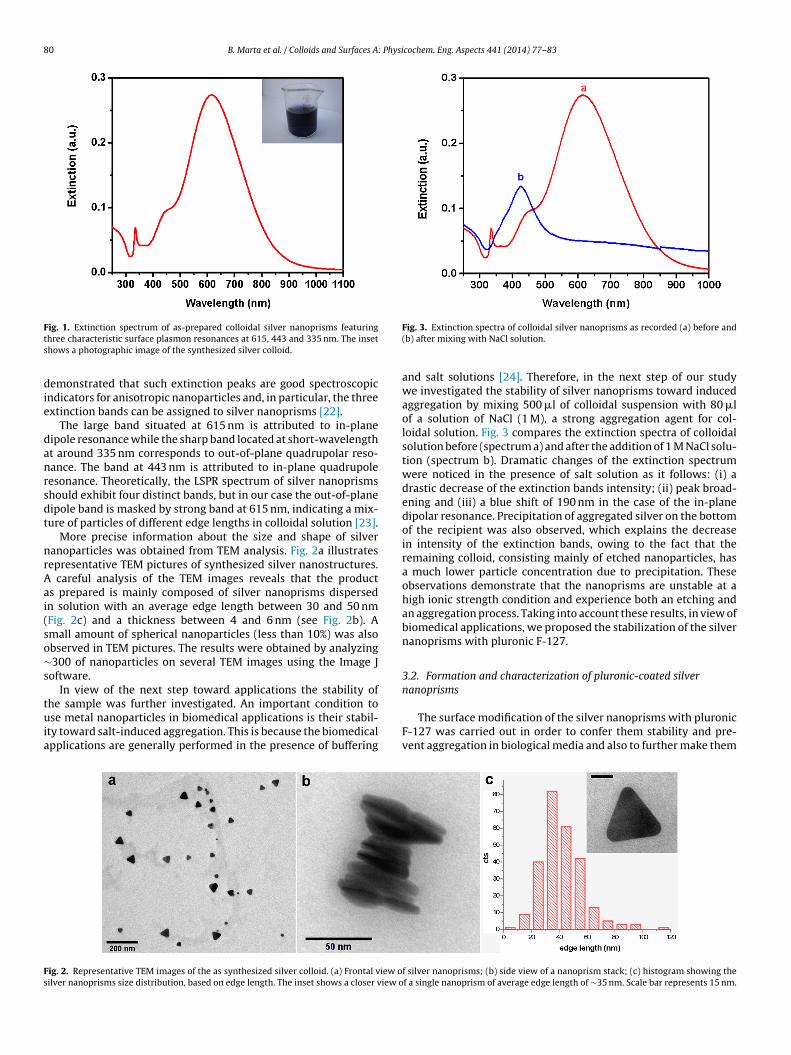
80 B. Marta et al. / Colloids and Surfaces A: Physicochem. Eng. Aspects 441 (2014) 77– 83
Fts
die
danrsdt
nrAai(so∼s
tuia
Fs
ig. 1. Extinction spectrum of as-prepared colloidal silver nanoprisms featuringhree characteristic surface plasmon resonances at 615, 443 and 335 nm. The insethows a photographic image of the synthesized silver colloid.
emonstrated that such extinction peaks are good spectroscopicndicators for anisotropic nanoparticles and, in particular, the threextinction bands can be assigned to silver nanoprisms [22].
The large band situated at 615 nm is attributed to in-planeipole resonance while the sharp band located at short-wavelengtht around 335 nm corresponds to out-of-plane quadrupolar reso-ance. The band at 443 nm is attributed to in-plane quadrupoleesonance. Theoretically, the LSPR spectrum of silver nanoprismshould exhibit four distinct bands, but in our case the out-of-planeipole band is masked by strong band at 615 nm, indicating a mix-ure of particles of different edge lengths in colloidal solution [23].
More precise information about the size and shape of silveranoparticles was obtained from TEM analysis. Fig. 2a illustratesepresentative TEM pictures of synthesized silver nanostructures.
careful analysis of the TEM images reveals that the products prepared is mainly composed of silver nanoprisms dispersedn solution with an average edge length between 30 and 50 nmFig. 2c) and a thickness between 4 and 6 nm (see Fig. 2b). Amall amount of spherical nanoparticles (less than 10%) was alsobserved in TEM pictures. The results were obtained by analyzing300 of nanoparticles on several TEM images using the Image J
oftware.In view of the next step toward applications the stability of
he sample was further investigated. An important condition tose metal nanoparticles in biomedical applications is their stabil-
ty toward salt-induced aggregation. This is because the biomedicalpplications are generally performed in the presence of buffering
ig. 2. Representative TEM images of the as synthesized silver colloid. (a) Frontal view oilver nanoprisms size distribution, based on edge length. The inset shows a closer view o
Fig. 3. Extinction spectra of colloidal silver nanoprisms as recorded (a) before and(b) after mixing with NaCl solution.
and salt solutions [24]. Therefore, in the next step of our studywe investigated the stability of silver nanoprisms toward inducedaggregation by mixing 500 �l of colloidal suspension with 80 �lof a solution of NaCl (1 M), a strong aggregation agent for col-loidal solution. Fig. 3 compares the extinction spectra of colloidalsolution before (spectrum a) and after the addition of 1 M NaCl solu-tion (spectrum b). Dramatic changes of the extinction spectrumwere noticed in the presence of salt solution as it follows: (i) adrastic decrease of the extinction bands intensity; (ii) peak broad-ening and (iii) a blue shift of 190 nm in the case of the in-planedipolar resonance. Precipitation of aggregated silver on the bottomof the recipient was also observed, which explains the decreasein intensity of the extinction bands, owing to the fact that theremaining colloid, consisting mainly of etched nanoparticles, hasa much lower particle concentration due to precipitation. Theseobservations demonstrate that the nanoprisms are unstable at ahigh ionic strength condition and experience both an etching andan aggregation process. Taking into account these results, in view ofbiomedical applications, we proposed the stabilization of the silvernanoprisms with pluronic F-127.
3.2. Formation and characterization of pluronic-coated silvernanoprisms
The surface modification of the silver nanoprisms with pluronicF-127 was carried out in order to confer them stability and pre-vent aggregation in biological media and also to further make them
f silver nanoprisms; (b) side view of a nanoprism stack; (c) histogram showing thef a single nanoprism of average edge length of ∼35 nm. Scale bar represents 15 nm.

B. Marta et al. / Colloids and Surfaces A: Physicochem. Eng. Aspects 441 (2014) 77– 83 81
Fs
br
tPswCtlt
cbcps−ipenaFpha
nanoprisms in NaCl solution presented in Fig. 6 reveals no aggre-gation, but a blue shift of the dipolar band occurs, indicating anetching process [23]. This result can be explained by an incomplete
ig. 4. Zeta potential of silver nanoprisms as function of pluronic concentrations inolution: (a) without pluronic; (b) 0.47 mM pluronic; (c) 0.8 mM pluronic.
iocompatible and reduce the toxicity and immune responseelated to the interaction with living organisms.
In order to avoid micellization of Pluronic in the colloidal solu-ion of silver nanoprisms, which would decrease the ability ofluronic to bind onto the nanoprisms’ surface, we developed a two-tep capping method. Colloidal solution of the silver nanoprismsas step-by-step supplied with quantities of pluronic below theMC (0.555 mM at room temperature [25]), allowing enough timeo bind before the addition of any further pluronic. This way mice-lization can be avoided in the solution, and even if micelles form,hey will mostly be on the surface of the nanoprisms.
To determine the optimal concentration of pluronic that ensuresolloid stability, an aqueous solution of 10 mM pluronic was step-y-step added to the silver nanoprisms colloid and the surfaceovering in each step was monitored by measuring the Zeta-otential of the colloidal solutions. Zeta potential distributionpectra the uncoated silver nanoprisms reveals a single peak at50 mV (curve a from Fig. 4) which can be attributed to the citrate
ons on the surface of silver nanoprisms. After the first step ofluronic incubation (final concentration 0.47 mM) a second peakmerges at −10 mV, corresponding to the pluronic-coated silveranoprisms. By increasing the pluronic concentration, the peakt −50 mV decreases in favor of peak at −10 mV (curve b from
ig. 4), and at a final concentration of 0.8 mM it disappears com-letely (curve c from Fig. 4), suggesting that almost all nanoprismsave been completely covered with the polymer. As a consequence,minimum pluronic concentration of 0.8 mM is required for the
Fig. 5. Normalized extinction spectra of colloidal silver nanoprisms as recorded (a)before and (b) after pluronic coating.
complete coating of the silver nanoprisms assuring colloidal stabi-lization.
Adsorption of pluronic onto the surface of the silver nanoprismswas further demonstrated by analyzing the LSPR spectrum of thecolloidal solution. Fig. 5 (curve b) shows the spectrum of pluronic-coated silver nanoprisms solution (final pluronic concentrationof 0.8 mM) recorded a day after adding the polymer to colloidalsolution in order to allow the polymer to bind onto the silvernanoprisms surfaces. For comparison, Fig. 5 (curve a) shows the ref-erence spectrum recorded from blank solution (without pluronic).A red shift of 14 nm in the maximum of the dipolar band wasobserved after incubation with pluronic, which can be assignedto the modification of the refractive index of the medium sur-rounding the particle, confirming the presence of the polymer onthe nanoprisms surface. The shape and the width of the extinc-tion spectrum recorded before and after the surface modificationwith pluronic solution remained almost unchanged, ruling out anypossible aggregation of the particles upon polymer adsorption.
Hereafter, aggregation tests were carried out in order to testthe stability of the pluronic-coated silver nanoprisms at high ionicstrength. The extinction spectrum of the pluronic-coated silver
Fig. 6. Extinction spectra of pluronic-coated silver nanoprisms as recorded (a)before and (b) after mixing with NaCl solution.

82 B. Marta et al. / Colloids and Surfaces A: Physicochem. Eng. Aspects 441 (2014) 77– 83
Fcc
ctsine
3
wtartigpcctccv
icaioocw
Table 1MIC and MBC data for the two S. aureus strains incubated with pluronic-coated silvernanoprisms and AgNO3 solutions.
Antibacterial agent Bacterium type MIC (�g ml−1) MBC (�g ml−1)
Pluronic-coated silvernanoprisms
1190R 24 >49UCLA 8076 18 37
ig. 7. Agar plates inoculated with bacteria and different concentrations of pluronic-oated silver nanoprisms after 24 h of incubation. The white dots represent theolonies of bacteria formed (CFUs).
apping by pluronic at the nanoprism edges. However, comparedo the behavior of the bare nanoprisms in similar conditions, theurface modification with pluronic offers an improved stabilityn biological medium. Furthermore, truncated triangular shapedanoparticles have been shown to posses a very strong bactericidalffect [3] which makes this change actually desirable.
.3. Antibacterial activity of pluronic-coated silver nanoprisms
The antimicrobial effects of pluronic-coated silver nanoprismsere evaluated determining the minimum inhibitory concentra-
ion (MIC) and minimum bactericidal concentration (MBC) whichre the standard microbiological techniques to evaluate the bacte-iostatic and bactericidal properties of antimicrobial agents. All theests were performed in duplicate, and repeated after several daysn order to ensure the reproducibility of our results. The bacterialrowth was evaluated by monitoring the culture turbidity. Table 1resents the mean MIC and MBC values obtained for pluronic-oated silver nanoprisms and AgNO3 solutions. In addition, theoncentration of pluronic in the solutions used did not influencehe bacterial growth in any way. Fig. 7 shows images of bacteriaultures in the presence of different concentrations of pluronic-oated silver nanoprisms, used for the determination of the MBCalues.
Both bacteriostatic and bactericidal activity show differencesn case of the two investigated strains (Table 1). Higher antimi-robial activity of pluronic-coated silver nanoprisms was noticedgainst the UCLA 8076 strain. The MIC and MBC values obtainedn the present study are lower than [26] or similar [27] with those
bserved previously. Smaller MIC and MBC values were recorded inur previous study for chitosan-coated triangular silver nanoparti-les where a synergistic activity of chitosan and silver nanoparticlesas observed [28]. As pointed out before, the pluronic polymerAgNO3 1190R 4 4UCLA 8076 4 20
itself doesn’t show any antimicrobial activity, therefore in thepresent study the antibacterial activity of pluronic-coated silvernanoprisms should be related only to the effect of silver nanopar-ticles.
Preliminary reports from the literature suggested that it isalmost impossible to separate the antibacterial effect of silvernanoparticles by that of silver ions. However, it was proposed thatphysical disruption of cell membrane integrity by silver nanopar-ticles might be the primary cause of antibacterial effect, while theaction of silver ions released by the particles or the accumulation ofnanoparticles in the cytoplasm occurs as secondary effects [29]. Inthis regard a reference test was performed to evaluate the antibac-terial activity of silver ions. For silver ions the MIC and MBC valuesin Table 1 are similar to those reported previously. However, thebactericidal activity shows an obvious difference in the two investi-gated strains. An interesting remark is that in case of the UCLA8076strain the MBC value for the pluronic-coated silver nanoprisms iscomparable with the MBC value obtained using silver ions. Thedifferences between these two tested strains reside in the cellscomposition. For example, 1190R strain is composed by a uniformpopulation of cells which expresses a high level of resistance. Incontrast, UCLA 8076 strain is composed by several subpopulations,and it can be considered a heterogeneous methicillin-resistantstrain (data not shown). In case of these type of strains majorityof cells (typically 99.9% or more) are susceptible to low drug con-centrations, and only few cells (e.g. 1 in 106) show high resistance tomethicillin. These strains lead to misidentification and often causetherapeutic problems [30].
A large number of previous studies evaluated the antibacterialeffect of silver ions and silver nanoparticles against Gram-positiveand Gram-negative organisms. These studies demonstrated a lowerantibacterial effect of silver nanoparticles toward Gram-positivebacteria as compared with the Gram-negative microorganisms.A possible explanation is that silver nanoparticles may penetratemore easily through the outer membrane of Gram-negative bacte-ria than the peptidoglycan layer of Gram-positive bacteria. Thisis because the rigid peptidoglycan layer specific to Gram-positivebacteria consists of linear polysaccharide chains crosslinked byshort peptides; architecture which provides only a small numberof anchoring sites. Therefore, the interaction between antibacte-rial agents and Gram-positive bacteria becomes very difficult. Incontrast to these studies, the present work reveals new resultswhich point out that the cell composition of the bacterium playsalso an important role on its mechanisms of defense toward sil-ver nanoparticles and silver ions. The encouraging results obtainedcan be related to the shape of nanoparticles, namely triangularnanoprisms, which is demonstrated to provide more {1, 1, 1} facetscompared to other shapes of nanoparticles [3]. Facets of {1, 1, 1}indexes have been proven to be more active, owing to the high reac-tivity of silver from facets with high atom density [31]. In addition, ithas been demonstrated that the antibacterial activity of sphericalsilver nanoparticles is size-dependent, smaller particles showinghigher antimicrobial efficiency [1]. In our case we suppose that
silver nanoprisms exhibit antibacterial activity in different waysdepending of their size. While small nanoprisms interact moreefficiently with the bacterial surface, the larger ones promote the
: Physi
rt
4
piaosfsvdcpon
A
peft1I
R
[
[
[
[
[
[
[
[
[
[
[
[
[
[
[
[
[
[
[
[
B. Marta et al. / Colloids and Surfaces A
elease of silver ions due to the high surface energy which makeshem chemically less stable.
. Conclusions
We successfully synthesized silver nanoprisms and fabricatedluronic-coated silver nanoprisms. In the first step we thoughtfully
nvestigated the morphology of the silver nanoprisms through TEMnd LSPR spectroscopy. Next, the formation and colloidal stabilityf pluronic-coated silver nanoprisms were analyzed through LSPRpectroscopy and Zeta potential measurements. In vitro tests per-ormed on two methicillin-resistant S. aureus strains have revealedtrong bacteriostatic and bactericidal effects of pluronic-coated sil-er nanoprisms. Both bacteriostatic and bactericidal activity showifferences in case of the two investigated strains, higher antimi-robial activity being noticed against the UCLA 8076 strain. Theositive results imply the continuation of the present study inrder to develop new therapeutics based on pluronic-coated silveranoprisms.
cknowledgments
This work was supported by CNCSIS–UEFISCSU, Romania,roject number PNII ID PCCE 129/2008. T. Simon also acknowl-dges the financial support of the Sectoral Operational Programmeor Human Resources Development 2007–2013, co-financed byhe European Social Fund, under the project number POSDRU/07/1.5/S/76841 with the title “Modern Doctoral Studies:
nternationalization and Interdisciplinarity”.
eferences
[1] J.R. Morones, J.L. Elechiguerra, A. Camacho, K. Holt, J.B. Kouri, J.T. Ramírez, M.J.Yacaman, The bactericidal effect of silver nanoparticles, Nanotechnology 16(2005) 2346.
[2] T. Iwase, Y. Uehara, H. Shinji, A. Tajima, H. Seo, K. Takada, T. Agata, Y. Mizunoe,Staphylococcus epidermidis Esp inhibits Staphylococcus aureus biofilm formationand nasal colonization, Nature 465 (2010) 346–349.
[3] S. Pal, Y.K. Tak, J.M. Song, Does the antibacterial activity of silver nanoparti-cles depend on the shape of the nanoparticle?: a study of the Gram-negativebacterium Escherichia coli, Appl. Environ. Microbiol. 73 (2007) 1712–1720.
[4] J.S. Kim, E. Kuk, K.N. Yu, J.-H. Kim, S.J. Park, H.J. Lee, S.H. Kim, Y.K. Park, Y.H. Park,C.-Y. Hwang, Y.-K. Kim, Y.-S. Lee, D.H. Jeong, M.-H. Cho, Antimicrobial effectsof silver nanoparticles, Nanomedicine 3 (2007) 95–101.
[5] I. Sondi, B. Salopek-Sondi, Silver nanoparticles as antimicrobial agent: a casestudy on E. coli as a model for Gram-negative bacteria, J. Colloid Interf. Sci. 275(2004) 177–182.
[6] L.F. Espinosa-Cristóbal, G.A. Martínez-Castanón, R.E. Martínez-Martínez, J.P.Loyola-Rodríguez, N. Patino-Marín, J.F. Reyes-Macías, F. Ruiz, Antibacterialeffect of silver nanoparticles against Streptococcus mutans, Mater. Lett. 63(2009) 2603–2606.
[7] S. Shrivastava, T. Bera, A. Roy, G. Singh, P. Ramachandrarao, D. Dash, Char-
acterization of enhanced antibacterial effects of novel silver nanoparticles,Nanotechnology 18 (2007) 225103.[8] C. Blanco-Andujar, L.D. Tung, N.T.K. Thanh, Synthesis of nanoparticles forbiomedical applications, Annu. Rep. Prog. Chem., Sect. A: Inorg. Chem. 106(2010) 553–568.
[
[
cochem. Eng. Aspects 441 (2014) 77– 83 83
[9] Q. Li, S. Mahendra, D.Y. Lyon, L. Brunet, M.V. Liga, D. Li, P.J.J. Alvarez, Antimi-crobial nanomaterials for water disinfection and microbial control: potentialapplications and implications, Water Res. 42 (2008) 4591–4602.
10] D. Li, Q. He, J. Li, Smart core/shell nanocomposites: intelligent polymers modi-fied gold nanoparticles, Adv Colloid Inter. Sci. 149 (2009) 28–38.
11] D. Li, Q. He, Y. Cui, K. Wang, X. Zhang, J. Li, Thermosensitive copolymer networksmodify gold nanoparticles for nanocomposite entrapment, Chem. Eur. J. 13(2007) 2224–2229.
12] D. Li, Y. Cui, K. Wang, Q. He, X. Yan, J. Li, Thermosensitive nanostructures com-prising gold nanoparticles grafted with block copolymers, Adv. Function. Mater.17 (2007) 3134–3140.
13] S. Fusco, A. Borzacchiello, P.A. Netti, Perspectives on: PEO–PPO–PEO triblockcopolymers and their biomedical applications, J. Bioact. Compat. Pol. 21 (2006)149–164.
14] T.I. Abdullin, O.V. Bondar, Y.G. Shtyrlin, M. Kahraman, M. Culha, Pluronic blockcopolymer-mediated interactions of organic compounds with noble metalnanoparticles for SERS analysis, Langmuir 26 (2010) 5153–5159.
15] T. Simon, S. Boca, A.-M. Gabudean, P. Baldeck, S. Astilean, LED-activated methy-lene blue-loaded Pluronic-nanogold hybrids for in vitro photodynamic therapy,J. Biophotonics (2013), http://dx.doi.org/10.1002/jbio.201300058.
16] T. Simon, S. Boca, D. Biro, P. Baldeck, S. Astilean, Gold–Pluronic core–shellnanoparticles: synthesis, characterization and biological evaluation, J.Nanopart. Res. 15 (15) (2013) 1578.
17] S.C. Boca, M. Potara, A.-M. Gabudean, A. Juhem, P.L. Baldeck, S. Astilean,Chitosan-coated triangular silver nanoparticles as a novel class of biocompati-ble, highly effective photothermal transducers for in vitro cancer cell therapy,Cancer Lett. 311 (2011) 131–140.
18] M. Singh, S. Singh, S. Prasad, Nanotechnology in medicine and antibac-terial effect of silver nanoparticles, Digital J. Nanomater. Bios. 3 (2008)115–122.
19] A.J. Frank, N. Cathcart, K.E. Maly, V. Kitaev, Synthesis of silver nanoprismswith variable size and investigation of their optical properties: a first-yearundergraduate experiment exploring plasmonic nanoparticles, J. Chem. Edu.87 (2010) 1098–1101.
20] The image processing toolkit ImageJ is freely available in the public domain at〈http://rsb.info.nih.gov/ij/〉.
21] E.C.L. Ru, P.G. Etchegoin, G. Pablo (Eds.), Principles of Surface-Enhanced RamanSpectroscopy: And Related Plasmonic Effects, Elsevier, Amsterdam, 2009,Chapter 3.
22] I. Pastoriza-Santos, L.M. Liz-Marzán, Colloidal silver nanoplates state of the artand future challenges, J. Mater. Chem. 18 (2008) 1724–1737.
23] P. Yu, J. Huang, J. Tang, Observation of coalescence process of silver nanospheresduring shape transformation to nanoprisms, Nanoscale Res. Lett. 6 (2010)46.
24] M. Zhou, S. Chen, S. Zhao, Synthesis of icosahedral gold nanocrystals: a thermalprocess strategy, J. Phys. Chem. B 110 (2006) 4510–4513.
25] P. Alexandridis, J.F. Holzwarth, T.A. Hatton, Micellization of poly(ethyleneoxide)–poly(propylene oxide)–poly(ethylene oxide) triblock copolymers inaqueous solutions: thermodynamics of copolymer association, Macro-molecules 27 (1994) 2414–2425.
26] B. Chudasama, A.K. Vala, N. Andhariya, R.V. Mehta, R.V. Upadhyay, Highly bac-terial resistant silver nanoparticles: synthesis and antibacterial activities, J.Nanopart. Res. 12 (2010) 1677–1685.
27] A. Panácek, L. Kvítek, R. Prucek, M. Kolár, R. Vecerová, N. Pizúrová, V.K.Sharma, T. Nevecná, R. Zboril, Silver colloid nanoparticles: synthesis, char-acterization, and their antibacterial activity, J Phys. Chem. B 110 (2006)16248–16253.
28] M. Potara, E. Jakab, A. Damert, O. Popescu, V. Canpean, S. Astilean, Synergis-tic antibacterial activity of chitosan–silver nanocomposites on Staphylococcusaureus, Nanotechnology 22 (2011) 135101.
29] A.L. Neal, What can be inferred from bacterium-nanoparticle interactions aboutthe potential consequences of environmental exposure to nanoparticles? Eco-
toxicology 17 (2008) 362–371.30] H. Rinder, Hetero-resistance: an under-recognised confounder in diagnosis andtherapy? J. Med. Microbiol. 50 (2001) 1018–1020.
31] D.W. Hatchett, H.S. White, Electrochemistry of sulfur adlayers on the low-indexfaces of silver, J. Phys. Chem.—US 100 (1996) 9854–9859.