Embed Size (px)
Citation preview
a
CRITICAL COMMENTARIES
Pluripotent Stem Cells as a Model for EmbryonicPatterning: From Signaling Dynamics to SpatialOrganization in a Dish
Idse Heemskerk1 and Aryeh Warmflash1,2*
1Department of Biosciences, Rice University, Houston, Texas2Department of Bioengineering, Rice University, Houston, Texas
Abstract: In vivo studies have identified the signaling pathways and transcription factors involved in patterning the verte-brate embryo, but much remains unknown about how these are organized in space and time to orchestrate embryogenesis.Recently, embryonic stem cells have been established as a platform for studying spatial pattern formation and differentiationdynamics in the early mammalian embryo. The ease of observing and manipulating stem cell systems promises to fill gaps inour understanding of developmental dynamics and identify aspects that are uniquely human. Developmental Dynamics000:000–000, 2016. VC 2016 Wiley Periodicals, Inc.
Key words: human embryonic stem cells; morphogen; gastrulation; dynamics; signaling
Submitted 11 March 2016; First Decision 29 June 2016; Accepted 6 July 2016; Published online 0 Month 2013
Introduction
Despite its importance, relative to other vertebrate classes, muchremains unknown about mammalian development in general,and human development in particular. Knowledge of humandevelopment has medical applications ranging from directedstem cell differentiation for regenerative medicine (Trounson andMcDonald, 2015) to treatment of childhood cancers (Hatten andRoussel, 2011). Moreover, an anthropocentric worldview makesour own development a fascinating subject of fundamentalresearch. Gaps in our knowledge result, to a large extent, fromthe difficulty in studying mammalian development: It is slow andtakes place in utero. Human development poses a particular chal-lenge because ethical considerations prevent experimentation onhuman embryos past the blastocyst stage.
Although mammals may be challenging to study in vivo, thisis balanced by the fact that pluripotent stem cells derived frommammalian embryos can be maintained in culture, enabling invitro study of early development. Embryonic stem cells (ESCs)provide a powerful complement to in vivo studies of develop-ment, as well as a unique model for human development past theblastocyst stage. However, the strengths of the stem cell model gofar beyond the insight it may provide into specifically mammali-an traits. The possibilities that cell culture provides for directobservation and experimental manipulation allow us to address
general questions about embryonic development that are difficultto answer in any in vivo model, and improve our understandingmore broadly.
In the early embryo, pluripotent cells differentiate into ecto-derm, mesoderm, and endoderm, and organize into a trilaminarstructure, a process known as gastrulation. Gastrulation isorchestrated by morphogens thought to form gradients across theembryo and specify the germ layers in a concentration-dependent manner. Much remains uncertain about the dynamicsand shape of these gradients, the way in which they determinecell fate and how they relate to the size and shape of the embryo.It is also unclear whether findings from model systems such asthe mouse can be applied to human development, or, more gener-ally, which aspects of mammalian development are conserved.
In this article, we will review what is known about the dynam-ics of differentiation and morphogen gradient formation in theearly embryo, in particular during gastrulation. Although ourfocus is on understanding mammalian embryogenesis, we alsoreview studies of embryonic patterning in non-mammalian mod-els, as quantitative studies of signaling and patterning dynamicshave not been performed in mammalian systems, and manyaspects are conserved across vertebrates or even bilaterians. Wewill discuss general gaps in our understanding of vertebrate pat-terning as well as those that are specific to mammals or tohumans. After expanding on the challenges of studying develop-ment in vivo, the strengths of the stem cell model, and the rela-tion between stem cells and the embryo, we will discussdifferentiation, spatial patterning, and interspecies differences.
DE
VE
LO
PM
EN
TA
L D
YN
AM
ICS
Grant sponsor: National Science Foundation; Grant number: MCB-1553228; Grant sponsor: Cancer Prevention Research Institute ofTexas; Grant number: RR140073.*Correspondence to: Aryeh Warmflash, Department of Biosciences, RiceUniversity, 6100 Main Street, Houston, Texas 77005. E-mail: [email protected]
Article is online at: http://onlinelibrary.wiley.com/doi/10.1002/dvdy.24432/abstractVC 2016 Wiley Periodicals, Inc.
DEVELOPMENTAL DYNAMICS 00:00–00, 2016DOI: 10.1002/DVDY.24432
1
Our emphasis will be on the importance of dynamics in morpho-gen gradient interpretation; the need for studying endogenousmorphogens to understand pattern formation; and the significantdifferences that exist between model organisms despite the con-servation of core pathways. We will discuss how stem cell modelshave contributed to our understanding of development to date,and examine the opportunities that they provide to complementin vivo studies and answer open questions in developmentalbiology.
Background and Perspective
Difficulties With In Vivo Models
Unraveling the complexity of embryonic development is a daunt-ing task. In vivo, patterning, growth, and morphogenesis of bothembryonic and extraembryonic tissues all happen simultaneouslywith little ability to separate processes in space or time. Imagingof three-dimensional (3-D) embryos and quantitative analysis ofthe resulting data is technically challenging. Moreover, quantita-tive understanding of development would be greatly aided bymaking subtle perturbations, such as changing the shape of a tis-sue or inhibiting and activating signaling in sequence over rela-tively short timescales, but these types of perturbations areimpossible in vivo. Much has been learned from genetic studies,but there is often difficulty in understanding the role of particulargenes due to partially penetrant phenotypes, even among litter-mates. For example, mouse mutants for the TGFb family ligandBMP4 (Winnier et al., 1995) and the Nodal inhibitors Lefty andCerberus (Perea-Gomez et al., 2002) show a wide range of mor-phological phenotypes, and even vary in whether gastrulationtakes place. Although such phenotypic diversity is hard to inter-pret when it concerns the overall development of an embryo, itmay contain additional information about the gene networkwhen intermediate steps such as the abundance of particular cellfates and expression of target genes are quantitatively evaluated(Corson and Siggia, 2012; Raj, et al., 2010).
Mammalian development poses special challenges because ittakes place in utero. The mouse is the mostly widely studiedmammalian model system due to several advantages: relativelylarge litter sizes, short generation times, and straightforwardgenetic manipulation. Nonetheless, technical difficulties stand inthe way of rapid progress, especially in going beyond identifyinggenes and pathways to a quantitative, systems-level understand-ing of differentiation and patterning. In vivo imaging is essen-tially impossible, and live imaging ex vivo is complicated by highsensitivity to phototoxicity (Piliszek et al., 2011). Moreover, keep-ing embryos healthy ex vivo for an extended period of time isextremely challenging. Although major achievements were madein embryo culture nearly 30 years ago, these predated the live-imaging era and were never widely adapted (Chen and Hsu,1982; Hsu, 1979; Tam, 1998). Recently, a technique allowingobservation of embryos as they progress through implantation invitro has been reported for both mouse and human (Bedzhov andZernicka-Goetz, 2014; Deglincerti et al., 2016; Shahbazi et al.,2016), which is a great step forward in the understanding of thisobscure stage of development. However, whether these embryoswill proceed through gastrulation remains unclear, and even ifthey can, in the human case they will not be permitted to do so,so alternative models remain essential.
In addition to experimental challenges, the mouse has limita-tions as a model for human development, as early mammaliandevelopment is diverse (Eakin and Behringer, 2004) and differ-ences between human and mouse are likely significant. Forexample, the development of extraembryonic tissues, and conse-quently the signaling environment of the embryo proper, is quali-tatively different; also, the mouse embryo is cup-shaped whereasthe human embryo is disc-shaped (Behringer et al., 2000;Dobreva et al., 2010; Rossant, 2015).
Stem Cell Culture to Complement In Vivo Studies
The flip side of slow in utero development is the possibility ofmaintaining ESCs in vitro. Moreover, unlike other model systems,in mammals the formation of the embryonic axes and germ layersegregation is independent of maternal transcripts. These featuresenable an alternative approach to the study of development. Fur-thermore, it has recently been shown that axis formation notonly is independent from maternal transcripts, but takes placewithout any maternal cues, which is encouraging for attempts toreproduce this process in vitro (Bedzhov and Zernicka-Goetz,2014; Bedzhov et al., 2015; Morris et al., 2012).
ESCs are pluripotent, meaning they are capable of differentiat-ing to all the cell types of the body. Traditionally, ESCs haveserved as a model for lineage specification but not patterning dueto the spatial disorganization that results from the majority ofESC differentiation methods. However, in recent years they havebeen made to reproduce spatial processes ranging from earlyembryonic patterning (ten Berge et al., 2008; van den Brink et al.,2014; Warmflash et al., 2014) to morphogenesis of organs likethe optic cup (Eiraku et al., 2011; Nakano et al., 2012). WhereasESCs are derived from blastocyst embryos (Martin, 1981; Thom-son et al., 1998), it is also possible to derive pluripotent cells byreprograming somatic cells (Takahashi and Yamanaka, 2006)(reviewed in Hochedlinger and Jaenisch, 2015). Such inducedpluripotent stem cells (iPSCs) are important for clinical applica-tions (Robinton and Daley, 2012), but we shall focus on ESCs asboth cell types behave similarly (Choi et al., 2015), with iPSCsoffering no clear advantage in studying early development.
ESCs ameliorate many of the in vivo challenges to studyingearly development. Clonal cell lines give reproducible pheno-types, or at least exclude genetics as the source of variation. Theenvironment of the cells can be controlled and systematicallyvaried in many ways. Micropatterns that restrict cell growth todesignated areas allow control over tissue shape and bring highreproducibility to spatial distributions of cells, and therefore topatterning (Ma et al., 2015; Warmflash et al., 2014). Microfluidicsallow precise spatial and temporal control of external signalingmolecules (Keenan and Folch, 2008; Moledina et al., 2012; Przy-byla and Voldman, 2012; Sorre et al., 2014; Tay et al., 2010).Moreover, protocols to maintain cells in particular states allow usto pause development at particular stages along a single lineage,e.g., mouse ESCs (mESCs), epiblast cells (mEpiSCs) (Brons et al.,2007; Tesar et al., 2007), and mesenchymal stem cells(mMSCs)(Bianco, 2014), or to study interaction between distinctlineages in a controlled manner, e.g. mESCs and extraembryonicendoderm (XEN) (Toh et al., 2011). From this perspective, twoimportant open questions are which intermediate states are suffi-ciently stable to be maintained by the right culture conditions,and whether this stability is related to naturally occurring
DE
VE
LO
PM
EN
TA
L D
YN
AM
ICS
2 HEEMSKERK AND WARMFLASH
checkpoints in development such as diapause in the mouse blas-tocyst. (Nichols et al., 2001; Scognamiglio et al., 2016)
Combining these technologies to control ESCs allows for a syn-thetic approach to embryogenesis and organogenesis. The field ofsynthetic biology has approached the complexity of genetic net-works by rebuilding functional circuits out of minimal compo-nents (Sprinzak and Elowitz, 2005), and multicellular circuitsinvolving distinct bacterial (Chen et al., 2015) or mammalian(Morsut et al., 2016; Roybal et al., 2016) cell types have recentlybeen constructed. Similarly, one could imagine engineering mini-mal versions of multicellular circuits such as those that existbetween embryonic and extraembryonic tissues to deconstructthe complex interactions involved in lineage specification. Extra-embryonic signals could be supplied to ESCs by extraembryoniccell lines, engineered cell lines with the required circuits inserted,or a completely artificial substitute, supplying the right signals atthe right place and time through microfluidics. The ability toreconstruct and manipulate these circuits in vitro would providea level of understanding impossible to achieve in a developingembryo.
Comparing Stem Cells to Embryos
To translate between the lessons learned in stem cells and embry-os, it is necessary to have a precise understanding of the in vivocounterparts of stem cell lines. Mammalian embryogenesisinvolves the extensive development of extraembryonic tissuesthat are crucial for supplying both nutrition and patterning sig-nals to the embryo proper. The first lineage segregation to takeplace is between inner cell mass (ICM) and trophectoderm (TE).Next, the inner cell mass forms a double layer consisting of hypo-blast or visceral endoderm (VE) and epiblast. It is predominantlythe epiblast that gives rise to the embryo, although in the mousethe VE has been found to contribute as well (Kwon et al., 2008).During gastrulation, the epiblast differentiates into ectoderm,mesoderm, and endoderm. This process is mainly orchestrated bythe BMP and Activin/Nodal branches of the TGFb pathway,through interplay with Wnt and FGF (Arnold and Robertson,2009). BMP and Nodal are morphogens (Green et al., 1992; Wil-son et al., 1997) thought to specify the germ layers in aconcentration-dependent manner through gradients across theembryo.
Human ESCs (hESCs) resemble the epiblast, which, based onthe mouse model, is believed not to contribute to the TE and VEin vivo. In vitro, hESCs still possess the potential to contribute toextraembryonic lineages. The vast majority of evidence now indi-cates the potential for hESCs to differentiate to trophectodermupon treatment with BMP4 (Li et al., 2013; Sudheer et al., 2012;Xu et al., 2002), although this remains controversial (Bernardoet al., 2011) (reviewed in Li and Parast, 2014). It is also possibleto revert hESCs to a more ICM-like state. (Gafni et al., 2013;Takashima et al., 2014; Theunissen et al., 2014), and it hasrecently been shown that it is possible to derive such na€ıvehuman stem cells directly from the ICM (Guo et al., 2016). In con-trast to hESCs, mESCs are equivalent to ICM cells (Hanna et al.,2010), and the murine equivalent of hESCs are mEpiSCs, whichare representative of the postimplantation epiblast (Brons et al.,2007; Tesar et al., 2007). It is important to note that hESCs,mESCs, and mEpiSCs are all pluripotent but represent differentdevelopmental stages with different signaling requirements(Warmflash et al, 2012a).
Cell Fate Determination
Directed by external signals, a single stem cell can give rise to anarray of different cell types. Signals are also required to maintaina progenitor cell in its state of stemness, and a number of differ-ent factors are required to maintain the pluripotent state in bothmouse (Ying et al., 2003; Ying et al., 2008) and human (James,2005; Vallier et al., 2005). Differentiation is traditionally dividedinto three stages: competence, when a cell is able to respond to asignal; specification, when the signal can be removed withoutchanging the resultant fate; and determination, when other sig-nals can no longer influence the fate. Signals are provided byother cells but can also consist of physical or chemical environ-mental factors such as substrate stiffness and oxygenation. Ideal-ly, differentiation would be probed experimentally by measuringthe response of a single cell or homogeneous population to awell-defined signaling environment, but this is difficult in vivo.For example, traditional experiments involving transplantationof tissue across the embryo (such as the pioneering experimentsof Spemann and Nieuwkoop in amphibians) (Kinder et al., 2001)leave much uncertain about both the identity of the transplantedcells and the environment they are moved into. Stem cells, how-ever, have a defined initial state and can be placed in a specifiedenvironment, allowing for exact determination of the differentia-tion trajectory as a function of the external stimuli.
Burning the French Flag
The paradigm for spatial differentiation in the early embryo is theFrench flag model of concentration-dependent response to amorphogen gradient, whereby cells closer to a localized source ofdiffusible ligands differentiate into different types than cells far-ther away (Wolpert, 1969). However, we will argue that there isessentially no direct evidence for patterning by level-dependentinterpretation of long-range morphogens in vertebrate systems.The issue can be broken into two parts: gradient formation andinterpretation. We will focus on the latter here and postpone dis-cussion of the former to the next section. In what follows, we willuse the word “morphogen” in a broad sense [as its originatorintended (Turing, 1952)], meaning any factor that determines aspectrum of cell fates. As discussed in detail in Green & Sharpe,2015, Turing’s model focused on how morphogen gradients areestablished and not how they are interpreted, and is potentiallyconsistent with any mechanism of gradient interpretation,including, but not limited to, the French flag.
A number of experiments in Xenopus animal cap cells provideevidence for level-dependent response to vertebrate morphogens.The animal cap cells are pluripotent and will default to a neuralfate if dissociated and cultured in isolation, but exposure of dis-sociated animal cap cells to exogenous Activin or FGF (Greenet al., 1992) induces the cells to differentiate to mesodermal fateswhose identity along the dorsal-ventral axis is determined by theconcentration of inducer. Similar treatment with BMP ligandsdoes not switch the germ layer but produces progressively moreventral fates within the ectoderm so that intermediate dosesinduce neural crest, whereas higher doses induce neural fates(Wilson et al., 1997). Of note, in all these experiments, each con-centration of ligand induced multiple fates within a shifting win-dow of available fates; there was not a one-to-one mappingbetween ligand concentration and fate. Experiments with artifi-cial Activin gradients created by mRNA injection or Activin-
DE
VE
LO
PM
EN
TA
L D
YN
AM
ICS
STEM CELLS AS A MODEL FOR EMBRYONIC PATTERNING 3

soaked beads subsequently showed that the response is direct(not due to an induced secondary morphogen) and that an Acti-vin gradient established through free diffusion can control thespatial organization of cell fates (Gurdon et al., 1994). One prob-lem with the dissociation experiments is that the heterogeneity inobtained cell types at each concentration could not be accountedfor, and could have been the result of heterogeneity in compe-tence before treatment, noisy signal reception, and transduction,or subsequent spatial organization due to endogenous signaling.The experiments with artificial gradients suggest the latter, as theartificial gradients produced coherent domains of expression,rather than random differentiation with a proportion of differentfates that depends on the distance from the source. Importantly,all experiments left undetermined how the dynamics of ligandpresentation are integrated into a cell fate decision. Mammalianmorphogen gradients, to the extent that they have been observedor inferred, are highly dynamic (Balaskas et al., 2012; Ferrer-Vaquer et al., 2010; Monteiro et al., 2008). Moreover, patterningof mammalian embryos takes place during a phase of rapidgrowth, and in all vertebrates the large-scale tissue rearrange-ments that occur during gastrulation result in the sources andreceivers of signals constantly moving relative to one another.How patterning takes place in this highly dynamic environmentis therefore left obscure by these early experiments. For thedynamic interpretation of Nodal, both the time integral of ligandlevels (Ben-Haim et al., 2006) and the maximal level (Bourillotet al., 2002; Dyson and Gurdon, 1998) have been proposed todetermine cell fate. Subsequently, both were ruled out byresponse of target genes to decreases in level, but the actual rela-tion to fate was left undetermined (Dubrulle et al., 2015).
A More Refined Picture of Cell Fate Determination
The difficulty in understanding the link between ligand dynamicsand cell fate may be due to the complexity of the relationship;there is likely no simple rule such as the proposed integral orratchet model. To obtain a more refined understanding of mor-phogen interpretation, it is necessary to separate ligand presenta-tion, signal transduction, transcription, and cell fate. Vertebratemorphogens are diffusible factors that generally signal by bind-ing receptors, leading to the translocation of signal transducersto the nucleus. These signal transducers can then complex withother nuclear factors to induce gene expression. Finally, mutuallyexclusive cell fates defined by the stable expression of a particu-lar set of genes are generally not a direct response to signal trans-ducers but a consequence of the regulatory logic between targets.In the following paragraphs, we review what is known abouteach of these steps and the opportunities for progress in stem cellmodels.
The Importance of Signaling Dynamics
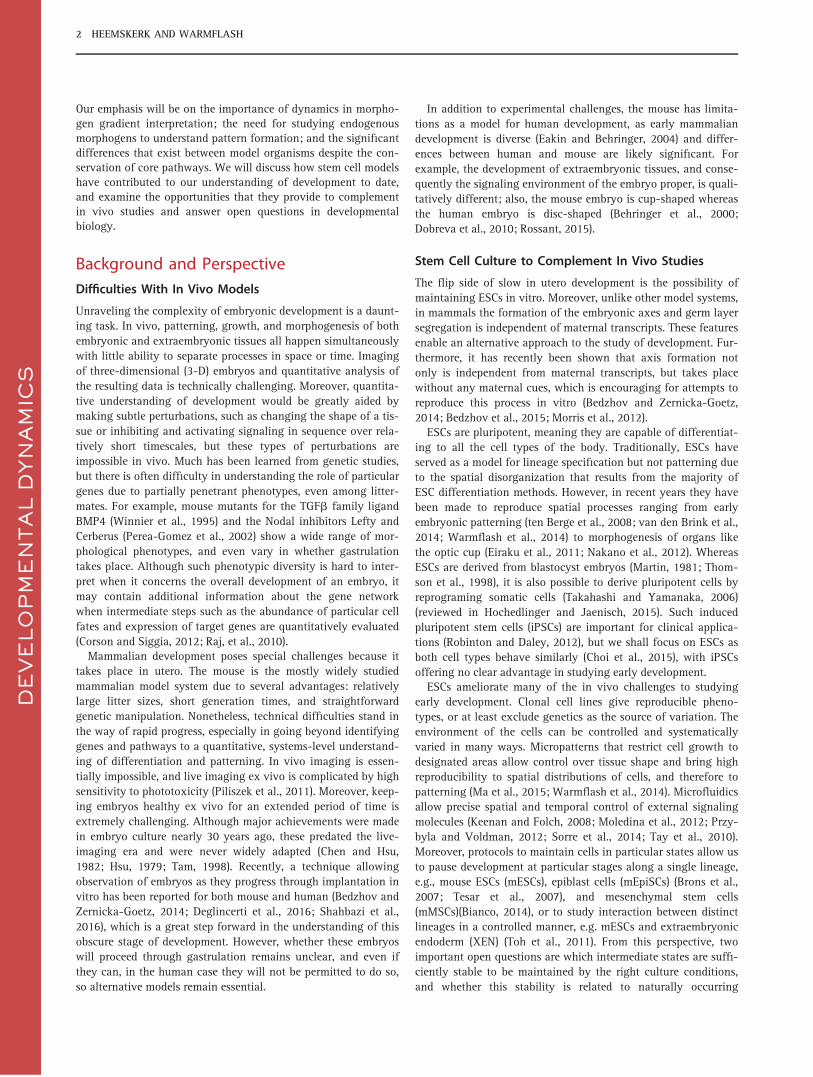
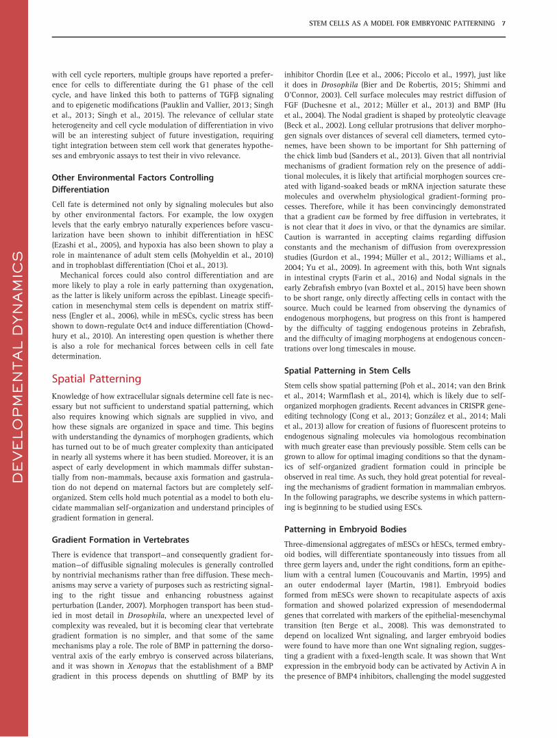
Figure 1 shows a theoretical example of how interpretation ofsignaling dynamics can play a fundamental role in pattern for-mation. Cells move across a morphogen gradient at differentspeeds. Three scenarios for the relation between pathway signal-ing and morphogen dynamics are explored: direct response toconcentration, temporal integration of ligand concentration, andresponse to rate of increase (adaptation). For simplicity, cell fateis assumed to be determined by thresholding the maximal signal-ing level in each case. The result differs from the French flag
model in two important ways: First, although pattern formationrequires a morphogen gradient, cell fates are patterned orthogo-nal to the gradient. Second, level-dependent response does notlead to spatial patterning, whereas the other two scenarios lead topatterns that are inverted relative to each other, with ligand inte-gration leading to the highest level of signaling in slow-movingcells, and response to ligand rate of increase leading to the high-est signaling in fast-moving cells. The example illustrates theeffect of cells moving relative to static morphogen levels, whichmay be relevant to the movement of mesendodermal precursorspast the organizer in various organisms. However, it is anextreme case chosen for its simplicity, and examples with staticcells and changing morphogen levels, or combinations of variousscenarios, can be constructed equally well. The general point isthat dynamics has the potential to uncouple both the axis of pat-terning and the arrangement of fates from the na€ıve expectationbased on the instantaneous morphogen gradient.
The importance of information processing at the level of thesignaling response to ligand was demonstrated for a number ofpathways in a handful of experimental systems. Although notdirectly relevant to early development, signaling dynamics areperhaps best understood for the NFkB pathway, where cell cultureexperiments have shown a complex relationship between ligandpresentation, signaling response, and target gene activation. Afast transcriptional feedback on the nuclear localization of thesignal transducer leads to an initial burst of signaling followedby a slower, possibly oscillatory phase (Hoffmann, 2002; Nelsonet al., 2004; Tay et al., 2010; Turner et al., 2010), and each ofthese responses may be important for the induction of particulargenes. Some transcriptional targets depend on sustained signal-ing at high doses, whereas others are transcribed with similaramplitude and finite duration regardless of the concentration andtemporal profile of stimulation (Hoffmann, 2002; Tay et al.,2010).
For the TGFb pathway, it was shown in C2C12 myoblasts(murine muscle progenitor cells) that the nuclear localization ofthe signal transducer Smad2 was sustained, while the nuclearlocalization of the co-transducer Smad4 was adaptive (Sorreet al., 2014; Warmflash et al., 2012b). There, too, adaptationdepends on a transcriptional feedback. Transcriptional dynamicsof many target genes follow those of Smad4 localization, and dif-ferentiation into myotubes was more effective with pulsed ratherthan sustained stimulation, consistent with adaptive but not sus-tained dynamics controlling differentiation. Adaptation in TGFb
signaling is also potentially consistent with the model thatgrowth in the Drosophila wing disc depends on the rate ofincrease of Dpp signaling (the Drosophila homologue of theTGFb superfamily member BMP4) (Wartlick et al., 2011); howev-er, this suggestion remains controversial (Harmansa et al., 2015).In vivo, an adaptive response, i.e., gradual desensitization, toSonic Hedgehog (Shh) was observed in patterning the chick neu-ral tube (Dessaud et al., 2007) through a negative feedback thatinduces transcription of the Shh inhibitor Patched-1.
Signaling Dynamics in Early Development
In Zebrafish, patterns of activation of both the TGFb transducerSmad2 (Dubrulle et al., 2015; Harvey and Smith, 2009) and anShh signaling reporter (Xiong et al., 2013) have been followed inindividual cells. However, in vivo, ligand levels cannot be con-trolled precisely or dynamically, and direct interpretation of an
DE
VE
LO
PM
EN
TA
L D
YN
AM
ICS
4 HEEMSKERK AND WARMFLASH
applied morphogen cannot be separated from response to endog-enous signaling. In mammalian systems, observing the dynamicsof signal production and transduction is much more challenging,and little work has been done quantifying signaling, morphogendistributions, and cell fate. Stem cells not only simplify under-standing differentiation, they may ultimately be the only systemthat allows the degree of control required to relate dynamics ofligand presentation to cell fate. For example, given that bothBMP and Nodal are TGFb family members, it is entirely possiblethat germ layer specification, like myoblast differentiation,depends on adaptive dynamics. It is now feasible to evaluate this
possibility using embryonic stem cells exposed to changingligand levels controlled by microfluidics.
Of course, it then remains to be determined which liganddynamics cells are actually exposed to during embryonic pattern-ing, and whether, for example, pulsed input has any in vivo rele-vance. We will return to this topic in the next section. However,regardless of in vivo relevance, it is of great value to know themost effective way to differentiate cells to a particular fate, eitherfor studying more advanced stages of development or for thera-peutic purposes. Current protocols for obtaining particular cellfates coarsely recapitulate the signaling environment known to
DE
VE
LO
PM
EN
TA
L D
YN
AM
ICS
Fig. 1. Importance of signaling dynamics for spatial patterning. Toy model demonstrating the dramatic effect signaling dynamics can have onpattern formation. For simplicity, cells move relative to a static morphogen gradient, and cell fate is assumed to be determined by the signalingmaximum in one of three possibilities for the relation between ligand dynamics and signaling response. A: Morphogen gradient from high (purple)to low (white) along Y direction, velocity field of cell trajectories (black) with two trajectories singled out (magenta and cyan). B: Signaling responsealong the cyan and magenta trajectories for three scenarios: response to ligand level (blue), response to increases in ligand (red), and response totime integral of ligand (green). Dashed lines indicate signaling maximum; note reversed positions of integral and increase response for the two tra-jectories. C: Maximal signaling response along X direction at late times for different scenarios. The maximal level for the trajectories in A and Bare indicated by cyan and magenta squares (ligand integration) and circles (ligand increase). D: Resultant pattern for all three scenarios requiresmorphogen gradient but is orthogonal to it, instead following the velocity gradient. Integral and increased response lead to opposite patterns,while concentration-dependent response produces no pattern at all.
STEM CELLS AS A MODEL FOR EMBRYONIC PATTERNING 5
achieve those cell fates in vivo (Chambers et al., 2009; McLeanet al., 2007; Mendjan et al., 2014). Lack of in vivo understandingchallenges the ability to obtain differentiated cell types at higherresolution, e.g., to consistently obtain lateral plate mesoderm. Forfurther progress, differentiation protocols cannot rely only on invivo studies. Instead, systematic study of stem cell responses indifferent environments should shed light on in vivo patterningand simultaneously allow for rational protocol design.
Gene Regulation and Cell Fate
Recent quantitative work in Zebrafish showed differentialresponse of target genes to signaling. In particular, transcriptionrates of target genes were shown to correlate with the ligand levelrequired to induce them stably (Dubrulle et al., 2015). In thisstudy, the complexity of the embryonic system was reduced byknocking down the genes for endogenous Nodal ligands andinjecting recombinant Nodal protein into the extraembryonicspace of the embryo, resulting in uniform exposure of the cells tothe supplied morphogen. In mammals, studies such as thesewould be much easier to perform in vitro with stem cells andwould provide similar information about pattern formation.Moreover, in cell culture, the activity of signaling reporters ofvarious pathways (Ferrer-Vaquer et al., 2010; Sorre et al., 2014),as well as reporters of target genes, can be followed in individualcells as the dynamics of morphogen stimulus are varied, allowinga more complete picture of the link between signaling dynamicsand the regulation of direct targets.
In vivo, the relation between signaling and cell fate is bestunderstood for Shh in the vertebrate neural tube (Cohen et al.,2013). In this system, it was shown that regulatory interactionsbetween Shh targets are essential for “thresholding” the signaland establishing coherent gene expression domains (Balaskaset al., 2012). Similar mechanisms were suggested for interpretingActivin levels in the Xenopus embryo (Saka and Smith, 2007)and are almost certainly involved in interpreting othermorphogens.
A great deal of progress has been made in understanding cellfate decisions at the level of the gene regulatory networks down-stream of signaling using stem cells. Small molecule interferencewith signaling, RNAi of particular genes, and, with the advent ofCRISPR technology, gene knockouts are more rapid and straight-forward to perform in stem cells than in developmental modelsystems. Furthermore, while interpreting whole body phenotypesis an advantage of animal systems, assessing signaling activity,gene expression, and cell fate proportions is performed muchmore easily and quantitatively using stem cells.
As one example, the gene regulatory network controllinghuman primitive streak (mesodermal and endodermal) fates isbeing unraveled using embryonic stem cells. Teo et al. establishedthe hierarchy of transcription factors in endoderm differentiation(Teo et al., 2011): The pluripotency-associated gene Nanog isessential for inducing Eomes early during differentiation, andEomes subsequently combines with Activin/Nodal signaling toinduce a network governing endoderm differentiation. Mendjanet al. (Mendjan et al., 2014) revealed how anterior and posteriorstreak fates depend on mutually repressing transcription factorsCdx2 and Nanog. Both studies were able to establish linksbetween the networks of transcription factors and key upstreamdevelopmental signals Activin/Nodal, BMP, and Wnt. Further-more, both studies checked consistency of their findings with
expression patterns in mouse, demonstrating the power of thisapproach for understanding development.
Differentiation vs. Pluripotency
In non-mammalian species, it is not known if a stable pluripotentstate exists or if differentiation is an unavoidable consequence ofthe onset of zygotic transcription. However, in mammals, differ-entiation requires destabilizing the pluripotent state. As such,understanding pluripotency is complementary to, and perhapsrequired for, understanding differentiation. Paradoxically, manyof the same factors that control differentiation also maintain plu-ripotency. Pluripotency in hESCs can be maintained by FGF andActivin (James, 2005; Vallier et al., 2005). In mESCs, BMP, LIF,and Wnt all play a role in maintaining the pluripotent state (tenBerge et al., 2011; Smith et al., 1988; Ying et al., 2003). These dif-ferences between mouse and human ESCs most likely result fromthe fact that mESCs model the ICM, whereas hESCs model theepiblast.
It has now become clear that the transcription factors essentialto maintain pluripotency are also involved in differentiation.Until recently, pluripotency was considered a stable state main-tained by a network of dedicated transcription factors in theabsence of destabilizing external signals. This was supported bythe maintenance of mESCs in conditions named 2i or 3i (Yinget al., 2008), ostensibly blocking all differentiation-promotingsignals. However, the core transcription factors Sox2, Oct4, andNanog are, respectively, required for differentiation toward ecto-derm, mesoderm, and endoderm (Malleshaiah et al., 2016; Teoet al., 2011; Thomson et al., 2011; Wang et al., 2012). Moreover,their overexpression can induce differentiation, as opposed towhat would be expected from factors simply blocking differentia-tion toward particular lineages. Pluripotency therefore seems tobe a delicate equilibrium between mutually inhibitory lineagespecifiers (Loh and Lim, 2011). This perhaps explains why thesame signaling pathways can both stabilize pluripotency andpromote differentiation. Further supporting this picture, lineagespecifiers can substitute for pluripotency-associated transcriptionfactors in reprograming differentiated cells to the pluripotentstate (Shu et al., 2013). This picture is complicated, however, bythe demonstration that combinatorial interactions between path-ways are important for influencing the balance between pluripo-tency and differentiation. It is not only the levels of Activin/Nodal signaling, but the state of the PI3K pathway that deter-mines whether Nodal signaling supports pluripotency or differen-tiation. When PI3K is activated, Nodal maintains pluripotency;but when it is inhibited, Nodal directs differentiation, and theseeffects are mediated by interactions between PI3K and the Wntand MAPK pathways (McLean et al., 2007; Singh et al., 2012).Consequently, nearly all modern protocols for endoderm differ-entiation include modulation of a second pathway in addition toActivin/Nodal (Pagliuca et al., 2014; Rezania et al., 2014; Tou-boul et al., 2010).
These in vitro studies elucidating the relationship between plu-ripotency and differentiation can also shed light on how thistransition occurs in the embryo; they would have been extremelydifficult to perform in vivo because, in that case, pluripotency isa transient state. Recently, the ability to stabilize cellular stateshas also been used to uncover the heterogeneity in those states(Hough et al., 2014; Singer et al., 2014) (earlier work reviewed inTorres-Padilla and Chambers, 2014). Finally, using stem cells
DE
VE
LO
PM
EN
TA
L D
YN
AM
ICS
6 HEEMSKERK AND WARMFLASH
with cell cycle reporters, multiple groups have reported a prefer-ence for cells to differentiate during the G1 phase of the cellcycle, and have linked this both to patterns of TGFb signalingand to epigenetic modifications (Pauklin and Vallier, 2013; Singhet al., 2013; Singh et al., 2015). The relevance of cellular stateheterogeneity and cell cycle modulation of differentiation in vivowill be an interesting subject of future investigation, requiringtight integration between stem cell work that generates hypothe-ses and embryonic assays to test their in vivo relevance.
Other Environmental Factors ControllingDifferentiation
Cell fate is determined not only by signaling molecules but alsoby other environmental factors. For example, the low oxygenlevels that the early embryo naturally experiences before vascu-larization have been shown to inhibit differentiation in hESC(Ezashi et al., 2005), and hypoxia has also been shown to play arole in maintenance of adult stem cells (Mohyeldin et al., 2010)and in trophoblast differentiation (Choi et al., 2013).
Mechanical forces could also control differentiation and aremore likely to play a role in early patterning than oxygenation,as the latter is likely uniform across the epiblast. Lineage specifi-cation in mesenchymal stem cells is dependent on matrix stiff-ness (Engler et al., 2006), while in mESCs, cyclic stress has beenshown to down-regulate Oct4 and induce differentiation (Chowd-hury et al., 2010). An interesting open question is whether thereis also a role for mechanical forces between cells in cell fatedetermination.
Spatial Patterning
Knowledge of how extracellular signals determine cell fate is nec-essary but not sufficient to understand spatial patterning, whichalso requires knowing which signals are supplied in vivo, andhow these signals are organized in space and time. This beginswith understanding the dynamics of morphogen gradients, whichhas turned out to be of much greater complexity than anticipatedin nearly all systems where it has been studied. Moreover, it is anaspect of early development in which mammals differ substan-tially from non-mammals, because axis formation and gastrula-tion do not depend on maternal factors but are completely self-organized. Stem cells hold much potential as a model to both elu-cidate mammalian self-organization and understand principles ofgradient formation in general.
Gradient Formation in Vertebrates
There is evidence that transport—and consequently gradient for-mation—of diffusible signaling molecules is generally controlledby nontrivial mechanisms rather than free diffusion. These mech-anisms may serve a variety of purposes such as restricting signal-ing to the right tissue and enhancing robustness againstperturbation (Lander, 2007). Morphogen transport has been stud-ied in most detail in Drosophila, where an unexpected level ofcomplexity was revealed, but it is becoming clear that vertebrategradient formation is no simpler, and that some of the samemechanisms play a role. The role of BMP in patterning the dorso-ventral axis of the early embryo is conserved across bilaterians,and it was shown in Xenopus that the establishment of a BMPgradient in this process depends on shuttling of BMP by its
inhibitor Chordin (Lee et al., 2006; Piccolo et al., 1997), just likeit does in Drosophila (Bier and De Robertis, 2015; Shimmi andO’Connor, 2003). Cell surface molecules may restrict diffusion ofFGF (Duchesne et al., 2012; M€uller et al., 2013) and BMP (Huet al., 2004). The Nodal gradient is shaped by proteolytic cleavage(Beck et al., 2002). Long cellular protrusions that deliver morpho-gen signals over distances of several cell diameters, termed cyto-nemes, have been shown to be important for Shh patterning ofthe chick limb bud (Sanders et al., 2013). Given that all nontrivialmechanisms of gradient formation rely on the presence of addi-tional molecules, it is likely that artificial morphogen sources cre-ated with ligand-soaked beads or mRNA injection saturate thesemolecules and overwhelm physiological gradient-forming pro-cesses. Therefore, while it has been convincingly demonstratedthat a gradient can be formed by free diffusion in vertebrates, itis not clear that it does in vivo, or that the dynamics are similar.Caution is warranted in accepting claims regarding diffusionconstants and the mechanism of diffusion from overexpressionstudies (Gurdon et al., 1994; M€uller et al., 2012; Williams et al.,2004; Yu et al., 2009). In agreement with this, both Wnt signalsin intestinal crypts (Farin et al., 2016) and Nodal signals in theearly Zebrafish embryo (van Boxtel et al., 2015) have been shownto be short range, only directly affecting cells in contact with thesource. Much could be learned from observing the dynamics ofendogenous morphogens, but progress on this front is hamperedby the difficulty of tagging endogenous proteins in Zebrafish,and the difficulty of imaging morphogens at endogenous concen-trations over long timescales in mouse.
Spatial Patterning in Stem Cells
Stem cells show spatial patterning (Poh et al., 2014; van den Brinket al., 2014; Warmflash et al., 2014), which is likely due to self-organized morphogen gradients. Recent advances in CRISPR gene-editing technology (Cong et al., 2013; Gonz�alez et al., 2014; Maliet al., 2013) allow for creation of fusions of fluorescent proteins toendogenous signaling molecules via homologous recombinationwith much greater ease than previously possible. Stem cells can begrown to allow for optimal imaging conditions so that the dynam-ics of self-organized gradient formation could in principle beobserved in real time. As such, they hold great potential for reveal-ing the mechanisms of gradient formation in mammalian embryos.In the following paragraphs, we describe systems in which pattern-ing is beginning to be studied using ESCs.
Patterning in Embryoid Bodies
Three-dimensional aggregates of mESCs or hESCs, termed embry-oid bodies, will differentiate spontaneously into tissues from allthree germ layers and, under the right conditions, form an epithe-lium with a central lumen (Coucouvanis and Martin, 1995) andan outer endodermal layer (Martin, 1981). Embryoid bodiesformed from mESCs were shown to recapitulate aspects of axisformation and showed polarized expression of mesendodermalgenes that correlated with markers of the epithelial-mesenchymaltransition (ten Berge et al., 2008). This was demonstrated todepend on localized Wnt signaling, and larger embryoid bodieswere found to have more than one Wnt signaling region, sugges-ting a gradient with a fixed-length scale. It was shown that Wntexpression in the embryoid body can be activated by Activin A inthe presence of BMP4 inhibitors, challenging the model suggested
DE
VE
LO
PM
EN
TA
L D
YN
AM
ICS
STEM CELLS AS A MODEL FOR EMBRYONIC PATTERNING 7
by in vivo data, according to which Wnt depends strictly on BMP(Ben-Haim et al., 2006), and suggesting the possibility that the invitro Wnt gradient is formed by interaction with a Nodal gradi-ent. A recent study made further advances by introducing adefined protocol to consistently produce patterned aggregates ofmESCs. This study also demonstrated that aggregates of a partic-ular size will both pattern and change shape to elongate alongthe axis of polarization (van den Brink et al., 2014). Similar self-organization and patterning have also been observed in P19embryonic carcinoma cells (Li and Marikawa, 2015; Marikawaet al., 2009).
The facts that embryoid bodies show features of patterning andmorphogenesis associated with gastrulation, and have alreadyrefined in vivo findings, show their potential as a model system.However, they also suffer from a number of limitations. Theirfeatures are not sufficiently reproducible to allow for quantitativecomparison between gene expression patterns in different embry-oid bodies, which makes it difficult to construct a detailed modelfor spatial pattern formation. Moreover, they are too large andopaque for in toto live imaging, and high-quality data requirefixation followed by optical clearing or cryosectioning.
Biologically, there are both pros and cons to using embryoidbodies as a model for gastrulation. The epiblast is a well-definedepithelial monolayer that is topologically a disc (as opposed togeometrically, i.e., it is a monolayer with a single connectedboundary) and receives signals from extraembryonic tissues at itsboundary and on its basal side. Embryoid bodies are able to formstructures that resemble the double-layered epithelium found invivo, but they do so slowly and incompletely, through a mecha-nism different from that of the embryo in vivo (Bedzhov andZernicka-Goetz, 2014), and result in a spherical topology. Undermany culture conditions, they simply remain solid 3-D aggre-gates. Therefore, two-dimensional (2-D) colonies can be arguedto more faithfully model the epiblast, as they form epithelialmonolayers and are topological discs.
Spatial Heterogeneity in 2-D hESC Colonies
When grown in two dimensions, removal of pluripotency factorsor treatment with BMP4 triggers differentiation to both embryon-ic and extraembryonic tissues in an apparently random fashion.This random differentiation is a consequence of endogenous gra-dients of morphogens and inhibitors that depend on random col-ony shape and density variations. One of the first studies toinvestigate endogenous signaling gradients in stem cell coloniesexamined differentiation in hESCs upon removal ofpluripotency-maintaining factors from the media (Peerani et al.,2007). Under these conditions, the cells differentiated to XENinhomogeneously with a local resistance to differentiation thatdepended on cell density. This resistance resulted from the secre-tion of the BMP inhibitor GDF3 by pluripotent cells, counteract-ing the differentiation signal from BMP2 secreted by all cells and,to a larger degree, the differentiated ones. By directly manipulat-ing colony size, and therefore average density, using micropat-terning, the authors demonstrated an inverse relationshipbetween density and likelihood of differentiation.
This study elegantly showed how quantitative analysis of evendisorganized stem cell colonies can provide information aboutthe mechanisms behind spatial organization, and how the levelof environmental control that is only possible in vitro can morerigorously test relationships between variables that are hard to
manipulate or separate in vivo. Quantitative analysis and repro-ducibility are key in understanding self-organization, because itis a highly nonlinear process with feedback both between signal-ing pathways and between signaling, cell density, and colonygeometry. Only by systematically varying one variable at a timeand evaluating the effects on gradient formation and patterningcan we hope to understand the underlying mechanisms.
Reproducible Spatial Differentiationon Micropatterned Surfaces
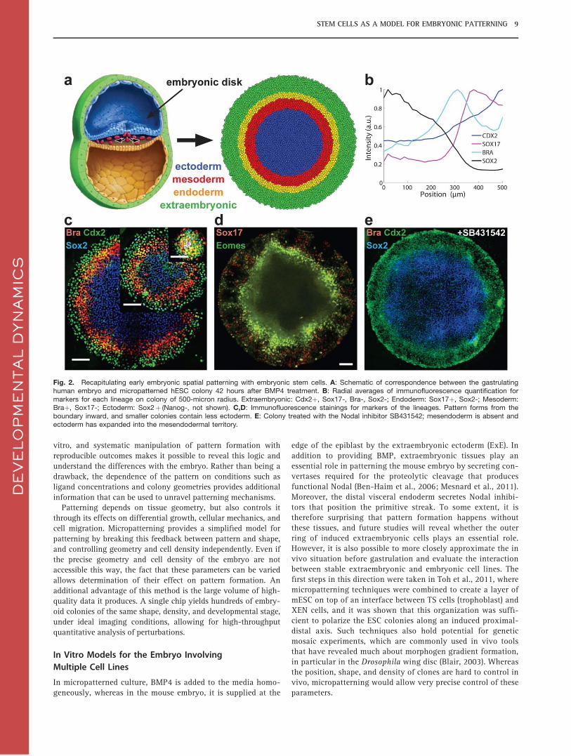
While the colonies in Peerani et al., 2007 showed a variety of cellfates, no reproducible pattern was formed. Recently it was foundby one of the authors that hESCs grown in circular colonies usingmicropatterned surfaces show reproducible spatial differentiationupon treatment with BMP4 (Warmflash et al., 2014). Cells posi-tive for markers of all three germ layers as well as extraembryon-ic tissue form concentric rings at particular radii in the colony(Fig. 2). Micropatterned surfaces contain defined regions of extra-cellular matrix proteins such as laminin or matrigel typically sur-rounded by a coating that prevents cell and protein adhesion,such as PLL-PEG. A number of techniques are available to pro-duce the surfaces, including photolithography (Azioune et al.,2009; Ma et al., 2015; Warmflash et al., 2014), microcontactprinting (Peerani et al., 2007; Th�ery and Piel, 2009), and stencilmicropatterning (Toh et al., 2011; Xing et al., 2015). These meth-ods were originally applied to study the biophysics of cell shapeand cell division (Th�ery et al., 2006; Th�ery et al. 2007) and weresubsequently adapted to study multicellular, colony-level events.
Although BMP4 is administered homogeneously in the experi-ments of Warmflash et al., 2014, the layout of cell fates appearsto be controlled by spontaneously formed gradients of BMPinhibitors and Nodal. Modulation of pathway activity with RNAiand small molecule inhibitors, as well as immunofluorescenceanalysis for activated signal transducers, revealed both a gradedresponse to the initial BMP stimulation and a Nodal activity gra-dient. These two signals are responsible for differentiation toextraembryonic and mesendodermal cell fates, respectively. Simi-lar to the embryo, this signaling gradient relied on secreted inhib-itors of Nodal and BMP, as patterning was lost upon knockdownof these inhibitors.
The length scale of the germ layer rings is independent of colo-ny size and the pattern forms from the boundary inward, assmaller colonies lose inner cell fates (Fig. 2). Such a fixed-lengthscale cannot be obtained by uniform production and degradationcombined with free diffusion, and implies a nontrivial mecha-nism for gradient formation, reaffirming the need for studyingendogenous morphogen gradients. Although the mechanismresponsible for this length scale is not yet known, its existenceprovides a hypothesis regarding the results of earlier work relat-ing colony size to cell fate. In Lee et al., 2009, micropatterningwas used to relate colony size to mesendoderm differentiation,and it was found that smaller colonies end up with higher frac-tions of endoderm relative to mesoderm. Evaluating the spatialpatterns shows that this effect was likely due to the relative posi-tions of these fates in the colony, as the more central-lying meso-derm is lost in smaller colonies.
The organization of cell fates into rings, rather than layers, dif-fers from the in vivo situation, and the cause of this remains tobe identified. However, all evidence indicates that the same regu-latory logic as in the embryo controls spatial organization in
DE
VE
LO
PM
EN
TA
L D
YN
AM
ICS
8 HEEMSKERK AND WARMFLASH
vitro, and systematic manipulation of pattern formation withreproducible outcomes makes it possible to reveal this logic andunderstand the differences with the embryo. Rather than being adrawback, the dependence of the pattern on conditions such asligand concentrations and colony geometries provides additionalinformation that can be used to unravel patterning mechanisms.
Patterning depends on tissue geometry, but also controls itthrough its effects on differential growth, cellular mechanics, andcell migration. Micropatterning provides a simplified model forpatterning by breaking this feedback between pattern and shape,and controlling geometry and cell density independently. Even ifthe precise geometry and cell density of the embryo are notaccessible this way, the fact that these parameters can be variedallows determination of their effect on pattern formation. Anadditional advantage of this method is the large volume of high-quality data it produces. A single chip yields hundreds of embry-oid colonies of the same shape, density, and developmental stage,under ideal imaging conditions, allowing for high-throughputquantitative analysis of perturbations.
In Vitro Models for the Embryo InvolvingMultiple Cell Lines
In micropatterned culture, BMP4 is added to the media homo-geneously, whereas in the mouse embryo, it is supplied at the
edge of the epiblast by the extraembryonic ectoderm (ExE). Inaddition to providing BMP, extraembryonic tissues play anessential role in patterning the mouse embryo by secreting con-vertases required for the proteolytic cleavage that producesfunctional Nodal (Ben-Haim et al., 2006; Mesnard et al., 2011).Moreover, the distal visceral endoderm secretes Nodal inhibi-tors that position the primitive streak. To some extent, it istherefore surprising that pattern formation happens withoutthese tissues, and future studies will reveal whether the outerring of induced extraembryonic cells plays an essential role.However, it is also possible to more closely approximate the invivo situation before gastrulation and evaluate the interactionbetween stable extraembryonic and embryonic cell lines. Thefirst steps in this direction were taken in Toh et al., 2011, wheremicropatterning techniques were combined to create a layer ofmESC on top of an interface between TS cells (trophoblast) andXEN cells, and it was shown that this organization was suffi-cient to polarize the ESC colonies along an induced proximal-distal axis. Such techniques also hold potential for geneticmosaic experiments, which are commonly used in vivo toolsthat have revealed much about morphogen gradient formation,in particular in the Drosophila wing disc (Blair, 2003). Whereasthe position, shape, and density of clones are hard to control invivo, micropatterning would allow very precise control of theseparameters.
DE
VE
LO
PM
EN
TA
L D
YN
AM
ICS
Fig. 2. Recapitulating early embryonic spatial patterning with embryonic stem cells. A: Schematic of correspondence between the gastrulatinghuman embryo and micropatterned hESC colony 42 hours after BMP4 treatment. B: Radial averages of immunofluorescence quantification formarkers for each lineage on colony of 500-micron radius. Extraembryonic: Cdx2þ, Sox17-, Bra-, Sox2-; Endoderm: Sox17þ, Sox2-; Mesoderm:Braþ, Sox17-; Ectoderm: Sox2þ (Nanog-, not shown). C,D: Immunofluorescence stainings for markers of the lineages. Pattern forms from theboundary inward, and smaller colonies contain less ectoderm. E: Colony treated with the Nodal inhibitor SB431542; mesendoderm is absent andectoderm has expanded into the mesendodermal territory.
STEM CELLS AS A MODEL FOR EMBRYONIC PATTERNING 9
Interspecies Differences
So far we have focused on the experimental strengths of stemcells as a model for early development and compared stem cellsto vertebrate in vivo systems including fish and frog. However,gastrulation differs substantially both between mammalian andnon-mammalian systems and between different mammals. Assuch, even the mouse may not be an accurate model for humangastrulation. Stem cells provide an opportunity to study humandevelopment and determine interspecies differences. We brieflyreview some of the known interspecies differences in gastrulationand, in particular, differences in the roles of conserved morpho-gens. This discussion is not intended to be complete, but serves toillustrate the extent of the dissimilarities. We then discuss howhESCs may help in better understanding uniquely human aspectsof embryogenesis.
Mouse vs. Frog
BMP has a conserved function in dorso-ventral and neural pat-terning across bilaterians, although the axis is inverted in verte-brates relative to arthropods (Bier and De Robertis, 2015; Holleyand Ferguson, 1997). Even though parts of the mechanism ofdorso-ventral gradient formation are conserved, both the func-tion and formation of the BMP gradient differ significantlybetween mouse and frog. In frog, BMPs are initially expressedthroughout the ectodermal and mesodermal parts of the embryo(Gilbert, 2014). In contrast, mouse BMP is not produced inembryonic cells: BMP2 is secreted from the VE and BMP4 fromthe ExE (Arnold and Robertson, 2009; Madabhushi and Lacy,2011; Winnier et al., 1995). Nodal, on the other hand, is broadlyexpressed in the mammalian embryo but restricted to a particulardomain in frog. In mouse, it is initially expressed throughout theepiblast and is required to maintain pluripotency, but in frog it isrestricted to the dorsal-vegetal side. Moreover, BMP is required inmouse to establish a Nodal gradient and initiate gastrulation(Mishina et al., 1995; Winnier et al., 1995) through a feedbackloop that also involves Wnt, whereas in frog, Nodal is activatedby VegT, a maternal gene with no mammalian ortholog. Overex-pression of BMPs in frog will cause cells to adopt progressivelymore ventral fates but will not convert cells between germ layers(Wilson et al., 1997), whereas in mammalian stem cells, BMPtreatment leads to conversion from pluripotency to both extraem-bryonic and mesodermal lineages, although this is likely throughfeedback loops with other pathways (Bernardo et al., 2011; Xuet al., 2002; Yu et al., 2011). Nodal signaling appears to inducemesendodermal fates in both species; however, the dose-dependent patterning of mesendoderm by Nodal (Green et al.,1992) has not been directly demonstrated in mammals.
Differences in Mammalian Developmentand Stem Cells
Many of the differences between mouse and frog involve theextraembryonic tissues that are absent in anamniotes [althoughequivalencies between the frog embryo and particular extraem-bryonic tissues may still exist (Beddington and Robertson, 1998)].It is precisely in the development of the extraembryonic tissuesthat mammalian species differ considerably. During the blasto-cyst stage, differences in the timing of lineage specification havebeen found: For example, a delay in expression of the
trophectodermal marker Cdx2 (Niakan and Eggan, 2013) and theabsence of Eomes expression in human trophectoderm (Blakeleyet al., 2015). The fact that it has been possible to derive stable celllines for all three lineages of the blastocyst in mouse but not inhuman is another indication that the very first cell fate decisionsmay be different. Immediately after the blastocyst stage, not onlymolecular but anatomical differences arise. It appears that thehuman amnion and extraembryonic mesoderm both form wellbefore gastrulation (Dobreva et al., 2010; Enders et al., 1986), incontrast to mouse, where these derive from the primitive streak.As a consequence, the human trophectoderm is separated fromthe epiblast by several tissue layers and a substantial distance bythe onset of gastrulation. Considering the importance of ExE-derived signals for early pattering in mouse, this observationraises the question of what takes the place of the feedback loopsbetween the ExE and epiblast in human.
In the context of these findings, it is particularly interesting todetermine which extraembryonic cell types hESCs can differenti-ate into, and what role they play in in vitro self-organization. If,as current data suggest, hESCs can differentiate into both tro-phectoderm and primitive endoderm, the genetic networkinvolved in these cell fate decisions can be quantitatively dissect-ed using the tools we discussed in previous sections. Moreover,the behavior of hESCs can be compared to that of mESCs ormEpiSCs under identical conditions to determine species differ-ences in molecular processes that are independent of embryonicgeometry or the relationship to extraembryonic lineages. Thevariety of readouts available in the stem cell platform can beused to straightforwardly determine whether different behavior isdue to signal input or response, e.g., whether a delay in Cdx2expression reflects a delay in cell signaling or in competence torespond. More generally, this opens up the possibility of a newkind of in vitro comparative embryology in which behavior ofembryonic cells from different species can be rigorously andquantitatively compared.
One of the most striking differences between human andmouse embryos is the geometry (but not topology) of the epiblast(Behringer et al., 2000). Whereas the mouse embryo is cup-shaped, the human embryo, like most mammalian embryos, isdisc-shaped, and it is not known how the shape of the pregastru-lation embryo is controlled or how it affects later events. Usingmicropatterning technology, one can grow human and mousestem cells in a variety of geometries to determine how shapeaffects pattern formation and how the self-organization of mor-phogen gradients in each species has adapted to a particulargeometry.
Discussion and Conclusion
Differentiation Outlook
Substantial evidence shows the importance of ligand and signal-ing dynamics in determining cell fate. Unraveling this relation-ship requires precise control of the dynamic environment, real-time imaging of signaling reporters and fate outcomes, and quan-titative analysis of the resulting data. In contrast to in vivo sys-tems, ESCs provide unparalleled control to probe the signalingdynamics and gene regulatory networks controlling early cellfate decisions. The largest conceptual challenge with thisapproach is to relate the lessons learned back to embryo. It isoften not clear which aspects of the embryonic environment need
DE
VE
LO
PM
EN
TA
L D
YN
AM
ICS
10 HEEMSKERK AND WARMFLASH
to be reproduced in vitro, and conversely, the physiological rele-vance of in vitro states can be difficult to discern. What is the rel-evance of the method of growing mESCs in a so-called “groundstate” (Ying et al., 2008)? Can the conversion of hESCs to tro-phectoderm by BMP4 treatment (Li and Parast, 2014; Xu et al.,2002) shed light on human extraembryonic development in vivo?The important differences in the role of BMP and Wnt signalingbetween mESC and mEpiScs and their in vivo correlates highlightthe need for caution when applying in vitro findings to theembryo (Biechele et al., 2013; Tang et al., 2010). Nonetheless,stem cells may offer the only opportunity to quantitatively mapthe relationship between external stimulus and fate. A combina-tion of careful evaluation of these relationships and confirmationof resulting hypotheses in vivo should ultimately allow us tounderstand how cell fate decisions in the embryo are shaped.
Spatial Patterning Outlook
Although still an emerging model, spatially controlled stem cellcolonies show great promise in revealing the mechanisms ofembryonic patterning and, more generally, in understanding theformation of self-organized morphogen gradients. The in vitroenvironment in current models differs substantially from the invivo environment and likely is the cause of the differences inspatial organization, but uncovering the underlying logic stillprovides essential information about the in vivo mechanism. Agap in the current approach is that results with hESCs cannot becompared directly to human embryos due to ethical and practicallimitations. Differences between hESC colonies and other mam-malian embryos could be due to culture artifacts or interspeciesdifferences. Comparing in vivo and in vitro patterning in mousecould clarify these issues by revealing the extent and nature ofculture artifacts. More generally, comparison to embryos to verifyin vitro findings will remain necessary, but regardless of the con-nection to in vivo development, improved understanding ofendogenous signaling and spatial heterogeneity in stem cells willdirectly impact regenerative medicine.
A substantial challenge for stem cell models of early develop-ment is to more faithfully reproduce aspects of morphogenesis.The recent demonstrations that optic cups (Eiraku et al., 2011),“mini-guts” (Sato and Clevers, 2013), patterned neural tubes(Meinhardt et al., 2014), and cerebral organoids (Lancaster et al.,2013) can be grown in vitro shows the potential of cells to under-go morphogenetic movements outside of the embryo. As 3-D
culture moves forward, it will be of particular importance to cre-ate models that undergo patterning and morphogenesis in threedimensions but retain the reproducibility of the 2-D micropat-terning models. The extensive progress made to date in organoidcultures and the bioengineering approaches for generating andmanipulating them are beyond the scope of this commentary,and the reader is referred to recent reviews of these topics (Gjor-evski et al., 2014; Turner et al., 2016).
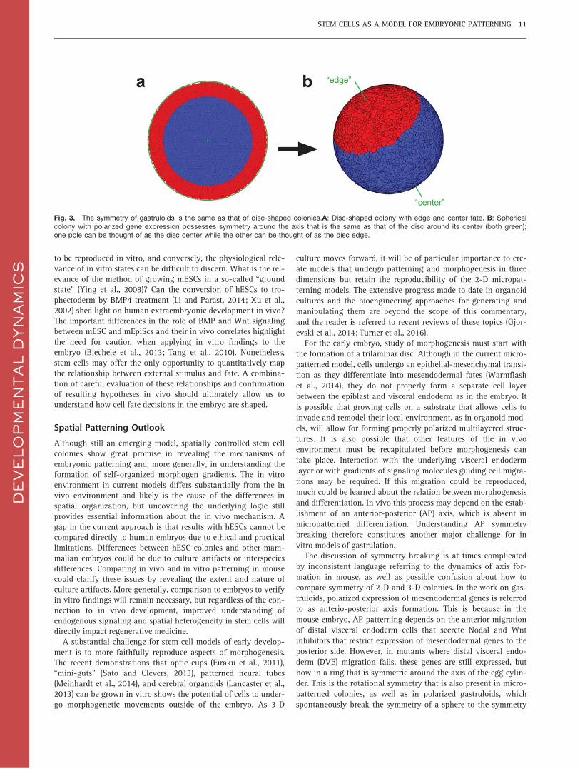
For the early embryo, study of morphogenesis must start withthe formation of a trilaminar disc. Although in the current micro-patterned model, cells undergo an epithelial-mesenchymal transi-tion as they differentiate into mesendodermal fates (Warmflashet al., 2014), they do not properly form a separate cell layerbetween the epiblast and visceral endoderm as in the embryo. Itis possible that growing cells on a substrate that allows cells toinvade and remodel their local environment, as in organoid mod-els, will allow for forming properly polarized multilayered struc-tures. It is also possible that other features of the in vivoenvironment must be recapitulated before morphogenesis cantake place. Interaction with the underlying visceral endodermlayer or with gradients of signaling molecules guiding cell migra-tions may be required. If this migration could be reproduced,much could be learned about the relation between morphogenesisand differentiation. In vivo this process may depend on the estab-lishment of an anterior-posterior (AP) axis, which is absent inmicropatterned differentiation. Understanding AP symmetrybreaking therefore constitutes another major challenge for invitro models of gastrulation.
The discussion of symmetry breaking is at times complicatedby inconsistent language referring to the dynamics of axis for-mation in mouse, as well as possible confusion about how tocompare symmetry of 2-D and 3-D colonies. In the work on gas-truloids, polarized expression of mesendodermal genes is referredto as anterio-posterior axis formation. This is because in themouse embryo, AP patterning depends on the anterior migrationof distal visceral endoderm cells that secrete Nodal and Wntinhibitors that restrict expression of mesendodermal genes to theposterior side. However, in mutants where distal visceral endo-derm (DVE) migration fails, these genes are still expressed, butnow in a ring that is symmetric around the axis of the egg cylin-der. This is the rotational symmetry that is also present in micro-patterned colonies, as well as in polarized gastruloids, whichspontaneously break the symmetry of a sphere to the symmetry
DE
VE
LO
PM
EN
TA
L D
YN
AM
ICS
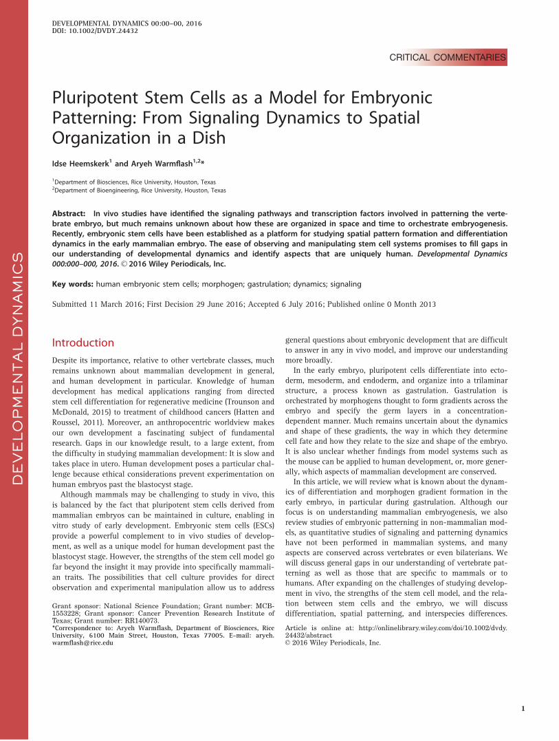
Fig. 3. The symmetry of gastruloids is the same as that of disc-shaped colonies.A: Disc-shaped colony with edge and center fate. B: Sphericalcolony with polarized gene expression possesses symmetry around the axis that is the same as that of the disc around its center (both green);one pole can be thought of as the disc center while the other can be thought of as the disc edge.
STEM CELLS AS A MODEL FOR EMBRYONIC PATTERNING 11
of a disc, as illustrated in Figure 3. When we refer to in vitro APaxis formation, we have in mind breaking rotational symmetry ofmicropatterned colonies to bilateral symmetry. Comparison ofmicropattern colonies to mouse mutants with failed DVE migra-tion could shed light on whether the defects in these models aresimilar (Migeotte et al., 2010; Nowotschin et al., 2013). Converse-ly, localized external supply of Nodal and Wnt inhibitors may besufficient to initiate AP patterning in stem cells. Current technol-ogies for patterning multiple cell types on surfaces (Toh et al.,2011) and delivering ligands with microfluidics (Cosson andLutolf, 2014; Wu et al., 2006) allow us to start testing thesehypotheses, and time will tell whether in vitro development canbe engineered to mimic the embryo. Of course, success in thisprogram raises ethical questions, and decisions must be reachedabout what experiments should be permitted with stem cell colo-nies that accurately mimic gastrulation and later stages (Peraet al., 2015).
Conclusion
Modern techniques in bioengineering, gene editing, and imaginghave now opened the door to using stem cells to address longintractable questions in developmental biology. Combining imag-ing of live cell reporters with exquisite control over the microen-vironment has the potential to reveal the relationship betweenthe cues cells receive and the fates they adopt. The ability of cellsto self-organize into patterns and morphologies in vitro willallow us to manipulate and study these processes with a resolu-tion that cannot be accessed in vivo. At all stages of this process,it is essential to use what is known about the embryo in vivo toguide the design of in vitro experiments, and to test the relevanceof the in vitro outcomes back in the embryo. By closing this loop,artificial stem cell systems have the potential to revolutionize ourunderstanding of patterning and morphogenesis in the embryo.
AcknowledgmentsThe authors thank Susana Chuva de Sousa Lopes for a helpful dis-cussion, and Daniel Wagner for careful reading of this article. Thiswork was supported Rice University.
ReferencesArnold SJ, Robertson EJ. 2009. Making a commitment: cell lineage
allocation and axis patterning in the early mouse embryo. NatRev Mol Cell Biol 10(2):91–103.
Azioune A, Storch M, Bornens M, Th�ery M, Piel M. 2009. Simpleand rapid process for single cell micro-patterning. Lab Chip9(11):1640–1642.
Balaskas N, Ribeiro A, Panovska J, Dessaud E, Sasai N, Page KM,et al. 2012. Gene regulatory logic for reading the Sonic Hedge-hog signaling gradient in the vertebrate neural tube. Cell 148(1-2):273–284.
Beck S, Le Good JA, Guzman M, Ben-Haim N, Roy K, BeermannF, Constam DB. 2002. Extraembryonic proteases regulate Nodalsignalling during gastrulation. Nat Cell Bio 4(12):981–985.
Beddington RS, Robertson EJ. 1998. Anterior patterning in mouse.Trends Genet 14(7):277–284.
Bedzhov I, Zernicka-Goetz M. 2014. Self-organizing properties ofmouse pluripotent cells initiate morphogenesis upon implanta-tion. Cell 156(5):1032–1044.
Bedzhov I, Bialecka M, Zielinska A, Kosalka J, Antonica F,Thompson AJ, et al. 2015. Development of the anterior-posterioraxis is a self-organizing process in the absence of maternal cuesin the mouse embryo. Cell Res 25(12):1368–1371.
Behringer RR, Wakamiya M, Tsang TE, Tam PPL. 2000. A flattenedmouse embryo: Leveling the playing field. Genesis 28(1):23–30.
Ben-Haim N, Lu C, Guzman-Ayala M, Pescatore L, Mesnard D,Bischofberger M, et al. 2006. The nodal precursor acting viaactivin receptors induces mesoderm by maintaining a source ofits convertases and BMP4. Dev Cell 11(3):313–323.
Bernardo AS, Faial T, Gardner L, Niakan KK, Ortmann D, Senner CE,et al. 2011. BRACHYURY and CDX2 mediate BMP-induced differ-entiation of human and mouse pluripotent stem cells into embryon-ic and extraembryonic lineages. Cell Stem Cell 9(2):144–155.
Bianco P. 2014. “Mesenchymal” stem cells. Annu Rev Cell and DevBiol 30:677–704.
Biechele S, Cockburn K, Lanner F, Cox BJ, Rossant J. 2013.Porcn-dependent Wnt signaling is not required prior to mousegastrulation. Development 140(14):2961–2971.
Bier E, De Robertis EM. 2015. BMP gradients: A paradigm formorphogen-mediated developmental patterning. Science348(6242):aaa5838.
Blair SS. 2003. Genetic mosaic techniques for studying Drosophiladevelopment. Development 130(21):5065–5072.
Blakeley P, Fogarty NME, del Valle I, Wamaitha SE, Hu TX, Elder K,et al. 2015. Defining the three cell lineages of the human blasto-cyst by single-cell RNA-seq. Development 142(18):3151–3165.
Bourillot P-Y, Garrett N, Gurdon JB. 2002. A changing morphogengradient is interpreted by continuous transduction flow. Develop-ment 129(9):2167–2180.
Brons IGM, Smithers LE, Trotter MWB, Rugg-Gunn P, Sun B,Chuva de Sousa Lopes SM, et al. 2007. Derivation of pluripotentepiblast stem cells from mammalian embryos. Nature 448(7150):191–195.
Chambers SM, Fasano CA, Papapetrou EP, Tomishima M, SadelainM, Studer L. 2009. Highly efficient neural conversion of humanES and iPS cells by dual inhibition of SMAD signaling. Nat Bio-technol 27(3):275–280.
Chen LT, Hsu YC. 1982. Development of mouse embryos in vitro:preimplantation to the limb bud stage. Science 218(4567):66–68.
Chen Y, Kim JK, Hirning AJ, Josic K, Bennett MR. 2015. Emergentgenetic oscillations in a synthetic microbial consortium. Science349(6251):986–989.
Choi HJ, Sanders TA, Tormos KV, Ameri K, Tsai JD, Park AM, et al.2013. ECM-dependent HIF induction directs trophoblast stemcell fate via LIMK1-mediated cytoskeletal rearrangement. PLoSOne 8(2):e56949.
Choi J, Lee S, Mallard W, Clement K, Tagliazucchi GM, Lim H,et al. 2015. A comparison of genetically matched cell linesreveals the equivalence of human iPSCs and ESCs. Nat Biotech-nol 33(11):1173–1181.
Chowdhury F, Na S, Li D, Poh Y-C, Tanaka TS, Wang F, Wang N.2010. Material properties of the cell dictate stress-inducedspreading and differentiation in embryonic stem cells. Nat Mater9(1):82–88.
Cohen M, Briscoe J, Blassberg R. 2013. Morphogen interpretation:the transcriptional logic of neural tube patterning. Curr OpinGenet Dev 23(4):423–428.
Cong L, Ran FA, Cox D, Lin S, Barretto R, Habib N, et al. 2013.Multiplex genome engineering using CRISPR/Cas systems. Sci-ence 339(6121):819–823.
Corson F, Siggia ED. 2012. Geometry, epistasis, and developmen-tal patterning. Proc Nat Acad Sci U S A 109(15):5568–5575.
Cosson S, Lutolf MP. 2014. Microfluidic patterning of protein gra-dients on biomimetic hydrogel substrates. Methods Cell Biol121, 91–102.
Coucouvanis E, Martin GR. 1995. Signals for death and survival: atwo-step mechanism for cavitation in the vertebrate embryo. Cell83(2):279–287.
Deglincerti A, Croft GF, Pietila LN, Zernicka-Goetz M, Siggia ED,Brivanlou AH. 2016. Self-organization of the in vitro attachedhuman embryo. Nature 533(7602):251–254.
Dessaud E, Yang LL, Hill K, Cox B, Ulloa F, Ribeiro A, et al. 2007.Interpretation of the sonic hedgehog morphogen gradient by atemporal adaptation mechanism. Nature 450(7170):717–720.
Dobreva MP, Pereira PNG, Deprest J, Zwijsen A. 2010. On the ori-gin of amniotic stem cells: of mice and men. Int J Dev Biol 54(5):761–777.
DE
VE
LO
PM
EN
TA
L D
YN
AM
ICS
12 HEEMSKERK AND WARMFLASH
Dubrulle J, Jordan BM, Akhmetova L, Farrell JA, Kim S-H, Solnica-Krezel L, Schier A. F. 2015. Response to Nodal morphogen gra-dient is determined by the kinetics of target gene induction. eLife4.
Duchesne L, Octeau V, Bearon RN, Beckett A, Prior IA, Lounis B,Fernig DG. 2012. Transport of fibroblast growth factor 2 in thepericellular matrix is controlled by the spatial distribution of itsbinding sites in heparan sulfate. PLoS Biol 10(7):e1001361.
Dyson S, Gurdon JB. 1998. The interpretation of position in a mor-phogen gradient as revealed by occupancy of activin receptors.Cell 93(4):557–568.
Eakin GS, Behringer RR. 2004. Diversity of germ layer and axis for-mation among mammals. Semin Cell Dev Biol 15(5):619–629.
Eiraku M, Takata N, Ishibashi H, Kawada M, Sakakura E, Okuda S,et al. 2011. Self-organizing optic-cup morphogenesis in three-dimensional culture. Nature 472(7341):51–56.
Enders AC, Schlafke S, Hendrickx AG. 1986. Differentiation of theembryonic disc, amnion, and yolk sac in the rhesus monkey. AmJ Anat 177(2):161–185.
Engler AJ, Sen S, Sweeney HL, Discher DE. 2006. Matrix ElasticityDirects Stem Cell Lineage Specification. Cell 126(4):677–689.
Ezashi T, Das P, Roberts RM. 2005. Low O2 tensions and the pre-vention of differentiation of hES cells. Proc Nat Acad Sci U S A102(13):4783–4788.
Farin HF, Jordens I, Mosa MH, Basak O, Korving J, Tauriello DVF,et al. 2016. Visualization of a short-range Wnt gradient in theintestinal stem-cell niche. Nature 530(7590):340–343.
Ferrer-Vaquer A, Piliszek A, Tian G, Aho RJ, Dufort D,Hadjantonakis A-K. 2010. A sensitive and bright single-cell reso-lution live imaging reporter of Wnt/ß-catenin signaling in themouse. BMC Dev Biol 10:121.
Gafni O, Weinberger L, Mansour AA, Manor YS, Chomsky E, Ben-Yosef D, et al. 2013. Derivation of novel human ground statenaive pluripotent stem cells. Nature 504(7479):282–286.
Gilbert SF. 2014. Developmental biology (10 ed.). Sunderland, MA:Sinauer Associates Incorporated.
Gjorevski N, Ranga A, Lutolf MP. 2014. Bioengineering approachesto guide stem cell-based organogenesis. Development 141(9):1794–1804.
Gonz�alez F, Zhu Z, Shi Z-D, Lelli K, Verma N, Li QV, Huangfu D.2014. An iCRISPR platform for rapid, multiplexable, and induc-ible genome editing in human pluripotent stem cells. Cell StemCell 15(2):215–226.
Green JB, New HV, Smith JC. 1992. Responses of embryonic Xen-opus cells to activin and FGF are separated by multiple dosethresholds and correspond to distinct axes of the mesoderm.Cell 71(5):731–739.
Green JB, Sharpe J. 2015. Positional information and reaction-dif-fusion: two big ideas in developmental biology combine. Devel-opment 142(7):1203–1211.
Guo G, von Meyenn F, Santos F, Chen Y, Reik W, Bertone P, et al.2016. Naive Pluripotent Stem Cells Derived Directly from IsolatedCells of the Human Inner Cell Mass. Stem Cell Reports 6(4):437–446.
Gurdon JB, Harger P, Mitchell A, Lemaire P. 1994. Activin signallingand response to a morphogen gradient. Nature 371(6497):487–492.
Hanna JH, Saha K, Jaenisch R. 2010. Pluripotency and cellularreprogramming: facts, hypotheses, unresolved issues. Cell143(4):508–525.
Harmansa S, Hamaratoglu F, Affolter M, Caussinus E. 2015. Dppspreading is required for medial but not for lateral wing discgrowth. Nature 527(7578):317–322.
Harvey SA, Smith JC. 2009. Visualisation and quantification of mor-phogen gradient formation in the zebrafish. PLoS Biol 7(5):e1000101.
Hatten ME, Roussel F. 2011. Development and cancer of the cere-bellum. Trends Neurosci 34(3):134–142.
Hochedlinger K, Jaenisch R. 2015. Induced Pluripotency and Epi-genetic Reprogramming. Cold Spring Harb Perspect Biol 7(12).
Hoffmann A. 2002. The Ikappa B-NF-kappa B Signaling Module:Temporal Control and Selective Gene Activation. Science298(5596):1241–1245.
Holley SA, Ferguson EL. 1997. Fish are like flies are like frogs: con-servation of dorsal-ventral patterning mechanisms. BioEssays19(4):281–284.
Hough SR, Thornton M, Mason E, Mar JC, Wells CA, Pera MF.2014. Single-cell gene expression profiles define self-renewing,pluripotent, and lineage primed states of human pluripotent stemcells. Stem Cell Reports 2(6):881–895.
Hsu YC. 1979. In vitro development of individually cultured wholemouse embryos from blastocyst to early somite stage. Dev Biol68(2):453–461.
Hu Q, Ueno N, Behringer RR. 2004. Restriction of BMP4 activitydomains in the developing neural tube of the mouse embryo.EMBO Rep 5(7):734–739.
James D. 2005. TGF/activin/nodal signaling is necessary for themaintenance of pluripotency in human embryonic stem cells.Development 132(6):1273–1282.
Keenan TM, Folch A. 2008. Biomolecular gradients in cell culturesystems. Lab Chip 8(1):34–57.
Kinder SJ, Tsang TE, Wakamiya M, Sasaki H, Behringer RR, NagyA, Tam PP. 2001. The organizer of the mouse gastrula is com-posed of a dynamic population of progenitor cells for the axialmesoderm. Development 128(18):3623–3634.
Kwon GS, Viotti M, Hadjantonakis A-K. 2008. The endoderm of themouse embryo arises by dynamic widespread intercalation ofembryonic and extraembryonic lineages. Dev Cell 15(4):509–520.
Lancaster MA, Renner M, Martin C.A, Wenzel D, Bicknell LS,Hurles ME, et al. 2013. Cerebral organoids model human braindevelopment and microcephaly. Nature 501(7467):373–379.
Lander AD. 2007. Morpheus unbound: reimagining the morphogengradient. Cell 128(2):245–256.
Lee HX, Ambrosio AL, Reversade B, De Robertis, EM. 2006.Embryonic dorsal-ventral signaling: secreted frizzled-related pro-teins as inhibitors of tolloid proteinases. Cell 124(1):147–159.
Lee LH, Peerani R, Ungrin M, Joshi C, Kumacheva E, Zandstra P.2009. Micropatterning of human embryonic stem cells dissectsthe mesoderm and endoderm lineages. Stem Cell Res 2(2):155–162.
Li AS. W, Marikawa Y. 2015. An in vitro gastrulation model recapit-ulates the morphogenetic impact of pharmacological inhibitors ofdevelopmental signaling pathways. Mol Reprod Dev 82(12):1015–1036.
Li Y, Parast MM. 2014. BMP4 regulation of human trophoblastdevelopment. Int J Dev Biol 58(2-4):239–246.
Li Y, Moretto-Zita M, Soncin F, Wakeland A, Wolfe L, Leon-GarciaS, et al. 2013. BMP4-directed trophoblast differentiation ofhuman embryonic stem cells is mediated through aDNp63þ cytotrophoblast stem cell state. Development 140(19):3965–3976.
Loh KM, Lim B. 2011. A precarious balance: pluripotency factorsas lineage specifiers. Cell Stem Cell 8(4):363–369.
Ma Z, Wang J, Loskill P, Huebsch N, Koo S, Svedlund FL, et al.2015. Self-organizing human cardiac microchambers mediatedby geometric confinement. Nat Commun 6, 7413.
Madabhushi M, Lacy E. 2011. Anterior visceral endoderm directsventral morphogenesis and placement of head and heart viaBMP2 expression. Dev Cell 21(5):907–919.
Mali P, Yang L, Esvelt KM, Aach J, Guell M, DiCarlo JE, et al.2013. RNA-guided human genome engineering via Cas9. Sci-ence 339(6121):823–826.
Malleshaiah M, Padi M, Ru�e P, Quackenbush J, Martinez-Arias A,Gunawardena, J. 2016. Nac1 Coordinates a Sub-network of Plu-ripotency Factors to Regulate Embryonic Stem Cell Differentia-tion. Cell Rep 14(5):1181–1194.
Marikawa Y, Tamashiro DAA, Fujita TC, Alarc�on VB. 2009. Aggre-gated P19 mouse embryonal carcinoma cells as a simple in vitromodel to study the molecular regulations of mesoderm formationand axial elongation morphogenesis. Genesis 47(2):93–106.
Martin GR. 1981. Isolation of a pluripotent cell line from earlymouse embryos cultured in medium conditioned by teratocarci-noma stem cells. Proc Nat Acad Sci U S A 78(12):7634–7638.
McLean AB, D’Amour KA, Jones KL, Krishnamoorthy M, Kulik MJ,Reynolds DM, et al. 2007. Activin a efficiently specifies definitiveendoderm from human embryonic stem cells only when
DE
VE
LO
PM
EN
TA
L D
YN
AM
ICS
STEM CELLS AS A MODEL FOR EMBRYONIC PATTERNING 13
phosphatidylinositol 3-kinase signaling is suppressed. Stem Cells25(1):29–38.
Meinhardt A, Eberle D, Tazaki A, Ranga A, Niesche M, Wilsch-Br€auninger M, et al. 2014. 3D Reconstitution of the PatternedNeural Tube from Embryonic Stem Cells. Stem Cell Reports 3(6):987–999.
Mendjan S, Mascetti VL, Ortmann D, Ortiz M, Karjosukarso DW,Ng Y, et al. 2014. NANOG and CDX2 pattern distinct subtypes ofhuman mesoderm during exit from pluripotency. Cell Stem Cell15(3):310–325.
Mesnard D, Donnison M, Fuerer C, Pfeffer, PL, Constam DB. 2011.The microenvironment patterns the pluripotent mouse epiblastthrough paracrine Furin and Pace4 proteolytic activities. GenesDev 25(17):1871–1880.
Migeotte I, Omelchenko T, Hall A, Anderson KV. 2010. Rac1-dependent collective cell migration is required for specificationof the anterior-posterior body axis of the mouse. PLoS Biol 8(8):e1000442.
Mishina Y, Suzuki A, Ueno N, Behringer RR. 1995. Bmpr encodesa type I bone morphogenetic protein receptor that is essentialfor gastrulation during mouse embryogenesis. Genes Dev 9(24):3027–3037.
Mohyeldin A, Garz�on-Muvdi T, Qui~nones-Hinojosa A. 2010. Oxygenin stem cell biology: a critical component of the stem cell niche.Cell Stem Cell 7(2):150–161.
Moledina F, Clarke G, Oskooei A, Onishi K, G€unther A, ZandstraPW. 2012. Predictive microfluidic control of regulatory ligand tra-jectories in individual pluripotent cells. Proc Nat Acad Sci U S A109(9):3264–3269.
Monteiro RM, de Sousa Lopes SMC, Bialecka M, de Boer S,Zwijsen A, Mummery CL. 2008. Real time monitoring of BMPSmads transcriptional activity during mouse development. Gene-sis 46(7):335–346.
Morris SA, Grewal S, Barrios F, Patankar SN, Straus B, Buttery L,et al. 2012. Dynamics of anterior-posterior axis formation in thedeveloping mouse embryo. Nat Commun 3:673.
Morsut L, Roybal KT, Xiong X, Gordley RM, Coyle SM, ThomsonM, Lim WA. 2016. Engineering Customized Cell Sensing andResponse Behaviors Using Synthetic Notch Receptors. Cell164(4):780–791.
M€uller P, Rogers KW, Jordan BM, Lee JS, Robson D, RamanathanS, Schier AF. 2012. Differential diffusivity of Nodal and Leftyunderlies a reaction-diffusion patterning system. Science336(6082):721–724.
M€uller P, Rogers KW, Yu SR, Brand M, Schier AF. 2013. Morpho-gen transport. Development 140(8):1621–1638.
Nakano T, Ando S, Takata N, Kawada M, Muguruma K, SekiguchiK, et al. 2012. Self-formation of optic cups and storable stratifiedneural retina from human ESCs. Cell Stem Cell 10(6):771–785.
Nelson DE, Ihekwaba AEC, Elliott M, Johnson JR, Gibney CA,Foreman BE, et al. 2004. Oscillations in NF-kappaB signalingcontrol the dynamics of gene expression. Science 306(5696):704–708.
Niakan KK, Eggan K. 2013. Analysis of human embryos fromzygote to blastocyst reveals distinct gene expression patternsrelative to the mouse. Dev Biol 375(1):54–64.
Nichols J, Chambers I, Taga T, Smith A. 2001. Physiological ratio-nale for responsiveness of mouse embryonic stem cells togp130 cytokines. Development 128(12):2333–2339.
Nowotschin S, Costello I, Piliszek A, Kwon GS, Mao C-A, KleinWH, et al. 2013. The T-box transcription factor Eomesodermin isessential for AVE induction in the mouse embryo. Genes Dev27(9):997–1002.
Pagliuca FW, Millman JR, G€urtler M, Segel M, Van Dervort A, RyuJH, et al. 2014. Generation of functional human pancreatic b
cells in vitro. Cell 159(2):428–439.Pauklin S, Vallier L. 2013. The cell-cycle state of stem cells deter-
mines cell fate propensity. Cell 155(1):135–147.Peerani R, Rao BM, Bauwens C, Yin T, Wood GA, Nagy A, et al.
2007. Niche-mediated control of human embryonic stem cellself-renewal and differentiation. EMBO J 26(22):4744–4755.
Pera MF, de Wert G, Dondorp W, Lovell-Badge R, Mummery CL,Munsie M, Tam PP. 2015. What if stem cells turn into embryos ina dish? Nat Methods 12(10):917–919.
Perea-Gomez A, Vella FDJ, Shawlot W, Oulad-Abdelghani M,Chazaud C, Meno C, et al. 2002. Nodal antagonists in the anteri-or visceral endoderm prevent the formation of multiple primitivestreaks. Dev Cell 3(5):745–756.
Piccolo S, Agius E, Lu B, Goodman S, Dale L, De Robertis EM.1997. Cleavage of Chordin by Xolloid metalloprotease suggestsa role for proteolytic processing in the regulation of Spemannorganizer activity. Cell 91(3):407–416.
Piliszek A, Kwon GS, Hadjantonakis A-K. 2011. Ex utero cultureand live imaging of mouse embryos. Methods Mol Biol 770:243–257.
Poh Y-C, Chen J, Hong Y, Yi H, Zhang S, Chen J, et al. 2014. Gen-eration of organized germ layers from a single mouse embryonicstem cell. Nat Commun 5:4000.
Przybyla L, Voldman J. 2012. Probing embryonic stem cell auto-crine and paracrine signaling using microfluidics. Annu Rev AnalChem (Palo Alto, Calif.), 5:293–315.
Raj A, Rifkin SA, Andersen E, van Oudenaarden A. 2010. Variabilityin gene expression underlies incomplete penetrance. Nature463(7283):913–918.
Rezania A, Bruin JE, Arora P, Rubin A, Batushansky I, Asadi A,et al. 2014. Reversal of diabetes with insulin-producing cellsderived in vitro from human pluripotent stem cells. Nat Biotech-nol 32(11):1121–1133.
Robinton DA, Daley, GQ. 2012. The promise of induced pluripotentstem cells in research and therapy. Nature 481(7381):295–305.
Rossant J. 2015. Mouse and human blastocyst-derived stem cells:vive les differences. Development 142(1):9–12.
Roybal KT, Rupp LJ, Morsut L, Walker WJ, McNally KA, Park JS,Lim WA. 2016. Precision Tumor Recognition by T Cells WithCombinatorial Antigen-Sensing Circuits. Cell 164(4):770–779.
Saka Y, Smith JC. 2007. A mechanism for the sharp transition ofmorphogen gradient interpretation in Xenopus. BMC Dev Biol 7:47.
Sanders TA, Llagostera E, Barna M. 2013. Specialized filopodiadirect long-range transport of SHH during vertebrate tissue pat-terning. Nature 497(7451):628–632.
Sato T, Clevers H. 2013. Growing self-organizing mini-guts from asingle intestinal stem cell: mechanism and applications. Science340(6137):1190–1194.
Scognamiglio R, Cabezas-Wallscheid N, Thier MC, Altamura S,Reyes A, Prendergast �AM, et al. 2016. Myc Depletion Induces aPluripotent Dormant State Mimicking Diapause. Cell 164(4):668–680.
Shahbazi MN, Jedrusik A, Vuoristo S, Recher G, Hupalowska A,Bolton V, et al. 2016. Self-organization of the human embryo inthe absence of maternal tissues. Nat Cell Bio.
Shimmi O, O’Connor MB. 2003. Physical properties of Tld, Sog,Tsg and Dpp protein interactions are predicted to help create asharp boundary in Bmp signals during dorsoventral patterning ofthe Drosophila embryo. Development 130(19):4673–4682.
Shu J, Wu C, Wu Y, Li Z, Shao S, Zhao W, et al. 2013. Induction ofpluripotency in mouse somatic cells with lineage specifiers. Cell153(5): 963–975.
Singer ZS, Yong J, Tischler J, Hackett JA, Altinok A, Surani MA,et al. 2014. Dynamic heterogeneity and DNA methylation inembryonic stem cells. Mol Cell 55(2):319–331.
Singh AM, Chappell J, Trost R, Lin L, Wang T, Tang J, et al. 2013.Cell-cycle control of developmentally regulated transcription fac-tors accounts for heterogeneity in human pluripotent cells. StemCell Reports 1(6):532–544.
Singh AM, Reynolds D, Cliff T, Ohtsuka S, Mattheyses AL, Sun Y,et al. 2012. Signaling network crosstalk in human pluripotentcells: a Smad2/3-regulated switch that controls the balancebetween self-renewal and differentiation. Cell Stem Cell 10(3):312–326.
Singh AM, Sun Y, Li L, Zhang W, Wu T, Zhao S, et al. 2015. Cell-Cycle Control of Bivalent Epigenetic Domains Regulates the Exitfrom Pluripotency. Stem Cell Reports 5(3):323–336.
Smith AG, Heath JK, Donaldson DD, Wong GG, Moreau J, StahlM, Rogers D. 1988. Inhibition of pluripotential embryonic stemcell differentiation by purified polypeptides. Nature 336(6200):688–690.
DE
VE
LO
PM
EN
TA
L D
YN
AM
ICS
14 HEEMSKERK AND WARMFLASH
Sorre B, Warmflas, A, Brivanlou AH, Siggia ED. 2014. Encoding oftemporal signals by the TGF-b pathway and implications forembryonic patterning. Dev Cell 30(3):334–342.
Sprinzak D, Elowitz MB. 2005. Reconstruction of genetic circuits.Nature 438(7067):443–448.
Sudheer S, Bhushan R, Fauler B, Lehrach H, Adjaye J. 2012. FGFinhibition directs BMP4-mediated differentiation of humanembryonic stem cells to syncytiotrophoblast. Stem Cells Dev,21(16):2987–3000.
Takahashi K, Yamanaka S. 2006. Induction of Pluripotent StemCells from Mouse Embryonic and Adult Fibroblast Cultures byDefined Factors. Cell 126(4):663–676.
Takashima Y, Guo G, Loos R, Nichols J, Ficz G, Krueger F, et al.2014. Resetting transcription factor control circuitry towardground-state pluripotency in human. Cell 158(6):1254–1269.
Tam PP. 1998. Postimplantation mouse development: wholeembryo culture and micro-manipulation. Int J Dev Biol 42(7):895–902.
Tang F, Barbacioru C, Bao S, Lee C, Nordman E, Wang X, et al.2010. Tracing the derivation of embryonic stem cells from theinner cell mass by single-cell RNA-Seq analysis. Cell Stem Cell6(5):468–478.
Tay S, Hughey JJ, Lee TK, Lipniacki T, Quake SR, Covert MW.2010. Single-cell NF-kappaB dynamics reveal digital activationand analogue information processing. Nature 466(7303):267–271.
ten Berge D, Koole W, Fuerer C, Fish M, Eroglu E, Nuss R. 2008.Wnt signaling mediates self-organization and axis formation inembryoid bodies. Cell Stem Cell 3(5):508–518.
ten Berge D, Kurek D, Blauwkamp T, Koole W, Maas A, Eroglu E,et al. 2011. Embryonic stem cells require Wnt proteins to preventdifferentiation to epiblast stem cells. Nat Cell Bio, 13(9):1070–1075.
Teo AKK, Arnold SJ, Trotter MWB, Brown S, Ang LT, Chng Z, et al.2011. Pluripotency factors regulate definitive endoderm specifi-cation through eomesodermin. Genes Dev 25(3):238–250.
Tesar PJ, Chenoweth JG, Brook FA, Davies TJ, Evans EP, MackDL, et al. 2007. New cell lines from mouse epiblast share defin-ing features with human embryonic stem cells. Nature 448(7150):196–199.
Theunissen TW, Powell BE, Wang H, Mitalipova M, Faddah DA,Reddy J, et al. 2014. Systematic identification of culture condi-tions for induction and maintenance of naive human pluripo-tency. Cell Stem Cell 15(4):471–487.
Th�ery M, Piel M. 2009. Adhesive micropatterns for cells: a micro-contact printing protocol. Cold Spring Harb Protoc 2009(7):pdb.prot5255.
Th�ery M, Jim�enez-Dalmaroni A, Racine V, Bornens M, J€ulicher F.2007. Experimental and theoretical study of mitotic spindle orien-tation. Nature 447(7143):493–496.
Th�ery M, Racine V, Piel M, P�epin A, Dimitrov A, Chen Y, et al.2006. Anisotropy of cell adhesive microenvironment governs cellinternal organization and orientation of polarity. Proc Nat AcadSci U S A 103(52):19771–19776.
Thomson JA, Itskovitz-Eldor J, Shapiro SS, Waknitz MA, SwiergielJJ, Marshall VS, Jones JM. 1998. Embryonic stem cell linesderived from human blastocysts. Science 282(5391):1145–1147.
Thomson M, Liu SJ, Zou L-N, Smith Z, Meissner A, RamanathanS. 2011. Pluripotency factors in embryonic stem cells regulatedifferentiation into germ layers. Cell 145(6):875–889.
Toh Y-C, Blagovic K, Yu H, Voldman J. 2011. Spatially organized invitro models instruct asymmetric stem cell differentiation. IntegrBiol (Camb) 3(12):1179–1187.
Torres-Padilla M-E, Chambers I. 2014. Transcription factor hetero-geneity in pluripotent stem cells: a stochastic advantage. Devel-opment 141(11):2173–2181.
Touboul T, Hannan NRF, Corbineau S, Martinez A, Martinet C,Branchereau S, et al. 2010. Generation of functional hepatocytesfrom human embryonic stem cells under chemically defined con-ditions that recapitulate liver development. Hepatology 51(5):1754–1765.
Trounson A, McDonald C. 2015. Stem Cell Therapies in Clinical Tri-als: Progress and Challenges. Cell Stem Cell 17(1):11–22.
Turing AM. 1952. The chemical basis of morphogenesis. PhilosTrans R Soc Lond B Biol Sci 237(641):37–72.
Turner DA, Baillie-Johnson P, Martinez-Arias A. 2016. Organoidsand the genetically encoded self-assembly of embryonic stemcells. BioEssays 38(2):181–191.
Turner DA, Paszek P, Woodcock DJ, Nelson DE, Horton CA, WangY, et al. 2010. Physiological levels of TNFalpha stimulationinduce stochastic dynamics of NF-kappaB responses in singleliving cells. J Cell Sci 123(Pt 16):2834–2843.
Vallier L, Alexander M, Pederse RA. 2005. Activin/Nodal and FGFpathways cooperate to maintain pluripotency of human embry-onic stem cells. J Cell Sci 118(Pt 19):4495–4509.
van Boxtel AL, Chesebro JE, Heliot C, Ramel M-C, Stone RK, HillCS. 2015. A Temporal Window for Signal Activation Dictates theDimensions of a Nodal Signaling Domain. Dev Cell 35(2):175–185.
van den Brink SC, Baillie-Johnson P, Balayo T, Hadjantonakis A-K,Nowotschin S, Turner DA, Martinez-Arias A. 2014. Symmetrybreaking, germ layer specification and axial organisation inaggregates of mouse embryonic stem cells. Development141(22):4231–4242.
Wang Z, Oron E, Nelson B, Razis S, Ivanova N. 2012. Distinct line-age specification roles for NANOG, OCT4, and SOX2 in humanembryonic stem cells. Cell Stem Cell 10(4):440–454.
Warmflash A, Arduini BL, Brivanlou AH. 2012a. The molecular cir-cuitry underlying pluripotency in embryonic stem cells. WileyInterdiscip Rev Syst Biol Med, 4(5):443–456.
Warmflash A, Sorre B, Etoc F, Siggia ED, Brivanlou AH. 2014. Amethod to recapitulate early embryonic spatial patterning inhuman embryonic stem cells. Nat Methods 11(8):847–854.
Warmflash A, Zhang Q, Sorre B, Vonica, A, Siggia ED, BrivanlouAH. 2012b. Dynamics of TGF-b signaling reveal adaptive andpulsatile behaviors reflected in the nuclear localization of tran-scription factor Smad4. Proc Nat Acad Sci U S A 109(28):E1947–56.
Wartlick O, Mumcu P, Kicheva A, Bittig T, Seum C, J€ulicher F,Gonz�alez-Gait�an M. 2011. Dynamics of Dpp signaling and prolif-eration control. Science 331(6021):1154–1159.
Williams PH, Hagemann A, Gonz�alez-Gait�an M, Smith JC. 2004.Visualizing long-range movement of the morphogen Xnr2 in theXenopus embryo. Curr Biol 14(21):1916–1923.
Wilson PA, Lagna G, Suzuki A, Hemmati-Brivanlou A. 1997. Con-centration-dependent patterning of the Xenopus ectoderm byBMP4 and its signal transducer Smad1. Development 124(16):3177–3184.
Winnier G, Blessing M, Labosky PA, Hogan BL. 1995. Bone mor-phogenetic protein-4 is required for mesoderm formation andpatterning in the mouse. Genes Dev 9(17):2105–2116.
Wolpert L. 1969. Positional information and the spatial pattern ofcellular differentiation. J Theor Biol 25(1):1–47.
Wu H, Huang B, Zare RN. 2006. Generation of complex, staticsolution gradients in microfluidic channels. J Am Chem Soc128(13):4194–4195.
Xing J, Toh Y-C, Xu S, Yu H. 2015. A method for human teratogendetection by geometrically confined cell differentiation andmigration. Sci Rep 5:10038.
Xiong F, Tentner AR, Huang P, Gelas A, Mosaliganti KR, Souhait L,et al. 2013. Specified neural progenitors sort to form sharpdomains after noisy Shh signaling. Cell 153(3):550–561.
Xu R-H, Chen X, Li DS, Li R, Addicks GC, Glennon C, et al. 2002.BMP4 initiates human embryonic stem cell differentiation to tro-phoblast. Nat Biotechnol, 20(12):1261–1264.
Ying Q-L, Nichols J, Chambers I, Smith A. 2003. BMP induction ofId proteins suppresses differentiation and sustains embryonicstem cell self-renewal in collaboration with STAT3. Cell 115(3):281–292.
Ying Q-L, Wray J, Nichols J, Batlle-Morera L, Doble B, WoodgettJ, et al. 2008. The ground state of embryonic stem cell self-renewal. Nature 453(7194):519–523.
Yu P, Pan G, Yu J, Thomson JA. 2011. FGF2 sustains NANOG andswitches the outcome of BMP4-induced human embryonic stemcell differentiation. Cell Stem Cell 8(3):326–334.
Yu SR, Burkhardt M, Nowak M, Ries J, Petr�asek Z, Scholpp S,et al. 2009. Fgf8 morphogen gradient forms by a source-sinkmechanism with freely diffusing molecules. Nature 461(7263):533–536.
DE
VE
LO
PM
EN
TA
L D
YN
AM
ICS
STEM CELLS AS A MODEL FOR EMBRYONIC PATTERNING 15


![10000005505-Maintenance of Human Pluripotent Stem Cells …€¦ · The maintenance and expansion of human pluripotent stem cells (human embryonic stem [ES] cells and human induced](https://img.pdfslide.us/doc/110x75/6033bf7fdddc672302645fcf/10000005505-maintenance-of-human-pluripotent-stem-cells-the-maintenance-and-expansion.jpg)