Embed Size (px)
Citation preview
T Cell Receptor-Independent Basal Signalingvia Erk and Abl KinasesSuppresses RAG Gene ExpressionJeroen P. Roose
1,2, Maximilian Diehn
3, Michael G. Tomlinson
1,2, Joseph Lin
1,2, Ash A. Alizadeh
3,¤1, David Botstein
4,¤2,
Patrick O. Brown3,5
, Arthur Weiss1,2,6,7*
1 Department of Medicine, University of California, San Francisco, San Francisco, California, United States of America, 2 Department of Microbiology and Immunology,
University of California, San Francisco, San Francisco, California, United States of America, 3 Department of Biochemistry, Stanford University School of Medicine, Stanford,
California, United States of America, 4 Department of Genetics, Stanford University School of Medicine, Stanford, California, United States of America, 5 Howard Hughes
Medical Institute, Stanford University School of Medicine, Stanford, California, United States of America, 6 Howard Hughes Medical Institute, University of California, San
Francisco, San Francisco, California, United States of America, 7 Rosalind Russell Medical Research Center for Arthritis, University of California, San Francisco, San Francisco,
California, United States of America
Signal transduction pathways guided by cellular receptors commonly exhibit low-level constitutive signaling in acontinuous, ligand-independent manner. The dynamic equilibrium of positive and negative regulators establishes sucha tonic signal. Ligand-independent signaling by the precursors of mature antigen receptors regulates development ofB and T lymphocytes. Here we describe a basal signal that controls gene expression profiles in the Jurkat T cell line andmouse thymocytes. Using DNA microarrays and Northern blots to analyze unstimulated cells, we demonstrate thatexpression of a cluster of genes, including RAG-1 and RAG-2, is repressed by constitutive signals requiring the adaptermolecules LAT and SLP-76. This TCR-like pathway results in constitutive low-level activity of Erk and Abl kinases.Inhibition of Abl by the drug STI-571 or inhibition of signaling events upstream of Erk increases RAG-1 expression. Ourdata suggest that physiologic gene expression programs depend upon tonic activity of signaling pathwaysindependent of receptor ligation.
Introduction
Considerable evidence supports the notion that in mostsignal transduction systems regulated by cellular receptorssome basal level of signaling occurs continuously in a ligand-independent manner, although the flux through such systemsmay vary considerably. The basal tone or the steady-state levelof signaling in unstimulated cells is the result of anequilibrium of positive and negative regulators within asignaling pathway. This dynamic equilibrium is often revealedwhen the functions of negative regulators of signal trans-duction are impaired. For instance, inactivation of tyrosinephosphatase function by inhibitors (e.g., by pervanadate)frequently leads to an increased level of tyrosine phosphor-ylation of cellular proteins, in a ligand-independent manner.Recent studies in the yeast mating pathway have shown thatinactivation of regulators of G-protein signaling (RGSproteins) can induce constitutive activation of downstreamsignaling pathways even in the absence of receptor expression(Siekhaus and Drubin 2003). Thus, the balanced actions ofpositive and negative regulators of signal transduction set thesteady-state equilibrium. Receptor stimulation then perturbsthe equilibrium state in various ways to initiate cellularresponses. The steady-state level of signaling in the unstimu-lated state may itself have functional consequences, forinstance, to maintain certain differentiated cellular proper-ties or functions.
In the immune system, signal transduction pathways thatare regulated by antigen receptors are functionally importantfor the appropriate development of properly selected T andB lymphocytes as well as in controlling responses to antigenby more mature cells. Ligand-independent signaling by the
pre-T and pre-B cell antigen receptors (pre-TCR and pre-BCR, respectively) promotes the developmental progression
Received July 28, 2003; Accepted September 17, 2003;; Published November 17,2003DOI: 10.1371/journal.pbio.0000053
Copyright: �2003 Roose et al. This is an open-access article distributed underthe terms of the Public Library of Science Open-Access License, which permitsunrestricted use, distribution, and reproduction in any medium, provided theoriginal work is properly cited.
Abbreviations: Abl, Abelson; AP-1, activating protein 1; b2M, b2-microglobulin;BCR, B cell receptor; Cbl, casitas B-lineage lymphoma; DAG, diacylglycerol; DMSO,dimethyl sulphoxide; DN, double negative; DP, double positive; Erk, extracellularsignal-regulated kinase; FACS, fluorescence-activated cell sorting; Fyn, Fgr/Yes-related novel protein tyrosine kinase; Gads, Grb-2-like adapter downstream of Shc;GEM, glycolipid-enriched microdomain; Grb-2, growth factor receptor-boundprotein 2; HM-I, human muscarinic receptor, subtype I; IP3, inositol trisphosphate;ITAM, immunoreceptor tyrosine-based activation motif; LAT, linker for activation inT cells; Lck, lymphocyte-specific protein tyrosine kinase; MAPK, mitogen-activatedprotein kinase; MEK, MAP kinase/Erk kinase; MHC, major histocompatibilitycomplex; mTOR, mammalian target of rapamycin; NFAT, nuclear factor foractivation of T cells; NGS, normal goat serum; PDBu, phorbol-12,13-dibutyrate;PDGF, platelet-derived growth factor; PI3K, phosphoinositide 39-kinase; PIP3,phosphatidylinositol 3,4,5-trisphosphate; PKA, protein kinase A; PKC, protein kinaseC; PLCc1, phospholipase Cc1; PMA, phorbol myristate acetate; PTEN, 39-phosphatase and tensin homolog deleted on chromosome 10; PTK, proteintyrosine kinase; RAG, recombinase-activating gene; RGS, regulator of G-proteinsignaling; RIPA, radioimmunoprecipitation assay; SH2, Src homology 2; SH3, Srchomology 3; SHIP, SH2-containing inositol polyphosphate 59-phosphatase; SLP-76,SH2 domain-containing leukocyte protein, 76 kDa; SMD, Stanford MicroarrayDatabase; SP, single positive; Syk, spleen tyrosine kinase; TCR, T cell receptor; ZAP-70, f-associated protein 70
Academic Editor: Philippa Marrack, National Jewish Medical and Research Center
*To whom correspondence should be addressed. E-mail: [email protected]
¤1Present address: Department of Microbiology and Immunology, University ofCalifornia, San Francisco, San Francisco, California, United States of America
¤2Present address: Lewis-Sigler Institute, Princeton University, Princeton, NewJersey, United States of America
PLoS Biology | http://biology.plosjournals.org Volume 1 | Issue 2 | Page 271
PLoS BIOLOGY
of immature cells (Irving et al. 1998; Saint-Ruf et al. 2000;Fuentes-Panana and Monroe 2001; Aifantis et al. 2002).Upregulation of receptor expression may account for theunique property of these unligated receptors to initiate thesignaling events that are required for maturation to the nextstage. An alternative explanation for induction of signalingevents by unligated receptors may be receptor clustering andlocalization to lipid rafts. However, unique cellular compart-mentalization of the receptor does not seem to explain therequirement for cell surface expression of the mature BCRfor B cell survival, as observed in a hapten-specific receptorsystem (Lam et al. 1997). This suggests that basal signalingtone by the unligated BCR that can interact, perhaps bychance, with cellular machinery may be sufficient to sendsignals downstream that are required for B cells’ survival. Theprecise nature of the signaling events that set the basalsteady-state level of signaling at any stage of differentiationhas not been studied in detail. However, the mechanisms bywhich the TCR and BCR initiate signaling in response toligand have been well studied. The mechanisms involved intransmitting the ligand-occupied state of the receptor arelikely to also contribute to the basal state of signaling sincethese same effectors must be regulated before and afterreceptor ligation. The magnitude and qualitative propertiesof the signals generated, however, are likely to differsubstantially.
The TCR consists of the antigen-binding TCRa and TCRbchains associated with the signal-transducing subunits CD3c,CD3d, CD3e, and TCRf chains (Weiss and Littman 1994). TCRstimulation (with or without CD4 or CD8 coreceptors) resultsin the activation of Lck and Fyn, Src protein tyrosine kinases(PTKs) that phosphorylate immunoreceptor tyrosine-basedactivation motifs (ITAMs) present in the cytoplasmic tail ofTCRf and CD3 chains. The Syk PTKs, Syk and ZAP-70, arerecruited to the doubly phosphorylated tyrosines in the ITAMand are subsequently tyrosine-phosphorylated themselves,resulting in their activation (Weiss and Littman 1994; Kane etal. 2000). Activated Src and Syk family kinases phosphorylatevarious substrates, including the adapter LAT (linker foractivation in T cells) (Zhang et al. 1998). LAT is palmitoylated,a modification that ensures its proper localization andfunction in the glycolipid-enriched microdomains (GEMs)(Lin et al. 1999). Phosphorylation of multiple tyrosineresidues in LAT creates distinct docking sites for manysignaling proteins, including phospholipase Cc1 (PLCc1),SLP-76 (via Gads), Grb2, Grap, Gads, the p85 regulatorysubunit of phosphoinositide 39-kinase (PI3K), Vav, and Cbl(reviewed in Tomlinson et al. 2000)). A pivotal role for LAT inTCR signaling was demonstrated in studies using Jurkat Tcells deficient in LAT, which display severely impaired PLCc1phosphorylation, calcium influx, and MAP kinase (MAPK)activation, as well as impaired activation of transcriptionfactors AP-1, NFAT, and NF-jB upon TCR engagement(Finco et al. 1998). A similar, though less severe, phenotypewas observed in SLP-76-deficient Jurkat T cells (Yablonski etal. 1998). These results point to a requirement for the LAT/SLP-76 module to transmit a signal(s) initiated by engage-ment of the TCR to the downstream calcium and MAPKpathways.
The thymic developmental blocks observed in micedeficient in LAT or SLP-76 highlight the physiologicimportance of the signaling events mediated by these
proteins (Clements et al. 1998; Pivniouk et al. 1998; Zhanget al. 1999). Mice rendered deficient in these proteins displayan early block in thymocyte development at the CD44�CD25þ
double-negative (DN) (CD4� and CD8�) stage due to a defectin ligand-independent pre-TCR signaling (see below). Sim-ilarly, thymocytes of mice lacking Rag-1 or Rag-2 are arrestedat the same stage, thus linking the genes responsible forrecombination and subsequent expression of the TCR withthe downstream signaling pathways (Mombaerts et al. 1992;Shinkai et al. 1992).Physiological Rag (in this manuscript we refer to both Rag-1
and Rag-2 as Rag) expression is strictly regulated, allowingsequential rearrangements of the TCRb and TCRa genes(Schatz et al. 1992). Rag gene expression can first be detectedwhen DN thymocytes become positive for CD25 and start torearrange the TCRb locus. Successful b rearrangement resultsin surface expression of the pre-TCR, made up of the b chainheterodimerized with a surrogate for the a chain (pre-Ta) andcomplexed with the signal transducing CD3 and f chains.Low-level surface expression of the pre-TCR by itself cansignal independently of the extracellular portions of theTCRb and pre-Ta chains leading to increased activities ofdownstream signaling pathways (Irving et al. 1998). Perhapsthis reflects the unique localization of the pre-TCR receptorto GEMs (Aifantis et al. 2002), although other studies suggestthat GEM localization may not be a unique property of thepre-TCR compared to the abTCR on thymocytes (Haks et al.2003). For the thymocyte, this results in increased survival,proliferation, progression to the double-postive (DP) (CD4þ
and CD8þ) stage, and TCRb allelic exclusion. This transition isat all times accompanied by termination of Rag geneexpression, although the exact mechanism is unclear (Nagao-ka et al. 2000). It is apparent that such termination is asimportant as the induction of Rag gene expression, asevidenced by altered thymocyte development in mice withtransgenic, aberrant Rag expression (Wayne et al. 1994). DPthymocytes undergo a second wave of Rag gene expression,required to rearrange the newly accessible TCRa locus(Wilson et al. 1994), but it remains unclear which signalsinduce reexpression of Rag genes. Productive rearrangementof the TCRa locus leads to expression of the mature abTCRon the surface. In contrast to pre-TCR b-chain selection inDN thymocytes, Rag expression and rearrangement in DPcells are only terminated after appropriate ligation of theTCR during positive selection by self-peptide and majorhistocompatibility complex (MHC) molecules (Nagaoka et al.2000). Positively selected thymocytes proceed to the single-positive stage (SP) (CD4þ or CD8þ), the direct precursors ofperipheral T cells.Rag-1 and Rag-2 are unusual genes in that they have
remained linked and conserved throughout evolution, areorientated in opposite directions, and are exclusively coex-pressed in lymphoid cells (Schatz et al. 1989, 1992; Oettingeret al. 1990). Two crucial regulatory regions in the Rag-1 andRag-2 loci were mapped in studies of transgenic mice carryingbacterial artificial chromosomes encompassing differentportions of the Rag locus (Yu et al. 1999). DN thymocytes ofthese mice required 10 kb of genomic sequence 59 of Rag-2essential for normal expression of both Rag-1 and Rag-2,whereas DP cells needed an 110 kb region upstream of Rag-2for both genes. Interestingly, Rag gene expression occurs intwo waves. This characteristic rules out transcriptional
PLoS Biology | http://biology.plosjournals.org Volume 1 | Issue 2 | Page 272
Tonic Erk and Abl Signals Repress RAG Expression
regulation by permanent, heritable silencing after the firstwindow of Rag gene expression (Fisher and Merkenschlager2002). Instead, it suggests a requirement for transientrepression by uncharacterized signaling pathways until Raggene expression is no longer needed and the genes can beheritably silenced, accompanied by locus translocation tocentromeric DNA, as has been described for SP thymocytes(Brown et al. 1999).
Basal signaling events in DN and DP thymocytes couldcontribute to the differentiated state of the thymocyte and tothe regulation of Rag gene expression. The exact nature ofbasal signaling in thymocytes is unknown, but it is probablymechanistically similar to that which is regulated by the pre-TCR and mature TCR. In unstimulated Jurkat T cells, basalsignaling activities can be visualized, for instance, bypervanadate treatment or expression of truncated signalingmolecules that block signal transduction. We used this modelcell line to initiate our studies to examine the potentialconstitutive signaling pathways in thymocytes. We firstanalyzed the basal signaling pathway that is downstream ofthe TCR using the Jurkat T cell line and TCR signalingpathway mutant clones, which were derived from wild-typeJurkat cells. We hypothesized that these well-characterizedmutants should have perturbations in their basal signalingmachinery. We compared gene expression patterns in wild-type Jurkat T cells with those in these somatic mutants. Ouridentification of RAG genes as targets of a basal signalingpathway defective in some Jurkat mutants led us to explorethe potential function of this pathway in regulating RAG geneexpression in Jurkat cells and in thymocytes, by usingchemical inhibitors to block basal signaling. We found that,in both Jurkat cells and in thymocytes, constitutive signalingrepresses expression of a group of genes that includes theRAG genes. Normal RAG gene expression depends on theadapter protein LAT, which transduces a signal to twopathways that induce low-level kinase activity of Erk and Abl.
Results
Evidence for Transcriptional Regulation by Tonic Activityof a TCR-Like Signaling Pathway in Unstimulated Jurkat TCells
The Jurkat T cell leukemic line E6–1 expresses high levelsof mature TCR and low levels of CD4, but no CD8 or MHCclass II. These cells therefore do not cross-present antigen orrespond to administration of superantigen alone (Fraser et al.1992). In this cell line, phospho-TCRf can be trapped andupregulated in resting cells that overexpress the tandem SH2domains of ZAP-70 in the absence of TCR stimulation (Qianet al. 1996). The tandem SH2 domains of ZAP-70 presumablycompete with endogenous phosphatases that oppose theaction of Lck and dephosphorylate TCRf under basalconditions. This lends support to the existence of aconstitutive but tonically regulated signaling pathway inunstimulated Jurkat cells. Therefore, we postulated that thesame tonic signaling pathway might regulate a gene expres-sion program in Jurkat cells and that a similar pathway mightexist in thymocytes or T cells.
We employed a panel of Jurkat T cell mutants lackingspecific TCR signaling proteins to study the transcriptionalconsequences of this constitutive signaling pathway, with thenotion that the loss of key components, even in unstimulated
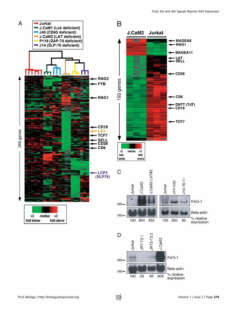
cells, might have consequences on the expression of a set ofgenes. Using DNA microarrays, we compared gene expressionprofiles of Jurkat-derived T cell lines deficient for Lck(J.CaM1) (Straus and Weiss 1992), LAT (J.CaM2) (Finco et al.1998), SLP-76 (J14) (Yablonski et al. 1998), ZAP-70 (P116)(Williams et al. 1998), and CD45 (J45) (Koretzky et al. 1991) tothose of wild-type Jurkat T cells. We set out to analyzeaberrant gene expression caused by the lack of constitutivesignaling in the mutant lines. Confirming original reports,LAT and SLP-76 (LCP2) mRNA levels were reduced in J.CaM2and J14 (Finco et al. 1998; Yablonski et al. 1998) (Figure 1A).In addition, all mutant lines demonstrated many uniquealterations in expression levels of a large number of genescompared to wild-type Jurkat cells, even though they are allderived from the parental Jurkat T cell line. It is possible thataltered gene expression resulting from random mutationscaused by chemical or radiation mutagenesis, unrelated to theTCR signaling defect, complicated this analysis (Figure 1A).We reasoned that the effects of disrupting a low-level, basal
signal would be most evident in a cell line with an absoluteblock in TCR signaling and thus initially focused on J.CaM2.Signaling events like calcium mobilization or activation ofMAPKs are severely impaired in LAT-deficient J.CaM2 evenwhen the TCR is heavily cross-linked (Finco et al. 1998) (datanot shown). We mathematically isolated the differences ingene expression profiles between J.CaM2 and Jurkat andidentified a large cluster of genes expressed at lower levels inJ.CaM2 than in wild-type Jurkat T cells (left three columns ingreen in Figure 1B). This cluster encompassed many cellsurface markers, including CD18 (b2 integrin), CD62L (L-selectin), CD28, and TCRa. We validated these observationsby fluorescence-activated cell sorting (FACS) analysis andnoticed that the expression levels of other cell surfacemarkers like CD2, CD5, and CD7 were also reduced. Stablereconstitution of J.CaM2 with LAT cDNA (J.CaM2-LAT)(Finco et al. 1998), however, did not result in reexpressionof these markers as analyzed by FACS or Northern blotanalysis (data not shown). Expression levels of two nuclearproteins, TCF7 (Tcf-1) and BIN2, were also reduced inJ.CaM2, but again not restored in J.CaM2-LAT, as evidencedby Northern blotting (data not shown). We concluded that thereduced expression levels of these genes could not beexplained by a lack of LAT expression. Instead, one or moreadditional key transcriptional regulators may be defective inthe J.CaM2 line.LAT-deficient cells also displayed a distinct cluster of
genes, including RAG-1 and RAG-2, expressed at higher levelsthan wild-type Jurkat (left three columns in red in Figure 1B).These findings were shared with SLP-76-deficient cells (Figure1A). The elevated expression levels of RAG-1 and RAG-2particularly interested us since it has been well establishedthat induction of the TCR signaling pathway is able toterminate Rag gene expression (Nagaoka et al. 2000). Wetherefore focused on this cluster of ‘‘derepressed’’ genesobserved in the J.CaM2 cells. We hypothesized that normalRAG gene expression in resting cells is held in check by abasal signal that requires both LAT and SLP-76.To test this hypothesis and to confirm the results obtained
in the DNA microarray experiments, we isolated RNA fromseveral cell lines and determined RAG-1 expression levels byNorthern blot analysis. Wild-type Jurkat T cells express verylow but detectable levels of RAG-1 mRNA (Figure 1C, lane 4).
PLoS Biology | http://biology.plosjournals.org Volume 1 | Issue 2 | Page 273
Tonic Erk and Abl Signals Repress RAG Expression
PLoS Biology | http://biology.plosjournals.org Volume 1 | Issue 2 | Page 274
Tonic Erk and Abl Signals Repress RAG Expression
The RAG locus is thus accessible to transcription factors,making this cell line a fortuitous model for our experiments.RAG-1 expression was markedly increased in J.CaM2 andmore moderately in J14 cells compared to that in wild-typeJurkat (Figure 1C). The reduced effect of missing SLP-76 inJ14 cells on RAG expression could reflect the leaky TCR-signaling phenotype of this mutant (Yablonski et al. 1998).Importantly, expression levels were reduced in both mutantcell lines when the repective mutations were complementedwith cDNAs encoding full-length LAT (J.CaM2-LAT) (Finco etal. 1998) or SLP-76 (J14–76-11) (Yablonski et al. 1998). Theelevated RAG-1 expression in the mutants was therefore notan aberrant characteristic caused by accidental mutations atother loci, but rather a result of the deficiency of the adaptersLAT or SLP-76. As on the microarray, RAG-1 expressionlevels analyzed by Northern blot analysis were not increasedin J.CaM1 (data not shown). Despite the Lck deficiency,J.CaM1-deficient cells flux calcium when the TCR is heavilycross-linked or in response to anti-CD3 antibodies (Strausand Weiss 1992). Expression of Fyn in these cells is a likelyexplanation for this signal. In a similar fashion, a low signalmay be capable to control RAG-1 expression in J.CaM2 cells.
We also made use of mutant Jurkat lines to test whethersurface abTCR expression was required for normal RAG-1expression, as observed in wild-type Jurkat cells. TCRa-deficient (J.RT-T3.1) or TCRb-deficient (J.RT3-T3.5) Jurkat Tcells (Ohashi et al. 1985) were generated in a screen forabTCR-negative cell lines (Weiss and Stobo 1984). Werepeated previously published FACS analyses and functionalexperiments on these lines. The TCRa- and TCRb-deficientJurkat lines express very low, if any, CD3e on the surface, butno abTCR (Weiss and Stobo 1984; data not shown). As aresult, these cells do not flux calcium, produce IL-2, ordemonstrate NFAT transcriptional activity in response toTCR stimulation (Ohashi et al. 1985; data not shown). TheTCRa- or TCRb-deficient Jurkat T cells did not displayelevated RAG-1 expression levels such as we observed in LAT-deficient cells (Figure 1D). Surface expression of the abTCR istherefore dispensable for maintenance of normal levels ofRAG-1 transcripts. Importantly, these results imply that thetonic signal regulating RAG gene expression is not generated
by (stimulation of) the extracellular portion of the abTCR,but rather by an intrinsically active signaling pathway. Atleast one of these mutants, JRT-T3.1, has been reported toexpress surface TCRf (Ono et al. 1995); we can therefore notcompletely exclude the possibility that signaling componentsof the TCR play a role in regulating this pathway.
Requirement for LAT in Tonic Repression of RAG-1Transcription Is Bypassed by Direct Activation of PKC orMAPK PathwaysRag-1 and Rag-2 are expressed in DP thymocytes and
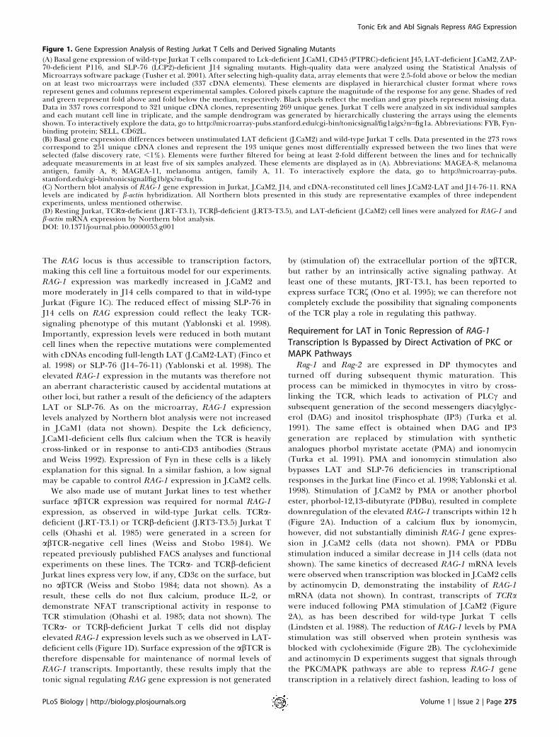
turned off during subsequent thymic maturation. Thisprocess can be mimicked in thymocytes in vitro by cross-linking the TCR, which leads to activation of PLCc andsubsequent generation of the second messengers diacylglyc-erol (DAG) and inositol trisphosphate (IP3) (Turka et al.1991). The same effect is obtained when DAG and IP3generation are replaced by stimulation with syntheticanalogues phorbol myristate acetate (PMA) and ionomycin(Turka et al. 1991). PMA and ionomycin stimulation alsobypasses LAT and SLP-76 deficiencies in transcriptionalresponses in the Jurkat line (Finco et al. 1998; Yablonski et al.1998). Stimulation of J.CaM2 by PMA or another phorbolester, phorbol-12,13-dibutyrate (PDBu), resulted in completedownregulation of the elevated RAG-1 transcripts within 12 h(Figure 2A). Induction of a calcium flux by ionomycin,however, did not substantially diminish RAG-1 gene expres-sion in J.CaM2 cells (data not shown). PMA or PDBustimulation induced a similar decrease in J14 cells (data notshown). The same kinetics of decreased RAG-1 mRNA levelswere observed when transcription was blocked in J.CaM2 cellsby actinomycin D, demonstrating the instability of RAG-1mRNA (data not shown). In contrast, transcripts of TCRawere induced following PMA stimulation of J.CaM2 (Figure2A), as has been described for wild-type Jurkat T cells(Lindsten et al. 1988). The reduction of RAG-1 levels by PMAstimulation was still observed when protein synthesis wasblocked with cycloheximide (Figure 2B). The cycloheximideand actinomycin D experiments suggest that signals throughthe PKC/MAPK pathways are able to repress RAG-1 genetranscription in a relatively direct fashion, leading to loss of
Figure 1. Gene Expression Analysis of Resting Jurkat T Cells and Derived Signaling Mutants
(A) Basal gene expression of wild-type Jurkat T cells compared to Lck-deficient J.CaM1, CD45 (PTPRC)-deficient J45, LAT-deficient J.CaM2, ZAP-70-deficient P116, and SLP-76 (LCP2)-deficient J14 signaling mutants. High-quality data were analyzed using the Statistical Analysis ofMicroarrays software package (Tusher et al. 2001). After selecting high-quality data, array elements that were 2.5-fold above or below the medianon at least two microarrays were included (337 cDNA elements). These elements are displayed in hierarchical cluster format where rowsrepresent genes and columns represent experimental samples. Colored pixels capture the magnitude of the response for any gene. Shades of redand green represent fold above and fold below the median, respectively. Black pixels reflect the median and gray pixels represent missing data.Data in 337 rows correspond to 321 unique cDNA clones, representing 269 unique genes. Jurkat T cells were analyzed in six individual samplesand each mutant cell line in triplicate, and the sample dendrogram was generated by hierarchically clustering the arrays using the elementsshown. To interactively explore the data, go to http://microarray-pubs.stanford.edu/cgi-bin/tonicsignal/fig1a/gx?n¼fig1a. Abbreviations: FYB, Fyn-binding protein; SELL, CD62L.(B) Basal gene expression differences between unstimulated LAT deficient (J.CaM2) and wild-type Jurkat T cells. Data presented in the 273 rowscorrespond to 251 unique cDNA clones and represent the 193 unique genes most differentially expressed between the two lines that wereselected (false discovery rate, ,1%). Elements were further filtered for being at least 2-fold different between the lines and for technicallyadequate measurements in at least five of six samples analyzed. These elements are displayed as in (A). Abbreviations: MAGEA-8, melanomaantigen, family A, 8; MAGEA-11, melanoma antigen, family A, 11. To interactively explore the data, go to http://microarray-pubs.stanford.edu/cgi-bin/tonicsignal/fig1b/gx?n¼fig1b.(C) Northern blot analysis of RAG-1 gene expression in Jurkat, J.CaM2, J14, and cDNA-reconstituted cell lines J.CaM2-LAT and J14-76-11. RNAlevels are indicated by b-actin hybridization. All Northern blots presented in this study are representative examples of three independentexperiments, unless mentioned otherwise.(D) Resting Jurkat, TCRa-deficient (J.RT-T3.1), TCRb-deficient (J.RT3-T3.5), and LAT-deficient (J.CaM2) cell lines were analyzed for RAG-1 andb-actin mRNA expression by Northern blot analysis.DOI: 10.1371/journal.pbio.0000053.g001
PLoS Biology | http://biology.plosjournals.org Volume 1 | Issue 2 | Page 275
Tonic Erk and Abl Signals Repress RAG Expression
detectable RAG-1 mRNA. The fact that RAG-1 expression iselevated in these cells, but can be reduced by a PMA signal,suggests that the LAT/SLP-76 module is essential for aconstitutive, suppressive signal that operates, in part, throughthe downstream signaling pathways. The MAPK pathway canalso be activated through seven-transmembrane receptorsignaling (Crespo et al. 1994). In J.CaM2 cells, this can beachieved by carbachol treatment of cells that stably expressthe human muscarinic receptor (J.CaM2-HM1-1.1) (Gold-smith et al. 1989). Carbachol stimulation decreased RAG-1and increased TCRa expression to the same extent asobserved with PMA (Figure 2C). Thus, pharmacologic andphysiologic activation of the PKC and MAPK pathways canlead to inhibition of RAG-1 gene expression.
Normal Expression of a Gene Cluster Including RAG-1 andRAG-2 Relies on a Signaling Competent LAT MoleculeWe next examined whether signaling via LAT is essential
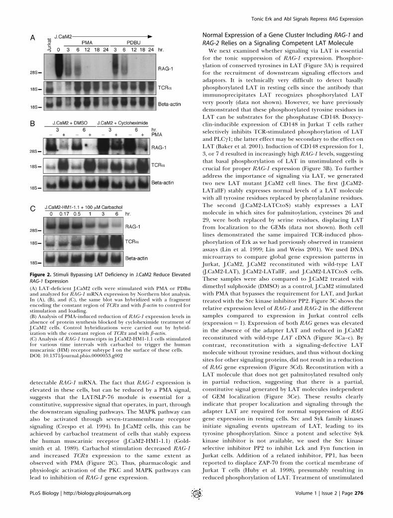
for the tonic suppression of RAG-1 expression. Phosphor-ylation of conserved tyrosines in LAT (Figure 3A) is requiredfor the recruitment of downstream signaling effectors andadaptors. It is technically very difficult to detect basallyphosphorylated LAT in resting cells since the antibody thatimmunoprecipitates LAT recognizes phosphorylated LATvery poorly (data not shown). However, we have previouslydemonstrated that these phosphorylated tyrosine residues inLAT can be substrates for the phosphatase CD148. Doxycy-clin-inducible expression of CD148 in Jurkat T cells ratherselectively inhibits TCR-stimulated phosphorylation of LATand PLCc1; the latter effect may be secondary to the effect onLAT (Baker et al. 2001). Induction of CD148 expression for 1,3, or 7 d resulted in increasingly high RAG-1 levels, suggestingthat basal phosphorylation of LAT in unstimulated cells iscrucial for proper RAG-1 expression (Figure 3B). To furtheraddress the importance of signaling via LAT, we generatedtwo new LAT mutant J.CaM2 cell lines. The first (J.CaM2-LATallF) stably expresses normal levels of a LAT moleculewith all tyrosine residues replaced by phenylalanine residues.The second (J.CaM2-LATCtoS) stably expresses a LATmolecule in which sites for palmitoylation, cysteines 26 and29, were both replaced by serine residues, displacing LATfrom localization to the GEMs (data not shown). Both celllines demonstrated the same impaired TCR-induced phos-phorylation of Erk as we had previously observed in transientassays (Lin et al. 1999; Lin and Weiss 2001). We used DNAmicroarrays to compare global gene expression patterns inJurkat, J.CaM2, J.CaM2 reconstituted with wild-type LAT(J.CaM2-LAT), J.CaM2-LATallF, and J.CaM2-LATCtoS cells.These samples were also compared to J.CaM2 treated withdimethyl sulphoxide (DMSO) as a control, J.CaM2 stimulatedwith PMA that bypasses the requirement for LAT, and Jurkattreated with the Src kinase inhibitor PP2. Figure 3C shows therelative expression level of RAG-1 and RAG-2 in the differentsamples compared to expression in Jurkat control cells(expression ¼ 1). Expression of both RAG genes was elevatedin the absence of the adapter LAT and reduced in J.CaM2reconstituted with wild-type LAT cDNA (Figure 3Ca–c). Bycontrast, reconstitution with a signaling-defective LATmolecule without tyrosine residues, and thus without dockingsites for other signaling proteins, did not result in a reductionof RAG gene expression (Figure 3Cd). Reconstitution with aLAT molecule that does not get palmitoylated resulted onlyin partial reduction, suggesting that there is a partial,constitutive signal generated by LAT molecules independentof GEM localization (Figure 3Ce). These results clearlyindicate that proper localization and signaling through theadapter LAT are required for normal suppression of RAGgene expression in resting cells. Src and Syk family kinasesinitiate signaling events upstream of LAT, leading to itstyrosine phosphorylation. Since a potent and selective Sykkinase inhibitor is not available, we used the Src kinaseselective inhibitor PP2 to inhibit Lck and Fyn function inJurkat cells. Addition of a related inhibitor, PP1, has beenreported to displace ZAP-70 from the cortical membrane ofJurkat T cells (Huby et al. 1998), presumably resulting inreduced phosphorylation of LAT. Treatment of unstimulated
Figure 2. Stimuli Bypassing LAT Deficiency in J.CaM2 Reduce Elevated
RAG-1 Expression
(A) LAT-deficient J.CaM2 cells were stimulated with PMA or PDBuand analyzed for RAG-1 mRNA expression by Northern blot analysis.In (A), (B), and (C), the same blot was hybridized with a fragmentencoding the constant region of TCRa and with b-actin to control forstimulation and loading.(B) Analysis of PMA-induced reduction of RAG-1 expression levels inabsence of protein synthesis blocked by cycloheximide treatment ofJ.CaM2 cells. Control hybridizations were carried out by hybrid-ization with the constant region of TCRa and with b-actin.(C) Analysis of RAG-1 transcripts in J.CaM2-HM1-1.1 cells stimulatedfor various time intervals with carbachol to trigger the humanmuscarinic (HM) receptor subtype I on the surface of these cells.DOI: 10.1371/journal.pbio.0000053.g002
PLoS Biology | http://biology.plosjournals.org Volume 1 | Issue 2 | Page 276
Tonic Erk and Abl Signals Repress RAG Expression
Jurkat cells with PP2 increased expression of RAG-1 and RAG-2 to a level similar to that in J.CaM2 cells, suggesting thatbasal Lck and/or Fyn kinase activity is required forconstitutive repression of these genes (Figure 3Ch).To investigate whether constitutive signaling through LAT
controls other genes in addition to RAG-1 and RAG-2, weplotted the expression profiles of genes whose expression waselevated in J.CaM2 relative to Jurkat cells and diminished byrestoration of LAT function in J.CaM2-LAT (Figure 3D).Roughly 20 genes demonstrated such an expression profile.As for the RAG genes, elevated levels of some but not of all ofthese genes could be reduced by PMA stimulation of J.CaM2or induced by PP2 treatment of wild-type Jurkat. Although atthis moment these genes have no obvious function inlymphocyte biology or gene rearrangement, these datasuggest that expression patterns of several other genes canbe influenced by basal signaling through components of theTCR signaling pathway in resting cells. It will be interesting toinvestigate what role this group of genes may play in T cellmaturation.
Chemical Inhibitors Map the Tonic Suppression of RAGGene Expression to a TCR-Like Signaling PathwayTo further trace specific molecular components of this
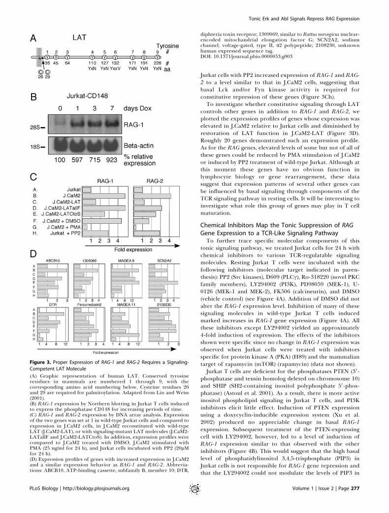
tonic signaling pathway, we treated Jurkat cells for 24 h withchemical inhibitors to various TCR-regulatable signalingmolecules. Resting Jurkat T cells were incubated with thefollowing inhibitors (molecular target indicated in paren-thesis): PP2 (Src kinases), D609 (PLCc), Ro-318220 (novel PKCfamily members), LY294002 (PI3K), PD98059 (MEK-1), U-0126 (MEK-1 and MEK-2), FK506 (calcineurin), and DMSO(vehicle control) (see Figure 4A). Addition of DMSO did notalter the RAG-1 expression level. Inhibition of many of thesesignaling molecules in wild-type Jurkat T cells inducedmarked increases in RAG-1 gene expression (Figure 4A). Allthese inhibitors except LY294002 yielded an approximately4-fold induction of expression. The effects of the inhibitorsshown were specific since no change in RAG-1 expression wasobserved when Jurkat cells were treated with inhibitorsspecific for protein kinase A (PKA) (H89) and the mammaliantarget of rapamycin (mTOR) (rapamycin) (data not shown).Jurkat T cells are deficient for the phosphatases PTEN (39-
phosphatase and tensin homolog deleted on chromosome 10)and SHIP (SH2-containing inositol polyphosphate 59-phos-phatase) (Astoul et al. 2001). As a result, there is more activeinositol phospholipid signaling in Jurkat T cells, and PI3Kinhibitors elicit little effect. Induction of PTEN expressionusing a doxycyclin-inducible expression system (Xu et al.2002) produced no appreciable change in basal RAG-1expression. Subsequent treatment of the PTEN-expressingcell with LY294002, however, led to a level of induction ofRAG-1 expression similar to that observed with the otherinhibitors (Figure 4B). This would suggest that the high basallevel of phosphatidylinositol 3,4,5-trisphosphate (PIP3) inJurkat cells is not responsible for RAG-1 gene repression andthat the LY294002 could not modulate the levels of PIP3 in
Figure 3. Proper Expression of RAG-1 and RAG-2 Requires a Signaling-
Competent LAT Molecule
(A) Graphic representation of human LAT. Conserved tyrosineresidues in mammals are numbered 1 through 9, with thecorresponding amino acid numbering below. Cysteine residues 26and 29 are required for palmitoylation. Adapted from Lin and Weiss(2001).(B) RAG-1 expression by Northern blotting in Jurkat T cells inducedto express the phosphatase CD148 for increasing periods of time.(C) RAG-1 and RAG-2 expression by DNA array analysis. Expressionof the two genes was set at 1 in wild-type Jurkat cells and compared toexpression in J.CaM2 cells, in J.CaM2 reconstituted with wild-typeLAT (J.CaM2-LAT), or with signaling-mutant LAT molecules (J.CaM2-LATallF and J.CaM2-LATCtoS). In addition, expression profiles werecompared to J.CaM2 treated with DMSO, J.CaM2 stimulated withPMA (25 ng/ml for 24 h), and Jurkat cells incubated with PP2 (20lMfor 24 h).(D) Expression profiles of genes with increased expression in J.CaM2and a similar expression behavior as RAG-1 and RAG-2. Abbrevia-tions: ABCB10, ATP-binding cassette, subfamily B, member 10; DTR,
diphteria toxin receptor; 1309069, similar to Rattus norvegicus nuclear-encoded mitochandrial elongation factor G; SCN2A2, sodiumchannel, voltage-gated, type II, a2 polypeptide; 2108230, unknownhuman expressed sequence tag.DOI: 10.1371/journal.pbio.0000053.g003
PLoS Biology | http://biology.plosjournals.org Volume 1 | Issue 2 | Page 277
Tonic Erk and Abl Signals Repress RAG Expression
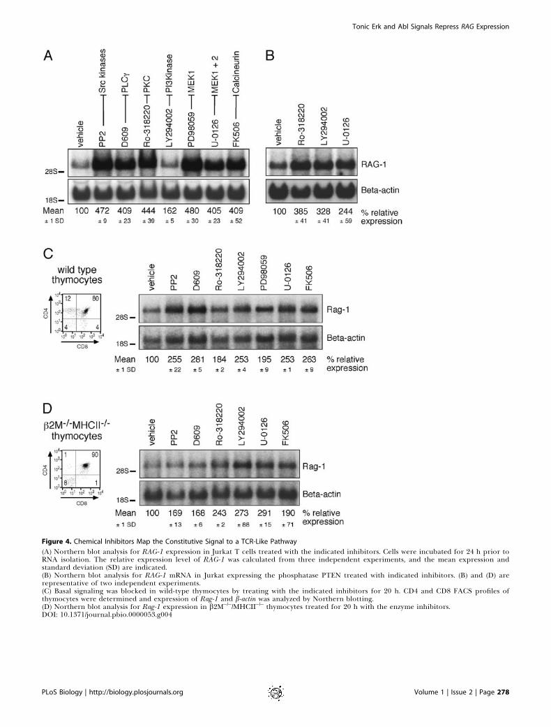
Figure 4. Chemical Inhibitors Map the Constitutive Signal to a TCR-Like Pathway
(A) Northern blot analysis for RAG-1 expression in Jurkat T cells treated with the indicated inhibitors. Cells were incubated for 24 h prior toRNA isolation. The relative expression level of RAG-1 was calculated from three independent experiments, and the mean expression andstandard deviation (SD) are indicated.(B) Northern blot analysis for RAG-1 mRNA in Jurkat expressing the phosphatase PTEN treated with indicated inhibitors. (B) and (D) arerepresentative of two independent experiments.(C) Basal signaling was blocked in wild-type thymocytes by treating with the indicated inhibitors for 20 h. CD4 and CD8 FACS profiles ofthymocytes were determined and expression of Rag-1 and b-actin was analyzed by Northern blotting.(D) Northern blot analysis for Rag-1 expression in b2M�/�/MHCII�/� thymocytes treated for 20 h with the enzyme inhibitors.DOI: 10.1371/journal.pbio.0000053.g004
PLoS Biology | http://biology.plosjournals.org Volume 1 | Issue 2 | Page 278
Tonic Erk and Abl Signals Repress RAG Expression
PTEN-deficient Jurkat cells, consistent with our previousfindings (Kane et al. 2002).
The effects of selective inhibitors, together with the resultsof our previous experiments with mutant cell lines, point to atonic signaling pathway that closely resembles a TCR-inducible pathway, with Src family kinase activity as the mostupstream component and MAPK as the most downstreamcomponent regulating RAG-1 and RAG-2 expression in Jurkatcells (for a model, see Figure 8). The same inhibitors yieldedvery similar results in two mouse thymocyte cell lines, SCB29and DPK (data not shown). The SCB29 cell line is derivedfrom a Scid mouse and expresses a pre-TCR (Groettrup et al.1992). DPK is a CD4þCD8þ thymocyte line expressing atransgene-encoded TCR (Kaye and Ellenberger 1992). SinceTCR signaling has been studied in great detail in Jurkat Tcells, we proceeded to use this model for our experiments andverified our results in primary thymocytes.
A Similar Tonic Signaling Mechanism Represses Rag-1Expression in Thymocytes of Wild-Type andb2M�/�/MHCII�/� Mice
We noticed that primary murine thymocytes were moresensitive to apoptosis induced by prolonged exposure tochemical inhibitors. Nevertheless, we were able to determinethe effects of the signaling inhibitors on Rag-1 expression inwild-type thymocytes by adding a general caspase inhibitor(Z-DEVD-FMK) to our incubations in order to blockapoptosis. Addition of Z-DEVD-FMK by itself had no effecton Rag-1 gene expression, but did improve survival withoutaffecting the relative proportions of the different thymocytesubsets (determined by CD4, CD8, CD25, and CD44 staining;data not shown). As observed in cell lines, inhibition ofcomponents of the downstream TCR signaling pathway intotal thymocytes significantly increased Rag-1 gene expres-sion (Figure 4C). The inhibitors delineated the same pathwayas in the Jurkat T cell and the thymocyte cell lines, includingSrc kinases, PLCc1, PKC, PI3K, MEK-1, and calcineurin. Themagnitude of induced Rag-1 expression was more moderatein thymocytes than the level observed in our model cell line.The fact that thymocytes in the process of receptorrearrangement already express Rag-1 may account for thesmaller induction over the existing level. We thereforetreated thymocytes from TCR transgenic OT-I mice withinhibitors in the same manner. OT-I transgenic mice have athymus containing substantial numbers of DP thymocytes. Inabsence of the peptide from chicken ovalbumin, these DPthymocytes are positively selected on self-antigens andeffectively downregulate Rag expression (McGargill et al.2000). As a result, the level of Rag-1 expression in thymocytesof these animals is lower than that in wild-type littermatethymi (Figure 5C, lanes 1 and 3). Treatment of OT-1thymocytes with PP2 or U-0126 led to a strong induction inRag-1 expression, comparable to the effects observed inJurkat T cells (Figure 5C). These data argue that detection ofconstitutive Rag repression in wild-type thymocytes ishindered by normal Rag expression during rearrangement.
To eliminate the possibility that we were studyinginhibition of a signal induced by an MHC–peptide inter-action with the TCR on the surface of the thymocyte, werepeated the same experiment in b2M�/�/MHCII�/� thymo-cytes (Grusby et al. 1993), which cannot encounter MHC–peptide interactions and as a result are blocked at the DP
stage. The pattern of effects of the inhibitors on Rag-1 geneexpression in these mutant thymocytes was very similar tothat observed in the wild-type thymocytes (see Figure 4D).The signal repressing Rag-1 gene expression is thus notinitiated by MHC–peptide stimulation, but instead appears tobe transmitted through a basal signaling pathway thatresembles the TCR-inducible signaling pathway. We utilizedthe same panel of inhibitors to determine whether the sameconstitutive repression of Rag-1 expression occurs in periph-eral T cells. To prevent stimulation by antibody binding,these cells were purified from lymph node and spleenthrough negative selection of other cell populations. Inseveral independent experiments, we did not observe anincrease in Rag-1 expression in the inhibitor-treated periph-eral T cells (data not shown).
Parallel Signaling Pathways Dependent on LAT MaintainBasal Phosphorylation of Erk and Abl KinasesThe inhibitor studies suggest that a constitutive signaling
pathway may use components very similar to those involvedin conventional TCR signaling triggered upon receptorligation. We wanted to confirm and identify the downstreameffector molecules that transduce the repressive signal. TheMAPK pathway was clearly implicated; however, recentstudies suggested that Abl kinase activity might also beinvolved.Human c-Abl and its homologue Abl-related gene (Arg) are
nonreceptor tyrosine kinases with structural homology to Srckinases (reviewed in Van Etten 1999). Kinase activity of c-Ablin normal cells is suppressed by autoinhibition, depending on80 N-terminal residues that are lost in translocations leadingto BCR–Abl fusion proteins and development of chronicmyelogenous leukemia (Pluk et al. 2002). The Abl inhibitorSTI-571/Gleevec/Imatinib (STI-571) recognizes c-Abl and Argbut not Src, displaces ATP from Abl, and traps the kinase inan inactive conformation (Schindler et al. 2000). STI-571 hasalso been reported to inhibit the receptor tyrosine kinases c-Kit and platelet-derived growth factor (PDGF) receptor athigher doses (Schindler et al. 2000). Activation of c-Ablrequires the Src family kinases (Plattner et al. 1999). Ablkinases have been linked to regulation of RAG geneexpression in studies of pre-B cells transformed by temper-ature-sensitive mutants of the Abelson virus (Chen et al.1994). Very recently, Muljo and Schlissel (2003) demonstratedthat treatment of Abelson virus-transformed pro-B cell linesexpressing active v-Abl with STI-571 activated transcriptionof Rag-1 and Rag-2. Jurkat T cells express moderate levels of c-Abl and low levels of Arg kinases, but are negative for c-Kitand PDGF receptor (Taylor et al. 2001; Bianchi et al. 2002;data not shown).These findings prompted us to examine whether Abl
kinases might play a role in constitutive repression of RAGgenes. Indeed, inhibition of Abl kinase activity by STI-571 inJurkat T cells induced RAG-1 expression to a level similar tothat induced by the inhibitors PP2 and Ro-318220 (Figure5A). Similarly, STI-571 induced elevated levels of Rag-1 inwild-type thymocytes (Figure 5B). This effect was morepronounced in TCR transgenic OT-1 thymocytes (Figure5C). Abl has been reported to induce MAPK signaling inresponse to stress (Kharbanda et al. 1995). To addresswhether STI-571 functions to elevate RAG-1 expressionindirectly by reducing a MAPK signal or whether there is a
PLoS Biology | http://biology.plosjournals.org Volume 1 | Issue 2 | Page 279
Tonic Erk and Abl Signals Repress RAG Expression
distinct Abl pathway, we carefully analyzed the specificphosphorylation status of Erk and Abl. Specific phosphor-ylation on threonine 202/tyrosine 204 residues reflects thekinase activities of Erk-1 and Erk-2. Active Abl is phosphory-lated on tyrosine and serine residues (Pluk et al. 2002).Phosphorylation of tyrosine 245 and tyrosine 412 in c-Abl are
crucial regulatory events required for its kinase activity(Brasher and Van Etten 2000). Phosphorylation of tyrosine245 reflects disruption of the inactive conformation and canbe detected with a phosphorylation-specific antibody.In radioimmunoprecipitation assay (RIPA) lysates of rest-
ing J.CaM2 cells, we consistently observed an absence of basalphosphorylated Erk-1 and Erk-2 proteins, signals that wereeasily detected in Jurkat or J.CaM2-LAT (Figure 5D). Inducedexpression of the phosphatase CD148 for 7 d also ablated thisconstitutive phospho-Erk signal (Figure 5D, lane 5). Expres-sion levels of c-Abl and the Erk-1 and Erk-2 kinases wereequal in all samples (upper and lower panels of Figure 5D).Treatment of wild-type Jurkat T cells with chemical
inhibitors of signaling components revealed that inhibitionof Src family kinases, PLCc, PKC, MEK-1 and MEK-2, andcalcineurin (using PP2, D609, Ro-318220, U-0126, and FK506,respectively) efficiently decreased the amount of phosphory-lated Erk-1 and Erk-2, the reciprocal effect of that on RAG-1expression (Figure 5E). Inhibition of calcineurin has beenreported to induce expression of the MAPK phosphatase 1and inhibit Erk signaling in neuronal cells (Zawadzka andKaminska 2003). A similar mechanism may account for theseeffects on Jurkat T cells and thymocytes. As seen for otherassays, the PI3K inhibitor LY294002 only has limited effect onphospho-Erk-1 and phospho-Erk-2 levels in Jurkat T cells. Toour surprise, treatment with STI-571 only slightly decreasedphospho-Erk (Figure 5E, lane 8), raising the possibility thatAbl and Erk kinases may act in separate, basally activepathways to repress RAG-1 expression.To resolve this question, we analyzed these low-level,
constitutive phospho-c-Abl and phospho-Erk signals byintracellular FACS staining (we were unable to use the site-specific phospho-Y245 for c-Abl in Western blot analysis; datanot shown). Panels I–III of Figure 6A demonstrate thefeasibility of this approach. PMA stimulation of Jurkat Tcells for 10 min resulted in 6-fold induction of phospho-Erksignal compared to unstimulated Jurkat (arbitrarily set at 100in the accompanying bar graph of Figure 6A, panel I). Asshown in the same panel, 24-h administration of a MEK-1 andMEK-2 inhibitor (U-0126) effectively blunted the phospho-Erk response, whereas the c-Abl inhibitor had a detectablebut consistently smaller effect. The STI-571 inhibitor was,however, very potent in blocking the phospho-Abl signal inK562 cells, which reflects the kinase activity of BCR–Abl inthis cell line (Figure 6A, panel II). Similar to the basal activityof the Erk kinases (Figure 6A, panel IV), phosphorylation oftyrosine 245 in Abl was consistently reduced in unstimulatedJ.CaM2 cells, resulting in a fluorescence signal not signifi-cantly above isotype control (Figure 6A, panels V and VI). Thephospho-Abl signal observed in Jurkat T cells probablyreflects both phosphorylated c-Abl and phosphorylated Argsince the two kinases are highly conserved in the tyrosine 245region. Importantly, phospho-Abl levels were restored in theJ.CaM2-LAT line, pointing to an essential role for LAT in Ablphosphorylation (Figure 6A, panel V; Figure 6B). Thereduction of c-Abl-specific phosphorylation in J.CaM2 cellswas confirmed by c-Abl immunoprecipitations followed bytotal phosphotyrosine immunoblotting and was restored inJ.CaM2 cells complemented with LAT cDNA (Figure 6B).Induced expression of the phosphatase CD148 for 7 d alsoablated this constitutive phospho-c-Abl signal (right panel inFigure 6B).
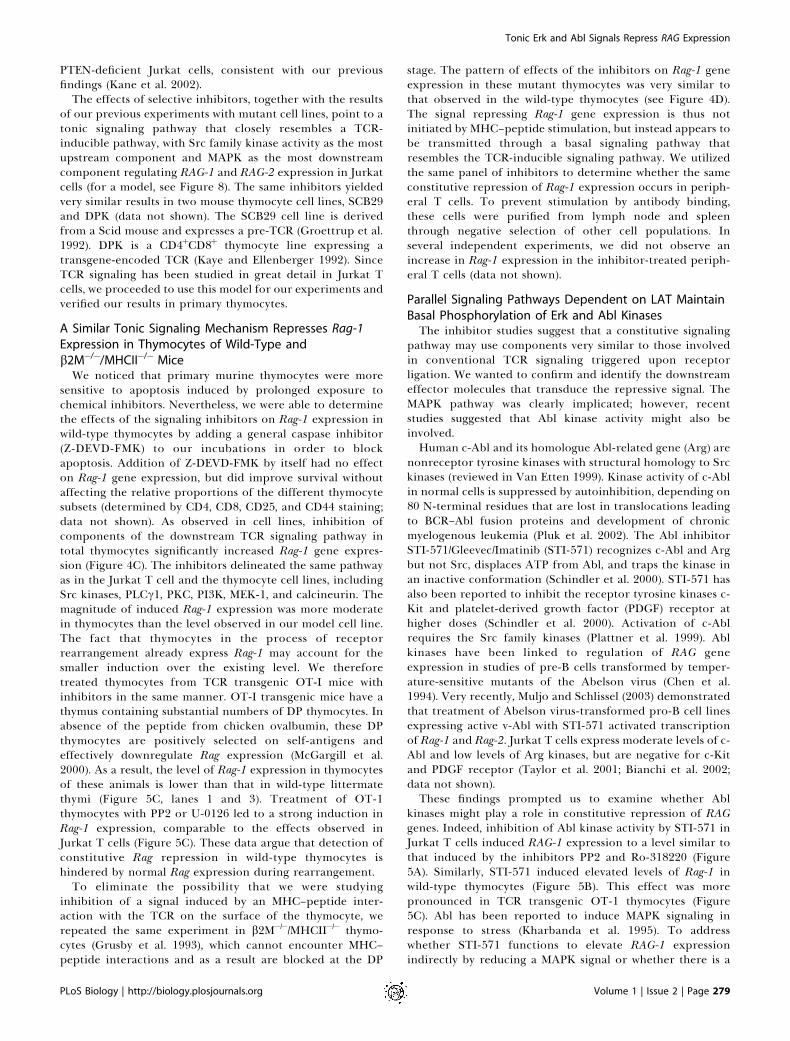
Figure 5. Erk and Abl Kinases Transduce Repressive Signals That Control
RAG Gene Expression
(A) Northern blot analysis for RAG-1 gene expression in STI-571-treated Jurkat T cells and comparison to PP2- or Ro-318220-treatedsamples.(B) Wild-type thymocytes were treated with the indicated inhibitorsfor 20 h and analyzed for Rag-1 expression.(C) Northern blot analysis of Rag-1 expression in inhibitor-treatedTCR transgenic OT-I thymocytes or wild-type thymocytes oflittermate controls. The relative expression level of Rag-1 wascalculated from two independent experiments, and the meanexpression and standard deviation (SD) are indicated.(D) Western blot analysis using RIPA lysates of the indicated cell lines.Protein levels of phosphorylated Erk-1 and Erk-2, Erk-1 and Erk-2,and c-Abl were determined in 43 106 resting cells per sample.(E) Analysis of phospho-Erk-1 and Erk-2 levels in Jurkat T cellstreated for 24 h with the indicated inhibitors prior to RIPA lysis.Equal loading is indicated by Erk-1 and Erk-2 levels determined bystripping and reprobing the same blot.DOI: 10.1371/journal.pbio.0000053.g005
PLoS Biology | http://biology.plosjournals.org Volume 1 | Issue 2 | Page 280
Tonic Erk and Abl Signals Repress RAG Expression

Figure 6. LAT Is Required for Two Largely
Separate Pathways Marked by Constitutive
Phosphorylation of Erk and Abl Kinases
(A) Intracellular FACS staining for phos-pho-Erk (I and IV), phospho-Abl (II andV), and isotype control (III and VI) in theindicated cell lines. Histograms are anexample of a representative experiment.The accompanying bar graphs are de-picted in the same color-coding andrepresent the mean levels of fluores-cence and standard deviation of threeindependent assays for all experiments.Mean levels of phosphoproteins meas-ured by fluorescence in a resting Jurkatpopulation were arbitrarily set at 100,and the mean fluorescence of the isotypecontrol samples is indicated by thedotted line as a point of reference.Specifics are mentioned in the text.(B) Analysis of tyrosine phosphorylationlevels of c-Abl in the indicated cellsamples. c-Abl or control immunoperci-pitations were immunoblotted for totaltyrosine phosphorylation by 4G10. Thesame blot was stripped and reprobed forc-Abl. Doxycyclin was administered for 7d to induce CD148 expression.(C) Resting Jurkat T cells were treatedwith the indicated inhibitors for 24 h,and intracellular FACS staining forphospho-Abl and isotype control wascompared to that detected in vehicle-treated Jurkat cells and the J.CaM2 line.Bar graphs (representing three experi-ments) display the four conditions de-picted in the histograms as well as threeadditional samples.DOI: 10.1371/journal.pbio.0000053.g006
PLoS Biology | http://biology.plosjournals.org Volume 1 | Issue 2 | Page 281
Tonic Erk and Abl Signals Repress RAG Expression
The signals that elicit Abl phosphorylation were limited toSrc and Abl kinases since blockade of PLCc, MEK-1 and MEK-2, or calcineurin activity had no effect on the phosphor-ylation status of tyrosine 245 (Figure 6C). PP2 and STI-571reduced the phospho-Abl level to that observed in J.CaM2,virtually equal to isotype control staining. It should be notedthat PP2 has recently been reported to inhibit to some extentc-Kit and BCR–Abl (Tatton et al. 2003). Therefore, the PP2effect may be the combined result of blocking Src familykinases and direct inhibition of Abl kinases. Together, thesefindings suggest that constitutive activity of Abl repressesRAG gene expression, requires the presence of LAT, and isblocked by Src and Abl kinase inhibitors, but is unaffected byErk activity.
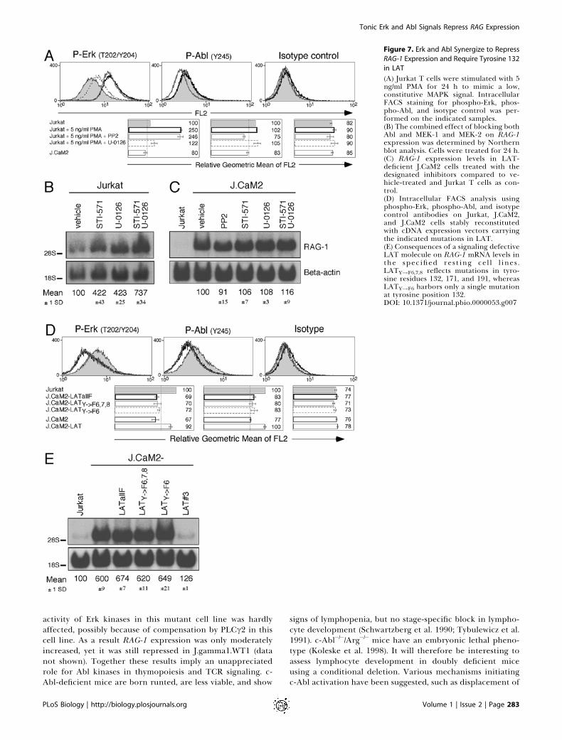
Both Erk and Abl Kinases Repress RAG-1 Expression andRequire Phosphorylation of Tyrosine 132 in LAT
The unaltered phosphorylation status of Abl in U-0126-treated cells suggested that the MEK–Erk pathway does notcontribute to basal Abl activity in our model. To further testthis, we elevated the basal MAPK signal in Jurkat T cells byaddition of 5 ng/ml PMA for 24 h. This treatment induced ahigher level of phospho-Erk that is largely abrogated by U-0126, but not by PP2 (left panel of Figure 7A). By contrast, lowamounts of PMA did not induce phosphorylation of Ablkinases (middle panel of Figure 7A). Therefore, whereasinhibition of Abl has modest effects on Erk phosphorylation,Erk activity does not enhance phosphorylation of tyrosine245 in Abl. These observations further support the notionthat Abl and Erk operate independently to regulate theexpression of RAG-1. This notion is illustrated by the additiveeffects of STI-571 and U-0126 in inducing RAG-1 expression(Figure 7B). Moreover, it appears that the regulatory roles ofboth kinases in this pathway depend on LAT. Addition ofSTI-571, U-0126, or a combination of these inhibitors, oreven targeting Src family kinases upstream of Abl and Erk,did not result in a hyperinduction of RAG-1 in the J.CaM2line (Figure 7C).
Finally, the results above demonstrated that the roles of Erkand Abl kinases in controlling RAG-1 gene expression arelargely parallel rather than sequential. MAPK activation byTCR stimulation has been known to rely on LAT (Finco et al.1998; Zhang et al. 1999). How does LAT transduce a uniquesignal to Abl? To study a possible LAT–Abl connection, wemade use of a panel of J.CaM2 cell lines reconstituted withLAT molecules that carry point mutations of tyrosineresidues (see Figure 3A). We and others have previouslydemonstrated that induction of phospho-Erk upon TCRstimulation is impaired in J.CaM2 lines reconstituted withLATY!F6,7,8 or LATY!F6 (Zhang et al. 2000; Lin and Weiss2001). These studies demonstrated the importance of PLCcrecruitment to tyrosine 132 (#6) in LAT for TCR-inducedcalcium flux and Erk activation. As demonstrated in Figure7D, constitutive phosphorylation of both Erk and Abl kinasesin these two lines was decreased compared to wild-type Jurkatand comparable to the levels seen in J.CaM2-LATallF andJ.CaM2 cells. Consistent with the reduced phosphorylation ofthese two kinases, RAG-1 expression was increased in J.CaM2cells reconstituted with LAT carrying only a single substitu-tion of tyrosine residue 132 by phenylalanine (Figure 7E).Thus, Abl, like Erk, depends upon phosphorylation oftyrosine 132 in LAT.
Discussion
Here we have demonstrated a ligand-independent con-stitutive signaling pathway that is functional in Jurkat T cells,two murine thymomas, and in thymocytes. Our studies revealthat even without TCR engagement, the signaling pathwaysnormally responsive to TCR stimulation are not inert. Insteadthe components of these pathways deliver unique constitutiveinstructions to the nucleus that maintains proper geneexpression programs. RAG gene expression is tightly regu-lated during thymopoiesis and is expressed in at least twowaves. The tonic basal signal we characterized in this studymaintains repression of RAG gene expression and involves aTCR-like signal transduction pathway. One arm of thepathway requires Src family kinase activity, presence ofLAT–SLP-76, and the enzyme activities of PLCc1, PKC, PI3K,MEK-1, and calcineurin, culminating in basal kinase activityof Erk (Figure 8). We also identified a novel pathway that isresponsible for constitutive phosphorylation of Abl. Propersignal transduction relies on Src family kinase activity and thepresence of tyrosine 132 in LAT and can be blocked by STI-571 (Figure 8). We postulate that these constitutive signalsmay function to repress RAG gene expression duringthymopoiesis and, as such, allow the locus to remainaccessible for later usage without inappropriate expression.Previous reports have demonstrated the importance of
constitutive signals triggered by the antigen receptor orcoreceptors in mature lymphocytes. In order to survive, Bcells require surface expression of the BCR that generates abasal signal independently of endogenous antigen (Lam et al.1997). Naive peripheral T cells appear to be less dependenton their receptors for survival (Polic et al. 2001), althoughthey do depend on either Lck or Fyn as well as CD4 and MHCmolecules for survival (Zamoyska et al. 2003). They alsomaintain a high degree of sensitivity for foreign antigen byhaving constitutive phosphorylation of TCRf (Stefanova et al.2002). Similar signals were suggested to occur in thymocytes(Nakayama et al. 1989; van Oers et al. 1994).In this study, we have identified a TCR-independent and
MHC-independent signaling pathway that maintains repres-sion of RAG gene expression in a constitutive fashion. It isperhaps not surprising that in Jurkat cells and in thymocytesthis pathway shares components with conventional TCRsignaling. Key effector molecules for this repressive signal areErk and Abl kinases. One branch of this constitutive signalingpathway involves Src family kinase activity, presumablyphosphorylation of TCRf and ZAP-70, phosphorylation ofLAT, and recruitment and activation of PLCc (Figure 8). Thisresults, most likely, through low-level generation of thesecond messengers IP3 and DAG in constitutive activationof Erk kinases. We also identified a novel second pathway thatresults in constitutive phosphorylation and activity of Abl,which requires phosphorylation of tyrosine residue 132 inLAT, but not PLCc, activation. The specific requirement oftyrosine residue 132 suggests that this pathway involves theformation of a LAT–PLCc–Abl or LAT–Abl complex.Recently a complex of c-Abl and PLCc1 has been reportedin growth factor receptor signaling (Plattner et al. 2003). Inagreement with these results, we observed a decrease in basalAbl phosphorylation in PLCc1-deficient Jurkat cells (J.gam-ma1) and normal Abl phosphorylation in PLCc1-reconsti-tuted cells (J.gamma1.WT1; data not shown). However, basal
PLoS Biology | http://biology.plosjournals.org Volume 1 | Issue 2 | Page 282
Tonic Erk and Abl Signals Repress RAG Expression
activity of Erk kinases in this mutant cell line was hardlyaffected, possibly because of compensation by PLCc2 in thiscell line. As a result RAG-1 expression was only moderatelyincreased, yet it was still repressed in J.gamma1.WT1 (datanot shown). Together these results imply an unappreciatedrole for Abl kinases in thymopoiesis and TCR signaling. c-Abl-deficient mice are born runted, are less viable, and show
signs of lymphopenia, but no stage-specific block in lympho-cyte development (Schwartzberg et al. 1990; Tybulewicz et al.1991). c-Abl�/�/Arg�/� mice have an embryonic lethal pheno-type (Koleske et al. 1998). It will therefore be interesting toassess lymphocyte development in doubly deficient miceusing a conditional deletion. Various mechanisms initiatingc-Abl activation have been suggested, such as displacement of
Figure 7. Erk and Abl Synergize to Repress
RAG-1 Expression and Require Tyrosine 132
in LAT
(A) Jurkat T cells were stimulated with 5ng/ml PMA for 24 h to mimic a low,constitutive MAPK signal. IntracellularFACS staining for phospho-Erk, phos-pho-Abl, and isotype control was per-formed on the indicated samples.(B) The combined effect of blocking bothAbl and MEK-1 and MEK-2 on RAG-1expression was determined by Northernblot analysis. Cells were treated for 24 h.(C) RAG-1 expression levels in LAT-deficient J.CaM2 cells treated with thedesignated inhibitors compared to ve-hicle-treated and Jurkat T cells as con-trol.(D) Intracellular FACS analysis usingphospho-Erk, phospho-Abl, and isotypecontrol antibodies on Jurkat, J.CaM2,and J.CaM2 cells stably reconstitutedwith cDNA expression vectors carryingthe indicated mutations in LAT.(E) Consequences of a signaling defectiveLAT molecule on RAG-1 mRNA levels inthe specified rest ing cel l l ines .LATY!F6,7,8 reflects mutations in tyro-sine residues 132, 171, and 191, whereasLATY!F6 harbors only a single mutationat tyrosine position 132.DOI: 10.1371/journal.pbio.0000053.g007
PLoS Biology | http://biology.plosjournals.org Volume 1 | Issue 2 | Page 283
Tonic Erk and Abl Signals Repress RAG Expression

the myristate group or the SH2 or SH3 domain, all disruptingthe inactive conformation and allowing tyrosine phosphor-ylation of residue 245 in the linker region or of 412 in theactivation loop (Hantschel et al. 2003). We hypothesize that inthis signaling pathway, binding of Abl to PLCc opens up theinactive conformation. In addition, formation of a LAT–PLCc–Abl complex could bring Abl in close proximity to Srcfamily kinases, facilitating phosphorylation of Abl by Src.
Two elegant studies have addressed the specific role oftyrosine residue 136 of LAT, the murine equivalent of humantyrosine 132, in vivo. Thymocytes of 2-wk-old LATY136F
knock-in mice demonstrate an incomplete block at the pre-TCR signaling stage (Aguado et al. 2002; Sommers et al. 2002).Paradoxically, a few weeks later the animals developlymphadenopathy and splenomegaly. Lymph node CD4þ cellsexhibit an activated phenotype, are of the T helper 2 type,and are autoreactive (Aguado et al. 2002; Sommers et al.2002). Interestingly, these peripheral cells and DP and SPthymocytes display dramatically reduced surface TCR levelsin comparison to their wild-type counterparts. This could
reflect selection of a different repertoire due to alteredstrength of signal, preferentially selecting autoreactive cells.Alternatively, our data suggest that these thymocytes mayaberrantly express Rag genes, resulting in undesirable furtherrearrangement leading to self-reacting TCRs.Rag loci seem to remain accessible at least up to the DP
stage when surface abTCR is expressed. When ex vivo DPthymocytes are stimulated overnight with anti-TCR antibody,half of the cells have inactivated the Rag locus by reposition-ing to centromeric regions (Brown et al. 1999). Incubation ofthese DP cells with stroma results in the development into SPthymocytes combined with complete centromeric reposition-ing of the Rag locus. Permanent, heritable silencing sub-sequently prevents further Rag expression when thymocytesexit the thymus and become peripheral T cells. In agreementwith this model, we failed to detect induced expression ofRag-1 in purified T cells from peripheral lymphoid organsusing our panel of inhibitors. Similarly, Yu et al. (1999) couldnot observe any fluorescent activity in T cells of the RAGreporter mice. In lymph node T cells, this cascade may very
Figure 8. Model of the Constitutive Signaling Pathway That Provides a Basal Repression of RAG Gene Transcription
Constitutive signaling in resting thymoytes and our model Jurkat T cell line represses RAG gene expression. Chemical inhibition of signalingmolecules, genetic modifications, or induced expression of genes that resulted in elevated RAG expression are summarized in the gray-shadedboxes. The tonic signal and therefore normal expression of RAG genes rely strongly on signaling through the adapter LAT. The effects of basalphosphorylation of LAT are twofold: (1) recruitment and activation of PLCc generates low levels of second messengers that signal throughcalcium and Ras pathways, culminating in Erk kinase activity; and (2) phosphorylation of tyrosine residue 6 of LAT, likely establishing a LAT–PLCc–Abl complex, is required for low-level kinase activity of Abl. Abl and Erk kinase activities deliver unique repressive signals to control RAGgene expression.DOI: 10.1371/journal.pbio.0000053.g008
PLoS Biology | http://biology.plosjournals.org Volume 1 | Issue 2 | Page 284
Tonic Erk and Abl Signals Repress RAG Expression
well regulate expression patterns of other genes, like thosedepicted in Figure 3D, and sensitize the T cell to respond toforeign antigens (Stefanova et al. 2002).
Repositioning of the Rag locus over time requires transientrepression, consistent with our findings here. In addition,repression may ensure physiologic Rag levels, allowing the DPthymocyte to test the newly assembled abTCR before addi-tional TCRa rearrangements are started. At the moment wecan only speculate on the exact timing of the constitutivesignal during thymopoiesis. Total thymocyte populations, asanalyzed in Figure 4C, typically contain 80% DP cells. Giventhe effects we observe with the various inhibitors on totalthymocytes with DP thymocytes being by far the largestpopulation, this is the most likely subset where basalrepression of Rag expression takes place.
Nevertheless, many components that transduce this re-pressive signal are expressed in thymocytes well before TCRor pre-TCR signaling occurs. It is therefore possible that Raggenes are repressed even before the first wave of Ragexpression occurrs in CD25-positive DN thymocytes. If so, acytokine signal may consequently be required to release theconstitutive repression of Rag genes to allow Rag expressionand rearrangement of the TCRb chain. We are currentlyadapting our intracellular FACS staining to determinephospho-Erk and phospho-Abl in thymocyte subsets. Inaddition, we are planning to study this pathway in Rag-GFPindicator mice (Yu et al. 1999).
Our results support the notion that receptor-independentsignaling occurs in T cells and thymocytes. This tonic basalsignaling pathway utilizes many of the same signalingcomponents as a TCR transduction pathway triggered byreceptor engagement does. However, the activity of thepathway is far less than is seen when the TCR is stimulated.The basal flux of phosphorylation, second messengers, orboth in the pathway is well compensated by negativeregulators, thereby establishing a basal, steady-state tone.Our results indicate that this basal level of activity, nonethe-less, has significant biological consequences and functions inT cells, as revealed here by the regulation of the RAG genes.The existence of a constitutive, ligand-independent regula-tory role of this pathway has implications for interpreting theeffects of mutations and chemical agents that interfere withits activity. It is likely that such basal activity in othersignaling systems and in other cell types likewise hassignificant biological functions.
Materials and Methods
Cell lines, inhibition assays, and stimulations. References to all celllines are cited in the text. To minimize variation in this and otherstudies, we isolated a single clone by limiting dilution of Jurkat E6-1 Tcells, designated Jurkat E6-1-clone1. Clone numbers of derived celllines are mentioned in the text or here: J.CaM2-LAT#3 (similar resultswere obtained with J.CaM2-LAT#70), J.CaM2-LATallF#11, J.CaM2-CtoS#2, Jurkat-CD148 (L12), J.CaM2-LATY!F6,7,8#55, and J.CaM2-LATY!F6#107. Jurkat T cells and derived cell lines were grown inRPMI (Cellgrow, Heydon, Virginia, United States) supplemented with5% FCS (Hyclone, South Logan, Utah, United States) and pen/strep–glutamine (Irvine Scientific, Santa Ana, California, United States) atdensities ranging between 0.43 106 and 1.03 106 cells per milliliter.SCB-29 cells were cultured in RPMI and DPK cells in CLICK’smedium (Irvine Scientific), both with 10% FCS, 50 lM b-mercapto-ethanol, and antibiotics. Thymi of 1- to 3-mo-old wild-type C57BL/6mice or b2M�/�/MHCII�/� mice on C57BL/6 background (Taconic,Germantown, New York, United States) were removed, and single-cellsuspensions were filtered, pelleted, and resuspended at approxi-
mately 253106 to 503106 cells per milliliter in RPMI with 20% FCS,50 lM b-mercaptoethanol, antibiotics, 20 lM Caspase-3 inhibitor II(Calbiochem, San Diego, California, United States), and inhibitors.Inhibitor assays were set up in the appropriate medium. Finalinhibitors concentrations were 20 lM PP2, 10 nM rapamycin(Calbiochem), 20 lM D609, 10 lM H89 (Sigma, St. Louis, Missouri,United States), 1 lM Ro 31-8220 (Alexis Biochemicals, Montreal,Quebec, Canada), 5 lM LY294002, 30 lM PD98059, 10 lMU0126 (CellSignaling Technology, Beverly, Massachusetts, United States), 100 ng/ml FK506, and 10 lM STI-571 (University of California San FranciscoSchool of Pharmacy, San Francisco, California, United States). Allwere dissolved in DMSO as a 1,000-fold stock, except STI-571, whichwas dissolved in water (pH 4). Cycloheximide (Sigma) was added at 10lg/ml and actinomycin D (ICN Biochemicals, Costa Mesa, California,United States) at 40 lg/ml. PMA and PDBu (Sigma) were added at 25ng/ml unless mentioned otherwise: ionomycin (Calbiochem) at 1 lM,carbachol (ICN Biochemicals) at 100 lM. Efficacy of inhibitors andstimulators was tested in independent assays, like phosphorylation ofErk and NFAT luciferase assays. CD148 expression was induced by 1lg/ml doxycyclin (Sigma), as was 48-h induction of PTEN expression.
DNA arrays. The cDNA microarrays contained 37,632 elements,representing approximately 18,000 different genes, and weremanufactured and hybridized as previously described (Eisen andBrown 1999; Alizadeh et al. 2000; see also http://brownlab.stanford.edu) and were scanned using a 4000B GenePix scanner at 10 mmresolution (Axon Instruments Inc., Union City, California, UnitedStates). In each analysis, mRNA from a cell line was used as a templatefor Cy-5-labeled cDNA synthesis. As a reference pool, RNA derivedfrom wild-type Jurkat cells was used to prepare Cy-3-labeled cDNA.The Cy-5-labeled cell line cDNA and the Cy-3-labeled referencecDNA were mixed and hybridized to microarrays. Comparison of allexperimental samples to the same reference allowed the relativeexpression level of each gene to be compared across all of theexperiments. The resulting images were processed using GenePix Pro3.0. The data were then normalized and indexed in the StanfordMicroarray Database (SMD). Expression data for some of the genes inFigure 3 did not make the selection in Figure 1A and 1B due to highstringency used in the experiments presented in Figure 1A and 1B.All of the raw microarray data are available through SMD, linked viahttp://microarray-pubs.stanford.edu/tonicsignal/.
RNA isolation and Northern blot analysis. RNA was isolated asdescribed (Chomczynski and Sacchi 1987). Total RNA (25 lg) wasseparated by electrophoresis, transferred to Hybond Nþ nylon(Amersham Biosciences, Piscataway, New Jersey, United States), andUV cross-linked, all according to standard protocols. Hybridizationwas carried out in ExpressHyb (Clontech, Palo Alto, California,United States) with randomly labeled fragments using the RediprimeII Random Prime Labeling System and [32P]a-dCTP (AmershamBiosciences). For human RAG-1, mixed 400 bp and 800 bp EcoRIcDNA fragments were used; for mouse Rag-1, a 500 bp EcoRIfragment; for TCRa, a 350 bp HindIII–PvuII fragment encompassingthe constant region; for b-actin, a 540 bp PCR fragment (primers: 59-gtgggccgctctaggcaccaa-39 and 59-ctctttgatgtcacgcacgatttc-39). Exposedfilms were quantified avoiding image saturation using a Kodak ImageStation 440CF and Kodak ID Image Analysis Software (EastmanKodak Corporation, Rochester, New York, United States). Relativeexpression of RAG-1 was calculated by subtracting backgroundintensity from all the hybridizing bands. RAG-1 intensity wassubsequently divided by b-actin intensity, and the result was adjustedto match 100% in vehicle-treated samples.
Western blot analysis and immunopercipitations. Western blotanalysis of RIPA lysates was performed according to standardprocedures (see Lin and Weiss 2001) using antibodies following themanufacturers’ suggestions: phospho-p44/42 MAP kinase (Thr204/Tyr204) antibody (Cell Signaling Technology), antibodies to Erk-1(C16) and Erk-2 (C14), and c-Abl (24-11) for immunopercipitationsand c-Abl (K12) for blotting (all Santa Cruz Biotechnology, SantaCruz, California, United States). c-Abl or control (using purifiedmouse IgG2b; ICN Biochemicals) immunopercipitations were per-formed on 500 ll RIPA lysates of 50 3 106 cells with 5 lg of 24-11antibody bound to protein G–sepharose (Pharmacia, Peapack, NewJersey, United States) in 500 ll of NP40 buffer (Lin and Weiss 2001).Total phosphotyrosine levels were detected by 4G10 (UpstateBiotechnology, Lake Placid, New York, United States) and visualizedusing SuperSignal ECL reagent (Pierce Biotechnology, Rockford,Illinois, United States) and a Kodak Imaging Station.
Surface and intracellular FACS analysis. To determine thecomposition of thymocyte populations, cells were stained in FACSbuffer (PBS, 1% BSA) with antibodies against CD4 (conjugated toFITC), CD8 (conjugated to tricolor) (CalTag Laboratories, Burlin-
PLoS Biology | http://biology.plosjournals.org Volume 1 | Issue 2 | Page 285
Tonic Erk and Abl Signals Repress RAG Expression
game, California, United States), CD3e (conjugated to PE) (PharMin-gen, San Diego, California, United States) and B220, DX-5, Gr-1, Mac-1 (eBioscience, San Diego, California, United States), and CD11b(Pharmigen), all conjugated to APC or Cy-5 using FL-4 as a negativegate. In addition, thymocytes negative for CD8, CD3, B220, DX-5,CD11b, and Mac-1 were analyzed for CD25 ( (FITC) (eBioscience) andCD44 (PE) (CalTag). Jurkat cell lines were analyzed for CD3e andabTCR staining using anti-CD3e (clone SK7; BD Biosciences, San Jose,California, Unied States) and anti-abTCR (clone IP26; eBioscience).
Intracellular FACS staining was performed using Fix & Perm(CalTag) according to the manufacturer’s protocol. In short, cellswere seeded in plates at 0.4 3 106 cells per milliliter, 5 ml per well.The next day, 43 106 cells were fixed in 100 ll of buffer A for 15 minand twice washed in FACS buffer, and one-sixth of each sample perstaining was transferred to a concave 96-well plate. Samples werepreblocked for 10 min in buffer B containing 5% normal goat serum(NGS) (Jackson Immunoresearch Laboratories, West Grove, Pennsyl-vania, United States). Subsequently, cells were stained in buffer Bwith 5% NGS for 1 h at 48C in the dark with 1:50 phospho-p44/42MAP kinase (Thr204/Tyr204) antibody, 1:10 phospho-c-Abl (Tyr245)antibody (Cell Signaling Technology), or control rabbit IgG. After twowashes in FACS buffer, cells were stained for 1 h in buffer B with 5%NGS and 1:50 PE-conjugated AffiniPure F(ab9)2 fragment donkeyanti-rabbit IgG (Jackson Immunoresearch Laboratories), washed twotimes, and analyzed.
Supporting Information
Figure 1A and 1B can interactively explored at http://microarray-pubs.stanford.edu/tonicsignal/.
All of the raw microarray data in this paper are available throughthe Stanford Microarray Database, linked via http://microarray-pubs.stanford.edu/tonicsignal/.
Accession NumbersLocus Link ID numbers (http://www.ncbi.nlm.nih.gov/LocusLink/) forthe loci discussed in this paper are ABCB10 (23456), c-Abl (25), Arg
(27), BIN2 (51411), Cbl (867), CD2 (914), CD3d (915), CD3e (916), CD3c(917), CD4 (920), CD5 (921), CD6 (923), CD7 (924), CD8 (925), CD18(281877), CD25 (3559), CD28 (940), CD44 (960), CD62L (6402), CD148(5795), DTR (1839), Erk-2 (5594), Fyn (2534), Fyn-binding protein(2533), Gads (9402), Grap (71520), Grb2 (2885), c-Kit (3815), Lck(3932), MAGEA-8 (4107), MAGEA-11 (4110), MEK-1 (5604), MEK-2(5605), MHC class II (85498), mTOR (2475), muscarinic receptor(1128), PDGF (5159), PI3K (5295), PKA (5566), PLCc1 (5335), PTEN(5728), RAG-1 (human) (5896), Rag-1 (mouse) (19373), RAG-2 (human)(5897), Rag-2 (mouse) (19374), SCN2A2 (6326), SHIP (3635), SLP-76(3937), Syk (6850), TCF7 (6932), TCRa (6955), TCRb (6957), TCRf(919), Vav (7409), and ZAP-70 (7535).
Acknowledgments
The authors express their gratitude to David Schatz for RAG cDNAs,Jonathan Kaye for providing DPK cells, and Joseph DeRisi and AdamCarroll for assistance with DNA microarray experiments. We thankNigel Killeen, Tony DeFranco, and Larry Kane for critically readingthe manuscript; Mark Schlissel for sharing data prior to publication;Rene Bernards for continuous support; and Kristin Hogquist and theWeiss lab for suggestions and comments. JPR is grateful for grantsfrom The Netherlands Organization for Scientific Research (NWO)and the Dutch Cancer Society (KWF). MD and AAA were supportedby the Medical Scientist Training Program. POB is an investigator ofthe Howard Hughes Medical Institute. This work was supported inpart by a grant from the National Cancer Institute (CA 72531).
Conflicts of Interest. The authors have declared that no conflicts ofinterest exist.
Author Contributions. JPR and AW conceived and designed theexperiments. JPR, MD, MGT, JL, and AAA performed the experi-ments. JPR, MD, MGT, and AAA analyzed the data. MD, JL, DB, andPOB contributed reagents/materials/analysis tools. JPR wrote thepaper. POB assisted in preparing the manuscript. AW guided JPR’sproject and assisted in writing the manuscript. &
ReferencesAguado E, Richelme S, Nuez-Cruz S, Miazek A, Mura AM, et al. (2002) Induction
of T helper type 2 immunity by a point mutation in the LAT adaptor.Science 296: 2036–2040.
Aifantis I, Borowski C, Gounari F, Lacorazza HD, Nikolich-Zugich J, et al. (2002)A critical role for the cytoplasmic tail of pTa in T lymphocyte development.Nat Immunol 3: 483–488.
Alizadeh AA, Eisen MB, Davis RE, Ma C, Lossos IS, et al. (2000) Distinct types ofdiffuse large B-cell lymphoma identified by gene expression profiling.Nature 403: 503–511.
Astoul E, Edmunds C, Cantrell DA, Ward SG (2001) PI 3-K and T-cell activation:Limitations of T-leukemic cell lines as signaling models. Trends Immunol22: 490–496.
Baker JE, Majeti R, Tangye SG, Weiss A (2001) Protein tyrosine phosphataseCD148-mediated inhibition of T-cell receptor signal transduction isassociated with reduced LAT and phospholipase Cc1 phosphorylation.Mol Cell Biol 21: 2393–2403.
Bianchi C, Muradore I, Corizzato M, Cornacchini G, Beretta L, et al. (2002) Theexpression of the nonreceptor tyrosine kinases Arg and c-Abl is differentlymodulated in B lymphoid cells at different stages of differentiation. FEBSLett 527: 216–222.
Brasher BB, Van Etten RA (2000) c-Abl has high intrinsic tyrosine kinaseactivity that is stimulated by mutation of the Src homology 3 domain and byautophosphorylation at two distinct regulatory tyrosines. J Biol Chem 275:35631–35637.
Brown KE, Baxter J, Graf D, Merkenschlager M, Fisher AG (1999) Dynamicrepositioning of genes in the nucleus of lymphocytes preparing for celldivision. Mol Cell 3: 207–217.
Chen YY, Wang LC, Huang MS, Rosenberg N (1994) An active v-Abl proteintyrosine kinase blocks immunoglobulin light-chain gene rearrangement.Genes Dev 8: 688–697.
Chomczynski P, Sacchi N (1987) Single-step method of RNA isolation by acidguanidinium thiocyanate–phenol–chloroform extraction. Anal Biochem162: 156–159.
Clements JL, Yang B, Ross-Barta SE, Eliason SL, Hrstka RF, et al. (1998)Requirement for the leukocyte-specific adapter protein SLP-76 for normalT cell development. Science 281: 416–419.
Crespo P, Xu N, Simonds WF, Gutkind JS (1994) Ras-dependent activation ofMAP kinase pathway mediated by G-protein bc subunits. Nature 369: 418–420.
Eisen MB, Brown PO (1999) DNA arrays for analysis of gene expression.Methods Enzymol 303: 179–205.
Finco TS, Kadlecek T, Zhang W, Samelson LE, Weiss A (1998) LAT is required
for TCR-mediated activation of PLCc1 and the Ras pathway. Immunity 9:617–626.
Fisher AG, Merkenschlager M (2002) Gene silencing, cell fate and nuclearorganisation. Curr Opin Genet Dev 12: 193–197.
Fraser JD, Newton ME, Weiss A (1992) CD28 and T cell antigen receptor signaltransduction coordinately regulate interleukin 2 gene expression inresponse to superantigen stimulation. J Exp Med 175: 1131–1134.
Fuentes-Panana EM, Monroe JG (2001) Ligand-dependent and -independentprocesses in B-cell-receptor-mediated signaling. Springer Semin Immuno-pathol 23: 333–350.
Goldsmith MA, Desai DM, Schultz T, Weiss A (1989) Function of a heterologousmuscarinic receptor in T cell antigen receptor signal transduction mutants.J Biol Chem 264: 17190–17197.
Groettrup M, Baron A, Griffiths G, Palacios R, von Boehmer H (1992) T cellreceptor (TCR) beta chain homodimers on the surface of immature but notmature alpha, gamma, delta chain deficient T cell lines. EMBO J 11: 2735–2745.
Grusby MJ, Auchincloss H Jr, Lee R, Johnson RS, Spencer JP, et al. (1993) Micelacking major histocompatibility complex class I and class II molecules. ProcNatl Acad Sci U S A 90: 3913–3917.
Haks MC, Belkowski SM, Ciofani M, Rhodes M, Lefebvre JM, et al. (2003) Lowactivation threshold as a mechanism for ligand-independent signaling inpre-T cells. J Immunol 170: 2853–2861.
Hantschel O, Nagar B, Guettler S, Kretzschmar J, Dorey K, et al. (2003) Amyristoyl/phosphotyrosine switch regulates c-Abl. Cell 112: 845–857.
Huby RD, Weiss A, Ley SC (1998) Nocodazole inhibits signal transduction bythe T cell antigen receptor. J Biol Chem 273: 12024–12031.
Irving BA, Alt FW, Killeen N (1998) Thymocyte development in the absence ofpre-T cell receptor extracellular immunoglobulin domains. Science 280:905–908.
Kane LP, Lin J, Weiss A (2000) Signal transduction by the TCR for antigen. CurrOpin Immunol 12: 242–249.
Kane LP, Mollenauer MN, Xu Z, Turck CW, Weiss A (2002) Akt-dependentphosphorylation specifically regulates Cot induction of NF-jB-dependenttranscription. Mol Cell Biol 22: 5962–5974.
Kaye J, Ellenberger DL (1992) Differentiation of an immature T cell line: Amodel of thymic positive selection. Cell 71: 423–435.
Kharbanda S, Ren R, Pandey P, Shafman TD, Feller SM, et al. (1995) Activationof the c-Abl tyrosine kinase in the stress response to DNA-damaging agents.Nature 376: 785–788.
Koleske AJ, Gifford AM, Scott ML, Nee M, Bronson RT, et al. (1998) Essentialroles for the Abl and Arg tyrosine kinases in neurulation. Neuron 21: 1259–1272.
PLoS Biology | http://biology.plosjournals.org Volume 1 | Issue 2 | Page 286
Tonic Erk and Abl Signals Repress RAG Expression
Koretzky GA, Picus J, Schultz T, Weiss A (1991) Tyrosine phosphatase CD45 isrequired for T-cell antigen receptor and CD2-mediated activation of aprotein tyrosine kinase and interleukin 2 production. Proc Natl Acad Sci US A 88: 2037–2041.
Lam KP, Kuhn R, Rajewsky K (1997) In vivo ablation of surface immunoglo-bulin on mature B cells by inducible gene targeting results in rapid celldeath. Cell 90: 1073–1083.
Lin J, Weiss A (2001) Identification of the minimal tyrosine residues requiredfor linker for activation of T cell function. J Biol Chem 276: 29588–29595.
Lin J, Weiss A, Finco TS (1999) Localization of LAT in glycolipid-enrichedmicrodomains is required for T cell activation. J Biol Chem 274: 28861–28864.
Lindsten T, June CH, Thompson CB (1988) Transcription of T cell antigenreceptor genes is induced by protein kinase C activation. J Immunol 141:1769–1774.
McGargill MA, Derbinski JM, Hogquist KA (2000) Receptor editing indeveloping T cells. Nat Immunol 1: 336–341.
Mombaerts P, Iacomini J, Johnson RS, Herrup K, Tonegawa S, et al. (1992) RAG-1-deficient mice have no mature B and T lymphocytes. Cell 68: 869–877.
Muljo SA, Schlissel MS (2003) A small molecule Abl kinase inhibitor inducesdifferentiation of Abelson virus-transformed pre-B cell lines. Nat Immunol4: 31–37.
Nagaoka H, Yu W, Nussenzweig MC (2000) Regulation of RAG expression indeveloping lymphocytes. Curr Opin Immunol 12: 187–190.
Nakayama T, Singer A, Hsi ED, Samelson LE (1989) Intrathymic signalling inimmature CD4þCD8þ thymocytes results in tyrosine phosphorylation of theT-cell receptor f chain. Nature 341: 651–654.
Oettinger MA, Schatz DG, Gorka C, Baltimore D (1990) RAG-1 and RAG-2,adjacent genes that synergistically activate V(D)J recombination. Science248: 1517–1523.
Ohashi PS, Mak TW, van den Elsen P, Yanagi Y, Yoshikai Y, et al. (1985)Reconstitution of an active surface T3/T-cell antigen receptor by DNAtransfer. Nature 316: 606–609.
Ono S, Ohno H, Saito T (1995) Rapid turnover of the CD3 f chain independentof the TCR–CD3 complex in normal T cells. Immunity 2: 639–644.
Pivniouk V, Tsitsikov E, Swinton P, Rathbun G, Alt FW, et al. (1998) Impairedviability and profound block in thymocyte development in mice lacking theadaptor protein SLP-76. Cell 94: 229–238.
Plattner R, Kadlec L, DeMali KA, Kazlauskas A, Pendergast AM (1999) c-Abl isactivated by growth factors and Src family kinases and has a role in thecellular response to PDGF. Genes Dev 13: 2400–2411.
Plattner R, Irvin BJ, Guo S, Blackburn K, Kazlauskas A, et al. (2003) A new linkbetween the c-Abl tyrosine kinase and phosphoinositide signalling throughPLC-c1. Nat Cell Biol 5: 309–319.
Pluk H, Dorey K, Superti-Furga G (2002) Autoinhibition of c-Abl. Cell 108:247–259.
Polic B, Kunkel D, Scheffold A, Rajewsky K (2001) How abT cells deal withinduced TCRa ablation. Proc Natl Acad Sci U S A 98: 8744–8749.
Qian D, Mollenauer MN, Weiss A (1996) Dominant-negative f-associatedprotein 70 inhibits T cell antigen receptor signaling. J Exp Med 183: 611–620.
Saint-Ruf C, Panigada M, Azogui O, Debey P, von Boehmer H, et al. (2000)Different initiation of pre-TCR and cdTCR signalling. Nature 406: 524–527.
Schatz DG, Oettinger MA, Baltimore D (1989) The V(D)J recombinationactivating gene, RAG-1. Cell 59: 1035–1048.
Schatz DG, Oettinger MA, Schlissel MS (1992) V(D)J recombination: Molecularbiology and regulation. Annu Rev Immunol 10: 359–383.
Schindler T, Bornmann W, Pellicena P, Miller WT, Clarkson B, et al. (2000)Structural mechanism for STI-571 inhibition of abelson tyrosine kinase.Science 289: 1938–1942.
Schwartzberg PL, Robertson EJ, Goff SP (1990) Targeted gene disruption of theendogenous c-abl locus by homologous recombination with DNA encoding aselectable fusion protein. Proc Natl Acad Sci U S A 87: 3210–3214.
Shinkai Y, Rathbun G, Lam KP, Oltz EM, Stewart V, et al. (1992) RAG-2-deficient mice lack mature lymphocytes owing to inability to initiate V(D)Jrearrangement. Cell 68: 855–867.
Siekhaus DE, Drubin DG (2003) Spontaneous receptor-independent hetero-trimeric G-protein signalling in an RGS mutant. Nat Cell Biol 5: 231–235.
Sommers CL, Park CS, Lee J, Feng C, Fuller CL, et al. (2002) A LAT mutation
that inhibits T cell development yet induces lymphoproliferation. Science296: 2040–2043.
Stefanova I, Dorfman JR, Germain RN (2002) Self-recognition promotes theforeign antigen sensitivity of naive T lymphocytes. Nature 420: 429–434.
Straus DB, Weiss A (1992) Genetic evidence for the involvement of the Lcktyrosine kinase in signal transduction through the T cell antigen receptor.Cell 70: 585–593.
Tatton L, Morley GM, Chopra R, Khwaja A (2003) The Src-selective kinaseinhibitor PP1 also inhibits Kit and Bcr–Abl tyrosine kinases. J Biol Chem278: 4847–4853.
Taylor ML, Dastych J, Sehgal D, Sundstrom M, Nilsson G, et al. (2001) The Kit-activating mutation D816V enhances stem cell factor-dependent chemo-taxis. Blood 98: 1195–1199.
Tomlinson MG, Lin J, Weiss A (2000) Lymphocytes with a complex: Adapterproteins in antigen receptor signaling. Immunol Today 21: 584–591.
Turka LA, Schatz DG, Oettinger MA, Chun JJ, Gorka C, et al. (1991) Thymocyteexpression of RAG-1 and RAG-2: Termination by T cell receptor cross-linking. Science 253: 778–781.
Tusher VG, Tibshirani R, Chu G (2001) Significance analysis of microarraysapplied to the ionizing radiation response. Proc Natl Acad Sci U S A 98:5116–5121.
Tybulewicz VL, Crawford CE, Jackson PK, Bronson RT, Mulligan RC (1991)Neonatal lethality and lymphopenia in mice with a homozygous disruptionof the c-abl proto-oncogene. Cell 65: 1153–1163.
Van Etten RA (1999) Cycling, stressed-out and nervous: Cellular functions of c-Abl. Trends Cell Biol 9: 179–186.
van Oers NS, Killeen N, Weiss A (1994) ZAP-70 is constitutively associated withtyrosine-phosphorylated TCRf in murine thymocytes and lymph node Tcells. Immunity 1: 675–685.
Wayne J, Suh H, Misulovin Z, Sokol KA, Inaba K, et al. (1994) A regulatory rolefor recombinase activating genes, RAG-1 and RAG-2, in T cell development.Immunity 1: 95–107.
Weiss A, Littman DR (1994) Signal transduction by lymphocyte antigenreceptors. Cell 76: 263–274.
Weiss A, Stobo JD (1984) Requirement for the coexpression of T3 and the T cellantigen receptor on a malignant human T cell line. J Exp Med 160: 1284–1299.
Williams BL, Schreiber KL, Zhang W, Wange RL, Samelson LE, et al. (1998)Genetic evidence for differential coupling of Syk family kinases to the T-cellreceptor: Reconstitution studies in a ZAP-70-deficient Jurkat T-cell line. MolCell Biol 18: 1388–1399.
Wilson A, Held W, MacDonald HR (1994) Two waves of recombinase geneexpression in developing thymocytes. J Exp Med 179: 1355–1360.
Xu Z, Stokoe D, Kane LP, Weiss A (2002) The inducible expression of the tumorsuppressor gene PTEN promotes apoptosis and decreases cell size byinhibiting the PI3K/Akt pathway in Jurkat T cells. Cell Growth Differ 13:285–296.
Yablonski D, Kuhne MR, Kadlecek T, Weiss A (1998) Uncoupling ofnonreceptor tyrosine kinases from PLC-c1 in an SLP-76-deficient T cell.Science 281: 413–416.
Yu W, Misulovin Z, Suh H, Hardy RR, Jankovic M, et al. (1999) Coordinateregulation of RAG1 and RAG2 by cell type-specific DNA elements 59 ofRAG2. Science 285: 1080–1084.
Zamoyska R, Basson A, Filby A, Legname G, Lovatt M, et al. (2003) Theinfluence of the Src-family kinases, Lck and Fyn, on T cell differentiation,survival and activation. Immunol Rev 191: 107–118.
Zawadzka M, Kaminska B (2003) Immunosuppressant FK506 affects multiplesignaling pathways and modulates gene expression in astrocytes. Mol CellNeurosci 22: 202–209.
Zhang W, Sloan-Lancaster J, Kitchen J, Trible RP, Samelson LE (1998) LAT: TheZAP-70 tyrosine kinase substrate that links T cell receptor to cellularactivation. Cell 92: 83–92.
Zhang W, Sommers CL, Burshtyn DN, Stebbins CC, DeJarnette JB, et al. (1999)Essential role of LAT in T cell development. Immunity 10: 323–332.
Zhang W, Trible RP, Zhu M, Liu SK, McGlade CJ, et al. (2000) Association ofGrb2, Gads, and phospholipase C-c1 with phosphorylated LAT tyrosineresidues: Effect of LAT tyrosine mutations on T cell antigen receptor-mediated signaling. J Biol Chem 275: 23355–23361.
PLoS Biology | http://biology.plosjournals.org Volume 1 | Issue 2 | Page 287
Tonic Erk and Abl Signals Repress RAG Expression


![Feliks Aret and Erk Reimnitz]](https://img.pdfslide.us/doc/110x75/6250121c52ec7931240ea117/feliks-aret-and-erk-reimnitz.jpg)