Embed Size (px)
Citation preview
Unkempt Is Negatively Regulated by mTOR andUncouples Neuronal Differentiation from Growth ControlAmelie Avet-Rochex1¤, Nancy Carvajal1, Christina P. Christoforou1, Kelvin Yeung2,
Katja T. Maierbrugger1, Carl Hobbs1, Giovanna Lalli1, Umut Cagin1, Cedric Plachot2, Helen McNeill2,
Joseph M. Bateman1*
1 Wolfson Centre for Age-Related Diseases, King’s College London, London, United Kingdom, 2 The Lunenfeld-Tanenbaum Research Centre, Toronto, Ontario, Canada
Abstract
Neuronal differentiation is exquisitely controlled both spatially and temporally during nervous system development. Defectsin the spatiotemporal control of neurogenesis cause incorrect formation of neural networks and lead to neurologicaldisorders such as epilepsy and autism. The mTOR kinase integrates signals from mitogens, nutrients and energy levels toregulate growth, autophagy and metabolism. We previously identified the insulin receptor (InR)/mTOR pathway as a criticalregulator of the timing of neuronal differentiation in the Drosophila melanogaster eye. Subsequently, this pathway has beenshown to play a conserved role in regulating neurogenesis in vertebrates. However, the factors that mediate the neurogenicrole of this pathway are completely unknown. To identify downstream effectors of the InR/mTOR pathway we screenedtranscriptional targets of mTOR for neuronal differentiation phenotypes in photoreceptor neurons. We identified theconserved gene unkempt (unk), which encodes a zinc finger/RING domain containing protein, as a negative regulator of thetiming of photoreceptor differentiation. Loss of unk phenocopies InR/mTOR pathway activation and unk acts downstream ofthis pathway to regulate neurogenesis. In contrast to InR/mTOR signalling, unk does not regulate growth. unk thereforeuncouples the role of the InR/mTOR pathway in neurogenesis from its role in growth control. We also identified the geneheadcase (hdc) as a second downstream regulator of the InR/mTOR pathway controlling the timing of neurogenesis. Unkforms a complex with Hdc, and Hdc expression is regulated by unk and InR/mTOR signalling. Co-overexpression of unk andhdc completely suppresses the precocious neuronal differentiation phenotype caused by loss of Tsc1. Thus, Unk and Hdc arethe first neurogenic components of the InR/mTOR pathway to be identified. Finally, we show that Unkempt-like is expressedin the developing mouse retina and in neural stem/progenitor cells, suggesting that the role of Unk in neurogenesis may beconserved in mammals.
Citation: Avet-Rochex A, Carvajal N, Christoforou CP, Yeung K, Maierbrugger KT, et al. (2014) Unkempt Is Negatively Regulated by mTOR and Uncouples NeuronalDifferentiation from Growth Control. PLoS Genet 10(9): e1004624. doi:10.1371/journal.pgen.1004624
Editor: Claude Desplan, New York University, United States of America
Received March 21, 2014; Accepted July 23, 2014; Published September 11, 2014
Copyright: � 2014 Avet-Rochex et al. This is an open-access article distributed under the terms of the Creative Commons Attribution License, which permitsunrestricted use, distribution, and reproduction in any medium, provided the original author and source are credited.
Data Availability: The authors confirm that all data underlying the findings are fully available without restriction. All relevant data are within the paper and itsSupporting Information files.
Funding: The work was funded by King’s College London, The Wellcome Trust (WT088460MA and WT089622MA, http://www.wellcome.ac.uk/) and the CanadianInstitutes of Health Research (FRN 102656 and 97933, http://www.cihr-irsc.gc.ca/e/193.html). The funders had no role in study design, data collection and analysis,decision to publish, or preparation of the manuscript.
Competing Interests: The authors have declared that no competing interests exist.
* Email: [email protected]
¤ Current address: Centre de Genetique et de Physiologie Moleculaire et Cellulaire, UMR 5534 CNRS, Universite Claude Bernard Lyon 1, Villeurbanne, France
Introduction
Neural progenitors in the developing human brain generate up
to 250,000 neurons per minute. After differentiating from these
neural progenitors, neurons migrate and are then integrated into
neural circuits. Temporal control of neurogenesis is therefore
critical to produce a complete and fully functional nervous system.
Loss of the precise temporal control of neuronal cell fate can lead
to defects in cognitive development and to neurodevelopmental
disorders such as epilepsy and autism.
Mechanistic target of rapamycin (mTOR) signalling has recently
emerged as a key regulator of neurogenesis [1]. mTOR is a large
serine/threonine kinase that forms two complexes, known as
mTORC1 and mTORC2 [2]. mTORC1 is rapamycin sensitive
and is regulated upstream by mitogen signalling, such as the insulin
receptor (InR)/insulin like growth factor (IGF) pathway, amino
acids, hypoxia, cellular stress and energy levels [3]. mTORC1
positively regulates a large number of cellular processes including
growth, autophagy, mitochondrial biogenesis and lipid biosynthesis
and activation of mTOR has been linked to cancer. Hyperactiva-
tion of mTOR signalling in neurological disease is best understood
in the dominant genetic disorder tuberous sclerosis complex (TSC),
which causes epilepsy and autism [4]. mTOR signalling has also
been shown to be activated in animal models of epilepsy and in
human cortical dysplasia [5–7].
The control of neurogenesis by the InR/mTOR pathway was
first discovered in the developing Drosophila melanogaster retina,
where activation of the pathway caused precocious differentiation
of photoreceptor neurons and inhibition caused delayed differen-
tiation [8–10]. Subsequent in vitro studies demonstrated that
insulin induces neurogenesis of neonatal telencephalonic neural
precursor cells in an mTOR dependent manner and that Pten
PLOS Genetics | www.plosgenetics.org 1 September 2014 | Volume 10 | Issue 9 | e1004624
negatively regulates neuronal differentiation of embryonic olfac-
tory bulb precursor cells [11,12]. More recently, in vivo studies
have shown that inhibition of mTOR suppresses neuronal
differentiation in the developing neural tube [13]. Furthermore,
knock-down of the mTOR pathway negative regulator RTP801/
REDD1 causes precocious differentiation of neural progenitors in
the mouse embryonic subventricular zone (SVZ), while overex-
pression of RTP801/REDD1 delays neuronal differentiation [14].
Loss of Pten, Tsc1, or overexpression of an activated form of Rheb,
also cause premature differentiation of neurons in the SVZ
[15–17]. These studies have demonstrated that InR/mTOR
signalling plays a conserved role in regulating neurogenesis in
several different neural tissues. However, the downstream effectors
of InR/mTOR signalling in neurogenesis are completely unknown.
To identify neurogenic downstream regulators of InR/mTOR
signalling we screened genes that were previously shown to be
transcriptionally regulated by mTOR in tissue culture cells [18], for
in vivo neurogenic phenotypes in the developing Drosophila retina.
From this screen we identified the zinc finger/RING domain protein
Unkempt (Unk) as a negative regulator of photoreceptor differenti-
ation. Loss of unk phenocopies the differentiation phenotype of InR/
mTOR pathway activation and Unk expression is negatively
regulated by InR/mTOR signalling. Importantly, unk does not
regulate cell proliferation or cell size and so uncouples the function of
InR/mTOR signalling in growth from its role in neurogenesis. We
also identified the evolutionarily conserved basic protein Headcase
(Hdc) [19], as a physical interactor of Unk and show that loss of hdccauses precocious differentiation of photoreceptors. Hdc expression is
regulated by the InR/mTOR pathway and by unk, demonstrating
that Hdc and Unk work together downstream of InR/mTOR
signalling in neurogenesis. Unk also regulates the expression of and
interacts with D-Pax2, suggesting a model for the regulation of
neurogenesis by the InR/mTOR pathway. We also show that one of
the mammalian homologs of Unk, Unkempt-like, is expressed in the
developing mouse retina and in the early postnatal brain. We have
thus identified the Unk/Hdc complex as the first component of the
InR/mTOR pathway that regulates the timing of neuronal
differentiation.
Results
unkempt negatively regulates the timing ofphotoreceptor differentiation
The eight photoreceptors (R1-R8) that constitute each omma-
tidium (facet) of the Drosophila compound eye (Figure S1A)
differentiate in a stereotypical sequence that is initiated by R8 in
response to signalling events around the morphogenetic furrow
(MF). The MF is a physical indentation that traverses the eye
imaginal disc from posterior to anterior during the final 48 hours
of larval development and the early pupal stage (Figure 1A) [20].
Photoreceptors differentiate posterior to the MF in rows that are
aligned along a differentiation front (Figure 1A, dotted line), with
each new row forming every two hours. Adult ommatidia are
organised in rows forming a mirror image about the equator
(Figure S1A, dotted line). We have previously shown that the
timing of differentiation of R1/6/7 and cone cells, but not R3/4
and R2/5, is regulated by InR/mTOR signalling [8,10]. In clones
that are mutant for Tsc1, in which mTOR signalling is activated,
R1/6 differentiate two to three rows ahead of the differentiation
front (Figure 1B, arrows), whereas differentiation is severely
delayed in clones that are mutant for Rheb (Figure 1J), in which
the pathway is inhibited. To identify novel factors that regulate
photoreceptor differentiation downstream of mTOR, we screened
genes that are transcriptionally regulated by mTOR in DrosophilaS2 cells [18] for differentiation phenotypes in vivo by RNAi. Flp-
out clones expressing dsRNAs against 28 mTOR regulated genes
(Table S1), were generated to test for photoreceptor differentiation
phenotypes. Using this approach we identified unkempt (unk) as a
potential negative regulator and three molecular chaperone
encoding genes (Hsc70Cb, Hsp60 and Hsp83) as potential positive
regulators of R1/6 differentiation (Table S1 and Figure S1B).
Hsp83 mutant clones are cell lethal, suggesting that the delayed
differentiation phenotype caused by RNAi of Hsp83 observed in
the screen, and potentially also Hsc70Cb and Hsp60, may be due
to cell death rather than a genuine delay in differentiation. These
chaperones were not studied further and we focused on unk as a
potential negative regulator of photoreceptor differentiation.
The Unk protein contains an N-terminal zinc finger domain
and C-terminal RING domain (Figure 1C). Unk physically
interacts with mTOR, Raptor and 4E-BP in Drosophila Kc167
cells and the strength of these interactions is regulated by insulin
[21]. Moreover, phosphopeptides corresponding to one of the two
mammalian homologs of Unk were identified in mouse and
human cells in both of the recent mTOR phosphoproteome
studies [22,23]. Null alleles for unk are lethal at around mid-pupal
development, while hypomorphic alleles develop to become adults
with an ‘unkempt’ phenotype (small rough eyes, held out wings
and crossed scutellar bristles) [24]. However, nothing else is known
about the function of unk. The previously isolated unk mutants are
no longer extant. Therefore, we generated novel mutations in unk(Figure 1C, see Materials and Methods). Animals homozygous for
unkex13, unkex24, unkDf, unke01984 or heteroallelic combinations of
these alleles are pupal lethal and mutant clones showed a complete
absence of Unk protein expression (Figure S1C), suggesting that
they are null alleles.
unk mutant clones cause precocious differentiation of R1/6/7
and cone cells (Figure 1D, E, F, arrows), very similar to the
phenotype seen in Tsc1 mutant clones (Figure 1B). Moreover, unkmutant clones have no effect on the differentiation of R3/4
(Figure 1G), or R2/5 (Figure 1H). Co-staining for the pan-
neuronal marker Elav, or markers of R3/4 and R8, with either
Bar or Prospero also demonstrated that the precocious differen-
tiation in unk mutant clones is not due to ectopic expression of Bar
Author Summary
The development of a functional nervous system requiresthat nerve cells are generated at exactly the right time andplace to be correctly integrated. Defects in the timing atwhich nerve cells are generated, or ‘differentiate’, lead toneurological disorders such as epilepsy and autism.However, very little is known about the identity of thegenes that control the timing of nerve cell differentiation.Using developing photoreceptor nerves in the eye of thefruit fly, Drosophila, as a model, we showed previously thata molecular pathway known as ‘mTOR signalling’ is a keyregulator of the timing of differentiation. In this study wehave identified two new genes, unkempt and headcase,which control the timing of photoreceptor differentiationin Drosophila. The activity of unkempt and headcase iscontrolled by mTOR signalling and it is through thesegenes that mTOR is able to control nerve cell differenti-ation. The proteins encoded by unkempt and headcaseform a complex and act synergistically to control thedevelopment of Drosophila photoreceptors. mTOR signal-ling controls a number of important cellular processes, butunkempt and headcase are the first components of thispathway to be identified that control nerve cell differen-tiation.
Unkempt and mTOR Signalling Regulate Neurogenesis
PLOS Genetics | www.plosgenetics.org 2 September 2014 | Volume 10 | Issue 9 | e1004624
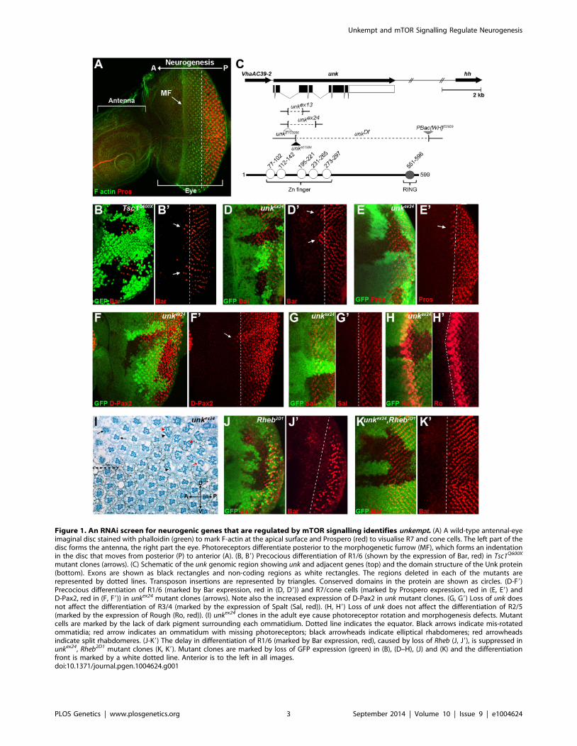
Figure 1. An RNAi screen for neurogenic genes that are regulated by mTOR signalling identifies unkempt. (A) A wild-type antennal-eyeimaginal disc stained with phalloidin (green) to mark F-actin at the apical surface and Prospero (red) to visualise R7 and cone cells. The left part of thedisc forms the antenna, the right part the eye. Photoreceptors differentiate posterior to the morphogenetic furrow (MF), which forms an indentationin the disc that moves from posterior (P) to anterior (A). (B, B9) Precocious differentiation of R1/6 (shown by the expression of Bar, red) in Tsc1Q600X
mutant clones (arrows). (C) Schematic of the unk genomic region showing unk and adjacent genes (top) and the domain structure of the Unk protein(bottom). Exons are shown as black rectangles and non-coding regions as white rectangles. The regions deleted in each of the mutants arerepresented by dotted lines. Transposon insertions are represented by triangles. Conserved domains in the protein are shown as circles. (D-F9)Precocious differentiation of R1/6 (marked by Bar expression, red in (D, D9)) and R7/cone cells (marked by Prospero expression, red in (E, E9) andD-Pax2, red in (F, F9)) in unkex24 mutant clones (arrows). Note also the increased expression of D-Pax2 in unk mutant clones. (G, G9) Loss of unk doesnot affect the differentiation of R3/4 (marked by the expression of Spalt (Sal, red)). (H, H9) Loss of unk does not affect the differentiation of R2/5(marked by the expression of Rough (Ro, red)). (I) unkex24 clones in the adult eye cause photoreceptor rotation and morphogenesis defects. Mutantcells are marked by the lack of dark pigment surrounding each ommatidium. Dotted line indicates the equator. Black arrows indicate mis-rotatedommatidia; red arrow indicates an ommatidum with missing photoreceptors; black arrowheads indicate elliptical rhabdomeres; red arrowheadsindicate split rhabdomeres. (J-K9) The delay in differentiation of R1/6 (marked by Bar expression, red), caused by loss of Rheb (J, J9), is suppressed inunkex24, Rheb2D1 mutant clones (K, K9). Mutant clones are marked by loss of GFP expression (green) in (B), (D–H), (J) and (K) and the differentiationfront is marked by a white dotted line. Anterior is to the left in all images.doi:10.1371/journal.pgen.1004624.g001
Unkempt and mTOR Signalling Regulate Neurogenesis
PLOS Genetics | www.plosgenetics.org 3 September 2014 | Volume 10 | Issue 9 | e1004624
or Prospero (Figure S2 A–C). Thus, loss of unk phenocopies the
precocious photoreceptor differentiation phenotype caused by
activation of InR/mTOR signalling. This suggests that unkactivity is normally repressed by mTOR during photoreceptor
differentiation. The R1/6 precocious differentiation phenotype in
unk mutant clones was rescued by overexpression of unk cDNA
(Figure S1D), demonstrating that the precocious differentiation
phenotype is specifically due to loss of unk.
To test whether increased expression of unk is sufficient to delay
photoreceptor differentiation, MARCM (mosaic analysis with a
repressible cell marker) clones were generated that overexpressed
unk. No change in the timing of differentiation of R1/6 was seen
in these clones (Figure S1E). Therefore, unk is necessary but not
sufficient to regulate the timing of differentiation of R1/6/7 and
cone cells.
In the adult eye unk mutant clones had a striking phenotype.
Mutant ommatidia in the anterior half of the eye had a normal
structure but had rotation defects (Figure 1I, black arrows),
similar to Tsc1 and Pten mutant clones [8]. Ommatidia in the
posterior half of the eye were missing photoreceptors (red arrow)
and contained photoreceptors with elliptical (black arrowheads)
and split (red arrowheads) rhabdomeres (Figure 1I). This
phenotype is typical of perturbation of the F-actin cytoskeleton
and Pten and Tsc1 mutant clones cause similar defects in
photoreceptor apical membrane morphogenesis ([25,26] and
Figure S1F). In summary these data demonstrate that unk is
necessary for the normal timing of differentiation and morpho-
genesis of photoreceptors.
Loss of unk suppresses the delay in photoreceptordifferentiation caused by inhibition of InR/mTORsignalling
To test whether unk acts genetically downstream of InR/
mTOR signalling, we generated double mutant clones that lacked
both unk and Rheb. Compared to Rheb clones, which cause a
strong delay in the differentiation of R1/6 (Figure 1J), differen-
tiation in unk, Rheb clones appeared normal (Figure 1K). Thus,
the strong delay caused by the loss of Rheb was suppressed, but
photoreceptors in these double mutant clones did not differentiate
precociously as in unk mutant clones (Figure 1D). In the adult eye
the elliptical and split rhabdomere phenotype seen in unk mutant
clones was suppressed in unk, Rheb mutant clones (Figure S1H).
However, both Rheb and unk, Rheb mutant clones contained mis-
rotated ommatidia and missing photoreceptors (Figure S1G, H).
Therefore, although unk suppresses the delay in photoreceptor
differentiation caused by inhibition of InR/mTOR signalling,
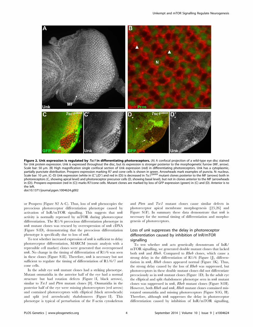
Figure 2. Unk expression is regulated by Tsc1 in differentiating photoreceptors. (A) A confocal projection of a wild-type eye disc stainedfor Unk protein expression. Unk is expressed throughout the disc, but its expression is stronger posterior to the morphogenetic furrow (MF, arrow).Scale bar: 50 mm. (B) High magnification single confocal section of Unk expression (red) in differentiating photoreceptors. Unk has a cytoplasmic,partially punctate distribution. Prospero expression marking R7 and cone cells is shown in green. Arrowheads mark examples of puncta. N: nucleus.Scale bar: 10 mm. (C–D) Unk expression (white in (C9),(D9) and red in (D)) is decreased in Tsc1Q600X mutant clones posterior to the MF (arrows) both inphotoreceptors (C, showing apical level) and photoreceptor precursor cells (D, showing basal level), but not in clones anterior to the MF (arrowheadsin (D)). Prospero expression (red in (C)) marks R7/cone cells. Mutant clones are marked by loss of GFP expression (green) in (C) and (D). Anterior is tothe left.doi:10.1371/journal.pgen.1004624.g002
Unkempt and mTOR Signalling Regulate Neurogenesis
PLOS Genetics | www.plosgenetics.org 4 September 2014 | Volume 10 | Issue 9 | e1004624
there may be an additional factor(s) that regulates R1/6/7 and
cone cell fate and acts in parallel with unk (see Discussion).
Unk expression is negatively regulated by InR/mTORsignalling in photoreceptor neurons
Unk is a ubiquitously expressed cytoplasmic protein (Figure 2A,
B). In the eye disc Unk is expressed in undifferentiated cells
anterior to the MF, in photoreceptor precursors posterior to the
MF, photoreceptors and cone cells (Figure 2A, B and 3B, C).
Moreover, Unk is expressed more strongly posterior to the MF in
the apical plane of the disc containing differentiated photorecep-
tors and cone cells (Figure 2A and S3B). Although localised to the
cytoplasm, Unk has a partially punctate distribution (Figure 2B).
In accordance with the negative regulation of unk expression by
mTOR in S2 cells ([18] and Table S1), Unk expression is reduced
in Tsc1 clones posterior to the MF both in differentiated
photoreceptors and undifferentiated photoreceptor precursors
(Figure 2C, D, arrows). However, Unk expression is unchanged
in Tsc1 clones anterior to the MF (Figure 2D, arrowheads). Thus,
Unk expression is negatively regulated by the InR/mTOR
pathway in differentiating photoreceptor neurons and photore-
ceptor precursors. Inhibition of InR/mTOR signalling, using
Dp110, or Rheb mutant clones, did not cause an increase in Unk
expression (Figure S4) and so Unk expression is not positively
regulated by inhibition of mTOR signalling.
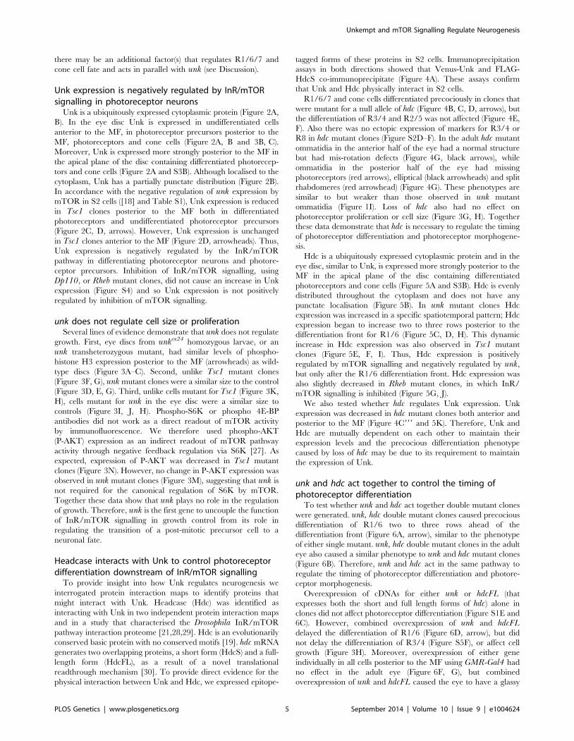
unk does not regulate cell size or proliferationSeveral lines of evidence demonstrate that unk does not regulate
growth. First, eye discs from unkex24 homozygous larvae, or an
unk transheterozygous mutant, had similar levels of phospho-
histone H3 expression posterior to the MF (arrowheads) as wild-
type discs (Figure 3A–C). Second, unlike Tsc1 mutant clones
(Figure 3F, G), unk mutant clones were a similar size to the control
(Figure 3D, E, G). Third, unlike cells mutant for Tsc1 (Figure 3K,
H), cells mutant for unk in the eye disc were a similar size to
controls (Figure 3I, J, H). Phospho-S6K or phospho 4E-BP
antibodies did not work as a direct readout of mTOR activity
by immunofluorescence. We therefore used phospho-AKT
(P-AKT) expression as an indirect readout of mTOR pathway
activity through negative feedback regulation via S6K [27]. As
expected, expression of P-AKT was decreased in Tsc1 mutant
clones (Figure 3N). However, no change in P-AKT expression was
observed in unk mutant clones (Figure 3M), suggesting that unk is
not required for the canonical regulation of S6K by mTOR.
Together these data show that unk plays no role in the regulation
of growth. Therefore, unk is the first gene to uncouple the function
of InR/mTOR signalling in growth control from its role in
regulating the transition of a post-mitotic precursor cell to a
neuronal fate.
Headcase interacts with Unk to control photoreceptordifferentiation downstream of InR/mTOR signalling
To provide insight into how Unk regulates neurogenesis we
interrogated protein interaction maps to identify proteins that
might interact with Unk. Headcase (Hdc) was identified as
interacting with Unk in two independent protein interaction maps
and in a study that characterised the Drosophila InR/mTOR
pathway interaction proteome [21,28,29]. Hdc is an evolutionarily
conserved basic protein with no conserved motifs [19]. hdc mRNA
generates two overlapping proteins, a short form (HdcS) and a full-
length form (HdcFL), as a result of a novel translational
readthrough mechanism [30]. To provide direct evidence for the
physical interaction between Unk and Hdc, we expressed epitope-
tagged forms of these proteins in S2 cells. Immunoprecipitation
assays in both directions showed that Venus-Unk and FLAG-
HdcS co-immunoprecipitate (Figure 4A). These assays confirm
that Unk and Hdc physically interact in S2 cells.
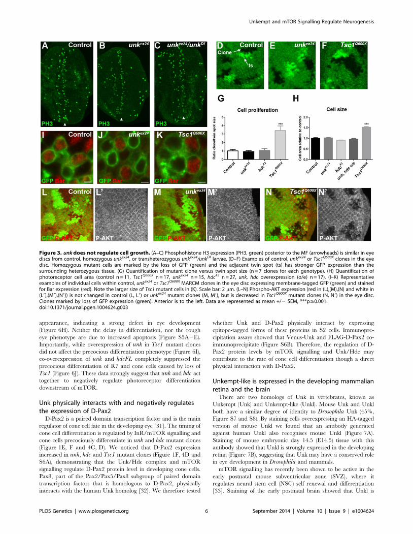
R1/6/7 and cone cells differentiated precociously in clones that
were mutant for a null allele of hdc (Figure 4B, C, D, arrows), but
the differentiation of R3/4 and R2/5 was not affected (Figure 4E,
F). Also there was no ectopic expression of markers for R3/4 or
R8 in hdc mutant clones (Figure S2D–F). In the adult hdc mutant
ommatidia in the anterior half of the eye had a normal structure
but had mis-rotation defects (Figure 4G, black arrows), while
ommatidia in the posterior half of the eye had missing
photoreceptors (red arrows), elliptical (black arrowheads) and split
rhabdomeres (red arrowhead) (Figure 4G). These phenotypes are
similar to but weaker than those observed in unk mutant
ommatidia (Figure 1I). Loss of hdc also had no effect on
photoreceptor proliferation or cell size (Figure 3G, H). Together
these data demonstrate that hdc is necessary to regulate the timing
of photoreceptor differentiation and photoreceptor morphogene-
sis.
Hdc is a ubiquitously expressed cytoplasmic protein and in the
eye disc, similar to Unk, is expressed more strongly posterior to the
MF in the apical plane of the disc containing differentiated
photoreceptors and cone cells (Figure 5A and S3B). Hdc is evenly
distributed throughout the cytoplasm and does not have any
punctate localisation (Figure 5B). In unk mutant clones Hdc
expression was increased in a specific spatiotemporal pattern; Hdc
expression began to increase two to three rows posterior to the
differentiation front for R1/6 (Figure 5C, D, H). This dynamic
increase in Hdc expression was also observed in Tsc1 mutant
clones (Figure 5E, F, I). Thus, Hdc expression is positively
regulated by mTOR signalling and negatively regulated by unk,
but only after the R1/6 differentiation front. Hdc expression was
also slightly decreased in Rheb mutant clones, in which InR/
mTOR signalling is inhibited (Figure 5G, J).
We also tested whether hdc regulates Unk expression. Unk
expression was decreased in hdc mutant clones both anterior and
posterior to the MF (Figure 4C999 and 5K). Therefore, Unk and
Hdc are mutually dependent on each other to maintain their
expression levels and the precocious differentiation phenotype
caused by loss of hdc may be due to its requirement to maintain
the expression of Unk.
unk and hdc act together to control the timing ofphotoreceptor differentiation
To test whether unk and hdc act together double mutant clones
were generated. unk, hdc double mutant clones caused precocious
differentiation of R1/6 two to three rows ahead of the
differentiation front (Figure 6A, arrow), similar to the phenotype
of either single mutant. unk, hdc double mutant clones in the adult
eye also caused a similar phenotype to unk and hdc mutant clones
(Figure 6B). Therefore, unk and hdc act in the same pathway to
regulate the timing of photoreceptor differentiation and photore-
ceptor morphogenesis.
Overexpression of cDNAs for either unk or hdcFL (that
expresses both the short and full length forms of hdc) alone in
clones did not affect photoreceptor differentiation (Figure S1E and
6C). However, combined overexpression of unk and hdcFLdelayed the differentiation of R1/6 (Figure 6D, arrow), but did
not delay the differentiation of R3/4 (Figure S5F), or affect cell
growth (Figure 3H). Moreover, overexpression of either gene
individually in all cells posterior to the MF using GMR-Gal4 had
no effect in the adult eye (Figure 6F, G), but combined
overexpression of unk and hdcFL caused the eye to have a glassy
Unkempt and mTOR Signalling Regulate Neurogenesis
PLOS Genetics | www.plosgenetics.org 5 September 2014 | Volume 10 | Issue 9 | e1004624
appearance, indicating a strong defect in eye development
(Figure 6H). Neither the delay in differentiation, nor the rough
eye phenotype are due to increased apoptosis (Figure S5A2E).
Importantly, while overexpression of unk in Tsc1 mutant clones
did not affect the precocious differentiation phenotype (Figure 6I),
co-overexpression of unk and hdcFL completely suppressed the
precocious differentiation of R7 and cone cells caused by loss of
Tsc1 (Figure 6J). These data strongly suggest that unk and hdc act
together to negatively regulate photoreceptor differentiation
downstream of mTOR.
Unk physically interacts with and negatively regulatesthe expression of D-Pax2
D-Pax2 is a paired domain transcription factor and is the main
regulator of cone cell fate in the developing eye [31]. The timing of
cone cell differentiation is regulated by InR/mTOR signalling and
cone cells precociously differentiate in unk and hdc mutant clones
(Figure 1E, F and 4C, D). We noticed that D-Pax2 expression
increased in unk, hdc and Tsc1 mutant clones (Figure 1F, 4D and
S6A), demonstrating that the Unk/Hdc complex and mTOR
signalling regulate D-Pax2 protein level in developing cone cells.
Pax8, part of the Pax2/Pax5/Pax8 subgroup of paired domain
transcription factors that is homologous to D-Pax2, physically
interacts with the human Unk homolog [32]. We therefore tested
whether Unk and D-Pax2 physically interact by expressing
epitope-tagged forms of these proteins in S2 cells. Immunopre-
cipitation assays showed that Venus-Unk and FLAG-D-Pax2 co-
immunoprecipitate (Figure S6B). Therefore, the regulation of D-
Pax2 protein levels by mTOR signalling and Unk/Hdc may
contribute to the rate of cone cell differentiation though a direct
physical interaction with D-Pax2.
Unkempt-like is expressed in the developing mammalianretina and the brain
There are two homologs of Unk in vertebrates, known as
Unkempt (Unk) and Unkempt-like (Unkl). Mouse Unk and Unkl
both have a similar degree of identity to Drosophila Unk (45%,
Figure S7 and S8). By staining cells overexpressing an HA-tagged
version of mouse Unkl we found that an antibody generated
against human Unkl also recognises mouse Unkl (Figure 7A).
Staining of mouse embryonic day 14.5 (E14.5) tissue with this
antibody showed that Unkl is strongly expressed in the developing
retina (Figure 7B), suggesting that Unk may have a conserved role
in eye development in Drosophila and mammals.
mTOR signalling has recently been shown to be active in the
early postnatal mouse subventricular zone (SVZ), where it
regulates neural stem cell (NSC) self renewal and differentiation
[33]. Staining of the early postnatal brain showed that Unkl is
Figure 3. unk does not regulate cell growth. (A–C) Phosphohistone H3 expression (PH3, green) posterior to the MF (arrowheads) is similar in eyediscs from control, homozygous unkex24, or transheterozygous unkex24/unkDf larvae. (D–F) Examples of control, unkex24 or Tsc1Q600X clones in the eyedisc. Homozygous mutant cells are marked by the loss of GFP (green) and the adjacent twin spot (ts) has stronger GFP expression than thesurrounding heterozygous tissue. (G) Quantification of mutant clone versus twin spot size (n = 7 clones for each genotype). (H) Quantification ofphotoreceptor cell area (control n = 11, Tsc1Q600X n = 17, unkex24 n = 15, hdc43 n = 27, unk, hdc overexpression (o/e) n = 17). (I–K) Representativeexamples of individual cells within control, unkex24 or Tsc1Q600X MARCM clones in the eye disc expressing membrane-tagged GFP (green) and stainedfor Bar expression (red). Note the larger size of Tsc1 mutant cells in (K). Scale bar: 2 mm. (L–N) Phospho-AKT expression (red in (L),(M),(N) and white in(L9),(M9),(N9)) is not changed in control (L, L9) or unkex24 mutant clones (M, M9), but is decreased in Tsc1Q600X mutant clones (N, N9) in the eye disc.Clones marked by loss of GFP expression (green). Anterior is to the left. Data are represented as mean +/2 SEM, ***p#0.001.doi:10.1371/journal.pgen.1004624.g003
Unkempt and mTOR Signalling Regulate Neurogenesis
PLOS Genetics | www.plosgenetics.org 6 September 2014 | Volume 10 | Issue 9 | e1004624
expressed throughout the brain (Figure S9A). Unkl is expressed
throughout the SVZ, but is most strongly expressed in the cells
close to the ventricle, similar to phosphorylated 4E-BP (P-4E-BP),
a marker of mTOR pathway activity (Figure 7C, D). Further
analyses showed that glial fibrillary acidic protein (GFAP) positive
NSCs, Mash1 positive transit amplifying progenitors (TAPs) and
neuroblasts (identified by the expression of doublecortin (Dcx)) all
express Unkl (Figure 7E–G and S9B–D). These data suggest that
Unkl may play a role in mTOR-dependent neural stem/
progenitor cell differentiation in the mammalian CNS. Thus,
Unk may act downstream of InR/mTOR signalling to regulate
neuronal differentiation in both Drosophila and mammals.
Discussion
Several lines of evidence together demonstrate that unk and hdcact downstream of InR/mTOR signalling to negatively regulate
the timing of photoreceptor cell fate. First, loss of either unk or
hdc causes precocious differentiation of the same cells and to the
same degree as activation of InR/mTOR signalling. Second,
the expression of both Unk and Hdc are regulated by InR/
mTOR signalling. Third, loss of unk suppresses the strong delay
in photoreceptor differentiation caused by inhibition of the
InR/mTOR pathway and combined overexpression of unk and
hdc suppresses the precocious photoreceptor differentiation
caused by loss of Tsc1. Fourth, although Unk has been shown
to physically interact with mTOR [21], neither unk nor hdcregulate cell or tissue growth. Taken together these data show
that unk and hdc are novel downstream components of the InR/
mTOR pathway that regulate the timing of neuronal differen-
tiation (Figure 8).
InR/mTOR signalling is a major regulator of cell growth. In
Drosophila activation of InR/mTOR signalling by loss of either
Figure 4. Hdc physically interacts with Unk and negatively regulates neurogenesis. (A) Hdc physically interacts with Unk. Venus-Unk orFLAG-HdcS were expressed alone or together in S2 cells and immunoprecipitated with GFP or FLAG antibodies. (B, B9) hdc43 mutant clones showingprecocious differentiation of R1/6 (arrows), marked by the expression of Bar (red). (C-C999) hdc43 mutant clones showing precocious differentiation(arrow) of R7 and cone cells (marked by the expression of Prospero (red)) and decreased expression of Unk (white in (C0) and (C999)). Thedifferentiation front is marked by a dotted line. (D, D9) Precocious differentiation of cone cells (marked by the expression of D-Pax2, red) in a hdc43
mutant clone. Arrow indicates cone cells that have differentiated precociously. Note also the increased expression of D-Pax2 in hdc mutant clones.(E, E9) Loss of hdc does not affect the differentiation of R3/4 (marked by the expression of Spalt (Sal, red)). (F, F9) Loss of hdc does not affect thedifferentiation of R2/5 (marked by the expression of Rough (Ro, red)). (G) hdc43 mutant clones in the adult eye cause defects in ommatidial rotationand morphogenesis. Mutant cells are marked by the lack of dark pigment. Black arrows indicate mis-rotated ommatidia; red arrows indicateommatidia with missing photoreceptors; black arrowheads indicate elliptical rhabdomeres; red arrowhead indicates split rhabdomeres. Mutantclones are marked by loss of GFP expression (green) in (B)–(F). Anterior is to the left.doi:10.1371/journal.pgen.1004624.g004
Unkempt and mTOR Signalling Regulate Neurogenesis
PLOS Genetics | www.plosgenetics.org 7 September 2014 | Volume 10 | Issue 9 | e1004624
Tsc1, Tsc2, Pten, or overexpression of Rheb causes increased cell
size and proliferation [34–37]. In the genetic disease TSC, which
is caused by mutations in Tsc1 or Tsc2, patients develop benign
tumours in multiple organs including the brain [4]. The previously
identified components of the InR/mTOR pathway regulate both
growth and neurogenesis in Drosophila and vertebrate model
systems [8,10,13,14,16,17,38]. unk and hdc therefore represent a
branchpoint in the pathway where its function in neurogenesis
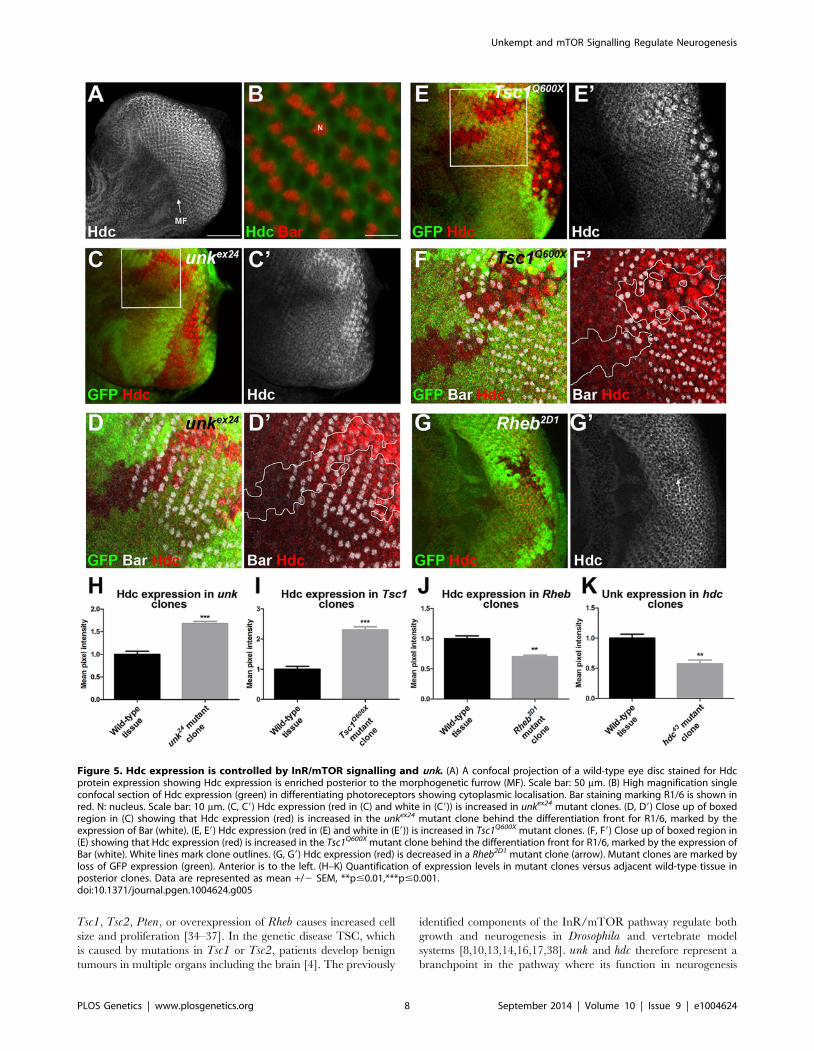
Figure 5. Hdc expression is controlled by InR/mTOR signalling and unk. (A) A confocal projection of a wild-type eye disc stained for Hdcprotein expression showing Hdc expression is enriched posterior to the morphogenetic furrow (MF). Scale bar: 50 mm. (B) High magnification singleconfocal section of Hdc expression (green) in differentiating photoreceptors showing cytoplasmic localisation. Bar staining marking R1/6 is shown inred. N: nucleus. Scale bar: 10 mm. (C, C9) Hdc expression (red in (C) and white in (C9)) is increased in unkex24 mutant clones. (D, D9) Close up of boxedregion in (C) showing that Hdc expression (red) is increased in the unkex24 mutant clone behind the differentiation front for R1/6, marked by theexpression of Bar (white). (E, E9) Hdc expression (red in (E) and white in (E9)) is increased in Tsc1Q600X mutant clones. (F, F9) Close up of boxed region in(E) showing that Hdc expression (red) is increased in the Tsc1Q600X mutant clone behind the differentiation front for R1/6, marked by the expression ofBar (white). White lines mark clone outlines. (G, G9) Hdc expression (red) is decreased in a Rheb2D1 mutant clone (arrow). Mutant clones are marked byloss of GFP expression (green). Anterior is to the left. (H–K) Quantification of expression levels in mutant clones versus adjacent wild-type tissue inposterior clones. Data are represented as mean +/2 SEM, **p#0.01,***p#0.001.doi:10.1371/journal.pgen.1004624.g005
Unkempt and mTOR Signalling Regulate Neurogenesis
PLOS Genetics | www.plosgenetics.org 8 September 2014 | Volume 10 | Issue 9 | e1004624

bifurcates from that in growth control (Figure 8). Moreover, our
analysis of unk and hdc demonstrates that regulation of cell growth
can be uncoupled from and is not required for the function of
InR/mTOR signalling in the temporal control of neuronal
differentiation.
At the protein level we show that Unk and Hdc physically
interact in S2 cells. Although this interaction remains to be
demonstrated in vivo, the additional observations that they both
regulate each other’s expression and act synergistically in vivo
strongly support the model that they physically interact (Figure 8).
Moreover, Unk and Hdc have also previously been shown to
physically interact by yeast-2-hybrid and co-immunoprecipitation
[21,28,29]. Unk and Hdc are both expressed in all developing
photoreceptors and so we hypothesise that they control the timing
of differentiation through the regulation of neurogenic factors
whose expression is restricted to R1/6/7 and cone cells (Figure 8).
Loss of unk causes increased expression of D-Pax2, the main
regulator of cone cell differentiation. hdc and Tsc1 mutant clones
also cause a similar increase in D-Pax2 expression. Overexpression
of D-Pax2 alone is insufficient to induce cone cell differentiation,
which requires overexpression of both D-Pax2 and Tramtrack88
(TTK88) [39]. Thus, regulation of D-Pax2 expression by mTOR
signalling may contribute to the rate of cone cell differentiation,
while overall control would require the regulation of additional
factors such as TTK88 (Figure 8). Pax8, part of the Pax2/Pax5/
Pax8 paired domain transcription factor subgroup that is
homologous to D-Pax2, has been shown to physically interact
with one of the two human homologs of Unkempt [32]. We find
that Drosophila Unk physically interacts with D-Pax2, demon-
strating that the physical interaction between Unk and this group
of transcription factors is conserved. We suggest that D-Pax2 may
be one of several neurogenic factors regulated by InR/mTOR
signalling, through a physical interaction with the Unk/Hdc
complex, to control the timing of R1/6/7 and cone cell fate
(Figure 8).
Unk has been shown to physically interact with mTOR and the
strength of this interaction is regulated by insulin [21]. This
suggests the intriguing possibility that the inhibition of Unk activity
by InR/mTOR signalling is dependent on the strength of the
physical interaction between Unk and the mTORC1 complex.
Unk was also identified as part of the mTOR-regulated
phosphoproteome in both human and murine cells [22,23]. Thus,
Unk may potentially be regulated by mTOR through phosphor-
ylation. Future studies will fully characterise the mechanism by
which mTORC1 regulates Unk activity.
Our study represents the first demonstration of a role for unk in
specific developmental processes. By contrast, hdc has previously
been shown to regulate dendritic pruning during metamorphosis
and to act as a branching inhibitor during tracheal development
[30,40]. A screen for genes affecting tracheal tube morphogenesis
and branching recently identified Tsc1 [41], suggesting that InR/
mTOR also regulates tracheal development. Thus, hdc and unkmay act repeatedly as downstream effectors of the InR/mTOR
pathway during Drosophila development.
The one previous study of either of the mammalian Unk
homologs showed that Unkl binds specifically to an activated form
of the Rac1 GTPase [42]. If this function is conserved in
Drosophila then the defects in photoreceptor apical membrane
morphogenesis caused by activation of mTOR signalling or loss of
unk/hdc may be mediated through Rac1.
The function of the two unk homologs, unk and unkl, in
mammalian development is not known, but unk has been shown
to be expressed in the mouse early postnatal mouse retina [43].
We find that Unkl is also expressed in the developing mouse
Figure 6. unk and hdc act together to control the timing ofphotoreceptor differentiation. (A, A9) An unkex24, hdc43 doublemutant clone causes a similar precocious differentiation of R1/6phenotype (arrow) to either single mutant. (B) An unkex24, hdc43 doublemutant clone in the adult eye causes similar ommatidial rotation andmorphogenesis defects to unk and hdc mutant clones. Mutant cells aremarked by the lack of pigment. Black arrow indicates a mis-rotatedommatidium; red arrows indicate ommatidia with missing photorecep-tors; black arrowheads indicate elliptical rhabdomeres; red arrowheadsindicate split rhabdomeres. (C, C9) Overexpression of hdcFL does notaffect the differentiation of R1/6. (D, D9) Combined overexpression ofunk and hdcFL cause a delay in the differentiation of R1/6 (arrow). (E2H)Combined overexpression of unk and hdcFL affects eye development.Eyes from GMR-Gal4 control (E), or GMR-Gal4 driving the expression ofunk (F), hdcFL (G), or unk, hdcFL (H) in female flies. Note the glassyappearance in (H). (I, I9) Overexpression of unk in a Tsc1Q600X clone doesnot affect the precocious differentiation of R7 and cone cells. (J, J9)Overexpression of unk and hdc in a Tsc1Q600X clone completelysuppresses the precocious differentiation of R7 and cone cells. MARCMwas used to generate clones in (A), (C), (D), (I) and (J) and so clonal cellsare marked by GFP expression (green). Bar (red) marks R1/6 in (A), (C)and (D), while Prospero expression (Pros, red) marks R7 and cone cells in(I) and (J). The differentiation front is marked by a dotted line. Anterior isto the left.doi:10.1371/journal.pgen.1004624.g006
Unkempt and mTOR Signalling Regulate Neurogenesis
PLOS Genetics | www.plosgenetics.org 9 September 2014 | Volume 10 | Issue 9 | e1004624
Unkempt and mTOR Signalling Regulate Neurogenesis
PLOS Genetics | www.plosgenetics.org 10 September 2014 | Volume 10 | Issue 9 | e1004624
retina, suggesting that Unk may play a conserved role in eye
development in both flies and mammals. InR/mTOR signalling
acts as a pro-survival pathway preventing retinal degeneration
[44], but its role in mammalian eye development has not been
characterised. By contrast InR/mTOR signalling has a well
characterised role in NSC self-renewal and differentiation in the
mouse SVZ. Loss of Tsc1 or expression of a constitutively active
form of Rheb in neural progenitor cells in the postnatal mouse
SVZ causes the formation of heterotopias, ectopic neurons and
olfactory micronodules [15,16]. Furthermore, individuals with
TSC, which results in activated mTOR signalling, have aberrant
cortical neurogenesis and develop benign cortical tumours during
foetal development and throughout childhood [4,45]. mTOR
signalling has been shown to be active in proliferative NSCs and
TAPs in the neonatal SVZ and inhibition of mTOR signalling
prevents NSC differentiation [33]. We find that Unkl is expressed
in both NSCs and TAPs in the early postnatal SVZ. Thus, Unkl
may regulate NSC differentiation downstream of mTOR
signalling in the mammalian brain. Unkempt may therefore play
a conserved role in regulating the timing of neural cell fate
downstream of mTOR signalling in both flies and mammals.
Materials and Methods
Fly strains and genetic crossesFlies were maintained on standard yeast, glucose, cornmeal,
agar food at 25uC unless stated otherwise. Fly stocks were
FRT82B, Dp110A [46], FRT82B, Tsc1Q600X [34], FRT82B,Tsc1Q87X [37], FRT82B, Rheb2D1 [36], FRT82B, hdc43 [19] and
UAS-hdcFL [30] and UAS-unk (this study). For clonal experi-
ments the stocks used were y, w, hs-flp; FRT82B, ubi-GFP, y, w,hs-flp; FRT82B, M[95A]Rps63, ubi-GFP and y, w, hs-flp;tub-Gal4, UAS-mCD8GFP;FRT82B, tub-Gal80 (MARCM stock).
RNAi lines were obtained from the Vienna Drosophila Resource
Centre. All other stocks were obtained from the Bloomington
Stock Centre. For mosaic analysis mutant clones were generated
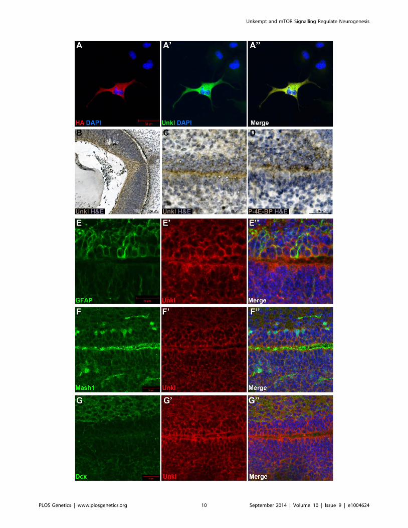
Figure 7. Unkl is expressed in the developing mammalian eye and SVZ. (A-A0) COS-7 cells overexpressing HA-tagged mouse Unkl stainedfor HA expression (red), Unkl expression (green) and DAPI (blue). (B) Hematoxylin and eosin (H&E) (blue) stained coronal section from a mouse E14.5retina showing strong Unkl expression (brown) in the retina. (C, D) Serial H&E (blue) stained sagittal sections of the lateral SVZ from a P0 mouseshowing Unkl expression (brown in (C)) and P-4E-BP expression (brown in (D)). (E-G0) Sagittal sections of the lateral SVZ from a P0 mouse showingUnkl expression (red in (E9, E0), (G9, G0), (F9, F0)) and NSCs (stained for GFAP, green in (E, E0)), TAPs (stained for Mash1, green in (F, F0)), or neuroblasts(stained for Dcx, green in (G, G0)). DAPI staining is shown in blue in (E0), (F0), (G0).doi:10.1371/journal.pgen.1004624.g007
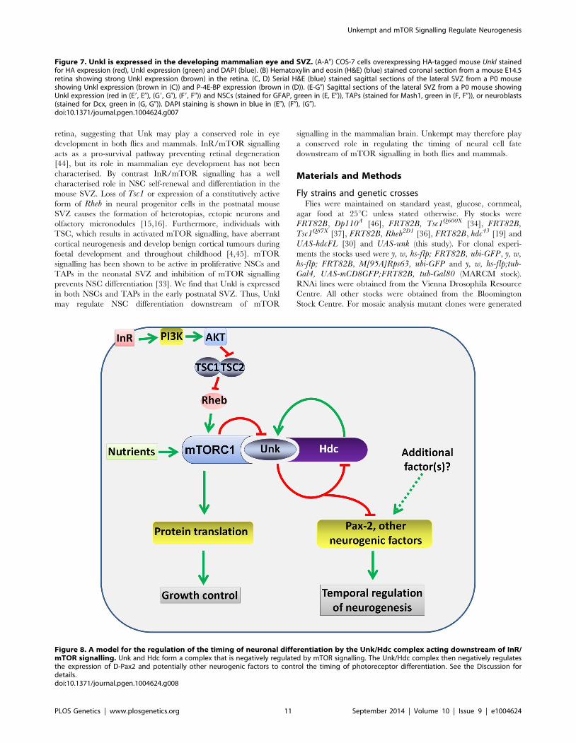
Figure 8. A model for the regulation of the timing of neuronal differentiation by the Unk/Hdc complex acting downstream of InR/mTOR signalling. Unk and Hdc form a complex that is negatively regulated by mTOR signalling. The Unk/Hdc complex then negatively regulatesthe expression of D-Pax2 and potentially other neurogenic factors to control the timing of photoreceptor differentiation. See the Discussion fordetails.doi:10.1371/journal.pgen.1004624.g008
Unkempt and mTOR Signalling Regulate Neurogenesis
PLOS Genetics | www.plosgenetics.org 11 September 2014 | Volume 10 | Issue 9 | e1004624
by Flp/FRT mediated recombination using heat-shock-flp or by
MARCM [47]. For clonal analysis in eye discs larvae were heat-
shocked for 1-1.5 hours at 37uC 24 hours after egg laying (AEL).
For adult clones larvae were heat-shocked for 30 minutes at 37uC24 hours and again at 48 hours AEL. Adult eye sections were
prepared as described previously [48].
The line P[EPgy2]unkEY03956 was used to generate mutants
unkex13 and unkex24 through imprecise P-element excision [49].
The deleted region in both unkex13 and unkex24 begins 557 bp into
the second intron and for unkex13 ends 219 bp into the third exon
and for unkex24 ends 150 bp into the fourth intron. The PiggyBac
lines, PBac[RB]unke01984 and PBac[WH]f03929 were used to
generate unkDf using a Flp/FRT –based precise excision strategy
[50]. Mutation break points and deleted regions were confirmed
or determined by PCR and sequencing.
Generation of the Unk antibody, staining andimmunofluorescence
The Unk antibody was generated using a GST-fusion protein as
described in Text S1. The antibodies used were: rat anti-Unk3
(1:500, this study), mouse anti-Hdc (a gift from Robert White, 1:5)
[19], rat anti-Bar (1:500) [10], rat anti-Elav (DSHB, 1:100), mouse
anti-Prospero (DSHB, 1:100), mouse anti-Rough (DSHB, 1:10),
rabbit anti-Spalt (a gift from R. Barrio, 1:500), guinea pig anti-
senseless (a gift from Hugo Bellen, 1:500), rabbit anti-cleaved
caspase 3 (Cell Signalling, 1:100), rabbit anti-D-Pax2 (a gift from
M. Noll, 1:20) [31], rabbit anti-P-AKT (Cell Signalling, 4045S,
1:200), rabbit or mouse anti-GFP (Life Technologies, 1:1000),
rabbit anti-PH3 (Upstate, 1:50), rabbit anti RING finger protein
unkempt like (Abcam ab155197, 1:300), rat anti HA (Roche),
mouse anti-Mash1 (a gift from Francois Guillemot, 1:20), mouse
anti GFAP (Sigma, G3893, 1:1000) and goat anti Dcx (Santa Cruz
Sc8066, 1:200).
For immunofluorescence, dissected third instar larvae were
fixed for 30 minutes in 4% paraformaldehyde, then washed five
times for 10 minutes in PBS/0.1% triton (PBST), before blocking
in PBST/1% normal goat serum (PBST-NGS) for an additional
hour. Eye discs were incubated with primary antibody overnight
in PBST-NGS at 4uC. After five to six washes of 10 minutes in
PBST discs were incubated for two hours with secondary antibody
at room temperature, washed five to six times in PBST and then
mounted in Vectashield (Vectalabs). Phospho-AKT staining was
performed exactly as described [51].
P0 mouse CNS tissue was fixed overnight in 4% paraformalde-
hyde then embedded in paraffin and 6 mm sections were cut. To
generate primary neural progenitor monolayers, subventricular
zone fragments from P1 mice were triturated with 2 ml of HBSS
containing 0.25% trypsin (Gibco) and 40 ml of DNAse l (1 mg/ml,
Worthington) and incubated at 37uC for 2 minutes. The trypsin was
inactivated with 5 ml of DMEM (Gibco) containing 10% FCS and
the solution was centrifuged at 1,500 rpm for 5 minutes. After
another two washes with DMEM/10% FCS to remove any traces
of trypsin, the pellet was re-suspended in pre-equilibrated (at 37uC/
5% CO2) Neurobasal complete medium (Gibco) containing B27
supplement, 2 mM L-glutamine (Invitrogen) and 0.6% glucose
(Sigma). Cells were plated onto 24 well plates (2.56106 cells/well)
on glass coated with polyornithine (0.5 mg/ml, Sigma). Cells were
maintained in Neurobasal complete medium at 37uC/5% CO2 for
24 hours then fixed and stained as for COS-7 cells (below).
COS-7 cells were fixed for 30 minutes in 4% paraformalde-
hyde, washed several times in PBS, blocked for one hour in PBS/
1% bovine serum albumen/0.2% triton/0.02% sodium azide,
then incubated overnight at 4uC in the primary antibody diluted in
blocking buffer. Secondary antibodies were FITC donkey anti-
mouse, Cy3 donkey anti-rat, Cy5 donkey anti-rat and Cy5 donkey
anti-mouse (Jackson Immunolabs); Alexa488 anti-rabbit, Alexa594
anti-rabbit and Alexa594 anti-mouse (Life Technologies). Images
were acquired on a Zeiss LSM710 confocal microscope and
processed in Photoshop CS4 (Adobe). The differentiation front
was marked with a dotted line positioned just ahead of the most
anterior row of photoreceptors.
Cloning of unk, hdc and D-Pax2Details of the cloning of unk, hdc and D-Pax2 are described in
Text S1. UAS-unk transgenic lines were generated by germline
transformation (BestGene Inc.). Overexpression of Unk using
these lines was confirmed by immunostaining.
Cell culture and immunoprecipitationFor immunoprecipitation (IP) experiments Venus-unk, FLAG-hdcS
and FLAG-D-Pax2 were expressed in S2 cells (DGRC) cultured in
SF9-S2 medium (PAA laboratories). For IP experiments, cells were
seeded in six well plates at a density of around 1.5 million cells per well.
Cells were then transfected with 4.5 mg of Venus-unk, with or without
0.5 mg FLAG-hdcS or 0.5 mg FLAG-D-Pax2 using transfectin (Biorad)
according to the manufacturer’s instructions. The IP protocol was
adapted from [52]. For each condition after 48 hours cells from two
wells were manually detached, washed in cold PBS and lysed for
1.5 hours in lysis buffer (25 mM Tris pH 8, 150 mM NaCl, 5%
glycerol, 1% triton, 1 mM PMSF, 16 protease inhibitor cocktail
(Roche)) at 4uC. 800 mg of protein was then used for IPs. After clearing,
lysates were incubated overnight with the antibody at 4uC. Venus-Unk
was immunoprecipitated with 4 mg of rabbit anti-GFP antibody (Life
Technologies). FLAG-HdcS and FLAG-D-Pax2 were immunoprecip-
itated with 3 mg of rabbit anti-FLAG (Fisher). The complexes were
then immunoprecipitated for 3 hours using Protein G agarose beads
(Thermo Scientific Pierce). After washes in 25 mM Tris pH 8,
150 mM NaCl, immunoprecipitated proteins were recovered by
boiling the beads in 26SDS-PAGE loading buffer and then subjected
to SDS-PAGE followed by immunoblot with rabbit anti-GFP (1:1000)
and mouse monoclonal anti-FLAG M2 (1:500, Agilent).
For overexpression of HA-Unkl COS-7 cells were grown in
Dulbecco’s modified Eagle’s medium (Life Technologies) supple-
mented with 10% fetal bovine serum (Life Technologies), in a
humidified atmosphere of 5% CO2 at 37uC. For transfection COS-7
cells were seeded at a density of 2.56105 cells per well in Opti-MEM
Reduced Serum Medium (Life Technologies) and transfected with
5 mg of pcDNA3-HA-unkl [42] using Lipofectamine 2000 (Life
Technologies) according to the manufacturer’s instructions.
Quantification and statistical analysisFor quantification of photoreceptor cell areas confocal images of
MARCM clones stained with Bar were used. CD8-GFP expres-
sion was used to identify individual cell membranes, which were
manually outlined at the level of R1/6 nuclei (identified by Bar
staining) and cell areas were calculated using ImageJ. Clone and
twin spot areas were manually outlined and the areas calculated
using ImageJ. The numbers of active caspase 3 positive cells in eye
discs were quantified using the quantification tool in Volocity
(Perkin Elmer) from three dimensional confocal images of the
whole disc with scans every 1 mm. Expression levels were
determined in ImageJ using the Measure tool and four mutant
clones/four adjacent areas of wild-type tissue were quantified for
each genotype. Statistical analysis was performed in Graphpad
Prism 5. Statistical significance was determined using an unpaired
Student’s t test for pairwise comparisons, or one way analysis of
Unkempt and mTOR Signalling Regulate Neurogenesis
PLOS Genetics | www.plosgenetics.org 12 September 2014 | Volume 10 | Issue 9 | e1004624
variance (ANOVA) with Dunnett’s multiple comparison post hoc
test for multiple comparisons to the control.
Supporting Information
Figure S1 Identification of unk and further analysis of
differentiation phenotypes. (A) Each ommatidium in the adult
eye consists of 8 photoreceptors arranged in a trapezoid, forming a
mirror image about the equator (dotted line). Anterior is to the left
and dorsal is up. (B, B9) Expression of a dsRNA against unk using
MARCM causes precocious differentiation of R1/6 (marked by
Bar expression (red)). Arrows indicate photoreceptors that have
differentiated ahead of the differentiation front. (C, C9) Complete
loss of Unk protein expression (red in (C) and white in (C9)) in an
unkex24 mutant clone. (D, D9) Expression of unk in unkex24 mutant
cells using MARCM rescues the R1/6 precocious differentiation
phenotype (marked by Bar expression (red)). (E, E9) Overexpres-
sion of unk using MARCM does not affect the differentiation of
R1/6 (marked by Bar expression (red)). (F) Tsc1Q87X mutant
clones in the adult eye showing elliptical (black arrowheads) and
split (red arrowheads) rhabdomeres and ommatidia with missing
photoreceptors (red arrows). (G) Rheb2D1 mutant clones in the
adult eye. (H) unkex24, Rheb2D1 mutant clones in the adult eye.
Ommatidia with missing photoreceptors are indicated by red
arrows. Mutant ommatidia are marked by the lack of surrounding
dark pigment in (F–H). Clonal cells are marked by the presence of
GFP (green) in panels (B), (D) and (E) and by the absence of GFP
in (C). The differentiation front is marked by a white dotted line.
Asterisk in (B), (D) and (E) indicates Bar staining in basal precursor
cells close to the MF. Anterior is to the left.
(TIF)
Figure S2 Precocious differentiation in unk or hdc mutant clones
is not due to ectopic expression of Bar or Prospero. (A-C9) unkex24
clones stained with: (A, A9) Elav (blue) and Prospero (Pros, red); (B,
B9) Spalt (Sal, blue) and Bar (red); (C, C9) Senseless (sense, blue)
and Bar (red). (D-F9) hdc43 clones stained with: (D, D9) Elav (blue)
and Prospero (Pros, red); (E, E9) Spalt (Sal, blue) and Bar (red); (F,
F9) Senseless (sense, blue) and Bar (red). Mutant clones are marked
by loss of GFP expression (green).
(TIF)
Figure S3 Unk and Hdc expression are increased in photore-
ceptors. (A-A0) A wild type late third instar eye disc stained for
expression of Prospero ((A), Pros, a projection image of the whole
disc) and Unk (A9, A0) showing single confocal section of an apical
plane showing differentiated photoreceptors (A9), or a basal plane
showing photoreceptor precursors (A0). (B-B0) A wild type late
third instar eye disc stained for expression of Bar ((B), a projection
image of the whole disc) and Hdc (B9, B0) showing single confocal
section of an apical plane showing differentiated photoreceptors
(B9), or a basal plane showing photoreceptor precursors (B0).
Arrows mark the position of the MF.
(TIF)
Figure S4 Unk expression is not affected by inhibition of InR/
mTOR signalling. Unk expression (red in (A) and white in (A9))
does not change in Dp110A mutant clones. Severely delayed
differentiation of R7 and cone cells is shown by the expression of
Prospero (white in (A)). Mutant clones are marked by loss of GFP
expression (green). The differentiation front is marked by a white
dotted line. Anterior is to the left.
(TIF)
Figure S5 Co-overexpression of unk and hdc does not in-
crease apoptosis or delay the differentiation of R3/4. (A-C9) A
GMR-Gal4 (heterozygous) control eye disc (A, A9), or GMR-Gal4driving the co-expression of unk and hdcFL (B, B9), or the pro-
apoptotic gene hid (C, C9). Active caspase 3 expression marking
apoptotic cells is shown in green and Elav expression marking
differentiated photoreceptors in red. (D) Quantification of the
number of apoptotic cells. N = 4 discs for each genotype. Data are
represented as mean +/2 SEM, ***p#0.001.n.s. not significant.
(E, E9) MARCM clones overexpressing unk and hdc stained for
active caspase 3 expression (Casp, red). (F, F9) MARCM clones
overexpressing unk and hdc stained for Spalt (Sal, red) marking
R3/4. The differentiation front is marked by a white dotted line.
Clones are marked by GFP expression in (E) and (F).
(TIF)
Figure S6 mTOR signalling negatively regulates D-Pax2
expression and Unk physically interacts with D-Pax2. (A, A9)
Increased D-Pax2 expression (red) in Tsc1 mutant clones marked
by loss of GFP expression (green). (B) Venus-Unk or FLAG-D-
Pax2 were expressed alone or together in S2 cells and
immunoprecipitated with FLAG antibody.
(TIF)
Figure S7 Alignment of the primary amino acid sequence of
Drosophila Unk (dmUnk and mouse Unk (mmUnk).
(TIFF)
Figure S8 Alignment of the primary amino acid sequence of
Drosophila Unk (dmUnk) and mouse Unk like (mmUnkL).
(TIF)
Figure S9 Expression of Unkl in primary SVZ cultures. (A, A9)
Sagittal section from the brain of a P0 mouse showing Unkl
expression (green in (A)) and DAPI (blue in (A9)). (B-D9) Primary
cultured cells from the SVZ of a P1 mouse stained for Unkl
(green in (B, B0), (C, C0), (D, D0)) and GFAP (red in (B9, B0)),
Mash1 (red in (C9, C0)) or Dcx (red in (D9, D0)); DAPI shown in
blue in (B0), (C0) and (D0). (E-E0) Primary cultured cells from the
SVZ of a P1 mouse stained in the same way as in (B–D), but
omitting the primary antibody to show the staining for Unkl is
not due to background fluorescence from the secondary
antibody.
(TIF)
Table S1 The 28 transcriptional targets of mTOR that were
screened for photoreceptor differentiation phenotypes. 1[18].2This study. Genes which gave a differentiation phenotype are
highlighted in yellow.
(PDF)
Text S1 Supplemental materials and methods and supplemental
reference.
(PDF)
Acknowledgments
We thank Ilaria Nisoli, Patricia Machado, Nicolas Loncle, Darren
Williams, Markus Noll, Rob White, Rosa Barrio, Hugo Bellen, Fiona
Howell and Phillipa Evans for reagents or technical assistance and Francois
Guillemot for reagents and helpful advice.
Author Contributions
Conceived and designed the experiments: AAR HM JMB. Performed the
experiments: AAR NC CPC KY KTM CH UC CP JMB. Analyzed the
data: AAR GL HM JMB. Contributed reagents/materials/analysis tools:
NC CPC. Contributed to the writing of the manuscript: AAR GL HM
JMB.
Unkempt and mTOR Signalling Regulate Neurogenesis
PLOS Genetics | www.plosgenetics.org 13 September 2014 | Volume 10 | Issue 9 | e1004624
References
1. Russell RC, Fang C, Guan KL (2011) An emerging role for TOR signaling in
mammalian tissue and stem cell physiology. Development 138: 3343–3356.2. Wullschleger S, Loewith R, Hall MN (2006) TOR signaling in growth and
metabolism. Cell 124: 471–484.3. Zoncu R, Efeyan A, Sabatini DM (2011) mTOR: from growth signal integration
to cancer, diabetes and ageing. Nat Rev Mol Cell Biol 12: 21–35.
4. Orlova KA, Crino PB (2010) The tuberous sclerosis complex. Ann N Y AcadSci 1184: 87–105.
5. Ljungberg MC, Bhattacharjee MB, Lu Y, Armstrong DL, Yoshor D, et al.(2006) Activation of mammalian target of rapamycin in cytomegalic neurons of
human cortical dysplasia. Ann Neurol 60: 420–429.
6. Russo E, Citraro R, Constanti A, De Sarro G (2012) The mTOR signaling pathwayin the brain: focus on epilepsy and epileptogenesis. Mol Neurobiol 46: 662–681.
7. Zeng LH, Rensing NR, Wong M (2009) The mammalian target of rapamycinsignaling pathway mediates epileptogenesis in a model of temporal lobe epilepsy.
J Neurosci 29: 6964–6972.8. Bateman JM, McNeill H (2004) Temporal control of differentiation by the
insulin receptor/tor pathway in Drosophila. Cell 119: 87–96.
9. Bateman JM, McNeill H (2006) Insulin/IGF signalling in neurogenesis. Cell MolLife Sci 63: 1701–1705.
10. McNeill H, Craig GM, Bateman JM (2008) Regulation of neurogenesis andepidermal growth factor receptor signaling by the insulin receptor/target of
rapamycin pathway in Drosophila. Genetics 179: 843–853.
11. Han J, Wang B, Xiao Z, Gao Y, Zhao Y, et al. (2008) Mammalian target ofrapamycin (mTOR) is involved in the neuronal differentiation of neural
progenitors induced by insulin. Mol Cell Neurosci 39: 118–124.12. Otaegi G, Yusta-Boyo MJ, Vergano-Vera E, Mendez-Gomez HR, Carrera AC,
et al. (2006) Modulation of the PI 3-kinase-Akt signalling pathway by IGF-I andPTEN regulates the differentiation of neural stem/precursor cells. J Cell Sci 119:
2739–2748.
13. Fishwick KJ, Li RA, Halley P, Deng P, Storey KG (2010) Initiation of neuronaldifferentiation requires PI3-kinase/TOR signalling in the vertebrate neural tube.
Dev Biol 338: 215–225.14. Malagelada C, Lopez-Toledano MA, Willett RT, Jin ZH, Shelanski ML, et al.
(2011) RTP801/REDD1 regulates the timing of cortical neurogenesis and
neuron migration. J Neurosci 31: 3186–3196.15. Feliciano DM, Quon JL, Su T, Taylor MM, Bordey A (2012) Postnatal
neurogenesis generates heterotopias, olfactory micronodules and corticalinfiltration following single-cell Tsc1 deletion. Hum Mol Genet 21: 799–810.
16. Lafourcade CA, Lin TV, Feliciano DM, Zhang L, Hsieh LS, et al. (2013) Rhebactivation in subventricular zone progenitors leads to heterotopia, ectopic
neuronal differentiation, and rapamycin-sensitive olfactory micronodules and
dendrite hypertrophy of newborn neurons. J Neurosci 33: 2419–2431.17. Zhu G, Chow LM, Bayazitov IT, Tong Y, Gilbertson RJ, et al. (2012) Pten
deletion causes mTorc1-dependent ectopic neuroblast differentiation withoutcausing uniform migration defects. Development 139: 3422–3431.
18. Guertin DA, Guntur KV, Bell GW, Thoreen CC, Sabatini DM (2006)
Functional genomics identifies TOR-regulated genes that control growth anddivision. Curr Biol 16: 958–970.
19. Weaver TA, White RA (1995) headcase, an imaginal specific gene required foradult morphogenesis in Drosophila melanogaster. Development 121: 4149–
4160.20. Wolff T, Ready D (1993) Pattern Formation in the Drosophila Retina. In: M B,
A M-A, editors. The Development of Drosophila melanogaster: CSHL Press.
pp.1277–1326.21. Glatter T, Schittenhelm RB, Rinner O, Roguska K, Wepf A, et al. (2011)
Modularity and hormone sensitivity of the Drosophila melanogaster insulinreceptor/target of rapamycin interaction proteome. Mol Syst Biol 7: 547.
22. Hsu PP, Kang SA, Rameseder J, Zhang Y, Ottina KA, et al. (2011) The
mTOR-regulated phosphoproteome reveals a mechanism of mTORC1-mediated inhibition of growth factor signaling. Science 332: 1317–1322.
23. Yu Y, Yoon SO, Poulogiannis G, Yang Q , Ma XM, et al. (2011)Phosphoproteomic analysis identifies Grb10 as an mTORC1 substrate that
negatively regulates insulin signaling. Science 332: 1322–1326.
24. Mohler J, Weiss N, Murli S, Mohammadi S, Vani K, et al. (1992) Theembryonically active gene, unkempt, of Drosophila encodes a Cys3His finger
protein. Genetics 131: 377–388.25. Goberdhan DC, Paricio N, Goodman EC, Mlodzik M, Wilson C (1999)
Drosophila tumor suppressor PTEN controls cell size and number byantagonizing the Chico/PI3-kinase signaling pathway. Genes Dev 13: 3244–
3258.
26. Pinal N, Goberdhan DC, Collinson L, Fujita Y, Cox IM, et al. (2006) Regulatedand polarized PtdIns(3, 4, 5)P3 accumulation is essential for apical membrane
morphogenesis in photoreceptor epithelial cells. Curr Biol 16: 140–149.
27. Harrington LS, Findlay GM, Gray A, Tolkacheva T, Wigfield S, et al. (2004)
The TSC1-2 tumor suppressor controls insulin-PI3K signaling via regulation of
IRS proteins. J Cell Biol 166: 213–223.
28. Giot L, Bader JS, Brouwer C, Chaudhuri A, Kuang B, et al. (2003) A protein
interaction map of Drosophila melanogaster. Science 302: 1727–1736.
29. Veraksa A, Bauer A, Artavanis-Tsakonas S (2005) Analyzing protein complexes
in Drosophila with tandem affinity purification-mass spectrometry. Dev Dyn
232: 827–834.
30. Steneberg P, Samakovlis C (2001) A novel stop codon readthrough mechanism
produces functional Headcase protein in Drosophila trachea. EMBO Rep 2:
593–597.
31. Fu W, Noll M (1997) The Pax2 homolog sparkling is required for development
of cone and pigment cells in the Drosophila eye. Genes Dev 11: 2066–2078.
32. Miyamoto-Sato E, Fujimori S, Ishizaka M, Hirai N, Masuoka K, et al. (2010) A
comprehensive resource of interacting protein regions for refining human
transcription factor networks. PLoS One 5: e9289.
33. Hartman NW, Lin TV, Zhang L, Paquelet GE, Feliciano DM, et al. (2013)
mTORC1 targets the translational repressor 4E-BP2, but not S6 kinase 1/2, to
regulate neural stem cell self-renewal in vivo. Cell Rep 5: 433–444.
34. Gao X, Pan D (2001) TSC1 and TSC2 tumor suppressors antagonize insulin
signaling in cell growth. Genes Dev 15: 1383–1392.
35. Potter CJ, Huang H, Xu T (2001) Drosophila Tsc1 functions with Tsc2 to
antagonize insulin signaling in regulating cell growth, cell proliferation, and
organ size. Cell 105: 357–368.
36. Stocker H, Radimerski T, Schindelholz B, Wittwer F, Belawat P, et al. (2003)
Rheb is an essential regulator of S6K in controlling cell growth in Drosophila.
Nat Cell Biol 5: 559–565.
37. Tapon N, Ito N, Dickson BJ, Treisman JE, Hariharan IK (2001) The Drosophila
tuberous sclerosis complex gene homologs restrict cell growth and cell
proliferation. Cell 105: 345–355.
38. Feliciano DM, Su T, Lopez J, Platel JC, Bordey A (2011) Single-cell Tsc1
knockout during corticogenesis generates tuber-like lesions and reduces seizure
threshold in mice. J Clin Invest 121: 1596–1607.
39. Shi Y, Noll M (2009) Determination of cell fates in the R7 equivalence group of
the Drosophila eye by the concerted regulation of D-Pax2 and TTK88. Dev Biol
331: 68–77.
40. Loncle N, Williams DW (2012) An interaction screen identifies headcase as a
regulator of large-scale pruning. J Neurosci 32: 17086–17096.
41. Ghabrial AS, Levi BP, Krasnow MA (2011) A systematic screen for tube
morphogenesis and branching genes in the Drosophila tracheal system. PLoS
Genet 7: e1002087.
42. Lores P, Visvikis O, Luna R, Lemichez E, Gacon G (2010) The SWI/SNF
protein BAF60b is ubiquitinated through a signalling process involving Rac
GTPase and the RING finger protein Unkempt. FEBS J 277: 1453–1464.
43. Blackshaw S, Harpavat S, Trimarchi J, Cai L, Huang H, et al. (2004) Genomic
analysis of mouse retinal development. PLoS Biol 2: E247.
44. Punzo C, Kornacker K, Cepko CL (2009) Stimulation of the insulin/mTOR
pathway delays cone death in a mouse model of retinitis pigmentosa. Nat
Neurosci 12: 44–52.
45. Wei J, Li P, Chiriboga L, Mizuguchi M, Yee H, et al. (2002) Tuberous sclerosis
in a 19-week fetus: immunohistochemical and molecular study of hamartin and
tuberin. Pediatr Dev Pathol 5: 448–464.
46. Weinkove D, Neufeld TP, Twardzik T, Waterfield MD, Leevers SJ (1999)
Regulation of imaginal disc cell size, cell number and organ size by Drosophila
class I(A) phosphoinositide 3-kinase and its adaptor. Curr Biol 9: 1019–1029.
47. Lee T, Luo L (1999) Mosaic analysis with a repressible cell marker for studies of
gene function in neuronal morphogenesis. Neuron 22: 451–461.
48. Yang CH, Simon MA, McNeill H (1999) mirror controls planar polarity and
equator formation through repression of fringe expression and through control
of cell affinities. Development 126: 5857–5866.
49. Robertson HM, Preston CR, Phillis RW, Johnson-Schlitz DM, Benz WK, et al.
(1988) A stable genomic source of P element transposase in Drosophila
melanogaster. Genetics 118: 461–470.
50. Thibault ST, Singer MA, Miyazaki WY, Milash B, Dompe NA, et al. (2004) A
complementary transposon tool kit for Drosophila melanogaster using P and
piggyBac. Nat Genet 36: 283–287.
51. Kockel L, Kerr KS, Melnick M, Bruckner K, Hebrok M, et al. (2010) Dynamic
switch of negative feedback regulation in Drosophila Akt-TOR signaling. PLoS
Genet 6: e1000990.
52. Machado P, Rostaing P, Guigonis JM, Renner M, Dumoulin A, et al. (2011)
Heat shock cognate protein 70 regulates gephyrin clustering. J Neurosci 31: 3–
14.
Unkempt and mTOR Signalling Regulate Neurogenesis
PLOS Genetics | www.plosgenetics.org 14 September 2014 | Volume 10 | Issue 9 | e1004624