Embed Size (px)
Citation preview
Targeted Inhibition of miRNA Maturationwith Morpholinos Reveals a Role for miR-375in Pancreatic Islet DevelopmentWigard P. Kloosterman
1, Anne K. Lagendijk
1, Rene F. Ketting
1*, Jon D. Moulton
2, Ronald H. A. Plasterk
1
1 Hubrecht Laboratory-KNAW, Utrecht, The Netherlands, 2 Gene Tools, Philomath, Oregon, United States of America
Several vertebrate microRNAs (miRNAs) have been implicated in cellular processes such as muscle differentiation,synapse function, and insulin secretion. In addition, analysis of Dicer null mutants has shown that miRNAs play a role intissue morphogenesis. Nonetheless, only a few loss-of-function phenotypes for individual miRNAs have been describedto date. Here, we introduce a quick and versatile method to interfere with miRNA function during zebrafish embryonicdevelopment. Morpholino oligonucleotides targeting the mature miRNA or the miRNA precursor specifically andtemporally knock down miRNAs. Morpholinos can block processing of the primary miRNA (pri-miRNA) or the pre-miRNA, and they can inhibit the activity of the mature miRNA. We used this strategy to knock down 13 miRNAsconserved between zebrafish and mammals. For most miRNAs, this does not result in visible defects, but knockdown ofmiR-375 causes defects in the morphology of the pancreatic islet. Although the islet is still intact at 24 hourspostfertilization, in later stages the islet cells become scattered. This phenotype can be recapitulated by independentcontrol morpholinos targeting other sequences in the miR-375 precursor, excluding off-target effects as cause of thephenotype. The aberrant formation of the endocrine pancreas, caused by miR-375 knockdown, is one of the first loss-of-function phenotypes for an individual miRNA in vertebrate development. The miRNA knockdown strategypresented here will be widely used to unravel miRNA function in zebrafish.
Citation: Kloosterman WP, Lagendijk AK, Ketting RF, Moulton JD, Plasterk RHA (2007) Targeted inhibition of miRNA maturation with morpholinos reveals a role for miR-375 inpancreatic islet development. PLoS Biol 5(8): e203. doi:10.1371/journal.pbio.0050203
Introduction
MicroRNAs (miRNAs) have a profound impact on thedevelopment of multicellular organisms. Animals lacking theDicer enzyme, which is responsible for the processing of theprecursor miRNA into the mature form, cannot live [1–3].MiRNA mutants have been described only for Caenorhabditiselegans and Drosophila, reviewed in [4]. From these studies, it isclear that invertebrate miRNAs are involved in a variety ofcellular processes, such as developmental timing [5,6],apoptosis [7,8], and muscle growth [9]. Analysis of conditionalDicer null alleles in mouse has indicated a general role formiRNAs in morphogenesis of the limb, skin, lung epithelium,and hair follicles [10–13]. Overexpression studies in mousehave implicated specific vertebrate miRNAs in cardiogenesisand limb development [14,15]. In zebrafish, embryos lackingboth maternal and zygotic contribution of Dicer have severebrain defects [2]. Strikingly, the brain phenotype of maternal-zygotic Dicer zebrafish can be restored by injection of miR-430, the most abundant miRNA in early zebrafish develop-ment. Despite all these studies describing functions formiRNAs in development, no vertebrate miRNA mutant hasbeen described to date. Genetically, it is challenging to obtainmutant miRNA alleles in zebrafish, because their small sizemakes them less prone to mutations by mutagens, and formany miRNAs, there are multiple alleles in the genome orthey reside in families of related sequence.
Temporal inhibition of miRNAs by antisense moleculesprovides another strategy to study miRNA function. 29-O-methyl oligonucleotides have been successfully used in vitroand in vivo to knock down miRNAs [16–18]. Morpholinos are
widely applied to knock down genes in zebrafish development[19] and have recently been used to target mature miR-214 inzebrafish [20]. However, off-target phenotypes are oftenassociated with the use of antisense inhibitors.Here, we show that morpholinos targeting the miRNA
precursor can knock down miRNAs in the zebrafish embryo.Several independent morpholinos can knock down the samemiRNA, and these serve as positive controls to filter out off-target effects. Morpholinos can block miRNA maturation atthe step of Drosha or Dicer cleavage, and they can inhibit theactivity of the mature miRNA. We show that inhibition ofmiR-375, which is expressed in the pancreatic islet andpituitary gland of the embryo [21], results in dispersed isletcells in later stages of embryonic development, whereas noeffects were observed in the pituitary gland. The morpholino-mediated miRNA knockdown strategy presented here, is anextremely fast and well-controlled method to study miRNAfunction in development.
Academic Editor: James C. Carrington, Oregon State University, United States ofAmerica
Received October 13, 2006; Accepted May 22, 2007; Published July 24, 2007
Copyright: � 2007 Kloosterman et al. This is an open-access article distributedunder the terms of the Creative Commons Attribution License, which permitsunrestricted use, distribution, and reproduction in any medium, provided theoriginal author and source are credited.
Abbreviations: dpf, days postfertilization; GFP, green fluorescent protein; hpf,hours postfertilization; LNA, locked nucleic acid; miRNA, microRNA; MO,morpholino oligonucleotide; RT-PCR, reverse transcriptase PCR
* To whom correspondence should be addressed. E-mail: [email protected]
PLoS Biology | www.plosbiology.org August 2007 | Volume 5 | Issue 8 | e2030001
PLoS BIOLOGY
Results
Morpholinos Targeting the Mature miRNA Deplete theEmbryo of Specific miRNAs
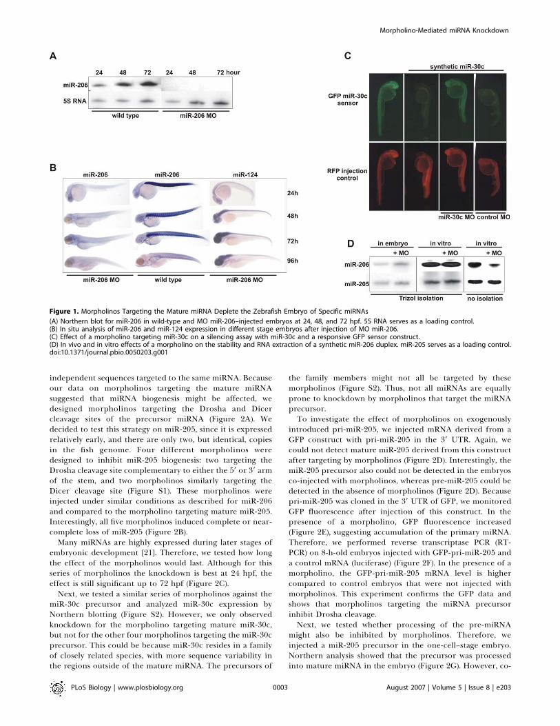
Since it is difficult to obtain a genetic mutant for a miRNAin zebrafish, we looked for alternative strategies to depletethe embryo of specific miRNAs. Antisense molecules such as29-O-methyl and locked nucleic acid (LNA) oligonucleotideshave been used to inhibit miRNAs in cell lines [16,18,22],Drosophila embryos [23], and adult mice [17]. We tried to usethese molecules to inhibit the function of endogenousmiRNAs in the zebrafish embryo. Although they can be usedto suppress the effects of miRNA overexpression [24],injection of higher concentrations required to obtain goodknockdown of endogenous miRNAs resulted in toxic effects,when injecting 1 nl solution at a concentration of approx-imately 10 lM and 50 lM for LNA and 29-O-methyloligonucleotides, respectively (unpublished data). Therefore,we switched to morpholinos because these are widely used toinhibit mRNA translation and splicing in zebrafish embryos[19], and have also been shown to target miRNAs in theembryo [2,20,24]. We injected 1 nl of 600 lM morpholinosolution with a morpholino complementary to the maturemiR-206 in one- or two-cell–stage embryos. Subsequently,embryos were harvested at 24, 48, 72, and 96 hourspostfertilization (hpf), and subjected to in situ hybridizationand Northern blotting (Figure 1A and 1B). This analysisshowed that the mature miRNA signal is suppressed up to 4 dafter injection of the morpholino. The knockdown effect wasspecific for this miRNA; parallel in situ analysis of the sameembryos with a probe for miR-124 did not show any effectson expression of this miRNA (Figure 1B). Thus, miRNAdetection can be specifically and efficiently suppressedduring embryonic and early larval stages of zebrafishdevelopment using morpholinos antisense to the maturemiRNA.
The zebrafish embryo can be used to monitor the effect ofmiRNAs on green fluorescent protein (GFP) reporters fused
to miRNA target sites [24]. To determine the effect of amorpholino in this assay system, we constructed a GFPreporter for miR-30c and tested it in the presence andabsence of a mature miR-30c duplex. Injected miR-30csilences this GFP reporter, which is in line with previousreports using similar strategies in the embryo (Figure 1C)[2,20,24]. Co-injection of the miR-30c duplex and a morpho-lino targeting mature miR-30c rescues the reporter signal,whereas injection of a control morpholino did not reverse thesilencing by miR-30c. These data indicate that a morpholinocan block the activity of a mature miRNA duplex in afunctional assay .There are three possible explanations for the observed
reduction in the detection signal for a miRNA that is targetedby a morpholino. First, the hybridization of a morpholinocould disturb isolation of the miRNA. Second, the morpho-lino could destabilize the miRNA. Third, the morpholinocould inhibit the maturation of the miRNA.To examine the effect of a morpholino on the isolation of a
mature miRNA, we incubated a mature miR-206 duplex and acontrol duplex (miR-205) with a morpholino against miR-206in vitro. After isolation, samples were analyzed by Northernblotting for the presence of miR-206 and miR-205. We couldstill detect miR-206, indicating that there is no effect of themorpholino on the RNA isolation procedure (Figure 1D).However, when morpholino and miRNA duplex wereincubated together in vitro and loaded on a denaturing gelwithout isolation, we observed a decrease in the signal formiR-206, indicating that the morpholino can bind to themiRNA in vitro and still does so in the denaturing gel.Next, we wanted to know whether a morpholino could
affect the stability of a mature miRNA in vivo. Therefore, weinjected a mature miR-206 and a control duplex (miR-205)together with a morpholino against miR-206 in the embryo.After incubation for 8 h, RNA was isolated and subjected toNorthern blot analysis to probe for injected miR-206 andinjected miR-205. In contrast to the data obtained forendogenous miR-206, there was no decrease observed in theamount of injected miR-206 in the morpholino-injectedembryos (Figure 1D) (endogenous miR-206 is not yet ex-pressed at this stage).Since these data show that there is no effect of a
morpholino on miRNA isolation or stability, we concludethat morpholinos deplete the embryo of miRNAs by inhibit-ing miRNA maturation. If this is the case, then we expectmorpholinos targeting other regions of the miRNA precursorto act as well as the morpholinos designed against the maturemiRNA, and this is indeed what we find (see next section).
Morpholinos Targeting the miRNA Precursor Interferewith Primary miRNA ProcessingInjection of antisense oligos in embryos might result in off-
target effects. Thus, phenotypic data retrieved from antisenseknockdown experiments should be treated with caution. InDrosophila, 29-O-methyl oligo–mediated knockdown of embry-onically expressed miRNAs caused defects that clearlydiffered from the phenotype of the corresponding knockoutfly [9,23]. In sea urchin experiments, off-target effects ofmorpholino knockdowns are well documented, though lowincubation temperatures favor off-target interactions [25].To filter out off-target effects, we sought a control strategy
that would allow us to compare effects of morpholinos with
PLoS Biology | www.plosbiology.org August 2007 | Volume 5 | Issue 8 | e2030002
Morpholino-Mediated miRNA Knockdown
Author Summary
The striking tissue-specific expression patterns of microRNAs(miRNAs) suggest that they play a role in tissue development.These small RNA molecules (;22 bases in length) are processedfrom long primary transcripts (pri-miRNA) and regulate geneexpression at the posttranscriptional level. There are hundreds ofdifferent miRNAs, many of which are strongly conserved. Vertebrateembryonic development is most easily studied in zebrafish, butgenetically disrupting miRNA genes to see which miRNA does whatis technically challenging. In this study, we interfere with miRNAfunction during the first few days of zebrafish embryonic develop-ment by introducing specific antisense morpholino oligonucleotides(morpholinos have been used previously to interfere with thesynthesis of the much larger mRNAs). We show that morpholinostargeting the miRNA precursor can block processing of the pri-miRNA or directly inhibit the activity of the mature miRNA. We alsoused morpholinos to study the developmental effects of miRNAknockdown. Although we did not observe gross phenotypic defectsfor many miRNAs, we found that zebrafish miR-375 is essential forformation of the insulin-secreting pancreatic islet. Loss of miR-375results in dispersed islet cells by 36 hours postfertilization,representing one of the first vertebrate miRNA loss-of-functionphenotypes.
independent sequences targeted to the same miRNA. Becauseour data on morpholinos targeting the mature miRNAsuggested that miRNA biogenesis might be affected, wedesigned morpholinos targeting the Drosha and Dicercleavage sites of the precursor miRNA (Figure 2A). Wedecided to test this strategy on miR-205, since it is expressedrelatively early, and there are only two, but identical, copiesin the fish genome. Four different morpholinos weredesigned to inhibit miR-205 biogenesis: two targeting theDrosha cleavage site complementary to either the 59 or 39 armof the stem, and two morpholinos similarly targeting theDicer cleavage site (Figure S1). These morpholinos wereinjected under similar conditions as described for miR-206and compared to the morpholino targeting mature miR-205.Interestingly, all five morpholinos induced complete or near-complete loss of miR-205 (Figure 2B).
Many miRNAs are highly expressed during later stages ofembryonic development [21]. Therefore, we tested how longthe effect of the morpholinos would last. Although for thisseries of morpholinos the knockdown is best at 24 hpf, theeffect is still significant up to 72 hpf (Figure 2C).
Next, we tested a similar series of morpholinos against themiR-30c precursor and analyzed miR-30c expression byNorthern blotting (Figure S2). However, we only observedknockdown for the morpholino targeting mature miR-30c,but not for the other four morpholinos targeting the miR-30cprecursor. This could be because miR-30c resides in a familyof closely related species, with more sequence variability inthe regions outside of the mature miRNA. The precursors of
the family members might not all be targeted by thesemorpholinos (Figure S2). Thus, not all miRNAs are equallyprone to knockdown by morpholinos that target the miRNAprecursor.To investigate the effect of morpholinos on exogenously
introduced pri-miR-205, we injected mRNA derived from aGFP construct with pri-miR-205 in the 39 UTR. Again, wecould not detect mature miR-205 derived from this constructafter targeting by morpholinos (Figure 2D). Interestingly, themiR-205 precursor also could not be detected in the embryosco-injected with morpholinos, whereas pre-miR-205 could bedetected in the absence of morpholinos (Figure 2D). Becausepri-miR-205 was cloned in the 39 UTR of GFP, we monitoredGFP fluorescence after injection of this construct. In thepresence of a morpholino, GFP fluorescence increased(Figure 2E), suggesting accumulation of the primary miRNA.Therefore, we performed reverse transcriptase PCR (RT-PCR) on 8-h-old embryos injected with GFP-pri-miR-205 anda control mRNA (luciferase) (Figure 2F). In the presence of amorpholino, the GFP-pri-miR-205 mRNA level is highercompared to control embryos that were not injected withmorpholinos. This experiment confirms the GFP data andshows that morpholinos targeting the miRNA precursorinhibit Drosha cleavage.Next, we tested whether processing of the pre-miRNA
might also be inhibited by morpholinos. Therefore, weinjected a miR-205 precursor in the one-cell–stage embryo.Northern analysis showed that the precursor was processedinto mature miRNA in the embryo (Figure 2G). However, co-
Figure 1. Morpholinos Targeting the Mature miRNA Deplete the Zebrafish Embryo of Specific miRNAs
(A) Northern blot for miR-206 in wild-type and MO miR-206–injected embryos at 24, 48, and 72 hpf. 5S RNA serves as a loading control.(B) In situ analysis of miR-206 and miR-124 expression in different stage embryos after injection of MO miR-206.(C) Effect of a morpholino targeting miR-30c on a silencing assay with miR-30c and a responsive GFP sensor construct.(D) In vivo and in vitro effects of a morpholino on the stability and RNA extraction of a synthetic miR-206 duplex. miR-205 serves as a loading control.doi:10.1371/journal.pbio.0050203.g001
PLoS Biology | www.plosbiology.org August 2007 | Volume 5 | Issue 8 | e2030003
Morpholino-Mediated miRNA Knockdown
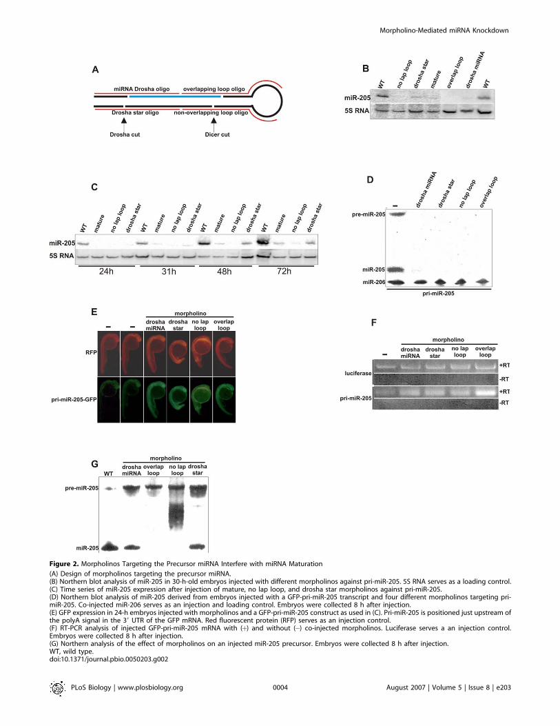
Figure 2. Morpholinos Targeting the Precursor miRNA Interfere with miRNA Maturation
(A) Design of morpholinos targeting the precursor miRNA.(B) Northern blot analysis of miR-205 in 30-h-old embryos injected with different morpholinos against pri-miR-205. 5S RNA serves as a loading control.(C) Time series of miR-205 expression after injection of mature, no lap loop, and drosha star morpholinos against pri-miR-205.(D) Northern blot analysis of miR-205 derived from embryos injected with a GFP-pri-miR-205 transcript and four different morpholinos targeting pri-miR-205. Co-injected miR-206 serves as an injection and loading control. Embryos were collected 8 h after injection.(E) GFP expression in 24-h embryos injected with morpholinos and a GFP-pri-miR-205 construct as used in (C). Pri-miR-205 is positioned just upstream ofthe polyA signal in the 39 UTR of the GFP mRNA. Red fluorescent protein (RFP) serves as an injection control.(F) RT-PCR analysis of injected GFP-pri-miR-205 mRNA with (þ) and without (�) co-injected morpholinos. Luciferase serves a an injection control.Embryos were collected 8 h after injection.(G) Northern analysis of the effect of morpholinos on an injected miR-205 precursor. Embryos were collected 8 h after injection.WT, wild type.doi:10.1371/journal.pbio.0050203.g002
PLoS Biology | www.plosbiology.org August 2007 | Volume 5 | Issue 8 | e2030004
Morpholino-Mediated miRNA Knockdown
injection of the overlap loop and non-overlapping loopmorpholinos blocked processing completely. There was onlya little effect of morpholinos targeting the Drosha cleavagesite, probably because they only partially overlap theprecursor.
A similar analysis was performed for miR-375, which isexpressed in the pancreatic islet and pituitary gland [21], andhas two copies in the zebrafish genome, which differ in theregions outside the mature miRNA.
Overlap loop and loop morpholinos were designed forboth miR-375–1 and miR-375–2, and a morpholino againstthe miRNA star sequence could be used to target both copiesof miR-375 simultaneously (Figure 3A). The efficacy of allmorpholinos was assessed by determining their effect oninjected pri-miR-375–1 or pri-miR-375–2 transcripts (Figure3B). As expected, each morpholino targeted the transcript towhich it was directed. However, the star miR-375 morpholinodid not knock down miR-375 completely. In addition,morpholino oligonucleotide (MO) miR-375 did not interferewith processing of miR-375 from pri-miR-375–1, possiblybecause this primary transcript forms a more stable hairpin.In all cases, the lack of a signal for mature miR-375 coincidedwith the absence of pre-miR-375, which could be detected inthe absence of a complementary morpholino.
Next, all morpholinos were injected separately and incombination, and embryos were subjected to Northernblotting to determine endogenous miR-375 expression at 24and 48 hpf (Figure 3C). In contrast to the results obtained byin situ hybridization (see last section), the morpholino tomature miR-375 only slightly decreased the expression ofmiR-375. However, MOmiR-375 could inhibit the activity of amature miR-375 duplex in a GFP-miR-375-target reporterassay (Figure 3E). The morpholinos targeting only one copy ofmiR-375 reduced miR-375 expression, with the strongesteffect for the morpholinos targeting pri-miR-375–1. How-ever, simultaneous injection of morpholinos targeting pri-miR-375–1 and pri-miR-375–2 completely knocked downmature miR-375, indicating that both transcripts are ex-pressed.
To further determine the contribution of each transcriptto mature miR-375 accumulation, we performed in situhybridization for pri-miR-375–1 and pri-miR-375–2 (Figure3D). Both transcripts could not be detected in wild-typeembryos. However, pri-miR-375–1 was detected in thepancreatic islet and the pituitary gland in embryos injectedwith the miR-375–1 loop morpholino and the morpholino tomiR-375 star. Similarly, pri-miR-375–2 was only detected inembryos injected with the miR-375–2 loop morpholino, themorpholino to miR-375 star and mature miR-375. Thus, bothtranscripts are expressed in the pituitary gland and thepancreatic islet, similar to miR-1 in the developing mouseheart [15]. Together, this indicates that these morpholinosinhibit primary miRNA processing and result in primarymiRNA accumulation, as we described for miR-205.
In conclusion, our data demonstrate that morpholinostargeting the miRNA precursor can interfere with primarymiRNA processing at either the Drosha or Dicer cleavage stepand that morpholinos targeting the mature miRNA caninhibit their activity in a functional assay. Taken together,our data show that different morpholinos targeting the samemiRNA may serve as positive controls for miRNA knockdownphenotypes in the embryo.
Knockdown of Many miRNAs Do Not Result in AnyObserved Developmental DefectsTo identify functions for individual miRNAs in zebrafish
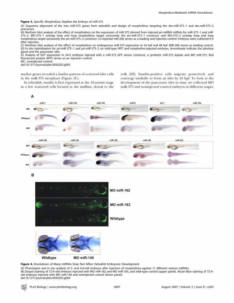
embryonic development, we knocked down a series of 11conserved vertebrate miRNAs (Table S1) and analyzed theirexpression after morpholino knockdown. Injected embryoswere monitored phenotypically by microscopic observationuntil four days postfertilization (dpf). Knockdown of mostmiRNAs resulted in loss of in situ staining for the respectivemiRNA. However, we could not observe gross morphologicalmalformations after knockdown of these miRNAs (Figure 4A).Therefore, we analyzed embryos injected with morpholinosagainst miR-182, miR-183, or miR-140 in more detail, becausewe could easily stain the tissues that express these miRNAs(Figure 4B). Embryos injected with morpholinos against miR-182 or miR-183, which are expressed in the lateral lineneuromasts and hair cells of the inner ear, were treated withDASPEI, which stains hair cells. Embryos injected with amorpholino against miR-140, which is expressed in cartilage,were subjected to Alcian Blue staining, a cartilage marker.However, staining of these specific cell types that express themiRNA did not uncover any defects upon knockdown (Figure4B).In conclusion, knockdown of many miRNAs does not
appear to significantly affect zebrafish embryonic develop-ment, at least not to the extent that can be visualized by themethods used in these examples.
Knockdown of miR-375 Affects Pancreatic IsletMorphologyMiR-375 is known to be expressed in the pancreatic islet
and the pituitary gland, and was first isolated from pancreaticbeta cells [21,26]. This miRNA is conserved in vertebrates andmay regulate insulin secretion by inhibiting myotrophin [26].We injected a morpholino against mature miR-375 into the
one-cell–stage embryo. This morpholino effectively knockeddown miR-375 in the first 4 d of development (Figure 5A), andit could also block the activity of an injected miR-375 duplex,as monitored by its effect on a GFP reporter silenced by miR-375 (Figure 3E).During the first 5 dpf, there was no clear developmental
defect except for a general delay in development. At around 7dpf, approximately 80% of the injected embryos died. Next,we analyzed the development of both the pituitary gland andthe pancreatic islet, by in situ hybridization with pit1 andinsulin markers. This analysis revealed no change in theformation of the pituitary gland (Figure 5B). However,analysis of insulin expression showed a striking malformationof the islet cells in 3-d-old morphant embryos (Figure 5B).Wild-type embryos have a single islet at the right side of themidline, whereas the miR-375 knockdown embryos havedispersed insulin-positive cells. The effect is sequencespecific, because a morpholino complementary to the maturemiR-375 morpholino inhibited the pancreatic islet pheno-type (Figure 5E).The pancreatic islet consists of four cell types, a, b, d, and
PP, expressing glucagon, insulin, somatostatin, and pancre-atic polypeptide, respectively. Insulin is the first hormoneexpressed, and somatostatin co-localizes partially with in-sulin, whereas glucagon-expressing cells are distinct [27]. Amore detailed analysis using somatostatin and glucagon as
PLoS Biology | www.plosbiology.org August 2007 | Volume 5 | Issue 8 | e2030005
Morpholino-Mediated miRNA Knockdown
PLoS Biology | www.plosbiology.org August 2007 | Volume 5 | Issue 8 | e2030006
Morpholino-Mediated miRNA Knockdown
marker genes revealed a similar pattern of scattered islet cellsin the miR-375 morphant (Figure 5C).
In zebrafish, insulin is first expressed at the 12-somite stagein a few scattered cells located at the midline, dorsal to the
yolk [28]. Insulin-positive cells migrate posteriorly andconverge medially to form an islet by 24 hpf. To look at thedevelopment of the pancreatic islet in time, we collected MOmiR-375 and noninjected control embryos at different stages,
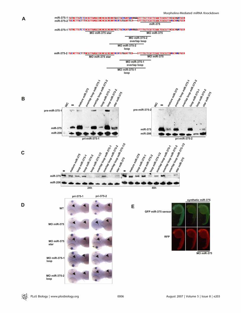
Figure 3. Specific Morpholinos Deplete the Embryo of miR-375
(A) Sequence alignment of the two miR-375 genes from zebrafish and design of morpholinos targeting the dre-miR-375–1 and dre-miR-375–2precursors.(B) Northern blot analysis of the effect of morpholinos on the expression of miR-375 derived from injected pri-miRNA mRNAs for miR-375–1 and miR-375–2. MO-375–1 overlap loop and loop morpholinos target exclusively the pri-miR-375–1 construct, and MO-375–2 overlap loop and loopmorpholinos target exclusively the pri-miR-375–2 construct. Co-injected miR-206 serves as a loading and injection control. Embryos were collected 8 hafter injection.(C) Northern blot analysis of the effect of morpholinos on endogenous miR-375 expression at 24 hpf and 48 hpf. MiR-206 serves as loading control.(D) In situ hybridization for pri-miR-375–1 and pri-miR-375–2 on wild-type (WT) and morpholino-injected embryos. Arrowheads indicate the pituitarygland and the pancreatic islet.(E) Analysis of GFP expression in 24-h embryos injected with a miR-375 GFP sensor construct, a synthetic miR-375 duplex and MO miR-375. Redfluorescent protein (RFP) serves as an injection control.NIC, noninjected control.doi:10.1371/journal.pbio.0050203.g003
Figure 4. Knockdown of Many miRNAs Does Not Affect Zebrafish Embryonic Development
(A) Phenotypes and in situ analysis of 3- and 4-d-old embryos after injection of morpholinos against 11 different mature miRNAs.(B) Daspei staining of 72-h-old embryos injected with MO miR-182 and MO miR-183, and wild-type control (upper panel). Alcian Blue staining of 72-h-old embryos injected with MO miR-140 and noninjected control (lower panel).doi:10.1371/journal.pbio.0050203.g004
PLoS Biology | www.plosbiology.org August 2007 | Volume 5 | Issue 8 | e2030007
Morpholino-Mediated miRNA Knockdown
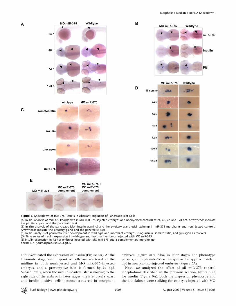
and investigated the expression of insulin (Figure 5D). At the16-somite stage, insulin-positive cells are scattered at themidline in both noninjected and MO miR-375–injectedembryos, and a presumptive islet is formed by 24 hpf.Subsequently, when the insulin-positive islet is moving to theright side of the embryo in later stages, the islet breaks apartand insulin-positive cells become scattered in morphant
embryos (Figure 5D). Also, in later stages, the phenotypepersists, although miR-375 is re-expressed at approximately 5dpf in morpholino-injected embryos (Figure 5A).Next, we analyzed the effect of all miR-375 control
morpholinos described in the previous section, by stainingfor insulin (Figure 6A). Both the dispersion phenotype andthe knockdown were striking for embryos injected with MO
Figure 5. Knockdown of miR-375 Results in Aberrant Migration of Pancreatic Islet Cells
(A) In situ analysis of miR-375 knockdown in MO miR-375–injected embryos and noninjected controls at 24, 48, 72, and 120 hpf. Arrowheads indicatethe pituitary gland and the pancreatic islet.(B) In situ analysis of the pancreatic islet (insulin staining) and the pituitary gland (pit1 staining) in miR-375 morphants and noninjected controls.Arrowheads indicate the pituitary gland and the pancreatic islet.(C) In situ analysis of pancreatic islet development in wild-type and morphant embryos using insulin, somatostatin, and glucagon as markers.(D) Time series of insulin expression in wild-type and morphant embryos injected with MO miR-375.(E) Insulin expression in 72-hpf embryos injected with MO miR-375 and a complementary morpholino.doi:10.1371/journal.pbio.0050203.g005
PLoS Biology | www.plosbiology.org August 2007 | Volume 5 | Issue 8 | e2030008
Morpholino-Mediated miRNA Knockdown
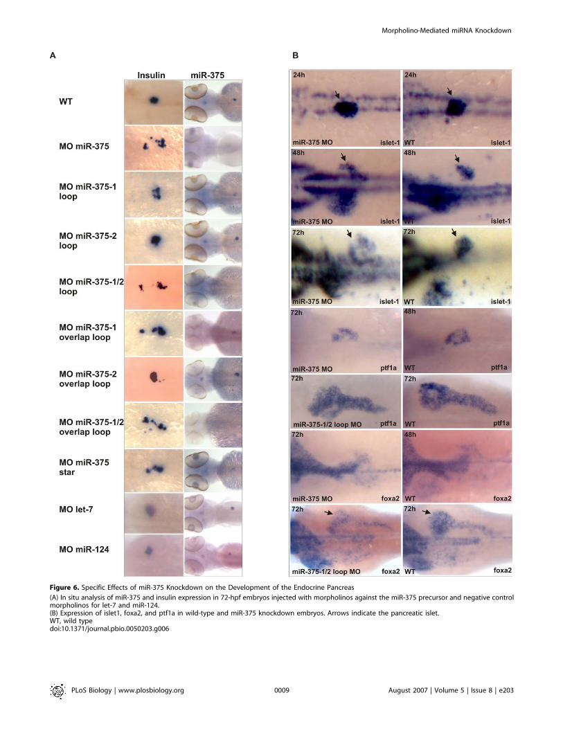
Figure 6. Specific Effects of miR-375 Knockdown on the Development of the Endocrine Pancreas
(A) In situ analysis of miR-375 and insulin expression in 72-hpf embryos injected with morpholinos against the miR-375 precursor and negative controlmorpholinos for let-7 and miR-124.(B) Expression of islet1, foxa2, and ptf1a in wild-type and miR-375 knockdown embryos. Arrows indicate the pancreatic islet.WT, wild typedoi:10.1371/journal.pbio.0050203.g006
PLoS Biology | www.plosbiology.org August 2007 | Volume 5 | Issue 8 | e2030009
Morpholino-Mediated miRNA Knockdown
miR-375. Injection of the overlap loop and loop morpholinostargeting pri-miR-375–1 also resulted in scattered insulin-positive cells at 72 hpf, although the effect was weakercompared to MO miR-375. The miR-375–2 loop and overlaploop morpholinos hardly induced any scattering of insulin-positive cells, whereas the effect was very strong in embryosinjected with morpholinos to pri-miR-375–1 and�2 simulta-neously. The effect of the miR-375 star morpholino oninsulin-positive cells was moderate compared to MO miR-375.
To further prove the specificity of the pancreatic isletphenotype, we injected two control morpholinos against let-7and miR-124 and analyzed these for miR-375 and insulinexpression. None of these control morpholinos showed lossof miR-375 expression or abnormal development of the isletcells (Figure 6A).
Next, we analyzed miR-375 knockdown embryos withmarkers staining the endocrine or exocrine pancreas (Figure6B). Similar to insulin staining, islet1 expression showeddispersed islet cells in embryos of 48 hpf and 72 hpf, but not24 hpf. Embryos injected with MO miR-375 exhibited delayeddevelopment of the exocrine pancreas, liver, and gut asshown by ptf1a and foxa2 staining. At 72 hpf, these markersshowed a similar pattern in MO miR-375–injected embryos asin noninjected embryos at 48 hpf. However, co-injection ofmiR-375–1/2 loop morpholinos did not delay development ofthe exocrine significantly, but these embryos still displayedthe scattered insulin-positive cells (Figure 6A). This showsthat loss of miR-375 mainly results in malformation of theendocrine pancreas, whereas surrounding tissues that do notexpress miR-375 are not affected.
Discussion
Functional data on miRNAs in vertebrate developmenthave been obtained mainly from overexpression studies andanalysis of conditional Dicer knockouts. For example, the roleof miR-430 in zebrafish brain morphogenesis has becomeclear from experiments that rescued Dicer null mutants byinjection of an miRNA duplex that mimicked a miR-430family member [2].
MiRNA expression can be conveniently studied in zebrafishembryos. However, dissecting miRNA function by disruptingmiRNA genes is difficult in zebrafish, because the miRNA istoo small to efficiently search for mutations by a target-selected mutagenesis approach [29]. In addition, it is unclearwhat such point mutations would do to processing orfunction of the miRNA.
It has been shown previously that morpholinos can targetmiRNAs in the zebrafish embryo [20,24]. In a recent study,mature miR-214 was targeted by a morpholino in zebrafish,and this resulted in a change in somite shape, reminiscent ofattenuated hedgehog signaling [20]. Although the phenotypecould be rescued by simultaneous inhibition of a negativeregulator of hedgehog signaling, no positive control mor-pholinos were reported that could mimic the phenotype. Inaddition, data were lacking that showed an effect of themorpholino on endogenous miR-214 levels.
The results in this paper show that morpholinos targetingthe miRNA precursor form a reliable and efficient tool todeplete the embryo of miRNAs during the first 4 d ofdevelopment, when most organ systems are formed and
miRNAs are expressed. We have shown that miRNA expres-sion can be inhibited by targeting the mature miRNA, theprecursor miRNA or the primary miRNA. Our data show thatsuch morpholinos can inhibit miRNA processing at theDrosha cleavage step or the Dicer cleavage step, probably bysteric blocking, although the exact mechanism is unclear. Inaddition, morpholinos targeting the mature miRNA caninhibit their activity, probably by preventing binding to atarget mRNA.We used morpholinos targeting the mature miRNA for a
set of 13 conserved vertebrate miRNAs to identify theirdevelopmental functions. By microscopic analysis, we couldnot observe clear defects associated with loss of 11 of thesemiRNAs during the first 4 d of embryonic development,although in situ hybridization revealed specific loss of mostknocked-down miRNAs. Because all the targeted miRNAs areexpressed in very specific tissues and we did not investigatemost morphants in much detail by marker analysis, we mayhave missed subtle defects. In addition, many miRNAs residein families of related sequence (e.g., let-7 and miR-182), andthese should possibly be targeted simultaneously by differentmorpholinos to obtain a biological effect. Furthermore, inthose instances in which miRNAs of unrelated sequencetarget a similar set of mRNAs when expressed in the sametissue [21], removing only one miRNA might not have aprofound impact on transcript levels or expression. Finally,microarray analysis and computational predictions haveshown that a single miRNA may regulate hundreds of mRNAs[30,31], but that some miRNAs act as a backup for mRNAsthat are already repressed transcriptionally [32]. Thus,knockdown of such miRNAs might not dramatically affectgene expression, but ensure robustness of protein interactionnetworks as for example miR-7 in Drosophila [33].In zebrafish, there are two copies of miR-375, and in human
and mouse only one copy has been identified [34]. To verifythe miR-375 knockdown phenotype, we designed controlmorpholinos targeting both precursors simultaneously (MOmiR-375 star) and separately. Complete knockdown was onlyobserved in those instances in which both miR-375 copieswere targeted simultaneously. This also led to scattered isletcells, proving the specificity of the phenotype. However,knockdown with miR-375–1/2 loop morpholinos did not delaydevelopment as seen in the knockdown with the mature miR-375 morpholino. This shows the strength of using controlmorpholinos and excludes the delayed development as arelevant miR-375 loss-of -function phenotype. A moderateversion of the phenotype was also observed in embryosinjected with a morpholino specifically targeting miR-375–1.Thus, a reduction in the level of miR-375 already disturbs isletintegrity. Similar to mouse miR-1 [15], miR-375 copiessurvived evolution and are expressed similarly in time andspace, probably to ensure the high intracellular concentra-tion of miR-375 necessary to repress many weakly bindingtargets.In a forward genetic screen, several mutants were identified
with improper development of the endocrine pancreas [35].These mutants fall into three classes: (1) mutants with severelyreduced insulin expression; (2) mutants with reduced insulinexpression and abnormal islet morphology; and (3) mutantswith normal levels of insulin expression and abnormal isletmorphology. However, in all of these mutants, islet cells donot merge into an islet from their first appearance at
PLoS Biology | www.plosbiology.org August 2007 | Volume 5 | Issue 8 | e2030010
Morpholino-Mediated miRNA Knockdown
approximately the 14-somite stage. Our miR-375 knockdownphenotype differs from this, because in the first instance, anislet is formed at approximately 24 hpf, but in later stages, theislet falls apart into small groups of cells. This rules out ageneral role for miR-375 in early endocrine formation as isseen for Wnt5 [36], but rather indicates a role in maintenanceof tissue identity, which is assumed to be a general function ofmiRNAs in development [21]. It is as yet unclear which miR-375 targets are involved in the phenotype. Work in cell lineshas implicated miR-375 in insulin secretion by targetingmyotrophin [26]. The zebrafish homolog of myotrophin alsocontains a seven-nucleotide seed match to miR-375 (unpub-lished data), but future studies should reveal whether thistarget or many other predicted targets are relevant to thephenotype. The specific expression of miR-375 in thepancreatic islet and its implication in insulin secretion makeit a candidate drug target in diabetes, e.g., to influence insulinlevels in the blood. However, our data show that if miR-375 isused as a drug target, developmental side effects need to betaken into account.
Materials and Methods
Morpholino and miRNA injections. Morpholinos were obtainedfrom Gene Tools LLC (http://www.gene-tools.com) and dissolved to aconcentration of 5 mM in water. Morpholinos were injected into one-or two-cell–stage embryos at concentrations between 200 lM and1,000 lM, and per embryo, one nl of morpholino solution wasinjected.
RNA oligos (Table S2) were obtained from Sigma (http://www.sigmaaldrich.com) and dissolved to a concentration of 100 lM indistilled water. Oligos were annealed using a 5x buffer containing 30mM HEPES-KOH (pH 7.4), 100 mM KCl, 2 mM MgCl2, and 50 mMNH4Ac. Typically, 1 nl of a 10 lM miRNA duplex solution wasinjected.
All morpholino sequences used in this study are listed in Table S1.Construction of miR-30c and miR-375 GFP reporters and pri-
miRNA constructs. The miR-30c and miR-375 reporter constructswere made by cloning two annealed oligos containing two perfectlycomplementary miRNA target sites into pCS2 (Clontech, http://www.clontech.com) containing a gfp gene between BamHI and ClaIrestriction sites. A construct containing pri-miR-205 was made byamplifying a genomic region (801 base pairs) containing the miR-205precursor (miR-205-hairpinF ggcattgaattcataaCCTCTTACCTGCAT-GACCTG; miR-205-hairpinR ggcatttctagaGTGTGTGCGTGTATT-CAACC). The resulting PCR fragment was cloned between XbaIand EcoRI restriction sites of PCS2GFP. Pri-miR-375–1 and pri-miR-375–2 constructs were made by amplifying genomic regions contain-ing miR-375–1 and miR-375–2 precursors (WKmiR-375–1F-pCS2gcccgggatccTGTGTCTTGCAGGAAAAGAG; WKmiR-375–1R-pCS2attacgaattcTCAAACTCTCCACTGACTGC; and WKmiR-375–2F-pCS2 gcccgggatccGCCCTCCCATTTGACTC; WKmiR-375–2R-pCS2attacgaattcAATGAGTGCACAAAATGTCC), and cloning of the re-sulting PCR fragments into the BamHI and EcoRI sites of pCS2.mRNA was synthesized using SP6 RNA polymerase. Luciferase mRNAwas derived from pCS2 containing luciferase between BamHI andEcoRI sites.
In situ hybridization, Northern blotting, and RT-PCR. In situhybridization was performed as described previously [37]. LNAprobes for miRNA detection were obtained from Exiqon (http://www.exiqon.com) and labeled using terminal transferase and DIG-11-ddUTP. cDNA clones for pri-miR-375–1, pri-miR-375–2, pit1, insulin,
somatostatin, and glucagon were used for antisense DIG-labeledprobe synthesis by T7 or Sp6 RNA polymerase.
For Northern blotting, total RNA was isolated from ten embryosper sample using Trizol reagent (Invitrogen, http://www.invitrogen.com). RNA was separated on a 15% denaturing polyacrylamide gel.Radiolabeled DNA probes complementary to miRNAs or 5S RNA(atcggacgagatcgggcgta) were used for hybridization at 37 8C.Stringency washes were done twice for 15 min at 37 8C using 2 3SSC 0.2% SDS. Alternatively, DIG-labeled LNA probes were used forhybridization at 60 8C and stringency washes were performed at 50 8Cwith 2x SSC 0.1% SDS for 30 min and 0.5x SSC 0.1% SDS for 30 min.
For RT-PCR, RNA was isolated with Trizol, treated with DNAse(Promega, http://www.promega.com) and subsequently purified againusing Trizol. cDNA was made with a poly dT primer. Primers used foramplification were miR-205-hairpinF and miR-205-hairpinR, andlucF (ATGGAAGACGCCAAAAACATAAAG) and lucR (ATTACATC-GATTTACACGGCGATCTTTCC).
Alcian Blue and Daspei staining. For Alcian Blue staining, embryoswere fixed for 1 h at room temperature in 4% PFA in PBS, rinsed for5 min in 50% MeOH, and stored overnight in 70% MeOH at 4 8C.Next, embryos were incubated for 5 min in 50% MeOH and for 5 minin 100% EtOH. Embryos were stained at room temperature withAlcian Blue (Sigma) for 90 min with continuous shaking. Subse-quently, embryos were rinsed in 80%, 50%, and 25% EtOH for 2 mineach and two times in water containing 0.2% Triton and neutralizedin 100% Borax solution. Finally, embryos were incubated for 60 minin digest solution (60% Borax solution, 1 mg/ml colleganase-free andelastase-free trypsin, 0.2% trypsin) and stored in 70% glycerol.
Staining of the hair cells was done by incubating live embryos for 5min in a 200 lM solution of Daspei (Sigma) inþchorion. After rinsingtwice in þ chorion, embryos were anesthetized using MS222 andmounted in methylcellulose.
Supporting Information
Figure S1. Design of Morpholinos Targeting the miR-205 Precursor
Found at doi:10.1371/journal.pbio.0050203.sg001 (224 KB TIF).
Figure S2. Morpholino-Mediated Knockdown of miR-30c
(A) Design of morpholinos targeting the miR-30c precursor.(B) Northern analysis of miR-30c expression in 24-h-old embryosinjected with different morpholinos targeting the miR-30c precursor.(C) Alignment of the precursor of miR-30 family miRNAs.
Found at doi:10.1371/journal.pbio.0050203.sg002 (784 KB TIF).
Table S1. Morpholinos Used in This Study
Found at doi:10.1371/journal.pbio.0050203.st001 (21 KB XLS).
Table S2. miRNA Sequences
Found at doi:10.1371/journal.pbio.0050203.st002 (13 KB XLS).
Acknowledgments
We thank B. Ason for reading the manuscript critically and F.Argenton and M. Hammerschmidt for providing cDNA clones forendocrine pancreas and pituitary markers.
Author contributions. WPK, AKL, RFK, JDM, and RHAP conceivedand designed the experiments. WPK and AKL performed theexperiments and analyzed data. JDM contributed reagents. WPK,RFK, JDM, and RHAP and wrote the paper.
Funding. This work was supported by the Council for Earth andLife Sciences of the Netherlands Organization for ScientificResearch.
Competing interests. The authors have declared that no competinginterests exist.
References
1. Bernstein E, Kim SY, Carmell MA, Murchison EP, Alcorn H, et al. (2003).Dicer is essential for mouse development. Nat Genet 35: 215–217.
2. Giraldez AJ, Cinalli RM, Glasner ME, Enright AJ, Thomson JM, et al. (2005).MicroRNAs regulate brain morphogenesis in zebrafish. Science 308: 833–838.
3. Wienholds E, Koudijs MJ, van Eeden FJ, Cuppen E, Plasterk RH (2003). ThemicroRNA-producing enzyme Dicer1 is essential for zebrafish develop-ment. Nat Genet 35: 217–218.
4. Ambros V (2004). The functions of animal microRNAs. Nature 431: 350–355.
5. Abbott AL, Alvarez-Saavedra E, Miska EA, Lau NC, Bartel DP, et al. (2005).The let-7 MicroRNA family members mir-48, mir-84, and mir-241 functiontogether to regulate developmental timing in Caenorhabditis elegans. Dev Cell9: 403–414.
6. Li M, Jones-Rhoades MW, Lau NC, Bartel DP, Rougvie AE (2005).Regulatory mutations of mir-48, a C. elegans let-7 family MicroRNA, causedevelopmental timing defects. Dev. Cell 9: 415–422.
7. Brennecke J, Hipfner DR, Stark A, Russell RB, Cohen SM (2003). bantam
PLoS Biology | www.plosbiology.org August 2007 | Volume 5 | Issue 8 | e2030011
Morpholino-Mediated miRNA Knockdown
encodes a developmentally regulated microRNA that controls cellproliferation and regulates the proapoptotic gene hid in Drosophila. Cell113: 25–36.
8. Xu P, Vernooy SY, Guo M, Hay BA (2003). The Drosophila microRNA Mir-14suppresses cell death and is required for normal fat metabolism. Curr Biol13: 790–795.
9. Sokol NS, Ambros V (2005) Mesodermally expressed Drosophila microRNA-1 is regulated by Twist and is required in muscles during larval growth.Genes Dev 19: 2343–2354.
10. Andl T, Murchison EP, Liu F, Zhang Y, Yunta-Gonzalez M, et al. (2006) ThemiRNA-processing enzyme dicer is essential for the morphogenesis andmaintenance of hair follicles. Curr Biol 16: 1041–1049.
11. Harfe BD, McManus MT, Mansfield JH, Hornstein E, Tabin CJ (2005) TheRNaseIII enzyme Dicer is required for morphogenesis but not patterning ofthe vertebrate limb. Proc Natl Acad Sci U S A 102: 10898–10903..
12. Harris KS, Zhang Z, McManus MT, Harfe BD, Sun X (2006) Dicer functionis essential for lung epithelium morphogenesis. Proc Natl Acad Sci U S A103: 2208–2213.
13. Yi R, O’Carroll D, Pasolli HA, Zhang Z, Dietrich FS, et al. (2006)Morphogenesis in skin is governed by discrete sets of differentiallyexpressed microRNAs. Nat Genet 38: 356–362.
14. Hornstein E, Mansfield JH, Yekta S, Hu JK, Harfe BD, et al. (2005) ThemicroRNA miR-196 acts upstream of Hoxb8 and Shh in limb development.Nature 438: 671–674.
15. Zhao Y, Samal E, Srivastava D (2005) Serum response factor regulates amuscle-specific microRNA that targets Hand2 during cardiogenesis. Nature436: 214–220.
16. Hutvagner G, Simard MJ, Mello CC, Zamore PD (2004) Sequence-specificinhibition of small RNA function. PLoS Biol 2: E98. doi:10.1371/journal.pbio.0020098
17. Krutzfeldt J, Rajewsky N, Braich R, Rajeev KG, Tuschl T, et al. (2005)Silencing of microRNAs in vivo with ‘antagomirs.’ Nature 438:: 685–689.
18. Meister G, Landthaler M, Dorsett Y, Tuschl T (2004) Sequence-specificinhibition of microRNA- and siRNA-induced RNA silencing. RNA 10: 544–550.
19. Nasevicius A, Ekker SC (2000) Effective targeted gene ‘knockdown’ inzebrafish. Nat Genet 26: 216–220.
20. Flynt AS, Li N, Thatcher EJ, Solnica-Krezel L, Patton JG (2007) ZebrafishmiR-214 modulates Hedgehog signaling to specify muscle cell fate. NatGenet 39: 259–263.
21. Wienholds E, Kloosterman WP, Miska E, Alvarez-Saavedra E, Berezikov E, etal. (2005) MicroRNA expression in zebrafish embryonic development.Science 309: 310–311.
22. Naguibneva I, Ameyar-Zazoua M, Nonne N, Polesskaya A, Ait-Si-Ali S, et al.
(2006) An LNA-based loss-of-function assay for micro-RNAs. BiomedPharmacother 60: 633–638.
23. Leaman D, Chen PY, Fak J, Yalcin A, Pearce M, et al. (2005) Antisense-mediated depletion reveals essential and specific functions of microRNAsin Drosophila development. Cell 121: 1097–1108.
24. Kloosterman WP, Wienholds E, Ketting RF, Plasterk RH (2004) Substraterequirements for let-7 function in the developing zebrafish embryo.Nucleic Acids Res 32: 6284–6291.
25. Coffman JA, Dickey-Sims C, Haug JS, McCarthy JJ, Robertson AJ (2004)Evaluation of developmental phenotypes produced by morpholino anti-sense targeting of a sea urchin Runx gene. BMC Biol 2: 6.
26. Poy MN, Eliasson L, Krutzfeldt J, Kuwajima S, Ma X, et al. (2004) Apancreatic islet-specific microRNA regulates insulin secretion. Nature 432:226–230.
27. Argenton F, Zecchin E, Bortolussi M (1999) Early appearance of pancreatichormone-expressing cells in the zebrafish embryo. Mech Dev 87: 217–221.
28. Biemar F, Argenton F, Schmidtke R, Epperlein S, Peers B, et al. (2001)Pancreas development in zebrafish: Early dispersed appearance ofendocrine hormone expressing cells and their convergence to form thedefinitive islet. Dev Biol 230: 189–203.
29. Wienholds E, Plasterk RH (2004) Target-selected gene inactivation inzebrafish. Methods Cell Biol 77: 69–90.
30. Lewis BP, Burge CB, Bartel DP (2005) Conserved seed pairing, often flankedby adenosines, indicates that thousands of human genes are microRNAtargets. Cell 120: 15–20.
31. Lim LP, Lau NC, Garrett-Engele P, Grimson A, Schelter JM, et al. (2005)Microarray analysis shows that some microRNAs downregulate largenumbers of target mRNAs. Nature 433: 769–773.
32. Farh KK, Grimson A, Jan C, Lewis BP, Johnston WK, et al. (2005) Thewidespread impact of mammalian MicroRNAs on mRNA repression andevolution. Science 310: 1817–1821.
33. Li X, Carthew RW (2005) A microRNA mediates EGF receptor signalingand promotes photoreceptor differentiation in the Drosophila eye. Cell 123:1267–1277.
34. Griffiths-Jones S (2006) miRBase: The microRNA sequence database.Methods Mol Biol 342: 129–138.
35. Kim HJ, Sumanas S, Palencia-Desai S, Dong Y, Chen JN, et al. (2006)Genetic analysis of early endocrine pancreas formation in zebrafish. MolEndocrinol 20: 194–203.
36. Kim HJ, Schleiffarth JR, Jessurun J, Sumanas S, Petryk A, et al. (2005) Wnt5signaling in vertebrate pancreas development. BMC Biol 3: 23.
37. Kloosterman WP, Wienholds E, de Bruijn E, Kauppinen S, Plasterk RH(2006) In situ detection of miRNAs in animal embryos using LNA-modifiedoligonucleotide probes. Nat Methods 3: 27–29.
PLoS Biology | www.plosbiology.org August 2007 | Volume 5 | Issue 8 | e2030012
Morpholino-Mediated miRNA Knockdown