Embed Size (px)
DESCRIPTION
Plga Microspheres
Citation preview

International Standard Serial Number (ISSN): 2249-6793
428 Full Text Available On www.ijupls.com
International Journal of Universal Pharmacy and Life Sciences 2(3): May-June 2012
IINNTTEERRNNAATTIIOONNAALL JJOOUURRNNAALL OOFF UUNNIIVVEERRSSAALLPPHHAARRMMAACCYY AANNDD LLIIFFEE SSCCIIEENNCCEESS
Review Article……!!!
Received: 01-06-2012; Revised; Accepted: 05-06-2012
PLGA MICROSPHERES AND NANOSPHERES AS DRUG CARRIERS
Kiruthika C*, Dr. Sivablan M
Mother Theresa Post Graduate and Research Institute of Health Sciences(Govt. of Puducherry Institution) Indira Nagar, Gorimedu, Puducherry- 605 006.Keywords:
PLGA, PLGA-PEG,
Dialysis method and Super
critical fluids method
For Correspondence:
Kiruthika C
Mother Theresa Post Graduate and Research Institute of Health Sciences(Govt. of Puducherry
Institution) Indira Nagar, Gorimedu, Puducherry- 605
006.E-mail:
ABSTRACT
Context: Poly (lactic-co-glycolic acid) PLGA and its co-polymers, have been extensively studied for a wide variety of pharmaceutical and biomedical applications. PLGA copolymer is one of the synthetic biodegradable and biocompatible polymers that has reproducible and slow-release characteristics in-vivo. It has been regarded as one of the few synthetic biodegradable polymers with controllable biodegradability, excellent biocompatibility, and high safety. PLGA can be used to prepare microspheres, nanospheres, sutures, surgical materials and scaffolds in tissue engineering. Objective: This review aims to compile the available applications of PLGA as microspheres and nanospheres and also the preparation techniques used to prepare the PLGA microspheres and nanospheres. Method: The microspheres and nanospheres were prepared by several methods using PLGA. All the methods have been summarized by collecting articles on the PLGA microspheres and nanospheres from various databases. The drugs that were formulated as the microspheres and nanospheres are collected from the several databases and included in the review. Conclusion: Various drugs (anti-cancer, antibiotics, NSIADs, etc.), peptides and vaccines have been formulated as the microspheres and nanospheres using PLGA. This review helps the further development of the polymer usage in the field of microspheres and nanocarriers.
Pharmaceutical Sciences
International Standard Serial Number (ISSN): 2249-6793
429 Full Text Available On www.ijupls.com
1. Introduction
1.1 Poly(lactic-co-glycolic acid) [1]
PLGA or poly(lactic-co-glycolic acid) is a copolymer which is used in therapeutic devices,
owing to its biodegradability and biocompatibility. PLGA, have generated tremendous interest
because of their excellent biocompatibility, biodegradability and mechanical strength. A number
of groups have published pioneering work on the utility of these polymers to make sutures/fibers.
Various polymeric devices like microspheres, microcapsules, nanoparticles, pellets, implants and
films have been fabricated using these polymers. They are also easy to formulate into various
delivery systems for carrying a variety of drug classes, such as vaccines, peptides, proteins and
micromolecules, which have been approved by the Food and Drug Administration for drug
delivery use.
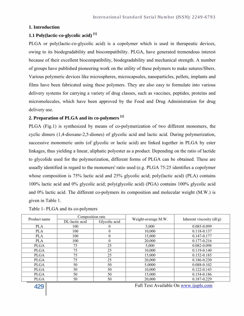
2. Preparation of PLGA and its co-polymers [1]
PLGA (Fig.1) is synthesized by means of co-polymerization of two different monomers, the
cyclic dimers (1,4-dioxane-2,5-diones) of glycolic acid and lactic acid. During polymerization,
successive monomeric units (of glycolic or lactic acid) are linked together in PLGA by ester
linkages, thus yielding a linear, aliphatic polyester as a product. Depending on the ratio of lactide
to glycolide used for the polymerization, different forms of PLGA can be obtained. These are
usually identified in regard to the monomers' ratio used (e.g. PLGA 75:25 identifies a copolymer
whose composition is 75% lactic acid and 25% glycolic acid; poly(lactic acid) (PLA) contains
100% lactic acid and 0% glycolic acid; poly(glycolic acid) (PGA) contains 100% glycolic acid
and 0% lactic acid. The different co-polymers its composition and molecular weight (M.W.) is
given in Table 1.
Table 1- PLGA and its co-polymers
Product nameComposition rate
Weight-average M.W. Inherent viscosity (dl/g)DL-lactic acid Glycolic acid
PLA 100 0 5,000 0.085-0.099PLA 100 0 10,000 0.118-0.137PLA 100 0 15,000 0.147-0.177PLA 100 0 20,000 0.177-0.216
PLGA 75 25 5,000 0.082-0.098PLGA 75 25 10,000 0.119-0.140PLGA 75 25 15,000 0.152-0.185PLGA 75 25 20,000 0.186-0.230PLGA 50 50 5,0000 0.088-0.102PLGA 50 50 10,000 0.122-0.143PLGA 50 50 15,000 0.154-0.186PLGA 50 50 20,000 0.187-0.229
International Standard Serial Number (ISSN): 2249-6793
430 Full Text Available On www.ijupls.com
3. PLGA-PEG block co-polymers [2]
The biodegradable polyesters are all strongly hydrophobic, and this has caused some limitations
in practical drug formulations. To add hydrophilic and other physico-chemical properties,
poly(ethylene glycol) (PEG) has been incorporated into the biodegradable polyesters. PEG is a
non-toxic, water-soluble polymer with proven biocompatibility. These are known as block
copolymers. A wide variety of drug formulations, such as microspheres/ nano-particles, micelles,
hydrogels, and injectable drug delivery systems have been developed using PLGA-PEG block
copolymers. They have been extensively investigated for use in a wide range of applications,
including implantable materials, drug delivery systems, and tissue engineering scaffolds. They
are very useful materials for pharmaceutical and biomedical applications, and thus they have
significant commercial potential.
The chemical composition and M.W. of block copolymers determines their water-solubility and
degradation kinetics. Polymers with low M.W. or composed of shorter hydrophobic blocks are
soluble in water, whereas high M.W. polymers and polymers with longer hydrophobic blocks are
not soluble but swell in water. In general, the degradation time will be shorter for low M.W.
polymers, more hydrophilic polymers, more amorphous polymers, and copolymers with higher
content of glycolide. Therefore, at identical conditions, low M.W. copolymers of lactide and
glycolide will degrade relatively rapidly, whereas the high M.W. homopolymers, PLA, and PGA
will degrade much more slowly.
4. Physicochemical properties of PLGA [3]
PLGA prepared from L-poly lactic acid (L-PLA) and L-poly glycolic acid (L-PGA) are
crystalline co-polymers while those from D,L-PLA and D,L-PGA are amorphous in nature. It has
been found that PLGAs containing >70% glycolide are amorphous in nature. The degree of
crystallinity and the melting point of the polymers are directly related to the M.W. of the
polymer. At lower M.Ws(19,000), a relatively constant release profile was obtained; increasing
the molecular weight to 23,000, 44,000 and 74,000 decreased the linearity of release. The rate of
drug release from particles containing higher M.W polymers was initially high, followed by a
decrease which was then followed again by an increase. The two-stage release profile suggested
the presence of two dominating release mechanisms in high M.W polymers. Degradation is the
main release mechanism for low M.W polymers after the initial burst stage. Spheres containing
high M.W polymers likely undergo initial slow drug release due to diffusion, followed by the
International Standard Serial Number (ISSN): 2249-6793
431 Full Text Available On www.ijupls.com
main drug release due to degradation. By correlating observed drug release with microscopic
observation of the microspheres; the drug release was fastest for the degradation of swollen
spheres. Physical properties such as the M.W. affect the mechanical strength of the polymer and
its ability to be formulated as a drug delivery device. Also, these properties may control the
polymer biodegradation rate and hydrolysis. Commercially available PLGA polymers are usually
characterized in terms of intrinsic viscosity, which is directly related to their M.W.s.
The mechanical strength, swelling behavior, capacity to undergo hydrolysis and subsequently the
biodegradation rate are directly influenced by the crystallinity of the PLGA polymer. The
resultant crystallinity of the PLGA co-polymer is dependent on the type and the molar ratio of
the individual monomer components (lactide and glycolide) in the copolymer chain. PLGA
polymers containing a 50:50 ratio of lactic and glycolic acids are hydrolyzed much faster than
those containing a higher proportion of either of the two monomers. PGA is highly crystalline
because it lacks the methyl side groups of the PLA. Lactic acid is more hydrophobic than
glycolic acid and, therefore, lactide-rich PLGA co-polymers are less hydrophilic, absorb less
water and subsequently degrade more slowly. It has a glass transition temperature (Tg) of 45°C
and an inherent viscosity of 0.5-0.8 mPa. The Tgs of the PLGA co-polymers are above the
physiological temperature of 37°C and hence they are normally glassy in nature. Thus, they have
a fairly rigid chain structure, which gives them significant mechanical strength to be formulated
as a degradable device. It has been reported that the Tgs of PLGA decrease with the decrease of
lactide content in the co-polymer composition with decreasing M.W. The PLGA polymers
chosen should also have considerable mechanical strength. Different factors like the M.W., co-
polymer composition (lactide/glycolide ratio), crystallinity and geometric regularity of individual
chains significantly affect the mechanical strength of the particular polymer.
5. Biodegradation of PLGA [3]
In both in-vitro and in-vivo, the PLGA co-polymer undergoes degradation in an aqueous
environment (hydrolytic degradation or biodegradation) through cleavage of its backbone ester
linkages. The polymer chains undergo bulk degradation and the degradation generally occurs at a
uniform rate throughout the PLGA matrix. It has been recorded that the PLGA biodegradation
occurs through random hydrolytic chain scissions of the swollen polymer. The carboxylic end
groups present in the PLGA chains increase in number during the biodegradation process as the
individual polymer chains are cleaved. These are known to catalyze the biodegradation process.
International Standard Serial Number (ISSN): 2249-6793
432 Full Text Available On www.ijupls.com
It has also been reported that large fragments are degraded faster internally and amorphous
regions degrade faster than crystalline regions. The biodegradation rates of the PLGA co-
polymers are dependent on the molar ratio of the lactic and glycolic acids in the polymer chain,
M.W. of the polymer, the degree of crystallinity and the Tg of the polymer.
A three-phase mechanism for PLGA biodegradation has been proposed:
1. Random chain scission process. The M.W. of the polymer decreases significantly, but no
appreciable weight loss and no soluble monomer products are formed.
2. In the middle phase, a decrease in M.W. accompanied by a rapid loss of mass and soluble
oligomeric and monomer products are formed.
3. Soluble monomer products formed from soluble oligomeric fragments. This phase is that of
complete polymer solubilization.
The role of enzymes in any PLGA biodegradation is unclear. Most of the literature indicates that
the PLGA biodegradation does not involve any enzymatic activity and is purely through
hydrolysis. However, some findings have suggested an enzymatic role in PLGA breakdown
based on the difference in the in-vitro and in-vivo degradation rates. It has also been found that
motion and buffers may affect their rate differences. However, it is known that PLGA
biodegrades into lactic and glycolic acids. Lactic acid enters the tricarboxylic acid cycle and is
metabolized and subsequently eliminated from the body as carbon dioxide and water. Glycolic
acid is either excreted unchanged in the kidney or it enters the tricarboxylic acid cycle and is
eventually eliminated as carbon dioxide and water.
6. Biocompatibility of PLGA [3]
The PLGA polymer had several advantages like good mechanical properties, low
immunogenicity and toxicity, excellent biocompatibility and predictable biodegradation kinetics.
The wide acceptance of the lactide/glycolide polymers as suture materials made them attractive
candidates for biomedical applications like ligament reconstruction, tracheal replacement,
surgical dressings, vascular grafts and nerve, dental and fracture repairs.
PLGA microspheres (average size 30 µm) induced a mild foreign body reaction and were
reported to be biocompatible. The volume of microspheres injected into the tissue may be
considered as an open porous implant, which induces an inflammatory response characterized by
the infiltration of macrophages, neutrophils, fibroblasts and some lymphocytes and by the
formation of fibrin, giant cells and new blood vessels. Tissue reaction to the PLGA microsphere
International Standard Serial Number (ISSN): 2249-6793
433 Full Text Available On www.ijupls.com
injection site after Week 1 showed heavy macrophage infiltration around the muscle due to a
systemic rise in the level of activated macrophages, which release cytokines, growth factors and
other bioactive agents to modulate the function of other cell types in the inflammatory milieu.
The release of octreotide acetate from PLGA microspheres has been tested in rabbits and
humans. Similar release patterns from rabbits and humans were observed.
7. Preparation of PLGA microsphere and nanospheres
The various methods used to prepare the microspheres and nanospheres using PLGA were
summarized in Fig.2.
7.1 Solvent evaporation method
7.1.1 Single emulsion: [4] [5]
Drug is dissolved in the organic phase containing polymer (PLGA). This organic phase is mixed
to the aqueous phase mostly poly(vinyl alcohol) (PVA) and mixed for 30mins and desired drop
size is obtained. The resulting emulsion is stirred on a magnetic stirrer and complete solvent
evaporation is achieved. The microspheres/nanospheres are collected by centrifugation. The
microspheres/nanospheres are washed with suitable solvent and dried. Another method of
preparing microspheres is by dissolving the drug and polymer in organic phase and the desired
size of the particles are obtained by using water as continuous phase. These two methods are the
oldest approach to prepare the microspheres and nanoparticles.
7.1.2 Double emulsion technique: [6]
First the drug for encapsulation is dissolved in water; this aqueous phase is dispersed in an
organic solvent (usually dichloromethane, DCM) which contains the degradable polymer
(PLGA) and the first W/O emulsion is formed. Dispersion of the first emulsion in a stabilized
aqueous medium (usually using poly(vinyl alcohol) as stabilizer) forms the final O/W emulsion.
Microspheres/nanospheres are formed as the DCM evaporates and the polymer hardens, trapping
the encapsulated drug. Sometimes the drug is dissolved in an organic solvent and the polymer is
dissolved in aqueous solvent and a primary o/w emulsion with a suitable emulsifier is prepared.
The primary emulsion is added to another organic phase which acts as a continuous phase for the
formation of microsphere/nanosphere and evaporation of the organic phase in the primary
emulsion. The microspheres/nanospheres are collected and dried[7].
7.1.3 Non-aqueous method: [8]
The drug is suspended in organic solvent and sonicated to form a suspension. To this suspension
polymer (PLGA) is added and sonicated for 30 min. This mixture is added to another organic
International Standard Serial Number (ISSN): 2249-6793
434 Full Text Available On www.ijupls.com
solvent and a primary emulsion is prepared. This emulsion is added slowly, drop-wise to the rest
of the organic solvent and stirred at a constant speed at room temperature for evaporation of the
organic solvent. The microspheres/nanospheres formed are filtered and washed with hexane. The
microspheres/nanospheres are dried in oven and stored.
7.1.4 Solid-in-oil-in-water (S/O/W) emulsion technique: [9]
The poorly soluble drugs are not dissolved in either organic phase and aqueous phase. Instead
they are dispersed in any one phase. The drug dispersed in the organic phase containing polymer
and it is cooled to form a S/O at homogenizer. This S/O dispersion is poured to the aqueous
phase and stirred to form the S/O/W emulsion. However, the S/O/W method requires a very low
drug particle size in order to allow a complete encapsulation of the drug crystals.
7.2 Dialysis Method for Modified PLGA [10]
This is a simple method that can be used for the preparation of microparticles/ nanoparticles with
block-copolymers, graft copolymers, and amphiphilic materials. Typically, this method consists
of using a dialysis device in which the organic solution is placed. The organic solution,
containing the polymer and the lipophylic active component is dialyzed for at least 12 hours
against distilled water to remove the organic solvent and the free active component.
7.3 Membrane Emulsion Evaporation Method [10]
The aqueous and organic phases are separated by a membrane which has a defined pore diameter
and distribution. The organic phase is forced through the pores to form an organic droplet which
is detached from the membrane by a certain movement of the aqueous phase. The membrane has
a hydrophobic or hydrophilic behavior as a function of the disperse phase (aqueous or organic
solvent). This can lead to very uniform size distribution of nanoparticles, but the main drawback
is the bigger size obtained compared to normal emulsion evaporation method. The pore diameter
affects the final size of the nanoparticles, and there is a relation pore to droplet diameter of 1:3.
There are a number of criteria that have to be met in order to obtain nanoparticles in the
nanometer range: the membrane must have a pore diameter between 100 and 200 nm, the applied
pressure difference should be slightly greater than the critical pressure, the contact angle should
be as small as possible, and the surfactant should be adsorbed fast at the oil water interface. SPG
(Shirasu Porous Glass) and PTFE (poly(tetrafluoroethylene)) are the main membranes used in
this technique. Microspheres are also prepared by this method.
International Standard Serial Number (ISSN): 2249-6793
435 Full Text Available On www.ijupls.com
7.4 Salting out/ coacervation phase separation [5]
Salting out is a method to precipitate dissolved polymers, i.e., most commonly proteins, by
attracting water molecules to the salt ions and therefore decreasing the number of water
molecules available for solvation of the polymer. Although the term “salting out” might be
misunderstood in case of water insoluble polymers, the described method utilizes the controlled
precipitation of PLGA from an organic phase of a water-miscible solvent while emulsified in a
viscous PVA/salt solution. By adding water to the system, the organic phase solvent is slowly
extracted, whereas the polymer is unable to follow the solvent and forms microparticles rather
than nanoparticles. However, this process seems to require a careful optimization of certain
process parameters, e.g., the salt type and concentration, the type of polymer and solvent, and the
ratios of these compounds in order to obtain microparticles at all. Microparticle preparation by
organic phase separation can be a temperature-induced process, but has mostly been performed
by adding a coacervation agent (ternary system of polymer + solvent + nonsolvent or second
polymer) to a suspension or emulsion of a drug in a PLGA solution and solidifying the resulting
liquid coacervate capsule in a hardening bath. Some of the steps can be performed at a reduced
temperature, e.g., the hardening in hexane cooled to−70 ◦C by a dry-ice/isopropanol mixture.
Such methods are commonly employed for water soluble compounds, but have been used for
hydrophobic drugs too.
7.5 Melting techniques [5]
Melting techniques represent another strategy to encapsulate drugs into biodegradable polymers
with some exceptions, avoid the use of organic solvents but require the dispersion or melting of
the drug in a polymer melt. In order to form microparticles the hot melt of the matrix polymer 1
can be dispersed in a second, molten, water soluble polymer 2, which is immiscible with the
matrix polymer 1. Then, the resulting emulsion will be solidified by cooling, and the polymer 1
microparticles will be collected after dissolving the continuous phase polymer 2 in water. More
commonly, the drug/matrix polymer melt is cooled down and then ground to form non-spherical
particles. To allow an easier grinding of otherwise unmanageable lumps of the congealed melt, it
may be advantageous to extrude the material before complete solidification, especially for large
batch sizes. Extrusion prior to grinding has also been used for the preparation of microparticles
from pre-casted films. If spherical particles and a smaller size distribution are desired, the ground
melt can be emulsified in a hot solution containing emulsifier.
International Standard Serial Number (ISSN): 2249-6793
436 Full Text Available On www.ijupls.com
Clear drawbacks of the melting technique are the thermal treatment of the drug and the multitude
of steps to obtain smooth microparticles. Moreover, the fear of residual solvents might be
exaggerated, since lyophilization was shown to reduce solvent impurities to a safe value for an
emulsion-based preparation technique and numerous commercial microparticle formulations are
prepared with toxic carrier solvents, but have met regulatory standards. Lastly, it should be
pointed out that melt-based encapsulation methods commonly develop highly nonporous
polymer matrices,which lead to undesirably slow release profile specially for hydrophobic drugs.
7.6 Spraying techniques[6]
In Spray Drying the polymer is first dissolved in a suitable volatile organic solvent such as
DCM, Acetone, etc. The drug in the solid form is then dispersed in the polymer solution under
high-speed homogenization. This dispersion is then atomized in a stream of hot air. The
atomization leads to the formation of the small droplets or the fine mist from which the solvent
evaporate instantaneously leading the formation of the microspheres in a size range 1-100μm .
Micro particles are separated from the hot air by means of the cyclone separator while the trace
of solvent is removed by vacuum drying. One of the major advantages of process is feasibility of
operation under aseptic conditions. This process is rapid and this leads to the formation of porous
microparticles This method is to improve the entrapment efficiency of hydrophilic drugs.
Nanoparticles with mean size 257 nm (182- 417 nm) and 240 nm (182-417 nm) for preparations
with 40% w/w and 100% of theoretical drug loading was obtained respectively. The main
advantage of this method is the high entrapment efficiency for hydrophilic drugs, which were
90.9% ± 0.16% and 100.03% ± 2.01% for the same preparations.
7.7 Methods using supercritical fluids (SCF) [5]
Substances become supercritical fluids (SCF) when placed above their critical point (i.e., T > Tc
and p > pc). SCF exhibit the flow properties of a gas (low viscosity) and the dissolving power of
a liquid. SCF can easily penetrate through materials because they do not show any surface
tension, and their solvent power is related to their density, which experiences large changes in
the vicinity of the critical point, and can be controlled by altering temperature and/or pressure.
SCF have a variety of applications including their suitability for the extraction of substances
from a large variety of materials, e.g., essential oils from plants. Most commonly CO2 is used for
such purposes, as it has a low critical point and is an easily accessible, environment-safe gas.
There are at least two techniques for the particle design by SCF, which can be roughly
differentiated by their concept to dissolve and precipitate the polymer and the drug.
International Standard Serial Number (ISSN): 2249-6793
437 Full Text Available On www.ijupls.com
In two common scenarios the drug and matrix polymer might be either dissolved or melted in the
SCF and afterwards form particles following the rapid expansion from supercritical solution or
precipitate into particles from the gas-saturated solutions/suspensions after spraying the melt and
releasing the gas, respectively.
8. Drugs formulated as microspheres using PLGA
8.1 Rifampicin (RFP) [11]
Rifampicin (RFP), 3-(4-methyl-l-piperazinyl-irninomethyl) is one of the most potent and broad
spectrum antibiotics against bacterial pathogens and is a key component of anti-TB therapy.
PLGA microspheres of RFP were prepared using o/w solvent evaporation method. Two methods
were chosen. In the first PLGA and RFP were dissolved in the organic phase and in the second
the drug was dissolved in the aqueous phase. Briefly twenty milligram of PLGA was dissolved in
1 ml of DCM. This was dispersed in 4 ml of an aqueous phase of 4% PVA. The emulsion was
homogenized for 10 min at 800 rpm. Subsequent evaporation of the DCM was carried out with
mechanical stirring over night at room temperature. Microparticles were collected by
centrifugation and washed by dispersion in water with subsequent centrifugation, this step was
repeated three times. Microspheres were than freeze dried. The microspheres were characterized
for particle size, surface charge, particle morphology, loading efficiency, nebulization of
microspheres, release studies and mucoadhesive studies.
PLGA coated chitosan microspheres containing RFP were also prepared. WSD Method was
used. Chitosan was dissolved in 50 ml of acetic acid buffer solution at pH 4.4, PVA (1%) was
also dissolved in this buffer. The PLGA (100 mg) and RFP (2mg/ml) were dissolved in 5 ml of
DCM which was poured into 50 ml of aqueous coating polymer solution prepared beforehand at
2 ml/min under stirring at 600 rpm using Ultraturrax® at room temperature. The PVA dissolved
in the aqueous solution of coating polymer prevented aggregation of the emulsion droplets and
sticking of the polymers to the propeller shaft during agitation. The entire dispersed system was
then centrifuged (4500 rpm 15 min) and the sediment was re-suspended in distilled water. This
process was repeated and the resultant dispersion was then subjected to freeze-drying overnight.
The same characteristics as that of the RFP loaded PLGA microspheres were evaluated for these
microspheres.[11] Inhalable RF-loaded PLGA microspheres for sustained lung delivery were
prepared using single emulsion-o/w and w/o/w double emulsion techniques and evaluated.[12]
International Standard Serial Number (ISSN): 2249-6793
438 Full Text Available On www.ijupls.com
8.2 Cyclosporine A [13]
Cyclosporine A (CyA), a highly lipophilic cyclic peptide, is the immunosuppressant of choice
for the prevention of allograft rejection after transplantation of bone marrow, kidney, liver, heart,
lung, pancreas and skin. Empty and CyA-loaded PLGA microspheres and nanospheres were
prepared by a solvent evaporation method. Briefly, 1.080 g of PLGA and 0.108 g of CyA were
dissolved in 15 ml of methylene chloride. This solution (organic phase) was then emulsified in
135 ml of an aqueous PVA solution (0.5%) using a stirring motor with a stainless-steel propeller
for 30 min or a high speed homogenizer for 1 min. Finally, the solvent was evaporated at 20°C
under reduced pressure. The resulting suspensions were washed with distilled water using a
filtration system (microspheres) or a tangential ultrafiltration system (nanospheres) and freeze-
dried. Surface morphology of microspheres and size determination, evaluation of the
encapsulation efficiency, physio-chemical characterization of CyA loaded microspheres and
nanospheres and in-vitro release studies were performed.
8.3 Dexamethasone [4] [14]
Dexamethasone, a glucocorticoid used to prevent or suppress inflammation in response to
multiple inciting events, including radiant, mechanical, chemical, infectious, and immunological
stimuli with its high potency and effectiveness on multiple organ systems was chosen for this
research. PLGA microspheres loaded with dexamethasone were prepared by an oil-in-water
(o/w) emulsion/solvent evaporation technique. The oil phase consisted of 20 mg of
dexamethasone added to 5ml of a mixture of 9 : 1 DCM to methanol in which was dissolved 100
mg of PLGA (2% w/v). This oil phase was added to 100 ml of 0.2% (w/v) PVA solution, which
was stirred at 1250 rpm for 30 min to achieve emulsification and the desired droplet size range.
Alternative formulations were made with the addition of polyethylene glycol (PEG) of 8000 (or
3350) molecular weight to the (2% w/v) oil phase. Ten percent of the PLGA dissolved in the
mixture of 9 : 1 DCM to methanol was replaced by PEG. The resulting microspheres were
collected from the PVA solution by centrifugation at 8000 rpm (6500g). The microspheres were
washed twice with distilled, deionized water, and lyophilized to dry, remove any trace of
solvents, and extend the storage life. Microspheres were analyzed for their particle size,
encapsulation analysis, in-vitro release of dexamethasone and dexamethasone degradation study.
8.4 Methotrexate [15]
Methotrexate (MTX) and formerly known as amethopterin, is an antimetabolite and antifolate
drug used in treatment of cancer and autoimmune diseases. PLGA microparticles containing

International Standard Serial Number (ISSN): 2249-6793
439 Full Text Available On www.ijupls.com
MTX were prepared by emulsion solvent diffusion technique. PLGA was dissolved in 9.5ml
DCM. Fifty milligram MTX was dissolved in 0.5ml distilled water, added to organic phase and
stirred to form w/o emulsion The primary emulsion was poured into 100ml of PVA (0.5%w/v)
solution with continuous stirring until complete evaporation of solvent. Microparticles were
filtered and washed under vacuum with solvent. Following modification was done to improve
stability and payload in subsequent batches.
Primary emulsion was not stable, so span-80 was employed as emulsifying agent for
improving its stability.
To improve drug loading, liquid paraffin was replaced to PVA aqueous solution as
continuous phase.
The percentage yield, particle size analysis by SEM analysis of particle size, drug content, in-
vitro drug release and time required for 50 (t50) and 70(t70) were evaluated.
8.5 Ipriflavone [16]
Ipriflavone is a synthetic flavonoid derivative that improves osteoblast cell activity inhibiting
bone resorption. This compound is usually employed in the systemic treatment of
postmenopausal and senile osteoporosis through oral administration .This drug can be
administered orally for the treatment of oesteopenia. Ipriflavone loaded PLGA microspheres
were prepared by O/W emulsion/solvent evaporation method. Ipriflavone and polymer were
dissolved in methylene chloride; this solution (8 g) was dropped into 160 ml of 1% (w/v) PVA
solution at 15 ◦C under mixing using a Vibromixer E1 (Chemap AG, Volketswil, Switzerland) at
60 vibrations/s. The O/W emulsion was then brought to 40 ◦C and stirred for 3 h to allow solvent
evaporation. The microsphere suspension was centrifugated at 4000 rpm for 20 min and the
microspheres washed twice with water; an additional ethanol washing step was sometimes
employed. Microspheres were then collected on a Millipore 0.8 _m membrane, and dried under
vacuum. Certain parameters in phase composition were evaluated to investigate microsphere
morphology and drug loading.
Molecular weight of the copolymer used.
Drug/polymer weight ratio (1/5; 1/10; 1/20).
Presence of an emulsifier agent (Span 20) in the polymeric solution.
SEM analysis, particle size analysis, drug content, in-vitro drug release and in-vivo studies with
rat jaws.

International Standard Serial Number (ISSN): 2249-6793
440 Full Text Available On www.ijupls.com
8.6 5-fluorouracil (5-FU) [17]
The 5-FU is water soluble. Several reports are available for encapsulation of hydrophilic drugs
into PLGA microspheres by a water-in-oil-in-water (w/o/w) emulsification solvent evaporation
technique. Hence PLGA microspheres containing 5-FU were prepared by this technique. PLGA
(150 mg) was dissolved in DCM, and 5-FU was dissolved in distilled water. The 5-FU solution
was added to the PLGA solution, and the mixture was sonicated for 2 minutes using a 250-W
probe-type sonicator to prepare a primary (w/o) emulsion. Subsequently, the resulting primary
emulsion was added to an aqueous solution of PVA (1% w/v). The resulting w/o/w emulsion was
stirred at maximum speed with a magnetic stirrer for 1.5 hours at room temperature to allow
solvent evaporation and microsphere formation. The microspheres were separated by vacuum
filtration. Most of the non-encapsulated 5-FU remained in the continuous aqueous phase and was
removed in the filtrate. The remaining loose drug and the PVA on the surface of the
microspheres was removed by washing the microspheres 3 times, using 100 ml of distilled water
each time. The microspheres were collected from the filter paper and dried at 37°C for 16 hours.
Microspheres were preserved in a desiccator kept in a refrigerator until the time of evaluation.
Yield of microspheres, morphology and encapsulation efficiency were evaluated.
8.7 N6-cyclopentyladenosine (CPA) [18]
The anti-ischemic drug CPA was prepared as microspheres by solvent evaporation technique.
Three different formulations were prepared by altering the drug content . Kinetic experiments,
cell culture, membrane preparation and receptor binding assays, CPA release studies and SEM
analysis were performed.
8.8 Bupivacaine (BU) [19]
BU was selected because it is a local anesthetic drug, usually administered by the parenteral
route for the regional control of major pain and regional anesthesia for decreasing systemic
administration of narcotic drugs. Microsphere preparation was performed by using the spray-
dryer. The microspheres were obtained by spraying 2% w/v feed made of PLGA and BU in
methylene chloride through a standard nozzle with inside diameter of 1 mm. Three
microparticulate systems were designed with the following BU/PLGA ratio: 10:90 w/w
(formulation 1); 25:75 w/w (formulation 2); and 40:60 w/w (formulation 3). The process
parameters were set as follows: inlet temperature 50 °C; outlet temperature 34–36 °C; and flow
rate 15 ml/min. Placebo microspheres were also prepared by spraying a 2% w/v PLGA solution
International Standard Serial Number (ISSN): 2249-6793
441 Full Text Available On www.ijupls.com
at the conditions described above. After preparation, the microspheres were stored at 4±1 °C
until use. γ-irradiation, β-irradiation, size distribution, thermal analysis, drug content assay,
dissolution test, FT-IR and HPLC analysis were done.
8.9 Cyclosporine A (CyA)-Cyclodextrin (CD) complex [20]
Cyclosporine A (CyA) poorly water soluble drug is used to prevent organ rejection after
transplantation and for the treatment of selected autoimmune diseases The aqueous solubility of
a drug-CD complex in particular, can be dramatically different from that of the free drug. The
water in oil in water (w/o/w) emulsification-solvent evaporation method was used to prepare the
microspheres loaded with the complex.Microsphere morphology and size analysis, encapsulation
efficiency, HPLC assay and in-vitro CyA release studies were carried out for the microspheres.
8.10 Proteins and peptidesProteins and peptides possess small half-lives and are incapable of diffusing through biological
membranes. Their instability in the stomach and intestines furthermore makes oral delivery of
these drugs difficult. The development of biodegradable polymeric microspheres has been seen
as a promising way to overcome the administering problems of these macromolecules.
8.10.1 Bovine serum albumin (BSA) [21]
BSA-loaded microspheres were fabricated using a water-in-oil-in-water (w/o/w) double-
emulsion solvent extraction/evaporation technique. Briefly, 20 mg of BSA was dissolved in 0.5
ml phosphate-buffered saline (pH 7.4) solution containing 0.05% (w/v) PVA as an emulsifier
and mixed with 12 ml methylene chloride containing 400 mg of PLGA. The emulsification was
carried out by sonication for 15 s using a VC 50T sonicator. The resulting emulsion was further
injected into a 250 ml PBS (pH 7.4) solution containing 0.05% (w/v) PVA as an emulsifier to
produce a double w/o/w emulsion. The dispersion was then stirred at a constant temperature for
30 min using a mixer. In order to extract methylene chloride from the first emulsion into the
external phase , a 640 ml PBS (pH 7.4) buffer solution containing 0.05% (w/v) PVA was added
continuously at a rate of 3 ml/min using a Dosimat pump. The temperature of the second
emulsion throughout the solvent extraction/evaporation stage was maintained constant using a
low-temperature circulator The resulting BSA-loaded microspheres were times with PBS. The
microspheres were then vacuum-dried overnight and stored at 40C. The microspheres were
evaluated for its shrinkage, in-vitro BSA release, BSA encapsulation efficiency, particle size
distribution, Scanning electron microscopy (SEM) analysis, polymer molecular masses and bulk
density. Effect of preparation temperature on release profiles of the protein was also evaluated.
International Standard Serial Number (ISSN): 2249-6793
442 Full Text Available On www.ijupls.com
8.10.2 Lysozyme [22]
Lysozyme, as a model protein, was precipitated through the formation of protein-Zn complex to
micronize for subsequent encapsulation within PLGA microspheres using a modified double
emulsion method. Various parameters including pH, type and concentration of added salts and
protein concentration, were modified to optimize the yield of protein complexation and
precipitation. The resulting protein particles (lysozyme-Zn complex as a freshly prepared
suspension or a freeze-dried solid) were then loaded into PLGA microspheres, using a double
emulsion technique and microspheres encapsulation efficiency and their sizes were determined.
8.10.3 Prolidase [6]
Deficiency of this enzyme results in chronic intractable ulcerations of the skin particularly of
lower limbs. The enzyme was encapsulated in PLGA microspheres by w/o/w multiple emulsion
technique. In-vitro and ex-vivo evaluation was done and results indicated that microencapsulation
stabilizes the enzyme activity. Other proteins formulated as PLGA microspheres were
recombinant human epidermal growth (rhEGF) factor , recombinant human erythropoietin
(rHEPO), protein-C, ribozymes, vapreotide (somatostatin analogue), ovalbumin, insulin like
growth factor-1, orntide acetate (LHRH antagonist), Human chorionic gonadotropin (hCG), β-
Lactoglobulin, Insulin and calcitonin.
8.10.4 Peptides [23]
Large-porous peptide-encapsulating polymeric particles with low residual solvent that retain
deslorelin integrity, sustain drug release, and exhibit reduced epithelial and macrophage uptake.
The supercritical carbon dioxide (SC CO2) pressure-quench treatment of microparticles prepared
using conventional approach expands these particles and extracts the residual organic solvent.
PLGA 50:50, 65:35, and 75:25 indicated that PLGA 50:50 was the most amenable to
morphological changes upon SC CO2 treatment. . The particles were then characterized for
morphology, polymer thermal properties, particle size, porosity, bulk density, and residual
solvent content. Also, deslorelin integrity, conformation, release and cellular uptake before and
after SC CO2 treatment was determined.
8.11 Vaccines
8.11.1 Diphtheria toxoid (DT) [6]
Diphtheria is a guanosine as potent adjuvant. Immunization communicable disease caused by
Corynebacterium diphtheriae which colonizes and forms a pseudo-membrane at the infection

International Standard Serial Number (ISSN): 2249-6793
443 Full Text Available On www.ijupls.com
site. This pathogen produces a potent protein toxin, diphtheria toxin, which is responsible for the
typical systemic toxemia. DT is required for active immunization against diphtheria. DT was
encapsulated in different types of PLA and PLGA microspheres by spray drying and
coacervation. Immunization of guinea pigs with DT microspheres made with relatively
hydrophilic PLGA 50:50 resulted in specific and sustained antibody responses to alum
adjuvanted toxoid in contrast to microspheres made with polymers where very low antibody
responses were determined confirming the feasibility of microsphere vaccines to induce strong,
long lasting protective antibody responses after single immunization.
8.11.2 Tetanus toxoid (TT) [6]
Tetanus is an intoxication manifested primarily by neuromuscular dysfunction. So vaccination is
required for prevention of this disease. TT was encapsulated using PLGA with different molar
compositions (50:50, 75:25) by w/o/w multiple emulsion technique and protein integrity was
evaluated during antigen release in-vitro in comparison to Al adsorbed TT for in-vivo induction
of tetanus-specific antibodies. TT microspheres elicited antibody titers as high as conventional
Al adsorbed TT which lasted for 29 weeks leading to the conclusion that TT microspheres can
act as potential candidates for single shot vaccine delivery system. Other vaccines prepared as
PLGA microparticles are SPf 66 malaria vaccine, Multivalent vaccines of Haemophilus
influenzae type b, Japanese encephalitis virus, pertussis toxin and rotavirus.
8.12 Non-halogenated solvent [24]
PLGA microspheres of vitamin B12 were prepared using emulsification solvent evaporation
technique. Halogenated solvents (e.g., methylene chloride) that are commonly used for the
preparation of the dispersed phase are considered harmful for the body and the environment.
Ethyl acetate, ethyl formate, and methyl ethyl ketone were substituted for the halogenated
solvents for the preparation of microspheres. In this research acetone was used as the solvent for
the dispersed phase. Acetone presents a low toxic potential to humans (class 3, according to the
ICH regulations). It is a water-miscible solvent with a higher boiling point than methylene
chloride, which allows its easy removal from formulations by washing. Microscopic observation,
determination of microsphere size, determination of vitamin B12 content of microspheres and in-
vitro release study was performed with the microspheres.
8.13 Mucoadhesive PLGA microparticles [25]
Hepatitis B surface antigen (HBsAg) loaded PLGA microparticles were prepared and coated
with chitosan and trimethyl chitosan (TMC). The microparticles were prepared by double

International Standard Serial Number (ISSN): 2249-6793
444 Full Text Available On www.ijupls.com
emulsion solvent evaporation process. A primary w/o emulsion of the antigen was prepared by
emulsifying it with trehalose and 2% (w/v) Mg(OH)2 with 4% (w/v) PLGA in methylene
chloride using a probe sonicator for 1 min. The coating polymers were dissolved in different
concentrations (0.1%, 0.25%, 0.5% and 0.75%, w/v) in 1% polyvinyl alcohol (PVA) solution.
Chitosan was dissolved in acetate buffer (pH 4.4), whereas TMC was dissolved in distilled
water. The water-in-oil-in-water emulsion was obtained by adding the primary emulsion
dropwise to the PVA solution containing different concentrations of coating polymers, followed
by probe sonication for 3 min. The resultant emulsion was stirred vigorously for 3 h to evaporate
the organic phase and to obtain the microparticles, which were collected by centrifuge at
22,000g and washed twice with distilled water to remove PVA. The microparticles were then
subjected to lyophilization. Uncoated PLGA microparticles were also prepared with 1% PVA
solution. The microspheres were evaluated for surface morphology, particle size, zeta potential,
protein loading efficiency, structural integrity, adsorption, in-vitro release and in-vivo
immunological response.
8.14 Surface modified PLGA microspheres [26]
Gelatin surface modified PLGA microspheres were prepared by the following methods.
Gelatin-Absorbed PLGA microspheres:
PLGA microspheres were dispersed in gelatin solution (1 mg/ml) for 4 hours at room
temperature. The microspheres were collected by centrifugation at 4000 rpm for 15 minutes at
25°C, washed with 500 ml of water, and freeze dried.
Gelatin-conjugated PLGA microspheres:
The amine groups of gelatin molecules were chemically attached to the carboxyl groups on the
surface of the microparticles. PLGA microspheres were dispersed into a gelatin/MOPS buffer (1
mg/ml), to which EDC (0.5 mg/ml) was added. After 4 hours at room temperature, the
microspheres were collected by centrifugation at 4000 rpm for 15 minutes at 25°C, washed twice
with 500 ml of water and freeze dried. Gelatin-Coated PLGA Microsphere using Spray Dryer:
Gelatin coating was achieved by dispersing the microspheres in 100 ml of gelatin (1 mg/ml) for 4
hours at room temperature, followed by spray drying. A BUCHI 190 mini spray dryer
(Brinkmann Instrument, Inc, Westbury, NY) was used at the following operating conditions:
pump intensity, 2; aspiration intensity, 18; inlet temp, 58°C to 60°C; outlet temp, 36°C -37°C;
and flow rate, 4.5 ml/min. Surface charge, morphology, encapsulation efficiency, particle size,
International Standard Serial Number (ISSN): 2249-6793
445 Full Text Available On www.ijupls.com
determination of surface modification and release studies were performed using a model drug
(dexamethasone) loaded microspheres.
9. Drug formulated as nanospheres using PLGA
9.1Triclosan [3]
Triclosan-loaded nanoparticles were prepared by the emulsification-diffusion process to obtain a
novel delivery system for the treatment of periodontal disease. The nanoparticles were prepared
using PLGA, PLA and cellulose acetate phthalate. Polyvinyl alcohol (PVA) was used as a
stabilizer. Batches were prepared with different amounts of triclosan in order to evaluate the
influence of the drug on nanoparticle properties. Solid nanoparticles of < 500 nm in diameter
were obtained. Entrapment efficiencies were higher than 63.8%. The characterization by
scanning electron microscopy and light scattering indicated that high concentrations of triclosan
seemingly caused the increase in the mean size of the nanoparticles. A decrease in the PLGA
glass transition temperature was observed by differential scanning calorimetry. This could
indicate that triclosan in PLGA nanoparticles behaves as a non-conventional plasticizer. A fast
release of triclosan from nanoparticles was detected. A preliminary in-vivo study in dogs with
induced periodontal defects suggested that triclosan-loaded nanoparticles penetrate through the
junctional epithelium.
9.2 Paclitaxel [3] [27]
A novel bioadhesive drug delivery system, PLGA/montmorillonite nanoparticles, for oral
delivery of paclitaxel. Paclitaxel-loaded PLGA/montmorillonite nanoparticles were prepared by
the emulsion/solvent evaporation method. Montmorillonite was incorporated in the formulation
as a matrix material component, which also plays the role of a co-emulsifier in the nanoparticle
preparation process. Paclitaxel-loaded PLGA/montmorillonite nanoparticles were found to be of
spherical shape with a mean size of around 310 nm and polydispersity of < 0.150. Adding the
montmorillonite component to the matrix material appears to have little influence on the particle
size and the drug encapsulation efficiency. The drug release pattern was found to be biphasic
with an initial burst followed by a slow, sustained release, which was not remarkably affected by
the montmorillonite component. Cellular uptake of the fluorescent coumarin 6-loaded
PLGA/montmorillonite nanoparticles showed that montmorillonite enhanced the cellular uptake
efficiency of the pure PLGA nanoparticles by 57-177% for Caco-2 cells and 11-55% for HT-29
cells, which was dependent on the amount of montmorillonite and the particle concentration in
International Standard Serial Number (ISSN): 2249-6793
446 Full Text Available On www.ijupls.com
incubation. Such a novel formulation is expected to possess extended residence time in the
gastrointestinal tract, which promotes oral delivery of paclitaxel. Paclitaxel loaded PEG-PLGA
microspheres were also prepared.[28] [29]
9.3 Ellagic acid [3]
PLGA nanoparticles loaded with ellagic acid were prepared by a method based on the concept of
emulsion-diffusion-evaporation using PEG 400 as a co-solvent for solubilizing the drug. While
developing this method, didodecyl dimethyl ammomium bromide (DMAB) and PVA, alone and
in combination with chitosan (CS) were employed. DMAB-stabilized particles were the smallest
of all the formulations, with a particle size of 148.5 nm. PVA alone gave particles of 269.7 nm
but a blend with CS (80:20) resulted in an increase in particle size (359.6 ± 23.6 nm). Initial
release of ellagic acid from nanoparticles in pH 7.4 phosphate buffer was rapid, followed by a
slower sustained release. Release rates followed the order PVA > PVA-CS > DMAB. Release
rate from the PLGA-DMAB particles was slowest, which is attributed to the higher
hydrophobicity of DMAB as compared with PVA, preventing diffusion of the drug out of the
polymeric matrix. Insolubility of CS at alkaline pH could have retarded the release in case of the
PVA-CS system. An in situ intestinal permeability study of pure drug and the drug encapsulated
in nanoparticles prepared using PVA, PVA-CS blend and DMAB as stabilizer in rats showed 66,
75, 73 and 87% permeation, respectively. Ellagic acid showed a good free-radical scavenging
effect in a yeast cell culture model as well as in a cell-free system.
9.4 Streptomycin [3]
An oral drug delivery system for an injectable antibiotic, streptomycin was developed. PLGA
nanoparticles encapsulating streptomycin were prepared by the multiple emulsion technique and
administered orally to mice for biodistribution and chemotherapeutic studies. The mean particle
size was 153.12 nm with 32.12 ± 4.08% drug encapsulation and 14.28 ± 2.83% drug loading.
Streptomycin levels were maintained for 4 days in the plasma and for 7 days in the organs
following a single oral administration of PLGA nanoparticles. There was a 21-fold increase in
the relative bioavailability of PLGA-encapsulated streptomycin compared with intramuscular
free drug. In Mycobacterium tuberculosis H (37)Rv-infected mice, eight doses of the oral
streptomycin formulation administered weekly were comparable to 24 intramuscular injections
of free streptomycin. Further, the nanoparticle formulation did not result in nephrotoxicity as
assessed on a biochemical basis. These results suggest that nanoencapsulation might be useful
International Standard Serial Number (ISSN): 2249-6793
447 Full Text Available On www.ijupls.com
for developing a suitable oral dosage form for streptomycin and perhaps for other antibiotics that
are otherwise injectable.
9.5 Estradiol [3]
Estradiol-loaded PLGA nanoparticulate formulations in the size range between 90 and 143 nm
for improving oral bioavailability and to sustain the release of estradiol by varying the M.W. and
co-polymer composition of PLGA. Nanoparticles were prepared following the emulsion-
diffusion-evaporation method employing DMAB as a stabilizer. The effect of polymer M.W. and
co-polymer composition on particle properties and release behavior (in-vitro and in- vivo) has
been reported. Drug release in-vitro decreased with increase in M.W. and lactide content of
PLGA. Zero order release was obtained with low M.W. (14,500 and 45,000 Da) PLGA, while
high M.W. (85,000 and 213,000 Da) and different co-polymer compositions followed square root
of time (Higuchi's pattern)-dependent release. The bioavailability of estradiol from nanoparticles
was assessed in male Sprague Dawley rats at a dose of 1 mg estradiol/rat. The in-vivo
performance of the nanoparticles was found to be dependent on the particle size, polymer M.W.
and copolymer composition. The C (max) of drug in the plasma was dependent on the polymer
M.W. and composition while particle size was found to influence the duration of release,
suggesting that smaller is better. The histopathological examination revealed absence of any
inflammatory response with the formulations prepared of low/high M.W. or high lactide content
polymers for the studied period. Together, these results indicate that nanoparticulate
formulations are ideal carriers for oral administration of estradiol, having a great potential to
address the dose-related issues of estradiol.
9.6 Cyclosporine [3]
Cyclosporine-loaded PLGA nanoparticles by the emulsion-diffusion-evaporation method were
prepred, which were optimized for particle size and entrapment efficiency. The optimized
particles were 143.3 ± 8.7 nm in size with narrow size distribution and 71.9 ± 1.7% entrapment
efficiency at 20% w/w initial drug loading when prepared with 0.1% w/v of
Didodecylmethylammonium bromide as stabilizer. These particulate carriers exhibited controlled
in-vitro release of cyclosporine for 23 days at a nearly constant rate and showed very good
hemocompatibility in-vitro. The nanoparticulate formulation showed significantly higher
intestinal uptake as compared with the SIM-Neoral® and cyclosporine suspensions. The relative
bioavailability of the nanoparticulate formulation was found to be 119.2% as compared with
International Standard Serial Number (ISSN): 2249-6793
448 Full Text Available On www.ijupls.com
SIM-Neoral® . A marked difference in the pharmacokinetic profile between nanoparticulate and
SIM-Neoral® formulations was observed. The nanoparticulate formulation showed controlled
release of cyclosporine over 5 days. On the other hand, the marketed formulation showed a sharp
C max with a 3-day release profile. The nanoparticulate formulation exerted significantly lower
nephrotoxicity in the rats as compared with SIM-Neoral® , which was evidenced by lower blood
urea nitrogen, plasma creatinine and malondialdehyde levels in the plasma and kidney. The
results were further supported by the histopathological changes in the kidneys. Together, these
results indicate that PLGA nanoparticles have a potential for oral delivery of cyclosporine.
9.7 Gentamicin [3]
Drug delivery systems containing gentamicin were studied as a treatment against
experimental Brucellosis in mice. Micro- and nanoparticles (with an average size of 310 nm)
prepared using PLGA 502H and microparticles made of PLGA 75:25H were successfully
delivered to the liver and the spleen, the target organs for Brucella melitensis. Both polymers
have the same M.W. but different lactic acid/glycolic acid ratios. Microparticles of PLGA 502H
and 75:25H released their contents in a sustained manner in contrast to PLGA 502H
nanoparticles, which were degraded almost completely during the first week post-administration.
The values of the pharmacokinetic parameters after administration of a single intravenous dose
of 1.5 mg/kg of body weight of loaded gentamicin revealed higher areas under the curve (AUC)
for the liver and the spleen and increased mean retention times (MRT) compared with those for
the free drug, indicating the successful uptake by phagocytic cells in both organs and the
controlled release of the antibiotic. Both gentamicin-loaded PLGA 502H and 75:25H
microparticles presented similar pharmacokinetic parameter values for the liver, but those made
of PLGA 75:25 H were more effective in targeting the antibiotic to the spleen (higher AUC and
MRT). The administration of three doses of 1.5 mg/kg significantly reduced the load associated
with the splenic B. melitensis infection. Thus, the formulation made with the 75:25H polymer
was more effective than that made with 502H microspheres. Therefore, both pharmacokinetic
and pharmacodynamic parameters showed the suitability of 75:25H microspheres to reduce the
infection of experimentally infected mice with B. melitensis.
9.8 Risperidone [3]
Extended-release PLGA nanoparticles of risperidone and a thermal-responsive in situ gel
containing risperidone nanoparticles for parenteral (subcutaneous) delivery and to reduce the
International Standard Serial Number (ISSN): 2249-6793
449 Full Text Available On www.ijupls.com
dose-dependent extrapyramidal side effects of risperidone were prepared. PLGA nanoparticles of
risperidone were designed by the nanoprecipitation method using polymeric stabilizer
(Poloxamer 407). The prepared nanoparticles were characterized for particle size by photon
correlation spectroscopy and atomic force microscopy. Poloxamer 407-based in situ gel
containing PLGA nanoparticles of risperidone was prepared by the modified cold method to
control the initial rapid release from the nanoparticles. The in-vivo efficacy (antipsychotic effect)
of the prepared formulations (nanoparticles and in situ gel containing nanoparticles) was studied
by administering them subcutaneously into mice. Extrapyramidal side effects of the formulations
were also studied. The particle size of the prepared nanoparticles ranged between 85 and 219 nm.
About 89-95% drug encapsulation efficiency was achieved when risperidone was loaded at 1.7-
8.3% by weight of the polymer. During in-vivo studies, prepared risperidone formulations
showed an antipsychotic effect that was significantly prolonged over that of risperidone solution
for up to 72 h, with fewer extrapyramidal side effects. The prolonged effect of risperidone was
obtained from the risperidone formulations administered subcutaneously, which may improve
the treatment of psychotic disorders by dose reduction.
9.9 Adriamycin [3]
PLGA-PEG co-polymers were synthesized by ring opening polymerization of the D L-lactide
and glycolide in the presence of PEG1000. The adriamycin-loaded nanoparticles were prepared
using a precipitation-solvent evaporation technique. The physical characteristics and drug-
loading efficiency of the PLGA-PEG nanoparticles were influenced by the composition of the
PLGA-PEG co-polymers used to prepare the nanoparticles. Particle sizes were between 65 and
100 nm for different compositions of PLGA-PEG co-polymers. PLGA-PEG nanoparticles
prepared from co-polymers having relatively high PLGA/PEG ratios were smaller. Entrapment
efficiency was 25-33%. Adriamycin release from the nanoparticles at pH 7.4 showed an initial
burst release and then sustained release phase. These results showed that PLGA-PEG
nanoparticles could be an effective carrier for cancer therapy.
9.10 Alendronate [3]
PLGA nanoparticles in the size range of 42-57 nm by the dialysis method, modified with both
alendronate and monomethoxy polyethylene glycol (mPEG) were prepared and evaluated the
potency for bone-targeted drug delivery. Alendronate, a targeting moiety that has a strong
affinity for bone, was conjugated to PLGA polymer via carbodiimide chemistry. mPEG-PLGA

International Standard Serial Number (ISSN): 2249-6793
450 Full Text Available On www.ijupls.com
block co-polymers with different M.W.s of mPEG (M(n) 550, 750 and 2000) were synthesized
and used for a hydrophilic layer on the surface of the nanoparticles to avoid RES. The surface-
modified PLGA nanoparticles with various ratios of alendronate and mPEG densities on their
surface were evaluated by adsorption study onto hydroxyapatite. It was confirmed that
alendronate-modified nanoparticles had a strong and specific adsorption to hydroxyapatite. The
amount of nanoparticles absorbed onto hydroxyapatite tended to be smaller when the content of
alendronate was decreased, block length of mPEG was found to reduce potency of alendronate.
9.11 Cisplatin [30]
Cisplatin nanoparticles with an average size of 150-160 nm and approximately 2% w/w cisplatin
content were prepared by a modified emulsification and solvent evaporation method. Normal
BALB/c mice tolerated three weekly intravenous injections of a relatively high dose of blank
PLGA-mPEG nanoparticles (500 mg/kg, equivalent to about 10 mg nanoparticles/mouse) and
three weekly intravenous injections of a high dose of nanoparticle-entrapped cisplatin (10
mg/kg). Also, histopathology examination indicated that there were no differences in the kidneys
or spleens from animals treated with cisplatin-loaded nanoparticles or blank nanoparticles
compared with the untreated control group. A moderate granulation of protoplasm of hepatic
cells was observed in the livers from mice treated with cisplatin-loaded nanoparticles and blank
nanoparticles; however, both the hepatic lobe and the portal hepatis maintained their normal
architecture. The cisplatin-loaded PLGA-mPEG nanoparticles appeared to be effective in
delaying tumor growth in HT 29 tumor-bearing SCID mice.Group of mice treated with cisplatin-
loaded nanoparticles exhibited a higher survival rate compared with free cisplatin group.
9.12 Docetaxel [30]
PLGA nanoparticles containing docetaxel with the desired size and drug-loading characteristics
suitable for intravenous administration can be prepared without using Tween®80. The cellular
cytotoxicity of the nanoparticles was higher than for the free drug. Docetaxel- loaded
nanoparticles reached good plasma levels in-vivo in comparison with a conventional formulation
of docetaxel (Taxotere®). The nanoprecipitation process has been applied for the formation of
docetaxel-loaded nanoparticles.66,73 Cheng et al showed that limiting drug loading to 1% (w/w)
minimized particle aggregation and yielded docetaxel-loaded PLGA nanoparticles.
9.13 Curcumin [30]
Curcumin has been used in traditional medicine for many centuries in India and China.108 It is
chemically diferuloylmethane, a yellow polyphenol extracted from the rhizomes of turmeric
International Standard Serial Number (ISSN): 2249-6793
451 Full Text Available On www.ijupls.com
(Curcuma longa). The only factor that limits the use of free curcumin for cancer therapy is its
poor solubility in water, which in turn limits its systemic bioavailability when administered
orally. Curcumin-loaded PLGA nanoparticles, and nanoparticle-based formulation of curcumin
has high potential as adjuvant therapy in prostate cancer. Another study demonstrated that
curcumin encapsulation in PLGA nanoparticles employing a nano precipitation approach in the
presence of polyvinyl alcohol and poly L-lysine stabilizers not only produced a very stable nano
formulation but also enhanced cellular drug uptake and retention, as well as sustained release of
curcumin. The optimized nanoparticle formulation has shown a greater inhibitory effect on the
growth of metastatic cancer (A2780CP and MDA-MB-231) cells than free curcumin.
9.14 Doxorubicin [31]
Nanoparticles by nanoprecipitation of acid-ended PLGA to control the release of doxorubicin in
a pH-dependent manner and deliver high loads of active drug to an MDA-MB-231 breast cancer
cell line was formulated. The pH- dependent release behavior could be a result of accelerated
degradation of the polymer and decreasing ionic interaction between the drug and the polymer at
an acidic pH.42 Another approach to improve the efficacy and selectivity of cancer treatment is
the application of hyperthermia in combination with traditional cancer therapeutics, such as
radiation therapy and chemotherapy.105,106 Hyperthermia makes some cancer cells more
sensitive to radiation and can also enhance the effect of certain anticancer drugs,106 thus
allowing the use of decreased chemotherapy doses. Indocyanine green is an optical tracer that
can generate heat by absorbing near-infrared light. The significance of the this study is the
synthesis of multifunctional PLGA nanoparticles and the incorporation of drugs with different
physical properties (indocyanine green being amphiphilic and doxorubicin being hydrophobic).
These indocyanine green-doxorubicin nanoparticles have potential applications as drug delivery
systems for combined chemotherapy and localized hyperthermia. PEGylated PLGA
nanoparticles encapsulating doxorubicin were also prepared. Surface modified drug loaded
nanoparticles were prepared by modified single emulsion method.
9.15 Proteins [32]
PEG–PLGA copolymer, which could be used to prepare the stealth nanoparticles or long-
circulating nanoparticles, was synthesized with methoxypolyethyleneglycol (MePEG) and
PLGA. Bovine serum albumin (BSA), chosen as model protein, was encapsulated within the
stealth nanoparticles with the double emulsion method. The particles were characterized in terms
International Standard Serial Number (ISSN): 2249-6793
452 Full Text Available On www.ijupls.com
of size, zeta potential and in-vitro release of the protein. The biological fate of the BSA-loaded
nanoparticles following intravenous administration was determined over 24 h in rats. The
experimental results showed that PEG–PLGA could be obtained by ring-opening polymerization
of lactide and glycolide in the presence of MePEG. The stealth nanoparticles loading BSA could
be prepared by the double emulsion technique. The entrapment efficiency was 48.6%, particle
size about 200 nm and zeta potential 216.1 mV. BSA release from the stealth nanoparticles
showed an initial burst release and then sustained release. PEG–PLGA nanoparticles could
extend half-life of BSA from 13.6 min of loaded in PLGA nanoparticles to 4.5 h and obviously
change the protein biodistribution in rats compared with that of PLGA nanoparticles.
9.16 Entrapped magnetite [10]
PLGA nanoparticles were prepared using emulsion evaporation method. Magnetite was prepared
by co-precipitation of ferrous salts (Fe(II) and Fe(III)) by addition of excess of ammonium
hydroxide. The attachment of oleic acid to the surface was done after the formation of magnetite
by addition of 15 ml of 20 %wt aqueous solution of oleic acid and 10% ammonium hydroxide.
The solution was stirred with a magnetic bar for 30 minutes at 80 °C in an oil bath. Following
stirring, the solution was placed on a magnet and washed three times, twice with distilled water
and once with ethanol. The solution was dried with nitrogen for two hours and stored for further
use.Nanoparticles were evaluated for their morphology, size, zeta potential, iron content analysis.
9.17 Etopside [33]
Etopside loaded PLGA nanoparticles for the sustained release of the drug which has the oral
bioavailability of 24-74%. Nanoparticles were prepared by emulsion solvent evaporation method
using high pressure homogenization. The formulations were optimized using double factorial
design. Particle size, entrapment efficiency, surface charge, in-vitro drug release studies were
carried out for the nanoparticles.
9.18 Estrogen [34]
PLGA nanoparticles of estrogen were prepared by emulsification diffusion method. One of the
most promising dosage forms as potential formulations for site-specific drug delivery system
including drug targeting has been nanoparticles. The evaluation was carried out for the
determination of estrogen content, particle size and effect of various factors on particle size.
9.19 Procaine Hydrochloride [35]
The nanoprecipitation technique for preparation of nanoparticles suffers the drawback of poor
incorporation of water soluble drugs. assess various formulation parameters to enhance the

International Standard Serial Number (ISSN): 2249-6793
453 Full Text Available On www.ijupls.com
incorporation of a water soluble drug (procaine hydrochloride) into (PLGA) nanoparticles
prepared by this technique. Particle size, zeta potential, entrapment efficiency, in-vitro release
studies were evaluated for the nanoparticles.
9.20 Others
Praziquantel[36], haloperidol[37], acylovir[38], carvedilol[39] and endostar- recombinant human
endostatin[40] .
10. Conclusion
PLGA is a biodegradable polymer. The advantage of biodegradation is the absence of an extra
procedure to remove the non-degraded particles from the body. The polymer is available in
various molecular weights. PLGA-PEG block co-polymers are soluble than PLGA and the
solubility depends on the PEG cross-linked. The microparticles and nanoparticles are used to
sustain the drug delivery, deliver macro molecules like proteins and vaccines, reduce the toxic
and side effects of cancer drugs by targeting and greatly increase the bioavailability of the drugs.
11. Conflict of interest
The authors report Conflict of Interest as Nil.
12. References
1. http://en.wikipedia.org/wiki/PLGA
2. Kang Moo Huh, Yong Woo Cho, Kinam Park. Drug Development & Delivery 2003;3:5.
3. Muthu MS. Nanoparticles based on PLGA and its co-polymer: An overview. Asian Journal
of Pharmceutics 2009;3:4:266-273.
4. Hickey T, Moussy F. Dexamethasone/PLGA microspheres for continuous delivery of an
anti-inflammatory drug for implantable medical devices. Biomaterials 2002;23:1649-1656.
5. Christian W, Steven P. Principles of encapsulating hydrophobic drugs in PLA/PLGA
microparticles. International Journal of Pharmaceutics 2008;364:298-327.
6. Sinha VR, Aman Trehan. Biodegradable microspheres for protein delivery. Journal of
Controlled Release 2003;90:261-280.
7. Freiberg S, Zhu SS. Polymer microspheres for controlled drug release. International Journal
of Pharmaceutics 2004;282:1-18.
8. Kumar G, Janoria, Ashim K. Mitra. Effect of lactide/glycolide ratio on the in vitro release of
ganciclovir and its lipophilic prodrug (GCV-monobutyrate) from PLGA microspheres.
International Journal of Pharmaceutics 2007;338:133-141.

International Standard Serial Number (ISSN): 2249-6793
454 Full Text Available On www.ijupls.com
9. Shigeyuki Takada, Yutaka Yamagata, Masafumi Misaki, Keiko Taira, Tomofumi Kurokawa
Sustained release of human growth hormone from microcapsules prepared by a solvent
evaporation technique. Journal of Controlled Release 2003;88:229-242.
10. Carlos Ernesto Astete R. Synthesis of Poly(Dl-Lactide-Co-Glycolide) nanoparticles with
entrapped magnetite. Louisiana State University: The Department of Biological &
Agricultural Engineering 2005:1-149.
11. Maria Letizia Manca. Chitosan and PLGA microspheres as drug delivery system against
pulmonary mycobacteria infections. University of Cagliari: Department Farmaco Chimico
Technologico 2006:10-134.
12. Doan TVP, Couet W, Olivier JC. Formulation and in vitro characterization of inhalable
rifampicin-loaded PLGA microspheres for sustained lung delivery. International Journal of
Pharmaceutics 2011;414:112-117.
13. Alejandro S, Jose L, Vila J, Maria J. Development of biodegradable microspheres and
nanospheres for the controlled release of cyclosporinA. International Journal of
Pharmaceutics 1993;99:263-273.
14. Banu S , Diane J. Evaluation of in vivo-in vitro release of dexamethsone from PLGA
microspheres. Journal of Controlled Release 2008;127:137-145.
15. Harsha V , Bharat G , Vishnu A. Preparation and physicochemical characterization of
Methotrexate PLGA Microparticles. International Journal of Pharmaceutical Research
2010;2:75-81.
16. Perugini P, Genta I, Conti B, Modena T, Cocchi D, Zaffe D, Pavanetto F. PLGA
microspheres for oral osteopenia treatment: preliminary “in vitro”/“in vivo” evaluation.
International Journal of Pharmaceutics 2003;256:153-160.
17. Rajesh H, Jolly R, Rajesh R, Heena N, Kishor N. Poly(D, L- Lactide- Co- Glycolide)
microspheres containing 5- Fluorouracil: Optimization of process parameters. AAPS
Pharmecutical Sciences and Technology 2003;4:1-8.
18. Dalpiaz A, Scatturin A, Pavan B, Biondi C, Vandelli VA, Forni F. Poly(lactic acid)
microspheres for the sustained release of antiischemic agents. Interrnational Journal of
Pharmaceutics 2002;242:115-120.
International Standard Serial Number (ISSN): 2249-6793
455 Full Text Available On www.ijupls.com
19. Montanari L, Cilurzo F, Selmin F, Conti B, Genta I, Poletti G, Orsini F, Valvo L.
Poly(lactide-co-glycolide) microspheres containing bupivacaine:comparison between gamma
and beta raiation effects. Journal of Controlled Release 2003;90:281-290.
20. Bizhan M, Sayyed A, Mahmoud R Nigel M. Preparation and characterization of PLGA
microspheres loaded by Cyclosporine-Cyclodextrin complex. Iranian Journal of
Pharmaceutical Sciences 2005;1:195-201.
21. Yi-Yan Yang, Hui-Hui Chia, Tai-Shung Chung. Effect of preparation temperature on the
characteristics and release profiles of PLGA microspheres containing protein fabricated by
double-emulsion solvent extraction / evaporation method. Journal of Controlled Release
2001;69:81-96.
22. Nastaran V, Vera L, Reza A, Hans M. Preparing Poly(Lactic-co-Glycolic acid) PLGA
microspheres containing Lysozyme-Zinc precipitate using a Modified Double Emulsion
Method. Iranian Journal of Pharmaceutical Research 2011;10:203-209.
23. Koushik K, Kompella UB. Preparation of large porous deslorelin-PLGA microparticles with
reduced residual solvent and cellular uptake using a supercritical carbon dioxide process.
Pharmaceutical Research 2004;21:524-535.
24. Akihiro M, Takeo K, Junichi M, Yuji H, Hiroshi Y. A novel preparation method for PLGA
microspheres using non-halogenated solvents. Journal of Controlled Release 2008;129:223-
227.
25. Dilip P, Amit K, Sharad M, Neeraj M, Bhuvaneshwar V, Shailja T, Arvind K, Suresh P.
Evaluation of Mucoadhesive PLGA Microparticles for Nasal Immunization. AAPS Journal
2010;12:130-137.
26. Jamie M, Diane J. Preparation and characterization of gelatin surface modified PLGA
microspheres. AAPS Journal of Pharmaceutical Sciences 2001;3:1-11.
27. Cristina F, Serigo S, Rogerio G. Paclitaxel-loaded PLGA nanoparticles: preparation,
physicochemical characterization and in vitro anti-tumoral activity. Journal of Controlled
Release 2002;83:273-286.
28. Fabienne D, Nathalie L, Benoît V, Christine J, Jacqueline M, Olivier F, Véronique P.
Paclitaxel-loaded PEGylated PLGA-based nanoparticles: In vitro and in vivo evaluation.
Journal of Controlled Release 2009;133:11-17.
International Standard Serial Number (ISSN): 2249-6793
456 Full Text Available On www.ijupls.com
29. Ya-Ping Lia, Yuan-Ying Peia, Xian-Ying Zhangb, Zhou-Hui Gub, Zhaob Hui Zhou , Wei-
Fang Yuan , Jian-Jun Zhou , Jian-Hua Zhu , Xiu-Jian Gao. PEGylated PLGA nanoparticles
as protein carriers: synthesis, preparation and biodistribution in rats. Journal of Controlled
Release 2001;71:203-211.
30. Dinarvand R, Sepehri N, Manoochehri1 S, Rouhani H,Atyabi F, Polylactide-co-glycolide
nanoparticles for controlled delivery of anticancer agents. International Journal of
Nanomedicine 2011;6:877–895.
31. Jason P, Peter M, Jing Lu, Kerry S, Carmen J, Mark W, Tarek M. PEGylated PLGA
nanoparticles for the improved delivery of doxorubicin. Nanomedicine 2009; 5(4):410-418.
32. Biswajit M, Kousik S, Gurudutta P, Soma G. Preparation, characterization and in-vitro
evaluation of sustained release protein-loaded nanoparticles based on biodegradable
polymers. International Journal of Nanomedicine 2008;3(4):487–496.
33. Kushwant S, Krutika K. Formulation optimization of Etopside loaded PLGA nanoparticles
by double factorial design and their evaluation. Current Drug Delivery 2010;7:51-64.
34. Hye-Young K, Jun-Young L, Sung-Wook C, Yangsoo J,Jung-Hyun K. Preparation of PLGA
nanoparticles containing estrogen by Emulsification-diffusion method. A:Physiochemical
and Engineering Aspects 2001;182:123-130.
35. Thirumala G, Snjezana S, Martin C, Lisbeth I, Stanely S. PLGA nanopartices prepared by
nanoprecipitation:drug loading and release studies of a water soluble drug. Journal of
Controlled Release 1999;57:171-185.
36. Rubiana M, Raul C. PLGA nanoparticles containing praziquantel:effect of formulation
variables on size distribution. International Journal of Pharmaceutics 2005;290:137-144.
37. Avinash B, Steven J. Production of haloperidol loaded PLGA nanoparticles for extended
controlled drug release of haloperidol. Journal of Microencapsulation 2005;22:773-785.
38. Devi Kusum V, Formulation and optimization of polymeric nano drug delivery system of
Acylovir using 32 full factorial design. International Journal of PharmTech 2009;1:644-653.
39. Jawahar N, Nagasamy D, Sureshkumar R, Senthil V, Ganesh G N K, Vinoth P, Sumeet S
Samanta M K. Development and characterization of PLGA-nanoparticles containing
carvedilol. Journal of Pharmaceutical Sciences and Research 2009;3:123-128.
40. Sayuan Hu, Yangde Zhang. Endostar-loaded PEG-PLGA nanoparticles: in vitro and in vivo
evaluation. International Journal of Nanomedicine 2010;5:1039-1048.