Embed Size (px)
Citation preview

CME
Platelet-Rich Plasma: A Review of Biology andApplications in Plastic Surgery
Barry L. Eppley, M.D.,D.M.D.
William S. Pietrzak, Ph.D.Matthew Blanton, M.D.
Indianapolis and Warsaw, Ind.; andChicago, Ill.
Learning Objectives: After studying this article, the reader should be able to:1. Define the role of platelets in hemostasis and wound healing. 2. Describe thetechnologies for platelet concentration and application. 3. Characterize theplatelet concentration and growth factor components of platelet-rich plasma. 4.List the potential applications of platelet-rich plasma in plastic surgery and howit may be applied intraoperatively. 5. Discuss the limitations of the use ofplatelet-rich plasma and its potential complications.Summary: Healing of hard and soft tissue is mediated by a complex array ofintracellular and extracellular events that are regulated by signaling proteins, aprocess that is, at present, incompletely understood. What is certain, however,is that platelets play a prominent if not deciding role. Controlled animal studiesof soft and hard tissues have suggested that the application of autogenousplatelet-rich plasma can enhance wound healing. The clinical use of platelet-richplasma for a wide variety of applications has been reported; however, manyreports are anecdotal and few include controls to definitively determine the roleof platelet-rich plasma. The authors describe platelet biology and its role inwound healing; the preparation, characterization, and use of platelet-rich plas-ma; and those applications in plastic surgery for which it may be useful. (Plast.Reconstr. Surg. 118: 147e, 2006.)
Healing of hard and soft tissue is mediated by acomplex array of intracellular and extracellu-lar events that are regulated by signaling pro-
teins, a process that is, at present, incompletelyunderstood.1–5 What is certain, however, is that plate-lets play a prominent if not deciding role.3,6 Plateletactivation in response to tissue damage and vascularexposure results in the formation of a platelet plugand blood clot to provide hemostasis and the secre-tion of biologically active proteins. These proteins, inturn, set the stage for tissue healing, which includescellular chemotaxis, proliferation, and differentia-tion; removal of tissue debris; angiogenesis; and thelaying down of extracellular matrix and regenerationof the appropriate type of tissue.2–4,6 In vitro, there isa dose–response relationship between platelet con-centration and the proliferation of human adult mes-enchymal stem cells, the proliferation of fibroblasts,and the production of type I collagen.7,8 This suggeststhat the application of autogenous platelet-rich
plasma can enhance wound healing, as has beendemonstrated in controlled animal studies for bothsoft and hard tissues.9,10
The clinical use of platelet-rich plasma for awide variety of applications has been reported,most prevalently in the problematic wound,maxillofacial, and spine literature.2,11–28 Collec-tively, these studies provide strong evidence tosupport the clinical use of platelet-rich plasma;however, many reports are anecdotal and fewinclude controls to definitively determine therole of platelet-rich plasma. In addition, there islittle consensus regarding platelet-rich plasmaproduction and characterization, which can im-pede the establishment of standards that arenecessary to integrate the vast literature in basicand clinical science on the subject.29–33 The ob-jectives of this review are to describe plateletbiology and its role in wound healing; the prep-aration, characterization, and use of platelet-richplasma; and those applications in plastic surgeryfor which it may be useful.
PLATELET ORIGIN, MORPHOLOGY,AND DISTRIBUTION
Platelets are cytoplasmic fragments ofmegakaryocytes (a type of white blood cell), areformed in the marrow, and are round or oval in
From the Division of Plastic Surgery, Indiana UniversitySchool of Medicine; Biomet, Inc.; and Department of Bio-engineering, University of Illinois at Chicago.Received for publication May 31, 2005; accepted January 5,2006.Copyright ©2006 by the American Society of Plastic Surgeons
DOI: 10.1097/01.prs.0000239606.92676.cf
www.PRSJournal.com 147e

shape, approximately 2 �m in diameter.34–36 Theyhave a trilaminar cell membrane with a glycopro-tein receptor surface overlying and partially inter-spersed with and penetrating a bilayer of phos-pholipids and cholesterol.26 Platelets lack nucleibut contain organelles and structures such as mi-tochondria, microtubules, and granules (�, �, and�).11,26,36,37 There are approximately 50 to 80�-granules per platelet, each bound by a unitmembrane and formed during megakaryocytematuration.38 The �-granules are approximately200 to 500 nm in diameter and contain over 30bioactive proteins, many of which have a funda-mental role in hemostasis and/or tissuehealing.3,38 The platelet cytoplasm contains anopen, canalicular system that increases the effec-tive surface area for intake of stimulatory agonistsand the discharge of effector secretions.
The submembrane region contains microfila-ments of actin and myosin that mediate morpho-logic alterations.26 These cells possess a tricarbox-ylic acid cycle and use glucose by means of theglycolytic and hexose monophosphate shuntpathways.36 Their function is closely linked to theirmetabolic activity.
Platelets reside intravascularly and are con-centrated in the spleen.36 The normal concentra-tion of platelets in blood is approximately 140,000to 400,000 platelets/mm3. These remain in thecirculation for an average of approximately 10days before removal by macrophages of the re-ticuloendothelial system.26,36
PLATELET FUNCTION IN HEMOSTASISAND WOUND HEALING
Functionally, platelets are involved with bothhemostasis and the initiation of wound healing.This, however, is a somewhat arbitrary divisionbecause hemostasis can be considered to be thefirst stage of healing.39
PLATELET ROLE IN HEMOSTASISAfter tissue injury, platelets become exposed
to damaged blood vessels, which places them indirect contact with collagen, the basement mem-branes of capillaries, and subendothelialmicrofibrils.36 This interaction causes the plateletsto aggregate at the site and change from arounded shape to one that includes large, stickyprotuberances, or pseudopodia.26,37 This processis called activation. During activation, the �-gran-ules fuse with the platelet plasma membrane andrelease their protein contents to the surround-ings, a topic that is described in more detail belowas related to the role of platelets in tissue healing.
Other factors that mediate activation includeadenosine diphosphate, which is released by acti-vated platelets, and thrombin and adrenalin.36,37
For small vascular defects, this platelet plug may besufficient to stop blood loss; however, if the defectis large, a blood clot may be required.
Blood clotting is initiated by one of two path-ways, namely, the intrinsic and extrinsicpathways.36 The intrinsic pathway is initiated bydamage or alteration to the blood, itself, whereasthe extrinsic pathway is initiated by contact ofblood with factors that are extraneous to the blood(e.g., damaged tissue). Both pathways involve acascaded reaction sequence whereby inactive fac-tors become activated which, in turn, catalyze theformation of other products from precursors thatgo on to catalyze subsequent reactions, leading tothe formation of a formal clot. Although bothpathways begin differently, they converge andshare many of the latter steps in the reaction se-ries, as shown in Figure 1. As is evident, calciumion is required for the reaction to proceed tocompletion. Platelets participate at multiple levelsin the reaction sequence that generates fibrinthreads, and are part of the final clot composition,which consists of a fibrin mesh, with the activatedplatelet aggregate and red and white blood cellsinterposed within. Within 20 minutes to 1 hourafter clot formation, the clot retracts by means ofcontraction of the platelet actin-myosin fibers.26,34
Such retraction helps to further close the vessel. Itis during this time that the platelet releasate,which includes the contents of the �-granules, isexpressed. Local vasoconstriction in response tothe release of thromboxane and serotonin fromthe platelet aggregate also aids hemostasis.2
The ability for blood to clot must be disabledfor blood to be maintained in the liquid state, exvivo, for transfusion or processing purposes. Be-cause free calcium ion is required for blood to clot,one effective means of preventing this is to bindthe calcium ion so it is unable to participate in thereaction sequence. Citrate ion is typically added,which binds with calcium ion, forming calciumcitrate, a soluble but un-ionizable substance. Typ-ical blood preservatives include acid citrate dex-trose and citrate phosphate dextrose which, inaddition to citrate, contain other substances tomaintain cellular viability.31,35
GENERAL WOUND-HEALINGCONCEPTS
There are three overlapping stages to woundhealing: (1) inflammatory, (2) proliferative, and(3) remodeling.2,4,5,39 Inflammation is the initial
Plastic and Reconstructive Surgery • November 2006
148e

response to tissue injury, whereby the goal is toprovide rapid hemostasis and begin the sequenceof events that leads to regeneration of tissue. Asblood escapes from the damaged vessels, a hema-toma forms, filling the tissue space, with plateletsplaying a key role, as described above. Growthfactors and cytokines are released by activatedplatelets and other cells, resulting in cell migra-tion, proliferation, differentiation, and matrixsynthesis.4,5 The fibrin mesh of the hematomafunctions as a provisional matrix to maintain theregenerative space and provide a scaffold for cellmigration and proliferation.5,16
The first inflammatory cells to invade thewound site are neutrophils, which provide rapid
protection against infection and removal of tissuedebris, having lifetimes measured in hours anddays.2,4–6,39 Next, there is an influx of monocytesand T lymphocytes.
The monocytes differentiate to macrophagesand become the predominant cell type. Macro-phages have lifetimes measured in days to monthsand assist the neutrophils in their function and insecreting factors that direct succeeding events.2,4–6
The role of the T lymphocytes in successful woundrepair is presently unclear.39 Mesenchymal stemcells migrate into the region, providing the un-committed cell line that will be responsible forformation of bone, cartilage, fibrous tissue, bloodvessels, and other tissues.4 Fibroblasts migrate into
Fig. 1. Schematic diagram of the role of platelets in clot formation.
Volume 118, Number 6 • Platelet-Rich Plasma
149e

the region and begin to proliferate, producingextracellular matrix.4,40 Blood vessel endothelialcells near the injury proliferate and form newcapillaries that extend into the injured site. Thisbegins the process of angiogenesis.2,4 Near the endof the inflammatory phase, granulation tissue,with a pink, soft, granular appearance, forms. Thisis a transient, well-vascularized tissue devoid ofnerves but rich in fibroblasts, capillaries, andchronic inflammatory cells that provides a meta-bolically rich environment to aid repair.5,41
During the second, or proliferative, phase ofwound healing, the damaged, necrotic tissue isremoved and replaced by living tissue that is spe-cific to the local tissue environment (e.g., bone,cartilage, fibrous tissue). The mesenchymal stemcells differentiate into osteoblasts, fibroblasts,chondrocytes, and other cell types as required togenerate the appropriate type of tissue.4 Localfactors, including the growth factor and cytokineprofile, hormones, nutrients, pH, oxygen tension,and the electrical and mechanical environment,mediate the appropriate differentiation.4
The third and final phase of wound healing isremodeling. During this phase, the newly gener-ated tissue reshapes and reorganizes to moreclosely resemble the original tissue. Changes thatoccur include a reduction in cell density and vas-cularity, removal of excess repair matrix, and ori-entation of the collagen fibers of the repair matrixalong lines of stress to maximize strength.2,4 Boneremodeling is generally described by Wolff’slaw.4,42 This final stage of healing can require yearsfor completion.2,4
Scar tissue differs from normal tissue in that itis regenerated tissue that consists primarily of fi-broblasts and matrix and may restore integrity butnot form and function.4 Soft tissue and skin healsby scar formation.4,43 The healed tissue, however,may consist of some components of the originaltissue that have reformed within the scar. Bone isunique in that it typically heals without scar (i.e.,the healed tissue cannot be distinguished fromuninjured bone).4 Tissue, patient, and treatmentvariables affect the rate and quality of the healingresponse.4
PLATELET ROLE IN WOUND HEALINGNumerous proteins are contained within the
�-granules of platelets that strongly influencewound healing, including platelet-derived growthfactor (PDGF) (including ��, ��, and �� iso-mers), transforming growth factor (TGF)-� (in-cluding �1 and �2 isomers), platelet factor 4(PF4), interleukin (IL)-1, platelet-derived angio-
genesis factor (PDAF), vascular endothelialgrowth factor (VEGF), epidermal growth factor(EGF), platelet-derived endothelial growth factor(PDEGF), epithelial cell growth factor (ECGF),insulin-like growth factor (IGF), osteocalcin, os-teonectin, fibrinogen, vitronectin, fibronectin,and thrombospondin (TSP)-1.2,6,16,23,26,34,38,44 Col-lectively, these proteins are members of the fam-ilies of growth factors, cytokines, and chemokineswhich, for the purpose of this review, are broadlyreferred to as secretory proteins.
Activation, also known as degranulation,causes the �-granules to fuse to the platelet cellmembrane, where at least some of the secretoryproteins (e.g., PDGF and TGF-�) are transformedto a bioactive state by the addition of histones andcarbohydrate side chains.6,11,38 The active proteinsare then secreted, allowing them to bind to thetransmembrane receptors of target cells (e.g.,mesenchymal stem cells, osteoblasts, fibroblasts,endothelial cells, and epidermal cells). Oncebound to the transmembrane receptors, intracel-lular signal proteins are activated, which results inthe expression of a gene sequence that directscellular proliferation, matrix formation, osteoidproduction, collagen synthesis, and so forth.6
Platelets begin to actively secrete these pro-teins within 10 minutes after clotting, with morethan 95 percent of the presynthesized growth fac-tors secreted within 1 hour.6 After this initial burstof protein release, the platelets synthesize and se-crete additional proteins for the balance of theirlife (5 to 10 days).6,16,33 As the direct platelet in-fluence begins to subside, macrophages, whicharrive by means of vascular ingrowth stimulated bythe platelets, assume responsibility for wound-healing regulation by secreting their own factors.Thus, the platelets at the repair site ultimately setthe pace for wound repair.6,35
The many proteins secreted by the activatedplatelets influence many aspects of wound heal-ing, and Anitua et al.3 have provided a recent,detailed review. For example, PDGF is chemotac-tic for macrophages whereas, collectively, PDGF,TGF-�, and IGF assist in chemotaxis and mitogen-esis of stem cells and osteoblasts, angiogenesis forcapillary ingrowth, bone matrix formation, andcollagen synthesis.3,16,24 TGF-� and PDGF also as-sist in bone mineralization.10 As a group, the ad-hesive proteins fibrinogen, fibronectin, vitronec-tin, and TSP-1 participate in thrombus formation,and some appear to have some mitogenic char-acteristics as well.3,45,46 Some of the secretory pro-teins released from platelets are absent in chronic,nonhealing wounds, providing further evidence
Plastic and Reconstructive Surgery • November 2006
150e
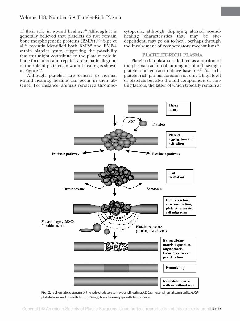
of their role in wound healing.26 Although it isgenerally believed that platelets do not containbone morphogenetic proteins (BMPs),6,35 Sipe etal.47 recently identified both BMP-2 and BMP-4within platelet lysate, suggesting the possibilitythat this might contribute to the platelet role inbone formation and repair. A schematic diagramof the role of platelets in wound healing is shownin Figure 2.
Although platelets are central to normalwound healing, healing can occur in their ab-sence. For instance, animals rendered thrombo-
cytopenic, although displaying altered wound-healing characteristics that may be site-dependent, may go on to heal, perhaps throughthe involvement of compensatory mechanisms.39
PLATELET-RICH PLASMAPlatelet-rich plasma is defined as a portion of
the plasma fraction of autologous blood having aplatelet concentration above baseline.31 As such,platelet-rich plasma contains not only a high levelof platelets but also the full complement of clot-ting factors, the latter of which typically remain at
Fig. 2. Schematic diagram of the role of platelets in wound healing. MSCs, mesenchymal stem cells; PDGF,platelet-derived growth factor; TGF-�, transforming growth factor beta.
Volume 118, Number 6 • Platelet-Rich Plasma
151e
their normal, physiologic levels. Other terms inthe literature that are sometimes used to describeplatelet preparations include platelet concen-trate, platelet gel, and platelet releasate.21,26,31,48
Ideally, there should be universal agreement re-garding definitions and terminology; however, atthe very least, the nature of the platelet derivativestudied should be precisely and unambiguouslydescribed. The processing required to concen-trate platelets in a portion of the plasma will leavethe balance essentially devoid of platelets. Suchplatelet-poor plasma can function clinically as afibrin sealant for hemostasis.2,20,23,35,49 Five impor-tant issues relating to platelets are discussed in thefollowing order: platelet concentration ratio, pro-cessing technique, quantification of secretory pro-tein concentration, handling, and application,and clinical use.
Platelet Concentration RatioTo a first approximation, the amount of he-
matoma that forms in response to trauma is pro-portional to the degree of tissue injury. In this way,delivery of platelet-rich plasma can be thought ofas responding with hematoma in excess of thatwhich would have been physiologically produced.It is likely that the effect of platelet-rich plasma onwound healing is a function of many variables,including the platelet concentration, platelet-richplasma volume delivered, the extent and type ofinjury, and the overall medical condition of thepatient. The large number of variables and theirpotential for interaction is probably the reasonthat there is no single recommendation for thedegree of increase of platelets in platelet-richplasma over baseline.
Some investigators have suggested that platelet-rich plasma should achieve a 3 to 5-fold increase inplatelet concentration over baseline,11,30,48 althoughthe dependence of clinical benefit on platelet con-centration versus total number of platelets deliveredmay need to await further investigation.32 Plateletconcentration ratios of less than 2-fold to 8.5-foldhave been reported.6,11,22,29,30,48,50 Weibrich et al.44
suggest that different individuals may require differ-ent platelet concentration ratios to achieve compa-rable biological effect.
Processing TechniqueDuring surgery, platelets will collect at the
surgical site to initiate clotting and healing,somewhat reducing the whole blood plateletcount.31 As such, blood should be drawn beforesurgery begins because the surgery itself will
lead to platelet activation that may interfere withpreparation.20,23
When anticoagulated blood is centrifuged,three layers form as a function of density: thebottom layer consisting of red blood cells (specificgravity, 1.09), the middle layer consisting of plate-lets and white blood cells (buffy coat; specific grav-ity, 1.06), and the top plasma layer (specific grav-ity, 1.03).26 Centrifugation forms the basis ofcurrent methods for producing platelet-richplasma, with the yield approximately 10 percent,by volume, of whole blood drawn. Platelet frag-mentation during processing should be avoided.Because it is the process of activation that resultsin the completion of the tertiary structure of someof the secretory proteins, such fragmentation dur-ing processing could result in the release of highlevels of proteins, with compromised bioactivity.48
The integrity of the platelet membrane can bepreserved by use of acid citrate dextrose type Aanticoagulant and low gravity forces duringcentrifugation.31,48
In addition, platelet activation occurring duringprocessing should be kept to a minimum. Althoughbioactive secretory proteins would be produced,they might be lost and not transferred to the surgicalbed when the clot is implanted, although the mag-nitude of this may be a function of the mode ofdelivery.29 P-selectin is a protein contained in theinner face of the �-granule membrane.38 On plateletactivation, the �-granule membrane fuses with theplatelet membrane and P-selectin becomes ex-pressed on the platelet surface, where it can be mea-sured and the amount of platelet activationdetermined.38,48 Thus, measurement of P-selectinprovides valuable information regarding the plate-let-rich plasma preparation.29,30
Although it is possible to use a standard lab-oratory centrifuge to produce platelet-richplasma, the process is labor intensive, generallyrequiring two spins and multiple transfers; conse-quently, sterility may be difficult to maintain.31,48,51
Furthermore, such techniques may not reliablymaximize the platelet concentration or the levelsof key secretory proteins.6
Standard cell separators and salvage devices canbe used to produce platelet-rich plasma. These de-vices operate on a unit of blood and typically usecontinuous-flow centrifuge bowl or continuous-flowdisk separation technology and both a hard (fast)and a soft (slow) spin, yielding platelet concentra-tions from two to four times baseline.30,52,53 Suchdevices include the CATS (Fresenius, Wilmington,Del.), Sequestra (Medtronic, Minneapolis, Minn.),
Plastic and Reconstructive Surgery • November 2006
152e

Haemonetics Cell Saver 5 (Haemonetics Corp.,Braintree, Mass.), and others.30,32,52
Many surgical procedures require use of rel-atively small volumes of platelet-rich plasma.20,23
Some of these procedures may be performed in anoffice setting, making draw of a full unit of bloodundesirable and legally precluding the reintro-duction of the unused portion of the blood to thepatient.31 Consequently, small, compact office sys-tems have been developed that produce approx-imately 6 ml of platelet-rich plasma from 45 to60 ml of blood, obviating the need for rein-fusion.6,20,31,54,55 There are many such systems, in-cluding the GPS (Biomet, Warsaw, Ind.), thePCCS (Implant Innovations, Inc., Palm Beach Gar-dens, Fla.), the Symphony II (DePuy, Warsaw,Ind.), the SmartPReP (Harvest TechnologiesCorp., Norwell, Mass.), and the Magellan(Medtronic, Minneapolis, Minn.).6,29,30,32,35,52,54 Al-though all operate on a small volume of drawnblood (45 to 60 ml) and on the principle of cen-trifugation, these systems differ widely in theirability to collect and concentrate platelets, withapproximately 30 to 85 percent of the availableplatelets collected and from a less than 2-fold to anapproximately 8-fold increase in the platelet con-centration over baseline.6,29,30,52
In general, most systems, whether large or smallvolume, do not concentrate the plasma proteins ofthe coagulation cascade.30,32,56 The concentration ofplasma protein levels above baseline can be achievedthough secondary ultrafiltration, as is done with theUltraConcentrator (Interpore Cross, Irvine, Calif.),and the Access System (Interpore Cross), in whichthe buffy coat collected from a centrifugation stageis passed through hollow fibers with an effective poresize of 30 kDa. With this system, up to two-thirds ofthe aqueous phase is removed by filtration; thus, theconcentrations of the retained plasma proteins andformed elements are correspondingly increased.56,57
Quantification of Secretory ProteinConcentration
The regenerative potential of platelet-richplasma depends, to large extent, on the levels ofsecretory proteins that are released on plateletactivation.29,44 These protein levels will depend onseveral factors, including (1) the concentrationsof these proteins contained in the platelets (apatient variable); (2) the processing technique,which will influence platelet concentration andwhether platelets are activated or fragmented dur-ing preparation; and (3) the completeness ofplatelet activation before measurement.29,31,44,58
The secretory proteins must first be releasedfrom the platelets before they can be measured.Release can be accomplished through platelet ac-tivation or through physical disruption of theplatelet/�-granule structure. The most commonmethod of platelet activation is to add calciumchloride and thrombin to the platelet-richplasma.23,29,35,48 The thrombin directly activatesplatelets, and the calcium ion replenishes thatwhich was bound by the acid citrate dextrose typeA anticoagulant. Although this method is oftenused to activate platelet-rich plasma clinically, theactivation that occurs during clot formation doesnot necessarily lead to complete release.33 An-other activation method uses adenosine diphos-phate, which acts directly on the platelets.59,60
Secretory protein levels are commonly ex-pressed in concentration units (e.g., measuredamount per milliliter of releasate or per 100,000platelets). Weibrich et al.,44 using a freeze/thawcycle to release proteins, measured the levels ofPDGF-��, PDGF-��, TGF-1, TGF-�2, and IGF-1 inspecimens of platelet-rich plasma derived from115 patients. Minimum and maximum values foreach typically spanned one to two orders of mag-nitude, with means � SD of 117.5 � 63.4 ng/ml,9.9 � 7.5 ng/ml, 169.4 � 84.5 ng/ml, 0.4 � 0.3ng/ml, and 84.2 � 23.6 ng/ml, respectively. Theyfound statistical correlations between the con-centrations of the following pairs of growth fac-tors: PDGF-��/PDGF-��, PDGF-��/TGF-�1, andPDGF-��/TGF-�1. There was little or no correla-tion between the levels of these individual proteinsand donor age and gender attributes. Eppley etal.29 used thrombin/calcium chloride to activateplatelets and release proteins from platelet-richplasma derived from 10 healthy volunteers. Secre-tory protein levels measured were 17 � 8 ng/ml(PDGF-��), 120 � 42 ng/ml (TGF-�1), 955 �1030 ng/ml (VEGF), 129 � 61 ng/ml (EGF), and72 � 25 ng/ml (IGF-1). Zimmermann et al.33 usedvarious methods to initiate platelet release andmeasured levels of PDGF-��, PDGF-��, andTGF-�1 in platelet-rich plasma preparations bothrich and deficient in white blood cells, expressinglevels both on a per-milliliter and a per-100,000platelet basis. For a given protein, there was typ-ically a 3- to 4-fold range in measured level versusrelease method, and the authors concluded thatthe release of each growth factor by a given samplepreparation method must be investigated and in-terpreted separately.
All else being equal, one would expect that theconcentration of released secretory proteinswould be linearly proportional to the platelet con-
Volume 118, Number 6 • Platelet-Rich Plasma
153e

centration ratio. Although such a relationship be-tween some secretory proteins (i.e., PDGF-��,TGF-�, VEGF, and EGF) and platelet count hasbeen reported,59 an additional study by the sameprincipal author confirmed this relationship onlyfor PDGF, TGF-�, and EGF, but not for VEGF andIGF.60 Although a general trend of increasing pro-tein content and platelet count for a variety ofsecretory proteins (PDGF-��, PDGF-��, TGF-1,TGF-�2, VEGF, EGF, and IGF-1) was demon-strated, Eppley et al.29 and Weibrich et al.44 foundlittle value in using platelet concentration ratio topredict resultant platelet-rich plasma secretoryprotein levels. Eppley et al.,29 using thrombin/calcium chloride to activate the platelets, foundvariable concentration ratios for several secretoryproteins, all lower than the platelet concentrationratio. Incomplete platelet activation and variablebinding of the expressed proteins to the clot,which would not have been measured in the plate-let-rich plasma supernatant, could be a partial ex-planation.
Handling and Application of Platelet-RichPlasma
After preparation, platelet-rich plasma is sta-ble, in the anticoagulated state, for 8 hours, orlonger, permitting the blood to be drawn beforesurgery and used, as needed, during lengthyoperations.6,31,61 The platelet-rich plasma must beactivated for the platelets to release their �-gran-ule contents, with the clot that forms providing avehicle to contain the secreted proteins and main-tain their presence at the wound site. This is mostcommonly performed by adding a solution of1000 units of topical bovine thrombin per milli-liter of 10% calcium chloride to the platelet-richplasma.2,20,22,29 Marx et al.22 described a techniquein which 6 ml of platelet-rich plasma, 1 ml of thecalcium chloride/thrombin mix, and 1 ml of air(to act as a mixing bubble) is introduced into a10-ml syringe. The syringe is agitated for 6 to 10seconds to initiate clotting, and then the clot de-livered. Alternatively, Man et al.20 described an-other technique for delivering the activated plate-let-rich plasma. The platelet-rich plasma andcalcium chloride/thrombin solution are mixed ina 10:1 (volume/volume) ratio by use of a dual-syringe mixing system. The platelet-rich plasma isdrawn into a 10-ml syringe and the activating so-lution is drawn into a 1-ml syringe. Both syringeplungers are connected to move together withboth output ports connected to a dual-spray ap-plicator tip that allows both solutions to be mixed
as they are applied to the wound. Platelet-poorplasma can be delivered similarly to function as afibrin glue or hemostatic agent.20,23 Because the�-granules quickly release their contents on acti-vation, Marx6 states that the clotted platelet-richplasma should be used within 10 minutes of clotinitiation.
This is not an issue with the dual-syringe spraydelivery, as the platelet-rich plasma is delivered tothe wound site immediately after activation. In thecase of other mixing techniques, it is important totransfer the clot to the surgical site before clotretraction; otherwise, the transferred clot may bedeficient in the secretary proteins that were ex-pressed.
In the early to mid 1990s, there were a fewreports of the development of antibovine antibod-ies (antibovine factor V) that cross-reacted withhuman clotting factors in response to use of thebovine product to provide hemostasis to open,bleeding vessels.62–64 Although bovine thrombin isoften used (along with calcium chloride) for clin-ical platelet activation, there is little evidence thatsuch cross-reaction occurs for this application.This may be because (1) current processing meth-ods remove much more bovine factor V contam-ination and (2) its use in platelet-rich plasma gelprecludes its exposure to the systemic circulation,possibly explaining why platelet-rich plasma hasnot produced postsurgical bleeding or shown el-evation in postoperative prothrombin time orthe development of detectable antibovineantibodies.10
Clinical UseProponents of platelet-rich plasma technol-
ogy suggest that benefits include an increase inhard- and soft-tissue wound healing and a de-crease in postoperative infection, pain, andblood loss.34 There have been numerous publi-cations on the use of platelet-rich plasma forseveral clinical applications, including peri-odontal and oral surgery,12,14,16,18,22–25,27 maxillo-facial surgery,27 aesthetic plastic surgery,2,20,26
spinal fusion,13,17,19 heart bypass surgery,15 andtreatment of chronic skin and soft-tissueulcers.21,65 The details of the quantity of platelet-rich plasma used and the methods of applicationare procedure-specific. Although the vast ma-jority of these studies have yielded excellent out-comes, most are only limited case studies orseries. As such, wound-healing enhancement byplatelet-rich plasma remains largely anecdotal.There exists, however, a small collection of
Plastic and Reconstructive Surgery • November 2006
154e

clinical studies with prospective or retrospec-tive controls that have demonstrated a sig-nificant enhancement of hard- and soft-tissue healing with the use of platelet-richplasma.11,12,15,17,21
In plastic surgery, the use of autologousblood–derived products has been largely limitedto fibrin glues, primarily used to obtain hemostasisand adherence of skin flaps. The first report wasin 1990 regarding a diverse use of fibrin glue (Tis-seel) for multiple aesthetic facial applications (23patients) applied on the undersurface of flaps.66
Application for a “minimal suture blepharoplasty”in 1992 with closure of incisions with autologousfibrin glue reported lower complications withmilia formation than standard suturetechniques.67 A study of 20 patients by Man et al.20
demonstrated that the use of autologous fibringlue and platelet gel in cosmetic surgical proce-dures involving the creation of flaps, such as inface and neck lifts and breast reductions and aug-mentations, resulted in numerous advantages.These included the elimination of the need fordrains, a reduction in postoperative pain andswelling, and improved wound healing. In an-other skin flap study, a clinical series of eight pa-tients were unilaterally treated with autologousplatelet–rich plasma mixed with thrombin and cal-cium chloride to form an autologous platelet gelto determine the effects on postoperative recoveryfrom deep-plane rhytidectomy.68 Staged postop-erative facial photographs were graded in ablinded fashion for postoperative ecchymosis andedema. Although no statistically significant differ-ences were identified in the data, trends suggestedthat treatment with autologous platelet gel wasbetter at preventing or improving ecchymosisrather than edema, and was chiefly demonstrablein the early phases of recovery. In a larger face-liftcase series, a single postoperative hematomaamong a cohort of 100 consecutive patients un-dergoing face lifting treated with bilateral autol-ogous platelet gel occurred, an outcome reportedas a significant reduction in postoperative com-plication incidence by the authors. No drains orpostoperative dressings were used in the fibringlue–treated group, which patients found mostfavorable.69 Similarly, Oliver et al. in 2001 re-ported on a prospective, randomized, double-blind trial of the use of fibrin sealant for face liftsin 20 patients. A significant difference in drainoutput occurred between control and treatedsides (average, 30 ml for the control side and 10ml for the fibrin glue side). It was suggested thatpostoperative drains may not be needed with this
technique, and that pain and bruising may like-wise be reduced.70 Favorable results were also re-ported by Fezza in 2002. In 24 consecutive pa-tients, fibrin glue was used in face lifts withoutdrains. These patients had less bruising and swell-ing, no incidence of hematoma, and shorter op-erative times compared with another group of 24face-lift patients where glue was not used.71 Themost recent study of the efficacy of a commercial(nonautologously derived) fibrin glue on face liftswas reported by Marchac in 2005, nearly 10 yearsafter his initial report in 1994. In a prospectivestudy of 30 patients, there were minimal differ-ences between glued and unglued sides in termsof drain output, bruising, swelling, or the inci-dence of hematomas.72 After 17 years of clinicaluse, this experienced author now concludes thatthe theoretical benefit of fibrin in face lifts is notas great as previously hoped. In a companion ex-perience, Jones et al. in 2004 did not find anystatistical difference in hematoma rates with theuse of fibrin glue compared with other preventiontechniques such as dressings, drains, or the use oftumescence.73
Results with fibrin glue fixation in foreheadendoscopy were reported in 1998. In 206 endo-scopic forehead procedures in 196 patients inwhich fibrin glue was used, good results were ob-tained in 86 percent of the cases, with minimalcomplications. Fibrin glue provided stable fixa-tion, with the possibility of displacement in severaldirections, including transversal expansion.74
Numerous clinical trials have reported favorableresults evaluating wounds treated with platelet re-leasate. Knighton et al.75 observed that 17 of 21chronic lower extremity ulcers reepithelialized dur-ing an 8.6-week (average) course of twice-dailywound treatment with platelet releasate suspendedon a collagen base (platelet-derived wound-healingfactor) compared with two of 13 similar woundstreated with placebo. After crossover of the placebogroup, all 11 nonhealed wounds achieved 100 per-cent epithelialization in an average of 7.1 weeks. Anearlier study by the same group, also using platelet-derived wound-healing factor, demonstrated a 93percent reepithelialization rate among 71 chronicwounds in 41 patients after daily treatments withautologous platelet concentrate. A similar clinicalprotocol was reported by Ganio et al. in a case seriesof 171 patients with 355 chronic (average, 75 weeksbefore presentation) lower extremity wounds.76 Re-sults included a 78 percent rate of limb salvage inpatients for whom amputation was initially recom-mended after daily 12-hour treatments with platelet-derived wound-healing factor for an average of 10
Volume 118, Number 6 • Platelet-Rich Plasma
155e

Tab
le1
.Su
mm
ary
ofC
linic
alSt
ud
ies
Inve
stig
atin
gU
seo
fPR
Pan
dP
PP
App
licat
ion
Stud
yD
esig
nR
esul
tsR
efer
ence
Skin
flap
scr
eate
ddu
rin
gfa
ce,
nec
k,an
dbr
east
surg
ery
20pa
tien
ts.
PRP
and
PPP
appl
ied
tofl
aps.
Dua
l-spr
ayap
plic
atio
n.
No
con
trol
sw
ith
out
PRP/
PPP.
Coa
gula
tion
ach
ieve
dw
ith
in45
sec.
Aut
hor
sfe
elbe
nef
its
incl
ude
the
follo
win
g:n
odr
ain
s,le
sspo
stop
erat
ive
pain
and
swel
ling,
shor
ter
oper
atin
gti
mes
.
20
Face
lift
Ran
dom
ized
,pr
ospe
ctiv
e,co
ntr
olle
d.8
pati
ents
,pl
atel
ets
appl
ied
toon
esi
dean
dn
otot
her
inea
chpa
tien
t.PR
Pge
lin
ject
edin
tow
oun
d.
Post
oper
ativ
ere
cove
ryph
otog
raph
sev
alua
ted
blin
ded
for
ecch
ymos
isan
ded
ema.
No
sign
ific
ant
effe
ctof
PRP,
but
tren
dssu
gges
tPR
Pm
igh
tre
duce
post
oper
ativ
esw
ellin
gan
dbr
uisi
ng.
68
Face
lift
100
con
secu
tive
pati
ents
trea
ted
wit
hbi
late
ral
PRP.
Sign
ific
ant
redu
ctio
nin
post
oper
ativ
eco
mpl
icat
ion
inci
den
ce.
69
Face
lift
30pa
tien
ts.
Pati
ents
pros
pect
ivel
yra
ndo
miz
edto
hav
ing
fibr
ingl
ue(c
omm
erci
al,
non
auto
logo
us)
appl
ied
tole
ftor
righ
tsi
deof
face
and
nec
k.N
eck
drai
ns,
but
not
face
drai
ns
used
.
Th
ere
was
evid
ence
ofle
ss24
-hr
drai
nag
eon
trea
ted
side
.T
her
ew
asn
osi
gnif
ican
tef
fect
oftr
eatm
ent
on1-
and
8-da
ysy
mm
etry
.A
uth
ors
hav
edi
scon
tin
ued
rout
ine
use
offi
brin
glue
.
70
Low
erex
trem
ity
chro
nic
,n
onh
ealin
gsk
inw
oun
ds32
(tot
al)
pati
ents
,pr
ospe
ctiv
e,ra
ndo
miz
ed,
con
trol
led,
blin
ded,
cros
sove
rst
udy.
Tre
ated
wit
hPD
WH
Fon
colla
gen
base
orpl
aceb
ofo
r8
wk.
By
8w
k,81
%of
PDW
HF-
trea
ted
pati
ents
had
100%
epit
hel
ializ
atio
n,
wh
erea
son
ly15
%of
con
trol
sdi
d(p
�0.
0001
).A
fter
cros
sove
r,un
hea
led
plac
ebo
pati
ents
hea
led
byav
erag
eof
7.1
wk.
65
Ch
ron
icw
oun
ds41
pati
ents
pres
enti
ng
wit
h71
chro
nic
wou
nds
aver
agin
g3.
8yr
ofco
nve
nti
onal
trea
tmen
t.D
aily
trea
tmen
tw
ith
PDW
HF
onco
llage
nba
se.
Succ
essf
ulre
epit
hel
ializ
atio
nac
hie
ved
in90
%of
pati
ents
and
93%
ofw
oun
dsat
anav
erag
eof
7.5
wk.
No
com
plic
atio
ns
from
PDW
HF
use.
71
Ch
ron
icn
onh
ealin
gw
oun
dsin
the
foot
171
pati
ents
pres
enti
ng
wit
h35
5w
oun
dsof
aver
age
dura
tion
75w
ktr
eate
dw
ith
PDW
HF.
100%
epit
hel
ializ
atio
nac
hie
ved
atan
aver
age
of10
wee
ks;
78%
limb
salv
age
for
“at-r
isk”
pati
ents
.72
Man
dibu
lar
con
tin
uity
defe
cts
Ran
dom
ized
,pr
ospe
ctiv
est
udy
of88
pati
ents
.U
sed
can
cello
usce
llula
rm
arro
wgr
afts
wit
hor
wit
hou
tPR
P.R
adio
grap
hic
and
his
tom
orph
omet
ryas
sess
men
tat
6m
o.
Gra
fts
wit
hPR
Ph
adap
prox
imat
ely
twic
eth
era
diog
raph
icm
atur
ity
ofth
ose
wit
hou
tan
da
sign
ific
antl
ygr
eate
rtr
abec
ular
bon
ede
nsi
ty.
Th
eau
thor
sco
ncl
uded
that
PRP
acce
lera
ted
bon
efo
rmat
ion
.
22
PRP,
plat
elet
-ric
hpl
asm
a;PP
P,pl
atel
et-p
oor
plas
ma;
PDW
HF,
plat
elet
-der
ived
wou
nd-
hea
ling
fact
or.
Plastic and Reconstructive Surgery • November 2006
156e
weeks. Marx et al.22 reported enhanced bone for-mation in mandibular bone grafts treated with plate-let-rich plasma, an effect lasting up to 6 monthspostoperatively that was documented by biopsy-proven enhanced bone deposition among platelet-rich plasma-treated grafts. Table 1 summarizes thesecited studies. Collectively, they provide existing evi-dence for the clinical use of platelet technology inthe healing of both soft- and hard-tissue wounds.
CONCLUSIONSPlatelets immediately appear at the site of tis-
sue injury in large numbers. As such, it makesevolutionary sense that platelets direct woundhealing because, by design, they will be presentexactly where and when needed, to create a localenvironment conducive to tissue regeneration. Byreleasing secretory proteins from their �-granuleson activation, platelets set the pace of wound heal-ing, with their effects remaining long after the clothas been cleared. The enhancement of healing bythe placement of a supraphysiologic concentra-tion of autologous platelets at the site of tissueinjury is supported by basic science studies. Thereexist a small number of controlled, clinical studiesthat provide evidence that the use of autologousplatelet-rich plasma does, indeed, accelerate soft-and hard-tissue healing in at least some applica-tions. The future role of platelet-rich plasma inplastic surgery largely remains both theoreticaland anecdotal at this time. Many more controlledclinical studies will be required to establish theconditions under which the application of plate-let-rich plasma has merit. Currently, platelet-richplasma use seems to offer some efficacy in certaintypes of acute and chronic wounds, although thetiming of application is not clear. In aesthetic plas-tic surgery, although theoretically appealing andnot harmful, the potential benefits of platelet-richplasma may be difficult to measure in these pa-tients with otherwise normal wound healing.
Barry L. Eppley, M.D., D.M.D.Division of Plastic Surgery
Indiana University School of Medicine702 Barnhill Drive, Suite 3540
Indianapolis, Ind. [email protected]
DISCLOSURE
None of the authors has a financial interest in anyof the products, devices, or drugs mentioned in thisarticle.
REFERENCES1. Beasley, L. S., and Einhorn, T. A. Role of growth factors in
fracture healing. In E. Canalis (Ed.), Skeletal Growth Factors .New York: Lippincott Williams & Wilkins, 2000. Pp. 311–322.
2. Bhanot, S., and Alex, J. C. Current applications of plateletgels in facial plastic surgery. Facial Plast. Surg. 18: 27, 2002.
3. Anitua, E., Andia, I., Ardanza, B., et al. Autologous plateletsas a source of proteins for healing and tissue regeneration.Thromb. Haemost. 91: 4, 2004.
4. Buckwalter, J. A., Einhorn, T. A., Bolander, M. E., et al.Healing of musculoskeletal tissues. In C. A. Rockwood, Jr.,R. W. Bucholz, and D. P. Green (Eds.), Fractures in Adults.Philadelphia: Lippincott-Raven, 1996. Pp. 261–304.
5. Anderson, J. M. The cellular cascades of wound healing. InJ. E. Davies (Ed.), Bone Engineering. Toronto: em squared inc.,2000. Pp. 81–93.
6. Marx, R. E. Platelet-rich plasma: Evidence to support its use.J. Oral Maxillofac. Surg. 62: 489, 2004.
7. Haynesworth, S. E., Kadiyala, S., Liang, L., et al. Mitogenicstimulation of human mesenchymal stem cells by plateletreleasate suggest a mechanism for enhancement of bonerepair by platelet concentrates (Poster no. 462). Transactionsof the 48th Annual Meeting of the Orthopaedic Research Society,Dallas, Texas, February 10–13, 2002.
8. Liu, Y., Kalen, A., Risto, O., et al. Fibroblast proliferation dueto exposure to a platelet concentrate in vitro is pH depen-dent. Wound Repair Regen. 10: 336, 2002.
9. Carter, C. A., Jolly, D. G., Worden, C. E., Sr., et al. Platelet-richplasma gel promotes differentiation and regeneration dur-ing equine wound healing. Exp. Mol. Pathol. 74: 244, 2003.
10. Fennis, J. P. M., Stoelinga, P. J. W., and Jansen, J. A. Man-dibular reconstruction: A clinical and radiographic animalstudy on the use of autogenous scaffolds and platelet-richplasma. J. Oral Maxillofac. Surg. 31: 281, 2002.
11. Marx, R. E. Platelet concentrate: A strategy for acceleratingand improving bone regeneration. In J. E. Davies (Ed.), BoneEngineering . Toronto: University of Toronto, 2000. Pp. 447–453.
12. Anitua, E. Plasma rich in growth factors: Preliminary resultsof use in the preparation of future sites for implants. J. OralImplantol. 14: 529, 1999.
13. Bose, B., and Balzarini, M. A. Bone graft gel: Autologousgrowth factors used with autograft bone for lumbar spinefusions. Adv. Ther. 19: 170, 2002.
14. Della Valle, A., Sammartino, G., Marenzi, G., et al. Preventionof postoperative bleeding in anticoagulated patients under-going oral surgery: Use of platelet-rich plasma gel. J. OralMaxillofac. Surg. 61: 1275, 2003.
15. DelRossi, A. J., Cernaianu, A. C., Vertrees, R. A., et al. Platelet-rich plasma reduces postoperative blood loss after cardio-pulmonary bypass. J. Thorac. Cardiovasc. Surg. 100: 281, 1990.
16. Froum, S. J., Wallace, S. S., Tarnow, D. P., et al. Effect ofplatelet-rich plasma on bone growth and osseointegration inhuman maxillary sinus grafts: Three bilateral case reports.Int. J. Periodontics Restorative Dent. 22: 45, 2002.
17. Hee, H. T., Majd, M. E., Holt, R. T., et al. Do autologousgrowth factors enhance transforaminal lumbar interbodyfusion? Eur. Spine J. 12: 400, 2003.
18. Kassolis, J. D., Rosen, P. S., and Reynolds, M. A. Alveolar ridgeand sinus augmentation utilizing platelet-rich plasma in com-bination with freeze-dried bone allograft: Case series. J. Peri-odontol. 71: 1654, 2000.
19. Lowery, G. L., Kulkarni, S., and Pennisi, A. E. Use of autol-ogous growth factors in lumbar spinal fusion. Bone 25 (2Suppl.): 47S, 1999.
Volume 118, Number 6 • Platelet-Rich Plasma
157e

20. Man, D., Plosker, H., and Winland-Brown, J. E. The use ofautologous platelet-rich plasma (platelet gel) and autolo-gous platelet-poor plasma (fibrin glue) in cosmetic surgery.Plast. Reconstr. Surg. 107: 229, 2001.
21. Margolis, D. J., Kantor, J., Santanna, J., et al. Effectiveness ofplatelet releasate for the treatment of diabetic neuropathicfoot ulcers. Diabetes Care 24: 483, 2001.
22. Marx, R. E., Carlson, E. R., Eichstaedt, R. M., et al. Platelet-rich plasma: Growth factor enhancement for bone grafts.Oral Surg. Oral Med. Oral Pathol. Oral Radiol. Endod. 85: 638,1998.
23. Petrungaro, P. S. Using platelet-rich plasma to accelerate softtissue maturation in esthetic periodontal surgery. Compend.Contin. Educ. Dent. 22: 729, 2001.
24. Robiony, M., Polini, F., Costa, F., et al. Osteogenesis distrac-tion and platelet-rich plasma for bone restoration of theseverely atrophic mandible: Preliminary results. J. Oral Max-illofac. Surg. 60: 630, 2002.
25. Soffer, E., Ouhayoun, J. P., and Anagnostou, F. Fibrin seal-ants and platelet preparations in bone and periodontal heal-ing. Oral Surg. Oral Med. Oral Pathol. Oral Radiol. Endod. 95:521, 2003.
26. Welsh, W. J. Autologous platelet gel: Clinical function andusage in plastic surgery. Cosmetic Derm. 11: 13, 2000.
27. Whitman, D. H., Berry, R. L., and Green, D. M. Platelet gel:An autologous alternative to fibrin glue with applications inoral and maxillofacial surgery. J. Oral Maxillofac. Surg. 55:1294, 1997.
28. Shanaman, R., Filstein, M. R., and Danesh-Meyer, M. J. Lo-calized ridge augmentation using GBR and platelet-rich plas-ma: Case reports. Int. J. Periodontics Restorative Dent. 21: 345,2001.
29. Eppley, B. L., Woodell, J. E., and Higgins, J. Platelet quan-tification and growth factor analysis from platelet-rich plas-ma: Implications for wound healing. Plast. Reconstr. Surg. 114:1502, 2004.
30. Kevy, S. V., and Jacobson, M. S. Comparison of methods forpoint of care preparation of autologous platelet gel. J. ExtraCorpor. Technol. 36: 28, 2004.
31. Marx, R. E. Platelet-rich plasma (PRP): What is PRP and whatis not PRP? Implant Dent. 10: 225, 2001.
32. Waters, J. H., and Roberts, K. C. Database review of possiblefactors influencing point-of-care platelet gel manufacture.J. Extra Corpor. Technol. 36: 250, 2004.
33. Zimmermann, R., Arnold, D., Strasser, E., et al. Sample prep-aration technique and white cell content influence the de-tectable levels of growth factors in platelet concentrates. VoxSang. 85: 283, 2003.
34. Guyton, A. C. Physiology of the Human Body. Philadelphia:Saunders College Publishing, 1979.
35. Tischler, M. Platelet rich plasma: The use of autologousgrowth factors to enhance bone and soft tissue grafts. N. Y.State Dent. J. 68: 22, 2002.
36. Conley, C. L. Hemostasis. In V. B. Mountcastle (Ed.), MedicalPhysiology. St. Louis: Mosby, 2004. Pp. 1137–1146.
37. Caro, C. D., Pedley, T. J., Schroter, R. C., et al. The Mechanicsof the Circulation. Oxford: Oxford University Press, 1978.
38. Harrison, P., and Cramer, E. M. Platelet alpha-granules. BloodRev. 7: 52, 1993.
39. Szpaderska, A. M., Egozi, E. I., Gamelli, R. L., et al. The effectof thrombocytopenia on dermal wound healing. J. Invest.Dermatol. 120: 1130, 2003.
40. Lowe, H. C., Rafty, L. A., Collins, T., et al. Biology of platelet-derived growth factor. In E. Canalis (Ed.), Skeletal GrowthFactors. Philadelphia: Lippincott Williams & Wilkins, 2000.Pp. 129–151.
41. Woodward, S. C., and Salthouse, T. N. The tissue responseto implants and its evaluation by light microscopy. In A. F.von Recum (Ed.), Handbook of Biomaterials Evaluation. NewYork: Macmillan, 1986. Pp. 364–378.
42. Frost, H. M. A 2003 update of bone physiology and Wolff’slaw for clinicians. Angle Orthod. 74: 3, 2004.
43. Roseborough, I. E., Grevious, M. A., and Lee, R. C. Preven-tion and treatment of excessive dermal scarring. J. Natl. Med.Assoc. 96: 108, 2004.
44. Weibrich, G., Kleis, W. K., Hafner, G., et al. Growth factorlevels in platelet-rich plasma and correlations with donorage, sex, and platelet count. J. Craniomaxillofac. Surg. 30: 97,2002.
45. Lariviere, B., Rouleau, M., Picard, S., et al. Human plasmafibronectin potentiates the mitogenic activity of platelet-de-rived growth factor and complements its wound healingeffects. Wound Repair Regen. 11: 79, 2003.
46. Zhou, Y. Q., Levesque, J. P., Hatzfeld, A., et al. Fibrinogenpotentiates the effect of interleukin-3 on early human he-matopoietic progenitors. Blood 82: 800, 1993.
47. Sipe, J. B.Waits, C. A.Skikne, B. et al. The presence of bonemorphogenetic proteins (BMPs) in megakaryocytes andplatelets. Presented at the 24th Annual Meeting of the Amer-ican Society for Bone and Mineral Research, San Antonio,Texas, September 20–24, 2002.
48. Gonshor, A. Technique for producing platelet-rich plasmaand platelet concentrate: Background and process. Int. J.Periodontics Restorative Dent. 22: 547, 2002.
49. Mann, K. G. Thrombin formation. Chest 124 (3 Suppl.): 4S,2003.
50. Weibrich, G., Kleis, W. K., Kunz-Kostomanolakis, M., et al.Correlation of platelet concentration in platelet-rich plasmato the extraction method, age, sex, and platelet count of thedonor. Int. J. Oral Maxillofac. Implant. 16: 693, 2001.
51. Slater, M., Patava, J., Kingham, K., et al. Involvement ofplatelets in stimulating osteogenic activity. J. Orthop. Res. 13:655, 1995.
52. Arm, D. M. Autologous platelet-based therapies for ortho-paedic tissue regeneration. Orthopedics 25: 169, 2002.
53. Hannon, T. J., Polston, G., Pekarske, W. J., et al. Determinationof Platelet Yields from Platelet Rich Plasma for Five AutotransfusionDevices. Cardiothoracic Research and Education Foundation,1999.
54. Marlovits, S., Mousavi, M., Gabler, C., et al. A new simplifiedtechnique for producing platelet-rich plasma: A short tech-nical note. Eur. Spine J. 13 (Suppl. 1): S102, 2004.
55. Lozada, J. L., Caplanis, N., Proussaefs, P., et al. Platelet-richplasma application in sinus graft surgery: Part I. Backgroundand processing techniques. J. Oral Implantol. 27: 38, 2001.
56. Hood, A. G., and Arm, D. M. Topical application of autog-enous tissue growth factors for augmentation of structuralbone graft fusion. Presented at the American Society of Ex-tra-Corporeal Technology 11th Annual Symposium on NewAdvances in Blood Management, Las Vegas, Nev. April 20–23, 2004.
57. Arm, D. M. Characterization of an autologous platelet gelcontaining multiple growth factors. Transactions of the 45thAnnual Meeting of the Orthopaedic Research Society, Anaheim,Calif., February 1–4, 1999.
58. Weibrich, G., Kleis, W. K., and Hafner, G. Growth factorlevels in the platelet-rich plasma produced by 2 differentmethods: Curasan-type PRP kit versus PCCS PRP system. Int.J. Oral Maxillofac. Implants 17: 184, 2002.
59. Kevy, S. V., Jacobson, M. S., Blasetti, L., et al. Preparation ofgrowth factor enriched autologous platelet gel (Paper no.
Plastic and Reconstructive Surgery • November 2006
158e
262). Transactions of the Society for Biomaterials 27th AnnualMeeting, St. Paul, Minn., April 24–29, 2001.
60. Kevy, S. V., Jacobson, M. S., and Kadiyala, S. Characterizationof growth factor levels in platelet concentrates. Presented atthe 5th Annual Hilton Head Workshop on Engineering Tis-sues, Hilton Head, S.C., February 21–25, 2001.
61. Anderson, N. A., Pamphilon, D. H., Tandy, N. J., et al. Com-parison of platelet-rich plasma collection using the Haemo-netics PCS and Baxter Autopheresis C. Vox Sang. 60: 155,1991.
62. Christie, R. J., Carrington, L., and Alving, B. Postoperativebleeding induced by topical bovine thrombin: Report of twocases. Surgery 121: 708, 1997.
63. Rapaport, S. I., Zivelin, A., Minow, R. A., et al. Clinical sig-nificance of antibodies to bovine and human thrombin andfactor V after surgical use of bovine thrombin. Am. J. Clin.Pathol. 97: 84, 1992.
64. Zehnder, J. L., and Leung, L. L. Development of antibodiesto thrombin and factor V with recurrent bleeding in a patientexposed to topical bovine thrombin. Blood 76: 2011, 1990.
65. Knighton, D. R., Ciresi, K., Fiegel, V. D., et al. Stimulation ofrepair in chronic, nonhealing, cutaneous ulcers using plate-let-derived wound healing formula. Surg. Gynecol. Obstet. 170:56, 1990.
66. Ellis, D. A., and Shaikh, A. The ideal tissue adhesive in facialplastic and reconstructive surgery. J. Otolaryngol. 19: 68, 1990.
67. Mandel, M. A. Minimal suture blepharoplasty: Closure ofincisions with autologous fibrin glue. Aesthetic Plast. Surg. 16:269, 1992.
68. Powell, D. M., Chang, E., and Farrior, E. H. Recovery fromdeep-plane rhytidectomy following unilateral wound treat-ment with autologous platelet gel. Arch. Facial Plast. Surg. 3:245, 2001.
69. Marchac, D., and Sandor, G. Facelifts and sprayed fibrin glue:An outcome analysis of 200 patients. Br. J. Plast. Surg. 47: 306,1994.
70. Oliver, D. W., Hamilton, S. A., Figle, A. A., Wood, S. H., andLamberty, B. G. A prospective, randomized, double-blindtrial of the use of fibrin sealant for facelifts. Plast. Reconstr.Surg. 108: 2102, 2001.
71. Fezza, J. P., Cartwrwight, M., Mack, W., and Flaharty, P. Thesue of aerosolized fibrin glue in face-lift surgery. Plast. Re-constr. Surg. 110: 658, 2002.
72. Marchac, D., and Greensmith, A. L. Early postoperative ef-ficacy of fibrin glue in facelifts: A prospective randomizedtrial. Plast. Reconstr. Surg. 115: 911, 2005.
73. Jones, B. M., and Grover, R. Avoiding hematoma in cervi-cofacial rhytidectomy: A personal 8-year quest. Reviewing910 patients. Plast. Reconstr. Surg. 113: 381, 2004.
74. Marchac, D., Ascherman, J., and Arnaud, E. Fibrin glue fix-ation in forehead endoscopy: Evaluation of our experiencein 206 cases. Plast. Reconstr. Surg. 100: 704, 1997.
75. Knighton, D. R., Fiegal, V. D., Doucette, M., et al. The use oftopically applied platelet growth factors in chronic nonheal-ing wounds: A review. Wounds 1: 71, 1989.
76. Ganio, C., Tenewitz, F. E., Wilson, R., et al. The treatment ofchronic nonhealing wounds using autologous platelet-de-rived growth factors. J. Foot Ankle Surg. 32: 263, 1993.
Volume 118, Number 6 • Platelet-Rich Plasma
159e