Embed Size (px)
Citation preview
MASSACHUSETTSGENERAL HOSPITAL
RADIATION ONCOLOGY
Plastimatch, 3D Slicer, Slicer-RT, RTK
Gregory C. Sharp, PhD
Department of Radiation OncologyMassachusetts General Hospital
AAPM Annual Meeting 2015
Disclosures
No conflict of interest
I participate in sponsored research and beta testing agreements with Elekta and IBA Dosimetry
Disclaimer
“The Software has been designed for research purposes only and has not been reviewed or
approved by the Food and Drug Administration or by any other agency”
What's on my desktop?
● CERR● ConquestDICOM● Cygwin● dcmtk● Emacs● Firefox● gcc● gimp
● KeePass● Inkscape● LibreOffice● Octave● perl● Synergy● VirtualBox● 3D Slicer
Why open source?
● Freedom!● Software can move with scientists● No license files needed
● Open development process● Documentation● Mailing lists● Source code management● Bug tracking
Today's outline
slicer.org
slicerrt.orgplastimatch.org
openrtk.org
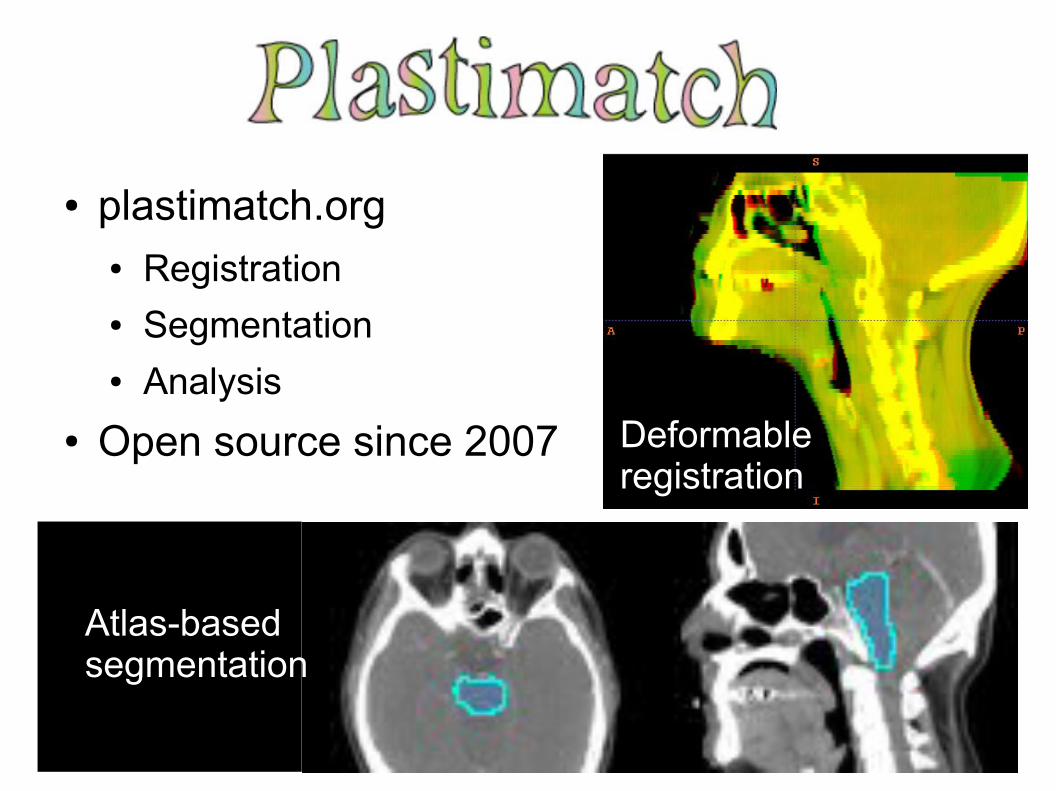
● plastimatch.org● Registration● Segmentation● Analysis
● Open source since 2007 Deformable registration
Atlas-basedsegmentation
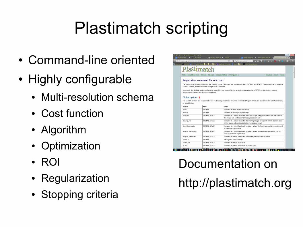
Plastimatch scripting
● Command-line oriented● Highly configurable
● Multi-resolution schema● Cost function● Algorithm● Optimization● ROI● Regularization● Stopping criteria
Documentation on
http://plastimatch.org
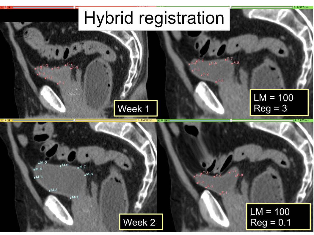
Week 1
Week 2LM = 100Reg = 0.1
LM = 100Reg = 3
Hybrid registration
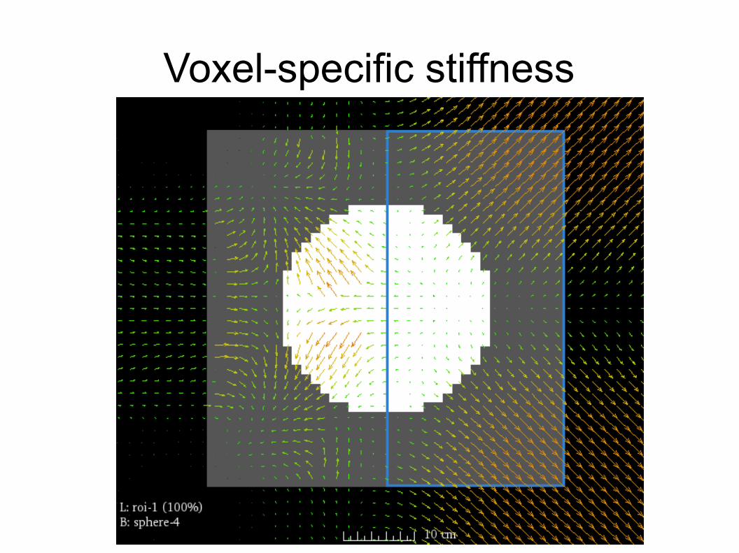
Voxel-specific stiffness
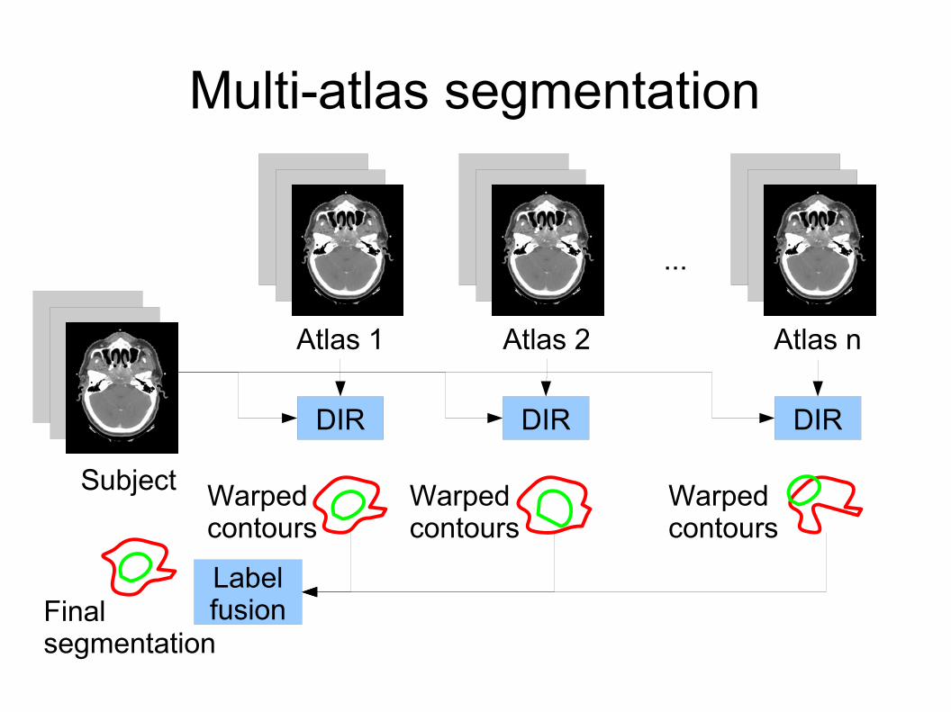
Multi-atlas segmentation
Atlas 1 Atlas 2
DIR
Warpedcontours
Subject
Atlas n
...
DIR DIR
Warpedcontours
Warpedcontours
LabelfusionFinal
segmentation
Laboratory for Percutaneous Surgery – Copyright © Queen’s University, 2015
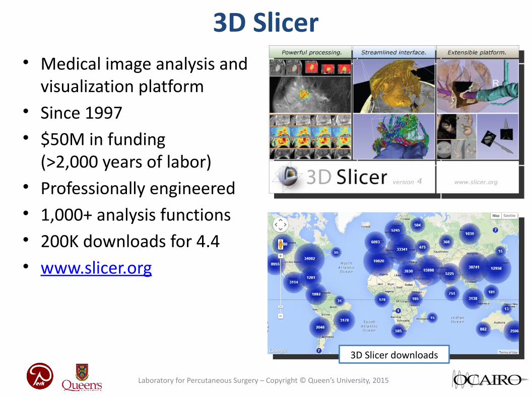
3D Slicer• Medical image analysis and
visualization platform• Since 1997• $50M in funding
(>2,000 years of labor)• Professionally engineered• 1,000+ analysis functions• 200K downloads for 4.4• www.slicer.org
3D Slicer downloads
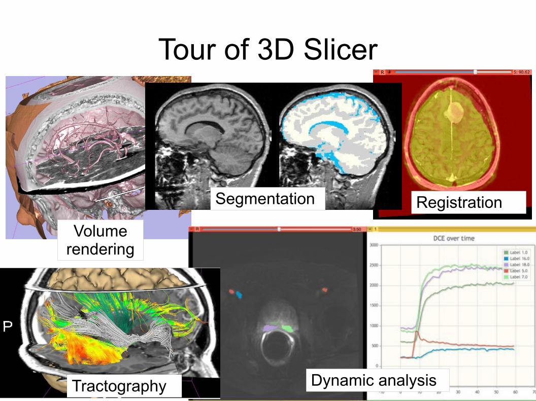
Tour of 3D Slicer
Tractography
Segmentation
Dynamic analysis
Registration
Volume rendering
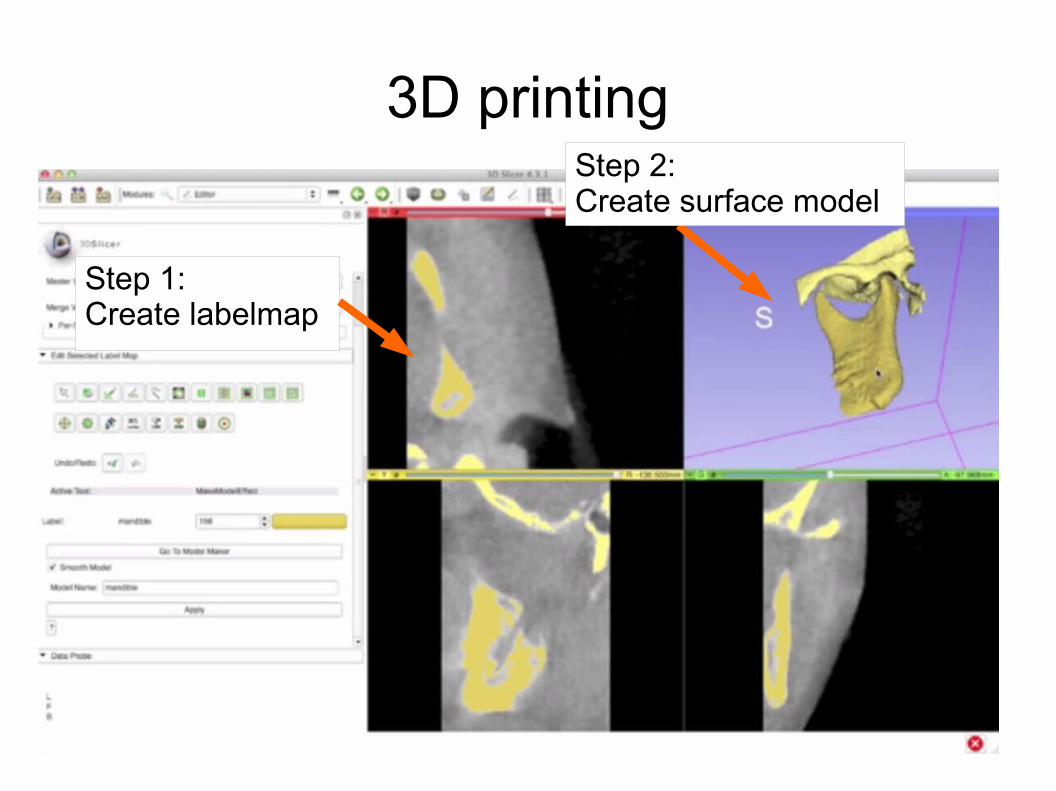
3D printing
Step 1:Create labelmap
Step 2:Create surface model
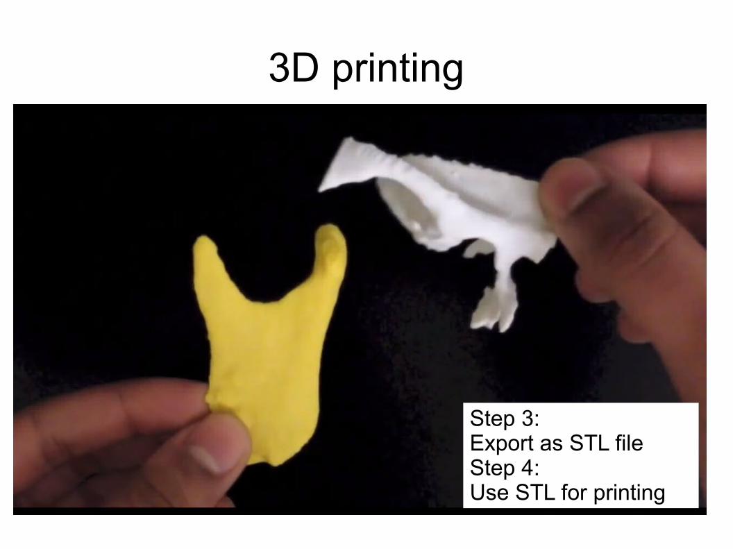
3D printing
Step 3:Export as STL fileStep 4:Use STL for printing
Developing slicer modules
● Three kinds of modules:● Command line module● Scripted module● Loadable module
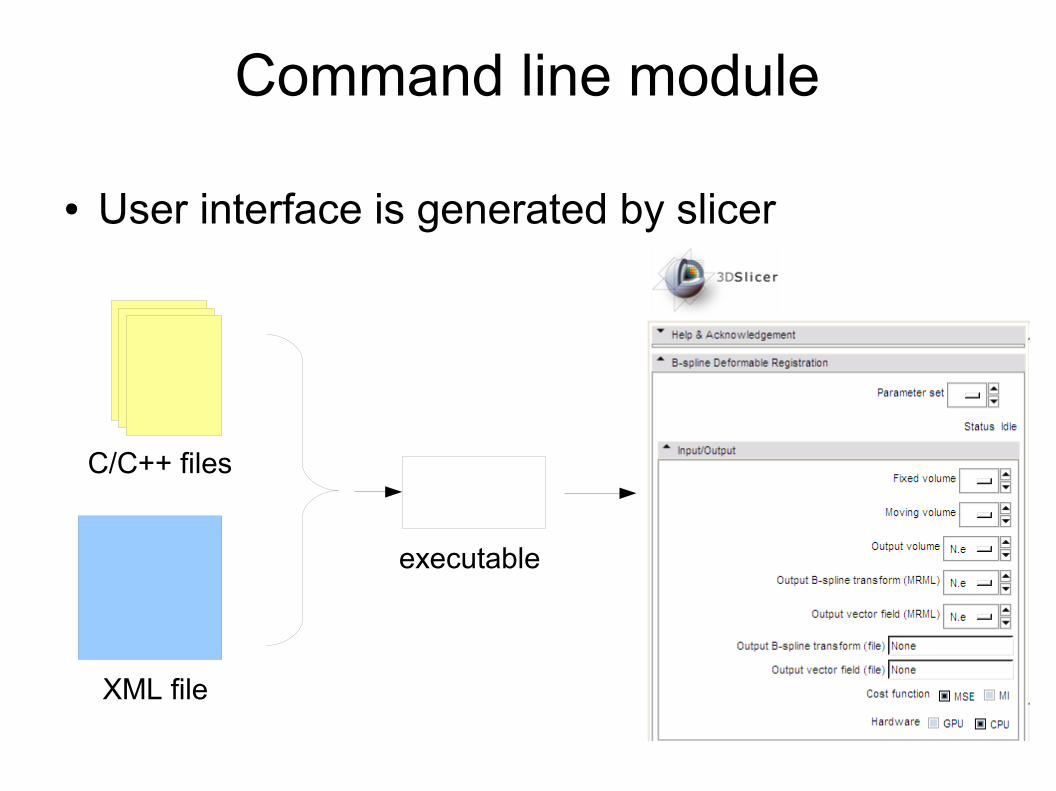
Command line module
● User interface is generated by slicer
XML file
C/C++ files
executable
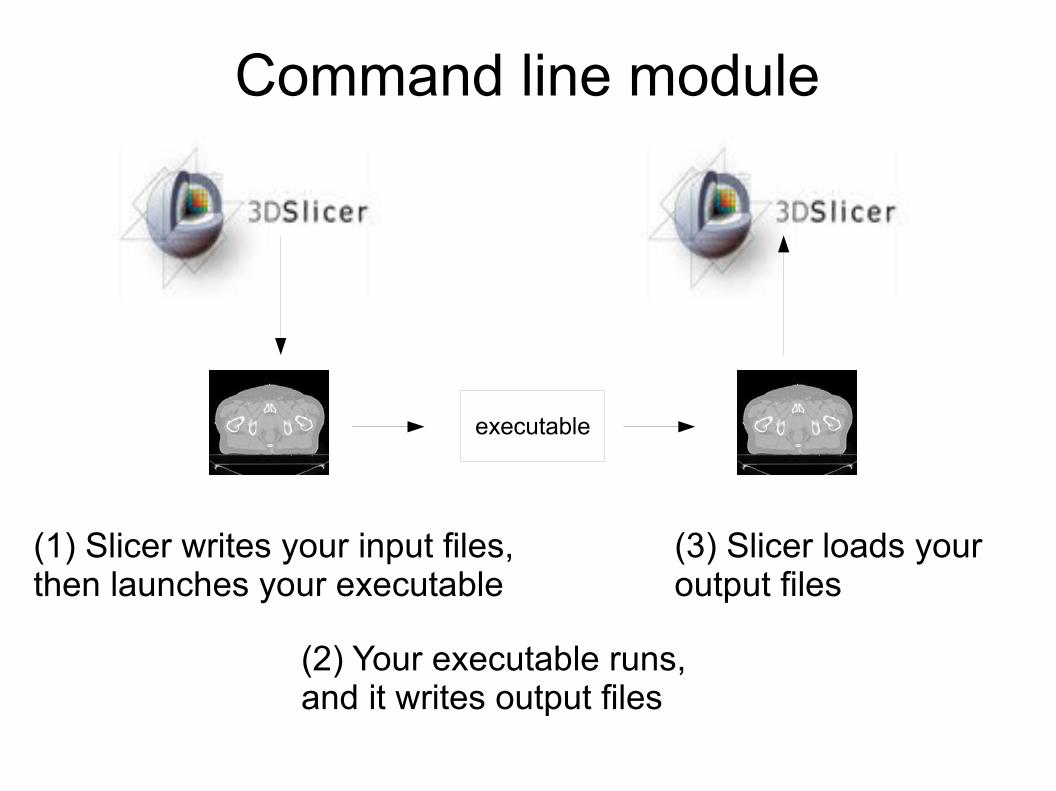
Command line module
executable
(1) Slicer writes your input files, then launches your executable
(3) Slicer loads your output files
(2) Your executable runs, and it writes output files
- 19 -Laboratory for Percutaneous Surgery – Copyright © Queen’s University, 2015
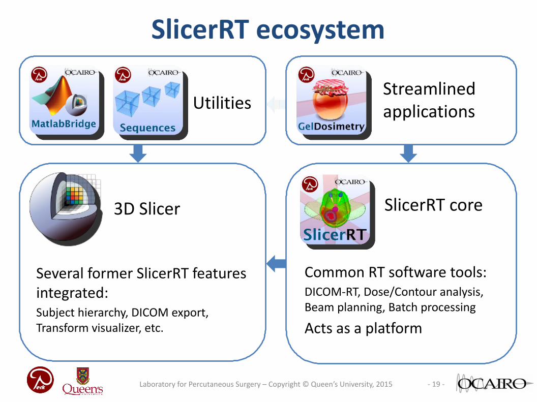
SlicerRT ecosystem
3D Slicer
UtilitiesStreamlined applications
SlicerRT core
Several former SlicerRT features integrated:Subject hierarchy, DICOM export, Transform visualizer, etc.
Common RT software tools:DICOM-RT, Dose/Contour analysis, Beam planning, Batch processing
Acts as a platform
- 20 -Laboratory for Percutaneous Surgery – Copyright © Queen’s University, 2015
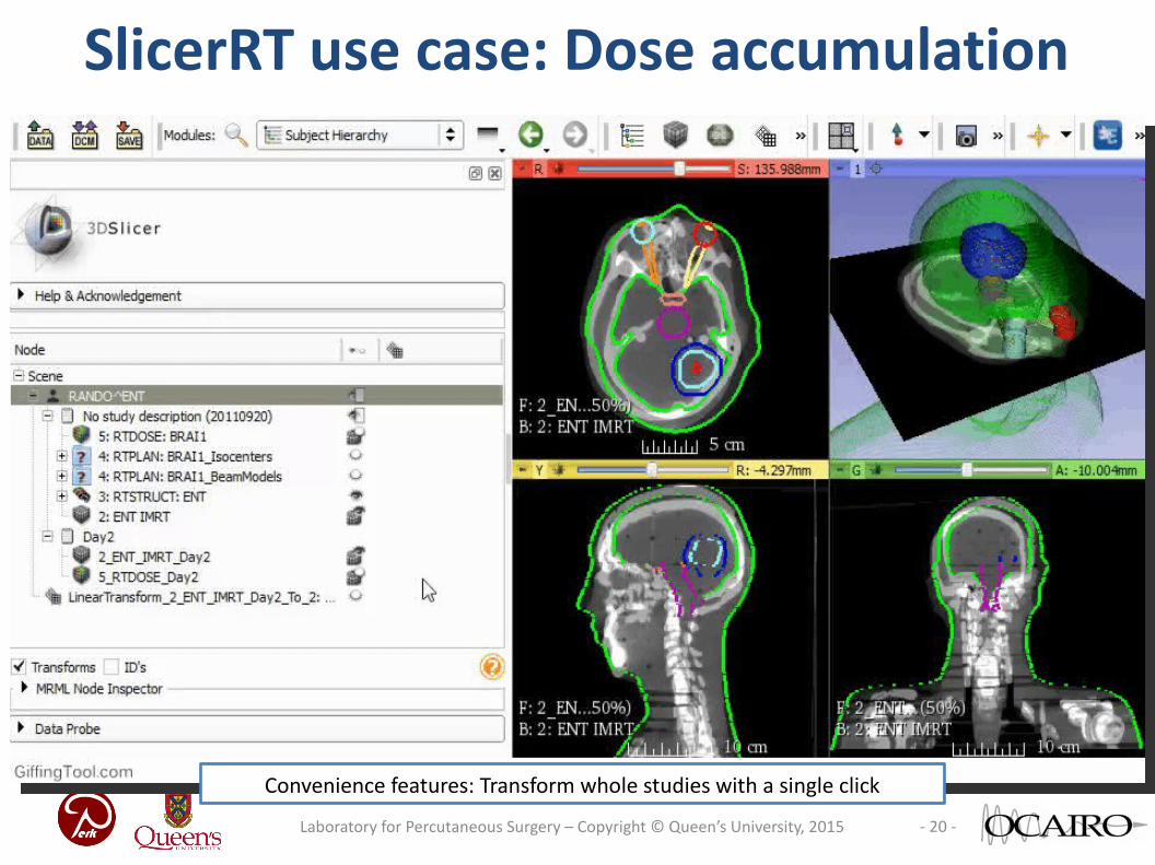
SlicerRT use case: Dose accumulation
• Register anatomical images• Transform whole studies• Accumulate dose• Compute DVH
Collaboration withPMH, TorontoPinter et al., MedPhys, 2012
Evaluate plans using DVH
Visualize transforms, anatomy in 3DConvenience features: Transform whole studies with a single click
- 21 -Laboratory for Percutaneous Surgery – Copyright © Queen’s University, 2015
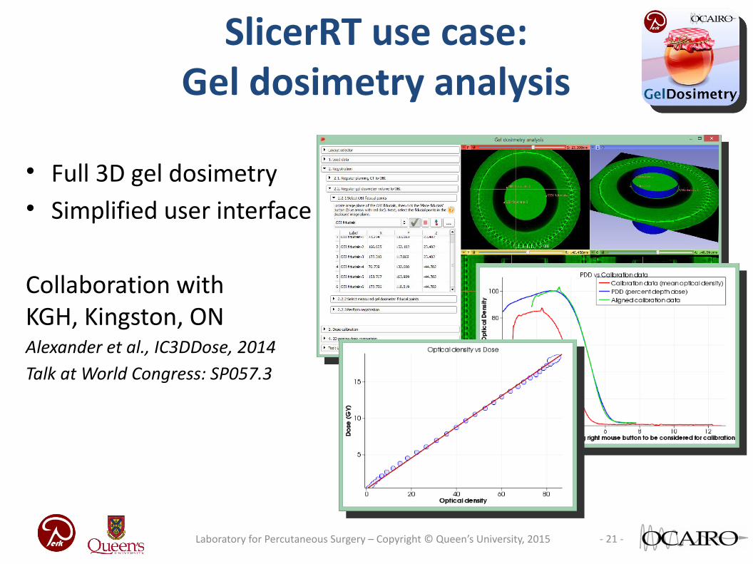
SlicerRT use case:Gel dosimetry analysis
• Full 3D gel dosimetry• Simplified user interface
Collaboration withKGH, Kingston, ONAlexander et al., IC3DDose, 2014Talk at World Congress: SP057.3
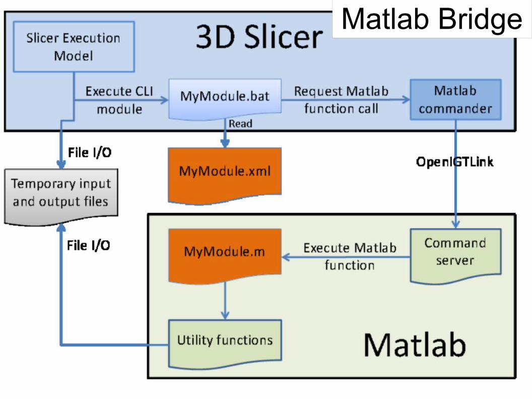
Matlab Bridge
- 23 -Laboratory for Percutaneous Surgery – Copyright © Queen’s University, 2015
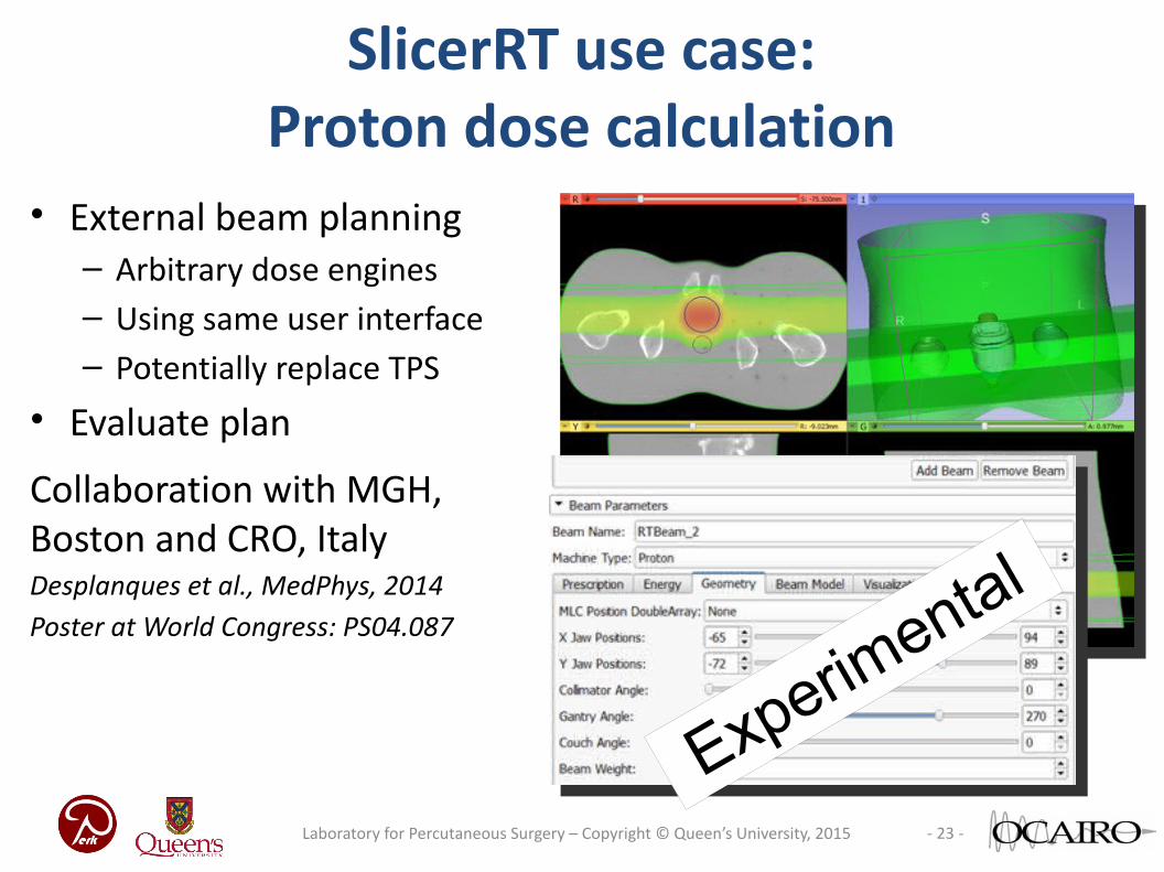
SlicerRT use case:Proton dose calculation
• External beam planning– Arbitrary dose engines– Using same user interface– Potentially replace TPS
• Evaluate plan
Collaboration with MGH, Boston and CRO, ItalyDesplanques et al., MedPhys, 2014Poster at World Congress: PS04.087
Experimental
● RTK = reconstruction toolkit
● openrtk.org
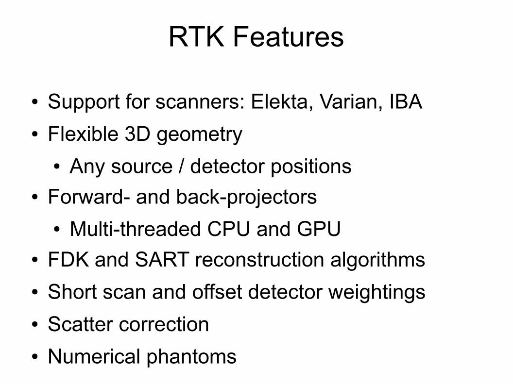
RTK Features
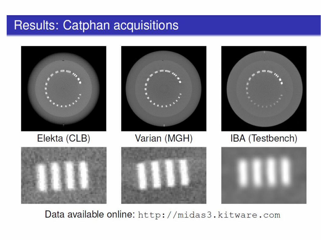
● Support for scanners: Elekta, Varian, IBA● Flexible 3D geometry
● Any source / detector positions● Forward- and back-projectors
● Multi-threaded CPU and GPU● FDK and SART reconstruction algorithms● Short scan and offset detector weightings● Scatter correction● Numerical phantoms
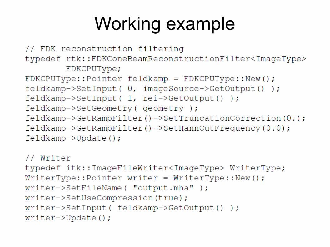
Working example
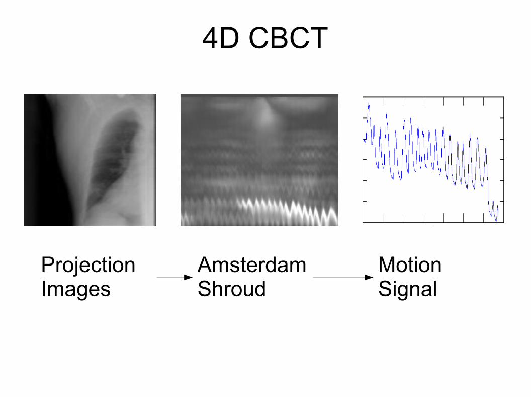
4D CBCT
Amsterdam Shroud
ProjectionImages
MotionSignal
Links
slicer.org
slicerrt.orgplastimatch.org
openrtk.org