Embed Size (px)
Citation preview
1 3
J Plant Res (2015) 128:7–15DOI 10.1007/s10265-014-0677-4
JPR SYMPOSIUM
Plasmodesmata of brown algae
Makoto Terauchi · Chikako Nagasato · Taizo Motomura
Received: 27 August 2014 / Accepted: 14 October 2014 / Published online: 17 December 2014 © The Botanical Society of Japan and Springer Japan 2014
Keywords Brown algae · Multicellularity · Pit field · Plasmodesmata · Primary plasmodesmata · Secondary plasmodesmata
Introduction
Brown algae are multicellular photosynthetic eukaryotes that are found in marine environments. They include spe-cies of various sizes from microscopic to large exceeding tens of meters, namely, giant kelps. In many cases, brown algae have complex life cycles that include sexual dimor-phism, and the alternation occurs between the sporophyte and gametophyte generations (Luthringer et al. 2014; Sil-berfeld et al. 2010; Wynne and Loiseaux 1976). The two generations are connected by asexual and sexual reproduc-tions with diverse modes. Brown algae have evolved unique systems such as a complex life cycle, during adaptation to coastal environments.
Brown algae, together with diatoms, belong to the het-erokontophyta and to the phylum of “Stramenopiles” with other heterotrophic organisms including Oomycetes. Stra-menopiles are phylogenetically distant from the Opis-tokonts (animals and fungi) and the Archaeplastida (green plants and red algae) (Yoon et al. 2004). Brown algae evolved complex multicellularity independently of animals, fungi, green plants and red algae. These five multicellular groups have developed intercellular connections, namely gap junctions in animals (Caspar et al. 1977; Hervé and Derangeon 2013; Kumar and Gilula 1996), septal pores in fungi (Bauer et al. 2006; Marchant 1976; Reichle and Alexander 1965), pit plugs in red algae (Pueschel 1977; Pueschel and Cole 1982) and plasmodesmata (PD) in green plants (Burch-Smith et al. 2011) and brown algae (Schmitz and Srivastava 1974; Schmitz 1981, 1990; Terauchi et al.
Abstract Plasmodesmata (PD) are intercellular connec-tions in plants which play roles in various developmental processes. They are also found in brown algae, a group of eukaryotes possessing complex multicellularity, as well as green plants. Recently, we conducted an ultrastructural study of PD in several species of brown algae. PD in brown algae are commonly straight plasma membrane-lined chan-nels with a diameter of 10–20 nm and they lack desmotu-bule in contrast to green plants. Moreover, branched PD could not be observed in brown algae. In the brown alga, Dictyota dichotoma, PD are produced during cytokinesis through the formation of their precursor structures (pre-plasmodesmata, PPD). Clustering of PD in a structure termed “pit field” was recognized in several species having a complex multicellular thallus structure but not in those having uniseriate filamentous or multiseriate one. The pit fields might control cell-to-cell communication and con-tribute to the establishment of the complex multicellular thallus. In this review, we discuss fundamental morphologi-cal aspects of brown algal PD and present questions that remain open.
Plasmodesmata: Function and Diversity in Plant Intercellular Communication
M. Terauchi Graduate School of Environmental Science, Hokkaido University, Sapporo 060-0810, Japane-mail: [email protected]
M. Terauchi Research Center for Inland Seas, Kobe University, Kobe 657-8501, Japan
C. Nagasato (*) · T. Motomura Muroran Marine Station, Field Science Center for Northern Biosphere, Hokkaido University, Muroran 051-0003, Japane-mail: [email protected]
8 J Plant Res (2015) 128:7–15
1 3
2012). Intercellular connections allow cell-to-cell com-munication through the transport of various molecules and contribute to the elaboration of the complex multicellular-ity (Bloemendal and Kück 2013).
In land plants, PD are plasma membrane-lined tubular channels with a diameter of 30–50 nm, creating symplastic continuity across the cell wall. Endoplasmic reticulum (ER, desmotubule), characteristically passes through the PD lumen. PD of land plants are categorized into two types: unbranched “simple PD” and branched “complex PD”. Molecules transported via PD include ions, small com-pounds, proteins and RNA (Kim 2005). Transport of these materials via PD is highly regulated and is involved in a number of developmental processes in land plants (Burch-Smith et al. 2011). The structure of PD is much different from that of gap junctions of animals; these 2–4 nm wide proteinous channels facilitate the transport of small mol-ecules up to about 1 kDa such as ions, secondary signal-ing messengers, nucleotides and metabolites (Hervé and Derangeon 2013; Maeda and Tsukihara 2011). Septal pores of fungi are 50–500 nm wide plasma membrane-lined pores co-localized with peroxisome-derived vesicles or ER-derived septal pore caps. They contribute to the cellular differentiation (Bauer et al. 2006; Reichle and Alexander 1965; van Peer et al. 2010). Pit plugs of red algae consist of a proteinaceous plug core occluding a pore lined by plasma membrane in the cell wall and cap membrane covering both sides of the plug core (Pueschel and Cole 1982). The structure of pit plug provides significant taxonomical infor-mation (Pueschel and Cole 1982).
In brown algae, studies on intercellular transport have focused on sieve elements in kelps. In some laminarialean algae, the differentiation of tissues consisting of epidermal, cortex and medullary cells is conspicuous, and medullary cells (sieve elements) are functionally analogous to those of land plants. The sieve elements of brown algae are con-tinuous to cortex cells via the complex filamentous cell net-work (Schmitz 1984). The monitoring of transport of iso-topes (14C, 32P, 125I) showed that the long-distance transport of photosynthetic products and iodine occurred through the sieve elements (Amat and Srivastava 1985; Schmitz and Srivastava 1975, 1979). The cross walls of sieve elements are perforated by numerous pores, linking adjacent sieve elements. The diameter of the pores ranges from 37.5 nm to 2.6 µm (Schmitz 1990). Although smaller pores can be regarded as PD, larger pores are predicted to be specialized structures of PD that are formed by the enzymatic digestion of the cross wall (Schmitz and Srivastava 1974; March-ant 1976; Schmitz 1981, 1990). The detail of the structure and the function of PD in other cell types and algal species remain obscure.
Considering the distant evolutionary relationship between brown algae and green plants, they must have
evolved PD independently (Raven 2008). The similarity of molecular components between brown algal PD and those of green plants might be low (Cock et al. 2010; Salmon and Bayer 2013). Structural and functional analyses of PD will give insights into how brown algae established indepen-dently complex multicellularity. Recently, we carried out ultrastructural observations of PD in the brown alga Dic-tyota dichotoma (Terauchi et al. 2012). We characterized their detail structure and formation during cytokinesis. In this review, we summarize our current knowledge on the structure of brown algal PD and compare them with green plants.
Ultrastructure of brown algal PD and its relationship to the molecular traffic
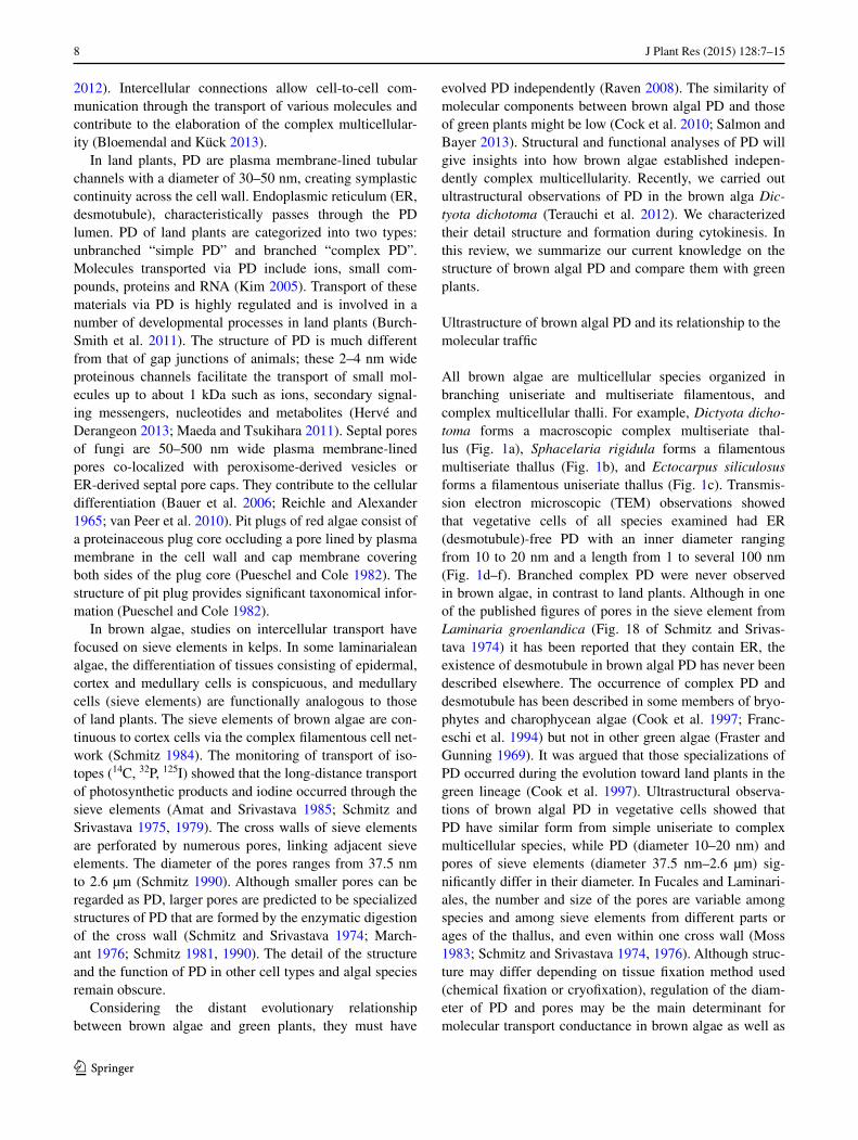
All brown algae are multicellular species organized in branching uniseriate and multiseriate filamentous, and complex multicellular thalli. For example, Dictyota dicho-toma forms a macroscopic complex multiseriate thal-lus (Fig. 1a), Sphacelaria rigidula forms a filamentous multiseriate thallus (Fig. 1b), and Ectocarpus siliculosus forms a filamentous uniseriate thallus (Fig. 1c). Transmis-sion electron microscopic (TEM) observations showed that vegetative cells of all species examined had ER (desmotubule)-free PD with an inner diameter ranging from 10 to 20 nm and a length from 1 to several 100 nm (Fig. 1d–f). Branched complex PD were never observed in brown algae, in contrast to land plants. Although in one of the published figures of pores in the sieve element from Laminaria groenlandica (Fig. 18 of Schmitz and Srivas-tava 1974) it has been reported that they contain ER, the existence of desmotubule in brown algal PD has never been described elsewhere. The occurrence of complex PD and desmotubule has been described in some members of bryo-phytes and charophycean algae (Cook et al. 1997; Franc-eschi et al. 1994) but not in other green algae (Fraster and Gunning 1969). It was argued that those specializations of PD occurred during the evolution toward land plants in the green lineage (Cook et al. 1997). Ultrastructural observa-tions of brown algal PD in vegetative cells showed that PD have similar form from simple uniseriate to complex multicellular species, while PD (diameter 10–20 nm) and pores of sieve elements (diameter 37.5 nm–2.6 µm) sig-nificantly differ in their diameter. In Fucales and Laminari-ales, the number and size of the pores are variable among species and among sieve elements from different parts or ages of the thallus, and even within one cross wall (Moss 1983; Schmitz and Srivastava 1974, 1976). Although struc-ture may differ depending on tissue fixation method used (chemical fixation or cryofixation), regulation of the diam-eter of PD and pores may be the main determinant for molecular transport conductance in brown algae as well as
9J Plant Res (2015) 128:7–15
1 3
in green plants. PD with a large diameter in the sieve ele-ment can be regarded as pores specialized for the long-dis-tance transport. In land plants, it has been reported that PD determine the upper limit of molecular weight of the cargo macromolecules (size exclusion limit, SEL) (Christensen et al. 2009; Zambryski 2004). Degradation and synthesis of callose (β1, 3-glucan) at the neck region of PD is one of the molecular mechanisms for controlling SEL; when the cell receives endogenous (e.g. developmental process) and exogenous (e.g. pathogen infection and cell injury) signals, callose is deposited at the neck region of PD, resulting in a decrease of the diameter of PD and SEL, while callose degradation reverses the effect (Zavaliev et al. 2011). SEL is varied in species, tissues, and developmental stages. In a study of Elodea canadensis leaf cells microinjected with fluorescent-labeled peptides, SEL was estimated to be less than 1 kDa (Goodwin 1983). In the analysis of tobacco leaf cells expressing a green fluorescent protein (GFP, fusion protein), SEL was estimated to be around 50 kDa in sink leaves while greatly reduced in source leaves (Oparka et al. 1999). In Zea mays, microinjection of GFP and fluorescent-labeled dextran in coleoptile epidermal cells showed that the intercellular movement of the dextran probe (4.4 kDa) was limited and movement of GFP was also observed to some extent (Wymer et al. 2001). SEL in these cells was predicted to be around 4.4 kDa. Theoretically, globular proteins with an approximate diameter of 9 nm correspond to a molecular weight of 45 kDa (Lucas and Wolf 1993),
which predicts that the macromolecular transport via PD in brown algae may be possible in terms of their diameter (i.e. 10–20 nm). FITC-dextran (10 kDa) microinjected in early developmental stage zygotes of the brown alga Fucus spiralis was transported throughout the young sporophyte, suggesting that PD provides the functional symplastic route for the intercellular transport of molecules in brown algae (Bouget et al. 1998). However, it is yet unclear whether the temporal or permanent alteration of diameter of PD regu-lates SEL in brown algae.
Formation of brown algal PD
In land plants, two types of PD exist: the primary PD formed during cytokinesis and the secondary PD which have a post-cytokinetic origin. Primary PD are generated by the physical obstruction of ER incorporated into the cell plate as shown by TEM observations (Hepler 1982). Sec-ondary PD are synthesized de novo by the local degrada-tion of the cell wall and protrusion of plasma membrane and desmotubule or by addition of branches to primary PD (Lucas and Wolf 1993; Faulkner et al. 2008). In several brown algae, PD-like structures were observed in the cell partition membrane during cytokinesis (La Claire 1981; Katsaros et al. 2009). Recently, we confirmed by elec-tron tomographic analysis that the precursor structures of brown algal PD, pre-plasmodesmata (PPD), occurred dur-ing cytokinesis in D. dichotoma (Terauchi et al. 2012). In
Fig. 1 General view of thal-lus and ultrastructure of PD in a, d Dictyota dichotoma (sporophyte, Dictyotales), a complex multicellular thallus; b, e Sphacelaria rigidula (male gametophyte, Sphacelariales), a multiseriate thallus; c, f Ecto-carpus siliculosus (male sporo-phyte, Ectocarpales), a filamen-tous uniseriate thallus. TEM samples were prepared by rapid freezing/freeze substitution. Insets show the transverse views of plasmodesmata. ER-free PD penetrate the electron-dense cell wall (cw). Abbreviations: chl, chloroplast; cw, cell wall; pd, plasmodema; pm, plasma membrane; py, pyrenoid. Scale bars: 20 µm (a–c), 100 nm (d–f), 20 nm (inset of d–f)
10 J Plant Res (2015) 128:7–15
1 3
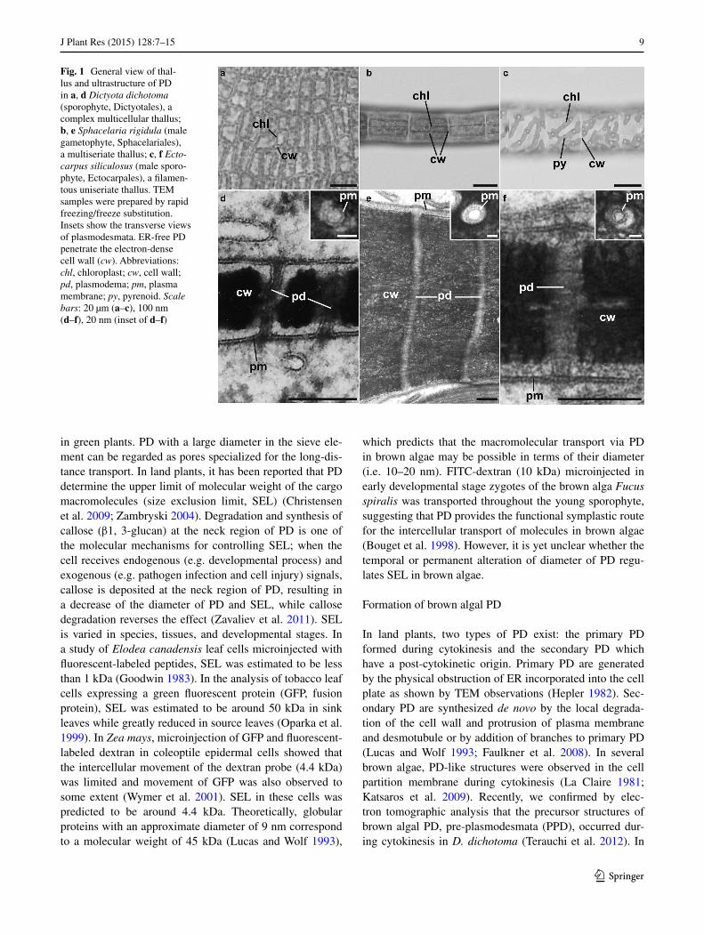
cytokinesis of brown algae, Golgi vesicles and flat cisternae take part in the formation of the cell partition membrane (Nagasato and Motomura 2002, 2009; Katsaros et al. 2009; Nagasato et al. 2010, 2014). Similarly, in D. dichotoma, cytokinesis proceeds by the expansion of patches of mem-branous sacs which are formed by the fusion of Golgi vesi-cles and flat cisternae (Fig. 2a). In the developing cell parti-tion membrane, tubular membranous structures (PPD) are recognized (Fig. 2b, c). It was suggested that PPD derived from the invagination of the membranous sac in D. dicho-toma (Fig. 3a). Their inner diameter ranges from 10 to 20 nm as in mature PD. They are evenly distributed in spe-cific areas of the membranous sacs and persist after com-pletion of the cell partition membrane (Fig. 3b). Mature PD are clustered in a part of the cell walls called the “pit fields”, cluster of PD corresponding to the deeper side of epidermal cells near the underlying medullary cells (see next paragraph for a detail description of the pit fields). PPD are preferentially formed in the restricted region of the cell wall where the mature PD are formed. This indicates
that the PPD distribution in the newly forming cell parti-tion membrane matches the sites of the ‘‘future’’ pit field (Fig. 3c). These data confirm that brown algae have pri-mary PD that are produced in a manner different from land plants. It is unclear how PPD are constructed at the molec-ular level and how their position in the cell partition mem-brane is determined.
Are secondary PD present in brown algae? In previ-ous studies of the first and second cytokinesis of zygotes of Scytosiphon lomentaria (Nagasato and Motomura 2002) and Silvetia babingtonii (Nagasato et al. 2010, 2014), PPD were not observed in the first and second cell parti-tion membrane. In the mature thalli of S. lomentaria and S. babingtonii, we observed dense PD in the cell wall.
Fig. 2 Formation of PD during cytokinesis in D. dichotoma. a Elec-tron micrograph of cytokinesis of a meristematic epidermal cell. Cytokinesis proceeds by expansion of the membranous sacs arranged on the cytokinetic plane as patches (arrowheads). b Longitudinal view of pre-plasmodesmata (PPD) in the developing cell partition membrane. Arrowhead indicates one PPD. c Transverse view of PPD in the developing cell partition membrane. Arrowhead indicates one PPD. Abbreviations: chl, chloroplast; n, nucleus; pm, plasma mem-brane. Scale bars: 500 nm (a), 100 nm (b), 200 nm (c)
Fig. 3 Schematic illustration of the formation of PD in D. dichotoma (drawn from Terauchi et al. 2012). a Early stage of cytokinesis. Golgi vesicles and flat cisternae (not shown) fuse to form the membranous sac (MS). Cytokinesis proceeds by the expansion of MS (arrows). PPD formation begins in the restricted region of MS by the protrusion (arrowheads) of MS membrane from either side of MS. b Late stage of cytokinesis before the initial cell wall development. The cell par-tition membrane contains PPD-rich region (PR). c Mature cell wall. Plasmodesmata (PD) are clustered in the thin cell wall (CW) region as a pit field, and then the PD-free cell wall gets thickened
11J Plant Res (2015) 128:7–15
1 3
Additionally, at later stages of S. babingtonii zygote devel-opment, PD are present in the newly formed cell wall (unpublished data). There is a high possibility that brown algae have the capability to generate secondary PD.
PD distribution and their implication for the body plan in brown algae
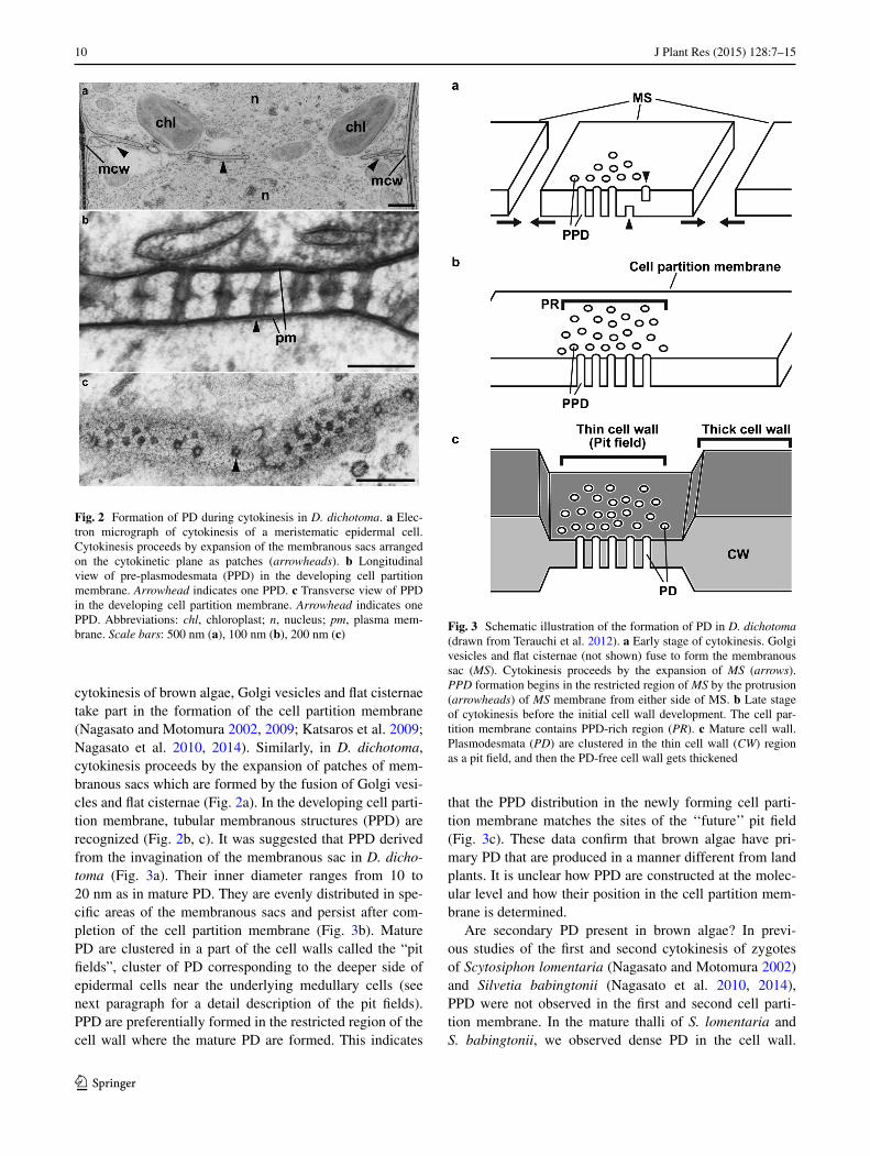
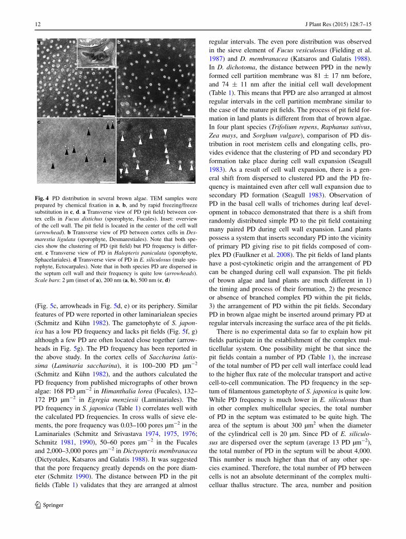
Frequency and distribution of PD in the cell wall should play important roles in cell-to-cell communication in brown algae. In D. dichotoma, PD are clustered at the pit fields and localized in the thin cell wall. Clustering appears during cytokinesis (Terauchi et al. 2012). In our study, the pit fields were observed in many species examined (Dic-tyotales, Laminariales, Fucales, Desmarestiales, Scyto-siphonales) (Table 1; Figs. 4, 5). The pit fields were pre-sent in the thin cell wall in Dictyotales, Laminariales and Fucales and in the relatively uniform cell wall in other spe-cies (Desmarestiales and Scytosiphonales). The pit fields are round or oval shaped and their number, mean area
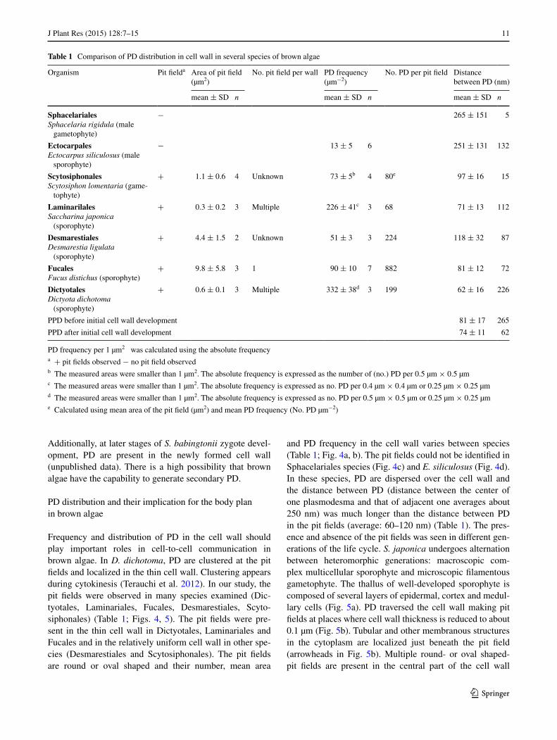
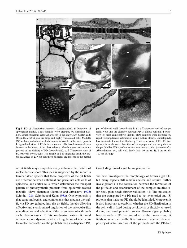
and PD frequency in the cell wall varies between species (Table 1; Fig. 4a, b). The pit fields could not be identified in Sphacelariales species (Fig. 4c) and E. siliculosus (Fig. 4d). In these species, PD are dispersed over the cell wall and the distance between PD (distance between the center of one plasmodesma and that of adjacent one averages about 250 nm) was much longer than the distance between PD in the pit fields (average: 60–120 nm) (Table 1). The pres-ence and absence of the pit fields was seen in different gen-erations of the life cycle. S. japonica undergoes alternation between heteromorphic generations: macroscopic com-plex multicellular sporophyte and microscopic filamentous gametophyte. The thallus of well-developed sporophyte is composed of several layers of epidermal, cortex and medul-lary cells (Fig. 5a). PD traversed the cell wall making pit fields at places where cell wall thickness is reduced to about 0.1 µm (Fig. 5b). Tubular and other membranous structures in the cytoplasm are localized just beneath the pit field (arrowheads in Fig. 5b). Multiple round- or oval shaped- pit fields are present in the central part of the cell wall
Table 1 Comparison of PD distribution in cell wall in several species of brown algae
PD frequency per 1 µm2 was calculated using the absolute frequencya + pit fields observed − no pit field observedb The measured areas were smaller than 1 µm2. The absolute frequency is expressed as the number of (no.) PD per 0.5 µm × 0.5 µmc The measured areas were smaller than 1 µm2. The absolute frequency is expressed as no. PD per 0.4 µm × 0.4 µm or 0.25 µm × 0.25 µmd The measured areas were smaller than 1 µm2. The absolute frequency is expressed as no. PD per 0.5 µm × 0.5 µm or 0.25 µm × 0.25 µme Calculated using mean area of the pit field (µm2) and mean PD frequency (No. PD µm−2)
Organism Pit fielda Area of pit field (µm2)
No. pit field per wall PD frequency (µm−2)
No. PD per pit field Distance between PD (nm)
mean ± SD n mean ± SD n mean ± SD n
SphacelarialesSphacelaria rigidula (male
gametophyte)
− 265 ± 151 5
EctocarpalesEctocarpus siliculosus (male
sporophyte)
− 13 ± 5 6 251 ± 131 132
ScytosiphonalesScytosiphon lomentaria (game-
tophyte)
+ 1.1 ± 0.6 4 Unknown 73 ± 5b 4 80e 97 ± 16 15
LaminarilalesSaccharina japonica
(sporophyte)
+ 0.3 ± 0.2 3 Multiple 226 ± 41c 3 68 71 ± 13 112
DesmarestialesDesmarestia ligulata
(sporophyte)
+ 4.4 ± 1.5 2 Unknown 51 ± 3 3 224 118 ± 32 87
FucalesFucus distichus (sporophyte)
+ 9.8 ± 5.8 3 1 90 ± 10 7 882 81 ± 12 72
DictyotalesDictyota dichotoma
(sporophyte)
+ 0.6 ± 0.1 3 Multiple 332 ± 38d 3 199 62 ± 16 226
PPD before initial cell wall development 81 ± 17 265
PPD after initial cell wall development 74 ± 11 62
12 J Plant Res (2015) 128:7–15
1 3
(Fig. 5c, arrowheads in Fig. 5d, e) or its periphery. Similar features of PD were reported in other laminarialean species (Schmitz and Kühn 1982). The gametophyte of S. japon-ica has a low PD frequency and lacks pit fields (Fig. 5f, g) although a few PD are often located close together (arrow-heads in Fig. 5g). The PD frequency has been reported in the above study. In the cortex cells of Saccharina latis-sima (Laminaria saccharina), it is 100–200 PD µm−2 (Schmitz and Kühn 1982), and the authors calculated the PD frequency from published micrographs of other brown algae: 168 PD µm−2 in Himanthalia lorea (Fucales), 132–172 PD µm−2 in Egregia menziesii (Laminariales). The PD frequency in S. japonica (Table 1) correlates well with the calculated PD frequencies. In cross walls of sieve ele-ments, the pore frequency was 0.03–100 pores µm−2 in the Laminariales (Schmitz and Srivastava 1974, 1975, 1976; Schmitz 1981, 1990), 50–60 pores µm−2 in the Fucales and 2,000–3,000 pores µm−2 in Dictyopteris membranacea (Dictyotales, Katsaros and Galatis 1988). It was suggested that the pore frequency greatly depends on the pore diam-eter (Schmitz 1990). The distance between PD in the pit fields (Table 1) validates that they are arranged at almost
regular intervals. The even pore distribution was observed in the sieve element of Fucus vesiculosus (Fielding et al. 1987) and D. membranacea (Katsaros and Galatis 1988). In D. dichotoma, the distance between PPD in the newly formed cell partition membrane was 81 ± 17 nm before, and 74 ± 11 nm after the initial cell wall development (Table 1). This means that PPD are also arranged at almost regular intervals in the cell partition membrane similar to the case of the mature pit fields. The process of pit field for-mation in land plants is different from that of brown algae. In four plant species (Trifolium repens, Raphanus sativus, Zea mays, and Sorghum vulgare), comparison of PD dis-tribution in root meristem cells and elongating cells, pro-vides evidence that the clustering of PD and secondary PD formation take place during cell wall expansion (Seagull 1983). As a result of cell wall expansion, there is a gen-eral shift from dispersed to clustered PD and the PD fre-quency is maintained even after cell wall expansion due to secondary PD formation (Seagull 1983). Observation of PD in the basal cell walls of trichomes during leaf devel-opment in tobacco demonstrated that there is a shift from randomly distributed simple PD to the pit field containing many paired PD during cell wall expansion. Land plants possess a system that inserts secondary PD into the vicinity of primary PD giving rise to pit fields composed of com-plex PD (Faulkner et al. 2008). The pit fields of land plants have a post-cytokinetic origin and the arrangement of PD can be changed during cell wall expansion. The pit fields of brown algae and land plants are much different in 1) the timing and process of their formation, 2) the presence or absence of branched complex PD within the pit fields, 3) the arrangement of PD within the pit fields. Secondary PD in brown algae might be inserted around primary PD at regular intervals increasing the surface area of the pit fields.
There is no experimental data so far to explain how pit fields participate in the establishment of the complex mul-ticellular system. One possibility might be that since the pit fields contain a number of PD (Table 1), the increase of the total number of PD per cell wall interface could lead to the higher flux rate of the molecular transport and active cell-to-cell communication. The PD frequency in the sep-tum of filamentous gametophyte of S. japonica is quite low. While PD frequency is much lower in E. siliculosus than in other complex multicellular species, the total number of PD in the septum was estimated to be quite high. The area of the septum is about 300 µm2 when the diameter of the cylindrical cell is 20 µm. Since PD of E. siliculo-sus are dispersed over the septum (average 13 PD µm−2), the total number of PD in the septum will be about 4,000. This number is much higher than that of any other spe-cies examined. Therefore, the total number of PD between cells is not an absolute determinant of the complex multi-celluar thallus structure. The area, number and position
Fig. 4 PD distribution in several brown algae. TEM samples were prepared by chemical fixation in a, b, and by rapid freezing/freeze substitution in c, d. a Transverse view of PD (pit field) between cor-tex cells in Fucus distichus (sporophyte, Fucales). Inset: overview of the cell wall. The pit field is located in the center of the cell wall (arrowhead). b Transverse view of PD between cortex cells in Des-marestia ligulata (sporophyte, Desmarestiales). Note that both spe-cies show the clustering of PD (pit field) but PD frequency is differ-ent. c Transverse view of PD in Halopteris paniculata (sporophyte, Sphacelariales). d Transverse view of PD in E. siliculosus (male spo-rophyte, Ectocarpales). Note that in both species PD are dispersed in the septum cell wall and their frequency is quite low (arrowheads). Scale bars: 2 µm (inset of a), 200 nm (a, b), 500 nm (c, d)
13J Plant Res (2015) 128:7–15
1 3
of pit fields may comprehensively influence the pattern of molecular transport. This idea is supported by the report in laminarialean species that those properties of the pit fields are different between anticlinal and periclinal cell walls of epidermal and cortex cells, which determines the transport pattern of photosynthetic products from epidermis toward medulla (sieve elements) (Schmitz and Srivastava 1975; Schmitz 1981; Schmitz and Kühn 1982). One hypothesis is that cargo molecules and components that mediate the traf-fic via PD are gathered into the pit fields, thereby allowing effective and synchronized regulation of the molecular flux rate, direction and selection of the cargo molecules through each plasmodesma. If this mechanism exists, it could achieve a more dynamic and strict regulation of intercellu-lar molecular traffic via the pit fields than via dispersed PD.
Concluding remarks and future perspective
We have investigated the morphology of brown algal PD, but many aspects still remain unclear and require further investigation: (1) the correlation between the formation of the pit fields and establishment of the complex multicellu-lar body plan needs further validation. (2) The molecules that are transported via PD need to be inventoried and (3) proteins that make up PD should be identified. Moreover, it is also important to establish whether the PD distribution in the cell wall is fixed during cytokinesis or flexibly adjusted during the developmental process. Brown algae probably have secondary PD that are added to the pre-existing pit fields or other cell walls. It is unknown whether de novo post-cytokinetic insertion of the pit fields into the PD-free
Fig. 5 PD of Saccharina japonica (Laminariales). a Overview of sporophyte thallus. TEM samples were prepared by chemical fixa-tion. Small epidermal cells (E) are seen in the upper side. Cortex cells (C) in the central part are large and highly vacuolated cells. Medulla (M) with expanded extracellular matrix is visible in the lower part. b Longitudinal view of PD between cortex cells. No desmotubule can be seen in the lumen of the plasmodesma. Membranous structures are present in the vicinity of PD (arrowheads). c, d Transverse view of PD between cortex cells. The image in d is magnified from the dot-ted rectangle in c. Note that three pit fields are present in the central
part of the cell wall (arrowheads in d). e Transverse view of one pit field. Note that the distance between PD is almost constant. f Over-view of male gametophyte thallus. TEM samples were prepared by rapid freezing/freeze substitution using culture strains. Gametophyte has uniseriate filamentous thallus. g Transverse view of PD. PD fre-quency is much lower than that of sporophyte and do not gather as the pit field but PD are often located near to each other (arrowheads). Abbreviations: cw, cell wall. Scale bars: 10 µm (a, f), 2 µm (c, d), 100 nm (b, e, g)
14 J Plant Res (2015) 128:7–15
1 3
cell wall takes place in brown algae. In land plants, pre-existing PD can be removed from the cell wall during the developmental process. For example, it is well known that cell walls in guard cells lose PD and become symplasti-cally isolated from surrounding cells during their matura-tion (Wille and Lucas 1984). It is not known whether the elimination of pre-existing PD occurs in brown algae. From the reports of the absence of PD in the first cell partition membrane of zygotes of several brown algae (Nagasato and Motomura 2002; Nagasato et al. 2010, 2014), we can infer that some vegetative cells of the developing thallus as well as zygotes might undergo PPD-free cytokinesis. However, cells of mature thalli of all species examined had PD. The complete symplastic isolation by the absence of PD may be rare in brown algae. In land plants, it was reported that the local grouping of cells by SEL of PD, called “symplas-tic field”, was the fundamental mechanism in creating the positional information, and achieving cell and tissue differ-entiation (Kim et al. 2004). In brown algae, the existence of a symplastic field is unknown. In the early developmental stage of zygotes in D. dichotoma, F. disticus and S. japon-ica, pit fields were not observed and the PD frequency was low (unpublished data). The onset of pit field formation might be regulated according to the developmental sched-ule. Brown algal PD still leaves many puzzles to be solved. The data presented here could serve as a framework to a detailed functional analyses of brown algal PD.
Acknowledgments We thank Drs. Toshiaki Ito (Hokkaido Uni-versity) and Naoko Kajimura (Osaka University) for their support in electron tomographic analysis of PD. This study was supported by KAKENHI (26440160) and Research Fellowship for Young Scientists from the Japan Society for the Promotion of Science (11J01697).
References
Amat MA, Srivastava LM (1985) Translocation of iodine in Lami-naria saccharina (Phaeophyta). J Phycol 21:330–333
Bauer R, Begerow D, Sampaio JP, Weiß M, Oberwinkler F (2006) The simple-septate basidiomycetes: a synopsis. Mycol Prog 5:41–66
Bloemendal S, Kück U (2013) Cell-to-cell communication in plants, animals, and fungi: a comparative review. Naturwissenschaften 100:3–19
Bouget FY, Berger F, Brownlee C (1998) Position dependent control of cell fate in the Fucus embryo: role of intercellular communica-tion. Development 125:1999–2008
Burch-Smith TM, Stonebloom S, Xu M, Zambryski PC (2011) Plas-modesmata during development: re-examination of the impor-tance of primary, secondary, and branched plasmodesmata struc-ture versus function. Protoplasma 248:61–74
Caspar DLD, Goodenough DA, Makowski L, Phillips WC (1977) Gap junction structures I. Correlated electron microscopy and x-ray diffraction. J Cell Biol 74:605–628
Christensen NM, Faulkner C, Oparka K (2009) Evidence for unidi-rectional flow through plasmodesmata. Plant Physiol 150:96–104
Cock JM, Sterck L, Rouze P, Scornet D, Allen AE, Amoutzias G, Anthouard V, Artiguenave F, Aury JM, Badger JH et al (2010)
The Ectocarpus genome and the independent evolution of multi-cellularity in the brown algae. Nature 465:617–621
Cook ME, Graham LE, Botha CEJ, Lavin CA (1997) Comparative ultrastructure of plasmodesmata of Chara and selected bryo-phytes: toward an elucidation of the evolutionary origin of plant plasmodesmata. Am J Bot 84:1169–1178
Faulkner C, Akman OE, Bell K, Jefree C, Oparka KJ (2008) Peeking into pit fields: multiple twinning model of secondary plasmodes-mata formation. Plant Cell 20:1504–1508
Fielding AH, Carter PL, Smith CA (1987) Sieve plates in Fucus: a reappraisal of size and pore distribution. Phycologia 26:501–504
Franceschi VR, Ding B, Lucas WJ (1994) Mechanism of plasmodes-mata formation in characean algae in relation to evolution of intercellular communication in higher plants. Planta 192:347–358
Fraster TW, Gunning BES (1969) The ultrastructure of plasmodes-mata in the filamentous green alga Bulbochaete hiloensis (Nor-dst.) Tiffany. Planta 88:244–254
Goodwin PB (1983) Molecular size limit for movement in the sym-plast of the Elodea leaf. Planta 157:124–130
Hepler PK (1982) Endoplasmic reticulum in formation of the cell plate and plasmodesmata. Protoplasma 111:121–123
Hervé JC, Derangeon M (2013) Gap-junction-mediated cell-to-cell communication. Cell Tissue Res 352:21–31
Katsaros C, Galatis B (1988) Thallus development in Dictyopteris membranacea (Phaeophyta, Dictyotales). Br Phycol J 23:71–88
Katsaros C, Motomura T, Nagasato C, Galatis B (2009) Diaphragm development in cytokinetic vegetative cells of brown algae. Bot Mar 52:150–161
Kim JY (2005) Regulation of short-distance transport of RNA and protein. Curr Opin Plant Biol 8:45–52
Kim I, Cho E, Crawford K, Hempel FD, Zambryski PC (2004) Cell-to-cell movement of GFP during embryogenesis and early seedling development in Arabidopsis. Proc Natl Acad Sci USA 102:2227–2231
Kumar NM, Gilula NB (1996) The gap junction review communica-tion channel. Cell 84:381–388
La Claire JW II (1981) Occurrence of plasmodesmata during infur-rowing in a brown alga. Biol Cell 40:139–142
Lucas WJ, Wolf S (1993) Plasmodesmata: the intercellular organelles of green plants. Trends Cell Biol 3:308–315
Luthringer R, Cormier A, Ahmed S, Peters AF, Cock JM, Coelho SM (2014) Sexual dimorphism in the brown algae. Perspect Phycol 1:11–25
Maeda S, Tsukihara T (2011) Structure of the gap junction channel and its implications for its biological functions. Cell Mol Life Sci 68:1115–1129
Marchant HJ (1976) Plasmodesmata in algae and fungi. In: Gunning BES, Robards TAW (eds) Intercellular communication in plants: studies on plasmodesmata. Springer, Berlin, pp 59–80
Moss BL (1983) Sieve elements in the Fucales. New Phytol 93:433–437
Nagasato C, Motomura T (2002) Ultrastructural study on mito-sis and cytokinesis in Scytosiphon lomentaria zygotes (Scyto-siphonales, Phaeophyceae) by freeze-substitution. Protoplasma 219:140–149
Nagasato C, Motomura T (2009) Effect of latrunculin B and brefel-din A on cytokinesis in the brown alga Scytosiphon lomentaria zygotes (Scytosiphonales, Phaeophyceae). J Phycol 45:404–412
Nagasato C, Inoue A, Mizuno M, Kanazawa K, Ojima T, Okuda K, Motomura T (2010) Membrane fusion process and assembly of cell wall during cytokinesis in the brown alga Silvetia babingtonii (Fucales, Phaeophyceae). Planta 232:287–298
Nagasato C, Kajimura N, Terauchi M, Mineyuki Y, Motomura T (2014) Electron tomographic analysis of cytokinesis in the brown alga Silvetia babingtonii (Fucales, Phaeophyceae). Protoplasma 251(6):1347–1357
15J Plant Res (2015) 128:7–15
1 3
Oparka KJ, Roberts AG, Boevink P, Santa Cruz S, Roberts IM, Pradel KS, Imlau A, Kotlizky G, Sauer N, Epel B (1999) Simple but not branched plasmodesmata allow the nonspecific trafficking of pro-teins in developing tobacco leaves. Cell 97:743–754
Pueschel CM (1977) A freeze-etch study of the ultrastructure of red algal pit plugs. Protoplasma 91:15–30
Pueschel CM, Cole KM (1982) Rhodophycean pit plugs: an ultrastruc-tural survey with taxonomic implication. Ame J Bot 69(5):703–720
Raven JA (2008) Evolution of plasmodesmata. In: Oparka KJ (ed) plasmodesmata. Blackwell, London, pp 33–50
Reichle RE, Alexander JV (1965) Multiperforate septations, Woronin bodies, and septal plugs in Fusarium. J Cell Biol 24:489–496
Salmon MS, Bayer EM (2013) Dissecting plasmodesmata molecular composition by mass spectrometry-based proteomics. Front Plant Sci. doi:10.3389/fpls.2012.00307
Schmitz K (1981) Translocation. In: Lobban CS, Wynne MJ (eds) The biology of seaweeds. Blackwell, London, pp 534–558
Schmitz K (1984) A cell system for symplastic transport of photoas-similate between meristoderm and sieve elements in Alaria tenui-folia. Marine Biol 78:209–214
Schmitz K (1990) Algae. In: Behnke HD, Sjolund RD (eds) Sieve elements. Comparative structure induction and development, Springer, Berlin, pp 1–18
Schmitz K, Kühn R (1982) Fine structure distribution and frequency of plasmodesmata and pits in the cortex of Laminaria hyperborea and L. saccharina. Planta 154:385–392
Schmitz K, Srivastava L (1974) Fine structure and development of sieve tubes in Laminaria groenlandica Rosenv. Cytobiologie 10:66–87
Schmitz K, Srivastava L (1975) On the fine structure of sieve tubes and the physiology of assimilate transport in Alaria marginata. Can J Bot 53:861–876
Schmitz K, Srivastava LM (1976) The fine structure of sieve elements of Nereocystis lutkeana. Am J Bot 63:679–693
Schmitz K, Srivastava LM (1979) Long distance transport in Mac-rocystis integrifolia. I. Translocation of 14C labelled assimilates. Plant Physiol 63:995–1002
Seagull RW (1983) Differences in the frequency and disposition of plasmodesmata resulting from root cell elongation. Planta 159:497–504
Silberfeld T, Leigh JW, Verbruggen H, Cruaud C, de Reviers B, Rous-seau F (2010) A multi-locus time-calibrated phylogeny of the brown algae (Heterokonta, Ochrophyta, Phaeophyceae): Investi-gating the evolutionary nature of the ‘‘brown algal crown radia-tion”. Mol Phylogenet Evol 56:659–674
Terauchi M, Nagasato C, Kajimura N, Mineyuki Y, Okuda K, Katsa-ros C, Motomura T (2012) Ultrastructural study of plasmodes-mata in the brown alga Dictyota dichotoma (Dictyotales, Phaeo-phyceae). Planta 236:1013–1026
van Peer AF, Wang FF, van Driel KGA, de Jong JF, van Donselaar EG, Müller WH, Boekhout T, Lugones LG, Wösten HAB (2010) The septal pore cap is an organelle that functions in vegetative growth and mushroom formation of the wood-rot fungus Schizo-phyllum commune. Environ Microbiol 12:833–844
Wille AC, Lucas WJ (1984) Ultrastructural and histochemical studies on guard-cells. Planta 160:129–142
Wymer CL, Fernandez-Abalos JM, Doonan JH (2001) Microinjection reveals cell-to-cell movement of green fluorescent protein in cells of maize coleoptiles. Planta 212:692–695
Wynne MJ, Loiseaux S (1976) Recent advances in life history studies of the Phaeophyta. Phycologia 15:435–452
Yoon HS, Hackett JD, Ciniglia C, Pinto G, Bhattacharya D (2004) A molecular timeline for the origin of photosynthetic eukaryotes. Mol Biol Evol 21:809–818
Zambryski P (2004) Cell-to-cell transport of proteins and fluorescent tracers via plasmodesmata during plant development. J Cell Biol 162:165–168
Zavaliev R, Ueki S, Epel BL, Citovsky V (2011) Biology of cal-lose (β-1, 3-glucan) turnover at plasmodesmata. Protoplasma 248:117–130