Embed Size (px)
Citation preview
2
Moss as a Model System for Plant Stress Responses
Andrew C. Cuming
2.1
Introduction
We live on a green planet, yet it was not always so. An observer from space sees the
blue of the ocean and the green of the land, but this is a comparatively recent
development in our planet’s history. Only in the last 450 million years has the
planet’s land surface become colonized by plants. Until this time, the land was
bare: rock, sand, and mud, devoid of organic matter and unable to support life. It is
difficult to determine precisely when the first eukaryotes gained a secure foothold
in the terrestrial environment. The most direct evidence derives from early fossil
spores indicating colonization by plants, dating from the mid-Ordovician period
(about 440–490 million years ago) [1, 2], although the earliest surviving megafossils
indicating the anatomical features of early plants occur about 50 million years later
in the fossil record [3]. What were these plants like and where did they come from?
It is now generally accepted that today’s land plants evolved from aquatic green
algae, and molecular systematic analysis has identified the charophytes [4] and
more specifically the Charales [5] as the likely ancestral taxon to all modern land
plants. The extant members of the Charales (e.g., Chara) remain aquatic and grow
as branched filaments. They have a haplontic life cycle in which the product of
sexual fusion immediately undergoes meiosis to generate haploid tissue. This is by
contrast with the land plants, which exhibit a diplobiontic life cycle, with an
alteration of haploid gametophyte and diploid sporophyte generations.
Among today’s land plants, it is the bryophytes that represent the first group to
diverge in the land plant lineage. The bryophytes comprise three distinct sub-
groupings: the mosses, liverworts, and hornworts. All display characters that
might be considered ‘‘primitive,’’ such as branched filamentous protonemal tis-
sues (in the mosses) and cells containing a single, algal-like pyrenoid-containing
chloroplast (in the hornworts). The earliest fossil sporangia have been suggested as
most similar to those found in extant liverworts [2]. All have a dominant haploid
gametophyte generation, in which the sporophyte is dependent on the gameto-
phyte for its development and nourishment. While the relationship between these
Plant Stress Biology. Edited by H. HirtCopyright r 2009 WILEY-VCH Verlag GmbH & Co. KGaA, WeinheimISBN: 978-3-527-32290-9
| 17
groups, in terms of their order of origin, remains disputed, it is agreed that, among
extant plant species, it is the bryophytes that most closely resemble the likely first
common ancestor of the land plants.
The ancestors of land plants were most likely found in the marginal areas of
bodies of water and were exposed periodically as the water receded. The successful
colonization of the land would have required a number of adaptations to permit
the survival of such plants. Terrestrial environments are necessarily more variable
in nature than aquatic environments. There are greater fluctuations of tempera-
ture, over both short and long timescales. The availability of water – a necessity for
life – is uncertain and there are greatly enhanced levels of radiation: a factor
contributing to both temperature and the availability of water, and also directly
damaging through the mutagenic effects of ultraviolet-induced DNA damage.
Modern plants have acquired multiple anatomical and developmental adaptations
to enable them to survive environmental extremes. The vascular plants that
dominate today’s planetary surface have extensive, branching root systems that
ramify throughout the soil, enabling them to scavenge water from the substratum.
Evaporative water loss is reduced through the presence of protective surfaces: waxy
cuticles, suberin, and lignin all act to retain water within the plant, whilst lignified
vascular systems both mechanically support the development of massive struc-
tures, and enable the distribution of water scavenged from the soil to all parts of
the plant. Evaporative water loss and gas exchange is facilitated through the pre-
sence of stomatal apertures on the leaf surfaces. The effects of incident radiation
are ameliorated through the accumulation of pigments that serve as sunscreens
(e.g., anthocyanins). Sexual reproduction no longer requires a layer of water for the
dissemination of swimming gametes.
The first land plants lacked these adaptations. Their ability to survive and
prosper therefore must necessarily have depended, at first, on biochemical adap-
tations to withstand such environmental variability. Such adaptations would be
expressed as metabolic responses, rather than as developmental in nature, and
such adaptations can still be recognized in today’s land plants. Although such
traits are commonly characterized as ‘‘primitive,’’ it should be emphasized that
this term reflects their ancient origins, rather than their efficacy. These ‘‘primi-
tive’’ traits have been instrumental in the conquest of the land by plants and in
their subsequent shaping of the terrestrial environment. Their importance is
highlighted by the diversity of the bryophytes, specifically the mosses, in modern
ecosystems.
The terrestrial flora is today dominated by the angiosperms, of which there are
thought to be about 250 000 species. This diversity originated from a massive
radiation in the mid-Cretaceous period (about 100 million years ago) and occurred
in concert with a similarly extensive radiation among insect species that act as
pollinators. Nevertheless, the bryophytes remain a large, diverse and successful
group: it is estimated that there are approximately 10 000 moss species, which is
second only in number to the angiosperms. Clearly, primitive traits retain their
value, even after 450 million years. Mosses retain many of the properties we
ascribe to the first successful colonists of the land. Most significantly, they are
18 | 2 Moss as a Model System for Plant Stress Responses
habitat-forming organisms – they are capable of colonizing bare surfaces, such as
bare rock and mud, and growing to envelop the surface. Their death and decay
generates the organic matter that distinguishes a true soil from a simple mineral
sludge, and provides the basis for colonization by other plants, more demanding in
their substrate requirements. Whilst many mosses thrive best under conditions of
shade and moisture, others are able to colonize and exploit exposed bare habitats
apparently inimical to plant growth. Even in urban environments, the sight of
mosses growing on bare roofs and walls is commonplace.
The study of mosses, therefore, provides insights into the conquest of land by
living organisms, and the origins of terrestrial life. Mosses provide a starting point
for unraveling the evolution of plant gene function through comparative, ‘‘evo-
devo’’ genomic strategies and for the identification of molecular strategies for
adaptations to abiotic stress.
Can a ‘‘systems biology’’ approach be applied to the study of these processes in
mosses? Until recently, this might have appeared an unlikely prospect. However,
in recent years, considerable progress has been made in developing genomic
resources for one moss species, Physcomitrella patens, whose biology makes it
particularly amenable for such an analysis. This species has now become a pow-
erful model for deploying systems-level approaches for comparative analyses of
plant stress responses.
2.2
Model Systems
Model systems provide us with tools to investigate processes common to entire
classes of living organism. They are chosen for their experimental amenity, and
vary according to their purpose and the particular expertise of the experimentalist,
but certain features are more desirable than others. They should be representative
of their class; thus, mice are good experimental models for the study of mam-
malian development, but zebrafish may be more tractable for wider applications
including all vertebrates. Arabidopsis thaliana is pre-eminent as a model plant,
having a small sequenced genome, a genetic linkage map densely marked by
molecular markers, a rapid life cycle, small size, and straightforward procedures
for genetic manipulation by transgenesis (http://www.Arabidopsis.org). It is an
outstanding workhorse for identifying how cellular organization and development
are programmed within the flowering plants, and also for how many fundamental
plant processes are regulated, common to all taxa of green plants. However, in
order to explore how these processes have evolved, we need to look beyond a single
species. Other model plants include rice – as an example of the monocotyledonous
plants and a representative cereal (the most important group of food crops on the
planet) – and several other species of angiosperm, whose genomes have recently
been deciphered (soybean, poplar, grape). Nevertheless, to undertake a wider
comparative analysis, models are also required from outside the flowering plants.
To date, these are fewer in number, but include the diatom Ostreococcus [6], the
2.2 Model Systems | 19
unicellular green alga, Chlamydomonas rheinhardtii [7], the moss P. patens [8], andthe lycophyte Selaginella moellendorfii (http://selaginella.genomics.purdue.edu/;
http://genome.jgi-psf.org/Selmo1/Selmo1.home.html).
What are the general features of mosses, how do they differ from the flowering
plants, and how can we use them experimentally? Anatomically, mosses are
simpler than the flowering plants [9]. Many of their structures are only a single cell
in thickness (a particularly attractive feature for cell biologists, since the processes
occurring within a single cell can be conveniently observed using modern imaging
techniques). Genetically, the mosses (like all the bryophytes) are also distinctively
different. All plants exhibit an alteration of generations, with a haploid gameto-
phyte generation and a diploid sporophyte generation. In the so-called ‘‘higher’’
plants (pteridophytes, lycopods, gymnosperms, and angiosperms), the diploid
sporophyte represents the dominant generation. The pollen grain and embryo sac
(the microspore and megaspore) represent the gametophyte generation that derive
from meiotic cell division and (in the case of the embryo sac) that depend on the
sporophyte for their transitory existence. By contrast, the bryophytes have a
dominant, haploid gametophyte generation, which produces gametes by mitotic
division. Upon fusion of gametes, a diploid sporophyte develops that is entirely
dependent on the gametophyte for its nourishment and growth. Within the
sporophyte, meiocytes are formed that generate haploid spores by meiotic division.
These spores are dispersed to initiate a new gametophyte. Stages in moss devel-
opment are illustrated in Figure 2.1.
When a moss spore germinates, it does so by extending a filamentous cell
known as a protonema. This cell divides, generating a uniseriate protonemal
filament in which the apical cell undergoes continuous mitotic divisions to extend
the filament. Whilst the apical cell remains continuously active in cell division – in
effect it is a unicellular meristem – the subapical cells are less active. Typically they
may only undergo one more mitotic division to generate a side-branch initial. This
initial can itself then divide to generate another uniseriate filament or it may
initiate the formation of a three-dimensional ‘‘bud’’ containing a more-or-less
tetrahedral meristematic apical cell that will proliferate the leafy shoots that are
characteristic of mature moss colonies.
There are thus a very limited number of cell types in a moss. The protonemal
tissue may comprise either slower growing, chloroplast-rich chloronemal cells or
rapidly growing caulonemal cells (the caulonemata enable the moss to spread
over the substrate). The cells of the leafy shoot may include specialized midrib
cells, in addition to those comprising the lamina, and in some mosses, such as
Sphagnum spp. specialized water storage cells (hyaline cells). The leafy shoots
represent the sexual organs of mosses, in that they bear the specialized male and
female reproductive structures – gametangia – at their apices. The female
gametangia are flask-shaped archegonia, each containing a single egg cell, and
the male gametangia – the antheridia – produce large numbers of motile fla-
gellate spermatozoids.
Although there are some rudimentary conducting tissues in some mosses, they
generally lack a vascular system and the analysis of the Physcomitrella genome
20 | 2 Moss as a Model System for Plant Stress Responses
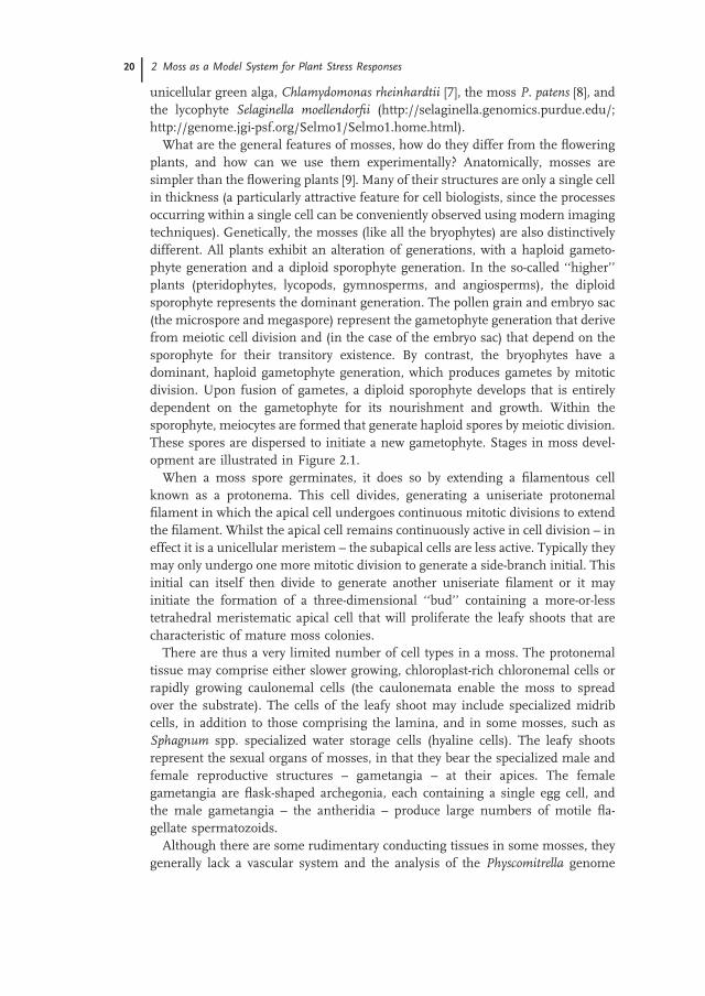
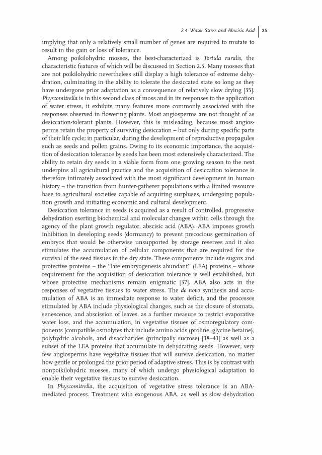
Figure 2.1 Stages in the development of the moss, P. patens. (a) The
mature sporophyte is borne at the tip of the leafy shoot (the
gametophore). (b) A mature spore. Each sporophyte contains between
2000 and 5000 haploid spores. The mature spores are enclosed in a
sporopollenin wall. (c) Spores germinate to produce chloronemal
filaments. This panel shows a germinated sporeling and an
ungerminated spore (top left). (d) Protonemal filaments grow by
repeated division and elongation of the apical cell. The subapical cells
divide to generate side-branch initials or buds. (e) A bud initial at an
early stage of development. Buds develop to form the leafy shoots. (f)
Gametophores, with rhizoids just discernable at the base.
(g) A mature gametophyte comprises many gametophores. This plant
was initiated on agar medium inoculated with a ‘‘spot inoculum’’ of
chloronemal tissue about 4 weeks earlier. (h) Following incubation at
low temperature, the gametangia develop at the gametophore apex.
The leaves have been stripped from the gametophore to reveal the
flask-shaped archegonium (the female structure containing the egg
cell) and the small ovoid antheridia that produce flagellate sperm. (i)
Following fertilization of the egg cell by a sperm, the sporophyte
develops within the archegonium. (j and k) Protoplasts are easily
obtained by digestion of chloronemal filaments with the cell-wall
degrading enzyme mixture ‘‘Driselase.’’ Each protoplast is totipotent
and capable of regenerating to produce a new plant. Protoplasts are
also readily transformed by uptake of exogenous DNA.
sequence indicates that this moss, at least, lacks the genes necessary to synthesize
lignin of the type found in the tracheophytes [8]. Neither do mosses produce
extensive ramifying root systems that are able to scavenge water from the sub-
strate, although the protonemal network may penetrate soils to a shallow depth.
Gametophores are often characterized by a proliferation of empty cells – rhizoids –
at their base and these have been postulated to have some root-like functions [10],
possibly in nutrient assimilation or support, but the gametophyte in general lacks
many of the adaptations used by the tracheophytes to restrict water loss, such as
cuticular wax and somatal apertures. Nevertheless, these adaptations are not
absent: they are restricted to the sporophyte phase of the life cycle, indicating their
early evolution among the land plants.
2.3
Physcomitrella as a Model System
Very few mosses have been studied intensively at the molecular or biochemical
level. However, one moss species, P. patens, has emerged as an excellent model
system for undertaking genome-level analyses. Physcomitrella has been studied
since the early years of the twentieth century and at the genetic level since 1968,
when the first mutants were isolated [11]. First, the cells of a fully differentiated
plant retain their totipotency. Mosses are easily grown in axenic tissue culture by
the simple expedient of fragmenting plants in water with a laboratory blender and
dispersing the suspension over the surface of an agar plate [12]. The resultant
suspension rapidly regenerates as a uniform mat of protonemal filaments, thus
providing an excellent source of homogeneous tissue for biochemical or molecular
analysis. Alternatively, small explants of filamentous tissue can be subcultured as
‘‘spot inocula’’ and they will recapitulate the entire developmental pathway to
generate new, clonal plants.
The haploid nature of the dominant gametophyte generation clearly facilitates
the recognition of mutant phenotypes immediately following mutagenic treatment
and this was exploited by Cove et al. in the isolation of auxotrophic mutants [13], as
well as mutants affecting a number of processes relating to cellular differentiation
[14], hormone responses [15], and the polar growth responses of single cells: the
protonemal apical cell is the site of perception of, transduction and response to
environmental stimuli such as light and gravity [16–19]. This provides the oppor-
tunity to elucidate how such processes operate within a single, easily observed cell,
rather than in a multicellular organ containing a number of different cell types, as
occurs in anatomically more complex plants. Furthermore, cellular differentiation
processes are easily studied and manipulated: the transitions between chloronemal
and caulonemal cells can be regulated by auxin [9, 20] and the carbohydrate
nutritional status of the cell [21] whilst bud formation leading to gametophore
development is triggered by the action of cytokinins [9, 22, 23, 24]. Owing to
the simple anatomy of the moss, allowing accurate and quantitative analysis, and
the predictable nature of the developmental transitions that occur, Physcomitrella
22 | 2 Moss as a Model System for Plant Stress Responses
developmental progression is highly amenable to a detailed computational analysis
that allows predictions to be made and tested [25]. Consequently, Physcomitrellaprovides a highly suitable organism for systems-level analysis – especially when the
genetic and genomic resources now available are considered.
Whilst the utility of Physcomitrella as a subject for cellular level investigations
was clear, its potential for comparative genomic analysis of the evolution of gene
function was not immediately apparent. Only after the establishment of routine
genetic transformation did one of the more remarkable properties of this organ-
ism emerge. Physcomitrella is routinely transformed by the polyethylene glycol-
mediated uptake of naked DNA by protoplasts [26]. This is not a particularly
efficient means of DNA delivery in any plant species, but development of the
technique was aided by the ability of Physcomitrella protoplasts to regenerate with
very high efficiency. During the early development of the protoplast transforma-
tion procedure, it was discovered that retransformation of a transgenic line with a
second DNA construct containing vector sequences homologous with the first
transgene resulted (i) in an increased frequency of transformation by the second
construct and (ii) genetic cosegregation of the two transgenes [27]. It was postu-
lated that this arose from a propensity for the second transgene to become inte-
grated at the first transgenic locus by homologous recombination – a form of gene
targeting. This was subsequently confirmed by a landmark molecular analysis [28],
which showed that transformation with a DNA construct containing sequence
homology with an endogenous genomic sequence resulted in targeting of the
transgene to the endogenous locus. This occurred at very high frequency (up to
100%) – an efficiency of gene targeting hitherto only observed in yeast and cer-
tainly very much higher than occurs in flowering plants.
Gene targeting technology thus allows ‘‘reverse genetic’’ analysis of gene
function to be undertaken with great facility in Physcomitrella – a concept proven
by the first predetermined targeted knockout phenotype to be described in any
plant, that of the Physcomitrella FTSZ gene. This gene encodes a plastid tubulin
required for chloroplast division, the cells of ftsZ mutants generated by targeted
disruption containing single giant chloroplasts [29].
Deployment of such a powerful tool for genetic manipulation can only be
effective if the sequences of the genes to be targeted are known. This requirement
stimulated a series of gene discovery programmes. Initially these comprised the
accumulation of expressed sequence tag (EST) collections [30, 31] and culminated
in the acceptance of the Physcomitrella genome for complete sequence determi-
nation by an international consortium based on the United States Department of
Energy’s Joint Genome Institute Community Sequencing Program. The first-draft
sequence assembly of the Physcomitrella genome was released in 2007 [8].
Currently, the use of Physcomitrella as a model is supported by the genome
sequence assembly – representing about 486 Mbp and encoding approximately
25 000 genes – complemented by a recently developed, molecular-marker-based
linkage map which is anchored to the genome sequence. This will facilitate
mutagenesis-based ‘‘forward genetic’’ screening to undertake the map-based
cloning of genes responsible for selected phenotypic traits [32]. The EST resource
2.3 Physcomitrella as a Model System | 23
comprises about 300 000 sequences, and microarray chips are available based on
the EST resource and on the genome sequence. The facility with which gene
targeting may be undertaken allows the ‘‘reverse genetic’’ analysis of gene func-
tion by gene disruption to generate knockout mutants. It is also possible to carry
out more sophisticated manipulations that include the ability to construct
‘‘knock-in’’ fusions – for example, with reporter genes (b-glucuronidase, Green
Fluorescent Protein, etc.) that allow gene expression to be visualized in lines where
the reporter is introduced into the correct genetic locus, rather than at an ectopic
site elsewhere in the genome, or with molecular tags to facilitate the isolation of
molecular complexes (e.g., by generating epitope-tagged or affinity-tagged pro-
teins). Additionally, it is a relatively simple matter to undertake site-directed
mutagenesis of specific genes, containing as little as a single base alteration. Such
surgical precision is not available in any other plant model.
2.4
Water Stress and Abscisic Acid
As has been indicated above, the mosses retain many features that must have been
characteristic of the first land plants. These features include relatively high levels
of tolerance to abiotic stresses. Most strikingly, many mosses exhibit a char-
acteristically high degree of dehydration tolerance [33]. At its most extreme, this
occurs in the form of desiccation tolerance – the ability to withstand dehydration to
about 5–10% of the plant’s original water content. At this point, it is important to
be clear about how this property is defined. In plain English, the term ‘‘desiccation
tolerance’’ simply refers to the ability to survive and recover from the desiccated
state. However, this expression provides insufficient precision, in that it does not
specify the timescale over which a tolerant state is achieved. Consequently, a
distinction must be made between plants that are able to achieve a viable, desic-
cated state only following a period of adaptation through relatively prolonged
exposure to conditions that cause dehydration to occur over a period of time, and
plants that can equilibrate rapidly with a dry atmosphere and reach the dehydrated
state without a need for prior physiological adaptation [34]. The term ‘‘poikilo-
hydric’’ is used to describe the latter class of plant [35]. Poikilohydry is a generally
rare phenomenon that remains relatively common among the bryophytes,
whereas the ability of plants to achieve a state of desiccation tolerance following a
longer adaptive period is more generally widespread. However, poikilohydry is
relatively rare among the tracheophytes, implying that it is a characteristic that has
been lost during the evolution of anatomical complexity [35, 36]. Nevertheless, a
small number of taxa among the tracheophytes still display desiccation tolerance.
These so-called ‘‘resurrection plants’’ are able to undergo complete dehydration of
the vegetative tissues and recover normal metabolic function rapidly following
rehydration. From the distribution of this character within the land plant phylo-
geny, it is apparent that it is has independently re-evolved several times [36],
24 | 2 Moss as a Model System for Plant Stress Responses
implying that only a relatively small number of genes are required to mutate to
result in the gain or loss of tolerance.
Among poikilohydric mosses, the best-characterized is Tortula ruralis, the
characteristic features of which will be discussed in Section 2.5. Many mosses that
are not poikilohydric nevertheless still display a high tolerance of extreme dehy-
dration, culminating in the ability to tolerate the desiccated state so long as they
have undergone prior adaptation as a consequence of relatively slow drying [35].
Physcomitrella is in this second class of moss and in its responses to the application
of water stress, it exhibits many features more commonly associated with the
responses observed in flowering plants. Most angiosperms are not thought of as
desiccation-tolerant plants. However, this is misleading, because most angios-
perms retain the property of surviving desiccation – but only during specific parts
of their life cycle; in particular, during the development of reproductive propagules
such as seeds and pollen grains. Owing to its economic importance, the acquisi-
tion of desiccation tolerance by seeds has been most extensively characterized. The
ability to retain dry seeds in a viable form from one growing season to the next
underpins all agricultural practice and the acquisition of desiccation tolerance is
therefore intimately associated with the most significant development in human
history – the transition from hunter-gatherer populations with a limited resource
base to agricultural societies capable of acquiring surpluses, undergoing popula-
tion growth and initiating economic and cultural development.
Desiccation tolerance in seeds is acquired as a result of controlled, progressive
dehydration exerting biochemical and molecular changes within cells through the
agency of the plant growth regulator, abscisic acid (ABA). ABA imposes growth
inhibition in developing seeds (dormancy) to prevent precocious germination of
embryos that would be otherwise unsupported by storage reserves and it also
stimulates the accumulation of cellular components that are required for the
survival of the seed tissues in the dry state. These components include sugars and
protective proteins – the ‘‘late embryogenesis abundant’’ (LEA) proteins – whose
requirement for the acquisition of desiccation tolerance is well established, but
whose protective mechanisms remain enigmatic [37]. ABA also acts in the
responses of vegetative tissues to water stress. The de novo synthesis and accu-
mulation of ABA is an immediate response to water deficit, and the processes
stimulated by ABA include physiological changes, such as the closure of stomata,
senescence, and abscission of leaves, as a further measure to restrict evaporative
water loss, and the accumulation, in vegetative tissues of osmoregulatory com-
ponents (compatible osmolytes that include amino acids (proline, glycine betaine),
polyhydric alcohols, and disaccharides (principally sucrose) [38–41] as well as a
subset of the LEA proteins that accumulate in dehydrating seeds. However, very
few angiosperms have vegetative tissues that will survive desiccation, no matter
how gentle or prolonged the prior period of adaptive stress. This is by contrast with
nonpoikilohydric mosses, many of which undergo physiological adaptation to
enable their vegetative tissues to survive desiccation.
In Physcomitrella, the acquisition of vegetative stress tolerance is an ABA-
mediated process. Treatment with exogenous ABA, as well as slow dehydration
2.4 Water Stress and Abscisic Acid | 25
imposed over a period of several days, results in the acquisition of desiccation
tolerance [42, 43]. Figure 2.2 shows some of the structural and anatomical
consequences of stress and ABA treatment. These experimental conditions likely
reflect the ecological situation of P. patens; typically, this species occupies the
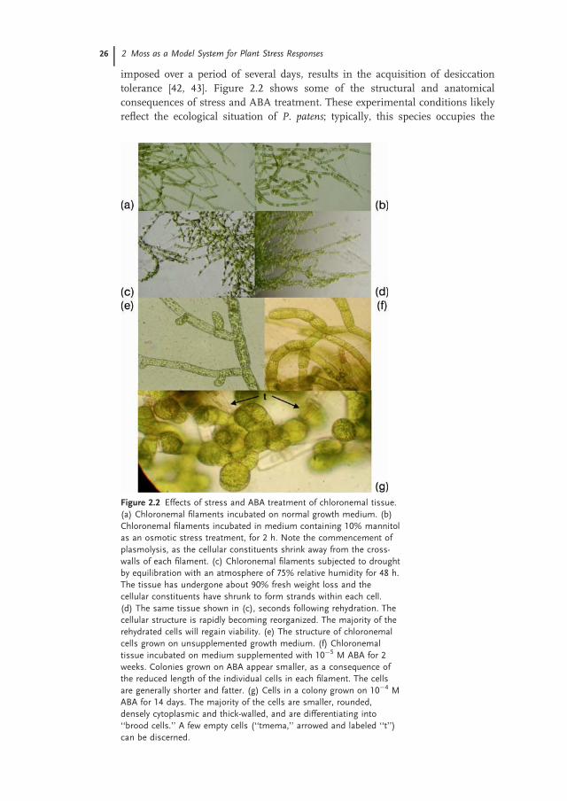
Figure 2.2 Effects of stress and ABA treatment of chloronemal tissue.
(a) Chloronemal filaments incubated on normal growth medium. (b)
Chloronemal filaments incubated in medium containing 10% mannitol
as an osmotic stress treatment, for 2 h. Note the commencement of
plasmolysis, as the cellular constituents shrink away from the cross-
walls of each filament. (c) Chloronemal filaments subjected to drought
by equilibration with an atmosphere of 75% relative humidity for 48 h.
The tissue has undergone about 90% fresh weight loss and the
cellular constituents have shrunk to form strands within each cell.
(d) The same tissue shown in (c), seconds following rehydration. The
cellular structure is rapidly becoming reorganized. The majority of the
rehydrated cells will regain viability. (e) The structure of chloronemal
cells grown on unsupplemented growth medium. (f) Chloronemal
tissue incubated on medium supplemented with 10�5 M ABA for 2
weeks. Colonies grown on ABA appear smaller, as a consequence of
the reduced length of the individual cells in each filament. The cells
are generally shorter and fatter. (g) Cells in a colony grown on 10�4 M
ABA for 14 days. The majority of the cells are smaller, rounded,
densely cytoplasmic and thick-walled, and are differentiating into
‘‘brood cells.’’ A few empty cells (‘‘tmema,’’ arrowed and labeled ‘‘t’’)
can be discerned.
26 | 2 Moss as a Model System for Plant Stress Responses
muddy margins of reservoirs, lakes, and ponds, and when exposed is probably
subjected to relatively slow dehydration due to its close association with the
underlying, generally water-retentive substratum (by contrast with species that
colonize bare rock surfaces and that likely undergo much more rapid drying/
wetting cycles). The acquisition of desiccation tolerance is associated with
membrane stabilization (cellular membranes do not undergo a phase transition
that would result in leakiness) and the physical transition of the cytosol from a
liquid to a glassy state [43]. Such changes are characteristic of organisms that
exhibit anhydrobiotic survival.
ABA induces a number of additional changes in Physcomitrella. Most striking is
the growth arrest and differentiation of the chloronemal cells to form rounded,
thick-walled ‘‘brood cells’’ (brachycytes). These cells become interspersed by
empty, fragile cells (‘‘tmema cells’’) that are easily fractured, thus releasing the
brachycytes that act, in effect, as vegetative spores, each able to initiate the for-
mation of a new colony following dispersal and rehydration [44, 20].
Analysis of the biochemical and metabolic changes that occur following ABA or
stress treatment highlights the similarities between the responses of Physcomitrellaand of the developing seeds of angiosperm species. Transcriptional profiling of
protonemal tissue subjected to such treatments has revealed a number of char-
acteristic changes in gene expression that are initiated very rapidly. Within min-
utes of the application of ABA, transcripts encoding a number of LEA proteins can
be observed to accumulate and these transcripts very rapidly become highly
abundant [45]. Many of the genes expressed in response both to application of ABA
and of the imposition of drought stress have homologs that are similarly regulated
in angiosperms: they include a substantial component encoding LEA proteins, as
well as a number of genes encoding proteins that have been identified as stress-
associated in flowering plants.
Additionally, the underlying mechanisms by which these genes are regulated in
response to ABA and to drought stress appear to have been conserved during the
evolution of the land plants. In flowering plants, drought-stress and ABA-induced
gene expression is regulated through two principal classes of transcription factor.
These are the abscisic-responsive element binding (AREB) factors [46, 47] and
dehydration-responsive element binding (DREB) factors [48, 49] The AREB factors
are members of the large and diverse basic domain/leucine zipper (bZIP) tran-
scription factor family that interacts with cis-acting sequences that include a core
ACGT motif. The DREB factors are members of the equally large APETALA2/
ethylene-responsive element binding (AP2/EREB) transcription factor family that
recognizes cis-acting sequences containing the CCGAC motif. These two classes
of factor interact in a number of the signal transduction pathways that lead to
stress-induced gene expression in the vegetative tissues of flowering plants [50,
51], and factors of both classes have also been identified as mediating both
ABA- and osmotic stress-induced expression of LEA genes during seed develop-
ment [52, 47, 48].
Many of the genes upregulated by ABA and drought stress in Physcomitrellacontain promoter motifs characteristic of these recognition sites [45], and the
2.4 Water Stress and Abscisic Acid | 27
function of the ACGT-core ABRE motif has been demonstrated to be required
both for the expression of a cereal Group 1 LEA gene in moss cells [53] and for the
homologous expression of its Physcomitrella ortholog [54]. Interestingly, the Group
1 LEA genes in flowering plants are highly seed-specific in their pattern of
expression. This is a consequence of their requirement for transcriptional activa-
tion by the ABI3 (ABA-INSENSITIVE3) transcription factor (encoded, in Arabi-dopsis, by the ABI3 gene) which is highly seed-specific in its pattern of expression,
and which is required both for the onset of embryonic dormancy and for the
acquisition of desiccation tolerance. Whereas in those flowering plant genomes
analyzed to date, there resides only a single copy of this key developmental reg-
ulatory gene, the Physcomitrella genome contains at least three ABI3 paralogs [8,
55], suggesting that the evolutionary origins of this gene were related to a primary
role in mediating the drought stress-tolerance pathway, and that recruitment for a
more specialized role in coordinating the seed developmental pathway was
accompanied by loss of the additional members of this gene family.
2.5
T. ruralis: A Model for Poikilohydry
The moss T. ruralis is probably the best-characterized poikilohydric moss. It is able
to survive rapid dehydration and recover full metabolic activity within minutes of
rehydration [56]. Unlike Physcomitrella, which stands at the polar opposite end of
the dehydration tolerance spectrum, Tortula does not show any striking alterations
in the pattern of gene expression during the period of prior dehydration [57].
Instead, this species appears to be constitutively prepared for dehydration. This
likely reflects its ecological distribution, being common in sand dunes and open
grassland [35]. Significant changes in gene expression associated with the desic-
cated state are apparent only following rehydration, when a number of novel
transcripts appear in the polysomal mRNA pool. Designated ‘‘rehydrins’’ [58],
these gene products were suggested to be involved in the repair of dehydration-
associated damage, early in the recovery phase of rehydration. However, it is
interesting that an EST-based analysis of the rehydration-associated transcriptome
of T. ruralis identified a significant number of transcripts encoding LEA proteins
among the most abundant class [59]. One rehydration associated gene product – a
protein designated ‘‘Tr288’’ – is a member of the Group 2 LEA proteins [60]: a class
of polypeptide initially defined as ‘‘dehydrins’’ following their identification as
ABA- and dehydration-induced gene products in cereals [61]. In Physcomitrella – asin angiosperms – the orthologous gene is expressed during drought stress, salt
stress, and in response to ABA treatment, [62] and the mutant strain derived by
gene knockout exhibited hypersensitivity to osmotic and salt stress.
Whilst the protective functions of LEA proteins are far from clearly understood,
at the molecular level, it is generally agreed that they serve to protect macro-
molecular constituents of the cell from the consequences of dehydration, which
include irreversible denaturation and the formation of inactive macromolecular
28 | 2 Moss as a Model System for Plant Stress Responses
aggregates. Dehydrins, like most LEA proteins, are highly hydrophilic, and retain a
high potential for sequestering water molecules [63]. It has been suggested that
they might retain a minimal level of hydration within the cell, offer their hydro-
philic side-chains as participants in hydrogen-bonding interactions with other
macromolecules as ‘‘replacement water’’ [37, 64] or associate to form macro-
molecular fibrillar structures that reinforce the structure of the cell as the compo-
nents undergo the vitrification that is characteristic of dry cells [65, 66]. In
considering why supposedly protective proteins might accumulate during the
recovery from dehydration, an alternative possibility is that such highly hydrophilic
proteins might act as moderators of dehydration/rehydration rates, restricting too-
rapid water loss during dehydration and acting to sequester incoming water upon
rehydration. In this model, water would be retained in an osmotically unresponsive
form: cells accumulating a significant quantity of such proteins would become
osmotically relatively stable – a property akin to that of camel erythrocytes (essential
for survival in an animal that can undergo prolonged periods of exercise in a desert
environment, punctuated by occasional, but substantial, intake of water), which has
been ascribed to the increased water-binding capacity of the unusually hydrophilic
form of hemoglobin found in this species [67].
A final consideration is that the resilience of the desiccated state in poikilohydric
mosses appears to be a feature only of the gametophyte generation. A study of the
desert moss, Tortula inermis, revealed that whereas the gametophyte was highly
tolerant of extreme desiccation, the sporophyte was far more sensitive [68]. Since
sporophytes display a number of anatomical features that are more characteristic
of the vascular plants (including the differentiation of conducting tissues, and the
presence of stomata and cuticles) their desiccation sensitivity may be correlated
with this additional anatomical complexity.
2.6
Cold Stress and Abscisic Acid
Many abiotic stresses elicit similar responses at the molecular and biochemical
level. This reflects the extent of cross-talk that exists between the various stress-
associated perception and signal transduction pathways (reviewed in Chapter 4),
but also some of the common cellular consequences of individual stresses and the
mechanisms that must subsequently be activated to bring about the amelioration
of these stresses or the repair of stress-induced damage. Thus, water-deficit stress
responses have much in common with low-temperature and freezing stress. Both
drought and freezing have the effect of reducing the available water in the cell.
Both require the accumulation of solutes that can act as either compatible
osmolytes or as antifreeze compounds, and both require an alteration of mem-
brane lipids (typically the desaturation of fatty acids) to alter membrane fluidity
and reduce the likelihood of membrane phase transitions. As with the drought
stress response, the freezing stress response and low-temperature acclimation
have been studied in some detail in Physcomitrella.
2.6 Cold Stress and Abscisic Acid | 29
Physcomitrella is highly susceptible to temperatures below freezing. However,
pretreatment with ABA for as little as 24 h substantially enhances freezing tol-
erance, with the temperature at which 50% of cells surviving freezing falling from
about �2 to �8 1C as a consequence of this treatment [43, 69]. Analysis of gene
expression by differential display, to identify novel genes, and of the expression of
candidate genes selected by their similarity with genes known to mitigate freezing
stress in flowering plants indicated a significant upregulation of these genes was
required for the acquisition of tolerance. As well as genes encoding LEA proteins,
these included genes for enzymes associated with sugar metabolism and the
detoxification of reactive oxygen species – the generation of which is a common
consequence of abiotic stress treatments. ABA-treated tissue additionally exhibited
a significant degree of membrane stabilization following freezing [69] suggesting
that modification of membrane lipids comprised a substantial component of the
mechanism of induced tolerance. Treatment with osmotically challenging con-
centrations of mannitol and NaCl also induces the expression of such genes [45,
69]. Oldenhof et al. [43] also demonstrated that ABA treatment increased tolerance
to freezing, and that this was associated with an increase in endogenous sucrose
levels. This is likely derived from the rapid breakdown of chloroplast-localized
starch following the administration of ABA [70] and is accompanied by the
accumulation of additional novel sugars such as the trisaccharide, theanderose
[71]. However, it is an open question as to whether such treatments reflect the true
mechanism by which freezing tolerance is mediated in Physcomitrella or simply
highlight the extensive overlap in the responses of a stress-induced gene set to
multiple agents. Thus, acclimation of Physcomitrella protonemal tissue by
incubation at low temperatures for prolonged periods (up to 1 week) will also
substantially enhance freezing tolerance, and induce the expression of many of the
same genes upregulated by ABA treatments. Although these changes are essen-
tially similar to those rapidly and massively induced by ABA treatment and
although mutants that are insensitive to ABA exhibit a reduced level of freezing
tolerance [72], the changes occurring during low-temperature acclimation do so
in the apparent absence of any measurable increase in the endogenous con-
centrations of ABA present in the moss tissue [73], thus implicating multiple,
parallel signal transduction pathways in the regulation of these stress responses.
2.7
Future Perspectives
The development of genomic resources for Physcomitrella has resulted in a con-
comitant burgeoning of interest among plant scientists for using this species for
comparative studies of plant processes. In the near future, we can expect to see a
more comprehensive accumulation of large transcriptomic, proteomic, and
metabolomic datasets of the type that have been established for other model
organisms. Proteomic approaches have already been applied to identify the poly-
peptide content of protonemal cells [74, 75] and to begin to decipher the
30 | 2 Moss as a Model System for Plant Stress Responses
Physcomitrella phosphoproteome – an important prerequisite for the dissection of
signal transduction pathways associated with cellular differentiation and stress
responses [76, 77].
Whilst Physcomitrella provides the most easily manipulated species among the
mosses, we should not focus on this species to the exclusion of others, whose study
may return insights into processes not evident in Physcomitrella. Thus, T. ruraliswill remain an exemplar for the analysis of poikilohydric desiccation tolerance and
we can expect analysis of other mosses adapted to specialized environments to be
of value in determining the basis of tolerance to these stresses. One such example
is adaptation to heavy metal ions deposited at industrially polluted sites. Physco-mitrella is generally susceptible to the toxic effects of heavy metal ions [78], unlike
other mosses such as Fontinalis antipyretica [79], Scopelophila cataractae [80, 81], orTaxithelium nepalense [82]. As well as being of academic interest, an insight into the
way in which nonmodel bryophytes adapt to such adverse conditions may provide
novel approaches to engineering stress tolerance in crop species, through the
identification of novel genes, and for strategies for the phytoremediation of pol-
luted environments [83]. The rapid advances in massively parallel DNA sequen-
cing technology can be expected to make transcriptomic analysis possible for
organisms that have previously not enjoyed the benefits of extensive research
funding. The use of technology such as the Roche 454 GS-FLX DNA sequencing
procedure enables the acquisition of about 400 000 sequence traces of about 500
bases each, in a single day. This technology has already been used to sample the
Physcomitrella epigenome [84]. When used to sequence ESTs derived from specific
tissues or treatments, quantitative analysis of the clustered sequences generates
mRNA abundance profiles over a wide dynamic range [85]. Whilst complete
genome sequencing for many organisms remains a relatively distant prospect,
transcriptome sampling by new-generation sequencing promises a much more
immediate return, that can generate a sequence database that will interface with
proteomic and metabolomic profiling for nonmodel species.
References
1 Strother, P.K., Al-Hajri, S., and Traverse,
A. (1996) New evidence for land plants
from the lower Middle Ordovician of
Saudi Arabia. Geology, 24, 55–58.2 Wellman, C.H., Osterloff, P.L., and
Mohiuddin, U. (2003) Fragments of the
earliest land plants. Nature, 425, 282–285.
3 Edwards, D. and Feehan, J. (1980)
Records of Cooksonia-type sporangia
from late Wenlock strata in Ireland.
Nature, 287, 41–42.4 Manhart, J.R. and Palmer, J.D. (1990)
The gain of two chloroplast introns
marks the green algal ancestors of land
plants. Nature, 345, 268–270.5 Karol, K.G., McCourt, R.M., Cimino,
M.T., and Delwiche, C.F. (2001) The
closest living relatives of land plants.
Science, 294, 2351–2353.6 Palenik, B., Grimwood, J., Aerts, A.,
Rouze, P., Salamov, A., Putnam, N.,
Dupont, C., Jorgensen, R., Derelle, E.,
Rombauts, S., Zhou, K., Otillar, R.,
Merchant, S.S., Podell, S., Gaasterland,
T., Napoli, C., Gendler, K., Manuell, A.,
Tai, V., Vallon, O., Piganeau, G., Jancek,
S., Heijde, M., Jabbari, K., Bowler, C.,
References | 31
Lohr, M., Robbens, S., Werner, G.,
Dubchak, I., Pazour, G.J., Ren, Q.,
Paulsen, I., Delwiche, C., Schmutz, J.,
Rokhsar, D., Van de Peer, Y., Moreau,
H., and Grigoriev, I.V. (2007) The tiny
eukaryote Ostreococcus provides genomic
insights into the paradox of plankton
speciation. Proc. Natl. Acad. Sci. USA,104, 7705–7710.
7 Merchant, S.S., Prochnik, S.E., Vallon,
O., Harris, E.H., Karpowicz, S.J.,
Witman, G.B., Terry, A., Salamov, A.,
Fritz-Laylin, L.K., Marechal-Drouard, L.,
Marshall, W.F., Qu, L.-H., Nelson, D.R.,
Sanderfoot, A.A., Spalding, M.H.,
Kapitonov, V.V., Ren, Q., Ferris, P.,
Lindquist, E., Shapiro, H., Lucas, S.M.,
Grimwood, J., Schmutz, J., Cardol, P.,
Cerutti, H., Chanfreau, G., Chen, C.-L.,
Cognat, V., Croft, M.T., Dent, R.,
Dutcher, S., Fernandez, E., Fukuzawa,
H., Gonzalez-Ballester, D., Gonzalez-
Halphen, D., Hallmann, A., Hanikenne,
M., Hippler, M., Inwood, W., Jabbari,
K., Kalanon, M., Kuras, R., Lefebvre,
P.A., Lemaire, S.D., Lobanov, A.V., Lohr,
M., Manuell, A., Meier, I., Mets, L.,
Mittag, M., Mittelmeier, T., Moroney,
J.V., Moseley, J., Napoli, C., Nedelcu,
A.M., Niyogi, K., Novoselov, S.V.,
Paulsen, I.T., Pazour, G., Purton, S.,
Ral, J.-P., Riano-Pachon, D.M., Riekhof,
W., Rymarquis, L., Schroda, M.,
Stern, D., Umen, J., Willows, R.,
Wilson, N., Zimmer, S.L., Allmer, J.,
Balk, J., Bisova, K., Chen, C.-J.,
Elias, M., Gendler, K., Hauser, C.,
Lamb, M.R., Ledford, L., Long, J.C.,
Minagawa, J., Page, M.D., Pan, J.,
Pootakham, W., Roje, S., Rose, A.,
Stahlberg, E., Terauchi, A.M., Yang, P.,
Ball, S., Bowler, C., Dieckmann, C.L.,
Gladyshev, V.N., Green, P.,
Jorgensen, R., Mayfield, S.M., Mueller-
Roeber, B., Rajamani, S., Sayre, R.T.,
Brokstein, P., Dubchak, I., Goodstein,
D., Hornick, L., Huang, Y.W., Jhaveri,
J., Luo, Y., Martınez, D., Ngau, W.C.A.,
Otillar, B., Poliakov, A., Porter, A.,
Szajkowski, L., Werner, G., Zhou, K.,
Grigoriev, I.V., Rokhsar, D.S., and
Grossman, A.R. (2007) The
Chlamydomonas genome reveals the
evolution of key animal and plant
functions. Science, 318, 245–250.8 Rensing, S.A., Lang, D., Zimmer, A.,
Terry, A., Salamov, A., Shapiro, H.,
Nishiyama, T., Perroud, P.-F., Lindquist,
E., Kamisugi, Y., Tanahashi, T.,
Sakakibara, K., Fujita, T., Oishi, K.,
Shin-I, T., Kuroki, Y., Toyoda, A.,
Suzuki, Y., Hashimoto, S., Yamaguchi,
K., Sugano, S., Kohara, Y., Fujiyama, A.,
Anterola, A., Aoki, S., Ashton, N.,
Barbazuk, W.B., Barker, E., Bennetzen,
J., Blankenship, R., Cho, S.H., Dutcher,
S., Estelle, M., Fawcett, J.A., Gundlach,
H., Hanada, K., Heyl, A., Hicks, K.A.,
Hughes, J., Lohr, M., Mayer, K.,
Melkozernov, A., Murata, T., Nelson, D.,
Pils, P., Prigge, M., Reiss, B., Renner,
T., Rombauts, S., Rushton, P.,
Sanderfoot, A., Schween, G., Shiu, S.-
H., Stueber, K., Theodoulou, F.L., Tu,
H., Van de Peer, Y., Verrier, P.J.,
Waters, E., Wood, A., Yang, L., Cove,
D.J., Cuming, A.C., Hasebe, M., Lucas,
S., Mishler, B.D., Reski, R., Grigoriev, I.,
Quatrano, R.S., and Boore, J.L. (2008)
The Physcomitrella genome reveals
insights into the conquest of land by
plants. Science, 319, 64–69.9 Reski, R. (1998) Development, genetics
and molecular biology of mosses.
Botanica Acta, 111, 1–15.10 Menand, B., Yi, K.K., Jouannic, S.,
Hoffman, L., Ryan, E., Linstead, P.,
Schaefer, D.G., and Dolan, L. (2007) An
ancient mechanism controls the
development of cells with a rooting
function in land plants. Science, 316,1477–1480.
11 Engel, P.P. (1968) Induction of
biochemical and morphological mutants
in moss Physcomitrella patens. Am. J.Bot., 55, 438–446.
12 Knight, C.D., Cove, D.J., Cuming, A.C.,
and Quatrano, R.S. (2002) Moss gene
technology, in Molecular Plant Biology,Vol. 2 (eds P.M. Gilmartin and C.
Bowler), Oxford University Press,
Oxford, pp. 285–299.
13 Ashton, N.W. and Cove, D.J. (1977) The
isolation and preliminary
characterisation of auxotrophic and
analogue resistant mutants of the moss.
32 | 2 Moss as a Model System for Plant Stress Responses
Physcomitrella patens. Mol. Genet.Genomics, 154, 87–95.
14 Ashton, N.W., Cove, D.J., and
Featherstone, D.R. (1979) The isolation
and physiological analysis of mutants of
the moss, Physcomitrella patens, whichover-produce gametophores. Planta, 144,437–442.
15 Ashton, N.W., Grimsley, N.H., and
Cove, D.J. (1979) Analysis of
gametophytic development in the moss,
Physcomitrella patens, using auxin and
cytokinin resistant mutants. Planta, 144,427–435.
16 Jenkins, G.I. and Cove, D.J. (1983)
Phototropism and polarotropism of
primary chloronemata of the moss
Physcomitrella patens: responses of thewild-type. Planta, 158, 357–364.
17 Jenkins, G.I. and Cove, D.J. (1983)
Phototropism and polarotropism of
primary chloronemata of the
moss Physcomitrella patens:responses of mutant strains.
Planta, 158, 432–438.18 Jenkins, G.I., Courtice, G.R.M., and
Cove, D.J. (1986) Gravitropic responses
of wild-type and mutant strains of the
moss Physcomitrella patens. Plant CellEnviron., 9, 637–644.
19 Knight, C.D., Futers, T.S., and Cove,
D.J. (1991) Genetic analysis of a mutant
class of Physcomitrella patens in which
the polarity of gravitropism is reversed.
Mol. Genet. Genomics, 230, 12–16.20 Decker, E.L., Frank, W., Sarnighausen,
E., and Reski, R. (2006) Moss systems
biology en route: phytohormones in
Physcomitrella development. Plant Biol.,8, 397–406.
21 Thelander, M., Olsson, T., and Ronne,
H. (2005) Effect of the energy supply on
filamentous growth and development in
Physcomitrella patens. J. Exp. Bot., 56,653–662.
22 Wang, T.L., Horgan, R., and Cove, D.J.
(1981) Cytokinins from the moss
Physcomitrella patens. Plant Physiol., 68,735–738.
23 Reski, R., and Abel, W.O. (1985)
Induction of budding on chloronemata
and caulonemata of the moss,
Physcomitrella patens, using
isopentenyladenine. Planta 165,354–358.
24 Reutter, K., Atzorn, R., Hadeler, B.,
Schmulling, T., and Reski, R. (1998)
Expression of the bacterial ipt gene in
Physcomitrella rescues mutations in
budding and in plastid division. Planta,206, 196–203.
25 Fracchia, F.D. and Ashton, N.W. (1995)
A visualization tool for studying the
development of the moss Physcomitrellapatens, in Proceedings of the IEEEConference on Visualization (eds G.M.
Nielson and D. Silver). IEEE, New York,
pp. 364–367.
26 Schaefer, D., Zryd, J.P., Knight, C.D.,
and Cove, D.J. (1991) Stable
transformation of the moss
Physcomitrella patens. Mol. Genet.Genomics, 226, 418–424.
27 Kammerer, W. and Cove, D.J. (1996)
Genetic analysis of the effects of re-
transformationof transgenic lines of the
moss Physcomitrella patens. Mol. Genet.Genomics, 250, 380–382.
28 Schaefer, D.G. and Zryd, J.P. (1997)
Efficient gene targeting in the moss
Physcomitrella patens. Plant J., 11,1195–1206.
29 Strepp, R., Scholz, S., Kruse, S.,
Speth, V., and Reski, R. (1998) Plant
nuclear gene knockout reveals a role in
plastid division for the homolog of the
bacterial cell division protein FtsZ, an
ancestral tubulin. Proc. Natl. Acad. Sci.USA, 95, 4368–4373.
30 Nishiyama, T., Fujita, T., Shin-I, T., Seki,
M., Nishide, H., Uchiyama, I., Kamiya,
A., Carninci, P., Hayashizaki, Y.,
Shinozaki, K., Kohara, Y., and Hasebe,
M. (2003) Comparative genomics
of Physcomitrella patens gametophytic
transcriptome and Arabidopsisthaliana: implication for land plant
evolution. Proc. Natl. Acad. Sci. USA, 100,8007–8012.
31 Lang, D., Eisinger, J., Reski, R., and
Rensing, S.A. (2005) Representation and
high-quality annotation of the
Physcomitrella patens transcriptome
demonstrates a high proportion of
proteins involved in metabolism in
moses. Plant Biol., 7, 238–250.
References | 33
32 Kamisugi, Y., von Stackelberg, M.,
Lang, D., Care, M., Reski, R., Rensing,
S.A., and Cuming, A.C. (2008) A
sequence-anchored genetic linkage map
for the moss Physcomitrella patens. PlantJ., 56, 855–866.
33 Dilks, T.J.K. and Proctor, M.C.F. (1974)
The pattern of recovery of bryophytes
after desiccation. J. Bryol., 8, 97–115.34 Alpert, P. (2006) Constrains of tolerance:
why are desiccation-tolerant organisms
so small or rare? J. Exp. Biol., 209,1575–1584.
35 Proctor, M.C.F. and Tuba, Z. (2002)
Poikilohydry and homoihydry: antithesis
or spectrum of possibilities? New Phytol.,156, 327–349.
36 Oliver, M.J., Tuba, Z., and Mishler, B.
(2000) The evolution of vegetative
desiccation tolerance in land plants.
Plant Ecol., 151, 85–100.37 Cuming, A.C. (1999) LEA proteins, in
Seed Proteins (eds P.R. Shewry and
R. Casey), Kluwer, Dordrecht,
pp. 753–780.
38 Yoshiba, Y., Kiyosue, T., Nakashima, K.,
Yamaguchi-Shinozaki, K., and
Shinozaki, K. (1997) Regulation of levels
of proline as an osmolyte in plants
under water stress. Plant Cell Physiol.,38, 1095–1102.
39 McKue, K.F. and Hanson, A.D. (1996)
Drought and salt tolerance: towards
understanding and application. TrendsBiotechnol., 8, 358–362.
40 Popp, M. and Smirnoff, N. (1995) Polyol
accumulation and metabolism during
water deficit, in Environment and PlantMetabolism (ed. N. Smirnoff), BIOS,
Oxford, pp. 199–215.
41 Bianchi, G., Gamba, A., Murelli, C.,
Salamini, F., and Bartels, D. (1991)
Novel carbohydrate metabolism in the
resurrection plant Craterostigmaplantagineum. Plant J., 1, 35–39.
42 Frank, W., Ratnadewi, D., and Reski, R.
(2005) Physcomitrella patens is highlytolerant against drought, salt and
osmotic stress. Planta, 220, 384–394.43 Oldenhof, H., Wolkers, W.F.,
Bowman, J.L., Tablin, F., and Crowe,
J.H. (2006) Freezing and desiccation
tolerance in the moss Physcomitrella
patens: an in situ Fourier transform
infrared spectroscopic study. Biochim.Biophys. Acta, 1760, 1226–1234.
44 Schnepf, E. and Reinhard, C. (1997)
Brachycytes in Funaria protonemata:induction by abscisic acid and fine
structure. J. Plant Physiol., 151, 166–175.45 Cuming, A.C., Cho, S.H., Kamisugi, Y.,
Graham, H., and Quatrano, R.S. (2007)
Microarray analysis of transcriptional
responses to abscisic acid and osmotic-
salt- and drought-stress in the moss,
Physcomitrella patens. New Phytol., 176,275–287.
46 Marcotte, W.R., Russell, S.H., and
Quatrano, R.S. (1989) Abscisic acid-
responsive sequences from the Em gene
of wheat. Plant Cell, 1, 969–976.47 Finkelstein, R.R. and Lynch, T.J. (2000)
The Arabidopsis abscisic acid response
gene ABI5 encodes a basic leucine
zipper transcription factor. Plant Cell,12, 599–609.
48 Shen, Q. and Ho, T.-H.D. (1995)
Functional dissection of an abscisic acid
(ABA)-inducible gene reveals two
independent ABA-responsive complexes
each containing a G-box and a novel cis-acting element. Plant Cell, 7, 295–307.
49 Sakuma, Y., Liu, Q., Dubouzet, J.G.,
Abe, H., Shinozaki, K., and Yamaguchi-
Shinozaki, K. (2002) DNA-binding
specificity of the ERF/AP2 domain of
Arabidopsis DREBs, transcription factors
involved in dehydration- and cold-
inducible gene expression. Biochem.Biophys. Res. Commun., 290, 998–1009.
50 Shinozaki, K. and Yamaguchi-Shinozaki,
K. (1997) Gene expression and signal
transduction in water-stress response.
Plant Physiol., 115, 327–334.51 Shinozaki, K., Yamaguchi-Shinozaki, K.,
and Seki, M. (2003) Regulatory network
of gene expression in the drought and
cold-stress response. Curr. Opin. PlantBiol., 6, 410–417.
52 Guiltinan, M.J., Marcotte, W.R., and
Quatrano, R.S. (1990) A plant leucine
zipper protein that recognizes an
abscisic acid response element. Science,250, 267–271.
53 Knight, C.D., Seghal, A., Atwal, K.,
Wallace, J.C., Cove, D.J., Coates, D.,
34 | 2 Moss as a Model System for Plant Stress Responses
Quatrano, R.S., Bahadur, S.,
Stockley, P.G., and Cuming, A.C. (1995)
Molecular responses to abscisic acid and
stress are conserved between moss and
cereals. Plant Cell, 7, 499–506.54 Kamisugi, Y. and Cuming, A.C. (2005)
The evolution of the abscisic acid-
response in land plants: comparative
analysis of group 1 LEA gene expression
in moss and cereals. Plant Mol. Biol., 59,723–737.
55 Marella, H.H., Sakata, Y., and
Quatrano, R.S. (2006) Characterization
and functional analysis of ABSCISICACID INSENSITIVE 3-like genes from
Physcomitrella patens. Plant J., 46,1032–1044.
56 Bewley, J.D. (1979) Physiological aspects
of desiccation-tolerance. Annu. Rev. PlantPhysiol., 30, 195–238.
57 Oliver, M.J. (1991) Influence of
protoplasmic water loss on the control
of protein synthesis in the desiccation-
tolerant moss Tortula ruralis:ramifications for a repair-based
mechanism of desiccation-tolerance.
Plant Physiol., 97, 1501–1511.58 Scott, H.B.I.I. and Oliver, M.J. (1994)
Accumulation and polysomal
recruitment of transcripts in response to
desiccation and rehydration of the moss
Tortula ruralis. J. Exp. Bot., 45, 577–583.59 Oliver, M.J., Dowd, S.E., Zaragoza, J.,
Mauget, S.A., and Payton, P.R. (2004)
The rehydration transcriptome of the
desiccation-tolerant bryophyte Tortularuralis: transcript classification and
analysis. BMC Genomics, 5, 89.60 Velten, J. and Oliver, M.J. (2001) Tr288:
a rehydrin with a dehydrin twist. PlantMol. Biol., 45, 713–722.
61 Close, T.J. (1996) Dehydrins: emergence
of a biochemical role of a family of plant
dehydration proteins. Physiol. Planta.,97, 795–803.
62 Saavedra, L., Svensson, J., Carballo, V.,
Izmendi, D., and Vidal, S. (2006) A
dehydrin gene in Physcomitrella patens isrequired for salt and osmotic stress
tolerance. Plant J., 45, 237–249.63 Campbell, S.A. and Close, T.J. (1997)
Dehydrins: genes, proteins and
associations with phenotypic traits. NewPhytol., 137, 61–74.
64 Hoekstra, F.A., Golovina, E.A., and
Buitink, J. (2001) Mechanisms of plant
desiccation tolerance. Trends Plant Sci.,6, 431–440.
65 Wise, M.J. (2003) Leaping to
conclusions: a computational reanalysis
of late embryogenesis abundant proteins
and their possible roles. BMCBioinformatics, 4, 52.
66 Wise, M. and Tunnacliffe, A. (2004)
POPP the question: what do LEA
proteins do? Trends Plant Sci., 9, 13–17.67 Bogner, P., Csutora, P., Cameron, I.L.,
Wheatley, D.N., and Miseta, A. (1998)
Augmented water binding and low
cellular water content in erythrocytes
of camel and camelids. Biophys J., 75,3085–3091.
68 Stark, L.R., Oliver, M.J., Mishler, B.D.,
and McLetchie (2007) Generational
differences in response to desiccation
stress in the desert moss Tortula inermis.Ann. Bot., 99, 53–60.
69 Minami, A., Nagao, M., Arakawa, K.,
Fujikawa, S., and Takezawa, D. (2003)
Abscisic acid-induced freezing tolerance
is accompanied by increased expression
of stress-related genes. J. Plant Physiol.,160, 475–483.
70 Nagao, M., Minami, A., Arakawa, K.,
Fujikawa, S., and Takezawa, D. (2005)
Rapid degradation of starch in
chloroplasts and concomitant
accumulation of soluble sugars
associated with ABA-induced freezing
tolerance in the moss Physcomitrellapatens. J. Plant Physiol., 162, 169–180.
71 Nagao, M., Oku, K., Minami, A.,
Mizuno, K., Sakurai, M., Arakawa, K.,
Fujikawa, S., and Takezawa, D. (2006)
Accumulation of theanderose in
association with the development of
freezing tolerance in the moss
Physcomitrella patens. Phytochemistry, 67,702–709.
72 Takezawa, D. (2007) Reduced sensitivity
to freezing and dehydration in the
abscisic acid-insensitive mutant in
mosses. Plant Cell. Physiol. Suppl., 48,893.
References | 35
73 Minami, A., Nagao, M., Ikegama, K.,
Koshiba, T., Arakawa, K., Fujikawa, S.,
and Takezawa, D. (2005) Cold
acclimation in bryophytes: low-
temperature-induced freezing tolerance
in Physcomitrella patens is associatedwith increases in expression levels of
stress-related genes but not with
increase in level of endogenous abscisic
acid. Planta, 220, 414–423.74 Sarnighausen, E., Wurtz, V., Heintz, D.,
van Dorsselaer, A., and Reski, R. (2004)
Mapping of the Physcomitrella patensproteome. Phytochemistry, b65,1589–1607.
75 Cho, S.H., Hoang, Q.T., Kim, Y.Y.,
Shin, H.Y., Ok, S.H., Bae, J.M., and
Shin, J.S. (2006) Proteome analysis of
gametophores identified a
metallothionein involved in various
abiotic stress responses in
Physcomitrella patens. Plant Cell Rep., 25,475–488.
76 Heintz, D., Wurtz, V., High, A.A., Van
Dorsselaer, A., Reski, R., and
Sarnighausen, E. (2004) An efficient
protocol for the identification of protein
phosphorylation in a seedless plant,
sensitive enough to detect members of
signalling cascades. Electrophoresis, 25,1149–1159.
77 Heintz, D., Erxleben, A., High, A.A.,
Wurtz, V., Reski, R., Van Dorsselaer, A.,
and Sarnighausen, E. (2006) Rapid
alteration of the phosphoproteome in
the moss Physcomitrella patens aftercytokinin treatment. J. Proteome Res., 5,2283–2293.
78 Rother, M., Krauss, G.-J., Grass, G., and
Wesenberg, D. (2006) Sulphate
accumulation under Cd2þ stress in
Physcomitrella patens – combined
transcript, enzyme and metabolite
profiling. Plant Cell Environ., 29,1801–1811.
79 Rau, S., Miersch, J., Neumann, D.,
Weber, E., and Krauss, J.-G. (2007)
Biochemical responses of the aquatic
moss Fontinalis antipyretica to
Cd, Cu, Pb and Zn determined by
chlorophyll fluorescence and
protein levels. Environ. Exp. Bot., 59,299–306.
80 Shaw, J.A. and Beer, S.C. (1989)
Scopelophila cataractae (Mitt.) Broth. in
Pennsylvania. Bryologist, 92, 112–115.81 Shaw, J.A. (1995) Genetic biogeography
of the rare ‘‘copper moss’’, Scopelophilacataractae (Pottiaceae). Plant Systemat.Evol., 197, 43–58.
82 Choudhury, S. and Panda, S.K. (2005)
Toxic effects, oxidative stress and
ultrastructural changes in moss
Taxithelium nepalense (Schwaegr.) Broth.under chromium and lead toxicity.
Water Air Soil Pollut., 167, 73–90.83 Prasad, M.N.V. and De Oliveira
Freitas, H.M. (2003) Metal
hyperaccumulation in plants –
biodiversity prospecting for
phytoremediation technology. Electron. J.Biotechnol., 6, 285–321.
84 Axtell, M.J., Snyder, J.A., and Bartell,
D.P. (2007) Common functions for
diverse small RNAs of land plants. PlantCell, 19, 1750–1769.
85 Weber, A.P.M., Weber, K.L., Carr, K.,
Wilkerson, C., and Ohlrogge, J.B. (2007)
Sampling the Arabidopsis transcriptome
with massively parallel pyrosequencing.
Plant Physiol., 144, 32–42.
36 | 2 Moss as a Model System for Plant Stress Responses

![Target of Rapamycin Signaling in Plant Stress …Update on Target of Rapamycin Signaling in Plant Stress Responses Target of Rapamycin Signaling in Plant Stress Responses1[OPEN] Liwen](https://img.pdfslide.us/doc/110x75/5f05e4b57e708231d4153f1e/target-of-rapamycin-signaling-in-plant-stress-update-on-target-of-rapamycin-signaling.jpg)