Embed Size (px)
Citation preview
Int.J.Curr.Microbiol.App.Sci (2014) 3(3): 960-968
960
Original Research Article
Plant leaf mediated synthesis of silver nanoparticles using Phyllanthus niruri and its antimicrobial activity against multi drug resistant human pathogens
P.Kathireswari1*, S.Gomathi1 and K.Saminathan2
1Department of Zoology, Kongunadu Arts and Science Collge, Coimbatore-641029, India 2Department of Nanotechnology, KSR College of Technology, Tiruchengode- 637 215, India
*Corresponding author
A B S T R A C T
Introduction
The use of environmentally benign materials like plant leaf extract, bacteria and fungi for the synthesis of silver nanoparticles offers numerous benefits of eco-friendliness and compatibility for pharmaceutical and biomedical applications as they do not use toxic chemicals in the synthesis protocols. Chemical synthesis methods lead to the presence of some toxic chemical species adsorbed on the surface that may have
adverse effects in medical applications. Bioinspired synthesis of nanoparticles provides advancement over chemical and physical methods as it is a cost effective and environment friendly and in this method there is no need to use high pressure, energy, temperature and toxic chemicals.
Silver nanoparticles are the metal of choice as they hold the promise to kill
ISSN: 2319-7706 Volume 3 Number 3 (2014) pp. 960-968 http://www.ijcmas.com
K e y w o r d s
Silver nanoparticles; Plant leaf extract; Nano-technology; Pathogens; Antimicrobial activity.
The work is being carried out for the applications which would help in the prevention of human pathogens. We describe the synthesis of silver nanoparticles using plant Phyllanthus niruri leaf extracts. A strong characteristic absorbance peak at around 420 nm was observed at different time intervals. Particle size analysis of these particles shows that they are 63.45 nm in range and also this paper deals with a thorough investigation on the characterization of the silver nanoparticles by XRD, SEM and FTIR analysis and result reveals that, the average grain size of the silver nanoparticles formed in the bioreduction process is determined by XRD pattern of the silver nanoparticles formed in the experiment and is estimated to be 60 nm and spherical in shape. FTIR spectral analysis showed array of absorbance bands in 400 cm-1 -1500 cm-1 finally tested the biocompatibility by antimicrobial test against multi drug resistant human pathogens like Staphylococcus aureus, Escherichia coli, Pseudomonous sp, Proteus vulgaris, and Salmonella typhi. The evaluation of agar diffusion test was made on the basis of zone of inhibition of bacteria around the test sample. The result reveals that the maximum antimicrobial activity was observed against the test organisms when compared to the control.
Int.J.Curr.Microbiol.App.Sci (2014) 3(3): 960-968
961
microbe s effectively (Sondi and Salopek-Sondi 2004). The silver nanoparticles act on a broad range of target sites both extracellularly as well intracellularly. In fact, microbes generally have a harder time developing resistance to silver than they do to antibiotics (Baker, et al. 2005 and Landsdown 2007) the oligodynamic effect that silver has on microbes, whereby silver ions bind to reactive groups in bacterial cells, resulting in their precipitation and inactivation. Some well known examples are, synthesis of gold nanotriangles using Lemmon grass and tamarind leaf extract (Shankar et al 2005) synthesis of silver nanoparticles using geranium leaf (Shankar et al 2003), fungus (Ajaykumar et al 2001 and Ahmad et al 2003) soluble starch (Vigneshwaran 2006) silver tolerant yeast strain. (Kowshik 2003) synthesis of bimetallic Au core-Ag shell nanoparticles using Neem leaf broth ( Shankar et al., 2004) biosynthesis of silver based crystalline nanoparticles of well defined composition and shapes as equilateral triangles and hexagons within the periplasmic space of bacteria Pseudomononas stutzery isolated from silver mines.
The Phyllanthus genus belongs to the Euphorbiaceae family. There are over 300 genera with over 5000 species in the Euphorbiaceae worldwide, while Phyllanthus has about 750 - 800 species, found in tropical and subtropical regions worldwide. A number of the Phyllanthus species have been reported to have extensive history in medicine systems. Substantial amount of the genus are used widely in traditional medicine for the treatment of flu, dropsy, diabetes, jaundice, gall and bladder calculus, liver disease (Unander et al., 1995;). Most of these species have pharmacological properties e.g. Phyllanthus niruri has
demonstrated in vitro antibacterial actions against Staphylococcus, Micrococcus and Pasteurella bacteria as well as in vivo and in vitro anti-malarial properties, which validates other traditional uses of the genus (Veeramuthu et al., 2006). Extracts of Phyllanthus had been used as antiviral source to treat hepatatis B studied the aqueous extracts of Phyllanthus niruri and found it to inhibit viral DNA in vitro. In addition, they eliminated detectable virus from the sera of woodchucks (Marmota monax) acutely or chronically infected with the woodchuck hepatitis virus (WHV). The methanol extracts of five Phyllanthus species from India was reported to have strong antioxidant activity). P. niruri has antifungal activity on ringworm, ulcers, scabies and jaundice.
Herein, we report synthesis of silver nanoparticles, reducing silver ions present in the aqueous solution of silver nitrate complex by the plant extract of Phyllanthus niruri. This plant are traditional folk medicine and occur throughout the tropical and it is using as a liver tonic for jaundice and chronic liver diseases. To the best of our knowledge we are the first to report its use in synthesizing silver nanoparticles, which can provide a new platform to this plant making it a value added nanotechnology product in future. Materials and Methods
Preparation of aqueous extract
Fresh plant materials of Phyllanthus niruri was collected and the aqueous extract of sample was prepared using the freshly collected leaves (5g), by washing in running tap water and then in double distilled water, followed by boiling with
Int.J.Curr.Microbiol.App.Sci (2014) 3(3): 960-968
962
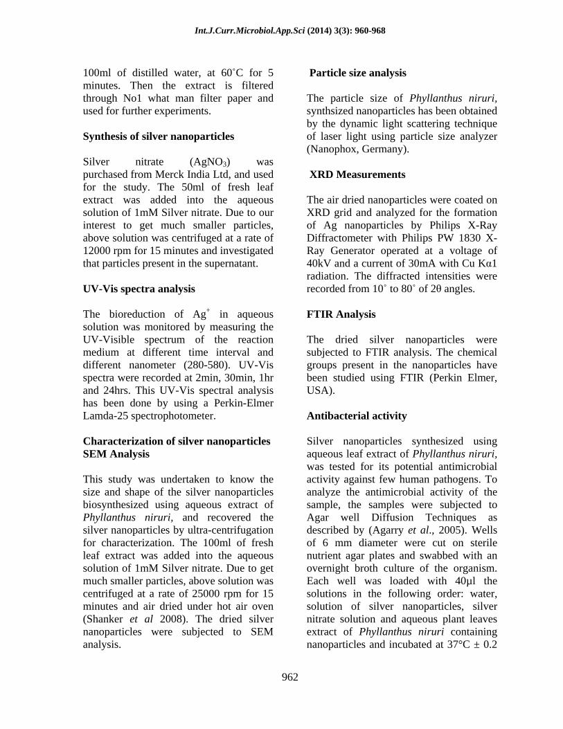
100ml of distilled water, at 60 C for 5 minutes. Then the extract is filtered through No1 what man filter paper and used for further experiments.
Synthesis of silver nanoparticles
Silver nitrate (AgNO3) was purchased from Merck India Ltd, and used for the study. The 50ml of fresh leaf extract was added into the aqueous solution of 1mM Silver nitrate. Due to our interest to get much smaller particles, above solution was centrifuged at a rate of 12000 rpm for 15 minutes and investigated that particles present in the supernatant.
UV-Vis spectra analysis
The bioreduction of Ag+ in aqueous solution was monitored by measuring the UV-Visible spectrum of the reaction medium at different time interval and different nanometer (280-580). UV-Vis spectra were recorded at 2min, 30min, 1hr and 24hrs. This UV-Vis spectral analysis has been done by using a Perkin-Elmer Lamda-25 spectrophotometer.
Characterization of silver nanoparticles SEM Analysis
This study was undertaken to know the size and shape of the silver nanoparticles biosynthesized using aqueous extract of Phyllanthus niruri, and recovered the silver nanoparticles by ultra-centrifugation for characterization. The 100ml of fresh leaf extract was added into the aqueous solution of 1mM Silver nitrate. Due to get much smaller particles, above solution was centrifuged at a rate of 25000 rpm for 15 minutes and air dried under hot air oven (Shanker et al 2008). The dried silver nanoparticles were subjected to SEM analysis.
Particle size analysis
The particle size of Phyllanthus niruri, synthsized nanoparticles has been obtained by the dynamic light scattering technique of laser light using particle size analyzer (Nanophox, Germany).
XRD Measurements
The air dried nanoparticles were coated on XRD grid and analyzed for the formation of Ag nanoparticles by Philips X-Ray Diffractometer with Philips PW 1830 X-Ray Generator operated at a voltage of 40kV and a current of 30mA with Cu K 1 radiation. The diffracted intensities were recorded from 10 to 80 of 2 angles.
FTIR Analysis
The dried silver nanoparticles were subjected to FTIR analysis. The chemical groups present in the nanoparticles have been studied using FTIR (Perkin Elmer, USA).
Antibacterial activity
Silver nanoparticles synthesized using aqueous leaf extract of Phyllanthus niruri, was tested for its potential antimicrobial activity against few human pathogens. To analyze the antimicrobial activity of the sample, the samples were subjected to Agar well Diffusion Techniques as described by (Agarry et al., 2005). Wells of 6 mm diameter were cut on sterile nutrient agar plates and swabbed with an overnight broth culture of the organism. Each well was loaded with 40µl the solutions in the following order: water, solution of silver nanoparticles, silver nitrate solution and aqueous plant leaves extract of Phyllanthus niruri containing nanoparticles and incubated at 37°C ± 0.2
Int.J.Curr.Microbiol.App.Sci (2014) 3(3): 960-968
963
C. Antimicrobial activity in terms of zones of inhibition (mm) was recorded after 24 h of incubation. The antagonistic action of plant leaf extracts of Phyllanthus niruri, was tested against test organisms in triplicates.
Test organisms
Pure cultures of bacteria namely Staphylococcus aureus, Escherichia coli, Pseudomonous sp, Proteus vulgaris, and Salmonell typhi and they cause most of the hospital infections. The evaluation of agar diffusion test was made on the basis of zone of inhibition of bacteria around the test sample.
Results and Discussion
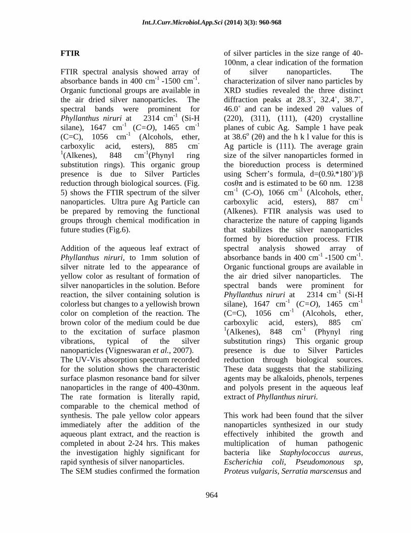
The formation of silver nanoparticles in the solution of 1mM silver nitrate and aqueous extract of the Phyllanthus niruri plant sample was confirmed by change in the color to yellowish after incubation with silver nitrate, while the controls retained the original color (Fig.1).
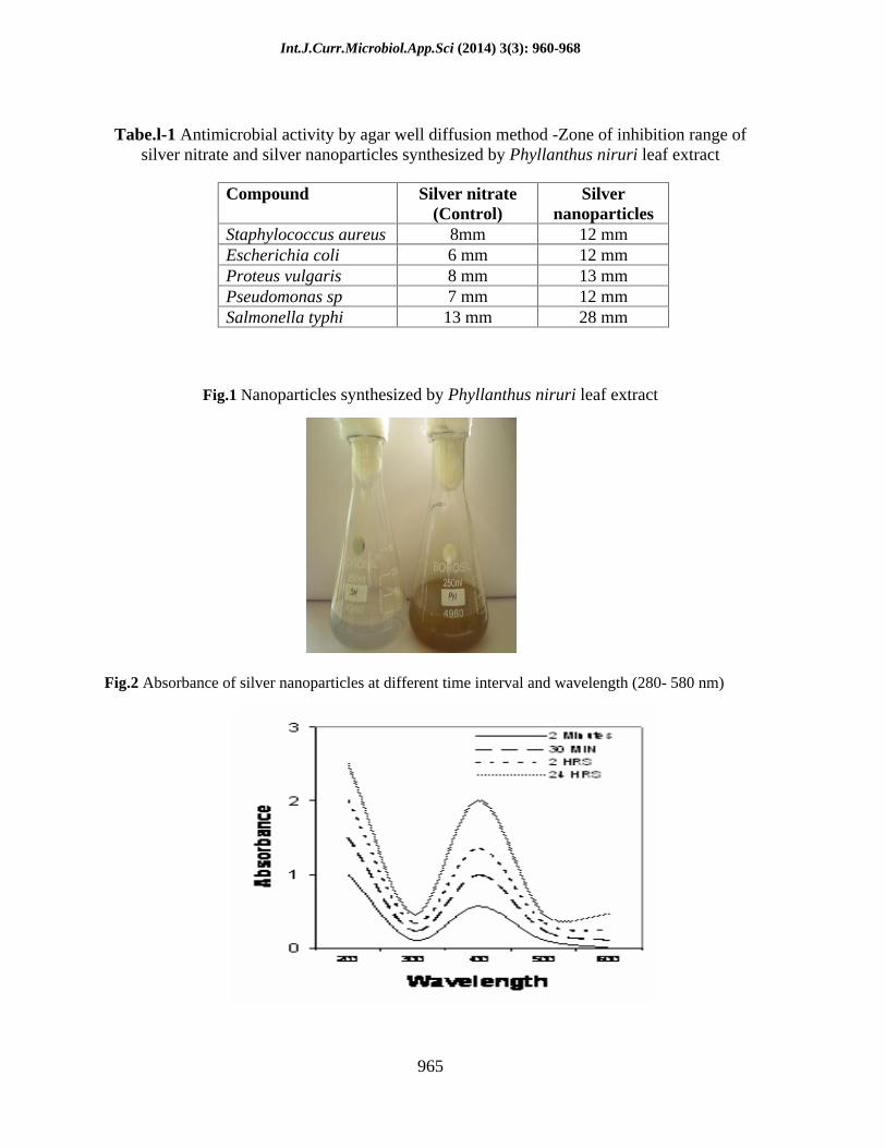
UV-Visible Spectral Analysis
The bioreduction of Ag+ in the aqueous extract was monitored by periodic sampling of the reaction mixture at regular intervals by using UV- Visible spectroscopy. The silver nanoparticles exhibits yellowish color in water and this arises due to excitation of surface plasmon vibrations in the metal nanoparticles (Mulvaney et al., 1996). The UV-Visible spectra recorded from the aqueous silver nitrate - Phyllanthus niruri leaf broth. A strong characteristic absorbance peak at around 420 nm was observed at different time intervals (Fig.2). Scanning electron microscopic analyses of the silver nano particles reduced form of silver nitrate
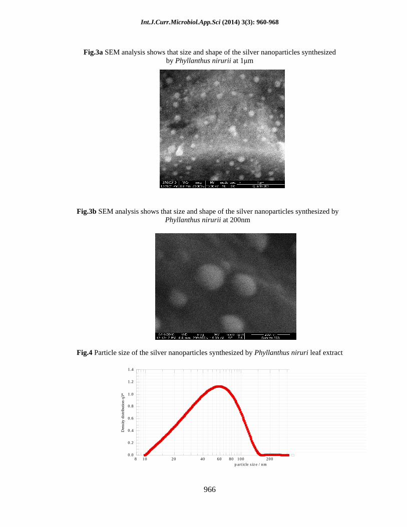
solution through bioreduction are clearly distinguishable owing to their size difference. It is clear from the SEM pictures that silver particles in the bioreduced colloidal suspensions measured 40- 70 nm in size. The particles are spherical in shape, well defined, separated as much as possible (Fig. 3a and 3b). This may be due to the reduction in liquid solution and some chelating action also available in the solution. Due to this silver particle nucleation is higher than the particle agglomeration.
Particle Size Analysis
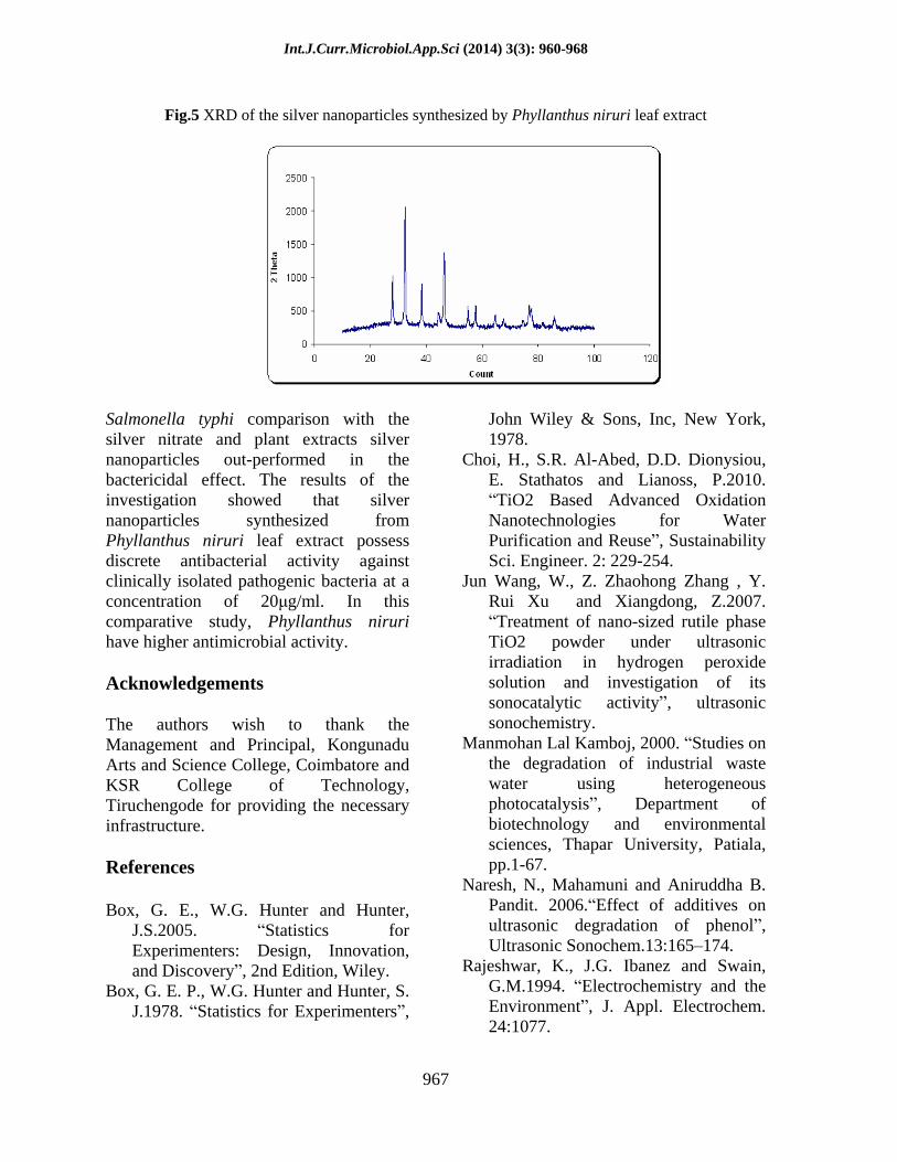
Particle size analysis shows that, when scanning from 1 nm, the particles count is very low and it s gradually reached the higher value at 63 nm and again it gradually decreased. So this indicates that the maximum nanoparticles in the range of 55 to 65 nm and only very few particles are present below and above this range (Fig. 4).
XRD Analysis
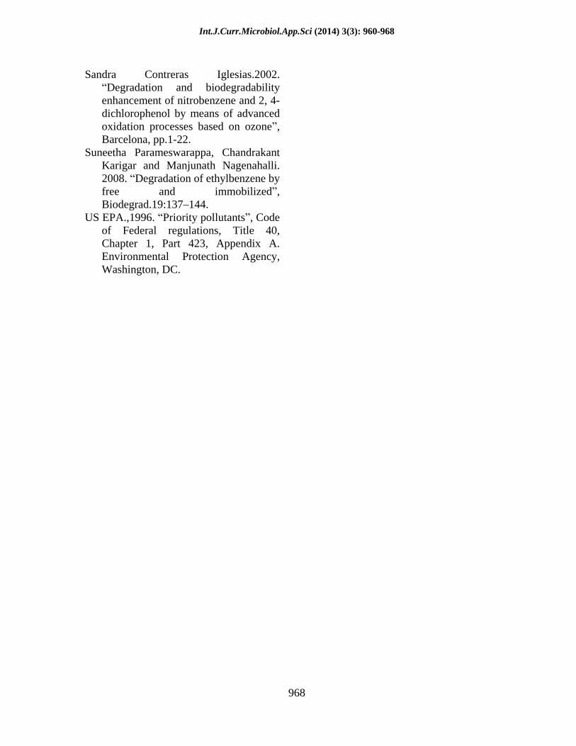
X-Ray Diffraction studies of the two samples are shown (Fig. 4). XRD analysis shows three distinct diffraction peaks at 28.3 , 32.4 , 38.7 , 46.0 and can be indexed 2 values of (220), (311), (111), (420) crystalline planes of cubic Ag. Sample 1 have peak at 38.6o (2 ) and the h k l value for this is Ag particle is (111). Corresponding JCPDS value are presented in Fig The average grain size of the silver nanoparticles formed in the bioreduction process is determined using Scherr s formula, d=(0.9 *180 )/ cos and is estimated to be 60 nm shows the XRD pattern of the silver nanoparticles formed in our experiment (Fig.5).
Int.J.Curr.Microbiol.App.Sci (2014) 3(3): 960-968
964
FTIR
FTIR spectral analysis showed array of absorbance bands in 400 cm-1 -1500 cm-1. Organic functional groups are available in the air dried silver nanoparticles. The spectral bands were prominent for Phyllanthus niruri at 2314 cm-1 (Si-H silane), 1647 cm-1 (C=O), 1465 cm-1
(C=C), 1056 cm-1 (Alcohols, ether, carboxylic acid, esters), 885 cm-
1(Alkenes), 848 cm-1(Phynyl ring substitution rings). This organic group presence is due to Silver Particles reduction through biological sources. (Fig. 5) shows the FTIR spectrum of the silver nanoparticles. Ultra pure Ag Particle can be prepared by removing the functional groups through chemical modification in future studies (Fig.6).
Addition of the aqueous leaf extract of Phyllanthus niruri, to 1mm solution of silver nitrate led to the appearance of yellow color as resultant of formation of silver nanoparticles in the solution. Before reaction, the silver containing solution is colorless but changes to a yellowish brown color on completion of the reaction. The brown color of the medium could be due to the excitation of surface plasmon vibrations, typical of the silver nanoparticles (Vigneswaran et al., 2007). The UV-Vis absorption spectrum recorded for the solution shows the characteristic surface plasmon resonance band for silver nanoparticles in the range of 400-430nm. The rate formation is literally rapid, comparable to the chemical method of synthesis. The pale yellow color appears immediately after the addition of the aqueous plant extract, and the reaction is completed in about 2-24 hrs. This makes the investigation highly significant for rapid synthesis of silver nanoparticles. The SEM studies confirmed the formation
of silver particles in the size range of 40-100nm, a clear indication of the formation of silver nanoparticles. The characterization of silver nano particles by XRD studies revealed the three distinct diffraction peaks at 28.3 , 32.4 , 38.7 , 46.0 and can be indexed 2 values of (220), (311), (111), (420) crystalline planes of cubic Ag. Sample 1 have peak at 38.6o (2 ) and the h k l value for this is Ag particle is (111). The average grain size of the silver nanoparticles formed in the bioreduction process is determined using Scherr s formula, d=(0.9 *180 )/ cos and is estimated to be 60 nm.
1238 cm-1 (C-O), 1066 cm-1 (Alcohols, ether, carboxylic acid, esters), 887 cm-1
(Alkenes). FTIR analysis was used to characterize the nature of capping ligands that stabilizes the silver nanoparticles formed by bioreduction process. FTIR spectral analysis showed array of absorbance bands in 400 cm-1 -1500 cm-1. Organic functional groups are available in the air dried silver nanoparticles. The spectral bands were prominent for Phyllanthus niruri at 2314 cm-1 (Si-H silane), 1647 cm-1 (C=O), 1465 cm-1
(C=C), 1056 cm-1 (Alcohols, ether, carboxylic acid, esters), 885 cm-
1(Alkenes), 848 cm-1 (Phynyl ring substitution rings) This organic group presence is due to Silver Particles reduction through biological sources. These data suggests that the stabilizing agents may be alkaloids, phenols, terpenes and polyols present in the aqueous leaf extract of Phyllanthus niruri.
This work had been found that the silver nanoparticles synthesized in our study effectively inhibited the growth and multiplication of human pathogenic bacteria like Staphylococcus aureus, Escherichia coli, Pseudomonous sp, Proteus vulgaris, Serratia marscensus and
Int.J.Curr.Microbiol.App.Sci (2014) 3(3): 960-968
965
Tabe.l-1 Antimicrobial activity by agar well diffusion method -Zone of inhibition range of
silver nitrate and silver nanoparticles synthesized by Phyllanthus niruri leaf extract
Fig.1 Nanoparticles synthesized by Phyllanthus niruri leaf extract
Fig.2 Absorbance of silver nanoparticles at different time interval and wavelength (280- 580 nm)
Compound Silver nitrate (Control)
Silver nanoparticles
Staphylococcus aureus 8mm 12 mm Escherichia coli 6 mm 12 mm Proteus vulgaris 8 mm 13 mm Pseudomonas sp 7 mm 12 mm Salmonella typhi 13 mm 28 mm
Int.J.Curr.Microbiol.App.Sci (2014) 3(3): 960-968
966
Fig.3a SEM analysis shows that size and shape of the silver nanoparticles synthesized
by Phyllanthus nirurii at 1 m
Fig.3b SEM analysis shows that size and shape of the silver nanoparticles synthesized by Phyllanthus nirurii at 200nm
Fig.4 Particle size of the silver nanoparticles synthesized by Phyllanthus niruri leaf extract
0.0
0.2
0.4
0.6
0.8
1.0
1.2
1.4
Den
sity
dis
trib
utio
n q3
*
8 10 20 40 60 80 100 200p art icle siz e / nm
Int.J.Curr.Microbiol.App.Sci (2014) 3(3): 960-968
967
Fig.5 XRD of the silver nanoparticles synthesized by Phyllanthus niruri leaf extract
Salmonella typhi comparison with the silver nitrate and plant extracts silver nanoparticles out-performed in the bactericidal effect. The results of the investigation showed that silver nanoparticles synthesized from Phyllanthus niruri leaf extract possess discrete antibacterial activity against clinically isolated pathogenic bacteria at a concentration of 20 g/ml. In this comparative study, Phyllanthus niruri have higher antimicrobial activity.
Acknowledgements
The authors wish to thank the Management and Principal, Kongunadu Arts and Science College, Coimbatore and KSR College of Technology, Tiruchengode for providing the necessary infrastructure.
References
Box, G. E., W.G. Hunter and Hunter, J.S.2005. Statistics for Experimenters: Design, Innovation, and Discovery , 2nd Edition, Wiley.
Box, G. E. P., W.G. Hunter and Hunter, S. J.1978. Statistics for Experimenters ,
John Wiley & Sons, Inc, New York, 1978.
Choi, H., S.R. Al-Abed, D.D. Dionysiou, E. Stathatos and Lianoss, P.2010. TiO2 Based Advanced Oxidation
Nanotechnologies for Water Purification and Reuse , Sustainability Sci. Engineer. 2: 229-254.
Jun Wang, W., Z. Zhaohong Zhang , Y. Rui Xu and Xiangdong, Z.2007. Treatment of nano-sized rutile phase
TiO2 powder under ultrasonic irradiation in hydrogen peroxide solution and investigation of its sonocatalytic activity , ultrasonic sonochemistry.
Manmohan Lal Kamboj, 2000. Studies on the degradation of industrial waste water using heterogeneous photocatalysis , Department of biotechnology and environmental sciences, Thapar University, Patiala, pp.1-67.
Naresh, N., Mahamuni and Aniruddha B. Pandit. 2006. Effect of additives on ultrasonic degradation of phenol , Ultrasonic Sonochem.13:165 174.
Rajeshwar, K., J.G. Ibanez and Swain, G.M.1994. Electrochemistry and the Environment , J. Appl. Electrochem. 24:1077.
Int.J.Curr.Microbiol.App.Sci (2014) 3(3): 960-968
968
Sandra Contreras Iglesias.2002.
Degradation and biodegradability enhancement of nitrobenzene and 2, 4-dichlorophenol by means of advanced oxidation processes based on ozone , Barcelona, pp.1-22.
Suneetha Parameswarappa, Chandrakant Karigar and Manjunath Nagenahalli. 2008. Degradation of ethylbenzene by free and immobilized , Biodegrad.19:137 144.
US EPA.,1996. Priority pollutants , Code of Federal regulations, Title 40, Chapter 1, Part 423, Appendix A. Environmental Protection Agency, Washington, DC.