Embed Size (px)
Citation preview
Dr. Ibrahim Jabber Plant groups
1
Ministry of Higher Education and
Scientific Research
University of Baghdad
College of Science
Department of Biology
Plant Groups
2020-2021
الدراسة الاولية الصباحية والمسائية
الفصل الدراسي الاول
تدريسي المادة:
محمد جاسم أ.م.د. رسل غرب . ابراهيم جابر عبد ا.م.د. لمياء عبد الزهرةأ.م.د

Dr. Ibrahim Jabber Plant groups
2
Lecture 1
Algae
The algae, it is a collective term for all those chlorophyll-bearing organisms,
which are thalloid. Pond scums, stoneworts, seaweeds and the like collectively
known as the algae. Many of them are small, and unattractive. However, they
are important members of the plant world and several of them are significant to
man in many ways. All are chlorophyll-bearing plants with a plant body
showing no differentiation into true tissue. It never form true roots, stems and
leaves and thus called a thallus. The term is used even if the plant is a
unicellular. The thallus is non-vascular and thus has no elements for transport of
fluids. The algae can afford this simplicity because with only a few exceptions
they are water dwellers. They have non- jacketed, either unicellular sex organs
or multicellular in which every cell produces a gamete. Most of them are among
the simplest in the plant kingdom. In all, there are about 30000 species. On the
basis of: (a) Thallus like non-vascular plant body. (b) Simple, unicellular
non-jacketed sex organs. (c) No embryo development after gametic union,
the algae and fungi have long been grouped together in Thallophyta. Sharma
(1987) defines algae as an assemblage of chlorophyll bearing autotrophic
thallophytes bounded by a cell wall made up of pure or mixed
carbohydrates. The study of algae is called phycology (phycos=algae, logos=
study for), phycos is a Greek word means sea weed and the references to algae
are available in the early Chinese, Roman and Greek literatures.
Habit and habitat
The algae are predominant aquatic and found in fresh or salt water forms occur
abundantly in ponds, lakes, slow flowing stream and water reservoirs. In habit,
they may be free swimming, free floating or attached to the bottom in the
shallow water. Some are terrestrial, grow in wet situations, such as, on damp
soil, damp shaded sides of trees and walls or even rocks, and thus have adapted
themselves to a life in the air. So according to the habitat, the algae may be
classified as follows:
Dr. Ibrahim Jabber Plant groups
3
1-Aquatic algae
2- Terrestrial algae
3-Aerophytes
4- Cryophytes
5- Thermophytes
6-Algae of unusual habitats
1-Aquatic algae: Majority of the algal genera is aquatic and is found either
completely submerged or free floating on the surface of water. Aquatic algae
usually occur in ponds, pools, tanks, ditches, streams or in slow running rivers
and are called freshwater forms. Marine algae are found in sea and
macroscopic large thalli of brown algae are commonly known as sea weeds.
Freshwater algal forms like Chlamydomonas, Volvox, Hydrodictyon are found
in stagnant waters, whereas Cladophora, Oedogonium, Ulothrix and few species
of Vaucheria occur in slow running water bodies. Most of the members of
Phaeophyta and Rhadophyta are found in sea either floating on the surface of
sea water or attached with rocks or on any substratum.
2- Terrestrial algae: Many algal genera are found on or beneath the moist soil
surface and are called terrestrial algae. The algal forms occurring on the surface
of soil e.g. few species of Vachuria, Botrydium and Fritschiella are called
Sapophytes.
3- Aerophytes: such algal forms are adapted for aerial mode of life and occur
on the tree trunks, moist walls, flower pots, rocks, fencing wires and get their
water and carbon dioxide requirement completely directly from atmosphere are
called Aerophytes.
4- Cryophytes: these algae are found on the mountain peaks covered with
snow and impart attractive colors to the mountains e.g., Haematococcus nivalis
gives red color to Arctic and Alpine regions.
5- Thermopytes: these algae occurring in hot springs at quite high temperature
are called thermophytes. There are certain algae which are known to tolerate the
temperature up to 85 C̊ e.g., Oscillatoria brevis.
Dr. Ibrahim Jabber Plant groups
4
6- Algae of unusual habitats: many algae are found at various interesting
places and according to their habitats may be following types:
a) Halophytes algae: these algae are found in saline water containing high
percentage of salts e.g., Chlamydomonas chrenbergii.
b) Lithophytic algae: Usually these forms of algae grow on moist rocks and
other rocky surface.blue geen algae Rivularia occur on exposed rocks.
c) Epiphytic algae: Such algal forms which grow on the other aquatic plants are
called Epiphytic algae. Example is green algae Coleochaete nitellarum
occurs on Chara and Nitella.
d) Epizoic algae: Many algae grow on the shells of mollusce, turtles and fins of
fishes and are known as epizoic algae. Cladophora is found on snails and
shells of bivalves.
e) Endozoic algae: contrary to epizoic algal forms endozoic algae found inside
the body of aquatic animals e.g., Zoochlorella is found inside Hydra viridis.
f) Parasitic algae: these forms of algae parasite on a certain organisms e.g.,
Zooxanthella is known to occur inside the fresh sponges.
g) Symbiotic algae:several members of cyanophceae grow in association with
other plants and lichen e.g., Nostoc is found within the thalli of Anthoceros.
Classification
The committee on the International Code of Botanical Nomenclature has
recommended certain suffixes for use in the classification of Algae. These are
phyta for division , phyceae for class, phycideae for subclass, ales for order,
inales for sub-order, aceae for family, oideae for sub-family, Greek name for
genus and Latin name for a species.
Algal characteristic basic to primary classification
The primary classification of algae based on certain morphological and
physiological features. The chief among these are:
a) Pigment constitution of the cell.
b) Chemical nature of stored food material.
Dr. Ibrahim Jabber Plant groups
5
c) Kind, number, point of insertion and relative length of the flagella on the
motile cell.
d) Chemical composition of cell wall.
e) Presence or absence of a definitely nucleus in the cell.
Strucutre of algal cell
The cells constituting the algal thalli are basically of two kinds: prokartotic and
eukaryotic. The prokaryotic cell which constitute thalli of cyanophytes (blue
green algae) it have a cell wall which contain a specific strengthening
component not found in the cell walls of other algae, it is called mucopeptide.
The prokaryotes cells lacking a membrane-bound nucleus, mitochondria and
plastids and do not divided by mitosis. Eukaryotes cells with a nucleus plus
typical membrane organelles and have the same structure as is typical of the
higher plants.
Algal pigments
The color of the algal thallus which varies in different classes of algae is due to
the presence of definite chemical compounds in their cells. There are called the
pigments. Each pigment has its own characteristic color. The particular color
that a thallus has is due to the predominance of one pigment in a combination of
several others. For example brown algae have predominance of fucoxanthin
and phycophein while red algae and blue green algae have excess of
phycoerythrin and phycocyanin pigments respectively. Each group has its
own particular combination of pigments and a characteristic color which is not
found in the other algae groups. Thus the natrure of the pigments present in the
algal cells forms a quick guide to the primary classification of algae into
divisions. The photosynthetic pigments in algae are of three kinds, namely:
Dr. Ibrahim Jabber Plant groups
6
chlorophylls, carotenoids and phycobilins or biliproteins. The algal
chlorophylls are characterized by green color. In solution show the phenomenon
of fluorescence and emit red light. Chlorophyll pigments are fat soluble
compounds and are five different types: chlorophyll a, b, c, d and e. out of these
chlorophyll-a is universally present in all groups of algae, chlorophyll-b is
found in Chlorophyta, Euglenophyta and Charophyta chlorophyll-c occurs in
Bacillariophyta, Pyrrophyta and Phaephyta whereas chlorophyll- d occurs in
Rhodophyta, and chlorophyll- e has been found in Xanthophyta.
Cartenoids are fat soulable yellow colored pigments and subdivided into
carotene, xanthophylls and cartenoid acids.
Phycobilins are water soluble blue (phycocyanin) and red (phycoerythrin)
colored pigments and are present in the members of Cyanophyceae and
Rhodophyceae.
Lecture 2
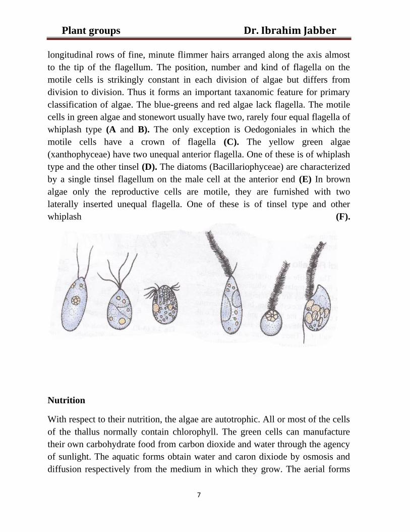
Algal flagella (Fig.1)
The motile cells of algae are provided with fine, protoplasmic, whip-like threads
called the flagella. The flagella on the cell may be equal (isokont) or unequal
(heterokont) in length. There are two main types of flagella, whiplash and
tinsel. The whiplash flagellum has a smooth surface. The tinsel flagellum bears
Dr. Ibrahim Jabber Plant groups
7
longitudinal rows of fine, minute flimmer hairs arranged along the axis almost
to the tip of the flagellum. The position, number and kind of flagella on the
motile cells is strikingly constant in each division of algae but differs from
division to division. Thus it forms an important taxanomic feature for primary
classification of algae. The blue-greens and red algae lack flagella. The motile
cells in green algae and stonewort usually have two, rarely four equal flagella of
whiplash type (A and B). The only exception is Oedogoniales in which the
motile cells have a crown of flagella (C). The yellow green algae
(xanthophyceae) have two unequal anterior flagella. One of these is of whiplash
type and the other tinsel (D). The diatoms (Bacillariophyceae) are characterized
by a single tinsel flagellum on the male cell at the anterior end (E) In brown
algae only the reproductive cells are motile, they are furnished with two
laterally inserted unequal flagella. One of these is of tinsel type and other
whiplash (F).
Nutrition
With respect to their nutrition, the algae are autotrophic. All or most of the cells
of the thallus normally contain chlorophyll. The green cells can manufacture
their own carbohydrate food from carbon dioxide and water through the agency
of sunlight. The aquatic forms obtain water and caron dixiode by osmosis and
diffusion respectively from the medium in which they grow. The aerial forms
Dr. Ibrahim Jabber Plant groups
8
obtain water from the damp substratum and carbon dioxide from the air. The
algae are also able to synthesize oil and proteins from the carbohydrates, which
they manufacture, and soluble forms of nitrogen and other minerals available in
solution in the water in which they grow.
Food reserves
The food materials, which accumulate as food reserves in the form of
polysaccharides, however, vary from group to group and thus provide useful
data for preliminary classification of algae. True starch is typical of only two
algal divisions namely, Chlorophyta and Charophyta. The two kindes of
characteristic starches are the cyanophycean starch and floridean starch. The
former is characteristic of division Cyanophyta and the latter of division
Rhodophyta. The three other important polysaccharides which accumulate as
reserve food are laminarin found in the brown algae, paramylon
characteristic of Euglenophyta and leucosin peculiar to the Xanthophyta,
Bacillariophyta and Chrysophyta. Besides, a proteinaceous compound
cyanophycin is found only in the cells of blue-green algae. Manitol which was
formerly considered to be unique to the brown algae has recently been reported
to occur in a few red algae
Reproduction in algae
Reproduction actually means the production of young ones like the parent.
There are two types of reproduction, asexual and sexual.
i) Asexual reproduction: it consists in the separation from the parent of a
highly specialized cell or a group of cells which directly develops into a new
individual resembling the parent. This type of reproduction take place in a
variety of ways:
Dr. Ibrahim Jabber Plant groups
9
1- Spore formation: the spore are reproductive units specialized for asexual
reproduction. Each unit can grow into a new organism by itself. The spores are
usually one-celled structure and formed by most of algae. They are produced
either within ordinary vegetative cells (Chlamydomonas, Ulothrix) or in
specially modified cells (Oedogonium,Vaucheria, Ectocarpus) called
sporangia. Each sporangium may produce a single large spore or more than
small spores. The spore may be motile or non-motile. They are able grow into
new individuals without fusion. When motile they are called the zoospores or
planospores. The non-motile spores are called the aplanospores. There are
many types of spores, the most important ones are:
a) Zoospores: formation of zoospores is the most characteristic method of
asexual reproduction in most of the green algae.
b) Aplanospores: they are non-motile and constitute a normal means of
asexual reproduction in the terrestrial species of the yellow green algae e.g.,
Vaucheria. In some green algae these spores produced under certain unusual
conditions (Ulothrix and Microspora).
c) Hypnospores: Under certain extreme environmental condition the
aplanospores in green algae secrete thicker walls around them and they called
hypnospores. The hypnospores germinate into new plants with the return of
suitable conditions for vegetative growth.
d) Akinetes: they are resting cells which mainly serve as means of
perennation rather than multiplication. During the sever conditions the cell
wall of akinetes considerably thickened. It highly resistant. The akinete contain
abundant reserve food. With the return of suitabll conditions the akinete
develop into new filaments.
e) Carpospores: spores produced directly in the carpogonium after
fertilization of the egg or directly from the cells of the filament following
fertilization are called carpospores. These carpospores are common in
polysiphonia and other red algae.
f) Tetraspores: in brown alga (phaeophyta) non-motile spores known as
tetraspores are produced in specialized cells known as tetrasporangia.
g) Pseudohormocytes: are clusters of cells formed terminally on erect
branches of westiellopsis- blue green alga. These are formed after repeated
transverse and longitudinal divisions of terminal cells and the content escape
as gonidia.
Dr. Ibrahim Jabber Plant groups
10
2- Fission: it is the simplest and the chief method of vegetative reproduction in
many one-celled green and blue green algae. It is simple division of a
unicellular alga into two new daughter cells.
3- Fragmentation: it is the breaking away of a few or many celled segments of
a filament. Such bits of living cells are called the fragments. The algae
(Spirogyra and Zygnema). The filamentous blue-green algae form specialized
fragments called the hormogonia. Hormogonia formation it is specialized
process of vegetative propagation characteristic of the Cyanophyta.
4- By the formation of adventitious branches.
5- By the formation of tubers.
6- Budding.
ii) Sexual reproduction: it involves the fusion of two specialized reproductive
cells called the gametes. Fusion may occur between two vgametes from the
same plant (monoecious) or from different plants (diocious). The process of
fusion called the fertilization. And the product of fusion called the zygote.
Sexual reproduction is absent in the blue-green bhb valgae. In general, it is of
two main types, isogamous and heterogamous.
a) Isogamy: It is the simplest and the most primitive type of sexual
reproduction. It is usually found among the lower or simple forms. It consists in
the fusion of morphologically identical gametes, which they are similar in size
and structure and are called the isogametes. The algae exhibiting isogamous
type of sexual reproduction are usually monoecious. The act of fusion in
isogamy is called conjugation and the product of fusion called zygospore.
b) Heterogamy: it is a more advanced type of sexual reproduction as compared
with isogamy. It includes the fusion of dissimilar gametes. The fusing gametes
differ in size, structure and physiology. The act of fusion in this type of sexual
reproduction is called fertilization and the product of fusion is termed the
zygote. Heterogamous sexual reproduction is of two types: anisogamous and
oogamous.
1- Anisogamous: It involves the fusion of gametes which are dissimilar and are
produced in modified vegetative cells called the gametangia. The larger,
passive gametes is called the female.
Dr. Ibrahim Jabber Plant groups
11
2- Oogamy: it is the highest and the most advanced stage of sexual
reptoduction. It is generally characteristic of the filamentous forms of algae.
The gametes are produced in special sexual organs which are very sharply
differentiation from ordinary vegetative cells. The female sex organ is enlarged,
it is called oogonium. While the male sex organ is smaller in size and is called
the antheridium. It produces one or more male cells or gametes furnished with
flagella. Exception being the red algae which exhibit oogamous sexual
reproduction of a very specialized kind. The male gametes are non-motile and
are called the spermatia. It is non –flagellated and produced singly in the male
sex organ called the spermatangium. The female sex organs called
carpogonium.
Lecture 3
Division: Cyanophyta
This division includes the blue-green algae which are the only known
oxygen producing prokaryotes. It is a small primitive group comprising of
about 2500 species placed under 150 genera. All of them are included in a
single class Cyanophyceae Sachs, 1874, or Myxophyceae Wallroth, 1833. The
members of this class are considered the simplest, living autotrophic plants.
Individually all blue-greens are microscopic.
Dr. Ibrahim Jabber Plant groups
12
Distribution
The blue-green algae are widely spread in the aquatic environment. Some
terrestrial species are also known. The aquatic forms mostly occur in
freshwater; a few however, are marine. A notable example of the marine blue-
greens is Trichodesmium erythraeum in which the red pigment predominates.
This species flourishes in the Sea Red and is responsible for the red color of its
water. The drastic increase in their number due to rapid algal growth and their
buoyancy result in the sudden appearance of large, dense algal population in
calm weather usually in the summer in the temperate regions and any time of
the year in the tropics. This accumulation or rapid appearance of planktonic blu-
green algae at the surface of lakes, ponds, and reservoirs of freshwater, which
attracts attention, is termed as algal bloom. This phenomena caused by
Eutrophication, The process by which a body of water acquires a high
concentration of nutrients, especially phosphates and nitrates. These typically
promote excessive growth of algae. As the algae die and decompose, high levels
of organic matter and the decomposing organisms deplete the water of available
oxygen, causing the death of other organisms, such as fish.
Some species of blue-greens occure as scums on dirty stagnant water full of
decaying organic materials. There is s sufficient number of thermal
Cyanbacteria. They grow on snow, hot springs which they are able to live in
water whose temperature is as high as 85C̊. The Cyanophyta thus provide good
example of adaptability of life to extreme of environments (high temperature of
hot springs and low temperature of Polar Regions). Because of their gelatinous
sheaths, they can withstand long periods of desiccation. The compactness of
protein molecules and their bonds in the protoplasm also help the cells to
face the extremes. Certain blue-greens lives intimately with other organisms as
Dr. Ibrahim Jabber Plant groups
13
symbionts. For examples, colonies of certain blue-greens grow in the root of
Cycas and within the thallus of Anthoceros. Anabaena occurs in cavities in
the fronds of Azolla (a water fern). Certain Cyanobacteria are associated with
the fungi in the formation of Lichen.
Organization of the thallus
The blue-green algae amongst the simplest photosynthetic plants living today.
Because of the variation in their habitats, so they have a range of vegetative
structure. Architecturally, the thallus may be a solitary cell or a colony.
1-Unicellular forms.
The thallus, in some species, is a unicellular which is usually spherical or oval
(Chroococcus, Synechococcus, Anacystis and Gloeocapsa).
2- Colonial forms.
In most blue-greens, the cells after division remain attached by their walls or are
held in a common gelatinous matrix to form a loose organization of cells, which
is termed a colony. The cells in the colonies are often aggregated into irregular,
palmelloid forms of great variability. Gloethece is an example of an aggregation
of a few cells. Microcystis are example of aggregation of numerous cells. The
colonies may be either filamentous or non-filamentous. Each colony is
generally enclosed in a gelatinous sheath.
Dr. Ibrahim Jabber Plant groups
14
a) Filamentous colonies: the filamentous colony is the result of repeated cell
division in a single plane and in a single direction forming a chain or thread, it
known as the trichome. The cells in the trichome may be held together by
either separation walls or a common gelatinous sheath around it. The trichome
with its enclosing sheath is called a filament, which either without heterocyst
and called filamentous non-heterocystous such as Lyngbya and Oscillatoria.
Or with heterocystous and called filamentous heterocystous such as Nostoc
and Anabaena
b) Non-filamentous colonies: the non-filamentous colonies are various forms.
They may be cubical, square or irregular depending on the planes and direction
in which cell divide.
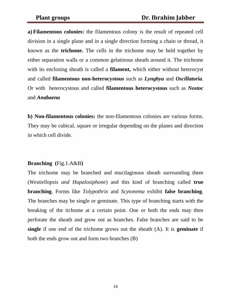
Branching (Fig.1.A&B)
The trichome may be branched and mucilaginous sheath surrounding them
(Westiellopsis and Hapalosiphone) and this kind of branching called true
branching. Forms like Tolypothrix and Scytonema exhibit false branching.
The branches may be single or geminate. This type of branching starts with the
breaking of the tichome at a certain point. One or both the ends may then
perforate the sheath and grow out as branches. False branches are said to be
single if one end of the trichome grows out the sheath (A). It is geminate if
both the ends grow out and form two branches (B)
Dr. Ibrahim Jabber Plant groups
15
Fig.1 (A-B). Scytonema. False branching;
A) Trichome with one false branch;
B) Trichome with two false branches.
Pigments
There is chlorophyll-a (but not chlorophyll-b) and a number of other pigments
such as β-carotene, xanthophylls and phycobilins. The best known phycobilins
are blue c-phycocyanin and red c-phycoerythrin. These two pigments are
unique to the cyanophytes and are not found in the other algal groups.
Differentiation of cells
The blue-green algae, in general show very little differentiation of cells.
However, the trichomes of certain filamentous genera show differentiation of

Dr. Ibrahim Jabber Plant groups
16
vegetative cells to either heterocysts or akinetes which possess special structural
and biochemical properties. The heterocyst differ from the vegetative cells occur
between them along the length of trichome with characteristic cellular spacing.
Heterocysts: the heterocysts differ markedly from vegetative cells and occur
between them along the length of the trichome at some regular intervals.
Heterocysts can be easily distinguished from the vegetative cells of the
trichome by their: 1- larger size, 2-thicker walls, 3- homogenous
transparent, pale yellowish content and distinct pore either at one or at
both ends.
Akinetes: specialized thick-walled resistant cells formed generally in some
heterocystous blue green algae. They are larger than vegetative cells. These
spore-like cells contain the entire protoplast of the cell enclosed by the original
parent cell wall and the thick spore envelope. The vegetative cell increase several
times in size and accumulates food reserves (cyanophycin). This is accompanied
by the secretion of a thick multi-layered spore wall envelope surrounding the
parent cell wall.
Reproduction
a- Sexual reproduction: formation of sex organs, gametes differentiation,
gametic union or formation of zygotes has not been observed in the
Cyanphyceae.
b- Asexual reproduction: it takes place by vegetative methods and by spore
formation.
1- Vegetative methods:- which includes as follows:
• Cell division or fission.
Dr. Ibrahim Jabber Plant groups
17
• Fragmentation.
• Hormogonia formation: this take place by two ways:
i-intercalary heterocysts
ii-by the formation of separation discs
2- Spore formation:-
• Endospores
• Akinetes
• Exospore
Economic importance
1- The blue-green algae furnish food for fish and other aquatic animals,
Oscillatoria is the most favourable blue-green alga consumed by 56
species of fishes.
2- The blue-green algae add organic matter to the soil and increase fertility.
3- The blue-green algae consider as nitrogen fixers. These algae have ability
to fix the atmospheric nitrogen. For examples: Nostoc, Anabaena and
Sytonema have high ability to fix nitrogen.
4- Some species of cyanophyceae such as Spirulina consider as a main
source for protein.
Dr. Ibrahim Jabber Plant groups
18
Spirulina health products
5-some species of cyanophyceae (Microcystis aeruginosa, Aphanizomenon and
others) produce toxins such as microcystin, neurotoxin that are poisonous to
human beings, animals (fish, cattle, sheep and other domesticated animals).
Lecture 4
Division: Chlorophyta
The division chlorophyta more appropriately called Chlorophycophyta includes
a large number of species. Prescott makes as many as 20000 species. There are
included in a single class Chlorophyceae. The Chlorophyta and the
Chlorophyceae have the same features. The cells consistuting the thallus are
eukaryotic and thus contain all the membrane bound cell organelles such as
definitely organized nucleus, plastids, mitochondaria, dictyosomes, endoplasmic
reticulum, and true vesicles. The thallus is typically green in colour due to the
presence of a green pigment called the chlorophyll which is contained in
plastids called the chloroplasts. Embedded in the chloroplasts are rounded,
proteinaceous bodies one or more in number, the pyrenoids. The pyrenoids are
intimately associated with the elaboration of starch, which is the principal
storage product. The reserve carbohydrate are usually stored in the form of
Dr. Ibrahim Jabber Plant groups
19
starch. The cell wall contains cellulose. Unlike the blue-green most of the green
algae produce motile reproductive bodies generally furnished with two to four
flagella. The flagella are of equal length and of whiplash. Occurance of sexual
reproduction is another feature, which distinguishes the Chlorophyceae from the
Cyanphyceae.
Occurrence
Most of Chlorophyceae are aquatic but some are subaerail. These forms are
generally found in moist situations. The aquatic forms , majority occur in fresh
cold water. They occur both in standing and flowing waters and may be
attached or planktonic (free-floating).they either form greenish scum on the
surface of quiet or stagnant water or grow firmly attached to the submerged
rocks, pieces of wood and other objects in water. Several species of the orders
Ulvales and Siphonales are marine. Very few parasitic forms are known to
occur among the green algae. Only one genus has been reported to cause
considerably economic loss to crops of tea and coffee. This is Cephaleuros
belonging to the order Chetophorales. Cephaleuros virescens is the cause of
red rust of tea in north east India.
Organization of thallus
The Chlorophyceae are a heterogenous group of plants exhibiting a wide range
of the body architecture. The plant body may be single-celled or many-celled
and the size it varies from minute unicells from one or two micron in diameter
to a few feet long, strand like structure. The plant body of algae called a thallus
because it shows no differentiation into true root, stem and leaves.
Dr. Ibrahim Jabber Plant groups
20
Cell wall structure
The protoplasmic content of the cell are bound by a definite cell wall, which is
typically stable in the green algae. It is a secretion product of the cell protoblast
deposited in the form of two concentric layers: the inner layer is composed
cellulose, while the outer layer is made of pectose.
Reproduction
In green algae, it takes place by asexual and sexual.
1. Asexual reproduction:
a- Vegetative reproduction
Propagation of the species by any method which uses only vegetative cells, is
known as vegetative reproduction. In this method, the parent cell wall is
retained. It may take place by:
i. Cell division.
ii. Fragmentation.
iii. Akinate formation.
b- Spore formation
It involves the multiplication of the species by the formation of highly
specialized reproductive cells called the asexual spores. Reproduction by
asexual spores is a very common method of propagation under normal
conditions of the life and is often called sporulation.
In green algae, the asexual spores are usually produced by the haploid plants
and thus are genetically haploid, they are usually produced endogenously in
Dr. Ibrahim Jabber Plant groups
21
more or less specialized cells called the sporangia and are differentiated by
mitosis from the protoplast of the sporangium and call the asexual spores as
mitospores in order to distinguish them from another kind of spores which are
differentiated by meiosis and are called the meiospores. The mitospores are
asexual spores. They may be motile or non-motile. The former are called
zoospores and the latter aplanospores.
2- Sexual reproduction
This kind of reproduction involves the fusion of two specialized reproductive
cells or units called the gametes.
In some algae the two fusing gamates may be produces by the same thallus and
in others by two separate thalli. The former are called monoecious or
homothallic species and the latter dioecious or hererothallic species. Sexual
reproduction always entails three steps namely formation of gametes, fusion of
gametes (syngamy or fertilization) and germination of zygote.
Life cycle of algae
Alternation of generations in green algae
In the single life cycle of certain sexually reproducing plants, there occurs two
individuals. One of these is sporophyte. It is characterized by the diploid
number of chromosomes in the nuclei of its cell. The diploid sporophyte is
concerned with the production of haploid spores called the meiospores.
They are differentiated by meiosis. The other individual is the gametophyte. It
characterized by haploid number of chromosome in the nuclei of its cell. It
responsible for sexual reproduction, which it bears the haploid gametes. The
two individuals normally follow each other in a single life cycle. This is called
Dr. Ibrahim Jabber Plant groups
22
alternation of generations. It means the alternation in the life cycle of two
distinct individual having not only different chromosome numbers but different
functions as well.
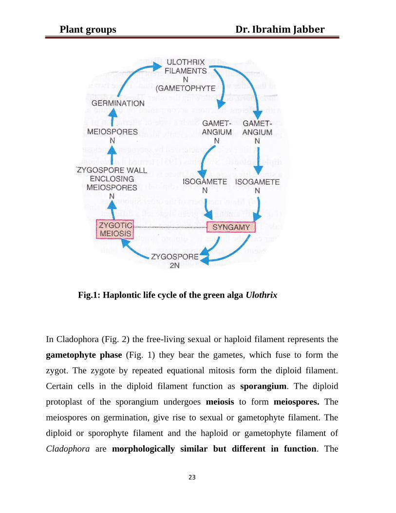
In the majority of the green algae such as Ulothrix the thallus (filament) is
haploid. It bears the gametes fuse to form the zygospore, is a diploid structure.
The diploid zygospore nucleus, prior to germination, undergoes meiosis. As a
result four haploid daughter nuclei are formed. Three of these disintegrate. The
surviving functional daughter nucleus becomes the nucleus of the first cell of
the new haploid or gametophyte filaments. The zygospore, therefore, is the only
diploid structure in the life cycle of these green algae, which represents a
simple, one-celled sporophyte. This results in the alternation of chromosome
numbers from haploid to diploid and back to haploid stage. There is no
alternation of individuals with different functions. A life cycle characterized
by a haploid thallus, and zygotic meiosis is called haplontic life cycle (Fig. 1).
Dr. Ibrahim Jabber Plant groups
23
Fig.1: Haplontic life cycle of the green alga Ulothrix
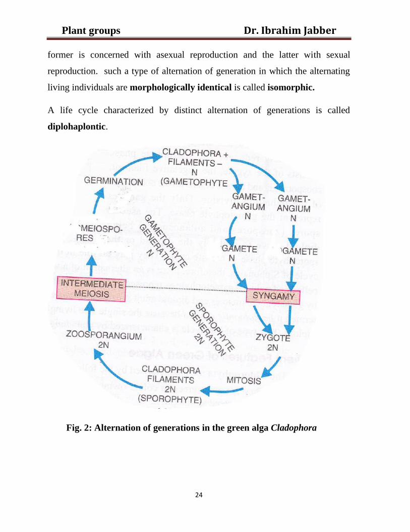
In Cladophora (Fig. 2) the free-living sexual or haploid filament represents the
gametophyte phase (Fig. 1) they bear the gametes, which fuse to form the
zygot. The zygote by repeated equational mitosis form the diploid filament.
Certain cells in the diploid filament function as sporangium. The diploid
protoplast of the sporangium undergoes meiosis to form meiospores. The
meiospores on germination, give rise to sexual or gametophyte filament. The
diploid or sporophyte filament and the haploid or gametophyte filament of
Cladophora are morphologically similar but different in function. The
Dr. Ibrahim Jabber Plant groups
24
former is concerned with asexual reproduction and the latter with sexual
reproduction. such a type of alternation of generation in which the alternating
living individuals are morphologically identical is called isomorphic.
A life cycle characterized by distinct alternation of generations is called
diplohaplontic.
Fig. 2: Alternation of generations in the green alga Cladophora
Dr. Ibrahim Jabber Plant groups
25
Economic importance
1- Some of the green algae constitute an important source of food for fish
and other aquatic animals.
2- The sea lettuce (Ulva) a marine algae used in salad and in soups,
Spirogyra and Oedogonium eaten directly by certain people..
3- Chlorella yields an antibiotic, Chlorellin.
4- Certain green algae as a source of biofuel, such as Chlorella vulgaris,
Botrycoccus brunii (characterized by a considerable production of lipids,
notably hydrocarbons) and Nanochloropsis oculata.
5- Green algae play important role in Phytoremidiation (biological
treatment by plant) for heavy metals, organic matters.
Dr. Ibrahim Jabber Plant groups
26
Phaeophyta (Brown algae) Lecture 5
General features
The brown algae are distinguish by their colour, which varies from olive green
through light golden to a rather deep shade of brown. This is due to the presence
of a golden brown xanthophyll pigment fucoxanthin. This is in addition to
chlorophyll a, chlorophyll c and carotens. The fucoxanthin, however, is
usually sufficiently strong to partially mask the chlorophyll and carotenoid
pigments in the cells. Typically the cells are uninucleate. The large thalli are
tough, leathery or rubbery in texture. Usually the thalli of brown algae secrete
abundant mucilage which readily absorbs moisture and retain it
tenaciously. This keeps the plants moist at low tide when they are exposed.
Mannitol and laminarin are the reserve photosynthetic products. Mannitol it is
a kind of complex alcohol widely distributed in the cells of almost all the brown
algae. The simple sugars are converted into mannitol type alcohol, therefore the
sugars are rare in Phaeophyceae. Starch is entirely absent. The plant body is
always immobile and multicellular.
The Phaeophyceae or brown algae, are a large group of mostly marine
multicellular algae, including many seaweeds. They play an important role in
marine environments, both as food and as habitats. For instance, Macrocystis, a
kelp of the order Laminariales, may reach 60 m (200 ft) in length and forms
Dr. Ibrahim Jabber Plant groups
27
prominent underwater kelp forests. Kelp forests like these contain a high level
of biodiversity. Another example is Sargassum, which creates unique floating
mats of seaweed in the tropical waters of the Sargasso Sea that serve as the
habitats for many species. Many brown algae, such as members of the order
Fucales, commonly grow along rocky seashores. Some members of the class,
such as kelp, are used as food for humans.
Motile reproductive cells are commonly found in the brown algae, generally
these are pyriform or spinle-shaped and biflagellate. The two flagella are of
unequal lengths and inserted laterally, one of these is whiplash type and the
other tinsel. Sexual reproduction ranges from isogamy to oogamy through
anisogamy. Oogamy, however, is the general rule. Altenation of generation is
frequently present.
Distribution
Unlike the Cyanophyceae and the Chlorophyceae, which are mainly fresh water
forms, the brown algae are almost exclusively marine. There are only few
rare ones, which are fresh water and usually grow in the streams that drain
directly into the ocean. The marine forms are found in the shallow waters along
the coasts of all seas but they attain their greatest development both in number
and large size in the cold waters of oceans and seas of northern latitudes.
They are benthonic and grow as lithophytes attached by holdfasts to the rocks,
stones or timbers beneath the surface. They usually develop air bladders to buoy
up the free portions of their thalli. At low tide they are exposed to air for several
hours. The algin coating protects them from desiccation by retaining
sufficient amount of water.
Dr. Ibrahim Jabber Plant groups
28
Range of thallus organization
All are multicellular and sessile. They have a definite form and are both the
largest and the most rugged of the algae.
They display the highest degree of body differentiation. Unicellular and
colonial forms (motile and non-motile) are unknown at present. The
unbranched filament so common in the Chlorophyceae has also not been
reported. The simplest type of the plant body in the brown algae is represented
by a heterotrichous filament which is the highest stage of development in
the green algae. Morphologically therefore the brown algae begin where
the green algae finish.
Visible structures
Brown algae exhibit marked morphological and anatomical complexity. The
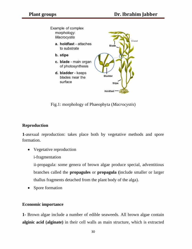
plant body consist of a forked, flattened, band-shaped upper part known as the
blade (photosynthetic and reproductive structures), a more or less
cylindrical lower part called the stipe and holdfast is a rootlike structure present
at the base of the alga (Fig.1).
A holdfast Like a root system in plants, a holdfast serves to anchor the alga in
place on the substrate where it grows, and thus prevents the alga from being
carried away by the current. Unlike a root system, the holdfast generally does
not serve as the primary organ for water uptake, nor does it take in nutrients
from the substrate. The overall physical appearance of the holdfast differs
among various brown algae and among various substrates. It may be heavily
branched, or it may be cup-like in appearance.
A stipe is a stalk or stemlike structure present in an alga. It may grow as a short
structure near the base of the alga (as in Laminaria), or it may develop into a
Dr. Ibrahim Jabber Plant groups
29
large, complex structure running throughout the algal body (as in Sargassum or
Macrocystis). In the most structurally differentiated brown algae (such as
Fucus), the tissues within the stipe are divided into three distinct layers or
regions. These regions include a central pith, a surrounding cortex, and an outer
epidermis, each of which has an analog in the stem of a vascular plant. In some
brown algae, the pith region includes a core of elongated cells that resemble the
phloem of vascular plants in both structure and function. In others (such as
Nereocystis), the center of the stipe is hollow and filled with gas that serves to
keep that part of the alga buoyant. The stipe may be relatively flexible and
elastic in species like Macrocystis pyrifera that grow in strong currents, or may
be more rigid in species like Postelsia palmaeformis that are exposed to the
atmosphere at low tide.
The terms blade and frond. The name blade is most often applied to a single
undivided structure, while frond may be applied to all or most of an algal body
that is flattened, but this distinction is not universally applied. The surface of the
blade may be smooth or wrinkled; its tissues may be thin and flexible or thick
and leathery. In species like Egregia menziesii, this characteristic may change
depending upon the turbulence of the waters in which it grows. In other species,
the surface of the blade is coated with slime to discourage the attachment of
epiphytes or to deter herbivores. Blades are also often the parts of the alga
that bear the reproductive structures.
Dr. Ibrahim Jabber Plant groups
30
Fig.1: morphology of Phaeophyta (Macrocystis)
Reproduction
1-asexual reproduction: takes place both by vegetative methods and spore
formation.
• Vegetative reproduction
i-fragmentation
ii-propagula: some genera of brown algae produce special, adventitious
branches called the propagules or propagula (include smaller or larger
thallus fragments detached from the plant body of the alga).
• Spore formation
Economic importance
1- Brown algae include a number of edible seaweeds. All brown algae contain
alginic acid (alginate) in their cell walls as main structure, which is extracted
Dr. Ibrahim Jabber Plant groups
31
commercially and used as an industrial thickening agent in food and for other
uses. One of these products is used in Lithium Ion batteries. Alginic acid is
used as a stable component of a battery anode. This polysaccharide is a major
component of brown algae.
2- Certain brown algae (Laminaria, Fucus) are a source of commercial
iodine. High concentrations of iodine, potassium, magnesium and other solutes
from seawater accumulate in the cells of brown algae.
3- Brown algae including kelp also fix a significant portion of the earth's carbon
dioxide yearly through photosynthesis.
4- Sargachromanol G, an extract of Sargassum, has been shown to have anti-
inflammatory effects.
Rhodoophyta (red algae) Lecture 6
General features and distribution
Dr. Ibrahim Jabber Plant groups
32
A majority of the seaweeds are red algae, and there are more Rhodophyceae
(about 4000 species) than all of the other major seaweed groups combined.
Although marine red algae occur at all latitudes, there is a marked shift in their
abundance from the equator to colder seas. There are few species in polar and
sub Polar Regions where brown algae predominate, but in temperate and
tropical regions they far outnumber these groups. The average size of the
plants differs according to geographical region. The larger species of fleshy red
algae occur in cool-temperate areas, whereas in tropical seas the Rhodophyceae
(except for massive calcareous forms) are mostly small, filamentous plants. The
Rhodophyceae also have the ability to live at greater depths in the ocean than do
members of the other algal classes. They live at depths as great as 200 m, an
ability related to the function of their accessory pigments in photosynthesis.
About 200 species of Rhodophyceae are found in fresh water, where they do not
reach as great a size as the red sea weeds. The majority of fresh water red algae
occur in running waters of small to mid-sized streams.
The Rhodophyceae lack flagellated cells have chlorophyll a, phycobiliproteins,
floridean starch as a storage product, and thylakoids occurring singly in the
chloroplast.
Morphology
In fact, they are the only eukaryotic algae, which produce no motile stage. Even
the reproductive cells are non-flagellated. Unicellular and colonial forms are
rare. Porphyridium is the common unicellular red algae. With the exception of
two genera, all red agae have a multicellular macroscopic
Dr. Ibrahim Jabber Plant groups
33
thallus which is very diverse in form. It may be filamentous (Goniotrichum),
ribbon-like or plate parenchymatous (poryphyra). The filamentous thallus is
most beautiful of all the algae. It is mostly branched and tufted.
Cell structure
The major features of a red algal cell are a chloroplast with one thylakoid per
band. floridean starch grains in the cytoplasm outside the chloroplast, no
flagella, pit connections between cells are present.
Pit connections
The higher orders of red algae are unique in having pits on the cell walls of
adjacent cells. These pits assist in maintaining protoplasmic continuity
allowing the passage of metabolites to the developing reproductive cells
between these cells. The pit connection may function as a site of structural
strength on the thallus.
A pit connection consists of a proteinaceous plug core in between two thallus
cells. There are two types of pit connections. Primary pit connections are
formed between two cells during cell division. Secondary pit connections
result when two cells fuse. Both types of pit connections have the same
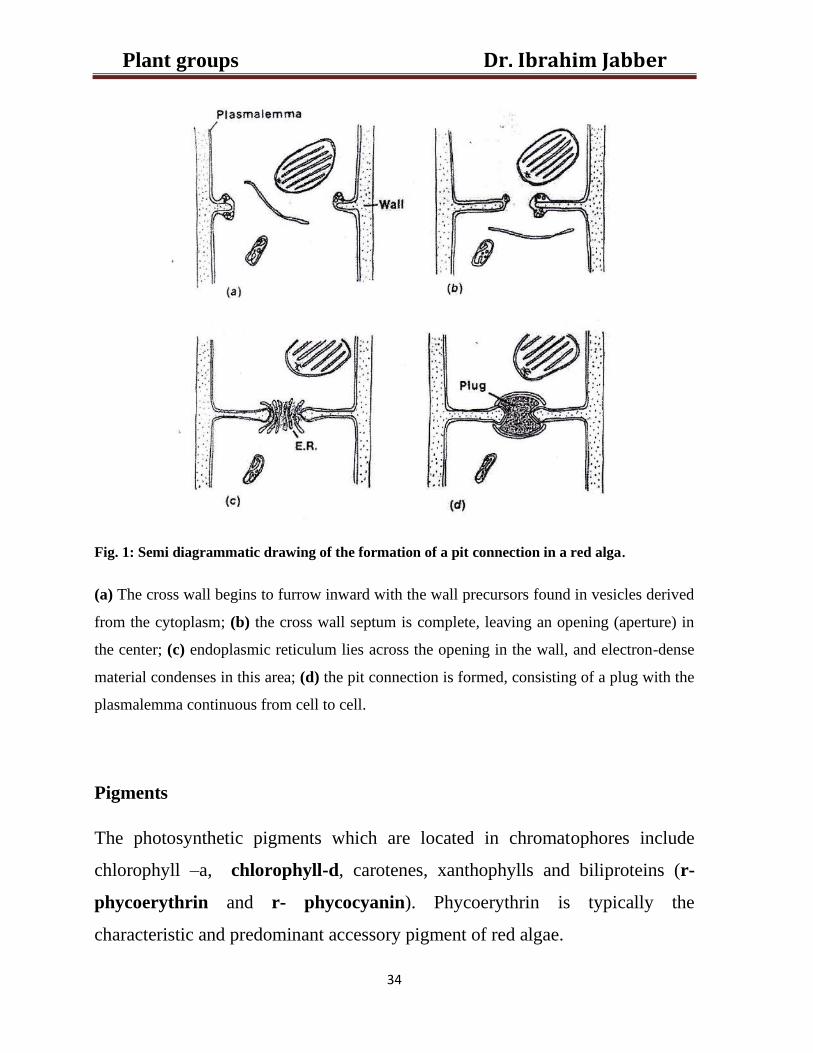
structure. Fig. 1 shows the formation of a pit connection in a red alga.
Dr. Ibrahim Jabber Plant groups
34
Fig. 1: Semi diagrammatic drawing of the formation of a pit connection in a red alga.
(a) The cross wall begins to furrow inward with the wall precursors found in vesicles derived
from the cytoplasm; (b) the cross wall septum is complete, leaving an opening (aperture) in
the center; (c) endoplasmic reticulum lies across the opening in the wall, and electron-dense
material condenses in this area; (d) the pit connection is formed, consisting of a plug with the
plasmalemma continuous from cell to cell.
Pigments
The photosynthetic pigments which are located in chromatophores include
chlorophyll –a, chlorophyll-d, carotenes, xanthophylls and biliproteins (r-
phycoerythrin and r- phycocyanin). Phycoerythrin is typically the
characteristic and predominant accessory pigment of red algae.
Dr. Ibrahim Jabber Plant groups
35
The red algae have the ability for self-adaptation for light intensity. Therefore,
these algae could live in the surface water or down in the benthic water.
However, in benthic position, the phycoerythrin will be increased and so it will
be decreased on the surface water which the green pigment chlorophyll will
increased. This phenomenon called the chromatic adaptation. Phycoerythin
reflects red light and absorbs blue light (bluish portion of the spectrum has the
shortest wavelength and the highest energy. So, this bluish component is able to
penetrate deep down in the ocean depths).Because blue light penetrates water to
a greater depth than light of longer wavelengths, these pigments allow red algae
to photosynthesize and live at somewhat greater depths than most other algae.
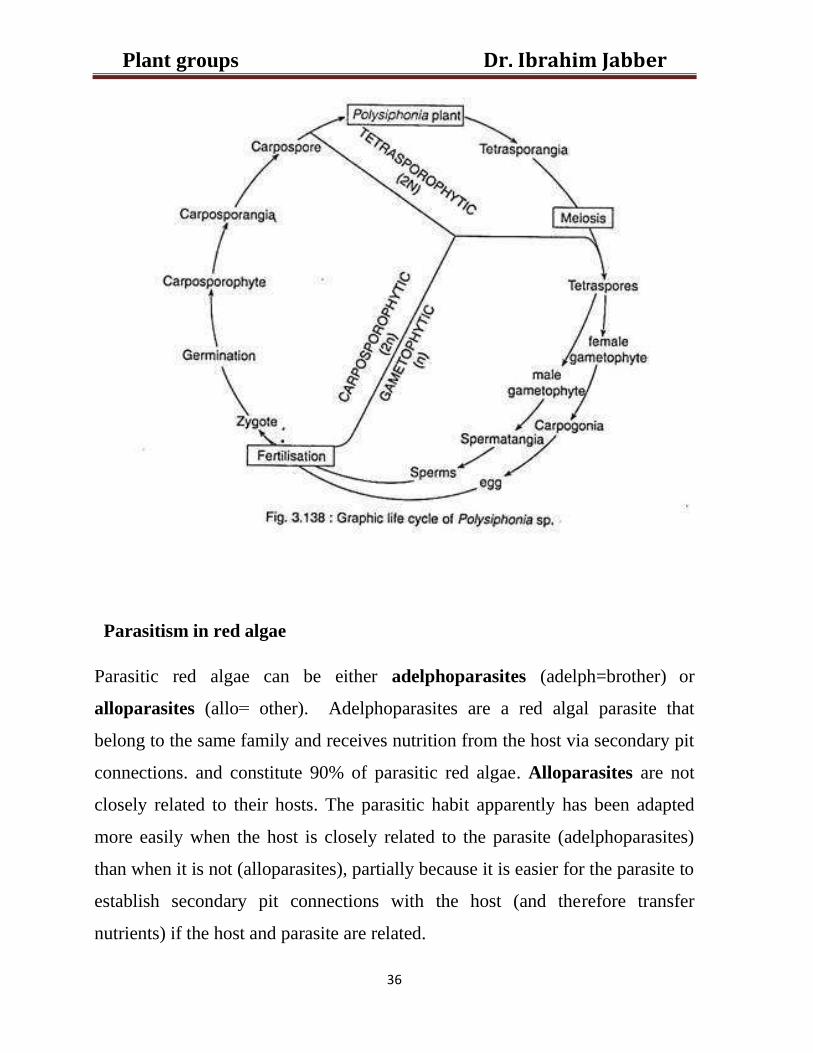
Reproductive structures
The Rhodophyceae have no flagellated cells. In the life cycle of Polysiphonia
occur three separate kinds of plants. These are the gametophyte, the
carposporophyte and the tetrasporophyte. The gametophyte is a free living
haploid plant.it is concerned with the sexual reproduction, the carposporophyte
is the diploid plant developed from the zygote and it remains attached to the
female gametophyte plant on which it is parasitic. The carposporophyte is
concerned with the production of diploid spores called the carpospores which
each carpospores germinated to give rise to the tetrasporophyte. The
tetrasporophyte is independent plant like the gametophyte. It is sexless and
concerned with the production of the haploid tetraspores. The haploid
gametophyte and the diploid tetrasporophyte plants are similar in their
vegetative structure but can be distinguished by the differentreproductive organs
(Fig. 2). spermatia (spherical or oblong cells produced in spermatangium,
which represents the male organ) which are carried passively by water currents
to the female organ, the carpogonium which contain the zygote
Dr. Ibrahim Jabber Plant groups
36
Parasitism in red algae
Parasitic red algae can be either adelphoparasites (adelph=brother) or
alloparasites (allo = other). Adelphoparasites are a red algal parasite that
belong to the same family and receives nutrition from the host via secondary pit
connections. and constitute 90% of parasitic red algae. Alloparasites are not
closely related to their hosts. The parasitic habit apparently has been adapted
more easily when the host is closely related to the parasite (adelphoparasites)
than when it is not (alloparasites), partially because it is easier for the parasite to
establish secondary pit connections with the host (and therefore transfer
nutrients) if the host and parasite are related.
Dr. Ibrahim Jabber Plant groups
37
Economic importance
1-Red algae used as food for both human being and animals.
2-The two most important polysaccharides derived from the Rhodophyceae are
agar and carrageenan. Agar (its melting point is between 90 and 100 C) is
obtained commercially from species of Gelidium. It is almost necessity in
research as it used as a base for culture media for bacteria.
Uses and applications of carrageenan
• Desserts, ice cream, cream, milkshakes, yogurts, salad dressings, sweetened
condensed milks.
• Sauces: to increase viscosity
• Toothpaste: stabilizer to prevent constituents separating.
• Fire-fighting foam: thickener to cause foam to become sticky.
• Shampoo and cosmetic creams: thickener.
• Marbling: the ancient art of paper and fabric marbling uses a carrageenan. mixture on
which to float paints or inks; the paper or fabric is then laid on it, absorbing the
colors.
• Shoe polish: to increase viscosity.
• Biotechnology: to immobilize cells and enzymes.
• Pharmaceuticals: used as an inactive excipient in pills and tablets.
• Soy milk and other plant milks: to thicken.
• Diet sodas: to enhance texture and suspend flavours.
Dr. Ibrahim Jabber Plant groups
38
Lecture -7
Bryophyta
Plants grow in two well-defined habitats. These are the water and the land. The
plants which grow in water are called the aquatics and the others terrestrial.
The best examples of aquatic plants are the algae and the land dwellers are the
seed plants (spermatophytes). Between these two extremes of habitats is a
transitional zone, it is represented by the swamps and the areas where water and
land meet. It may well be called the amphibious zone which inhabited by the
mosses, liverworts and hornworts which collectively constitute a group of
non-vascular land plants called the bryophytes. The latter are simple, thallus-
Dr. Ibrahim Jabber Plant groups
39
like plants. Which suggest the stages through wich the green algae may have
evolved to become terrestrial. Most of the bryophytes are land dwellers which
inhabit damp, shaded and humid localities. A few of them, however, live in or
float on water. The aquatic habit, of course, has been acquired by these plants
secondarily.
Evidence supports the view that these early land plants descended from algae-
like ancestors which were probably green. Adaptation to land environment or
sub-aerial life involved the development of certain features that were not
possessed by their aquatic ancestors. These are:
1- Development of organs for attachment and absorption of water.
2- Protection against desiccation.
3- Absorption of carbon dioxide from the atmosphere for photosynthesis.
4- Protection of reproductive cells from drying and mechanical injury.
5- The fertilized egg is retained with archegonium.
6- The thick-walled, wind-disseminated spores and primitive vascular system
in the form of a conducting strand are the other adaptations to land habit.
The bryophytes, however, cannot carry on their reproductive activities without
sufficient moisture. Presence of water is necessary. Without it the sex organs do
not reach maturity and do not dehisce. Water is essential for the transfer of
sperms to the archegonium. The retention of swimming sperms is an algal
characteristic. The bryophytes thus rely on water for the act of fertilization.
Life cycle
The bryophytes have evolved a life cycle which comprises two phases:
gametophyte and sporophyte. The former is the most conspicuous of the two.
It is green, long-lived, freely branching and independent.
1) Gametophyte:
a) Plant body
The bryophyte is a small group of most primitive land dwellers. A few of
them, however, are strictly aquatic. Example of aquatic genera is Ricciocarpus.
The bryophytes number about 24000 species which are grouped under nearly
Dr. Ibrahim Jabber Plant groups
40
960 genera. All of them are small and inconspicuous plants ranging in size from
about a millimeter in length to 30 centimeter or more.
The majority of the bryophytes, however, have the plant body differentiated
into stem and leaves. The leafy gametophyte of the liverworts is dorsiventral but
in the mosses it is erect. The erect, leafy moss gametophyte has a stem-like
central axis which bears leaf-like appendages. It is fixed to the substratum by
means of branched multicellular rhizoids apparently resembling the roots. The
thallus-like plant body of bryophytes bears the gametes. For this reason it is
called the gametophyte plant.
b) Reproduction
The gametes are produced in complex sex organs. They have attained a
degree of complexity far above that of the thallophytes. In the thallophytes the
sex organs are generally simple and unicellular. They are devoid of any wall of
sterile cells. The gametes are formed directly out of the protoplasts of these sex
cells. The bryophytes, on other hand, have multicellular, jacketed sex organs.
Each of these sex organs consist of an outer, protective wall of sterile cells
surrounding the cell or groups of cells which produce the gametes. The male sex
organ is called the antheridium. The female, however, is known as the
archegonium.Both kinds of sex organs may be developed on the same
individual and called monoecious or on distinct plants and called dioecious.
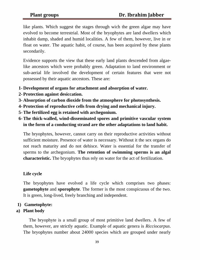
i) Antheridium
The antheridium(Fig.1 A) is a multicellular object ellipsoidal or club-shaped
sometimes spherical in form. It is borne on a short stalk which attaches it to the
gametophyte tissue.often it is embedded in the latter. The body of a ntheridium
has a wall of a single layer of sterile cells. It surrounds a mass of small squarish
or cubical cells called the androcytes. The latter produce the biflagellate male
gametes called the sperms. Several sperms are produced in each antheridium.
They are motile structure. Each sperm usually consists of a minute, slender,
spirally curved body furnished with two long, terminal, whiplash type flagella
(Fig.1 B).
Dr. Ibrahim Jabber Plant groups
41
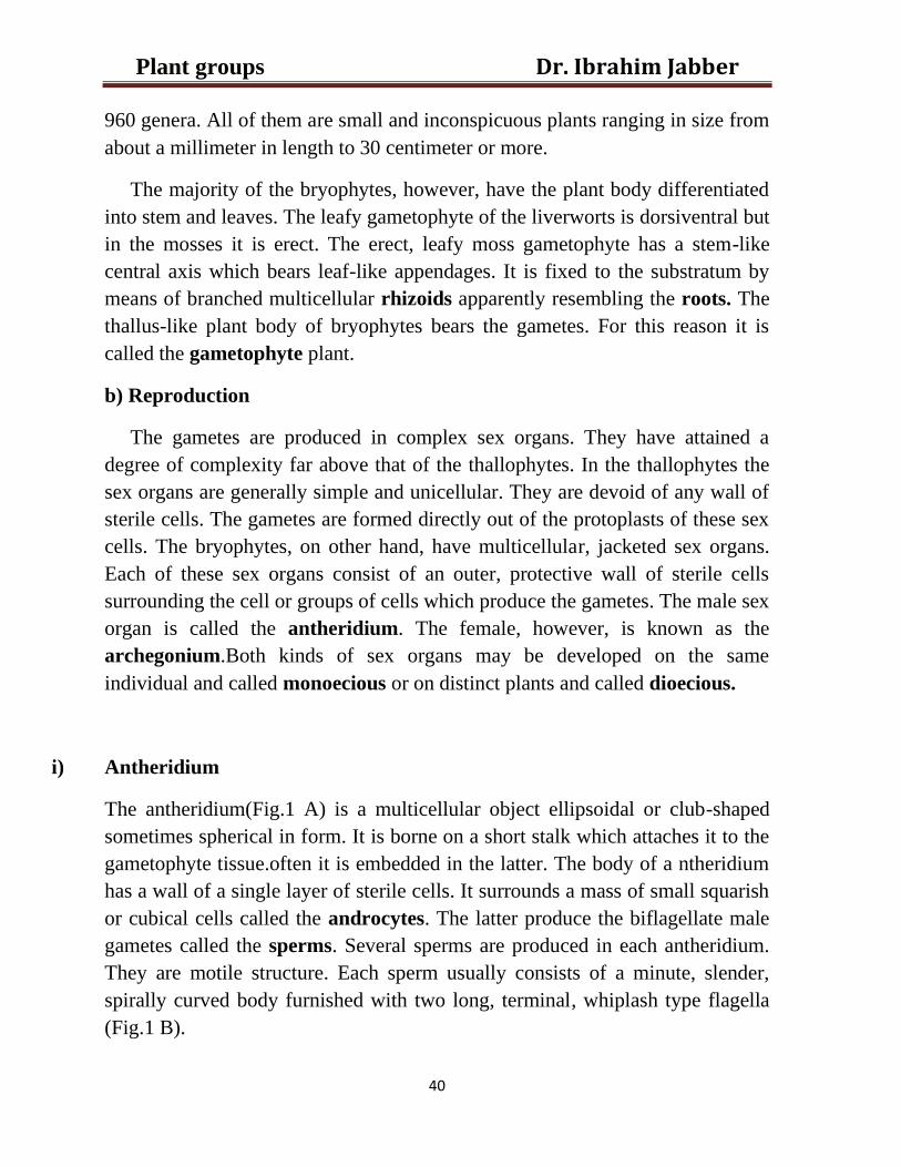
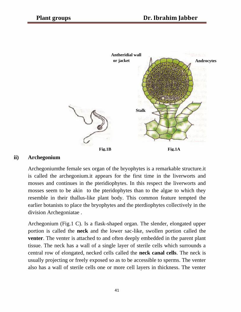
ii) Archegonium
Archegoniumthe female sex organ of the bryophytes is a remarkable structure.it
is called the archegonium.it appears for the first time in the liverworts and
mosses and continues in the pteridiophytes. In this respect the liverworts and
mosses seem to be akin to the pteridophytes than to the algae to which they
resemble in their thallus-like plant body. This common feature tempted the
earlier botanists to place the bryophytes and the pterdiophytes collectively in the
division Archegoniatae .
Archegonium (Fig.1 C). Is a flask-shaped organ. The slender, elongated upper
portion is called the neck and the lower sac-like, swollen portion called the
venter. The venter is attached to and often deeply embedded in the parent plant
tissue. The neck has a wall of a single layer of sterile cells which surrounds a
central row of elongated, necked cells called the neck canal cells. The neck is
usually projecting or freely exposed so as to be accessible to sperms. The venter
also has a wall of sterile cells one or more cell layers in thickness. The venter
Antheridial wall
or jacket
Stalk
Androcytes
Fig.1A Fig.1B
Dr. Ibrahim Jabber Plant groups
42
wall encloses two cells, the larger called egg cell or the ovum and the smaller
ventral canal cell.
Although the bryophytes resemble the pteridophytes in some feature but they
are unlike in other features. The resemblances and differences between
bryophytes and pteridophytes will discussed in the next lectures.
Fig.1C.
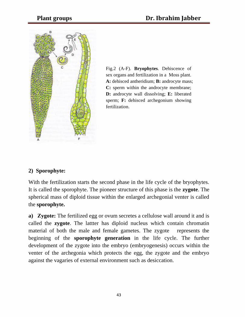
iii) Fertlization:
It occurs when the sex organs are mature. Moisture is essential for the maturing
of the sex organs and also for the movement of the sperms to the archegonia.
The mature antheridium ruptures at its apex liberating the sperms (A). At the
same time the axial row of neck canal cells including the ventral canal cell in
the mature archegonium disorganize (F). The tip of the archegonium also opens.
A narrow canal opening to the exterior is formed. It acts as a passage way to the
ovum in the venter. The liberated sperms swimming in a thin film of water
reach the archegonia (E).They enter through the open necks and swim down the
canals of the archegonia (Fig. 2). Reaching the venter one of them, probably the
first one to reach there penetrates the ovum (F). It fuses with the nucleus of the
ovum to accomplish fertilization. With the act of fertilization the gametophyte
generation ends and sporophyte generation starts. The gametes (sperms and
eggs) are the last structures of the gametophyte generation.
Neck
Lid or cover cells
Neck canal cells
Venter Egg
Ventral canal cell
Dr. Ibrahim Jabber Plant groups
43
2) Sporophyte:
With the fertilization starts the second phase in the life cycle of the bryophytes.
It is called the sporophyte. The pioneer structure of this phase is the zygote. The
spherical mass of diploid tissue within the enlarged archegonial venter is called
the sporophyte.
a) Zygote: The fertilized egg or ovum secretes a cellulose wall around it and is
called the zygote. The lattter has diploid nucleus which contain chromatin
material of both the male and female gametes. The zygote represents the
beginning of the sporophyte generation in the life cycle. The further
development of the zygote into the embryo (embryogenesis) occurs within the
venter of the archegonia which protects the egg, the zygote and the embryo
against the vagaries of external environment such as desiccation.
Fig.2 (A-F). Bryophytes. Dehiscence of
sex organs and fertilization in a Moss plant.
A: dehisced antheridium; B: androcyte mass;
C: sperm within the androcyte membrane;
D: androcyte wall dissolving; E: liberated
sperm; F: dehisced archegonium showing
fertilization.
Dr. Ibrahim Jabber Plant groups
44
b) Embryo: within venter of the archegonium the zygote undergoes
segmentation and develops without a resting period into a multicellular,
undifferentiated structure called an embryo.
c) Sporogonium: the embryo by further segmentation and differentiation
finally develops into a fully fledged sporophyte individual. It called the
sporogonium. The sporogonium in bryophytes is leafless and
rootless.generally,it is projecting and consist of three parts, the foot, the seta
and the capsule.
Alternation of generations
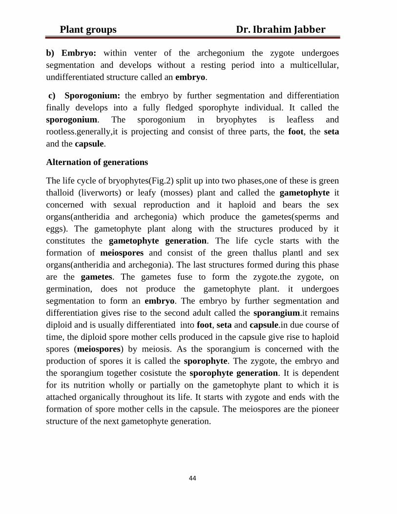
The life cycle of bryophytes(Fig.2) split up into two phases,one of these is green
thalloid (liverworts) or leafy (mosses) plant and called the gametophyte it
concerned with sexual reproduction and it haploid and bears the sex
organs(antheridia and archegonia) which produce the gametes(sperms and
eggs). The gametophyte plant along with the structures produced by it
constitutes the gametophyte generation. The life cycle starts with the
formation of meiospores and consist of the green thallus plantl and sex
organs(antheridia and archegonia). The last structures formed during this phase
are the gametes. The gametes fuse to form the zygote.the zygote, on
germination, does not produce the gametophyte plant. it undergoes
segmentation to form an embryo. The embryo by further segmentation and
differentiation gives rise to the second adult called the sporangium.it remains
diploid and is usually differentiated into foot, seta and capsule.in due course of
time, the diploid spore mother cells produced in the capsule give rise to haploid
spores (meiospores) by meiosis. As the sporangium is concerned with the
production of spores it is called the sporophyte. The zygote, the embryo and
the sporangium together cosistute the sporophyte generation. It is dependent
for its nutrition wholly or partially on the gametophyte plant to which it is
attached organically throughout its life. It starts with zygote and ends with the
formation of spore mother cells in the capsule. The meiospores are the pioneer
structure of the next gametophyte generation.
Dr. Ibrahim Jabber Plant groups
45
Fig.3.Bryophytes. Alternation of generations.
Lecture -8
Class: Hepaticopsida or Hepaticae (Liverworts).
In this class, the gametophyte is the dominant phase in the heteromorphic
alternation of the life cycle. They are classified according to the differences in
the structure of gametophytic and sporophytic phases.
The class Hepatopsida has been divided into the following seven well
recognized orders:
1- Takakiales 2- Calobryales 3-Jungermanniales 4-Metzgeriales 5-Marchantiales
6-Sphaerocarpales.
Dr. Ibrahim Jabber Plant groups
46
Order: Marchantiales
It is a well-defined, widely distributed group comprising more than 420 species
placed under 35 genera. The well known members are Marchantia and Riccia.
Generally, they inhabit moist situations and are strictly hygrophilous growing
on damp soil or rocks.
Family: Ricciaceae
Genus: Riccia
1-Gametophyte
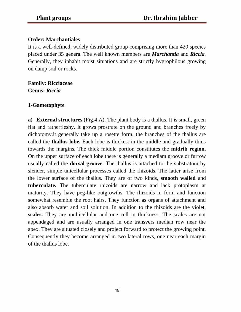
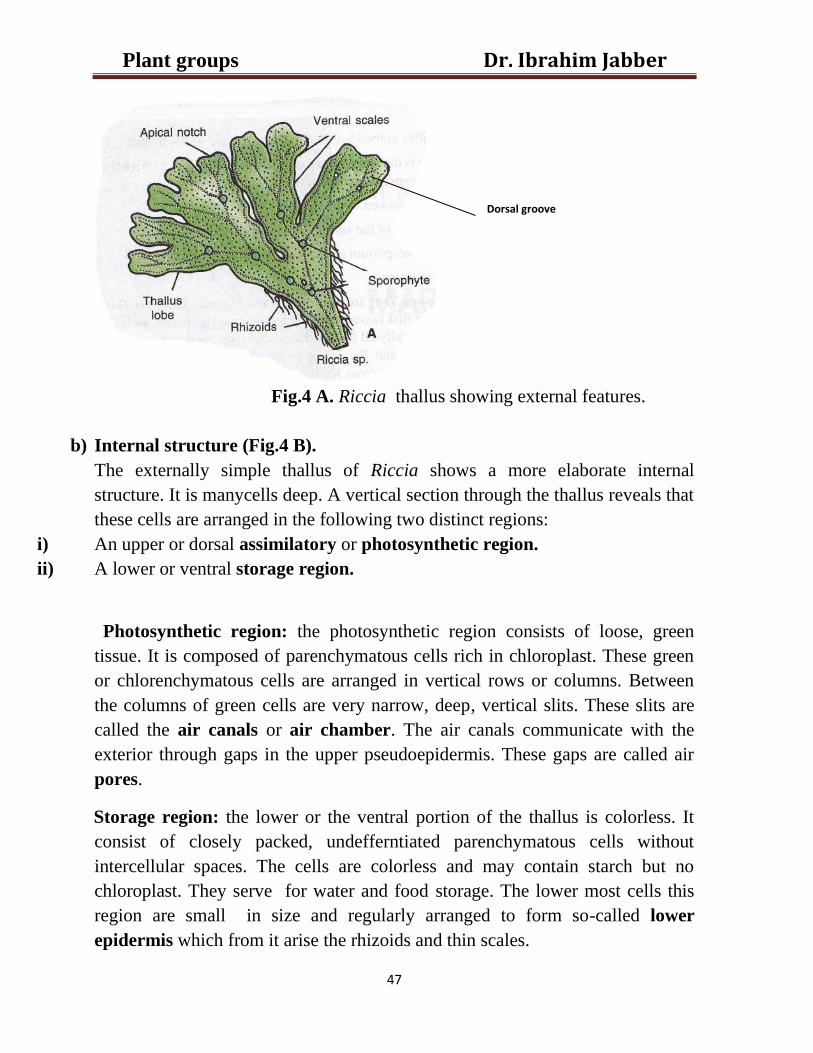
a) External structures (Fig.4 A). The plant body is a thallus. It is small, green
flat and ratherfleshy. It grows prostrate on the ground and branches freely by
dichotomy.it generally take up a rosette form. the branches of the thallus are
called the thallus lobe. Each lobe is thickest in the middle and gradually thins
towards the margins. The thick middle portion constitutes the midrib region.
On the upper surface of each lobe there is generally a mediam groove or furrow
usually called the dorsal groove. The thallus is attached to the substratum by
slender, simple unicellular processes called the rhizoids. The latter arise from
the lower surface of the thallus. They are of two kinds, smooth walled and
tuberculate. The tuberculate rhizoids are narrow and lack protoplasm at
maturity. They have peg-like outgrowths. The rhizoids in form and function
somewhat resemble the root hairs. They function as organs of attachment and
also absorb water and soil solution. In addition to the rhizoids are the violet,
scales. They are multicellular and one cell in thickness. The scales are not
appendaged and are usually arranged in one transvers median row near the
apex. They are situated closely and project forward to protect the growing point.
Consequently they become arranged in two lateral rows, one near each margin
of the thallus lobe.
Dr. Ibrahim Jabber Plant groups
47
Fig.4 A. Riccia thallus showing external features.
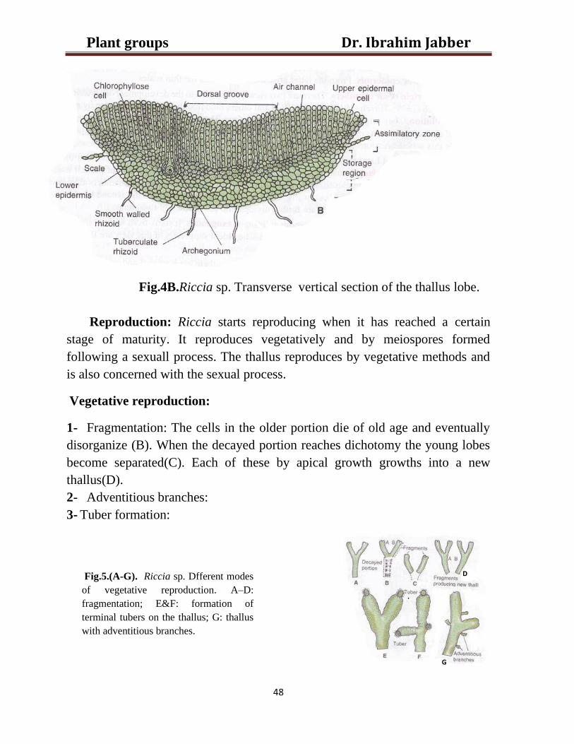
b) Internal structure (Fig.4 B).
The externally simple thallus of Riccia shows a more elaborate internal
structure. It is manycells deep. A vertical section through the thallus reveals that
these cells are arranged in the following two distinct regions:
i) An upper or dorsal assimilatory or photosynthetic region.
ii) A lower or ventral storage region.
Photosynthetic region: the photosynthetic region consists of loose, green
tissue. It is composed of parenchymatous cells rich in chloroplast. These green
or chlorenchymatous cells are arranged in vertical rows or columns. Between
the columns of green cells are very narrow, deep, vertical slits. These slits are
called the air canals or air chamber. The air canals communicate with the
exterior through gaps in the upper pseudoepidermis. These gaps are called air
pores.
Storage region: the lower or the ventral portion of the thallus is colorless. It
consist of closely packed, undefferntiated parenchymatous cells without
intercellular spaces. The cells are colorless and may contain starch but no
chloroplast. They serve for water and food storage. The lower most cells this
region are small in size and regularly arranged to form so-called lower
epidermis which from it arise the rhizoids and thin scales.
Dorsal groove
Dr. Ibrahim Jabber Plant groups
48
Fig.4B.Riccia sp. Transverse vertical section of the thallus lobe.
Reproduction: Riccia starts reproducing when it has reached a certain
stage of maturity. It reproduces vegetatively and by meiospores formed
following a sexuall process. The thallus reproduces by vegetative methods and
is also concerned with the sexual process.
Vegetative reproduction:
1- Fragmentation: The cells in the older portion die of old age and eventually
disorganize (B). When the decayed portion reaches dichotomy the young lobes
become separated(C). Each of these by apical growth growths into a new
thallus(D).
2- Adventitious branches:
3- Tuber formation:
D
Fig.5.(A-G). Riccia sp. Dfferent modes
of vegetative reproduction. A–D:
fragmentation; E&F: formation of
terminal tubers on the thallus; G: thallus
with adventitious branches.
G
Dr. Ibrahim Jabber Plant groups
49
Sexual reproduction:
The sex organs are developed on the thallus lobes which are not in any way
specialized for the purpose. They are developed in lines extending back from
the growing points. Generally they
lie in the dorsal furrow or groov sunk
deeply, each in a separate cavity. They
are developed in an acropetal order.
They younger ones are thus near the tip
or near the growing point and the
older are away from it. the antheridia
and archegonia, in some species, are
developed on the same thallus, they
are known as monoecious. In other
the two kindes of sex organs are
developed on different thalli,They are
referred to as dioecious.
Structure and development of sex organs:
a- Antheridia:
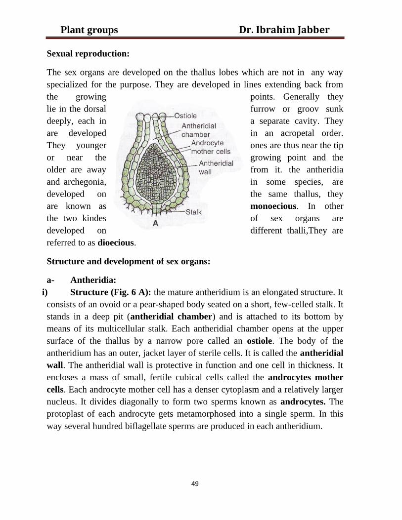
i) Structure (Fig. 6 A): the mature antheridium is an elongated structure. It
consists of an ovoid or a pear-shaped body seated on a short, few-celled stalk. It
stands in a deep pit (antheridial chamber) and is attached to its bottom by
means of its multicellular stalk. Each antheridial chamber opens at the upper
surface of the thallus by a narrow pore called an ostiole. The body of the
antheridium has an outer, jacket layer of sterile cells. It is called the antheridial
wall. The antheridial wall is protective in function and one cell in thickness. It
encloses a mass of small, fertile cubical cells called the androcytes mother
cells. Each androcyte mother cell has a denser cytoplasm and a relatively larger
nucleus. It divides diagonally to form two sperms known as androcytes. The
protoplast of each androcyte gets metamorphosed into a single sperm. In this
way several hundred biflagellate sperms are produced in each antheridium.
Dr. Ibrahim Jabber Plant groups
50
(Fig. 6 A)
ii) Dehiscence: presence of moisture is essential for the dehiscence of a mature
antheridium which the walls of the androcytes have dissolved and the sperms lie
free in the viscous fluid in the cavity of the antheridium surrounded by the
antheridial wall. Water enters the ostiole and finds its way into the antheridial
chamber. The cells at the apex of the antheridium absorb this water by
imbibition. They get softened and eventually disintegrated to form a distal pore.
Then they will escape through the canal of the antheridial chamber to the upper
surface of the thallus. Here they swim freely in a thin film of water in the dorsal
furrow.
iii) Development of the antheridium: Eeach antheridium develops from a
single superficial cell called the antheridial initial.
b- Archegonium: (Fig. 6 B):
i) Structure: the archegonium is a flask-shaped organ. It consists of two
parts, the basal swollen portion called the venter and a long, slender neck. The
venter is directly attached to the tissue of the thallus. There is usually no visible
stalk.The next consists of a vertical row of four cells called the neck canal cells
surrounded by a layer of sterile cells forming a protective jacket. The jacket or
neck cells are arranged in six longitudinal rows. Each row is 6-9 cells in height.
Dr. Ibrahim Jabber Plant groups
51
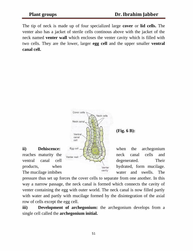
The tip of neck is made up of four specialized large cover or lid cells. The
venter also has a jacket of sterile cells continous above with the jacket of the
neck named venter wall which encloses the venter cavity which is filled with
two cells. They are the lower, larger egg cell and the upper smaller ventral
canal cell.
(Fig. 6 B):
ii) Dehiscence: when the archegonium
reaches maturity the neck canal cells and
ventral canal cell degenerated. Their
products, when hydrated, form mucilage.
The mucilage imbibes water and swells. The
pressure thus set up forces the cover cells to separate from one another. In this
way a narrow passage, the neck canal is formed which connects the cavity of
venter containing the egg with outer world. The neck canal is now filled partly
with water and partly with mucilage formed by the disintegration of the axial
row of cells except the egg cell.
iii) Development of archegonium: the archegonium develops from a
single cell called the archegonium initial.
Dr. Ibrahim Jabber Plant groups
52
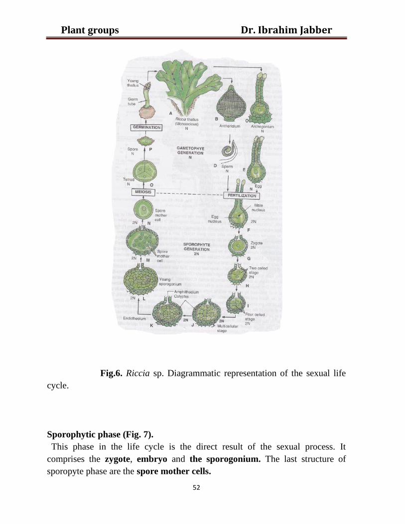
Fig.6. Riccia sp. Diagrammatic representation of the sexual life
cycle.
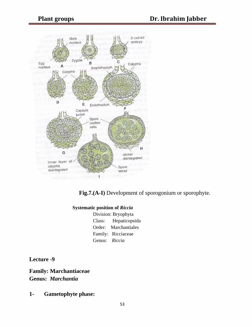
Sporophytic phase (Fig. 7).
This phase in the life cycle is the direct result of the sexual process. It
comprises the zygote, embryo and the sporogonium. The last structure of
sporopyte phase are the spore mother cells.
Dr. Ibrahim Jabber Plant groups
53
Fig.7.(A-I) Development of sporogonium or sporophyte.
Systematic position of Riccia
Division: Bryophyta
Class: Hepaticopsida
Order: Marchantiales
Family: Ricciaceae
Genus: Riccia
Lecture -9
Family: Marchantiaceae
Genus: Marchantia
1- Gametophyte phase:
Dr. Ibrahim Jabber Plant groups
54
a) External morphology of the thallus
It is a dark, green, somewhat fleshy, flat, once or a few times dichotomously
branch or lobe is traversed by a broad, thick, central midrib. It also has a notch
at its apex. At the bottom of notch is located the growing point. The upper
surface of the thallus is marked by rhomboidal to polygonal areas called the
areola (Fig.4).the boundaries between these areas mark the limit of the
underlying air chamber. Each area has a tiny an air pore in the center. The
pore is visible on the surface as light dot. The air pores permit aeration of
thallus with minimum dehydration. From the lower or the ventral surface of the
thallus arise numerous elongated, single celled, hair-like outgrowths called the
rhizoids. The latter anchor the plant to the substratum. In addition they absorb
water and minerals in solution. The rhizoids are of two kinds smooth walled &
tuberculate, the tuberculate peg rhizoids are thick walled, narrow and
appressed to the surface of the thallus and its fuction as a capillary conducting
system which serves to carry water to all the absorptive parts of the thallus. The
smooth-walled rhizoids stand out from the thallus and penetrate the substratum
to absorb water and to fix the thallus to it. They are broad, thin-walled with
colorless content. They are the first to be formed on germination. Besides the
rhizoids the ventral surface bears purplish flattered scales they are usually
arranged in two to four on either side of the midrib. At times little cup-like
structures, the gemma cups, are seen on the surface of the thallus. That arises in
the midrib region. The margin of the cup is toothed and membranous.
Dr. Ibrahim Jabber Plant groups
55
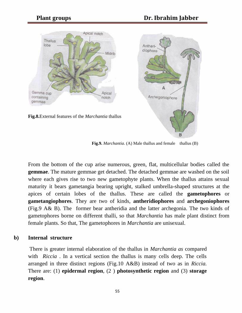
Fig.8.External features of the Marchantia thallus
Fig.9. Marchantia. (A) Male thallus and female thallus (B)
From the bottom of the cup arise numerous, green, flat, multicellular bodies called the
gemmae. The mature gemmae get detached. The detached gemmae are washed on the soil
where each gives rise to two new gametophyte plants. When the thallus attains sexual
maturity it bears gametangia bearing upright, stalked umbrella-shaped structures at the
apices of certain lobes of the thallus. These are called the gametophores or
gametangiophores. They are two of kinds, antheridiophores and archegoniophores
(Fig.9 A& B). The former bear antheridia and the latter archegonia. The two kinds of
gametophores borne on different thalli, so that Marchantia has male plant distinct from
female plants. So that, The gametophores in Marchantia are unisexual.
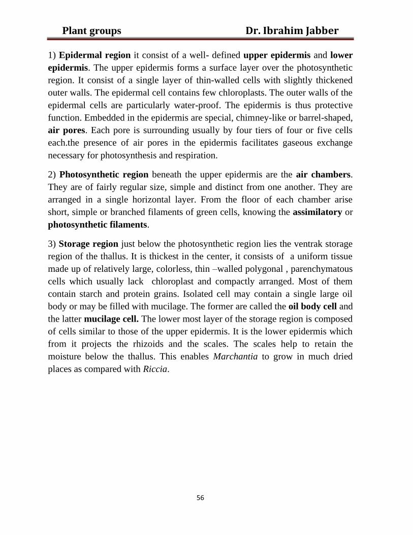
b) Internal structure
There is greater internal elaboration of the thallus in Marchantia as compared
with Riccia . In a vertical section the thallus is many cells deep. The cells
arranged in three distinct regions (Fig.10 A&B) instead of two as in Riccia.
There are: (1) epidermal region, (2 ) photosynthetic region and (3) storage
region.
Dr. Ibrahim Jabber Plant groups
56
1) Epidermal region it consist of a well- defined upper epidermis and lower
epidermis. The upper epidermis forms a surface layer over the photosynthetic
region. It consist of a single layer of thin-walled cells with slightly thickened
outer walls. The epidermal cell contains few chloroplasts. The outer walls of the
epidermal cells are particularly water-proof. The epidermis is thus protective
function. Embedded in the epidermis are special, chimney-like or barrel-shaped,
air pores. Each pore is surrounding usually by four tiers of four or five cells
each.the presence of air pores in the epidermis facilitates gaseous exchange
necessary for photosynthesis and respiration.
2) Photosynthetic region beneath the upper epidermis are the air chambers.
They are of fairly regular size, simple and distinct from one another. They are
arranged in a single horizontal layer. From the floor of each chamber arise
short, simple or branched filaments of green cells, knowing the assimilatory or
photosynthetic filaments.
3) Storage region just below the photosynthetic region lies the ventrak storage
region of the thallus. It is thickest in the center, it consists of a uniform tissue
made up of relatively large, colorless, thin –walled polygonal , parenchymatous
cells which usually lack chloroplast and compactly arranged. Most of them
contain starch and protein grains. Isolated cell may contain a single large oil
body or may be filled with mucilage. The former are called the oil body cell and
the latter mucilage cell. The lower most layer of the storage region is composed
of cells similar to those of the upper epidermis. It is the lower epidermis which
from it projects the rhizoids and the scales. The scales help to retain the
moisture below the thallus. This enables Marchantia to grow in much dried
places as compared with Riccia.
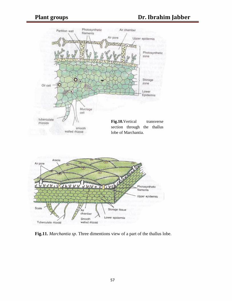
Dr. Ibrahim Jabber Plant groups
57
Fig.11. Marchantia sp. Three dimentions view of a part of the thallus lobe.
Fig.10.Vertical transverse
section through the thallus
lobe of Marchantia.
Dr. Ibrahim Jabber Plant groups
58
Reproduction
Marchantia thallus reproduces vegetatively and also by a sexual process.
Vegetation reproduction: the thallus plant reproduces vegetatively in the
growing season by the following three methods:
a) Fragmentation. It is brought about by the ageing of the vegetative cells. The
aged cells in the rear or basal part of the thallus rot and disintegrate.when this
decay cell reaches dichotomy the lobes become separated and each of the
detached lobes by apical growth dichotomy develops into an independent.
b) Formation of adventitious branches: these develop from any part of the
thallus particularly its ventral surface. In Marchantia plamata the development
of adventitious branches from the stalk and disk of the female gametophores.
The branches when detached by the decay of connecting tissue develop into
new individuals.
c) Gemmae formation (Fig.12): Another interesting and elegant method of
vegetative reproduction in Marchantia is by the formation of multicellular
bodies called the gammae. gemmae develop inside little, shallow, cup-like
growths with fingered margins (A). The cups are developed on the upper
surface of the thallus, generally in midrib. The gemmae are small,green, disc-
like, slighty bi-convex structures. Each gemma stands on edge on a short,
delicate single-celled stalk (B). The stalk attaches it to the bottom of the gemma
cup (C). Intermingled with the gemmae in the cup are the club-shaped
mucilages hairs which secrete mucilage copiously(C). With the absorbtion of
water provided by dew or rain the mucilage swells and causes the gemmae to
get detached from their stalk rather easily. The gemmae then washed on the soil
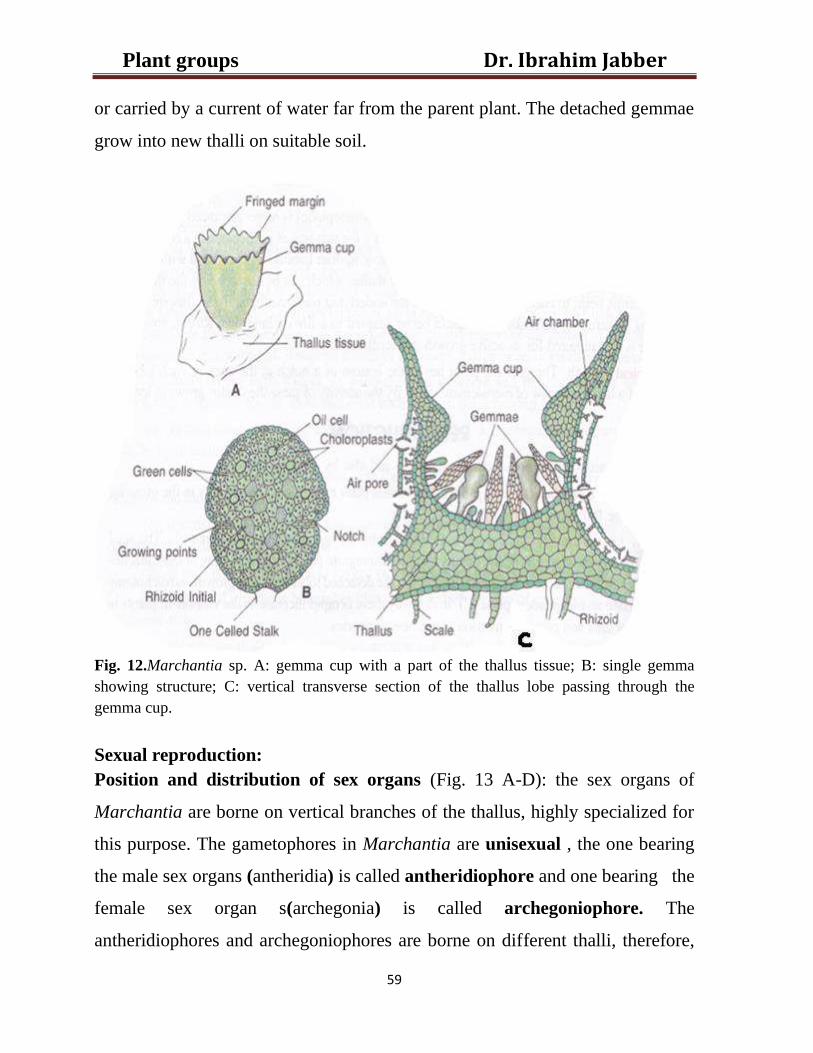
Dr. Ibrahim Jabber Plant groups
59
or carried by a current of water far from the parent plant. The detached gemmae
grow into new thalli on suitable soil.
Fig. 12.Marchantia sp. A: gemma cup with a part of the thallus tissue; B: single gemma
showing structure; C: vertical transverse section of the thallus lobe passing through the
gemma cup.
Sexual reproduction:
Position and distribution of sex organs (Fig. 13 A-D): the sex organs of
Marchantia are borne on vertical branches of the thallus, highly specialized for
this purpose. The gametophores in Marchantia are unisexual , the one bearing
the male sex organs (antheridia) is called antheridiophore and one bearing the
female sex organ s(archegonia) is called archegoniophore. The
antheridiophores and archegoniophores are borne on different thalli, therefore,
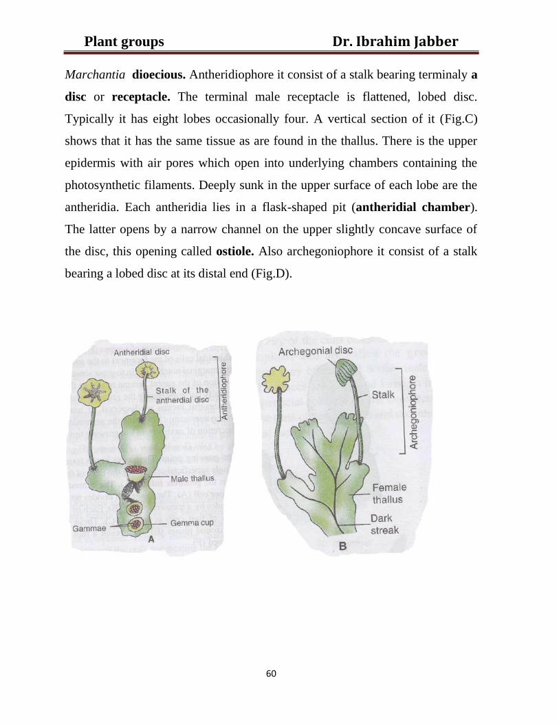
Dr. Ibrahim Jabber Plant groups
60
Marchantia dioecious. Antheridiophore it consist of a stalk bearing terminaly a
disc or receptacle. The terminal male receptacle is flattened, lobed disc.
Typically it has eight lobes occasionally four. A vertical section of it (Fig.C)
shows that it has the same tissue as are found in the thallus. There is the upper
epidermis with air pores which open into underlying chambers containing the
photosynthetic filaments. Deeply sunk in the upper surface of each lobe are the
antheridia. Each antheridia lies in a flask-shaped pit (antheridial chamber).
The latter opens by a narrow channel on the upper slightly concave surface of
the disc, this opening called ostiole. Also archegoniophore it consist of a stalk
bearing a lobed disc at its distal end (Fig.D).
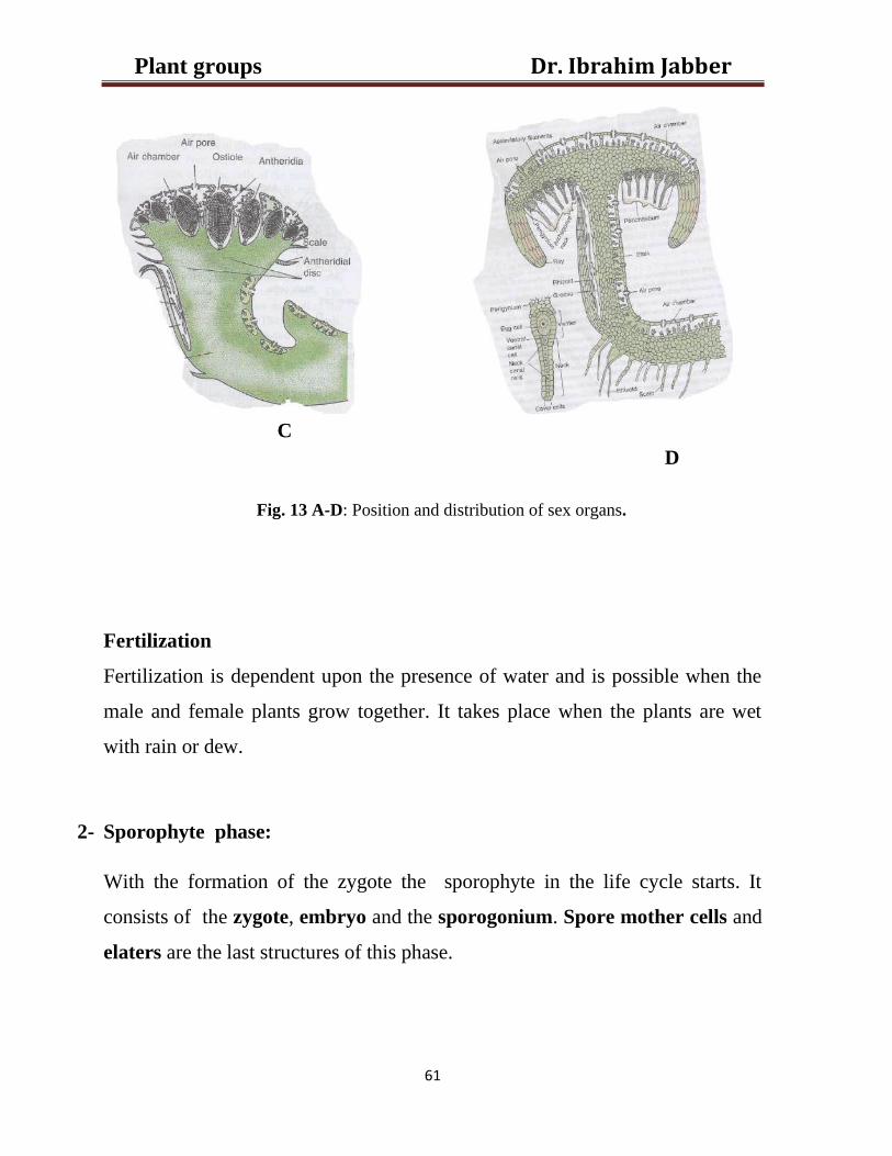
Dr. Ibrahim Jabber Plant groups
61
C
D
Fig. 13 A-D: Position and distribution of sex organs.
Fertilization
Fertilization is dependent upon the presence of water and is possible when the
male and female plants grow together. It takes place when the plants are wet
with rain or dew.
2- Sporophyte phase:
With the formation of the zygote the sporophyte in the life cycle starts. It
consists of the zygote, embryo and the sporogonium. Spore mother cells and
elaters are the last structures of this phase.
Dr. Ibrahim Jabber Plant groups
62
Structure of sporogonium: (Fig.14).it is differentiated into the following three
regions:
a) Foot: It is absorptive anchoring organ forming the basal broad portion of the
sporogonium. It consists of parenchmatous cells and is embedded in the tissue
of the female receptacle on its lower surface. It absorbs all the nourishment
from the gametophyte for the developing sporogonium.
b) Seta: it is at first very short and constriction-like. It connects the foot and the
capsule. The cells constituting it arranged in vertical row. With the formation of
spore tetrads in the capsule the seta elongates slightly. The slight and sudden
increase in the length of the seta ruptures the calyptra. It pushes the mature
capsule through the sheath enclosing the venter. These are perigynuim and
prichaetuim (involucre).
c) Capsule:it is oval in form and yellow in color when mature. It has an outer
layer of sterile cells consistuting the capsule wall. A cap of cells more than one
cell-layer thick is developed in some species at the apex of the capsule. It is
formed from the sporogenous cells. It projects into cavity of the capsule.
Enclosed within the capsule wall, at maturity, is a mass of meiospores and
elaters. The elaters are exceptionally long, slender cells with tapering ends.
They have double spirally thickened bands on their walls. The mature elaters
lack protoplasm. They are dead and are hygroscopic.
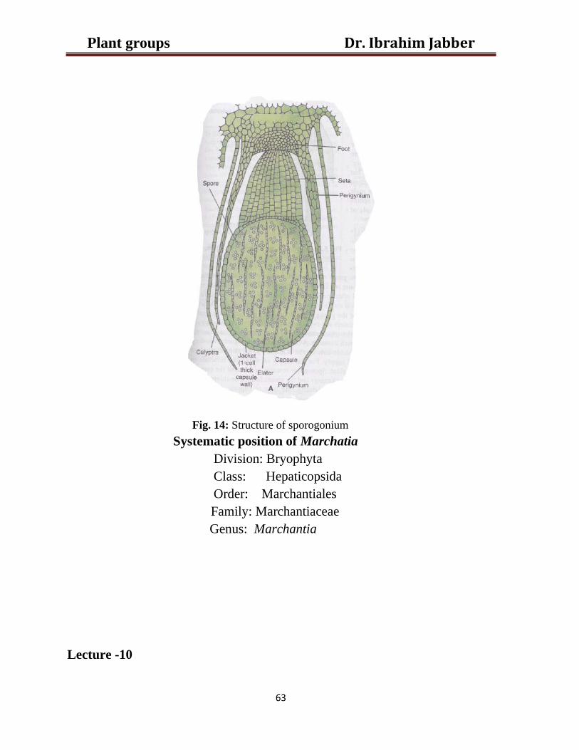
Dr. Ibrahim Jabber Plant groups
63
Fig. 14: Structure of sporogonium
Systematic position of Marchatia
Division: Bryophyta
Class: Hepaticopsida
Order: Marchantiales
Family: Marchantiaceae
Genus: Marchantia
Lecture -10

Dr. Ibrahim Jabber Plant groups
64
Class: Anthocerotopsida
This class embraces a small but very clearly defined group of plants which
differ in many ways, especially in its sporophyte. Popularly the members of this
group are called hornworts. This common name of Anthocerotopsida is
derived from the shape of its sporophyte.
Order: Anthocerotales
Family: Anthocerotaceae
Genus: Anthoceros
1) The gametophyte
a) External structure (Fig. 15)
The plant body, as in the bryophytes, is the gametophyte. It is a small,
yellowish green or dark green, the lobes of the thallus are thick and fleshy
generally more ore less divided to form irregularly lobed and folded margins
overlap and it is attached to the substratum by means of rhizoids which are only
smooth walled, where as tuberculate rhizoids and scales are absent. Ventral
surface of the thallus revealed small, rounded bluish green thickened spots
which called the Nostoc colonies, as each is inhabited by a blue-geen alga
Nostoc.

Dr. Ibrahim Jabber Plant groups
65
Fig. 15: External structure of Anthoceros
b) Internal structure
Internally, the thallus without a midrib. There is no tissue differentiation and
little cell specialization. Thus the photosynthetic and storage zones are not
recognizable. The entire thallus consists of soft parenchyma. Air containing
chamber and air pores are lacking. In some species there are stoma-like
pores on the ventral surface of the thallus. these are called slime pores, these
pores leads inwards often into rounded, intercellular spaces or cavities which
are filled with slime(mucilage). The mucilage cavities open on the ventral
surface of the thallus through the slime pores which serve as outlets for the
mucilage. Very often these mucilage cavities are inhabited by Nostoc colonies
(cyanobacteria). These colonies are visible to the naked eye as small, deep
blue- green rounded spots on the underside of the thallus. The sex organs
(antheridia & archegonia) are sunken in the thallus.
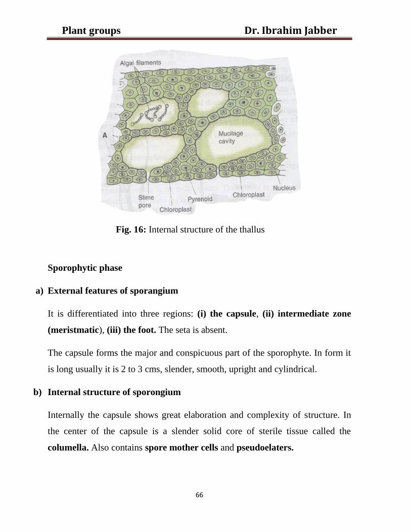
Dr. Ibrahim Jabber Plant groups
66
Fig. 16: Internal structure of the thallus
Sporophytic phase
a) External features of sporangium
It is differentiated into three regions: (i) the capsule, (ii) intermediate zone
(meristmatic), (iii) the foot. The seta is absent.
The capsule forms the major and conspicuous part of the sporophyte. In form it
is long usually it is 2 to 3 cms, slender, smooth, upright and cylindrical.
b) Internal structure of sporongium
Internally the capsule shows great elaboration and complexity of structure. In
the center of the capsule is a slender solid core of sterile tissue called the
columella. Also contains spore mother cells and pseudoelaters.
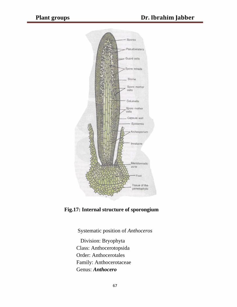
Dr. Ibrahim Jabber Plant groups
67
Fig.17: Internal structure of sporongium
Systematic position of Anthoceros
Division: Bryophyta
Class: Anthocerotopsida
Order: Anthocerotales
Family: Anthocerotaceae
Genus: Anthocero


![Plant Parameters for Plant Functional Groups of …Functional groups have been used for managing rare plants [20] and for looking at drivers of soil biota [21]. Functional groups have](https://img.pdfslide.us/doc/110x75/5f080f347e708231d42020d7/plant-parameters-for-plant-functional-groups-of-functional-groups-have-been-used.jpg)