Embed Size (px)
Citation preview

LETTERS
374 NATURE CELL BIOLOGY VOLUME 7 | NUMBER 4 | APRIL 2005
Plant formin AtFH5 is an evolutionarily conserved actin nucleator involved in cytokinesisMathieu Ingouff1,4,8, Jonathan N. Fitz Gerald1,5,8, Christophe Guérin2, Hélène Robert1,6, Mikael Blom Sørensen1,7, Daniel Van Damme3, Danny Geelen3, Laurent Blanchoin2,9 and Frédéric Berger1,5,9
Formins are actin-organizing proteins that are involved in cytokinesis and cell polarity1. In the plant Arabidopsis thaliana, there are more than 20 formin homologues, all of which have unknown roles2. In this study, we characterize specific cellular and molecular functions of the Arabidopsis formin AtFH5. Despite the low identity of AtFH5 to yeast and mammalian formins, the AtFH5 protein interacts with the barbed end of actin filaments and nucleates actin-filament polymerization in vitro, as is the case in yeast and mammals. In vivo, the AtFH5–GFP fusion protein localizes to the cell plate, a plant-specific membranous component that is assembled at the plane of cell division. Consistent with these data, loss of function of atfh5 compromises cytokinesis in the seed endosperm. Furthermore, endogenous AtFH5 transcripts accumulate in the posterior pole of the endosperm and loss of function of atfh5 perturbs proper morphogenesis of the endosperm posterior pole. Although cytokinesis in animals, yeast and plants occurs through morphologically distinct mechanisms, our study finds that formin recruitment to sites of actin assembly is a common feature of cell division among eukaryotes.
Formins are highly conserved proteins that are implicated in many actin-requiring processes1, such as cytokinesis, establishment of egg polarity and formation of stress fibres. In the enhancer-trap KS117 line, the expression pattern of which provides a polarity marker for endosperm development3, we have identified the corresponding T-DNA insertion that is localized in the upstream region of AtFH5 and that belongs to the Arabidopsis formin homology (FH) family2 (Fig. 1a). The FH2 domain of AtFH5 shares ~35% identity with other eukaryote formins and retains many highly conserved residues (see Supplementary Information, Fig. S1). By contrast,
1EMBO YIP team, Unité Mixte de Recherche 5667, Institut Fédératif de Recherche 128 BioSciences Lyon-Gerland, Ecole Normale Supérieure de Lyon, 46 allée d’Italie, F-69364 Lyon cedex 07, France. 2Laboratoire de Physiologie Cellulaire Végétale, Commissariat à l’Energie Atomique, Centre National de la Recherche Scientifique, Université Joseph Fourier, 17 rue des martyrs, F-38054 Grenoble, France. 3Department of Plant Systems Biology, VIB-Ghent University, Technologiepark 927, B-9052 Gent, Belgium. 4Present address: Laboratoire de Physiologie Cellulaire Végétale, Commissariat à l’Energie Atomique, Centre National de la Recherche Scientifique, Université Joseph Fourier, 17 rue des martyrs, F-38054 Grenoble, France. 5Present address: Temasek Lifesciences Laboratory, 1 Research Link, National University of Singapore, Singapore 117604. 6Present address: Institute of Biology of Leiden, Clusius Laboratory, Wassenaarseweg 64, AL 2333 Leiden, The Netherlands. 7Present address: Plant Research Department, PRD-301, Risø National Laboratory, PO Box 49, DK-4000 Roskilde, Denmark. 8These authors contributed equally to this work.9Correspondence should be addressed to F.B. (e-mail: [email protected]) or L.B. (e-mail: [email protected])
Published online: 13 March 2005; DOI: 10.1038/ncb1238
the amino terminus of AtFH5 is highly divergent from non-plant formins and shares the predicted domains that are typical of type-I Arabidopsis formins2 (Fig. 1b, c). In the homologous AtFH1, this N-terminal domain is required for localization to the cell membrane4.
In yeasts, actin patches depend on Arp2/3 complexes and cables of actin filaments depend on formins. The FH2 domain can nucleate actin filaments that grow at their barbed ends in vitro5 and can control actin assembly in vivo6,7. Recent studies in Arabidopsis showed that extensive actin filaments can be nucleated in the absence of Arp2/3 complexes8,
indicating that a potential role may exist for formins in plants. To test whether AtFH5 nucleates actin polymerization in vitro, we purified two carboxy-terminal constructs of AtFH5 fused to glutathione S-transferase (GST) (Fig. 2a). FH1–FH2–Cter consists of FH1, FH2 and C-terminal domains, and FH2–Cter consists of the FH2 and C-terminal domains. Pyrene-labelled actin monomers were incubated alone or in the presence of FH1–FH2–Cter and assembly was monitored by pyrene fluorescence (Fig. 2b). FH1–FH2–Cter decreased the initial lag of the actin polym-erization curve in a concentration-dependent manner, corresponding to active nucleation (Fig. 2b). Elongation of actin filaments nucleated by FH1–FH2–Cter with actin monomers bound to profilin confirms that these filaments are growing at their barbed ends (data not shown). The concentration of new barbed ends is strongly dependent on the presence of FH1–FH2–Cter (Fig. 2c). The curve plateaus at a high concentra-tion of FH1–FH2–Cter because elongation of actin filaments rapidly consumes actin monomers before each formin can form new filaments (Fig. 2c). This observation is in agreement with a previous report of actin nucleation by Arp2/3 complexes9. Filament length is inversely correlated to the concentration of nucleation centres. Accordingly, actin filaments are markedly shorter in the presence of FH1–FH2–Cter (mean length, 5.3 ±5 µm) (Fig. 2e) than alone (mean length, 18 ±17 µm) (Fig. 2d).
ncb1238.indd 374ncb1238.indd 374 15/3/05 2:34:20 pm15/3/05 2:34:20 pm
Nature Publishing Group© 2005

NATURE CELL BIOLOGY VOLUME 7 | NUMBER 4 | APRIL 2005 375
L E T T E R S
To initially ascertain barbed-end dynamics in the presence of AtFH5, a constant concentration of actin filaments was incubated with increasing concentrations of FH1–FH2–Cter before the addition of labelled actin monomers (Fig. 2f). The initial rate of elongation, which is proportional to the number of available free actin-filament barbed ends, decreased as we increased the FH1–FH2–Cter concentration. This indicates that the barbed ends of preformed filaments were capped with high affinity (Kd ~32 nM) by pre-incubation with FH1–FH2–Cter (Fig. 2f). In addi-tion, pyrene-labelled actin filaments were diluted below the barbed-end critical concentration (0.1 µM) in the presence of increasing amounts of FH1–FH2–Cter (Fig. 2g). The initial rate of depolymerization decreased with increased FH1–FH2–Cter concentration, again in agreement with a barbed-end capping activity. Interestingly, the FH2–Cter protein has no effect on actin nucleation (Fig. 2h). However, FH2–Cter inhibits the actin nucleation that is driven by FH1–FH2–Cter, indicating that FH2–Cter competes with FH1–FH2–Cter for binding actin monomers.
The biochemical analysis of AtFH5 indicates that the actin-organizing activity of formins is conserved throughout eukaryotes. To determine if such nucleating activity could also be related to cytokinesis, we examined AtFH5 subcellular localization in roots in which AtFH5 is expressed in the wild type. AtFH5 was tagged with green fluorescent protein (GFP) and expressed in transgenic Arabidopsis plants. We used the constitutive CaMV 35S promoter that enables dynamic observations in roots, with enhanced optical resolution in comparison to that seen in endosperm. Fluorescence microscopy showed that AtFH5–GFP fluorescence accu-mulates in the cell plate of dividing root cells (Fig. 3). The AtFH5–GFP signal follows the expanding plate over time and its intensity gradu-ally reduces after contact is made between the cell plate and the parent cell wall. The dynamic localization of AtFH5 protein to the cell plate
indicates that AtFH5 has a role in cytokinesis. A fluorescent dotted pat-tern of undetermined identity was also observed in the cytoplasm (Fig. 3) and might not reflect the true localization of AtFH5, as potential artifacts may be associated with overexpression of AtFH5–GFP.
The endosperm in the Arabidopsis plant seed provides a simple and tractable system for studying cytokinesis within a multinucleate single cell10. Endosperm development involves a series of successive nuclear divisions that are devoid of cytokinesis, generating a syncytium11,12. After eight rounds of division, the syncytium contains approximately 200 nuclei that become partitioned into mononucleate cells by a cytokinetic mechanism that is referred to as cellularization12. Cellularization initi-ates from the anterior pole, where the embryo develops, and continues towards peripheral endosperm, ending at the posterior pole that remains syncytial11,13. Nuclei at this pole are included in large multinucleate struc-tures called nodules that gradually migrate and eventually fuse with the cyst at the most posterior location14.
As the GFP activity in the AtFH5-tagged KS117 line indicated a role for this gene in endosperm development3, we examined the AtFH5 expression pattern during seed development. AtFH5 mRNAs were local-ized in the endosperm by in situ hybridization on seed sections. A strong expression was specific to the posterior pole in the cyst (Fig. 4a) and in nodules on top of the cyst (Fig. 4a, inset). No expression was detectable in the anterior and peripheral endosperm or in other seed compartments (such as the embryo and the seed coat). Expression of AtFH5 was further confirmed in transgenic Arabidopsis expressing the nuclear fluorescent reporter Histone2B–Yellow Fluorescent Protein (H2B–YFP) under the control of the AtFH5 upstream region. H2B–YFP is first detected in the posterior endosperm early in development (Fig. 4c) and remains restricted to the posterior pole (Fig. 4d) throughout the syncytial phase.
FH2FH1TMSP
AtFH5
Formin
GBD FH3 FH2FH1 D
a b
c
ATG TAA
1,000 bpKS117
AtFH5
−541
Figure 1 AtFH5 encodes a formin protein. (a) Schematic drawing of the AtFH5 locus. The T-DNA insertion in the KS117 enhancer-trap green fluorescent protein (GFP) line is localized 541 base pairs upstream of the putative translation (ATG) start site. Exons and introns are represented by black boxes and lines, respectively, and the AtFH5 upstream region is represented by a dotted line. The 5′- and 3′-untranslated regions are boxed in grey. (b) Predicted domains of the putative AtFH5 formin protein. The amino-terminus of AtFH5 has a putative signal peptide (double line) followed by a repeated SPPP motif (shaded) and a predicted trans-membrane domain (circled in grey). The AtFH5 protein features the two
typical carboxy-terminal domains of formins: the formin homology 1 (FH1) domain (dotted line), which is rich in proline (in bold), and the formin homology 2 (FH2) domain (underlined). (c) Domain organization in AtFH5 and other non-plant formins. The typical domains encountered in non-plant formins are depicted. Apart from the FH1–FH2 unit, AtFH5 shows no homology with the identified targeting (FH3) or regulatory domains (d, GBD) of formin. Note that formin domains are not drawn to scale. D, diaphanous autoregulatory domain; FH1, formin homology 1; FH2, formin homology 2; FH3, formin homology 3; GBD, Rho-GTPase binding domain; SP, signal peptide; TM, transmembrane domain.
ncb1238.indd 375ncb1238.indd 375 15/3/05 2:34:24 pm15/3/05 2:34:24 pm
Nature Publishing Group© 2005

376 NATURE CELL BIOLOGY VOLUME 7 | NUMBER 4 | APRIL 2005
L E T T E R S
This pattern persists after endosperm cellularization (Fig. 4e). Together, these two experiments indicate that AtFH5 expression is restricted to the posterior pole throughout endosperm development. However, GFP was also detected in the anterior and peripheral syncytial endosperm in the KS117 line3, indicating that the enhancer activity detected by KS117 GFP does not entirely mimic the endogenous AtFH5 pattern.
To gain insight into AtFH5 function during endosperm develop-ment, we examined two atfh5 insertional mutant lines (Fig. 5a, top). Reverse transcription PCR (RT-PCR) expression analyses with primers downstream of the insertions revealed no AtFH5 transcripts (Fig. 5a,
bottom). A barely detectable level of AtFH5 transcripts was obtained with primers that were positioned upstream of the insertions. These atfh5 mutations probably result in loss of function as neither of the two mutants expresses the actin-organizing domains of AtFH5. Although AtFH5 is expressed in most tissues, we could not detect any pheno-types in vegetative organs, probably due to the redundant function with other formins. Microscopic examination of atfh5;atfh5 seeds revealed perturbation of endosperm cellularization. Cleared siliques were used to score the onset of cellularization in wild-type and atfh5 mutant lines (Fig. 5b). Whereas most wild-type endosperm is cellularized by the heart
1,000Time (sec)
b
End
s ap
par
ent
(nM
)
FH1−FH2−Cter (nM)
c
FH2FH1
AtFH5COOH
1 327430 439 900849
341
433
FH1−FH2−Cter
FH2 Cter
N
a
Fluo
resc
ence
(a.u
.)
f
h
Fluo
resc
ence
(a.u
.)
Fluo
resc
ence
(a.u
.)Time (sec)
Time (s)Time (s)
0.225
0.45
0.675
0.9
0 200 400 600 800
50 nM UC FH1−FH2−Cter200 nM FH1−FH2−Cter
1,000 nM FH1−FH2−Cter500 nM FH1−FH2−Cter
Actin alone20 nM FH1−FH2−Cter50 nM FH1−FH2−Cter
0.1
0.2
0.3
0.4
0.5
0.6
0.7
0 200 400 600 800
d
0.05
0.1
0.15
0.2
0 100 200 300 400 500 600
100 nM
Actin alone
20 nM 50 nM
200 nM 300 nM
10 nM
0.098
0.112
0.126
0.14
0.154
0 100 200 300 400 500
g
100 nM 200 nM
Actin alone5 nM 10 nM 20 nM Fl
uore
scen
ce (a
.u.)
400 nM FH1−FH2−Cter + 4 µM FH2−Cter
800 nM UC FH2−Cter2 µM FH2−CterActin alone
400 nM FH1−FH2−Cter + 3 µM FH2−Cter
400 nM FH1−FH2−Cter + 5 µM FH2−Cter
400 nM FH1−FH2−Cter
e
0.2
0.4
0.6
0.8
1
1.2
0 200 400 600 800
400 nM FH1−FH2−Cter + 5 µM GST
Figure 2 AtFH5 (FH1–FH2–Cter) nucleates and caps actin filaments in vitro. (a) Schematic representation of domain organization of AtFH5, FH1–FH2–Cter and FH2–Cter constructs. (b) Actin (4 µM, 10% pyrene-labelled) was assembled in the presence of the indicated concentrations of the FH1–FH2–Cter protein. (c) The apparent concentration of barbed ends, as previously reported9. (d,e) Fluorescence micrographs of the products of actin polymerization. (d) Mg–ATP–actin alone; (e) 500 nM FH1–FH2–Cter. (f) Kinetics of the elongation
of actin-filament barbed ends in the presence of the indicated concentrations of FH1–FH2–Cter protein. (g) Kinetics of depolymerization of actin-filament barbed ends in the presence of the indicated concentrations of FH1–FH2–Cter protein. (h) Actin (4 µM, 10% pyrene-labelled) was assembled in the presence of the indicated concentrations of FH2–Cter and/or FH1–FH2–Cter proteins. UC symbol represents unfrozen glutathione S-transferase (GST)-cleaved proteins. All others proteins were used as GST-tagged proteins.
ncb1238.indd 376ncb1238.indd 376 15/3/05 2:34:25 pm15/3/05 2:34:25 pm
Nature Publishing Group© 2005

NATURE CELL BIOLOGY VOLUME 7 | NUMBER 4 | APRIL 2005 377
L E T T E R S
embryo stage, cellularization was delayed at that embryo stage in atfh5 endosperm. Nevertheless, cellularization occurred in the endosperm of both mutant alleles later — during the late heart embryo stage when wild-type endosperm is completely cellularized. To ascertain the origin of the delay of cellularization in atfh5 endosperm, we compared the onset of cellularization between wild-type and atfh5 endosperm. In the wild-type endosperm, cellularization occurs after nuclei division and is initially marked by a cell plate between separating sister nuclei13. This is rarely seen in fixed tissue (circa 0.02%; n > 5,000), as complete cellulari-zation is rapidly achieved by the formation of cell plates between non-sister nuclei (Fig. 5c, d; also see Supplementary Information, Fig. S2). In contrast to the wild type, this early cellularization step was frequently observed in atfh5 endosperm (a few seeds per silique at appropriate stages; n > 500), indicating a delay of this cytokinetic process (Fig. 5g; also see Supplementary Information, Figs S2,S3a, b). This would indicate that the time required for cell-plate formation and maturation is prolonged without wild-type AtFH5. Accordingly, binucleate cells were detected in atfh5-2 endosperm (see Supplementary Information, Fig. S2). As AtFH5 transcripts seem to be confined to the posterior pole, the effects of atfh5 mutations on cellularization in other parts of the endosperm could be explained if AtFH5 protein is translocated from the posterior pole towards the other domains of the endosperm.
In addition to defective cellularization, atfh5 mutations per-turb the ontogenesis of the cyst at the endosperm posterior pole (Fig. 5f, h; also see Supplementary Information, Fig. S3c). At the embryo heart stage in the wild-type endosperm, cyst forma-tion was evident in most seeds, with only 8.1% of seeds (n = 62)
containing a weakly discernible cyst (Fig. 5c, e). However, a com-plete absence of cyst structure was observed in 18.8% of the atfh5-1 seeds (n = 64), although nodules were consistently apparent around the site at which a cyst would normally form (Fig. 5f, h).
5 min
20 min10 min
10 µm
Figure 3 Formin AtFH5–GFP is targeted to the developing cell plate. Time series of a confocal section of the cortical layer near the tip of an Arabidopsis root expressing AtFH5–GFP. AtFH5–GFP fluorescence was concentrated in cells undergoing cytokinesis, and accumulated in young and maturing cell plates. Fluorescence was strongly reduced in cell walls that were just completed. In the cytoplasm, fluorescent dot-like organelles were observed. The contours of the root cells were prestained with propidium iodide for 10 min (red signal). Scale bar, 10 µm; time points are indicated in min.
ba
c d
e
cn c
n
cc
Figure 4 AtFH5 expression is restricted to the posterior pole during endosperm development. (a–b) In situ hybridization of AtFH5 transcripts in the endosperm. Sections hybridized with an antisense (a) or sense (b) AtFH5 probe. AtFH5 mRNAs are specifically detected in the cyst (arrow) and in the nodules (see inset a) of posterior endosperm in the sections that had been hybridized with the antisense probe. Scale bars, 40 µm. (c–e) AtFH5::H2B–YFP expression in the endosperm analysed by confocal laser microscopy. H2B–YFP fusion protein is first detected in one nucleus of posterior endosperm at the 16-nuclei stage (c), and becomes localized in the cyst consisting of multiple nuclei (arrow) and mono- and binucleate nodules (arrowheads) at the 48-nuclei stage (d). By the heart-embryo stage, H2B–YFP activity remains in mono- and multi-nucleate nodules and in the cyst (e). Scale bars, 20 µm for c; 30 µm for d; 40 µm for e. c, cyst; n, nodule.
ncb1238.indd 377ncb1238.indd 377 15/3/05 2:34:26 pm15/3/05 2:34:26 pm
Nature Publishing Group© 2005

378 NATURE CELL BIOLOGY VOLUME 7 | NUMBER 4 | APRIL 2005
L E T T E R S
A similar reduction of cyst was observed in atfh5-2 endosperm (see Supplementary Information, Fig. S3c). Cyst formation requires directional migration of nuclei towards the basal posterior endosperm to generate the cyst14. The cytoskeletal tracks that are required for directional nuclei movement could involve either the microtubule or actin-based network, as both kind of cytoskeleton are present at the posterior pole in an Arabidopsis relative15. We hypothesize that AtFH5 acts on the dynamics of specialized cytoskeletons, thereby enabling local nuclei migration towards the cyst. We attempted dynamic visu-alization of actin cytoskeleton in the wild-type endosperm, with the GFP fused to the actin-binding domain of mouse talin protein16 under the control of the ACTIN11 promoter17. This generated diffuse pat-terns of fluorescence in the posterior pole. This may be due to insuf-ficient optical resolution or to artefacts generated by GFP–Talin16. No labelling was detected in other parts of the endosperm. Dynamic localization of AtFH5 will be necessary to further understand the role of formin during endosperm development.
We find that Arabidopsis type-I formin AtFH5 has biochemical activity that is similar to yeast and mammalian formins5. In vitro purified FH1–FH2–Cter fragments of AtFH5 nucleate actin filaments that grow at their barbed ends. Surprisingly, in contrast to mam-malian18 and yeast formins19,20, this activity is not supported by the FH2–Cter fragment of AtFH5 alone, indicating that FH1 domain and the few amino acids between the FH1 and FH2 domains are necessary for nucleating activity.
Cytokinesis in plants has been thought to involve mostly microtubule-dependent structures and vesicular transport21,22, in contrast to animal cytokinesis in which actin and contractile forces have a major role23. However, during plant cytokinesis, short actin filaments are assembled de novo at the cell plate24,25. The role of actin in this compartment is unclear, but microinjection of profilin, a formin-interacting protein, delays or completely inhibits cell-plate formation26, and actin-depolymerizing drugs affect cell-plate guidance27,28. Based on AtFH5 localization and its biochemical properties, these short actin filaments would be assembled
e
c d
wt
c
ec
a
f g
h
atfh5-1
nn
FH2FH1
AtFH5
CN
atfh5-2
100 aa
atfh5-1
atfh5-1 atfh5-2wt
AtFH5
GADPH
b
Embryo stage
See
ds
with
cel
lula
rized
end
osp
erm
(%
)
Heart Late heart Early heart
wtatfh5-1atfh5-2
100
80
60
40
20
0
Figure 5 Molecular characterization of the AtFH5 locus. (a) Structure of the putative AtFH5 formin. The T-DNA insertion site in atfh5 mutant alleles is indicated by an arrow. Expression of AtFH5 was analysed by real-time PCR in siliques from wild-type (wt) and atfh5;atfh5 mutants. GADPH is the PCR control. (b) Delayed endosperm cellularization in atfh5;atfh5 mutants. Cleared siliques were used to score the onset of cellularization in wild-type and atfh5 lines with differential interference contrast microscopy. Wild-type (n = 269; 97 heart embryo stages), atfh5-1 (n = 206; 68 heart embryo stages) and atfh5-2 (n = 178; 79 heart embryo stages) seeds were scored for embryo stage and the presence of visible cellularized endosperm. Percentages represent percent of embryo class, per plant, that were completely cellularized. (c–e) Wild-type endosperm development. (c) At
the heart embryo stage, the endosperm cellularization is initiated at the anterior pole around the embryo (e) and progresses toward the posterior pole where a cyst (c) and nodules (n, see arrows) on top of the cyst differentiate. (d) Cellularization is easily distinguished by the double cell-wall layer (black arrow) surrounding the nuclei (white arrowhead). (e) Enlarged view of the cyst (c). (f–h) atfh5-1 endosperm development. (f) In atfh5-1, seed endosperm cellularization is slightly delayed and differentiation of structures in the posterior endosperm is affected. (g) Cellularizing cells with cell plates (black arrows) crossing between sister nuclei (white arrowheads). (h) At the posterior pole, nodules are formed but the cyst, normally resulting from oriented migration of nodules, is absent. Scale bars, 50 µm for c, f; 20 µm for d, g; 50 µm for e, h.
ncb1238.indd 378ncb1238.indd 378 15/3/05 2:34:29 pm15/3/05 2:34:29 pm
Nature Publishing Group© 2005
NATURE CELL BIOLOGY VOLUME 7 | NUMBER 4 | APRIL 2005 379
L E T T E R S
through a formin-dependent mechanism that is not exclusive to AtFH5, as atfh5 loss of function shows a delayed cytokinesis in endosperm and not in vegetative tissues.
In endosperm, atfh5 loss of function partially affects cytokinesis and morphogenesis. The eventual cellularization and formation of a cyst at the posterior endosperm in atfh5 mutants could be attributed to func-tional redundancy with other related formins2. In the atfh5 mutants, cell-plate expansion between sister and non-sister nuclei was conspicu-ously stalled during cellularization, indicating that AtFH5 is required for completion of cytokinesis. Subcellular localization of AtFH5–GFP to the incipient and expanding cell plate in roots further supports a role in cell-plate maturation. Similar to atfh5 mutants, completion of the cell plate during cytokinesis is delayed in plant cells that overexpress phrag-moplastin29. Phragmoplastin is a dynamin-like protein that is associated with membranous vesicle fusion at the cell plate29. One hypothesis to explore is that newly polymerized short actin filaments at the cell plate would participate in fine-tuning vesicle trafficking and fusion, enabling optimized membrane reconstitution as the cell plate expands.
Our study highlights the importance of actin and associated regula-tors, including formins, for the completion of higher-plant cytokinesis (as is the case in animals). It therefore becomes probable that, together with microtubules and vesicle trafficking, actin filaments participate in higher-plant and animal cytokinesis but use distinct scaffolds.
METHODSPlant material and growth conditions. The KS117 (C24 accession) line was identified in the Jim Haseloff ’s enhancer-trap GFP line collection (http://www.plantsci.cam.ac.uk/Haseloff). The atfh5-1 (Ws accession, FST 542F06) and atfh5-2 (Columbia accession, SALK_044464) mutants were obtained from the T-DNA mutant collections of INRA Versailles and the SALK institute, respectively. Plants were grown as reported previously14.
Cloning of AtFH5. Sequences of flanking regions of the KS117 (C24 acces-sion) line were identified and used to determine the position of the T-DNA insertion by homology searches against the Arabidopsis thaliana (Columbia accession) genome (http://www.arabidopsis.org/). The KS117 insertion is located in the upstream region of the At5g65540 gene. The corresponding gene in the C24 accession was amplified by PCR and entirely sequenced; it features the same exon–intron structure.
Biochemical methods. Two deleted AtFH5 fragments — FH1–FH2–Cter and FH2–Cter — were amplified by PCR from a RAFL clone containing AtFH5 cDNA (pda02156; RIKEN BioResource Center, Japan) using SalI-R (5΄-TTAGTCTGAATCTGAACTAGACTGATCCACG-3΄) with SalI-FH1 (5΄-AGGTCGACGATCAGGGAAGTCTTTTTCTGGA-3΄) or SalI-FH2 (5΄-AGGTCGACGATTAGATGATGATGCTCCCAAAA-3΄) primers. Error-free SalI-PCR fragments were ligated to a pGEX-4T1 vector (Promega, Charbonnieres, France) that was linearized by a SalI restriction enzyme. These constructs were overexpressed in the RosettaBlue (DE3)pLysS strain of Escherichia coli (Novagen, Madison, WI). Cultures were resuspended in extraction buffer (25 mM Tris (pH 7.5), 500 mM NaCl, 5% glycerol, 0.1% Triton X-100, 1 mM EDTA and 1 mM dithio-threitol), with protease inhibitor cocktail. After sonication and centrifugation (at 23,000 g for 30 min), supernatants were incubated with gluthatione-sepharose 4B (Amersham, Piscataway, NJ), washed with TBSE (25 mM Tris (pH 7.5), 500 mM NaCl, 1 mM EDTA and 1 mM dithiothreitol) and eluted with 100 mM glutathione in TBSE or cleaved with thrombin before elution with TBSE. Eluted proteins were dialysed against storage buffer (25 mM Tris (pH 8.0), 50 mM KCl, 1 mM EDTA, 1 mM dithiothreitol and 1 mM azide) and concentrated with Centriprep YM-30 (Amicon, Bedford, MA) and used directly or flash frozen in liquid nitrogen.
Fluorescence assay for actin polymerization or depolymerization. Actin (10% pyrene-labelled) was polymerized by the addition of 1:9 dilution of 10x KMEI
(500 mM KCl, 10 mM MgCl2, 10 mM EGTA and 100 mM imidazole at pH 7.0). Actin polymerization in the presence of AtFH5 purified proteins was followed by changes in pyrene fluorescence (excitation at 365 nm and emission at 407 nm) using a MOS450 Bio-Logic fluorimeter (Bio-Logic-Science Instruments SA, Claix, France) or a Xenius SAFAS (Safas SA, Monaco). Elongation or depolymeriza-tion assays to follow the capping activity of purified AtFH5 constructs have been described previously30. Both purified FH1–FH2–Cter and FH2–Cter proteins lacking the GST tag showed properties similar to those of GST–FH1–FH2–Cter and GST–FH2–Cter, indicating that the GST tag does not contribute to any of the tested activities (Fig. 2b, h). All purified proteins were tested before or after freezing with no change in activities (Fig. 2b, h).
Plasmid construction and transformation. PCR reactions were performed with the Expand High Fidelity kit (Roche Diagnostics, Brussels, Belgium). All the promoter fusion (AtFH5::H2B–YFP) and protein fusion (35S::AtFH5–EGFP) constructs were generated with the Gateway cloning technology (Invitrogen, Carlsbad, CA). Primers with attB1 and attB2 sequences were purchased from Invitrogen. Details of plasmids can be found in Supplementary Methods. Arabidopsis plants were transformed using the Agrobacterium-mediated flo-ral dip method31. Primary seed transformants were selectively grown on MS medium supplemented with kanamycin (50 µg ml–1).
Characterization of atfh5 mutant alleles. Expression of AtFH5 in both mutant lines was analysed by RT-PCR. Briefly, total RNA was extracted from siliques using RNeasy kit (Qiagen, Hilden, Germany). cDNA was produced with oligo(dT) primer and SuperScript II reverse transcriptase (Invitrogen). One-tenth of each cDNA sample was added to PCR reactions using prim-ers F-FH52F (5΄-AACAATCATTAGCACCAACTCTTC-3΄) and R-FH52R (5΄-ATTGCCGATTCTACAGCTTTTTG-3΄) to amplify the 5΄untranslated region of the AtFH5 transcript, and FH5F2754 (5΄-GTGCAAGCAAACCCAGAACATTCAATGGT-3΄) and FH5R3074 (5΄-GTCTGAATCTGAACTAGACTGATCCACG-3΄) to amplify the 3΄ untranslated region of the AtFH5 transcript.
Microscopy. Fluorescence of H2B–YFP and AtFH5–GFP protein fusions was analysed using confocal laser scanning microscopy on a LSM-510 CLSM or an inverted Axiovert Zeiss 100 M microscope (Zeiss, Jena, Germany).
Seeds of transgenic F2 lines for AtFH5::H2B–YFP constructs were analysed with the GFP excitation setting. The two valves of siliques were removed and lines of seeds still connected to the septum were freshly mounted in Murashige and Skoog culture medium (Sigma, Saint-Quentin Fallavier, France) supplemented with 0.3% (w/v) plant agar (Duchefa, The Netherlands). For each construct, H2B–YFP activity showed similar expression patterns. Seedlings (F2 generation) overexpressing AtFH5–GFP were imaged with excitation settings (488 nm for GFP, 543 nm for propidium iodide 2 d after germination.
The images were captured with the LSM510 image acquisition software (Zeiss). Digital image processing was performed with Photoshop 7 and Illustrator 9 (Adobe Systems, San Jose, CA).
Actin filaments were observed in vitro by epifluorescence illumination, as previously reported9, with a Zeiss-Axioplan microscope. Digital images were collected with a Hamamatsu ORCA charge-coupled camera using Axiovision software (Zeiss).
To study the phenotype of endosperm in atfh5 mutants, seeds were fixed and analysed by confocal microscopy as described previously3.
To quantify the delay in endosperm cellularization of atfh5 mutants, seeds at selected stages of embryo development — ranging from the early heart to the torpedo stage — were dissected from individual siliques and cleared in Hoyer’s medium12. The phenotype was analysed by microscopy using differential interfer-ence contrast (DIC) optics (Optiphot, Nikon, Japan) coupled with a AxioCam MRc digital camera (Zeiss). Images were processed with Axiovision software (Zeiss).
In situ hybridization. Seeds were fixed overnight in 4% paraformaldehyde. The tissues were dehydrated by means of a graded series of ethanol and embedded in Paraplast Plus (Sigma).
Two PCR products containing T7 polymerase promoter in either orientation were obtained with T7-F2754 (5΄T7TGCAAGCAAACCCAGAACATTCAATGG3΄ and R3384 5΄CAAAAGCCATTTCTTCATGGAGTGTACACA3΄ or F2754 and T7-R3384 primers. The sense and antisense RNA probes were
ncb1238.indd 379ncb1238.indd 379 15/3/05 2:34:31 pm15/3/05 2:34:31 pm
Nature Publishing Group© 2005

380 NATURE CELL BIOLOGY VOLUME 7 | NUMBER 4 | APRIL 2005
L E T T E R S
synthesized from purified PCR fragments with incorporation of digoxigenin (DIG RNA Labelling Kit, Roche Biochemicals, Brussels, Belgium).
The prehybridization, hybridization and washes were performed as described previously32. The hybridization signals were analysed with a Nikon (Tokyo, Japan) microscope using DIC optics.
Accession numbers. The GenBank accession number of At5g54650 (C24 acces-sion) is Y581683.
Note: Supplementary Information is available on the Nature Cell Biology website.
ACKNOWLEDGEMENTSWe gratefully acknowledge the Institut National de la Recherche Agronomique Versailles and the SALK Institute for providing the insertion mutant lines; and the RIKEN Bioresource Center for prodiving the AtFH5 full-length cDNA clones. We thank C. Dumas for hosting F. B.’s team in his Reproduction et Développement des Plantes laboratory (Lyon, France). We thank A. Chaboud and S. Corneille for sharing Gateway plasmid constructs and S. Madi for valuable technical advice. We thank F.-Y. Bouget for his help in setting up a screen to identify phragmoplast-localized GFP-tagged proteins. This work was supported by the French Génoplante II programme (M.I.). The work was sponsored by the International Research Fellowship Program of the National Science Foundation (Grant INT 0301886; J.FG.), by the Fund for Scientific Research-Flanders (D.G. and D.V.D.), by a Marie Curie postdoctoral fellowship from the European Commission (M.B.S.), by the Institut National de la Recherche Agronomique (F.B., J.FG.) and by ATIPE from the Centre National de la Recherche Scientifique (C.G. and L.B.). F.B. is part of the European Molecular Biology Organisation Young Investigator Program. We thank D. Kovar, H. Higgs and T. Pollard for helpful suggestions.
COMPETING FINANCIAL INTERESTSThe authors declare that they have no competing financial interests.
Received 4 January 2005; accepted 15 January 2005Published online at http://www.nature.com/naturecellbiology
1. Evangelista, M., Zigmond, S. & Boone, C. Formins: signaling effectors for assembly and polarization of actin filaments. J. Cell Sci. 116, 2603–2611 (2003).
2. Deeks, M. J., Hussey, P. J. & Davies, B. Formins: intermediates in signal-transduc-tion cascades that affect cytoskeletal reorganization. Trends Plant Sci. 7, 492–498 (2002).
3. Sørensen, M. B., Chaudhury, A. M., Robert, H., Bancharel, E. & Berger, F. Polycomb group genes control pattern formation in plant seed. Curr. Biol. 11, 277–281 (2001).
4. Cheung, A. Y. & Wu, H. M. Overexpression of an Arabidopsis formin stimulates supernu-merary actin cable formation from pollen tube cell membrane. Plant Cell 16, 257–269 (2004).
5. Zigmond, S. H. Formin-induced nucleation of actin filaments. Curr. Opin. Cell Biol. 16, 1–7 (2004).
6. Evangelista, M., Pruyne, D., Amberg, D. C., Boone, C. & Bretscher, A. Formins direct Arp2/3-independent actin filament assembly to polarize cell growth in yeast. Nature Cell Biol. 4, 32–41 (2002).
7. Kovar, D. R., Kuhn, J. R., Tichy, A. L. & Pollard, T. D. The fission yeast cytokinesis formin Cdc12p is a barbed end actin filament capping protein gated by profilin. J. Cell Biol. 161, 875–887 (2003).
8. Deeks, M. J. & Hussey, P. J. Arp2/3 and ‘the shape of things to come’. Curr. Opin. Plant Biol. 6, 561–567 (2003).
9. Blanchoin, L. et al. Direct observation of dendritic actin filament networks nucleated by Arp2/3 complex and WASp/Scar proteins. Nature 404, 1007–1011 (2000).
10. Berger, F. Endosperm: the crossroad of seed development. Curr. Opin. Plant Biol. 6, 42–50 (2003).
11. Brown, R. C., Lemmon, B. E., Nguyen, H. & Olsen, O. A. Development of endosperm in Arabidopsis thaliana. Sex Plant Reprod. 12, 32–42 (1999).
12. Boisnard-Lorig, C. et al. Dynamic analyses of the expression of the HISTONE::YFP fusion protein in Arabidopsis show that syncytial endosperm is divided in mitotic domains. Plant Cell 13, 495–509 (2001).
13. Sørensen, M. B. et al. Cellularisation in the endosperm of Arabidopsis thaliana is cou-pled to mitosis and shares multiple components with cytokinesis. Development 129, 5567–5576 (2002).
14. Guitton, A. E. et al. Identification of new members of Fertilisation Independent Seed Polycomb Group pathway involved in the control of seed development in Arabidopsis thaliana. Development 131, 2971–2981 (2004).
15. Nguyen, H., Brown, R. C. & Lemmon, B. E. Patterns of cytoskeletal organization reflect distinct developmental domains in endosperm of Coronopus didymus (Brassicaceae). Intl J. Plant Sci. 162, 1–14 (2001).
16. Ketelaar, T., Anthony, R. G. & Hussey, P. J. Green fluorescent protein-mTalin causes defects in actin organization and cell expansion in Arabidopsis and inhibits actin depolymerizing factor’s actin depolymerizing activity in vitro. Plant Physiol. 136, 3990–3998 (2004).
17. Huang, S., An, Y. Q., McDowell, J. M., McKinney, E. C. & Meagher, R. B. The Arabidopsis ACT11 actin gene is strongly expressed in tissues of the emerging inflorescence, pollen, and developing ovules. Plant Mol. Biol. 33, 125–139 (1997).
18. Shimada, A. et al. The core FH2 domain of diaphanous-related formins is an elongated actin binding protein that inhibits polymerization. Mol. Cell 13, 511–522 (2004).
19. Pruyne, D. et al. Role of formins in actin assembly: nucleation and barbed-end associa-tion. Science 297, 612–615 (2002).
20. Xu, Y. et al. Crystal structures of a Formin Homology-2 domain reveal a tethered dimer architecture. Cell 116, 711–723 (2004).
21. Verma, D. P. Cytokinesis and building of the cell plate in plants. Annu. Rev. Plant Physiol. Plant Mol. Biol. 52, 751–784 (2001).
22. Mayer, U. & Jürgens, G. Microtubule cytoskeleton: a track record. Curr. Opin. Plant Biol. 5, 494–501 (2002).
23. Robinson, D. N. & Spudich, J. A. Mechanics and regulation of cytokinesis. Curr. Opin. Cell Biol. 16, 182–188 (2004).
24. Staehelin, L. A. & Hepler, P. K. Cytokinesis in higher plants. Cell 84, 821–824 (1996).
25. Endle, M. C., Stoppin, V., Lambert, A. M. & Schmit, A. C. The growing cell plate of higher plants is a site of both actin assembly and vinculin-like antigen recruitment. Eur. J. Cell Biol. 77, 10–18 (1998).
26. Valster, A. H., Pierson, E. S., Valenta, R., Hepler, P. K. & Emons, A. Probing the plant actin cytoskeleton during cytokinesis and interphase by profilin microinjection. Plant Cell 9, 1815–1824 (1997).
27. Palevitz, B. & Hepler, P. K. The control of the plane of division during stomatal dif-ferentiation in Allium. II. Drug studies. Chromosoma 46, 327–341 (1974).
28. Schmit, A. C. & Lambert, A. M. Characterization and dynamics of cytoplasmic F-actin in higher plant endosperm cells during interphase, mitosis, and cytokinesis. J. Cell Biol. 105, 2157–2166 (1987).
29. Gu, X. & Verma, D. P. Dynamics of phragmoplastin in living cells during cell plate formation and uncoupling of cell elongation from the plane of cell division. Plant Cell 9, 157–169 (1997).
30. Huang, S., Blanchoin, L., Kovar, D. R. & Staiger, C. J. Arabidopsis capping protein (AtCP) is a heterodimer that regulates assembly at the barbed ends of actin filaments. J. Biol. Chem. 278, 44832–44842 (2003).
31. Clough, S. J. & Bent, A. F. Floral dip: a simplified method for Agrobacterium-mediated transformation of Arabidopsis thaliana. Plant J. 16, 735–743 (1998).
32. Jackson, D. Plant Pathology, Vol I: A Practical Approach (eds Gurr, S. J., McPherson, M. J. & Bowles, D. J.) 163–174 (IRL Press, Oxford, 1992).
ncb1238.indd 380ncb1238.indd 380 15/3/05 2:34:35 pm15/3/05 2:34:35 pm
Nature Publishing Group© 2005
S U P P L E M E N TA RY I N F O R M AT I O N
WWW.NATURE.COM/NATURECELLBIOLOGY 1
AtFH5 :PKTKLKPFFWDKVQANPEHSMVWNDIR~~SGSFQFNEEMIESLFGYAAADKNKNDKKGS~SGQ~~AALP~OsFH :PLHWIKVTRALQGSLWEELQRNDDSQ~~~SVSE~FDLSELESLFPAAVPKPNDSSK~~SDS~RRK~~~~~CeCYK1 :PKKVPKVDGPMRKFPWGAHTINPRDIPRESFWVGTNEEQLTSDRMFDRLRTKFATKPAANSGTLGGVLNSDmDIA :PKKKWDVKNPMKRANWKA~~IVPAKMSDKAFWVKCQEDKLAQDDFLAELAVKFSSKPV~~~~~KKE~~QKScBNI1p:FEKYPRPHKKLKQLHWEKLDCTDNSIWGTGKAEKFADDLYEKGVLADLEKAFAAREIKSLASKRKEDLQK
AtFH5 :~~~~~~~~~~~~~~QF~VQILEPKKGQNLSILLRAL~~~~~~~~~~~~NATTEEVCDALREGNELP~~~~OsFH :~~~~~~~~~~~~~~~S~~~LGSKPEKVHLIELRRA~~~NNTEIMLTKVKMPLPDLVSAALALDQS~~TLDCeCYK1 :KK~~~~~~~~~~KVKT~AQVIHDDKLLQKLGILQGSI~~~~~~~~~~~KMSHSELKLAILEVNEK~~VLTDmDIA :~~DAVDKPTTLTKKNVDLRVLDSKTAQNLAIMLGGSLK~~~~~~~~~~HLSYEQIKICLLRCDTD~~ILSScBNI1p:ITFLSRDISQQFGINLHMYSSLSVADLVKKILNCDRDFLQTPSVVEFLSKSEIIEVSVNLARNYAPYSTD
AtFH5 :VEFIQT LKMA TPEEELKLRLYCGEIAQLG~~~SA RFLKAVVDIPFAFKRLEALL~~~FMCTLHEEMAOsFH :VDQVEN IKFC TKEEMELLKNYTGDKENLG~~~KC QFFLELMKVPRMESKLR~VFS~~FKIQFGSQVACeCYK1 :VGFLEQ RSAM VEKE~LIDKLRAVNKAQFEEMPEG QFVTRLLQIQGLPLRLDLVL~~~FKMRFSEVLNDmDIA :SNILQQ IQYL PP~E~~HLKRLQEIKAKGEPLPPI QFAATIGEIKRLSPRLHNLN~~~FKLTYADMVQScBNI1p:WEGVRN EDAK PEKDPNDLQRADQIYLQLMVN ~~~SYWG~~~~~~SRMRALTVVTSYEREYNELL
AtFH5 :FVKESFQTLEVACK LRG RLFLKLLEAV KT RMNDGTFRG~G QA KLDT LK ADVKGTDGKT LLOsFH :DLRKSLNTIDSSCD IRS LKLKEIMKKI LL TLNQGTARG~A VG RLDS LK TDTRATNNKM LMCeCYK1 :ELKPAMSSVMEACE VRA EGFRTFLKLV AT FMGGATKNYSS YA DMRM TR VDTKDVDNRH LLDmDIA :DIKPDIVAGTAACE IRN KKFSKILELI LL YMNSGSKNE~A FG EISY TK SNTKDADNKQ LLScBNI1p:AKLRKVDKAVSALQ ~~~ DNLRNVFNVI AV FMNDTSKQ~~~ QG KLST QR TFIKDTTNSM FL
AtFH5 :HFVVQEIIRTEGVRAARTIRESQSFSSVKTEDLLVEETSEESEENYRNLGLEKVSGLSSELEHVKKSANIOsFH :HYLCKVLAAKSS~QLLDFYM~~~~~~~~~~~~~~~~~~~~~~~~~~~~~~~~~~~~~~~DLVSLEATSKICeCYK1 :HHLIEEMKRIDPRRARFALT~~~~~~~~~~~~~~~~~~~~~~~~~~~~~~~~~~~~~~~DFHHCIESSRVDmDIA :HYLADLVEKKFP~DALNFYD~~~~~~~~~~~~~~~~~~~~~~~~~~~~~~~~~~~~~~~DLSHVNKASRVScBNI1p:NYVEKIVRLNYP~SFNDFLS~~~~~~~~~~~~~~~~~~~~~~~~~~~~~~~~~~~~~~~ELEPVLDVVKV
AtFH5 :DADGLTGTVLKMGHALSKARDFVNSEMKSSGEES~~~~GFREALEDFIQNAEGSIMSILEEEKRIMALVKOsFH :Q~~~L~KMLAEEMQAVSKGLEKVQLEYNASESDGPVSEIFREKLKEFTDNAGADVQSLSSLFSEVGKKADCeCYK1 :NADEIRKTVQLTENNIKKLENCLKVYKIQGERD~~~~~LFDEKMRPFHEKAVKEFSTVSSMCGKMKNDWEDmDIA :NMDAIQKAMRQMNSAVKNLETDLQNNKVPQCDDD~~~~KFSEVMGKFAEECRQQVDVLGKMQLQMEKLYKScBNI1p:SIEQLVNDCKDFSQSIVNVERSVEIGNLSDSSKFHPLDKVLIKTLPVLPEARKKGDLLEDEVKLTIMEFE
AtFH5 :STGDYFHGKAG~KDEGLRLFVIVRD LIILDKSCKEVREALEDFIQNAEGSIMSILEOsFH :ALIKYFGEDPV~RCPFEQVISTLLT VTMFRKAHEENRKQA~ELDKKRAEKEAEAEKCeCYK1 :SLVKYYAFNDK~KYPMEEFFADIRT SEQYSNAWKELDAEA~EA~KRKEAEFETQKRDmDIA :DLSEYYAFDPS~KYTMEEFFADIKT KDAFQAAHNDNVRVREELEKKRRLQEAREQSScBNI1p:SLMHTYGEDSGDKFAKISFFKKFAD
LL PL PL PL PL P
E S L GN A F L L TE S L GN A F L L TE S L GN A F L L TE S L GN A F L L TE S L GN A F L L T
FFFFFINEYKKAQAQNLAA~EEEERLYIKHKKIVEE
~LE~
EEEE
Figure S1 Sequence alignment of the FH2 domain in formins from highly divergent organisms. The crystallised FH2 domain from S. cerevisiae Bni1p formin is as a reference for the alignment1. Identical residues are shaded in grey when found in at least three of the five proteins. The residues conserved among all sequences are marked in white. Accession numbers (Acc. Num.): A. thaliana Formin Homology 5 (AtFH5, Acc. Num. AY581683; C. elegans
cytokinesis defect-1 (CeCYK-1, Acc. Num. NP_741211); D. melanogaster diaphanous (DmDia, Acc. Num. P48608); O. sativa predicted formin homology protein (OsFH, Acc. Num. BAC55695); S. cerevisiae bud neck involved 1 (ScBni1p, Acc. Num. NC_14128).1. Xu, Y. et al. Crystal structures of a Formin Homology-2 domain reveal a tethered dimer
architecture. Cell 116, 711–723 (2004).
© 2005 Nature Publishing Group
S U P P L E M E N TA RY I N F O R M AT I O N
2 WWW.NATURE.COM/NATURECELLBIOLOGY
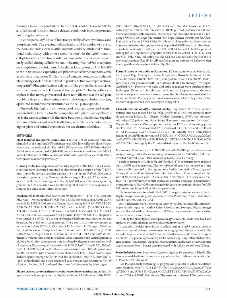
Figure S2 Delayed cellularisation in atfh5 mutant alleles. DIC images of cleared heart stage embryo seeds from wild type and atfh5 plants were taken to illustrate the cellularisation defects in these mutant alleles. In wild type (top panels), cellularisation is demonstrated by the double lines in regular polygonal patterns (black arrow) around a single nucleus (white arrow). The atfh5-1/atfh5-1 mutant (middle panels) at the same stage was notable for the incomplete cellularisation and the occurrence of single walls (black arrow) between nuclei. The atfh5-2/atfh5-2 mutant also features this phenotype (black arrows, left bottom panel). Further, in some of the atfh5-2 seeds where cellularisation occurred, binucleate cells were observed (white arrows, right bottom panel). Scale bar represents 20 µm.
b
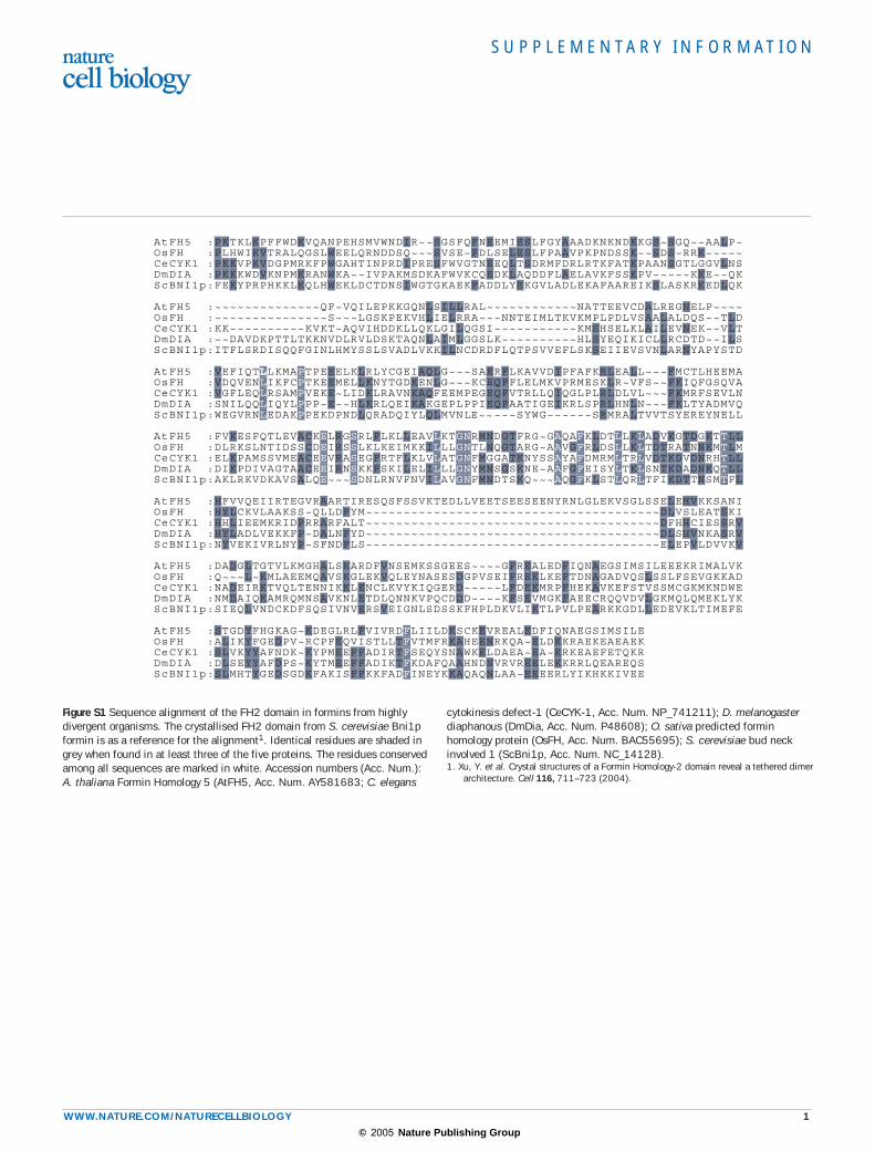
c
atfh5-2a
c
Figure S3 Abnormal endosperm development in atfh5-2 mutant allele. (a) Confocal sections of atfh5-2 seeds showing a slight delay of endosperm and reduction of cyst size in the posterior endosperm. (b) As in atfh5-1 cellularising cells appear with cell plates crossing between sister nuclei
(white arrows). (c) At the posterior pole, nodules are formed but the cyst (c) can be reduced in size compared to wild type. Scale bars represent 50 µm for a; 20 µm for b; 50 µm for c.
© 2005 Nature Publishing Group

S U P P L E M E N TA RY I N F O R M AT I O N
WWW.NATURE.COM/NATURECELLBIOLOGY 3
SUPPLEMENTARY METHODSPlasmid construction. PCR reactions were performed with the Expand High Fidelity kit (Roche Diagnostics, Brussels, Belgium). All the final constructs were gener-ated with the Gateway((cloning technology (Invitrogen, Carlsbad, CA). Primers with attB1 and attB2 sequences were purchased from Invitrogen.
AtFH5::H2B-YFP constructs. Two fragments of the upstream region of AtFH5 were amplified by PCR with attB1-P1 (5’-attB1-gagagaaattgacgaatga-3’) and attB2-R1 (5’-attB2-ATCTTTTGCTTCTCATTTCATCAG-3’) or attB1-P3 (5’-attB1-AAGGTCCGATGCCTAATGGA-3’) and attB2-R1 (5’-attB2-ATCTTTTGCTTCTCATTTCATCAG-3’). Both PCR products contain the 5’-UTR (460 bp long) and 1840 bp of the putative promoter region for the P1-R1 fragment (long FH5 construct, FH5L) and 850 bp of the putative promoter region for the P3-R1 fragment (short FH5 construct, FH5S). The pDONR207-FH5L and pDONR207-FH5S entry vectors were obtained by BP reaction cloning (Invitrogen).
The cassette Histone2B-Yellow Fluorescent Protein and the nopaline synthase terminator (H2B-YFP-nos) was amplified from pBI121-35S::H2B-YFP-nos vec-tor1 with primers H2B-YFP (5’-GGGCCCGGGATGGCGAAGGCAGATAAGAAAC-3’) and nos (5’-CCCGGGCCATGCCTACAGGTCACTGGAT-3’) containing respectively SmaI and ApaI restriction sites at their 5’ end. The SmaI/ApaI H2B-YFP-nos fragment was cloned into the SmaI/ApaI sites of the binary vector pGREE-NII (pGII) (http://www.pGREEN.ac.uk/; ref. 2). The Gateway recombination cassette with reading frame B (rfB) was inserted into SmaI site of pGII-H2B-YFP-nos to generate the “destination vector” pGII-rfB-H2B-YFP-nos. Recombination reactions (attL x attR) were performed between the “destination vector” pGII-rfA-H2B-YFP-nos and either the pDONR207-FH5L or pDONR207-FH5S entry vectors to produce respectively the vectors pGII-FH5L::H2B-YFP and pGII-FH5S::H2B-YFP.
Spatial and temporal expression pattern of H2B-YFP in transgenic Arabidopsis plants containing pGII-FH5L::H2B-YFP or pGII-FH5S::H2B-YFP was identical.35S::AtFH5-GFP constructTo construct a GFP-tagged version of the formin AtFH5 (At5g54650) putative start and stop codons were used to design the forward primer (5’-
ATGGTTGGAATGATTCGAGGAGG-3’) and the reverse primer (5’- GTCTGAATCTGAACTAGACTGATCCACG-3’) to which attB1 and attB2 extensions for Gateway® (Invitrogen, Carlsbad, CA) were added. The stop codon was replaced by a tyrosine codon for in-frame cloning with the Enhanced Green Fluorescent Protein (EGFP). cDNA was cloned from poly(A+) RNA extracted from Arabidopsis suspension cells following a one step RT-PCR reaction (Titan one tube RT-PCR kit; Roche Diagnostics, Brussels, Belgium) with high-fidelity DNA polymerase. PCR products were gel-purified (High pure PCR purification kit; Roche Diagnostics), and subcloned into pDONR207 via BP reaction cloning. The AtFH5 insert was transferred to the pK7WGF2 “destination vector”3 to generate EGFP C-terminal fusions downstream of the strong constitutive CaMV 35S promoter.1. Boisnard-Lorig, C. et al. Dynamic analyses of the expression of the HISTONE::YFP fusion protein in Arabidopsis show that syncytial endosperm is divided in mitotic domains. Plant
Cell 13, 495–509 (2001).2. Hellens, R. P., Edwards, E. A., Leyland, N. R., Bean, S. & Mullineaux, P. M. pGreen: a versatile and flexible binary Ti vector for Agrobacterium-mediated plant transformation. Plant
Mol. Biol. 42, 819–832 (2000).3. Karimi, M., Inze, D. & Depicker, A. GATEWAY vectors for Agrobacterium-mediated plant transformation. Trends Plant Sci. 7, 193–195 (2002).
© 2005 Nature Publishing Group